Embed Size (px)
Citation preview

Seattle and King County
2012 EMT Patient Care
Protocols
Seattle and King County
2012 EMT Patient Care
Protocols

UPDATES AND NEW CONTENT IN THE 2012
SEATTLE AND KING COUNTY PATIENT CARE PROTOCOLS
Glucometry age >1 CPR Protocols for King County and
Seattle Fire Department EPI use by EMT or health care
professional is an ALS indicator Pulse oximetry age >2 New material on Sepsis Call required to hospital for CVA
patients

TABLE OF CONTENTS
ACKNOWLEDGEMENTS/INTRODUCTION 5
TRAUMA 57
PROCEDURES & POLICIES 70
APPENDIX 144
INDEX 152
MEDICINE 9
ADVANCE LIFE SUPPORT (ALS) CRITERIA 6
TA
BL
E O
F C
ON
TE
NT
S

2
Acknowledgements....................................... 5
Introduction.................................................... 5
Advance Life Support (ALS) Indicators ....... 6
Medicine Abdominal Complaints............................... 9 Altered Level of Consciousness .............. 10 Anaphylaxis.............................................. 12 Ephinephrine (EpiPen).............................. 13 Asthma..................................................... 15 Behavioral ................................................ 16 Chest Discomfort ..................................... 17 Code ACS (Acute Coronary Syndrome)... 19 Cold-Related ............................................ 22 Congestive Heart Failure ......................... 25 Diabetic .................................................... 27 Glucometry................................................ 31 Drowning.................................................. 33 Excited Delirium........................................ 35 Heat-Related............................................. 37 Obstetric................................................... 39
APGAR................................................. 42 Gynecologic .............................................. 44 Peds Fever and Infection.......................... 45 Respiratory............................................... 47 Seizures ................................................... 49 Sepsis ....................................................... 50 Stroke....................................................... 52
FAST Exam.......................................... 54 Code CVA (Cerebral Vascular Accident).. 55
Trauma Bleeding Control ....................................... 57
TABLE OF CONTENTS T
AB
LE O
F C
ON
TE
NT
S

3
Dressing and Bandaging...........................58 Amputation ............................................59 Burns .....................................................59 Burns .........................................................60 Eye Injuries............................................... 61 Head and Neck......................................... 62 Orthopedic................................................ 64 Soft Tissue ............................................... 67
Procedures & Policies Airway Management .................................70 Oropharyngeal Airway...........................70 Suctioning..............................................71 Bag-Valve Mask ........................................72 Cardiac Arrest ...........................................74 Philips AED...........................................74 Physiocontrol AED................................76 Cardiac Arrest Footnotes......................78 Cardiac Arrest Seattle FD.....................80 CPR ..........................................................83 CPR For Adults ...................................83 CPR For Children & Infants .................84 CPR For Newborn ................................85 ECG Monitoring.........................................86 Epistaxis (Nosebleed) ...............................86 End of Life Issues......................................87 Group Health Consult Option....................89 Helicopter Procedures.............................. 91 Medication Administration ........................92 Activated Charcoal................................93 Inhalers (MDIs) .....................................93 Nitroglycerin ..........................................93 Oral Glucose.........................................94 Multi-Casualty Incident (MCI)................... 96 The Triage Team ................................. 96
TABLE OF CONTENTS (CONT.)
TA
BL
E O
F C
ON
TE
NT
S

4
Treatment Team Leader ....................... 97 Transportation Team Leader ................ 97 Field Triage Algorithm............................ 98 Neurological Assessment........................ 100 AVPU................................................... 100 Glasgow Coma Scale.......................... 101 Noxious Stimuli ........................................ 102 Oxygen Delivery ...................................... 103 Patient Positioning................................... 107 Recovery ............................................. 107 Semi-Reclining (Semi-Fowler’s) ......... 109 Shock Position .................................... 110 Patient Restraint ...................................... 111 Personal Protective Equipment (PPE) ... 114 Reportable Exposures ............................. 116 Physical Abuse and Neglect.................... 117 Postural Vital Signs ................................. 119 Psychiatric Evaluations............................. 120 Pulse Oximetry ........................................ 121 SICK/NOT SICK ...................................... 124 Adult ..................................................... 126 Pediatric ............................................... 128 Spinal Immobilization............................... 130 Splinting ................................................... 132 Taser Dart Removal and Care ............... 135 Teeth........................................................ 137 Transport and Destination ....................... 138 Final Disposition Options.......................... 140
Appendix Abbreviations............................................ 144 Normal Vital Signs By Age ....................... 145 Temperature Conversions........................ 145 Telephone Numbers ................................. 146
Index ........................................................... 152
TABLE OF CONTENTS (CONT.) T
AB
LE O
F C
ON
TE
NT
S

5
Thanks to all of the EMS providers for their help in the development of these protocols. Special thanks to Betty Hurtado for formatting and production.
Thanks to Jonathan Larsen and Norm Nedell for their meticulous review of the protocols, and to the training officers of Seattle and King County fire departments for their helpful comments.
INTRODUCTION
These patient care protocols are intended to help you in your job. Additional information and documents are on the EMS training site at: www.emsonline.net. These protocols define best practices for EMT care in Seattle & King County. It is important to realize that adherence to these protocols provides quality care to patients and protects you and your department.
You have a very challenging job - but a very rewarding one. There can be nothing more satisfying than providing help to the wounded, sympathy to the distressed, relief to the anxious, comfort to the frightened, and most importantly therapy and aid to the sick and injured. Your skills and training literally bring life back from the brink of death.
We applaud the fine job you do.
ACKNOWLEDGEMENTS
Michael K. Copass, MD Medical Director Medic One Seattle Fire Department
Mickey Eisenberg, MD, PhD King County Medical Program Director
AC
KN
OW
LE
DG
EM
EN
TS

6
The following list is offered as a summary guide and is not comprehensive. Nor does it take into account your IOS or the MOI
Abdominal Pain Discomfort or pain or unusual sensations
between the navel and jaw if the patient is > or = to 40 y/o and/or has cardiac history
Severe unremitting abdominal pain Breathing
Respirations >30 min Failure to respond to repeated inhalers Asthma attack with history of previous
intubation Audible wheezing not improved with inhaler Abnormal respiratory patterns Respiratory related with patient in the tripod
position Burns
Burns with possible airway involvement Burns with associated injuries: electrical
shock, fracture, airway 2nd or 3rd degree burns to face/head 2nd or 3rd degree burns >20% of body
Cardiac Suspected ACS (see page 19)
CVA Progression of stroke symptoms
Diabetic Diabetic that is unable to swallow Diabetic with rapid respirations Diabetic that fails to respond to oral glucose Suspected ketoacidosis
ADVANCE LIFE SUPPORT (ALS) INDICATORS
AD
VA
NC
E L
IFE S
UP
PO
RT (
AL
S)
CR
ITE
RIA

7
Hypothermia Temperature <95 degrees oral or tympanic Hypothermia with significant co-morbidity
(e.g. elderly, illness, circumstances, trauma, alcohol, drugs)
LOC GCS < or = 12 Hypoglycemia with decreased LOC Abnormal behavior with unstable vitals Abnormal behavior associated with possible
drug or alcohol overdose Pulse / BP
Hypotension (systolic <90 with appropriate clinical settings)
Signs of shock: pulse generally >120/minute, BP <90
Positive posturals (decrease in systolic BP >20 or increase in pulse >20)
Sustained tachycardia (generally >120/minute in appropriate clinical setting)
Systolic >200 or diastolic >110 with associated symptoms
Pregnancy with systolic <90 or >140 Hypotension and severe bradycardia
OB/GYN Female with severe unremitting pelvic pain Excessive vaginal bleeding Possible ectopic pregnancy Dispatched to birthing center/midwife Pregnancy complications: placenta previa,
abruptio placenta, diabetes, multiple birth, breech or limb presentation, prolapsed cord, shoulder dystocia, uncontrolled postpartum hemorrhage
Imminent birth
ADVANCE LIFE SUPPORT (ALS) INDICATORS (CONT.)
AD
VA
NC
E L
IFE S
UP
PO
RT (
AL
S)
CR
ITE
RIA

8
Pregnancy 3rd trimester with abdominal trauma
Pregnancy with significant MOI. Other
Use of epipen by EMT or healthcare professional
Suspected meningitis Sepsis
Decreased LOC Respiratory distress Respirations greater or RR > 30 per minute Signs and symptoms of shock
Seizure Multiple seizures Single seizure >5 minutes or >15 minutes
postictal with no LOC improvement Pregnant female Severe headache Associated with trauma Associated with drugs or alcohol Associated with hypoglycemia
Trauma Falls >2 times the body height Thrown >10-15 feet Penetrating injury to the head, eyes or box Pelvic fx, bilateral femur fx, or multisystem fx Femur fx with excessive swelling Open fx except hands and feet Severe pain Any underwater rescue Paresis and or paresthesia due to trauma
ADVANCE LIFE SUPPORT (ALS) INDICATORS (CONT.)
AD
VA
NC
E L
IFE S
UP
PO
RT (
AL
S)
CR
ITE
RIA

9
ALS Indicators
Signs and symptoms of shock which include: Poor skin signs (pale, sweaty) Sustained tachycardia (see page 7) Hypotension (systolic BP less than 90
mmHg) with an appropriate clinical setting
Unstable vital signs
Positive postural changes (see page 119)
Evidence of ongoing bleeding
Severe unremitting pain
BLS Indicators
Stable cardiac and respiratory functions
Stable vital signs
BLS Care
Request paramedics if indicated.
Provide supplemental oxygen and/or ventilatory assistance as necessary.
Position of comfort (Shock Position if hypotensive).
Prepare to suction patient if vomiting, estimate volume and describe character (color and consistency) of vomitus.
Reassure patient.
Monitor vital signs every five minutes.
ABDOMINAL COMPLAINTS
ME
DIC
INE —
AB
DO
MIN
AL C
OM
PL
AIN
TS

10
ALS Indicators
Decreased LOC
Respiratory distress or airway compromise
Signs and symptoms of shock which include: Poor skin signs (pale, sweaty) Sustained tachycardia (see page 7) Hypotension (systolic BP less than 90
mmHg) with an appropriate clinical setting
Unstable vital signs
Multiple seizures (status seizures)
Single seizure longer than five (5) minutes or with more than 15 minutes postictal with no improvement in LOC
Cyanosis
Hypoglycemia with decreased LOC
Seizure in pregnant female
Seizure with severe headache
Seizure associated with trauma
Drug or alcohol related seizures
BLS Indicators
Adequate respirations
Transient symptoms including seizure with stable vital signs
First time or typical seizure pattern for the patient with stable vital signs
ALTERED LOC M
ED
ICIN
E —
A
LT
ER
ED
LE
VE
L O
F C
ON
SC
IOU
SN
ES
S (
LO
C)

11
BLS Care
Provide supplemental oxygen and/or ventilatory assistance as necessary.
Protect patient from injury, remove objects from mouth and upper airway, do not restrain patient during seizure, remove hazardous objects near patient.
Position patient in position of comfort if alert and airway is secure; if not, then use recovery position.
Perform blood glucometry.
Loosen restrictive clothing.
Retain relevant drug containers and notes for transport with patient.
ALTERED LOC (CONT.)
ME
DIC
INE —
A
LT
ER
ED
LE
VE
L O
F C
ON
SC
IOU
SN
ES
S (
LO
C)

12
ALS Indicators
Respiratory distress
Signs and symptoms of shock which include: Poor skin signs (pale, sweaty) Sustained tachycardia (see page 7) Hypotension (systolic BP less than 90
mmHg) with an appropriate clinical setting
Unstable vital signs
Use of EpiPen by EMT or healthcare professional.
BLS Indicators
Bite or sting with local reaction or usual reaction to medication or food
Stable vital signs
No anaphylaxis
BLS Care
EpiPen for anaphylaxis (see page 13).
Oxygen as needed.
Reassure patient.
Remove stinger by scraping away from puncture site.
Any patient who receives an EpiPen (pre or post EMS arrival) should be transported (mode of transport depends on clinical findings and symptoms) and evaluated in a hospital.
ANAPHYLAXIS M
ED
ICIN
E —
A
NA
PH
YL
AX
IS
13
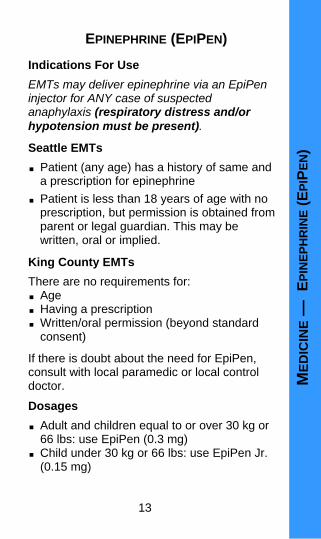
Indications For Use
EMTs may deliver epinephrine via an EpiPen injector for ANY case of suspected anaphylaxis (respiratory distress and/or hypotension must be present).
Seattle EMTs
Patient (any age) has a history of same and a prescription for epinephrine
Patient is less than 18 years of age with no prescription, but permission is obtained from parent or legal guardian. This may be written, oral or implied.
King County EMTs
There are no requirements for: Age Having a prescription Written/oral permission (beyond standard
consent)
If there is doubt about the need for EpiPen, consult with local paramedic or local control doctor.
Dosages
Adult and children equal to or over 30 kg or 66 lbs: use EpiPen (0.3 mg)
Child under 30 kg or 66 lbs: use EpiPen Jr. (0.15 mg)
EPINEPHRINE (EPIPEN)
ME
DIC
INE —
E
PIN
EP
HR
INE (
EP
IPE
N)

14
Injection Procedure
Confirm that patient is experiencing anaphylaxis and meets above criteria.
1. Check medication date and that the EpiPen dose matches to patient’s size.
2. Remove clothing and prep area of thigh with alcohol pad.
3. Remove safety cap and locate injection site on lateral thigh.
4. Place black tip of injector against thigh and push hard until injector activates.
5. Hold in place for 10 seconds. Note and document time of injection.
6. Remove injector, place in sharps container and massage site for 10 seconds.
7. Reassure patient and monitor for response/side effects to injection.
8. Continue to provide oxygen. Ventilate if necessary.
9. Monitor and document vitals every 5 minutes.
10. Update incoming medics on patient status and response to injection.
Use of EpiPen by EMT or healthcare professional is an ALS indicator. Any patient who receives an EpiPen (pre or post EMS arrival) should be transported (mode of transport depends on clinical findings and symptoms) and evaluated in a hospital.
EPINEPHRINE (EPIPEN) (CONT.) M
ED
ICIN
E —
E
PIN
EP
HR
INE (
EP
IPE
N)

15
ALS Indicators
Decreased LOC
Extreme anxiety and agitation Ashen color, cyanosis
Failure to respond to repeated inhalers
History of previous intubation
Respiratory distress—unable to speak normally
Labored respirations regardless of rate when found with other indicators
Audible wheezing not improved with inhalers
Sustained tachycardia (see page 7).
BLS Indicators Responds to self-administered metered-dose inhaler (MDI)
Normal vital signs
Able to speak normally
BLS Care Assist patient with his or her medications.
Provide supplemental oxygen and/or ventilatory assistance as necessary.
Reassure patient and urge calmness.
Obtain oximetry reading (see page 121).
Monitor vital signs every five to ten minutes.
ASTHMA
ME
DIC
INE —
A
ST
HM
A

16
ALS Indicators
Decreased LOC
Abnormal behavior with unstable vitals
Abnormal behavior with serious co-morbidity (e.g., drug or alcohol OD)
BLS Indicators
Abnormal behavior with stable vital signs
BLS Care
Secure safety of personnel and patient.
Provide support, reassurance to patient.
Provide supplemental oxygen and/or ventilatory assistance as necessary.
Wound or trauma care if indicated.
Call police if necessary (if patient refuses transport but EMTs feel patient needs further evaluation).
Use restraints when warranted (see page 111).
Monitor patient behavior and physiological changes, do not leave patient alone or unobserved.
BEHAVIORAL
Incapacitated or impaired patients or patients with mental or behavioral problems should be
evaluated in local hospital emergency departments.
ME
DIC
INE —
B
EH
AV
OR
IAL

17
ALS Indicators
Chest pain or discomfort of suspected myocardial ischemia (angina or MI)
(See Code ACS page 19)
Altered LOC
Use of nitroglycerin
Unstable vital signs
Signs and symptoms of shock which include: Poor skin signs (pale, sweaty) Sustained tachycardia (see page 7) Hypotension (systolic BP less than 90
mmHg) with an appropriate clinical setting
Discomfort, pain, or unusual sensations between the navel and the jaw if the patient is 40 years old or older and/or has a history of heart problems
BLS Indicators Apparent non-cardiac or minor traumatic chest pain if patient is less than 40 years old and no cardiac history and stable vital signs and no associated symptoms
Stable/normal vital signs
CHEST DISCOMFORT
ME
DIC
INE —
C
HE
ST D
ISC
OM
FO
RT

18
BLS Care
Request paramedics if indicated.
Provide supplemental oxygen and/or ventilatory assistance as necessary.
Assist patient with nitroglycerin if indicated (see page 93).
Provide aspirin if indicated (see Code ACS page 19).
Position of comfort.
Reassure patient.
Monitor vital signs every 5 minutes.
Monitor ECG if authorized, record strip.
CHEST DISCOMFORT (CONT.)
Special Instructions For Chest Pain
Patients with possible cardiac chest pain require ALS evaluation
Maintain high index of suspicion that atypical chest pain may be cardiac in origin
Elderly patients, women, and diabetics may present with atypical findings such as fatigue, weakness, shortness of breath, or syncope See Code ACS page 19
ME
DIC
INE —
C
HE
ST D
ISC
OM
FO
RT

19
Acute coronary syndrome (ACS) requires rapid assessment by EMTs and paramedics and expedited transport to a cath-ready hospital.
This policy applies to all patients presenting with possible ACS and who are initially evaluated by EMTs.
Evaluation for ACS
1. Patient exhibits any of the following signs or symptoms: a. Uncomfortable pressure, fullness,
squeezing or pain in the center of the chest that lasts more than a few minutes, or goes away and comes back.
b. Pain that spreads to the shoulders, neck, or arms.
c. Chest discomfort with lightheadedness, fainting, sweating, nausea, or shortness of breath.
-OR-
2. Patient exhibits any of the two following signs or sympthoms, when ACS is suspected: a. Atypical chest pain, stomach, or
abdominal pain. This may include discomfort that can be localized to a point, that is “sharp” in nature, that is reproducible by palpitation, or that is in
CODE ACS (ACUTE CORONARY SYNDROME)
ME
DIC
INE —
C
OD
E A
CS

20
the “wrong” location (such as the upper abdomen).
b. Unexplained nausea (without vomiting) or lightheadedness (not vertigo) without chest pain.
c. Shortness of breath and difficulty breathing (without chest pain).
d. Unexplained anxiety, weakness, or fatigue.
e. Palpitations, cold sweat, or paleness.
Administer Aspirin (currently not authorized for Seattle EMTs)
1. Administer one 325 mg aspirin tablet (or four 81 mg baby aspirins) for patients with ACS. Patients may chew or swallow (with a small amount of water) the aspirin(s). Do not use enteric coated aspirin.
2. Be sure that the patient is alert and responsive and meets indications and has no contraindications.
Contraindications For Use
1. Patient is allergic to aspirin. 2. If they have taken 325 mg aspirin within 60
minutes for this event, do not administer additional aspirin.
3. Blood pressure >180/>110. 4. Active or suspected GI bleeding.
CODE ACS (CONT.) (ACUTE CORONARY SYNDROME)
ME
DIC
INE —
C
OD
E A
CS

21
M
ED
ICIN
E —
C
OD
E A
CS
Additional Procedures
1. If the patient has his/her own nitroglycerin and meets the criteria for administration, do not delay assisting with nitroglycerin administration.
2. Request paramedics if not already dispatched.
3. Record your actions, including the dosage and the time of administration.
4. Record the time of onset of symptoms. The time of onset should be the time that symptoms began which prompted the patient to call 911.
5. EMTs should limit on scene time to no more than 15 minutes.
CODE ACS (CONT.) (ACUTE CORONARY SYNDROME)

22
ALS Indicators
Decreased/altered LOC
Temperature less than 95° F (35°C) oral or tympanic
Cessation of shivers in a cold patient
Significant co-morbidities (e.g., elderly, illness, circumstances, trauma, alcohol, drugs)
Cardiac arrest
Hypotension (systolic BP less than 90 mmHg) with an appropriate clinical setting
BLS Indicators
Cold exposure, temperature greater than 95°F, normal vital signs and no abnormal LOC
Frostbite with temperature greater than 95°F, normal vital signs and no abnormal LOC
BLS Care (Hypothermia)
Remove patient from the cold environment and protect the patient from further heat loss.
Provide supplemental oxygen and/or ventilatory assistance as necessary.
Provide high flow oxygen via NRB or bag-valve mask (see page 103).
COLD-RELATED M
ED
ICIN
E —
C
OL
D-R
EL
AT
ED

23
Remove wet clothing.
Position of comfort. If decreased LOC, place in recovery position.
Warm the patient.
Warm the aid unit.
Monitor patient’s vital signs, use ECG monitor if authorized, repeat temperature measurements.
BLS Care (Hypothermic Cardiac Arrest Or Profound Bradycardia)
If no pulse is detected after one minute, begin CPR and apply AED. If breathing, assume there is cerebral perfusion. Therefore, “NO” CPR.
If AED states “Shock Indicated”, follow cardiac arrest protocol.
BLS Care (Frostbite)
Protect cold-injured part from further injury.
Remove any constricting or wet clothing or shoes and replace with a dry bulky dressing.
Splint the affected extremity and do not let the patient walk on or use it.
COLD-RELATED (CONT.)
If pulse is present, withhold CPR regardless of rate or BP. M
ED
ICIN
E —
C
OL
D-R
EL
AT
ED

24
Remove constricting jewelry (e.g., rings, watchbands).
Do not rub or massage injured tissue.
Transport to an emergency room.
Do not rewarm frozen tissue unless transport time will exceed two hours and it is certain that the thawed tissue will not refreeze. Obtain medical direction prior to initiating rewarming. Rewarming should be done with 100oF - 105oF water. Do not use dry heat; it heats unevenly and may burn frozen tissue. Stop rewarming when the tissue turns red-purple and becomes pliable.
COLD RELATED (CONT.) M
ED
ICIN
E —
CO
LD
-RE
LA
TE
D

25
Congestive heart failure (CHF) can range from the very mild to very severe (pulmonary edema). Usually patients with congestive heart failure call EMS for worsening shortness of breath and/or worsening fatigue.
ALS Indicators
Decreased LOC
Signs and symptoms of shock which include: Poor skin signs (pale, sweaty) Sustained tachycardia (see page 7) Hypotension (systolic BP less than 90
mmHg) with an appropriate clinical setting
Extreme anxiety and agitation
Unable to lie flat
Ashen color, cyanosis
Respiratory distress—unable to speak normally
Respirations greater than 30 per minute
Labored respirations regardless of rate
BLS Indicators Normal vital signs without respiratory distress
Able to speak normally
BLS Care Provide supplemental oxygen and/or assist ventilation with a BVM as necessary.
CONGESTIVE HEART FAILURE
ME
DIC
INE —
CO
NG
ES
TIV
E H
EA
RT F
AIL
UR
E

26
Position patient upright with legs dangling (dependent) unless hypotensive.
If hypotensive, place patient in supine position.
Reassure patient and urge calmness.
Obtain oximetry reading (see page 121).
Monitor vital signs every 5 to10 minutes depending on patient’s condition.
CONGESTIVE HEART FAILURE (CONT.) M
ED
ICIN
E —
CO
NG
ES
TIV
E H
EA
RT F
AIL
UR
E

27
ALS Indicators
Altered LOC
Absent or depressed gag reflex, as indicated by inability to swallow
Patient unable to protect airway
Unstable vital signs
Rapid respiration
Signs and symptoms of shock which include: Poor skin signs (pale, sweaty) Sustained tachycardia (see page 7) Hypotension (systolic BP less than 90
mmHg) with an appropriate clinical setting
Failure to respond to oral glucose unit with continued glucose <60 despite repeated treatment.
Suspected diabetic ketoacidosis (glucometry reading >400 or “high” with associated symptoms)
Seizures
BLS Indicators
Normal or mild reduction in LOC
Gag reflex intact, as indicated by swallowing
Patient can protect airway
Normal vital signs
Symptoms of hypoglycemia relieved by oral glucose
Hyperglycemia with normal vital signs
DIABETIC
ME
DIC
INE —
DIA
BE
TIC

28
BLS Care
Request paramedics if indicatedPerform blood glucometry (see page 31).
Provide supplemental oxygen and/or ventilatory assistance as necessary.
If hypoglycemic and patient is able to swallow, position upright and give oral glucose.
If hypoglycemic, and patient is unable to swallow, position on side, give oxygen, ventilation and await paramedics.
Maintain normal body temperature.
Monitor vital signs in response to sugar.
Diabetic patients with symptom of hyperglycemia should be evaluated in an emergency room. Transport decision based on clinical presentation.
If in doubt whether symptoms are due to hypoglycemia and swallowing ability is intact, position upright and give oral glucose.
Special Instructions For Diabetic Patients
Patients with hypoglycemia who have responded to oral glucose may be left at scene (see page 32). These patients must have a repeat glucose level of 60 mg/dl or higher documented and after-care instructions must be left with the patient.
DIABETIC (CONT.) M
ED
ICIN
E —
DIA
BE
TIC
CO
NT.)

29
Distinguishing hyperglycemia from hypoglycemia can be difficult without a blood glucose. Recent medical history can help.
History Suggesting Hypoglycemia
Insufficient food intake
Excessive insulin dosage
Normal to excessive activity level
Rapid onset
Absent thirst
Intense hunger
Headache
May have seizures
History Suggestion Hyperglycemia
Recent infection
Polyphagia (excessive food intake)
Polydipsia (intense thirst)
Polyuria (excessive frequency and amount of urine)
Vomiting, abdominal pain
“Flu-like” symptoms, nausea
Insufficient insulin dosage
Gradual onset
Normal activity level
DIABETIC (CONT.)
ME
DIC
INE —
DIA
BE
TIC
(C
ON
T.)

30
ME
DIC
INE —
DIA
BE
TIC
(C
ON
T.)
Signs and Symptoms of Diabetic Coma (Hyperglycemia with Ketoacidosis)
Altered LOC (restless to coma)
Warm and dry skin
Hypotension (systolic BP less than 90 mmHg) with an appropriate clinical setting
Sustained tachycardia
Reduced circulation in extremities
Vomiting
Sweet, fruity breath
Kussmaul breathing (deep, rapid)
High blood glucose Greater than 200 mg/dl (mild
hyperglycemia) Greater than 300 mg/dl (moderate
hyperglycemia) Greater than 400 mg/dl (severe
hyperglycemia)
Signs and Symptoms of Hypoglycemia
Hypoglycemia may be due to excessive insulin or decreased food intake, or increased activity.
Irritability, confusion, seizures or coma
Pale, moist skin
Normal or rapid pulse
Low blood glucose (usually less than 60 mg/dl) with glucometry
DIABETIC (CONT.)

31
Indications For Use
Any time an EMT encounters a patient with an altered level of consciousness. This may include patients with the following: - Unconsciousness - Suspected diabetic-related problem - Signs and symptoms of stroke - Suspicion of drug or alcohol intoxication
Any time EMTs feel that the blood sugar level may assist patient care.
Contraindications
Children less than 1 (one) year of age.
Use and application
Perform the testing procedure as outlined in the instructions for your specific device. All reading should be recorded on the incident response form.
GLUCOMETRY
Under no circumstances should the presence of a blood glucose monitor
detract from basic patient care. (e.g., ABCs)
Glucometry is an approved protocol but optional by individual departments.
ME
DIC
INE —
GL
UC
OM
ET
RY

32
Perform blood glucose evaluation after the ABCs and initial assessment have been completed. *If a patient is treated with oral glucose you must perform a second glucose level check.
Patients on oral hypoglycemic agents who are initially found to be hypoglycemic should be strongly advised to seek further evaluation by a physician due to the high likelihood of repeated hypoglycemia secondary to long medication half-life.
Patients on insulin may be safely left at home when ALL THREE of the following conditions are met: 1. Patient is able to eat and drink normally. 2. Patient responds completely as evidence
by BOTH: Blood glucose reaches greater than 60
mg/dl, AND Patient is conscious and alert with
appropriate behavior. 3. A responsible person can remain with the
patient.
These patients must receive after-care instructions if they are not being transported to the hospital. You must document pre and post treatment glucose and that after-care instructions were given to patient.
*If glucometry is available
GLUCOMETRY (CONT.) M
ED
ICIN
E —
GL
UC
OM
ET
RY (
CO
NT.)

33
ALS Indicators
Any underwater rescue
Altered LOC
Respiratory distress
Labored breathing
Hypotension (systolic BP less than 90 mmHg) with an appropriate clinical setting
Temperature less than 95°F
Significant co-morbidity (e.g., injury, intoxication)
Cardiac or respiratory arrest
BLS Indicators
Water-related accident including aspiration of water, injury in diving or swimming, with normal CNS function and vital signs
BLS Care
Request paramedics if indicated.
Remove the victim from the water; do not become a victim.
Neutral in-line cervical stabilization during removal from water with a backboard if spine injury is suspected or patient is unresponsive.
DROWNING
ME
DIC
INE —
DR
OW
NIN
G

34
If there is no suspected spinal injury, consider recovery position.
Provide supplemental oxygen and/or ventilatory assistance as necessary.
Prepare suction, expect vomiting.
Follow resuscitation protocols if cardiac or pulmonary arrest.
Warm aid unit.
Monitor vital signs.
All immersion incidents should be transported to the hospital for further evaluation.
Care For Scuba Diving Accidents
Request paramedics
High flow oxygen by NRM and/or BVM as necessary
Position patient flat (supine) or on side to avoid cerebral edema
DROWNING (CONT.) M
ED
ICIN
E —
DR
OW
NIN
G (
CO
NT.)

35
Definition:
A state of extreme mental and physiological excitement, characterized by extreme agitation, hyperthermia, hostility, exceptional strength and endurance without apparent fatigue. This condition is usually associated with illicit stimulant drug use and is associated with in-custody deaths.
ALS Indicators: Extreme agitation, disorientation Hyperthermia, diaphoresis, seeking water Stripping off of clothing, or no clothing Shouting, or keening (making animal
noises), unintelligible speech Eyes wide open, lid lift Paranoia, hallucinations Panic Violence toward others Unexpected physical strength and stamina Insensitivity to pain Violence or attraction to glass, reflection or
lights
BLS indicators: No BLS indicators if Excited Delirium is suspected. ALS must evaluate these patients.
EXCITED DELIRIUM
ME
DIC
INE —
EX
CIT
ED
DE
LIR
IUM

36
BLS Care: Secure safety of personnel, assure scene
safety before proceeding Request Police if not already on scene Restrain patient as necessary. See use of
Restraints page 111. Provide supplemental oxygen and or
ventilatory assistance as necessary. Wound or trauma care as necessary Package patient for ALS transport Be vigilant for changes in patient LOC, and
ABC’s Patients can decompensate quickly, without
warning and may suffer cardiac arrest CPR as per protocol
EXCITED DELIRIUM (CONT.) M
ED
ICIN
E —
EX
CIT
ED
DE
LIR
IUM
(C
ON
T.)

37
ALS Indicators
Decreased/altered LOC
Hot dry skin in the presence of elevated temperature
Sustained tachycardia (see page 7)
Hypotension (systolic blood pressure less than 90 mmHg)
Positive postural changes
BLS Indicators
Heat related cramps
Minor to moderate heat-related complaint with stable vital signs
BLS Care
Request paramedics if indicated.
Remove patient from the hot environment and place patient in a cool environment (back of air-conditioned transport vehicle or aid unit with air conditioner running on high).
Reassure and cool patient.
Provide supplemental oxygen and/or ventilatory assistance as necessary.
Loosen or remove clothing.
Apply cool packs to neck, groin and armpits for the heat-stroke patient.
HEAT-RELATED
ME
DIC
INE —
HE
AT-R
EL
AT
ED

38
ME
DIC
INE —
HE
AT-R
EL
AT
ED
(C
ON
T.)
Keep skin wet by applying cool water with sponge or wet towels.
Fan aggressively.
Place patient in Shock position.
If patient is responsive and not nauseated, have patient drink water.
If the patient is vomiting, place in recovery position.
Monitor patient’s vital signs and temperature (oral or tympanic).
HEAT-RELATED (CONT.)

39
ALS Indicators
Imminent birth
Decreased/altered LOC of mother/newborn baby
Abnormal blood pressure (less than 90 mmHg systolic or greater than 140 mmHg systolic) with neurologic symptoms
Complications with this pregnancy such as: Placenta previa Abruptio placenta Diabetes
Excessive vaginal bleeding
Suspected ectopic pregnancy
Any abdominal trauma to mother during third trimester
Trauma with significant MOI
Known or anticipate delivery of twins or more
Breech or limb presentation
Prolapsed cord
Shoulder dystocia
Uncontrolled postpartum hemorrhage
Seizures
Dispatch to birthing center/midwife
OBSTETRIC
ME
DIC
INE —
OB
ST
ET
RIC
40
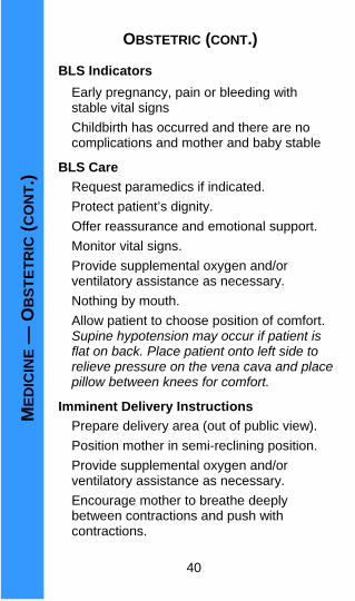
BLS Indicators
Early pregnancy, pain or bleeding with stable vital signs
Childbirth has occurred and there are no complications and mother and baby stable
BLS Care
Request paramedics if indicated.
Protect patient’s dignity.
Offer reassurance and emotional support.
Monitor vital signs.
Provide supplemental oxygen and/or ventilatory assistance as necessary.
Nothing by mouth.
Allow patient to choose position of comfort. Supine hypotension may occur if patient is flat on back. Place patient onto left side to relieve pressure on the vena cava and place pillow between knees for comfort.
Imminent Delivery Instructions
Prepare delivery area (out of public view).
Position mother in semi-reclining position.
Provide supplemental oxygen and/or ventilatory assistance as necessary.
Encourage mother to breathe deeply between contractions and push with contractions.
OBSTETRIC (CONT.) M
ED
ICIN
E —
OB
ST
ET
RIC
(C
ON
T.)

41
Prepare OB equipment and don sterile gloves, gowns, and eye protection.
As baby crowns, support head with gentle pressure to avoid explosive birth.
If membrane is still intact, rupture with your fingers to allow amniotic fluid to leak out.
If cord is around the baby’s neck, gently slip it over the head. Do not force it!
If the cord is too tight to slip over the head, apply umbilical cord clamps and cut.
As soon as baby’s head emerges, suction the mouth and nose with bulb syringe.
Allow the mother to push and support the head as it rotates.
Caution: Babies are slippery as they exit the birth canal; be careful and alert.
After delivery, wait for cord pulsation to cease, then place two clamps on the cord two inches apart and six inches away from the baby. Cut the cord between the clamps. Re-suction the baby’s mouth and nostrils only if baby is not breathing or is having res-piratory distress.
Dry and inspect the cord for bleeding.
Wrap baby in warm blanket.
Place baby on its side to facilitate drainage.
Inform the mother of the baby’s gender.
Note the time of birth, APGAR score of baby and gender.
OBSTETRIC (CONT.)
ME
DIC
INE —
OB
ST
ET
RIC
(C
ON
T.)

42
Clin
ical
Sig
n
0 p
oin
ts
1 p
oin
t 2
po
ints
A
App
ear
anc
e B
lue,
pa
le
Bod
y pi
nk, e
xtre
miti
es b
lue
Com
plet
ely
pink
P
Pul
se
Abs
ent
Less
than
100
/min
ute
Mor
e th
an
100/
min
ute
G
Grim
ace
No
resp
onse
G
rimac
es to
stim
ulat
ion
Crie
s
A
Act
ivity
Li
mp
Som
e fle
xion
of
extr
emiti
es
Act
ive
mot
ion
R
Res
pira
tory
E
ffort
A
bsen
t S
low
, irr
egu
lar
Str
ong
cry
or
resp
iratio
ns
AP
GA
R S
CO
RIN
G
Sco
re a
t 1
and
5 m
inu
tes
afte
r bi
rth.
OBSTETRIC (CONT.) M
ED
ICIN
E —
OB
ST
ET
RIC
(C
ON
T.)

43
Post Delivery Instructions
Observe perineum for bleeding.
Normally there should be a small to moderate amount of bloody material that will ooze from the vagina.
Apply oxygen to the mother as indicated via nasal cannula or nonrebreather mask to mother.
Do not pull on the umbilical cord.
The placenta should be delivered spontaneously within 20 minutes.
If delivered, wrap the placenta in the bag supplied in the OB Kit and send with the mother and baby to the hospital.
Massage the uterus with moderate firmness on the lower abdomen to stimulate uterine contraction.
Monitor vital signs of both mother and infant.
Maintain body temperature of both patients.
BLS transport of mother and baby to hospital, if no ALS indicators.
OBSTETRIC (CONT.)
ME
DIC
INE —
OB
ST
ET
RIC
(C
ON
T.)

44
ALS Indicators Decreased/altered LOC
Hypotension (systolic BP less than 90 mmHg) with an appropriate clinical setting
Sustained tachycardia (see page 7)
Moderate to severe hypertension (140 mmHg systolic or greater) in a pregnant woman with neurologic symptoms
Seizures
Severe unremitting pelvic pain
Excessive vaginal bleeding
Possible ectopic pregnancy
BLS Indicators Limited vaginal bleeding with stable vitals
Pelvic pain or discomfort with stable vitals
BLS Care Request paramedics if indicated.
Protect patient’s dignity.
Offer reassurance and emotional support.
Monitor vital signs.
Direct pressure over lacerations.
Provide supplemental oxygen.
Obtain focused history.
Allow patient to choose position of comfort.
GYNECOLOGIC M
ED
ICIN
E —
GY
NE
CO
LO
GIC

45
ALS Indicators
Decreased LOC
Respiratory distress
Seizure Respiratory distress or airway
compromise Recurrent seizure Prolonged, depressed LOC
Fever/Infection High index of suspicion for sepsis or
meningitis
BLS Indicators
Febrile seizure (generalized tonic/clonic—see page 46)
Fever/infection with low index of suspicion
BLS Care
Use Pediatric Assessment Triangle. (Page 128, 129)
Request paramedics if indicated.
Provide supplemental oxygen and/or ventilatory assistance as necessary.
Monitor vital signs.
Position of comfort.
For seizures, place child on side to protect airway.
PEDS FEVER AND INFECTION
ME
DIC
INE —
PE
DIA
TR
IC F
EV
ER
AN
D IN
FE
CT
ION

46
May assist caregiver with medication to reduce temperature (e.g., Tylenol [acetaminophen], not aspirin).
If febrile, attempt to reduce patient’s temperature with cool towels.
Remove clothes. Cover loosely with one layer. Do not allow to chill.
Special Instructions for Febrile Seizures
Patient with a history of a previous febrile seizure, who is now neurologically intact with stable vital signs, and a competent caregiver requests home care, may be left at home with a suggestion to follow-up with a physician.
First time febrile seizures must be evaluated in an emergency department
Febrile seizures are always generalized tonic/clonic in nature. Any focal seizure is not a febrile seizure until proven otherwise.
PEDS FEVER AND INFECTION (CONT.) M
edic
ine
— P
edia
tric
FE
VE
R A
ND
INF
EC
TIO
N (
CO
NT.)

47
ALS Indicators
Decreased LOC
Extreme anxiety and agitation
Tripod position
Respiratory distress—unable to speak normally
Respirations greater than 30 per minute
Ashen color, cyanosis, retractions
Failure to respond to usual treatments
Labored respirations regardless of rate when found with other indicators
Audible wheezing, rales when found with other indicators
Use of EpiPen injector
Sustained tachycardia (see page 7)
BLS Indicators
Respiratory complaints due to common causes such as a cold, flu, bronchitis
Respiratory complaints of a chronic but stable nature
Respiratory complaints with normal vital signs and adequate oxygenation with treatment
Patent airway
RESPIRATORY
ME
DIC
INE —
RE
SP
IRA
TO
RY

48
BLS Care
Provide supplemental oxygen and/or ventilatory assistance as necessary.
Obtain oximetry reading (see page 121).
Reassure patient and urge calmness.
Assist patient with his or her medications.
Administer EpiPen if indicated for anaphylaxis (see page 13).
Monitor vital signs every 5 to 10 minutes depending on patient’s condition.
RESPIRATORY (CONT.)
ME
DIC
INE —
RE
SP
IRA
TO
RY (
CO
NT.)

49
ALS Indicators
Multiple seizures (status seizures)
Single seizure longer than five (5) minutes or more than 15 minutes postictal with no improvement in LOC
Seizure due to hypoglycemia
Seizure due to hypoxia
Seizure following head trauma
Drug or alcohol associated seizures
BLS Indicators
History of seizure, and seizure is similar to prior episodes and patient is awake
BLS Care
Seizures that last more than 5 minutes require paramedic care.
After patient awakens, perform exam to determine if any injuries occurred or if any neurologic abnormalities exist.
During seizure, position the patient on his/her side.
During and after seizure, provide oxygen.
Perform blood glucometry.
Obtain oximetry reading after seizure.
SEIZURES
ME
DIC
INE —
SE
IZU
RE
S
50
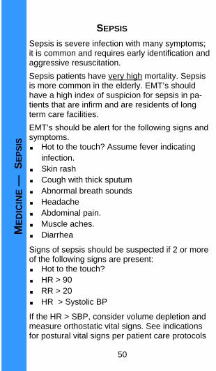
SEPSIS Sepsis is severe infection with many symptoms; it is common and requires early identification and aggressive resuscitation.
Sepsis patients have very high mortality. Sepsis is more common in the elderly. EMT’s should have a high index of suspicion for sepsis in pa-tients that are infirm and are residents of long term care facilities.
EMT’s should be alert for the following signs and symptoms. Hot to the touch? Assume fever indicating
infection. Skin rash Cough with thick sputum Abnormal breath sounds Headache Abdominal pain. Muscle aches. Diarrhea
Signs of sepsis should be suspected if 2 or more of the following signs are present: Hot to the touch? HR > 90 RR > 20 HR > Systolic BP
If the HR > SBP, consider volume depletion and measure orthostatic vital signs. See indications for postural vital signs per patient care protocols
ME
DIC
INE —
S
EP
SIS

51
and treat patients appropriately.
ALS Indicators Request Paramedics for all “Sick” patients. Decreased LOC Airway problems Respiratory distress Respirations greater than 30 per minute Signs and symptoms of shock which in-
clude: Poor skin signs (pale, sweaty) Sustained tachycardia (see page 7) Hypotension BP < 90 or positive postural vital signs (20 point drop in SBP or 20 beats per minute increase in HR) with appropriate clinical setting. (see page 119)
BLS indicators “Not Sick” patients. Conscious and alert Stable airway Stable vital signs No orthostatic changes in vital signs
BLS Care Use PPE Maintain airway, provide supplemental oxy-
gen as necessary Monitor vital signs Place patient in position of comfort Notify transport agency and or receiving
hospital of possible sepsis patient Document findings of infection and possible
sepsis on MIRF
SEPSIS (CONT.)
ME
DIC
INE —
S
EP
SIS
(C
ON
T.)

52
ME
DIC
INE —
ST
RO
KE
ALS Indicators Unconsciousness
Decreased LOC
Severe hypertension (blood pressure greater than 200 mmHg systolic or 110 mmHg diastolic with neurologic signs)
Hypotension and severe bradycardia
Seizures
Severe headache/vomiting
Uncontrolled airway and respiratory problems
Progression of stroke symptoms
BLS Indicators
Vital signs and condition stable
Stroke history
Stroke signs
Airway secure
BLS Care
Call paramedics if indicated.
Determine time onset of stroke if possible
Position patient in upright position.
Open and manage airway.
STROKE

53
Deliver oxygen and/or ventilatory assistance as necessary.
Maintain normal body temperature.
Protect paralyzed limbs.
Monitor vital signs.
Perform FAST exam.
Perform blood glucometry.
STROKE PLAN Revascularization by clot dissolving
medication should be initiated within several hours of a stroke. EMTs should attempt to limit scene times to fifteen (15) minutes.
If a stroke is of recent onset, very short scene times and transport times are critical.
Precisely document the time of onset of symptoms or time when the patient was last seen normal.
In general, arrival at hospital within several hours of onset of symptoms is critical as it will allow the ED to determine possible eligibility for thrombolytic or other therapy.
For patients with possible stroke, you must call the hospital ED and confer with a nurse or physician about the proper destination. Depending upon access to specialty care, the hospital may advise transport to their ED or may recommend another facility.
ME
DIC
INE —
ST
RO
KE (
CO
NT.)
STROKE (CONT.)

54
The FAST exam is used in the field to detect stroke. An abnormal finding strongly indicates a stroke.
FAST EXAM
Face Ask the patient to show teeth or smile
Normal: Both sides of the face move equally.
Abnormal: One side of the face does not move as well as the
Arm Ask the patient to close eyes and extend both arms straight out, palms up, for 10 seconds
Normal: Both arms move the same, or both arms do not move at all.
Abnormal: One arm drifts down compared to the other.
Speech Ask the patient to say "The sky is blue in Seattle"
Normal: The patient says correct words with no slurring of words
Abnormal: The patient slurs words, says the wrong words, or is unable to speak
Time Determine the time of onset of symptoms or when the patient was last known normal.
ME
DIC
INE —
FA
ST
EX
AM

55
Selected patients with CVA (cerebral vascular accident—stroke) can benefit from rapid thrombolytic therapy designed to dissolve the clot causing the CVA. For thrombolytic therapy to be effective, it generally should be given within 4.5 hours of the onset of the stroke. Since the hospital requires one hour for evaluation and CT this means that symptoms onset to arrival at hospital should generally be <3.5 hours. Patients who present with longer duration of symptoms may be eligible for other types of therapy including intra-arterial therapy. All hospitals in King County are designated as level I, II, or III stoke centers. You must call ahead to your usual receiving hospital and inform the staff of a code CVA patient.
The following policy is designed to assist EMTs in their evaluation of possible stroke patients. The policy stresses the need for rapid evaluation and rapid transport. For the stable patient not requiring paramedic evaluation, the EMTs should expedite transport to the hospital. Expedite does not mandate code red but rather requires rapid decision making, patient loading into the aid vehicle, and notification of hospital while enroute. You may transport code red when confined by traffic or transport time is > 15 minutes.
CODE CVA (CEREBRAL VASCULAR ACCIDENT—STROKE)
ME
DC
INE —
CO
DE C
VA

56
You must document the following information in your narrative:
1. Face: Is it symmetrical? YES or NO Arm: Symmetrical strength? YES or NO Speech: Is it slurred or abnormal? YES or NO Time: What time was the patient last known
to be normal? 2. Is the patient on Coumadin (Warfarin)?
3. Glucometry. Glucose should be over 60. Severe hypoglycemia can present like a stroke.
4. Glasgow Coma Scale Score (see page 101)
5. Time of hospital notification
6. Time you left the scene enroute to hospital
The following information must be provided to the destination hospital:
Incoming CVA patient, age, gender
Time of last known normal
Vital signs and symptoms
ETA
CODE CVA (CONT.) (CEREBRAL VASCULAR ACCIDENT—STROKE)
ME
DIC
INE —
CO
DE C
VA
(C
ON
T.)

57
To stop external bleeding:
Apply direct pressure on the open wound with sterile gauze or clean material.
Apply additional pressure if bleeding continues. A pressure dressing can be used to apply direct pressure. If blood soaks through the dressings, add new dressings—do not remove the old dressings.
If not contraindicated by the injury, elevate the bleeding extremity above the level of the heart.
If bleeding is uncontrolled by direct pressure and elevation, apply pressure at the appropriate pressure point. Hold pressure only as long as necessary to control bleeding. Reapply pressure if bleeding recurs. If pressure is held for a long period of time, tissue damage can result.
A “pressure device” may be used for control of severe, uncontrolled bleeding when all other methods of bleeding control have failed. When necessary, an oversized blood pressure cuff may be used. Inflate it no more than is necessary to stop bleeding.
Once stopped, you may need to immobilize the extremity and apply cold packs.
BLEEDING CONTROL
TR
AU
MA
—
BL
EE
DIN
G C
ON
TR
OL

58
If a patient’s condition and time permits, perform dressing and bandaging as follows:
Maintain body substance isolation (BSI) by wearing appropriate personal protective equipment.
Control bleeding with direct pressure or pressure points. Use a pressure device or pressure dressing for severe, uncontrolled bleeding. Military style trauma dressing may also be considered.
Do not remove the dressing once applied. If bleeding continues, put new dressings over the blood-soaked ones.
Secure the dressing with a bandage that is snug but does not impair circulation.
Large, easily removed debris, such as glass, splinters, or gravel can be removed before bandaging. Secure large, deeply imbedded fragments or projectiles in place with the bandage.
If possible, leave patient’s fingers or toes exposed.
Check circulation by feeling for a distal pulse or checking capillary refill.
Elevate or immobilize the injured extremity, if possible.
Cover eviscerated abdominal contents with a large multi-trauma dressing soaked with
DRESSING AND BANDAGING T
RA
UM
A —
D
RE
SS
ING
AN
D B
AN
DA
GIN
G

59
sterile saline (or clean water if saline unavailable). Then apply an occlusive dressing, if available, to retain heat and moisture. Secure with tape.
AMPUTATION
Wrap amputated parts in sterile dressings.
Place amputated part in a watertight container and then in a second container.
Place the container on ice.
Do not submerge the amputated part in water or place directly on ice.
Rapid transport of the patient and the severed part is critical to the success of re-implantation. If transport of a patient is delayed, consider sending the amputated part ahead to be surgically prepared.
Do not use dry ice to cool a severed part. Ice and chemical cold packs are acceptable.
BURNS
For burned areas, easily removed debris should be taken off the burn. Cover the area with dry, sterile dressings.
Remove wet chemicals, such as acid, with repeated flushing. Remove dry substance by first brushing the area and then flushing.
DRESSING AND BANDAGING (CONT.)
TR
AU
MA
—
DR
ES
SIN
G A
ND
BA
ND
AG
ING
(C
ON
T.)

60
ALS Indicators
Possible airway involvement including singed facial hair, soot in mouth/nose or hoarseness
Burns with associated injuries: electrical shock, fracture, or respiratory problems
Second or third degree burns to face/head
Second or third degree burns covering greater than 20% of the body
Severe pain (request ALS for pain control)
BLS Indicators
All other burns
BLS Care
First degree burn Cool, moist pads
Second degree burn Cover with dry dressing (commercial burn
sheets are acceptable)
DO NOT apply ointment or creams
Always be alert to possible airway involvement.
BURNS
TR
AU
MA
—
BU
RN
S

61
ALS Indicators
Major mechanism of injury
Penetrating injuries to eye
BLS Indicators
Minor mechanism of injury
Eyelid laceration with intact vision
Ultraviolet burns
BLS Care
Request paramedics if indicated.
Stabilize an impaled object in place and bandage both eyes.
Flush chemical burns to the eyes for 15 minutes with normal saline or water if saline is not available.
Ultraviolet burns to the eyes: treat with cool compresses over closed eyes.
EYE INJURIES
TR
AU
MA
—
EY
E IN
JUR
IES

62
ALS Indicators
Compromised airway
Abnormal respiratory patterns
Major mechanism of injury
Glasgow Coma Scale of 12 or less
Decreased LOC, unstable vital signs
Paresis (partial or complete paralysis) and/or paresthesia (abnormal sensation, e.g., tingling)
Evidence of injury to brain or spinal cord
Significant alcohol or drug use
BLS Indicators
Minor mechanism of injury
Intact airway, stable vital signs
No evidence of injury to brain or spinal cord
No significant drug or alcohol use
BLS Care
Request paramedics if indicated.
Ensure a patent airway.
Provide supplemental oxygen and/or ventilatory assistance as necessary.
Provide neutral, in-line cervical stabilization with proper sized cervical collar and padding.
TR
AU
MA
—
HE
AD
AN
D N
EC
K
HEAD AND NECK

63
Secure to backboard. Bandage as necessary. Monitor vital signs and neurologic status.
Special Instructions For Suspected Cervical Injury
Suspected cervical injury with non-alignment One attempt to realign neck to the neutral, in-line position unless new pain, additional numbness, tingling or weakness, additional compromise of airway or ventilation or resistance encountered.
Apply cervical collar and backboard (see page 130). If unable to realign then secure in the original position.
Helmet Removal As long as the airway is not affected and remains patent AND the c-spine can be secured in a neutral, in-line position, leave football and motorcycle helmets on. Pad the backboard/torso to maintain neutral alignment.
All other non-fitted helmets may be removed as soon as possible (e.g., bicycle helmets, skateboard helmets, rollerblade helmets).
If helmet needs to be removed, two EMTs should stabilize head and neck, remove chinstrap, remove helmet while stabilizing head, and apply cervical collar. Secure the patient to a backboard (see page 130).
TR
AU
MA
—
HE
AD
AN
D N
EC
K (
CO
NT.)
HEAD AND NECK (CONT.)

64
ALS Indicators
Decreased/altered LOC
Signs or symptoms of shock
Excessive uncontrolled bleeding
Pelvic fracture, bilateral femur fracture, or multi-system injury/fractures
Femur fracture with excessive swelling
Open fractures except for hands and feet
High index of suspicion based on mechanism of injury
Contact paramedic for severe pain (patient needs pain control)
BLS Indicators
Single extremity fracture with stable vital signs
Single joint injury with stable vital signs
BLS Care
Request paramedics if indicated.
Protect cervical spine if indicated.
Reassure and maintain normal body temperature.
Apply direct pressure and sterile dressing over major bleeding.
ORTHOPAEDIC T
RA
UM
A —
O
RT
HO
PE
DIC

65
Provide supplemental oxygen and/or ventilatory assistance as necessary.
Nothing by mouth.
Gently support injured part (see page 132).
Allow patient to choose position of comfort.
Check and record distal circulation, motor, and sensory (nerve function) before and after splinting.
Immobilize and splint if indicated.
Apply cold/ice pack to injured part (for closed tissue injury only).
Elevate fractured limb.
Prepare patient for transport (backboard).
Monitor patient’s vital signs every 5 to 10 minutes.
Realignment of Long Bone Fractures
Attempt to realign (open or closed) long bones that are angulated in the middle 1/3 then splint.
Long-bone fractures, which occur in the proximal or distal 1/3, that may or may not involve a joint, may be realigned if compromise of distal circulation or nerve function is detected and definitive care is delayed.
ORTHOPAEDIC (CONT.)
TR
AU
MA
—
OR
TH
OP
ED
IC (
CO
NT.)

66
Realignment may sometimes be necessary to facilitate packaging for transport.
Check and document CMS before and after splinting and/or realignment.
Pelvic Fractures (see page 133)
Multiple Extremity Fractures
These patients should be secured to a backboard which will serve as a general body splint for several sites.
Falls In Elderly Patients
In addition to consideration of orthopaedic injuries, consider head trauma and possible CNS bleeding (especially if they are on coumadin). Elderly patients on coumadin with head injury or suspected head injury MUST be evaluated in an emergency department.
ORTHOPAEDIC (CONT.) T
RA
UM
A —
O
RT
HO
PE
DIC
(C
ON
T.)
Rapid packaging and transport of the unstable patient or patient with multiple fractures takes priority over definitive
splinting at the scene.

67
ALS Indicators
Significant head injury
Signs and symptoms of shock which include: Poor skin signs (pale, sweaty) Sustained tachycardia (see page 7) Hypotension (systolic BP less than 90
mmHg) with an appropriate clinical setting
Soft tissue injuries that might compromise the airway
Excessive uncontrolled bleeding
Altered LOC
High index of suspicion based on mechanism of injury
BLS Indicators
Conscious and alert
Stable vital signs
Soft tissue injuries limited to the superficial layer of the skin (epidermis and dermis)
Single digit amputations (see page 59)
Soft tissue injuries, with bleeding controlled by direct pressure and/or elevation
SOFT TISSUE
TR
AU
MA
—
SO
FT T
ISS
UE

68
BLS Care for OPEN Soft Tissue Injuries
Request ALS if indicated.
Provide supplemental oxygen and/or ventilatory assistance as necessary.
Maintain an open airway.
Ensure adequate breathing.
Control bleeding.
Maintain normal body temperature.
Monitor vital signs.
Cervical spine protection, if indicated.
Special Instructions for OPEN Soft Tissue Injuries
Control bleeding with direct pressure on the area or upon pressure points. Use pressure dressings or pressure device (like a BP cuff) for severe, uncontrolled bleeding. Military style trauma dressing may also be considered.
Amputation (see page 59)
Removal of Foreign Objects
Large, easily removed debris, such as glass, splinters, or gravel must be removed before bandaging.
TR
AU
MA
— S
OF
T T
ISS
UE (
CO
NT.)
SOFT TISSUE (CONT.)

69
TR
AU
MA
—
SO
FT T
ISS
UE (
CO
NT.)
Large, deeply imbedded fragments or projectiles must be secured in place by the bandage.
Decontamination
Remove wet chemicals (e.g., acid) by repeated flushing with water.
Remove dry substances by first brushing the area and then by flushing with water.
Burns
Easily removed debris should be taken off the burned area, then cover the area with dry, sterile dressings.
Remove rings for hand burns.
SOFT TISSUE (CONT.)

70
OROPHARYNGEAL (OP) AIRWAY
An oropharyngeal airway rests in the patient's oropharynx, lifting the tongue away from the back of the throat preventing it from occluding the airway. The OP airway is used only on unconscious patients and generally those without respirations.
To size an oropharyngeal airway:
Choose correct size by measuring from the corner of the mouth to the ear lobe or from the chin to the angle of the jaw.
In infants and children, insert the airway tip down or sideways along with a tongue blade. Rotate down when you are halfway in the mouth or approaching the curve on the tongue.
AIRWAY MANAGEMENT
Do not use this device if a patient gags when inserted. Use of an airway on a patient with a gag reflex may cause
retching, vomiting, or spasm of the vocal cords.
PR
OC
ED
UR
ES &
PO
LIC
IES —
A
IRW
AY A
MA
NA
GE
ME
NT
An oropharangeal (OP) airway is not necessary if ventilation via BVM is easily
accomplished.

71
PR
OC
ED
UR
ES &
PO
LIC
IES —
A
IRW
AY M
AN
AG
EM
EN
T (
CO
NT.)
SUCTIONING
The Yankauer suction tip is preferred for most suctioning. If the holes on the Yankauer get plugged repeatedly, remove the tip and use larger bore tubing.
To suction with a Yankauer tip: Measure the same as for an oropharyngeal airway—approximately from the corner of the mouth to the ear lobe.
Do not suction while inserting; suction only after the Yankauer (or similar device) is in place and as you withdraw.
Suction for no more than 15 seconds at a time.
In rare cases, copious vomiting that threatens the airway may require a longer period of suctioning.
Oxygenate the patient well before and after suctioning.
AIRWAY MANAGEMENT (CONT.)

72
PR
OC
ED
UR
ES &
PO
LIC
IES —
BA
G-V
AL
VE M
AS
K
Successful ventilation with a BVM requires a good seal between the mask and the patient's face and maintaining an open airway.
To properly place a BVM:
Choose appropriate size for the patient.
Place the apex of the mask on the bridge of the nose (between the eyebrows).
Settle the base of the mask between the lower lip and the prominence of the chin.
TECHNIQUE
Kneel with a knee on each side of the patient's head.
Hold the mask firmly in position by placing the heel of the hand on top of the mask, extending the fingers and thumb forward forming a “C”, and grasping the lower jaw with the middle two or three fingers.
Squeeze the bag to ventilate.
If necessary, a second EMT may be needed to secure seal and assist with bagging.
Each ventilation should take one second and achieve chest rise.
Correct ventilation generates only modest chest rise.
BAG-VALVE MASK

73
NOTES
NO
TE
S

74
KIN
G C
OU
NT
Y E
ME
RG
EN
CY M
ED
ICA
L S
ER
VIC
ES
CA
RD
IAC
AR
RE
ST
IN
AD
UL
TS
AN
D C
HIL
DR
EN
≥ 8
YR
S O
LD
A
FO
R P
HIL
IPS
AE
D A
GE
NC
IES
AP
PR
OA
CH
TO
CA
RD
IAC
AR
RE
ST
FO
R K
ING
CO
UN
TY
EM
S A
GE
NC
IES
(C
AB
B:
Ch
est
co
mp
ress
ion
s à
Airw
ay
à B
rea
thin
g)
In t
he p
atie
nt
wh
o is
un
con
scio
us/
unre
spo
nsi
ve,
no
t b
rea
thin
g n
orm
ally
an
d in
wh
om
no
pul
se is
de
tect
ed,C
imm
edia
tely
p
erf
orm
ch
est
com
pre
ssio
ns,
D w
hile
turn
ing
on a
nd a
ttach
def
ibril
lato
r (A
ED
). O
nce
AE
D is
app
lied,
giv
e v
erb
al r
epo
rt a
nd
cou
nt c
omp
ress
ion
s. A
t co
mp
letio
n o
f 30
com
pre
ssio
ns,
ana
lyze
rh
yth
m,E
cle
ar
patie
nt a
nd s
hock
if in
dica
ted.
F R
esum
e ch
est
co
mp
ress
ion
s a
nd c
ont
inue
fo
r ~
2 m
inu
tes
bef
ore
ne
xt r
hyt
hm
an
aly
sis.
E, G
Alw
ays
com
ple
te a
ny
star
ted
cycl
e of
30
com
pre
ssio
ns
prio
r to
an
y rh
yth
m a
naly
sis
an
d a
lwa
ys r
esu
me
che
st c
om
pre
ssio
ns
imm
edia
tely
aft
er
rhyt
hm a
na
lysi
s/sh
ock
. D
o n
ot c
reat
e a
n a
dded
pa
use
by
ven
tila
ting
imm
ed
iate
ly b
efo
re a
ny
rhyt
hm
an
aly
sis.
E P
alp
ate
fem
ora
l pu
lse
(o
r
CARDIAC ARREST
Aft
er
2 m
inu
tes
of C
PR
, An
aly
ze r
hyt
hm
C
he
ck f
emo
ral p
uls
e w
hile
ana
lyzi
ng
rhyt
hm
Beg
in C
AB
. If
un
con
scio
us/
unre
spon
sive
, n
ot b
reat
hin
g n
orm
ally
and
no
pu
lse
imm
edia
tely
pe
rfo
rm c
hest
co
mp
res-
sio
ns,
turn
on
an
d a
ttach
def
ibril
lato
r. C
omp
lete
30
com
pre
ssio
ns;
an
aly
ze r
hyt
hm
. E
xcep
tio
n: W
hen
the
pa
tien
t go
es
into
VF
wh
ile m
on
itore
d o
r a
ttach
ed
to a
n A
ED
a d
efib
rilla
tory
sh
ock
ma
y b
e ad
min
-
iste
red
imm
ed
iate
ly.
No
Sh
ock
Ad
vise
d/I
nd
icat
ed
Imm
edi
atel
y be
gin
ches
t co
mp
ress
ion
. P
erf
orm
2 m
inut
es
of u
nin
terr
upt
ed
CP
R
Do
no
t d
ela
y C
PR
fo
r p
uls
e ch
eck
Sh
ock
Ad
vise
d/I
nd
ica
ted
(V
F o
r p
uls
ele
ss V
T)
Imm
ed
iate
ly d
eliv
er
SIN
GL
E s
hock
. Im
me
dia
tely
re
sum
e u
nin
terr
upt
ed
CP
R x
2 m
inu
tes.
D
o n
ot
del
ay
CP
R f
or
po
st-s
ho
ck p
uls
e c
hec
k o
r rh
yth
m a
nal
ysis
.
PR
OC
ED
UR
ES &
PO
LIC
IES —
CA
RD
IAC
AR
RE
ST

75
PR
OC
ED
UR
ES &
PO
LIC
IES —
CA
RD
IAC
AR
RE
ST (
CO
NT.)
CARDIAC ARREST (CONT.)
Aft
er
2 m
inu
tes
of C
PR
, a
naly
ze r
hyt
hm
C
he
ck f
emo
ral p
uls
e w
hile
ana
lyzi
ng
rhyt
hm
C
all
Ho
tlin
e a
fter
eve
ry c
ard
iac
eve
nt:
1-8
00
-60
7-2
92
6
Pro
vid
e y
ou
r n
am
e, a
gen
cy,
com
pan
y, d
ate
, tim
e, m
ed
ic u
nit,
pat
ien
t age
a
nd
gen
de
r, a
nd y
ou
r ca
ll b
ack
num
be
r.
Aft
er
2 m
inu
tes
of C
PR
, a
naly
ze r
hyt
hm
C
he
ck f
emo
ral p
uls
e w
hile
ana
lyzi
ng
rhyt
hm
Sh
ock
Ad
vise
d/I
nd
ica
ted
(V
F o
r p
uls
ele
ss V
T)
Imm
ed
iate
ly d
eliv
er
SIN
GL
E s
hock
. Im
me
dia
tely
re
sum
e u
nin
terr
upt
ed
CP
R x
2 m
inu
tes.
D
o n
ot
del
ay
CP
R f
or
po
st-s
ho
ck p
uls
e c
hec
k o
r rh
yth
m a
nal
ysis
.
No
Sh
ock
Ad
vise
d/I
nd
icat
ed
If p
uls
e,
ass
ess
blo
od
pre
ssu
re a
irwa
y a
nd
bre
ath
ing
If n
o p
uls
e,
perf
orm
2 m
inu
tes
of u
nin
terr
upte
d C
PR
Sh
ock
Ad
vise
d/I
nd
ica
ted
(V
F o
r p
uls
ele
ss V
T)
Imm
ed
iate
ly d
eliv
er
SIN
GL
E s
hock
. Im
me
dia
tely
re
sum
e u
nin
terr
upt
ed
CP
R x
2 m
inu
tes.
D
o n
ot
del
ay
CP
R f
or
po
st-s
ho
ck p
uls
e c
hec
k o
r rh
yth
m a
nal
ysis
.
No
Sh
ock
Ad
vise
d/I
nd
icat
ed
If p
uls
e,
ass
ess
blo
od
pre
ssu
re a
irwa
y a
nd
bre
ath
ing
If n
o p
uls
e,
perf
orm
2 m
inu
tes
of u
nin
terr
upte
d C
PR

76
KIN
G C
OU
NT
Y E
ME
RG
EN
CY M
ED
ICA
L S
ER
VIC
ES
CA
RD
IAC
AR
RE
ST
IN
AD
UL
TS
AN
D C
HIL
DR
EN
≥ 8
YR
S O
LD
A
FO
R P
HY
SIO
-CO
NT
RO
L A
ED
AG
EN
CIE
S
AP
PR
OA
CH
TO
CA
RD
IAC
AR
RE
ST
FO
R K
ING
CO
UN
TY
EM
S A
GE
NC
IES
(C
AB
B:
Ch
est
co
mp
ress
ion
s à
Airw
ay
à B
rea
thin
g)
In t
he p
atie
nt
wh
o is
un
con
scio
us/
unre
spo
nsi
ve,
no
t b
rea
thin
g n
orm
ally
an
d in
wh
om
no
pul
se is
de
tect
ed,C
imm
edia
tely
p
erf
orm
ch
est
com
pre
ssio
ns,
D w
hile
turn
ing
on a
nd a
ttach
def
ibril
lato
r (A
ED
). O
nce
AE
D is
ap
plie
d,
give
ve
rba
l re
po
rt
an
d c
oun
t co
mp
ress
ion
s. A
t com
ple
tion
of
30 c
om
pre
ssio
ns,
an
aly
ze r
hyt
hm
.E If
sho
ck is
ad
vise
d/in
dica
ted
perf
orm
30
ch
est
co
mp
ress
ion
s w
hile
AE
D is
ch
arg
ing,
cle
ar
pa
tien
t, a
nd s
hock
.F R
esum
e ch
est
com
pres
sion
s an
d co
ntin
ue fo
r ~
2 m
inu
tes
befo
re n
ext
rh
yth
m a
naly
sis.
E, G
Alw
ays
com
ple
te a
ny s
tart
ed c
ycle
of
30 c
omp
ress
ion
s p
rior
to a
ny
rhyt
hm
a
na
lysi
s a
nd
alw
ays
re
sum
e ch
est
com
pre
ssio
ns
imm
ed
iate
ly a
fte
r rh
yth
m a
na
lysi
s/sh
ock
. Do
no
t cre
ate
an
ad
ded
p
au
se b
y ve
ntil
atin
g im
me
dia
tely
bef
ore
an
y rh
yth
m a
na
lysi
s.E W
hen
po
ssib
le p
alp
ate
fem
ora
l pu
lse
(o
r ca
rotid
pu
lse
if
fem
ora
l is
ina
cce
ssib
le)
du
ring
CP
R a
nd
pa
rtic
ula
rly p
rior
to a
nd
du
ring
an
y p
au
se in
CP
R.H
PR
OC
ED
UR
ES &
PO
LIC
IES —
CA
RD
IAC
AR
RE
ST (
CO
NT.)
CARDIAC ARREST (CONT.)
Beg
in C
AB
. If
un
con
scio
us/
unre
spon
sive
, n
ot b
reat
hin
g n
orm
ally
and
no
pu
lse
imm
edia
tely
pe
rfo
rm c
hest
co
mp
res-
sio
ns,
turn
on
an
d a
ttach
def
ibril
lato
r. C
omp
lete
30
com
pre
ssio
ns;
an
aly
ze r
hyt
hm
. E
xcep
tio
n: W
hen
the
pa
tien
t go
es
into
VF
wh
ile m
on
itore
d o
r a
ttach
ed
to a
n A
ED
a d
efib
rilla
tory
sh
ock
ma
y b
e a
dm
inis
tere
d im
med
iate
ly.
No
Sh
ock
Ad
vise
d/I
nd
icat
ed
Imm
ed
iate
ly b
egi
n c
hest
com
pre
ssio
n.
Pe
rfo
rm 2
min
ute
s of
un
inte
rru
pte
d C
PR
D
o n
ot
del
ay
CP
R f
or
pu
lse
chec
k
Sh
ock
Ad
vise
d/I
nd
ica
ted
(V
F o
r p
uls
ele
ss V
T)
Ch
arg
e A
ED
. P
erf
orm
30
che
st c
om
pre
ssio
ns
du
ring
AE
D c
harg
ing
De
live
r S
ING
LE
sh
ock
. Im
med
iate
ly r
esu
me
un
inte
rru
pte
d C
PR
x 2
m
inu
tes.
D
o n
ot
del
ay
CP
R f
or
po
st-s
ho
ck p
uls
e c
hec
k o
r rh
yth
m a
nal
ysis
.
Aft
er
2 m
inu
tes
of C
PR
, An
aly
ze r
hyt
hm
C
he
ck f
emo
ral p
uls
e w
hile
ana
lyzi
ng
rhyt
hm

77
CARDIAC ARREST (CONT.)
PR
OC
ED
UR
ES &
PO
LIC
IES —
CA
RD
IAC
AR
RE
ST (
CO
NT.)
Aft
er
2 m
inu
tes
of C
PR
, a
naly
ze r
hyt
hm
C
he
ck f
emo
ral p
uls
e w
hile
ana
lyzi
ng
rhyt
hm
Ca
ll H
otli
ne
afte
r e
very
ca
rdia
c e
ven
t: 1
-80
0-6
07
-29
26
P
rovi
de
yo
ur
na
me
, age
ncy
, co
mpa
ny,
dat
e, t
ime
, me
dic
un
it, p
atie
nt a
ge
an
d g
end
er,
and
yo
ur
call
ba
ck n
umb
er.
Aft
er
2 m
inu
tes
of C
PR
, a
naly
ze r
hyt
hm
C
he
ck f
emo
ral p
uls
e w
hile
ana
lyzi
ng
rhyt
hm
No
Sh
ock
Ad
vise
d/I
nd
icat
ed
If p
uls
e,
ass
ess
blo
od
pre
ssu
re a
irwa
y a
nd
bre
ath
ing
If n
o p
uls
e,
perf
orm
2 m
inu
tes
of u
nin
terr
upte
d C
PR
Sh
ock
Ad
vise
d/I
nd
ica
ted
(V
F o
r p
uls
ele
ss V
T)
Ch
arg
e A
ED
. P
erf
orm
30
che
st c
om
pre
ssio
ns
du
ring
AE
D
cha
rgin
g D
eliv
er
SIN
GL
E s
ho
ck. I
mm
edia
tely
re
sum
e u
nin
terr
upt
ed
CP
R x
2 m
inu
tes.
D
o n
ot
del
ay
CP
R f
or
po
st-s
ho
ck p
uls
e c
hec
k o
r rh
yth
m
anal
ysis
.
No
Sh
ock
Ad
vise
d/I
nd
icat
ed
If p
uls
e,
ass
ess
blo
od
pre
ssu
re a
irwa
y a
nd
bre
ath
ing
If n
o p
uls
e,
perf
orm
2 m
inu
tes
of u
nin
terr
upte
d C
PR
Sh
ock
Ad
vise
d/I
nd
ica
ted
(V
F o
r p
uls
ele
ss V
T)
Ch
arg
e A
ED
. P
erf
orm
30
che
st c
om
pre
ssio
ns
du
ring
AE
D
cha
rgin
g D
eliv
er
SIN
GL
E s
ho
ck. I
mm
edia
tely
re
sum
e u
nin
terr
upt
ed
CP
R x
2 m
inu
tes.
D
o n
ot
del
ay
CP
R f
or
po
st-s
ho
ck p
uls
e c
hec
k o
r rh
yth
m a
nal
ysis
.

78
PR
OC
ED
UR
ES &
PO
LIC
IES —
CA
RD
IAC
AR
RE
ST (
CO
NT.)
CARDIAC ARREST (CONT.)
A. If age is not known, the presence of secondary sexual characteristics (development of axillary hair in males and breast tissue in females) define a child who has reached puberty and who should be treated as an “adult”. In ages < 8yrs (see below) continue uninter-rupted CPR until medics arrive. If a public access defi-brillator (PAD) is attached prior to your arrival, you may use it.
B. CAB refers to “Chest compressions followed by Airway followed by Breathing” interventions, and has displaced “ABC” in 2010.
C. If no pulse felt within 10 seconds, begin chest compres-sions. Count out loud for chest compressions.
D. Each CPR cycle (including the very first) begins with chest compression (at 100/min, ≥ 2 inches, with full recoil and 50%/50% compression/recoil duty cycle). Except in obvious cases of asphyxia (e.g. known drown-ing victim), opening the airway and ventilation (2 breaths) are not performed until completion of the first 30 chest compressions or after rhythm analysis.
E. To minimize the hands off (no chest compression) interval before a rhythm analysis/shock, complete 30 chest compressions, but do not create an added pause by ventilating (or checking pulse) just before rhythm analysis. That is, any CPR cycle immediately before shock is 30 compressions followed immediately by rhythm analysis and shock.
F. Philips AEDs: MRx, ForeRunner AEDs charge simul-taneously while analyzing rhythm (unless the “Pause (for CPR)” soft key is pressed). If a shock is advised during analysis, proceed to immediate shock, then resume CPR.
Physio Control AEDs: If shock is advised, resume CPR for 30 compressions while AED is charging. Then pause CPR briefly for shock, and immediately resume CPR thereafter.
G. 2 minutes in this protocol refers to 2 minutes or slightly longer depending on when 30 compressions before a

79
rhythm analysis are complete. During 2 minute CPR cycles, give 2 breaths (each over 1 sec) after every 30 compressions. Periods of CPR should not be inter-rupted except in cases of need to manage airway (emesis, etc.)
H. Whenever possible, a designated provider should maintain a finger(s) on the femoral pulse (or carotid pulse if femoral pulse is inaccessible) during CPR. This permits a qualitative assessment of the adequacy of chest compressions and an immediate pulse check without a pulse “hunt” (by already having a hand on its location) whenever CPR is paused for rhythm analysis, or at any other time that the protocol calls for a pulse check.
Additional Points:
Any patient found unconscious, unresponsive with a pulse but with systolic BP <60 should have CPR initi-ated. If a pulse is detected during resuscitation but systolic blood pressure < 60, resume CPR.
In children 1-8 yrs (absence of secondary sexual characteristics; usually <55 lbs), perform chest com-pressions at 100/min, ≥ 2 inches or 1/3 of chest depth at compression to ventilation ratio of 15:2
In infants < 1 yr compress chest at 100 min, to 1½ inches or 1/3 of chest depth with compression to venti-lation ratio of 15:2.
In newborns, compress to 1/3 chest depth. Perform 90 compressions and 30 ventilations/minute (compression: ventilation ratio of 3:1), taking ½ second for each compression or ventilation. If cardiac arrest suspected to be of cardiac etiology, may perform 15:2 CPR.
If at anytime 3 consecutive “no shocks” are advised and there is no pulse, continue CPR without interrup-tion until medics arrive.
Cardiac arrest protocols may change. Always follow current agency protocols.
CARDIAC ARREST (CONT.)
PR
OC
ED
UR
ES &
PO
LIC
IES —
CA
RD
IAC
AR
RE
ST (
CO
NT.)

80
Seattle Fire Department Cardiac Arrest AFTER Arrival
Adults ≥ 8 Years Old
CARDIAC ARREST (SEATTLE FD) P
RO
CE
DU
RE
S &
PO
LIC
IES —
CA
RD
IAC
AR
RE
ST (
SE
AT
TL
E F
D)
APPLY LP500
2 MINUTES OF CPR
BEGIN CPR 10:1 @ 100 PER MIN
VERIFY CARDIAC ARREST
ANALYZE AS SOON AS POSSIBLE
“SHOCK ADVISED” “NO SHOCK ADVISED”
30 Compressions while LP500 charges
SHOCK (no pulse check)
ANALYZE and rotate
compressor (Repeat cycle)
If “NO SHOCK”, con-tinue to ANALYZE
each
2 minute cycle
Check for pulse (< 10 seconds)
Rotate compressor If no pulse
Repeat until Medic arri-
val (no analysis)
On Medic Arrival Continue CPR until Medics ready to Charge/
Analyze/Shock with LP12
2 MINUTES OF CPR

81
CARDIAC ARREST (SEATTLE FD) Seattle Fire Department
Cardiac Arrest Before Arrival Adults ≥ 8 Years Old
PR
OC
ED
UR
ES &
PO
LIC
IES —
CA
RD
IAC
AR
RE
ST (
SE
AT
TL
E F
D)
2 MINUTES OF CPR
10:1 @ 100 PER MIN
VERIFY CARDIAC ARREST
1st ANALYSIS ROTATE COMPRESSOR
“SHOCK ADVISED” “NO SHOCK ADVISED”
30 Compressions while LP500 charges
SHOCK (no pulse check)
ANALYZE and rotate compressor
(Repeat cycle)
If “NO SHOCK”, continue to
ANALYZE each 2 minute cycle
Check for pulse (< 10 seconds)
Rotate compressor If no pulse
Repeat until Medic arrival
(no analysis)
On Medic Arrival Continue CPR until Medics ready to Charge/
Analyze/Shock with LP12
2 MINUTES OF CPR
2 MINUTES OF CPR

82
Seattle Fire Department Cardiac Arrest
Pediatrics < 8 Years Old (Adults ≥ 8 use LP500)
CARDIAC ARREST (SEATTLE FD) P
RO
CE
DU
RE
S &
PO
LIC
IES —
CA
RD
IAC
AR
RE
ST (
SE
AT
TL
E F
D)
2 MINUTES OF CPR
2 MINUTES OF CPR
BEGIN CPR 10:1 @ 100 PER MIN
VERIFY CARDIAC ARREST
Check for pulse (less than 10 seconds) Rotate compressor If no pulse
Repeat until Medic arrival

83
CPR FOR ADULTS
PR
OC
ED
UR
ES &
PO
LIC
IES —
CP
R F
OR
AD
UL
TS
MANUVER ADULT HCP: Adolescent and older
RECOGNITION Unresponsive (for all ages) No breathing or no normal breathing (ie, only gasping) No pulse palpated within 10 seconds for all ages (HCP only)
ACTIVATE: Emergency Response Number (lone rescuer)
Activate when victim found unre-sponsive HCP: if asphyxial arrest likely, call after 5 cycles (2 minutes) of CPR
CPR Sequence C-A-B Compression Rate At least 100/min Compression Depth At least 2 inches (5cm) Chest Wall Recoil Allow complete recoil between
compressions HCP rotate compressors every 2 minutes
Compression Interruptions Minimize interruptions in chest compressions Attempt to limit interruptions to <10 seconds
Airway Head tilt-chin lift (HCP suspected trauma: jaw thrust)
Compression-to-ventilation ratio (until advanced airway placed)
30:2 1 or 2 rescuers
Ventilations: When rescuer untrained or trained and not proficient
Compressions only
Ventilations with advanced airway (HCP)
1 breath every 6-8 seconds (8-10 breaths/min)
Asynchronous with chest compres-sions
About 1 second per breath Visible chest rise
Foreign-body airway obstruction
Responsive: Abdominal thrusts Unresponsive: CPR with airway check
Defibrillation Attach and use AED as soon as possible. Minimize interruptions in chest compressions before and after shock; resume CPR beginning with compressions immediately after each shock.

84
CPR FOR CHILDREN AND INFANTS P
RO
CE
DU
RE
S &
PO
LIC
IES —
CP
R F
OR
CH
ILD
RE
N A
ND
INF
AN
TS MANUVER CHILD
HCP: 1 year to Adolescent
INFANT Under 1 year of
age RECOGNITION Unresponsive (for all ages)
No breathing or only gasping) No pulse palpated within 10 seconds for all
ages (HCP only) ACTIVATE: Emergency Response Number (lone rescuer)
Activate after performing 5 cycles of CPR For sudden, witnessed collapse, activate after verifying that victim unresponsive
CPR Sequence C-A-B Compression Rate At least 100/min Compression Depth
At least 1/3 AP diame-ter About 2 inches (5cm)
At least 1/3 AP diameter About 1 ½ inches (4cm)
Chest Wall Recoil Allow complete recoil between compressions HCP rotate compressors every 2 minutes
Compression Interruptions
Minimize interruptions in chest compressions Attempt to limit interruptions to <10 seconds
Airway Head tilt-chin lift (HCP suspected trauma: jaw thrust)
Compression-to-ventilation ratio (until advanced airway placed)
30:2 2 rescuers
15:2 2HCP rescuers
Ventilations: When rescuer untrained or trained and not proficient
Compressions only
Ventilations with advanced airway (HCP)
1 breath every 6-8 seconds (8-10 breaths/min)
Asynchronous with chest compressions About 1 second per breath
Visible chest rise Foreign-body airway obstruction
Responsive: Abdominal thrusts Unresponsive: CPR with airway
check
Responsive: Back slaps and chest
thrusts Unresponsive: CPR
with airway check Defibrillation Attach and use AED as soon as possible.
Minimize interruptions in chest compressions before and after shock; resume CPR beginning
with compressions immediately after each shock.

85
CPR FOR NEWBORN
PR
OC
ED
UR
ES &
PO
LIC
IES —
CP
R F
OR
NE
WB
OR
N
Maneuver HCP = Health Care
Provider
Newborn
AIRWAY Head tilt/chin lift (Minimal, only as needed) (Suction only as needed)
BREATHS Initial
2 effective breaths at 1 second/breath (obtain chest rise)
Rescue breathing without chest compression
40-60 breaths/minutes (~1 breath every 1 to 1.25 seconds)
CIRCULATION Check pulse at umbilical cord stub or over the heart
Compression land-marks
Just below the nipple line
Compression Method (allow full recoil)
2 rescuers perform skill: “two thumb-encircling hands” technique
Compression Depth 1/3 depth of the chest
Compression Rate 120 per minute
Compression/Ventilation Ratio and events/minute
3:1 (2 rescuer) Deliver 90 compressions and 30 vents/minute
DEFIB: AED Not performed on children less than 8 years

86
The indications for ECG monitoring include: chest pain, arrhythmia, congestive heart failure, syncope or hypotension.
For BLS providers to perform ECG monitoring the following criteria must be met:
An approved course in ECG monitoring techniques and rhythm recognition.
Specific defibrillation/monitoring equipment, which will provide hard copy ECG rhythm strips for use by paramedics and others.
Medical Program Director approval for addition of ECG monitoring to EMT care plans.
EPISTAXIS (NOSEBLEED)
Stop a non-traumatic, “everyday” nosebleed by asking the patient to sit, leaning forward. This prevents blood from being swallowed or aspirated into the lung.
Apply direct pressure by pinching just below the bridge of the nose.
Apply pressure for 10 to 15 minutes.
Additionally, you can apply a cold pack to the bridge of the nose.
ECG MONITORING
PR
OC
ED
UR
ES &
PO
LIC
IES
—
EC
G M
ON
ITO
RIN
G /
EP
IST
AX
IS

87
EMTs have the responsibility to determine a patient’s resuscitation wishes, and honor them if possible.
Resuscitation efforts may be withheld or stopped in ANY of the following:
Injuries incompatible with life
Lividity, rigor mortis
A Do Not Attempt Resuscitation (DNAR) directive. This directive may be in the POLST (Physician Orders For Life-Sustaining Treatment) format. This is based on patient’s wishes.
“Compelling reasons” to withhold resuscitation can be invoked when written information is not available, yet the situation suggests that the resuscitation effort will be futile, inappropriate, and inhumane. A resuscitation effort may be withheld when the following two conditions are BOTH met:
End stage of a terminal illness Family indicates that the patient would not
wish to have a resuscitation effort
If a resuscitation effort has been initiated and the EMT is provided with a DNAR directive or compelling reasons that such an effort should be withheld, the resuscitation should be stopped.
END OF LIFE ISSUES
PR
OC
ED
UR
ES &
PO
LIC
IES —
EN
D O
F L
IFE I
SS
UE
S

88
Documentation is important. On the Incident Report Form, describe the patient’s medical history, presence of a DNAR directive if any, or verbal request to withhold resuscitation efforts.
“Do not attempt resuscitation” does not mean “do not care.” A dying patient for whom no resuscitation effort is indicated can still be provided with supportive care, which may include the following:
Clear the airway (including stoma) of secretions with suction device.
Provide oxygen using a cannula or non-rebreather.
Control any bleeding.
Provide emotional support to patient and family.
Contact the patient’s private physician.
Contact hospice if involved.
Paramedics should be called if additional judgment or support is needed.
END OF LIFE ISSUES (CONT.)
When in doubt, initiate resuscitation.
PR
OC
ED
UR
ES &
PO
LIC
IES —
EN
D O
F L
IFE IS
SU
ES (
CO
NT.)

89
EMTs and paramedics are authorized to consult with Group Health (GH) On-Scene Physician Support (OPS) for non-critical Group Health patients. Specifically OPS is intended for Group Health patients who are stable and do not require immediate paramedic transport. OPS does not replace paramedic contact with medical control doctors for protocol plans care or other required contact.
The OPS program is designed to give EMTs and paramedics in King County telephone access to a consulting GH physician who has immediate access to the patient’s full medical record. The physician can assist in determining the most appropriate care, most appropriate destination, and most appropriate transportation. In addition the physician can arrange a variety of care options including a short-notice appointment ot GH clinics, arrangements for medications to be filled at a GH pharmacy, emergency oxygen refills, and visiting nurse services. When consultation is provided by the GH physician he or she assumes medical responsibility for the decisions made.
The GH consulting physician may be reached 24/7 at 1-800-851-8684.
GROUP HEALTH CONSULT OPTION
PR
OC
ED
UR
ES &
PO
LIC
IES —
CO
NS
UL
T O
PT
ION

90
Identify yourself as a King County EMT/paramedic and provide the following information:
1. Your name, agency 2. Patient name, age, gender, regular doctor,
and GH number (if known) 3. Provide the CC and medical history 4. Describe the current clinical situation
including vital signs and relevant findings 5. Describe what you have done so far 6. Describe what you think is an appropriate
plan of care
Document on the MIRF that consultation was made with the Group Health OPS and what decisions were made.
GROUP HEALTH CONSULT OPTION (CONT.)
PR
OC
ED
UR
ES &
PO
LIC
IES —
CO
NS
UL
T O
PT
ION
(C
ON
T.)

91
The following are guidelines for the use of medical helicopters. In King County, Airlift Northwest is the primary medical helicopter. The use of medical helicopters may be considered when estimated ground transport times are likely to be excessive, due to traffic, distance.
Use of medical helicopters may be considered for any critical ill of injured patient requiring care at a facility outside of the local area when transport times are likely to be excessive. A medic unit must be dispatched anytime a medical helicopter is being considered. It is suggested that consultation with the responding medic unit take place prior to requesting a medical helicopter. Requests for helicopters are made through dispatch. Normally, there should only be one patient per helicopter. If two patients need to be flown, request a second helicopter.
HELICOPTER PROCEDURES
PR
OC
ED
UR
ES &
PO
LIC
IES —
HE
LIC
OP
TE
R P
RO
CE
DU
RE
S

92
ASSISTING WITH ADMINISTRATION OF PRESCRIBED MEDICATION
Initiate assessment and treatment of the patient as indicated by the signs and symptoms.
Verify the following when possible: medication has been prescribed by a
physician for the patient medication inside the container is the one
indicated on the prescription label medication has not passed the expiration
date on the prescription label
Determine the last time the patient self-administered the medication and the number of doses taken.
If in doubt, contact a medical control doctor, patient’s personal physician, or paramedic for medical direction. Administer the medication as directed.
Document the administration of the medication by recording the drug, dose, method, time and name of physician ordering the assistance with medication.
After five (5) minutes, reassess and document the patient’s vital signs and any changes.
MEDICATION ADMINISTRATION
Follow departmental protocol regarding the administration of medication
PR
OC
ED
UR
ES &
PO
LIC
IES —
ME
DIC
AT
ION
AD
MIN
IST
RA
TIO
N

93
ACTIVATED CHARCOAL Only administer activated charcoal after conferring with the medical control doctor or paramedic. In addition, feel free to consult with Poison Control at 1-800-222-1222. Recommended dosage is 1 gram/kg.
INHALERS (MDIs) Patients with chronic respiratory diseases such as asthma and COPD will often have prescriptions for bronchodilator, anticholinergic, and/or steroid inhalers. The EMT may locate the inhaler and hand it to the patient. The patient should be able to self-administer the medication. EMTs are authorized to assist in one treatment only. If the patient has already used the medication in excess of the prescription, do not assist in additional treatment. If the patient is unable to self-administer the medication, you should focus on airway management and oxygenation. This would qualify as an ALS indicator.
NITROGLYCERIN The patient should not have taken Viagra or Levitra within the past 24 hours or Cialis within the past 48 hours.
The patient may be assisted in taking prescribed nitroglycerin (NTG or nitro) if the pain is the same type of pain for which he or she normally takes nitroglycerin (i.e., typical
MEDICATION ADMINISTRATION (CONT.)
PR
OC
ED
UR
ES &
PO
LIC
IES —
ME
DIC
AT
ION
AD
MIN
IST
RA
TIO
N (
CO
NT.)

94
angina) and systolic BP greater than 100 mmHg. The EMT may locate the nitro (pill or spray), open the container, and offer it to the patient. Do not administer the drug into the patient's mouth. If in doubt, consult with the medical control doctor or paramedic before assisting with nitro.
The following conditions must be met before assisting with nitro:
Complaint of pain similar to that normally experienced as angina or cardiac pain
Blood pressure greater than 100 mmHg systolic
Patient takes no more than three doses total (5 minutes apart)
Prescription expiration date should not have passed
Patient should be sitting or lying down before assisting with nitro
Must be the patient’s prescribed nitroglycerin
ORAL GLUCOSE Prompt recognition and treatment of hypoglycemia is an important EMT skill. Indications for oral glucose: Suspected hypoglycemia in a diabetic (confirm through blood glucometry when available) Patient is awake and able to swallow
MEDICATION ADMINISTRATION (CONT.) P
RO
CE
DU
RE
S &
PO
LIC
IES —
ME
DIC
AT
ION
AD
MIN
IST
RA
TIO
N (
CO
NT.)

95
Contraindications for oral glucose: Unconsciousness Patient is unable to swallow
Procedure Help the patient sip or chew any sugar containing substance such as honey, orange juice, candy, or granulated sugar or place a bead of the commercial sugar preparation under the patient’s tongue.
Monitor patient’s response to the sugar.
Repeat blood glucometry (when available).
If the patient is left at home, you must leave aftercare instructions.
MEDICATION ADMINISTRATION (CONT.)
PR
OC
ED
UR
ES &
PO
LIC
IES —
ME
DIC
AT
ION
AD
MIN
IST
RA
TIO
N (
CO
NT.)
96
MEDICAL GROUP SUPERVISOR (MGS) Major Responsibilities of the MGS:
Assign triage, treatment, and transportation team leaders.
The MGS may initiate specific tasks: Notify Disaster Medical Control Center
(DMCC). Consider initiating the call-up of off-shift
personnel and the activation of Special Assignment Units through the IMS.
Request additional supplies and equipment through the IC.
Maintain records.
Medical Positions within the MCI Plan The Medical team leaders include:
Triage Team Leader Treatment Team Leader Transportation Team Leader
THE TRIAGE TEAM Major Responsibilities:
Triage may be accomplished using “Sick or Not Sick”, or agency specific triage method.
Obtaining the initial patient count for the IC.
Performing the initial triage of all patients and applying tape.
Confirming patient count and triage colors.
MULTI-CASUALTY INCIDENT
PR
OC
ED
UR
ES &
PO
LIC
IES —
MU
LT
I-CA
SU
AL
TY IN
CID
EN
T

97
TREATMENT TEAM LEADER
Major Responsibilities:
Set up treatment areas: red, yellow, and green. Assign leaders to each.
Assure that all patients are triaged and taped.
Direct and supervise treatment area. Prioritize patients for transportation.
TRANSPORTATION TEAM LEADER Major Responsibilities:
Set up ambulance staging area. Designate an Ambulance Staging Manager. Maintain medical communications. Document patient destination.
Communication with DMCC should be brief but should include:
What patient color or colors are loaded in each transport vehicle and are ready to transport
Special information (pediatric, pregnant, burns, or OB trauma).
Confirm hospital destination
Primary (DMCC) is Harborview Medical Cen-ter: 206-744-3074 Call and ask for the “Charge Nurse.” In the event that HMC is un-available, the secondary (DMCC) is Overlake: 425-455-6941
MULTI-CASUALTY INCIDENT (CONT.)
PR
OC
ED
UR
ES &
PO
LIC
IES —
MU
LT
I-CA
SU
AL
TY IN
CID
EN
T (
CO
NT.)

98
FIELD TRIAGE ALGORITHM
“Can
you
wal
k?
Com
e ov
er h
ere
!”
Ope
n A
irw
ay!
B
reat
hing
?
Dec
onta
min
atio
n
(Haz
Mat
)
Leve
l of C
onsc
ious
ness
(I
s th
e pa
tient
aw
ake?
)
Yes
N
o
Haz
ardo
us M
ate
rial
s In
volv
ed?
If y
es, t
hen
pat
ient
s m
ust
be d
econ
ned
befo
re a
ny
me
dica
l car
e!
Whi
te T
riage
tap
e —
”Dec
onta
min
ated
/Cle
an”
AIR
WA
Y
Mec
hani
sm o
f Inc
iden
t (A
sses
s fo
r In
jury
Pot
entia
l)
PR
OC
ED
UR
ES &
PO
LIC
IES
- F
IEL
D T
RIA
GE A
LG
OR
ITH
M (
AB
C)
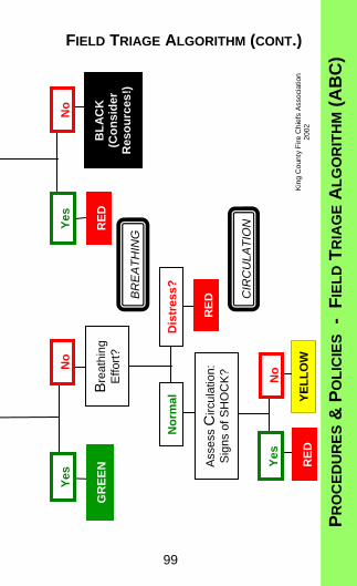
99
FIELD TRIAGE ALGORITHM (CONT.)
RE
D
BL
AC
K
(Co
nsi
der
R
eso
urc
es!)
No
Y
es
Kin
g C
oun
ty F
ire C
hie
fs A
ssoc
iatio
n 20
02
Yes
Dis
tres
s?
No
RE
D BR
EA
TH
ING
CIR
CU
LAT
ION
YE
LL
OW
R
ED
GR
EE
N
Bre
athi
ng
Effo
rt?
No
Y
es
Ass
ess
Circ
ulat
ion:
S
igns
of S
HO
CK
?
No
rmal
PR
OC
ED
UR
ES &
PO
LIC
IES
- F
IEL
D T
RIA
GE A
LG
OR
ITH
M (
AB
C)

100
NEUROLOGICAL ASSESSMENT P
RO
CE
DU
RE
S &
PO
LIC
IES —
NE
UR
OL
OG
ICA
L A
SS
ES
SM
EN
T
AV
PU
A
Ale
rt –
The
pat
ient
’s e
ye o
pen
spo
ntan
eous
ly a
s yo
u ap
proa
ch. T
he
patie
nt is
aw
are
and
resp
ons
ive
to th
e en
viro
nme
nt. T
he p
atie
nt
appr
opri
atel
y fo
llow
s co
mm
and
s.
V
Ver
bal
sti
mu
lus
resp
on
se –
The
pat
ient
’s e
yes
do n
ot o
pen
sp
onta
neou
sly.
The
pat
ient
’s e
yes
open
to v
erba
l com
man
d an
d th
e pa
tient
is a
ble
to r
espo
nd in
som
e m
eani
ngf
ul w
ay
wh
en a
sked
.
P
Pai
nfu
l st
imu
lus
resp
on
se –
The
pat
ient
do
es n
ot r
espo
nd to
you
r qu
estio
ns b
ut m
oves
or
crie
s ou
t w
hen
a pa
infu
l (no
xiou
s) s
timul
us is
ap
plie
d: e
arlo
be
pinc
h or
pre
ssur
e be
hin
d ea
rlob
e.
U
Un
resp
on
sive
– th
e pa
tient
doe
s no
t res
pon
d to
an
y st
imul
us.

101
NEUROLOGICAL ASSESSMENT (CONT.) G
LA
SG
OW
CO
MA
SC
AL
E
The
Gla
sgo
w C
om
a S
cale
(G
CS
) is
a m
eans
of m
easu
rin
g an
d m
oni
tori
ng le
vel
of c
onsc
ious
ne
ss b
y ca
lcul
atin
g a
scor
e ba
sed
on th
e b
est e
ye, v
erba
l, an
d m
o-to
r re
spon
se.
The
low
est
sco
re p
ossi
ble
is 3
, th
e hi
ghes
t is
15. T
he G
CS
is p
art
of C
ode
CV
A (
see
page
55)
.
Eye
Res
po
nse
B
est
Ver
bal
Res
po
ns
e B
est
Mo
tor
Res
po
ns
e
Spo
ntan
eous
ly o
pens
4
Ope
ns to
voi
ce
3
Ope
ns to
pai
n
2
No
resp
onse
1
Orie
nted
and
talk
ing
5
Dis
orie
nted
and
con
fuse
d
4
Inap
prop
riate
wo
rds
3
Inco
mpr
ehen
sibl
e
2
No
resp
onse
1
Obe
ys c
om
ma
nd
s
6
Loca
tes
pain
5
With
dra
ws
from
pai
n
4
Fle
xes
to p
ain
3
Ext
ends
to p
ain
2
No
resp
onse
1
PR
OC
ED
UR
ES &
PO
LIC
IES —
NE
UR
OL
OG
ICA
L A
SS
ES
SM
EN
T (
CO
NT.)

102
Indications
Any patient with decreased LOC.
The only approved methods of delivering noxious stimuli:
- Firm earlobe pressure (Figure 1) - Firm pressure behind earlobe (Figure 2)
Apply firm pressure to the earlobe for up to five seconds in order to assess a response to painful stimulation. This stimulation may be applied once or twice for no longer than 15 seconds during the initial evaluation and infrequently thereafter, if monitoring of the level of consciousness is necessary.
Prolonged application of stimuli, excessive applications, chemical stimuli, sternal rubs or eyeball pressure are not indicated nor approved by the Medical Program Director.
NOXIOUS STIMULI
PR
OC
ED
UR
ES &
PO
LIC
IES —
NO
XIO
US S
TIM
UL
I
Figure 1 Figure 2

103
The amount of oxygen given and the method of administration depend on many factors including a patient's medical history and the type of problem.
CONSCIOUS PATIENT WITHOUT RESPIRATORY DISTRESS
Begin with 2 liters per minute via nasal cannula as history is obtained. If no contraindications, you may increase to 4 liters per minute. Some patients may not require oxygen at all (e.g., a lacerated finger), but it is always best to provide oxygen when in doubt.
CONSCIOUS PATIENT WITH RESPIRATORY DISTRESS
Increase oxygen delivery according to the patient’s condition moving from nasal cannula to nonrebreathing mask. Use respiratory rate,
OXYGEN DELIVERY
Flow Volume Device
Low flow 2 - 4 liters/minute
Nasal cannula
High flow 10 - 15 liters/minute
Nonrebreathing mask
High flow with ventilation
15+ liters/minute
Bag-valve mask with reservoir
PR
OC
ED
UR
ES &
PO
LIC
IES —
OX
YG
EN
DE
LIV
ER
Y

104
effort, exchange, ease of speaking, skin signs, and level of consciousness as a guide. When using a nonrebreathing mask, remember to use a liter flow that is high enough to keep the bag inflated at least 1/3 full with the patient’s deepest inspiration.
CONSCIOUS PATIENT WITH SEVERE RESPIRATORY DISTRESS
Patients in severe respiratory distress may need assistance to breathe, as provided by a BVM with high flow oxygen. These patients may present with inability to speak, extreme exhaustion, minimal air movement, cyanosis, agitation, sleepiness, or a decreasing LOC. Examples include patients with chest or throat injury, airway obstruction, CHF, COPD, asthma, and near drowning. To assist respirations in a conscious patient, first explain the treatment to the patient then gently place the mask over the patient's nose and mouth and begin ventilations. Observe chest and abdomen and time the assisted breaths to coincide with the patient's or coach the patient to breathe with bag compressions.
UNCONSCIOUS PATIENT WITH SUFFICIENT RESPIRATORY EFFORT
Oxygen delivery may range from low-flow with a nasal cannula to high-flow with a nonrebreathing mask. Patient’s level of
OXYGEN DELIVERY (CONT.) P
RO
CE
DU
RE
S &
PO
LIC
IES —
OX
YG
EN
DE
LIV
ER
Y (
CO
NT.)

105
consciousness and vital signs (especially respiratory rate and effort), color, and nature of illness should determine oxygen flow level. Continually evaluate respiratory rate and effort and do not hesitate to assist respirations if necessary.
UNCONSCIOUS PATIENT WITH INSUFFICIENT OR NO RESPIRATORY EFFORT
Ventilate patient or assist ventilations with a BVM and high flow oxygen. If the patient resists the attempts to ventilate, try to time breaths with the patient's by compressing the bag as the patient inhales.
SPECIAL NOTE: COPD (emphysema, bronchitis, asthma)
The physiology of a person with COPD differs from that of a healthy person in that the primary stimulus to breathe comes from a decrease of oxygen in the blood rather than an increase in carbon dioxide. Providing the COPD patient with high concentrations of oxygen could theoretically depress their respiratory drive. Therefore, it is advisable to provide COPD patients with lower levels of oxygen initially, as long as they are not in severe respiratory distress. Two liters per minute by nasal cannula is usually sufficient in
OXYGEN DELIVERY (CONT.)
PR
OC
ED
UR
ES &
PO
LIC
IES —
OX
YG
EN
DE
LIV
ER
Y (
CO
NT.)

106
this situation. If a patient with COPD presents in respiratory distress and does not improve with low levels of oxygen, increase oxygen up to four (4) liters per minute.
A COPD patient whose respiratory drive is diminished due to excessive oxygen may present with increasing lethargy, confusion, and decreasing respiratory rate and effort. If this occurs, be prepared to assist ventilations.
If a COPD patient becomes unresponsive and/or stops breathing, ventilate via BVM with a high flow oxygen.
CAUTION: Over ventilation may worsen ‘air trapping’ and could cause pneumothorax.
SPECIAL NOTE: Infant And Young Child For an infant or young child with mild to moderate respiratory distress consider the “blow-by” technique. Hold the end of a supply tube or a nonrebreather mask approximately two inches away from the patient’s face. Another method to supply “blow-by” is with a paper cup. This can be done by pushing a supply tube through the bottom of the cup. Set the flow rate to 4-6 liters per minute.
OXYGEN DELIVERY (CONT.) P
RO
CE
DU
RE
S &
PO
LIC
IES —
OX
YG
EN
DE
LIV
ER
Y (
CO
NT.)
EMTs have the option of using a non-rebreather if nasal cannula at four (4) liter per minute is inadequate or patient has signs of hypoxia.

107
The treatment plan for every patient should include consideration for patient positioning. Proper positioning can reduce pain, improve physiological function, and improve the patient’s sense of well-being.
There are three positions to consider:
Recovery Semi-reclining Shock position
RECOVERY POSITION
This position is used for non-traumatic patients who are unresponsive but breathing. It protects the airway from vomit and secretions. (Figure 3, page 109)
The following steps are recommended:
Kneel beside the patient and straighten the legs.
Place the patient’s arm that is nearest to you at a right angle to body, elbow bent, palm up.
Place the other arm across the chest/abdomen (Figure 1, page 108).
If the patient is small, bring this arm farther across so that the back of the hand can be held against the patient’s nearest cheek.
PATIENT POSITIONING
PR
OC
ED
UR
ES &
PO
LIC
IES —
PA
TIE
NT P
OS
ITIO
NIN
G

108
Grasp the patient’s far-side thigh above the knee; pull the thigh up towards the patient’s body (Figure 1).
Place your other hand on the patient’s far-side shoulder and roll the patient toward you (Figure 2). Begin moving the patient’s uppermost hand toward the patient’s nearest cheek.
PATIENT POSITIONING (CONT.) P
RO
CE
DU
RE
S &
PO
LIC
IES
— P
AT
IEN
T P
OS
ITIO
NIN
G (
CO
NT.)
Figure 1
Figure 2

109
Adjust the leg you are holding until both the hip and knee are bent at right angles.
Tilt the patient’s head back and place the uppermost hand under the patient’s cheek. Use this hand to maintain head tilt (Figure 3). Use chin lift if necessary.
In suspected spinal cord trauma/injury first immobilize the patient with the appropriate size c-collar and backboard. If the patient is unconscious, monitor and protect the airway, if necessary, turn patient and backboard 90 degrees to facilitate drainage.
SEMI-RECLINING (SEMI-FOWLER’S)
In the semi-reclining position (Figure 4) a patient is usually sitting at a forty-five degree angle. A gentle knee bend adds comfort and helps to maintain the upright position. Additional pillows behind the head and knees may improve comfort. Patients with mild to moderate respiratory symptoms may benefit from this position.
PATIENT POSITIONING (CONT.)
PR
OC
ED
UR
ES &
PO
LIC
IES —
PA
TIE
NT P
OS
ITIO
NIN
G (
CO
NT.)
Figure 3
Monitor respirations closely.

110
SHOCK POSITION
In this position the feet are elevated up to twelve inches and the patient is supine (Figure 5). The use of this position will increase venous return to the heart resulting in increased blood pressure.
PATIENT POSITIONING (CONT.)
PR
OC
ED
UR
ES &
PO
LIC
IES —
PA
TIE
NT P
OS
ITIO
NIN
G (
CO
NT.)
Figure 5
Figure 4

111
If the reason for use of a device is to prevent movement and it is done without the consent of the patient, it is a restraint.
Generally, restraints are used in the prehospital environment whenever dangerous behavior (especially danger to self or others) is encountered. The provider has a clear duty to exercise increased vigilance for the safety of the patient, because the patient is unable to self protect while restrained. Likewise, the safety of the responders should be ensured. PROCESS OF RESTRAINT
Safety and the prevention of injuries are the major concerns in the process of restraint application. It is imperative to maximize the patient's self-control before deciding to apply restraints.
Self-control. The first step is to encourage the patient to exercise all the self-control he or she possesses. A statement such as "I know you don't want to hurt yourself or anyone else. I want you to try to stay in control. I know you can do it" is an example. Offer to help. Anxiety can interfere with concentration and an offer of assistance should reduce anxiety. A statement such as "I want to assure you that we will help you. We will not let you hurt yourself or someone else" is an example of an offer to help.
PATIENT RESTRAINT
PR
OC
ED
UR
ES &
PO
LIC
IES —
PA
TIE
NT R
ES
TR
AIN
T

112
Be ready and able to overpower patient. Never attempt physical restraint without the resources needed to safely overpower a patient.
Physical restraint. This is the time when most injuries tend to occur. Plan the actions so that each provider involved clearly understands his or her role. Typically, one person is assigned to each limb. One provider should communicate with the patient continuously. Once a decision is made to restrain, act quickly. Use only the force necessary for restraint. Depending on local requirements, it may be helpful to have the police present during restraint. EMTs should be aware of their own personal safety.
TYPES OF RESTRAINTS
The kinds of restraints used in the prehospital environment vary tremendously. Handcuff and cable ties should only be applied and removed by law enforcement personnel.
Once a patient is restrained, he or she should be carefully monitored to avoid airway obstruction. An NRM with appropriate oxygen flow may be applied to protect the EMS personnel from spit. Alternatively a “spit sock” may be used.
PATIENT RESTRAINT (CONT.) P
RO
CE
DU
RE
S &
PO
LIC
IES —
PA
TIE
NT R
ES
TR
AIN
T (
CO
NT.)

113
DOCUMENTATION
It is important to document the behavior that made restraints necessary as well as the restraint technique used. The documentation must reflect continual concern for the patient's safety and well-being as well as descriptions of the patient’s ongoing mental status and behavior.
PATIENT RESTRAINT (CONT.)
PR
OC
ED
UR
ES &
PO
LIC
IES —
PA
TIE
NT R
ES
TR
AIN
T (
CO
NT.)
Do not remove restraints until directed by the hospital emergency
department personnel.

114
INFECTIOUS DISEASE PREVENTION
HANDWASHING Handwashing is the most effective way to prevent transmission of infectious disease.
Wash Hands After patient contact Before eating, drinking, smoking or
handling food Before & after using the bathroom After cleaning or checking equipment
PERSONAL PROTECTIVE EQUIPMENT (PPE)
Gloves and eye protection must be worn for every patient.
FULL PPE for possible infectious contacts
Donning Sequence (MEGG) Mask* > Eye Protection > Gown > Gloves Mask patient (if possible)
Doffing (removal) Sequence Gloves > Gown > Hand cleaner Eye Protection > Mask > Hand cleaner Handle as contaminated waste Decon Eye Protection *Fit tested
PERSONAL PROTECTIVE EQUIPMENT (PPE)
PR
OC
ED
UR
ES &
PO
LIC
IES —
PE
RS
ON
AL P
RO
TE
CT
IVE E
QU
IPM
EN
T
115
INFECTIOUS DISEASE
Febrile Respiratory Illness Dispatchers will notify units of -
Infectious symptoms or locations Dispatch info or fever w/ cough or illness
or possible infectious disease
Full PPE (MEGG) Mask*, Eye Protection, Gowns, Gloves Mask patient (if possible) Limit patient contacts
After Patient Contact Remove PPE – (approved sequence) Dispose of PPE as contaminated waste On scene decon - eye protection &
equipment w/ germicidal cleaner Hospital decon - eye protection,
equipment and apparatus
At station Decon affected equipment & contacts (kits,
BP/steth, radios, clipboards, etc.) Wash hands before leaving apparatus
floor.
*Fit tested
PERSONAL PROTECTIVE EQUIPMENT (PPE) (CONT.)
PR
OC
ED
UR
ES &
PO
LIC
IES —
PE
RS
ON
AL P
RO
TE
CT
IVE E
QU
IPM
EN
T (
CO
NT.)

116
Bloodborne Exposure This is an exposure or potential exposure to Bloodborne Pathogens such as Hepatitis B, Hepatitis C, HIV or other pathogens that may be transmitted through contaminated body fluids or tissues. Examples include: blood, bloody body fluids including semen, vaginal secretions, cerebrospinal fluid, synovial, pleural, pericardial, and amniotic fluids.
An exposure only occurs if: There is a needle stick or cut with a possibly
contaminated needle or object. There is contact with non-intact skin (e.g.
skin that is cut, chapped, abraded, or afflicted with dermatitis.)
There is fluid contact with your mucous membranes such as eyes, nose, mouth.
Steps to take following exposure: Initiate self-care which includes washing
the site thoroughly with soap and water. Flush mucous membranes with water only.
Immediately report exposure to immediate supervisor and exposure control officer for risk assessment and follow-up.
Follow individual department’s exposure control policy. (see also PPE page 114).
For all other exposures follow your department’s infection/exposure control policy.
REPORTABLE EXPOSURES P
RO
CE
DU
RE
S &
PO
LIC
IES —
RE
PO
RT
AB
LE E
XP
OS
UR
ES

117
Child Abuse Signs and Symptoms of suspected abuse and neglect include:
Multiple bruises in various stages of healing
Bilateral/symmetrical injuries and/or bruises
Injury inconsistent with mechanism described
Repeated calls to the same patient or address
New suspicious injuries Parents, guardian or caregiver
inappropriately concerned Conflicting stories Fear on the part of the patient to discuss
the incident Lack of proper supervision of the patient Malnourished appearance Unsafe living environment Untreated chronic illness
Vulnerable Adults Defined as adults age 60 and older who cannot care for themselves and adults age 18 and older who, have a legal guardian, are developmentally delayed, live in a DSHS licensed facility, receive in home care
PHYSICAL ABUSE AND NEGLECT OF CHILDREN AND VULNERABLE ADULTS
PR
OC
ED
UR
ES &
PO
LIC
IES —
AB
US
E A
ND
NE
GL
EC
T

118
services, or have personal care aide who is paid for their services. Signs of abuse and neglect include:
Unexplained injuries or behavior Reports of physical, mental, or sexual
abuse Reports of being abandoned or deserted
without basic necessities Failing to provide basic life necessities,
not taking action to prevent harm or pain Failure to provide safe living conditions Untreated injuries or health problems Intentionally taking advantage of a
vulnerable adult either financially, or personally
Undue influence or coercion By Washington state law, Fire Fighters, Paramedics, and EMT’s are mandatory reporters.
REPORT NEGLECT/ABUSE OF VULNERABLE ADULTS TO DSHS: 1-866-363-4276 (1-866-ENDHARM)
Involve local Police in all suspicious cases. Call 911.
PHYSICAL ABUSE AND NEGLECT OF CHILDREN AND VULNERABLE ADULTS
(CONT.)
PR
OC
ED
UR
ES &
PO
LIC
IES —
AB
US
E A
ND
NE
GL
EC
T (
CO
NT.)

119
Indications For Measurement Of Posturals
Acute volume loss (such as suspected GI bleeding or internal hemorrhage)
Generalized weakness
Complaint of dizziness, lightheadedness, or fainting
Prolonged vomiting or diarrhea
Contraindications
Symptomatic hypotension while supine (systolic blood pressure less than 90 mmHg)
Third trimester bleeding
Trauma patients
Patient with suspected cardiac chest pain
To Check For Postural Vital Signs
Obtain blood pressure and heart rate after two (2) minutes in supine position. Then bring patient to seating position.
Next, stand patient upright slowly (caution: lay down patient promptly if he or she becomes dizzy or lightheaded when seated or standing).
After patient stands for one (1) minute obtain blood pressure and heart rate.
If fainting or light headedness develops return patient to supine position.
POSTURAL VITAL SIGNS
PR
OC
ED
UR
ES &
PO
LIC
IES —
PO
ST
UR
AL V
ITA
L S
IGN
S

120
Positive findings
Increase in pulse of 20/minute or more or a 20 mmHg or more drop in systolic BP from supine to standing with associated symptoms
Dizzy, lightheaded, or fainting while sitting or standing
PSYCHIATRIC EVALUATIONS
Assisting Police
Police may call EMS for assistance in determining whether a psychiatric patient is stable enough to go to jail. Your evaluation must be based on Sick/Not Sick and MOI and IOS. You must document vital signs.
POSTURAL VITAL SIGNS (CONT.)
PR
OC
ED
UR
ES &
PO
LIC
IES
—
PO
ST
UR
AL V
ITA
L S
IGN
S (
CO
NT.)
P
SY
CH
IAT
RIC
EV
AL
UA
TIO
NS
A positive postural is an ALS indicator in an appropriate clinical setting

121
Indications For Use
Pulse oximetry may be used anytime oxygen is in use or is to be administered to a patient based upon complaint or condition. This may include:
Shortness of breath Chest pain Altered level of consciousness (LOC) Pregnancy/active labor Chest trauma Any time the EMT believes the oxygen
saturation level needs to be assessed
Contraindications
Patients less then 2 (two) years of age.
Use and Administration
Place the probe on a clean digit. This should be accomplished simultaneously with the initial administration of oxygen allowing for a “room air” estimate.
PULSE OXIMETRY
Pulse oximetry is an approved protocol but optional by individual departments.
PR
OC
ED
UR
ES &
PO
LIC
IES —
PU
LS
E O
XIM
ET
RY
Under no circumstances should oxygen administration be delayed to
obtain an oximetry reading.

122
NOTE Pulse oximetry is inaccurate in the following clinical situations:
Cardiac arrest Shock Hypothermia Carbon monoxide poisoning Jaundice Presence of nail polish
Decisions about patient care should be based on a patient’s complaint and presentation and should not be based solely on a pulse oximeter reading.
Under no circumstances should the presence of a pulse oximeter detract from
patient care.
PULSE OXIMETRY (CONT.) P
RO
CE
DU
RE
S &
PO
LIC
IES —
PU
LS
E O
XIM
ET
RY (
CO
NT.)
Pulse Ox device should NOT be used to acquire distal pulse readings. This should
always be done by palpating the radial pulse.

123
NOTES
NO
TE
S
124
PR
OC
ED
UR
ES &
PO
LIC
IES —
SIC
K/N
OT S
ICK
The SICK/NOT SICK approach to rapid patient assessment has become a mainstay in determining the physiologic status of a patient in Seattle/King County. Whether it is medical or trauma, adult or pediatric, SICK/NOT SICK is the tool of choice for rapid patient assessment and appropriate patient care.
This revised edition of the Patient Care Protocols incorporates the SICK/NOT SICK approach which leads to the early recognition of critical (Sick) and non-critical (Not Sick) patients and, ultimately, rapid and appropriate patient care.
The clinical indicators used in the adult SICK/NOT SICK approach provide clarity and offer clear and CONCISE indicators for determining a patient’s physiologic stability. Often, these indicators are observable from across the room without even touching the patient.
Additional considerations that need to be incorporated into your SICK/NOT SICK decision- process include: mechanism of injury (MOI), nature of illness (NOI) and index of suspicion (IOS). These CONSIDERATIONS will help you in determining SICK/NOT SICK and may alone determine into which category the patient is placed.
NOTE MOI - Mechanism of Injury NOI - Nature of Illness IOS - Index of Suspicion
SICK/NOT SICK

125
PR
OC
ED
UR
ES &
PO
LIC
IES —
SIC
K/N
OT S
ICK
(C
ON
T.)
Adult SICK/NOT SICK Clinical Indicators:
Chief complaint and MOI/NOI/IOS Respirations Pulse (circulation) Mental status Skin signs (color, moisture, temperature) Body position/obvious trauma
The pediatric SICK/NOT SICK approach uses an innovative triad of indicators collectively called the “pediatric assessment triangle.” The triangle defines key indicators of physiologic stability, allowing the EMS provider to make an accurate and timely decision on the status of a pediatric patient.
First, determine the chief complaint and consider MOI, NOI, IOS
Then assess the elements of the Pediatric Assessment Triangle:
SICK/NOT SICK (CONT.)
Appearance Work of Breathing Circulation
Alertness Retractions Color
Color Nasal flaring Temperature
Distractiblity Body position Capillary refill time
Consolability Abdomen sounds Pulse
Eye contact
Motor activity
Speech/cry

126
DECIDE
Rapid Transport/ALS
SICK/NOT SICK Medical Rapid Patient Assessment
Considerations: BSI, scene size-up, family member, additional staffing
Chief Complaint/NOI* Respirations Pulse Mental Status Skin Signs/Color Body Position (Primary Assessment)
NOT SICK
Short report to incoming units Treat any life-threatening conditions immediately
100% 02 nonrebreather mask or BVM
Reassessment
Appropriate Position
History Taking Baseline vitals Rapid medical survey SAMPLE history
Secondary Assessment Includes a complete set of vitals
Secondary assessment *Includes a complete
set of vitals
Appropriate Transport
Reassessment
Low/Moderate Flow 02
*NOI - Nature of Illness
PR
OC
ED
UR
ES &
PO
LIC
IES —
SIC
K/N
OT S
ICK
(A
DU
LT)
SICK
The Clinical Picture
Short Report to incoming
units
History Taking Baseline vitals Rapid medical survey SAMPLE history OPQRST
Care for any obvious conditions as needed
Appropriate Treatment

127
SICK/NOT SICK Trauma Rapid Patient Assessment
Considerations: BSI, scene size-up, family member, additional staffing
The Clinical Picture
Chief Complaint/MOI* Respirations Pulse Mental Status Skin Signs/Color Obvious Trauma (Primary Assessment)
PR
OC
ED
UR
ES &
PO
LIC
IES —
SIC
K/N
OT S
ICK
(A
DU
LT)
*MOI—Mechanism of Injury
DECIDE
Rapid Transport/ALS
NOT SICK
Short report to incoming units
Rapid extrication Treat any life-threatening conditions immediately
Reassessment
History Taking Baseline vitals Rapid medical survey SAMPLE history
Secondary Assessment Includes a complete set of vitals
Secondary assessment *Includes a complete
set of vitals
Appropriate Treatment
Appropriate Transport
Reassessment
Low/Moderate Flow 02
SICK
Short Report to incoming
units
History Taking Baseline vitals Rapid trauma survey SAMPLE history
Spinal stabilization Care for any obvious or
additional injuries as needed 100% 02
nonrebreather mask or BVM
Immobilize spine

128
App
eara
nce B
reathing
Pediatric Triangle (Primary Assessment)
Considerations: Scene size-up/NOI*
Family member Additional staffing
SICK/NOT SICK Medical Rapid Pediatric Patient Assessment
Circulation/Skin Color
*NOI - Nature of Illness
PR
OC
ED
UR
ES &
PO
LIC
IES
— S
ICK
/NO
T S
ICK
(P
ED
IAT
RIC
)
DECIDE
Rapid Transport/ALS
NOT SICK
Short report to incoming units Treat any life-threatening conditions immediately
100% 02 nonrebreather mask or BVM
Reassessment (Keep Warm)
Appropriate Position
History Taking Baseline vitals Rapid medical survey SAMPLE history
Secondary Assessment *Includes a complete set of vitals
Low/Moderate Flow 02
SICK
Request ALS. Short Report to incoming
units
Secondary assessment Includes a complete set
of vitals
Appropriate Transport
Reassessment
History Taking Baseline vitals Rapid medical survey SAMPLE history OPQRST
Care for any obvious conditions as needed
Appropriate Treatment

129
DECIDE
PR
OC
ED
UR
ES &
PO
LIC
IES —
SIC
K/N
OT S
ICK
(P
ED
IAT
RIC
)
.
*MOI—Mechanism of Injury
App
eara
nce B
reathing
Pediatric Triangle (Primary Assessment)
Circulation/Skin Color
Rapid Transport/ALS
Short report to incoming
Rapid extrication Treat any life-threatening conditions immediately
Reassessment (Keep Warm)
History Taking Baseline vitals Rapid medical survey SAMPLE history
Secondary Assessment Includes a complete set of vitals
Secondary assessment Includes a complete set
of vitals
Appropriate Treatment
Appropriate Transport
Reassessment (Keep Warm)
Low/Moderate Flow 02
History Taking Baseline vitals Rapid trauma survey SAMPLE history
100% 02 non-rebreather mask or BVM
Immobilize spine
SICK/NOT SICK Trauma Rapid Pediatric Patient Assessment
Request ALS. Short Report to incoming
units
Considerations: Scene size-up/MOI*
Forcible entry Family member
Additional staffing
NOT SICK SICK
Spinal stabilization Care for any obvious or
additional injuries as needed

130
The following summary of spinal immobilization assumes that the ABCs and a distal circulation, motor, and sensory (CMS) exam have been assessed before and after splinting and treated accordingly.
Certain parts of this procedure may need to be modified in a critically injured patient whose airway, breathing, or circulation problems need to be treated immediately.
This summary also assumes that a patient is sitting upright in a car. The procedure will need to be modified if a patient is found in a different position or situation.
Stabilize head in neutral, in-line position. (Do not release stabilization until the patient is completely secured to a long backboard, as described below, or until another EMT takes over. There should be no pulling or traction taken.)
Measure and apply, properly-sized cervical collar.
Apply extrication device, using a short backboard, long board, or other device. The technique used will depend on the equipment available and the patient’s condition.
Extricate, maintain spinal alignment with head and neck stabilization in a neutral, in-line position.
SPINAL IMMOBILIZATION P
RO
CE
DU
RE
S &
PO
LIC
IES —
SP
INA
L I
MM
OB
ILIZ
AT
ION

131
Place patient on a long backboard and
immobilize chest by crisscrossing over shoulders, across chest to the hips.
Assess ventilation after tightening straps to ensure that respiratory effort is not impaired.
Immobilize the pelvis by crisscrossing or by strapping straight across. Use caution with pelvic or abdominal injuries.
Put one strap across the thighs above the knees and one strap across the lower extremities. An additional strap may be placed across the feet.
Stabilize the patient's head using a commercial immobilization device, rolled towels, or blankets. Secure patient's head to the backboard with two-inch adhesive tape across forehead.
Check CMS before and after immobilization.
Continue to monitor airway, breathing, circulation, vital signs, and level of consciousness.
SPINAL IMMOBILIZATION (CONT.)
PR
OC
ED
UR
ES &
PO
LIC
IES —
SP
INA
L I
MM
OB
ILIZ
AT
ION
(C
ON
T.)

132
Appropriate splinting can reduce or minimize dislocation, motion, hemorrhage, swelling, and pain.
GENERAL PRINCIPLES
The following general principles apply to splinting:
Remove or cut away clothing.
Dress and bandage significant wounds, using a sterile dressing.
Check CMS distal to injury before and after splinting.
Immobilize joints above and below injured bones.
For joint injuries, leave in place and immobilize the bone above and below the joint
It may be necessary on a mid-shaft (center 1/3) fracture to realign angulated injuries.
Pad splints well.
Elevate extremity after splinting, if possible.
Monitor CMS after splinting.
GUIDELINES FOR SPECIFIC INJURIES
Realignment of Long Bone Fractures
Attempt to realign (open or closed) long bones that are angulated in the middle 1/3 then splint.
SPLINTING
PR
OC
ED
UR
ES &
PO
LIC
IES —
SP
LIN
TIN
G

133
Long-bone fractures, which occur in the proximal or distal 1/3, that may or may not involve a joint, may be realigned if compromise of distal circulation or nerve function is detected and definitive care is delayed.
Realignment may sometimes be necessary to facilitate packaging for transport.
Check and document CMS before and after splinting and/or realignment.
Dislocations/Sprains
Splint dislocations or other joint injuries in the position found. Exception: Loss of a distal pulse and neurological function and definitive care is delayed. In that case, attempt to straighten into anatomical position until the pulse returns, excessive pain is felt, or resistance is encountered. Support with blanket, pillow, or well-padded splint. Elevate the limb. Pack the injured area in ice or use an ice pack.
Pelvic Fractures
Immobilization of these fractures can be accomplished by use of a bed sheet, disposable blanket, or a commercial device.
Fold sheet lengthwise into 8” to 14” width.
SPLINTING (CONT.)
PR
OC
ED
UR
ES &
PO
LIC
IES —
SP
LIN
TIN
G (
CO
NT.)

134
Place beneath patient; twist then wrap ends around patient, crossing over pelvic area.
Secure sheet with square knot, tape, or zip ties.
Secure the ends to the backboard.
TRACTION SPLINTING A lower extremity traction splint stabilizes fractures of the femur. This reduces motion, hemorrhage, swelling, and pain. Traction splints are indicated in midshaft femoral fractures without involvement of the hip joint, knee, or lower leg.
General Guidelines For Applying A Traction Splint
At least two EMTs are required to apply a traction splint.
Remove or cut away clothing. Dress and bandage significant wounds using a sterile dressing. Manually immobilize the injured extremity prior to dressing/bandaging. Check distal CMS before and after manipulation.
Objectives:
Determine SICK/NOT SICK Control Bleeding Properly measure splint Apply traction Apply splint Reassess CMS and vital signs
SPLINTING (CONT.)
PR
OC
ED
UR
ES &
PO
LIC
IES —
SP
LIN
TIN
G (
CO
NT.)

135
The TASER dart usually penetrates the skin only a few millimeters. EMTs can safely remove a dart simply by pulling it out. The only exception is involvement of the eye, face, neck, breast or groin. In this case, leave the dart in place and transport the patient to the hospital for dart removal.
Consider scene safety and measures to protect yourself and other rescuers from a potentially violent patient in situations when a TASER gun has been used. You do not need to transport a person to the hospital based solely on TASER dart exposure. If a patient has no need for further medical evaluation, you can leave him or her in police custody.
This skill may be performed by EMTs and ALS providers. (Depending on local protocol.)
ALS Indicators Compromise in ABCs
BLS Indicators Taser dart imbedded in skin
BLS Care Assure the scene is safe Wear PPE including gloves and eye
protection—consider mask and gown if blood is present
Remove TASER cartridge from gun or cut wires before removing darts
TASER DART REMOVAL AND CARE
PR
OC
ED
UR
ES &
PO
LIC
IES —
TA
SE
R D
AR
T R
EM
OV
AL &
CA
RE

136
TASER DART REMOVAL AND CARE (CONT.) P
RO
CE
DU
RE
S &
PO
LIC
IES —
TA
SE
R D
AR
T R
EM
OV
AL &
CA
RE (
CO
NT.)
- Dispose of darts in sharps container or TASER cartridge
Police must be in custody of patient Restrain if needed
Removal Procedure DO NOT REMOVE darts if:
- Patient is not under control - Eye, face, neck, breast or groin are
involved—patient must be transported to hospital for dart removal in this case
Grasp firmly with one hand and pull to remove, one dart at a time
Reassess patient Consider medical or behavioral problems
as the original cause of violent behavior - Drug/alcohol intoxication
- Behavioral problems - Trauma, etc.
Bandage wounds as appropriate Document situation and patient contact
thoroughly Patient Disposition Release to law enforcement if indicated Transport with law enforcement support if:
- Eye, face, neck, breast or groin are involved
- ALS indicated - Law enforcement officer requires
medical evaluation. Police protocol may require transport. This may be by PD or ambulance.

137
TASER DART REMOVAL AND CARE (CONT.)
Follow Patient Care Guidelines regarding restraint of aggressive or violent patients
TEETH
Place avulsed/dislodged tooth/teeth in milk or patient saliva and transport.
Burn Hazard
When a TASER is used in the presence of pepper spray propellant, there is a burn hazard. Electrical arcing from imperfect (but effective) dart contact can ignite the
propellant. The resulting combustion may not be visible, but can lead to complaints of heat and burning. If a patient complains of heat or burning, evaluate for possible
minor burns.
PR
OC
ED
UR
ES &
PO
LIC
IES —
TA
SE
R D
AR
T R
EM
OV
AL &
CA
RE /T
EE
TH

138
Transport Options
In deciding what is best for the patient, you have several transport options: Paramedic Transport All “Sick” patients and all patients with
unstable vital signs should be transported by medic unit (when available). If no medic unit is available, begin transport and rendezvous. All patients transported by paramedics must go to a hospital.
BLS Transport (via private ambulance or fire department BLS unit). Stable patients who require medical attention or oxygen during transport may be transported with a BLS vehicle. In deciding whether to call for private ambulance or transport via fire department BLS unit, departmental policies should be followed.
Before requesting a private ambulance, always inform the patient that you would like to call for a private ambulance, and that the ambulance company will charge a fee for transportation and service.
TRANSPORT AND DESTINATION
When requesting an ambulance for BLS transport, the default mode in King County for ambulance travel to the scene is non-
emergency response unless specific written protocols or contracts require code-red
response.
PR
OC
ED
UR
ES &
PO
LIC
IES —
TR
AN
SP
OR
T &
DE
ST
INA
TIO
N

139
Private Vehicle Transport Patients with minor alterations in vital signs
and stable conditions not requiring oxygen may be advised that travel to the hospital or clinic via private vehicle is safe. Obviously the patient should not be the driver.
Taxi Transport Some departments utilize a taxi voucher
program for patients who travel to a clinic, urgent care clinic, free-standing emergency department, hospital based emergency department. These patients must meet the following criteria: 1. Paramedic care is NOT required 2. Patient is ambulatory 3. Patient has a non-urgent condition
(clinically stable) including low index of suspicion for: a. Cardiac problem b. Stroke c. Abdominal aortic aneurysm d. GI bleed problems e. Major mechanism of injury
4. Patient must not have a. Need for a backboard b. Uncontrolled bleeding c. Uncontrolled pain d. Need for oxygen (except patient self
administered oxygen)
TRANSPORT AND DESTINATION (CONT.)
PR
OC
ED
UR
ES &
PO
LIC
IES —
TR
AN
SP
OR
T &
DE
ST
INA
TIO
N (
CO
NT.)

140
5. The EMT considers a taxi to be an
appropriate and safe method of transportation for the particular clinical problem.
6. Patient should be masked if there are respiratory symptoms.
Final Disposition Options In deciding what is best for the patient you have four disposition options: 1. Leave at Scene
Generally, patients with normal vital signs and minor injuries or illness may be left at the scene. Always caution the patient to seek medical care (or call 911) if the condition should worsen.
2. Urgent Care Clinic Selected patients may be transported to
a clinic or urgent care clinic by fire department EMTs if they meet the following criteria: A. Paramedic care is NOT required B. Patient is ambulatory C. Patient has a non-urgent condition
(clinically stable) including a. Low index of suspicion for:
Cardiac problem Stroke Abdominal aortic aneurysm GI bleed problems
b. Low index of suspicion for major mechanism of injury
TRANSPORT AND DESTINATION (CONT.) P
RO
CE
DU
RE
S &
PO
LIC
IES —
TR
AN
SP
OR
T &
DE
ST
INA
TIO
N (
CO
NT.)

141
D. Patient must not have
a. Need for a backboard b. Uncontrolled bleeding c. Uncontrolled pain d. Need for high flow oxygen
For guidance regarding transport decisions EMTs may consult with paramedics or with emergency department personnel at the medical control hospital. The EMT must notify the destination facility of the clinical problem and the facility must agree to accept the patient.
3. Free-standing Emergency Department Selected patients may be transported to a free-standing emergency department by EMTs if they meet the following criteria: Paramedic care is NOT required Patient has a non-urgent condition
(clinically stable) including: A. Low index of suspicion for cardiac,
stroke, abdominal aortic aneurysm, or GI bleed problems
B. Low index of suspicion for major mechanism of injury
C. Patient is willing to be transported to the free-standing emergency department.
For transport decisions guidance EMTs may consult with paramedics or with emergency department personnel at the medical control hospital. If a free-standing
TRANSPORT AND DESTINATION (CONT.)
PR
OC
ED
UR
ES &
PO
LIC
IES —
TR
AN
SP
OR
T &
DE
ST
INA
TIO
N (
CO
NT.)
142
emergency department destination is selected, that facility must be notified prior to transport and agree to accept the patient.
4. Hospital Emergency Department Transferring Patients At Rendezvous
Site Patients (particularly patients in less populated parts of King County) who are evaluated by EMTs and are in need of paramedic level care, may require transfer of care at a rendezvous site. This should be accomplished in the following fashion: A. The first arriving EMT team will
decide whether paramedic care is required according to EMT protocols. In some instances the dispatch center will have already dispatched paramedics to begin travel toward a rendezvous point.
B. If the EMT team determines paramedic care is not warranted then they will cancel the paramedics according to protocols.
If continued paramedic care is warranted then the EMTs will contact their dispatch center to request a paramedic rendezvous and a suggested site.
Upon meeting at the rendezvous site the paramedics will decide if the patient
TRANSPORT AND DESTINATION (CONT.) P
RO
CE
DU
RE
S &
PO
LIC
IES —
TR
AN
SP
OR
T &
DE
ST
INA
TIO
N (
CO
NT.)

143
should be transferred to the paramedic vehicle for further evaluation and treatment. The paramedics may, depending on the clinical situation, decide to assess the patient in the EMT vehicle. The paramedics will decide on the most suitable mode of transport to the hospital.
TRANSPORT AND DESTINATION (CONT.)
PR
OC
ED
UR
ES &
PO
LIC
IES —
TR
AN
SP
OR
T &
DE
ST
INA
TIO
N (
CO
NT.)
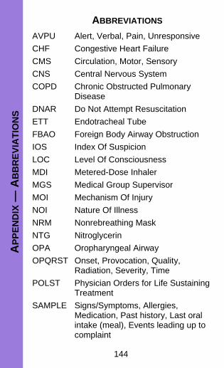
144
AVPU Alert, Verbal, Pain, Unresponsive
CHF Congestive Heart Failure
CMS Circulation, Motor, Sensory
CNS Central Nervous System
COPD Chronic Obstructed Pulmonary Disease
DNAR Do Not Attempt Resuscitation
ETT Endotracheal Tube
FBAO Foreign Body Airway Obstruction
IOS Index Of Suspicion
LOC Level Of Consciousness
MDI Metered-Dose Inhaler
MGS Medical Group Supervisor
MOI Mechanism Of Injury
NOI Nature Of Illness
NRM Nonrebreathing Mask
NTG Nitroglycerin
OPA Oropharyngeal Airway
OPQRST Onset, Provocation, Quality, Radiation, Severity, Time
POLST Physician Orders for Life Sustaining Treatment
SAMPLE Signs/Symptoms, Allergies, Medication, Past history, Last oral intake (meal), Events leading up to complaint
ABBREVIATIONS A
PP
EN
DIX
— A
BB
RE
VIA
TIO
NS

145
Age
R
espi
-ra
tions
(b
reat
hs/
min
ute)
Pul
se
(bea
ts/
min
ute)
Sys
tolic
B
lood
P
ress
ure
(m
m H
g)
Ne
wbo
rn:
0 to
1 m
onth
40
to 6
0 12
0 to
16
0 50
to 7
0
Infa
nt:
1 m
onth
to
1 y
ear
30 to
60
100
to
160
70 to
95
Tod
dler
: 1
to 3
ye
ars
24 to
40
90 to
150
80
to 1
00
Pre
scho
ol:
3 to
6 y
ear
s 22
to 3
4 80
to 1
40
80 to
100
Sch
ool a
ge:
6 to
12
year
s 18
to 3
0 70
to 1
20
80 to
110
Ado
lesc
ent:
12
to 1
8 yr
s 12
to 1
6 60
to 1
00
90 to
140
Ove
r 18
ye
ars
12 to
20
60 to
100
90
to 1
40
NORMAL VITAL SIGNS BY AGE
Temperature Conversions
AP
PE
ND
IX —
NO
RM
AL V
ITA
L S
IGN
S B
Y A
GE /
TE
MP
ER
AT
UR
E C
ON
VE
RS
ION
S

146
ALS PROVIDERS A
PP
EN
DIX
— T
EL
EP
HO
NE N
UM
BE
RS —
AL
S P
RO
VID
ER
S
Org
an
izat
ion
Ad
dre
ss
Tel
eph
on
e B
elle
vue
Fire
De
part
men
t 45
0 11
0th
Ave
nue
NE
B
elle
vue,
WA
98
004
(425
) 4
52-6
892
(P
hone
)
Kin
g C
ount
y M
edic
One
70
64 S
outh
22
0th S
tree
t #9
Ken
t, W
A 9
8032
(2
06)
296
-85
50 (
Pho
ne)
R
edm
ond
Fire
Dep
artm
ent
8450
- 1
61s
t Ave
nue
NE
R
edm
ond,
WA
980
52
(425
) 5
56-2
200
(P
hone
)
Sea
ttle
Fire
Dep
artm
ent
Med
ic O
ne
325
Nin
th A
venu
e
Sea
ttle,
WA
98
104
(206
) 3
86-1
483
(P
hone
)
Sho
relin
e F
ire D
epar
tmen
t 17
525
Aur
ora
Ave
nue
N.
Sho
relin
e, W
A
9813
3
(206
) 5
33-6
500
(P
hone
)
Vas
hon
Isla
nd F
ire &
Res
cue
1002
0 S
W B
ank
Roa
d
Vas
hon,
WA
98
070-
1150
(2
06)
463
-24
05 (
Pho
ne)

147
AMBULANCE AND COMMUNITY RESOURCES
AP
PE
ND
IX —
TE
LE
PH
ON
E N
UM
BE
RS —
AM
BU
LA
NC
E &
CO
MM
UN
ITY R
ES
OU
RC
ES
Org
an
izat
ion
A
dd
res
s T
elep
ho
ne
Tri
-Me
d A
mb
ula
nce
1
882
1 E
. V
alle
y H
igh
wa
y K
en
t, W
A 9
803
2
(206
) 2
43-5
622
(Ph
one
)
Am
eri
can
Me
dic
al
Re
spo
nse
13
075
Ga
tew
ay
Dri
ve S
E
Su
ite 1
00
Tu
kwila
, WA
9
816
8
(206
) 4
44-4
440
(Ma
in)
(206
) 6
23-1
111
(Dis
patc
h)
or
1-8
00-
542-
7701
KC
Sh
eri
ffs O
ffice
S
ea
rch
& R
escu
e 7
300
Per
imet
er
Ro
ad
S.,
Roo
m 1
43
Se
att
le, W
A
9810
8-3
849
(206
) 2
96-3
853
(Ph
one
) S
pe
cia
l Op
era
tions
Ru
ral/M
etr
o A
mbu
lanc
e
64
05 –
21
8th S
tree
t S
W
Mt.
La
ke T
err
ace,
WA
9
8043
(4
25)
672
-111
1 (P
ho
ne)
1-8
00-
989-
999
3
C
risis
Clin
ic o
f Kin
g C
ount
y 20
6-46
1-3
222
(206
) 4
61-8
368
(F
ax)
Men
tal h
ealth
res
ourc
e ag
ency
for
conc
erne
d pa
rent
s, r
elat
ives
, etc
. D
omes
tic V
iole
nce
Hot
lines
-
Kin
g C
ount
y -
Was
hing
ton
Sta
te
- N
atio
nal
(2
06)
205
-55
55
1-80
0-56
2-6
025
1-80
0-79
9-7
233

148
COMMUNITY RESOURCES A
PP
EN
DIX
— T
EL
EP
HO
NE N
UM
BE
RS —
CO
MM
UN
ITY R
ES
OU
RC
ES
Ag
en
cy
Ph
on
e N
um
ber
R
easo
n t
o C
all
Kin
g C
ount
y 24
hou
r C
risis
Lin
e
(206
) 4
61-3
222
1-
866-
427-
474
7
Em
otio
nal,
Ph
ysic
al o
r D
rug
Abu
se, S
uici
de
Kin
g C
ount
y E
MS
Div
isio
n
(206
) 2
96-4
693
A
dmin
istr
atio
n of
EM
S s
ervi
ces
Lang
uage
Ban
k A
mer
ican
Red
Cro
ss
(206
) 3
23-2
345
F
orei
gn la
ngua
ge
tran
slat
ion
Med
ical
Exa
min
er –
Kin
g C
ount
y (2
06)
731
-32
32
Rep
ort e
xpec
ted
natu
ral d
eath
; re
ques
t dea
th in
vest
igat
ion
Nat
iona
l Sui
cide
Pre
vent
ion
Life
-lin
e 1-
800-
273-
825
5
Sui
cide
, em
otio
nal,
fam
ily
Sex
ual A
ssau
lt -
Kin
g C
ount
y R
eso
urce
Cen
ter
- 24
hr
Res
ourc
e L
ine
(425
) 2
26-5
062
1-
888-
998-
642
3
Sup
port
for
rap
e vi
ctim
s
Sea
ttle
Men
tal H
ealth
(2
06)
302
-23
00
All
men
tal h
ealth
ser
vice
s
incl
udin
g 24
hr C
risis
Res
pons
e S
ervi
ce
Was
hing
ton
Poi
son
Cen
ter
1-80
0-22
2-1
222
Inge
stio
n of
sub
stan
ces

149
DISPATCH CENTERS
AP
PE
ND
IX —
TE
LE
PH
ON
E N
UM
BE
RS —
DIS
PA
TC
H C
EN
TE
RS
Airport Communication Center SeaTac International Airport
Phone: (206) 433-5229 FAX: (206) 439-5167
Norcom Communications Center
Phone: (425) 577-5656 FAX: (425) 577-5629
Enumclaw Police Department
Phone: (360) 825-3505 FAX: (360) 825-0184
Seattle Fire Department Dispatch
Phone: (206) 386-1493 FAX: (206) 684-7276
Valley Communications Center
Phone: (253) 852-2121 FAX: (253) 372-1506

150
EMERGENCY DEPARTMENTS A
PP
EN
DIX
— T
EL
EP
HO
NE N
UM
BE
RS —
EM
ER
GE
NC
Y D
EP
AR
TM
EN
TS
Ho
spit
al
Cit
y T
elep
ho
ne
Do
or
Co
de
Aub
urn
Re
gion
al M
edic
al C
ente
r A
ubur
n 25
3-33
3-2
561
Chi
ldre
n's
Hos
pita
l S
eatt
le
206-
987
-222
2
Enu
mcl
aw R
egi
onal
Hos
pita
l E
num
claw
36
0-80
2-3
208
Eve
rgre
en H
ospi
tal
Kirk
land
42
5-89
9-1
711
Goo
d S
ama
ritan
Hos
pita
l P
uya
llup
253-
697
-420
0
Gro
up
Hea
lth -
Cen
tral
S
eatt
le
206-
326
-322
3
Gro
up
Hea
lth -
Eas
tsid
e
Bel
levu
e 42
5-50
2-4
120
Har
borv
iew
Med
ical
Cen
ter
Sea
ttle
20
6-74
4-3
074
Hig
hlin
e C
omm
uni
ty H
ospi
tal
Bur
ien
206-
431
-531
6
Hig
hlin
e C
omm
. Hos
pita
l - R
iver
ton
Tuk
wila
20
6-24
8-4
730
Mar
y B
ridge
Chi
ldre
n’s
Hos
pita
l T
acom
a 25
3-40
3-1
418
M
onro
e V
alle
y H
ospi
tal
Mon
roe
360-
794
-140
2
Nor
thw
est H
ospi
tal
Sea
ttle
20
6-36
8-1
765
O
verla
ke H
osp
ital
Bel
levu
e 42
5-68
8-5
100

151
AP
PE
ND
IX —
TE
LE
PH
ON
E N
UM
BE
RS —
EM
ER
GE
NC
Y D
EP
AR
TM
EN
TS
EMERGENCY DEPARTMENTS (CONT.)
Pro
vide
nce
Hos
pita
l – C
olb
y E
vere
tt 42
5-26
1-3
000
Pro
vide
nce
Hos
pita
l – P
acifi
c E
vere
tt 42
5-25
8-7
555
Sno
qual
mie
Val
ley
Hos
pita
l S
noqu
alm
ie
425-
831
-232
3
St.
Cla
re H
ospi
tal
Lake
woo
d 25
3-58
9-8
700
St.
Fra
ncis
Hos
pita
l F
eder
al W
ay
253-
944
-797
1
St.
Jos
eph
Med
ical
Cen
ter
Tac
oma
253-
426
-696
3
Sw
edis
h H
ospi
tal
Edm
onds
42
5-64
0-4
682
S
wed
ish
Hos
pita
l - B
alla
rd
Sea
ttle
20
6-78
1-6
341
S
we
dish
Hos
pita
l - C
entr
al
Sea
ttle
20
6-38
6-2
573
S
we
dish
Hos
pita
l - P
rovi
denc
e S
eattl
e 20
6-32
0-2
111
Tac
oma
Gen
eral
Hos
pita
l T
acom
a 25
3-40
3-1
050
U
W M
edic
al C
ente
r S
eatt
le
206-
598
-400
0
VA
Pug
et S
ound
Hea
lth C
ente
r S
eatt
le
206-
762
-101
0
Val
ley
Me
dica
l Cen
ter
Ren
ton
206-
575
-257
4
Virg
inia
Mas
on H
ospi
tal
Sea
ttle
20
6-58
3-6
433

152
A ACS........................................................................ 19
AED Protocols.................................................. 74, 76
ALS Criteria.............................................................. 6 Abdominal Pain.................................................... 6 Breathing.............................................................. 6 Burns.................................................................... 6 CVA...................................................................... 6 Cardiac................................................................. 6 Diabetic ................................................................ 6 Hypothermia......................................................... 7 LOC...................................................................... 7 OB/GYN ............................................................... 7 Pulse/BP .............................................................. 7 Seizure................................................................. 8 Sepsis .................................................................. 8 Trauma ................................................................ 8 Other.................................................................... 8
APGAR Scoring ..................................................... 42
AVPU ................................................................... 100
Abbreviations ....................................................... 144
Abdominal Complaints............................................. 9
Abuse & Neglect ................................................. 117
Activated Charcoal................................................. 93
Airway Management .............................................. 70 Oropharyngeal Airway ....................................... 70 Suctioning .......................................................... 71
Airway Obstruction................................................. 83
Altered Level of Consciousness ............................ 10
Amputation ............................................................ 59
INDEX IN
DE
X

153
Anaphylaxis ......................................... 12, 13, 14, 48
Aspirin (ASA) ............................................. 18, 20, 46
Asthma................................................................... 15
B Bag-Valve Mask (BVM) ......................................... 72
Behavioral Emergencies ....................................... 16
Bleeding Control .................................................... 57
Bloodborne Exposure .......................................... 116
Bradycardia (Hypothermia)......................... 7, 22, 52
Bradycardia (Profound).......................................... 23
Breathing ................................................. 6, 103, 106
Burns ................................................... 6, 60, 69, 137 Chemical .......................................................... 59 Ultraviolet ......................................................... 61
C Cardiac Arrest .................................... 22, 23, 74, 122
Cardiac Arrest Seattle FD...................................... 80
Cervical Injury ........................ 33, 62, 63, 64, 68, 130
Charcoal (Activated) .............................................. 93
Chest Discomfort ............................................. 17, 19
Child Abuse & Neglect......................................... 117
Childbirth, Delivery................................................. 39 Post Delivery .................................................... 43 Cold-Related ......................................................... 22
Congestive Heart Failure (CHF) .............. 25, 86, 144
CPR ...................................................................... 83 Adult ................................................................ 83 Child and Infant................................................ 84 Newborn........................................................... 85 CVA........................................................................ 55
INDEX (CONT.)
IND
EX

154
D Decontamination ............................................. 69, 98
Defibrillation ........................................................... 74
Phillips.............................................................. 74
Physio-Control ................................................. 76
Delivery (Childbirth) ............................................... 39 Post Delivery (Childbirth) ................................. 43
Destination Options.............................................. 138
Diabetic ........................................... 6, 14, 27, 31, 94
Diabetic Coma (Hyperglycemia) ...................... 27, 30
Do Not Attempt Resuscitation (DNAR) .......... 87, 144
Dressing and Bandaging ..................... 23, 57, 58, 60
Drowning........................................................ 33, 104
E ECG Monitoring ..................................................... 86
Elderly Abuse & Neglect ...................................... 117
End of Life Issues .......................................... 87, 144
Epinephrine (EpiPen)............................. 8, 12, 13, 47
EpiPen (Epinephrine)............................. 8, 12, 13, 47
Epistaxis (Nosebleed) ............................................ 86
Exposure Policy ................................................... 116
Extremity Fractures.......................... 64, 58, 132, 134 Femur................................................... 8, 34, 64 Long Bone.............................................. 65, 132
Eye Injuries ................................................ 8, 61, 135
F FAST Exam............................................................ 54
Febrile Seizures ..................................................... 45
INDEX (CONT.)
IND
EX

155
Fever and Infection (Pediatric) .............................. 45
Foreign Objects (Soft Tissue) ............................... 68
Foreign Objects (Airway Obstruction) ........... 83, 144
Fractures ............................................................... 65 Extremity ................................. 58, 64, 132, 134 Femur ................................................ 8, 64, 134 Long Bone ............................................. 65, 132 Multiple ......................................................... 66 Pelvic .................................. 8, 64, 66, 131, 133
Frostbite .......................................................... 22, 23
G GI Bleeding ................................................... 20, 115
Glasgow Coma Scale ......................................... 101
Glucometry ............................................................ 31
Group Health Consult Option................................. 89
Gynecologic ........................................................... 44
H Head and Neck ..................................................... 62
Heat Related ......................................................... 37
Helicopter Procedures .......................................... 91
Helmet Removal ................................................... 63
Hyperglycemia (Diabetic Coma) ..................... 27, 30
Hypoglycemia ..................... 7, 10, 27, 32, 49, 55, 94
Hypotension (Shock) ............................................... 7
Hypothermia ...................................................... 6, 22
I IOS (Index of Suspicion) ............................. 124, 144
Inhaler ................................................................... 93
Insulin .............................................................. 29, 31
INDEX (CONT.)
IND
EX

156
L LOC ........................................................... 7, 10, 144
Long Bone Fractures ..................................... 65, 132
M MOI (Mechanism of Injury)........................... 124, 144
Medication Administration...................................... 92 Meningitis........................................................... 8, 45
Multi-Casualty Incident (MCI)................................. 96 Field Triage Algorithm ..................................... 98 Medical Group Supervisor .............................. 96 Transportation Team Leader ........................... 97 Treatment Team Leader .................................. 97 Triage Team..................................................... 96 N NOI (Nature of Illness) ................................ 124, 144
Neurological Assessment ................................... 100 AVPU ............................................................ 100 Glasgow Coma Scale ................................... 101 Nitroglycerin .................................................. 93, 144
Normal Vital Signs by Age .................................. 145
Nose Bleed (Epistaxis) .......................................... 86
Noxious Stimuli ................................................... 102
O Obstetric Delivery Instructions .............................. 40
Obstetrics .............................................................. 39
Open Soft Tissue Injuries....................................... 67
Oral Glucose.......................................... 6, 27, 32, 94
Oropharyngeal Airway ........................................... 70
Orthopedic Injuries................................................. 64
Oxygen Delivery................................................... 103
INDEX (CONT.) IN
DE
X

157
P Patient Positioning ............................................... 107 Recovery........................................................ 107 Semi-Reclining (Semi-Fowler’s)..................... 109 Shock ............................................................. 110
Pediatric Fever and Infection ................................. 45
Pelvic Fractures ........................... 8, 64, 66, 131, 133
Personal Protective Equipment (PPE)................. 114 Handwashing ................................................. 114 Infectious Disease.......................................... 115
Physical Abuse and Neglect ................................ 117
POLST (Physician Orders For Life-Sustaining Treatment)................................................ 87, 144
Post Delivery Instructions (Childbirth).................... 43 Delivery Instructions (Childbirth) ...................... 39
Postural Vital Signs.............................................. 119
Profound Bradycardia ............................................ 23
Psychiatric Evaluations .................................. 16, 120
Pulse/BP .................................................................. 7
Pulse Oximetry..................................................... 121
R Recovery Position ................................................ 107
Reportable Exposures ......................................... 116
Respiratory............................................................. 47
Restraint of Patients............................................. 111
S Scene Size Up .................................................... 124
Scuba Diving Accidents ........................................ 34
Seizures ............................................................ 8, 49
INDEX (CONT.)
IND
EX

158
Semi-Reclining (Semi-Fowler’s) ......................... 109
Sepsis ............................................................... 8, 50
Shock Position .................................................... 110
SICK/NOT SICK.................................................. 124
Soft Tissue ........................................................... 67 Open ............................................................... 68 Spinal Immobilization.......................................... 130
Splinting .............................................................. 132
Stings and Bites (Anaphylaxis) ............................. 12
Stroke.................................................................... 52
Suctioning ............................................................. 71
Sustained Tachycardia ........................................... 7
T Taser Dart Removal and Care............................ 135
Teeth................................................................... 137
Temperature Conversions .................................. 145
Traction Splinting ................................................ 134
Transport and Destination .................................. 138 Transport Options ......................................... 138 Disposition Options ....................................... 140 Transferring Patients At Rendezvous Site.... 142
Trauma.................................................................... 8
Y Yankauer Tip......................................................... 71
INDEX (CONT.) IN
DE
X

159
NOTES
NO
TE
S

160
NOTES N
OT
ES

Public Health - Seattle & King County Emergency Medical Services Division
401 Fifth Avenue, Suite 1200 Seattle, WA 98104-1818
T (206)296-4693 F (206)205-5160 www.kingcounty.gov/healthservices/ems
EMS Training Website: www.emsonline.net
© 2012 King County EMS JANUARY 2012
Public Health - Seattle & King County Emergency Medical Services Division
401 Fifth Avenue, Suite 1200 Seattle, WA 98104-1818
T (206)296-4693 F (206)205-5160 www.kingcounty.gov/healthservices/ems
EMS Training Website: www.emsonline.net
© 2012 King County EMS JANUARY 2012