Embed Size (px)
Citation preview
Scanning Electron Microscopy and X-Ray Microanalysis A Text for Biologists, Materials Scientists, and Geologists

Scanning Electron Microscopy and X-Ray Microanalysis A Text for Biologists, Materials Scientists, and Geologists
Joseph I. Goldstein Lehigh University Bethlehem, Pennsylvania
Dale E. Newbury National Bureau of Standards Washington, D.C.
Patrick Echlin University of Cambridge Cambridge, England
David C. Joy Bell Laboratories Murray Hill, New Jersey
Charles Fiori National Institutes of Health Bethesda, Maryland
Eric Lifshin General Electric Corporate Research and Development Schenectady, New York
PLENUM PRESS • NEW YORK AND LONDON

Library of Congress Cataloging in Publication Data
Main entry under title:
Scanning electron microscopy and X-ray microanalysis.
Bibliography: p. Includes index. 1. Scanning electron microscope. 2. X-ray microanalysis. I. Goldstein, Joseph,
1939-QH212.S3S29 502'8'25 81-13766 AACR2 ISBN-13: 978-1-4613-3275-6 e-ISBN-13: 978-1-4613-3273-2 DOl: 10.1007/978-1-4613-3273-2
First Printing-November 1981 Second Printing - April 1984
© 1981 Plenum Press, New York Softcover reprint of the hardcover 1 st edition 1981
A Division of Plenum Publishing Corporation 233 Spring Street, New York, N.Y. 10013
All rights reserved
No part of this book may be reproduced, stored in a retrieval system, or transmitted, in any form or by any means, electronic, mechanical, photocopying, microfilming, recording, or otherwise, without written permission from the publisher

Preface
This book has evolved by processes of selection and expansion from its predecessor, Practical Scanning Electron Microscopy (PSEM), published by Plenum Press in 1975. The interaction of the authors with students at the Short Course on Scanning Electron Microscopy and X-Ray Microanalysis held annually at Lehigh University has helped greatly in developing this textbook. The material has been chosen to provide a student with a general introduction to the techniques of scanning electron microscopy and x-ray microanalysis suitable for application in such fields as biology, geology, solid state physics, and materials science. Following the format of PSEM, this book gives the student a basic knowledge of (1) the user-controlled functions of the electron optics of the scanning electron microscope and electron microprobe, (2) the characteristics of electron-beam-sample interactions, (3) image formation and interpretation, (4) x-ray spectrometry, and (5) quantitative x-ray microanalysis. Each of these topics has been updated and in most cases expanded over the material presented in PSEM in order to give the reader sufficient coverage to understand these topics and apply the information in the laboratory. Throughout the text, we have attempted to emphasize practical aspects of the techniques, describing those instrument parameters which the microscopist can and must manipulate to obtain optimum information from the specimen. Certain areas in particular have been expanded in response to their increasing importance in the SEM field. Thus energy-dispersive x-ray spectrometry, which has undergone a tremendous surge in growth, is treated in substantial detail. Moreover, we have come to realize the importance of developing careful procedures for qualitative x-ray microanalysis, that is, the identification of the elemental constituents present in a sample; suitable procedures for both energydispersive and wavelength-dispersive x-ray spectrometry are described.
v

vi PREFACE
The most conspicuous addition to the book is the material on biological specimen preparation and coating. Because of the great difficulties in properly preparing a biological sample for SEM examination and analysis, this topic has been considered in detail. It should be recognized that this material is of value not only to biologists, but also to many nonbiological disciplines in which fragile samples, often containing water or other fluids, must be prepared for the SEM. These include polymers, pigments, corrosion products, textiles, and many others.
For the convenience of readers who are confronted with a need for numerical information on important parameters for SEM and x-ray microanalysis calculations, we have included a data base of frequently used information, including x-ray energies of principal lines, mass absorption coefficients, backscattering factors, and others.
Some material from PSEM has been deleted in preparing this book. Some chapters, including "Contrast Mechanisms of Special Interest in Materials Science" and "Ion Microprobe Mass Analysis," have been removecL The~e tonic~ and several others will be nresented in a comnanion
Contents
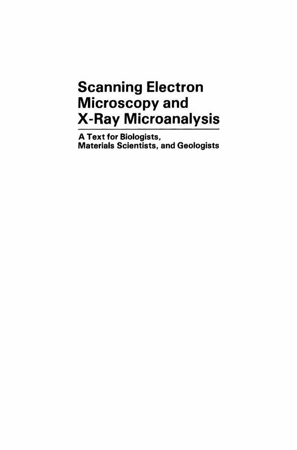
1. Introduction . 1.1. Evolution of the Scanning Electron Microscope 1.2. Evolution of the Electron Probe Microanalyzer 1.3. Outline of This Book . . . . . . . . .
2. Electron Optics. 2.1. Electron Guns
2.1.1. Thermionic Emission. . 2.1.2. Tungsten Cathode. . . 2.1.3. The Lanthanum Hexaboride (LaB6) Cathode. 2.1.4. Field Emission Gun . . . . . .
2.2. Electron Lenses. . . . . . . . . . 2.2.1. General Properties of Magnetic Lenses 2.2.2. Production of Minimum Spot Size 2.2.3. Aberrations in the Electron Optical Column
2.3. Electron Probe Diameter, dp' vs. Electron Probe Current i . 2.3.1. Calculation of dmin and i max • • • • • • • . •
2.3.2. Measurement of Microscope Parameters (dp' i, a) 2.3.3. High-Resolution Scanning Electron Microscopy.
3. Electron-Beam-Speclmen Interactions 3.1. Introduction . . . . 3.2. Scattering. . . . . .
3.2.1. Elastic Scattering . 3.2.2. Inelastic Scattering.
3.3. Interaction Volume. . . 3.3.1. Experimental Evidence 3.3.2. Monte Carlo Calculations
3.4. Backscattered Electrons . . . 3.4.1. Atomic Number Dependence 3.4.2. Energy Dependence 3.4.3. Tilt Dependence. . 3.4.4. Angular Distribution
vii
2 9
· 17
. 19 · 19 · 19 .24 .25 .29 .31 .31 .35 .40 .44 .44 .47 .49
.53 .53 .53 .54 .57 .59 .59 .60 .75 .75 .77 .78 .79

vIII
3.4.5. Energy Distribution . . 3.4.6. Spatial Distribution 3.4.7. Sampling Depth. . . .
3.5. Signals from Inelastic Scattering 3.5.1. Secondary Electrons . 3.5.2. X-Rays. . . . . . 3.5.3. Auger Electrons. . . 3.5.4. Cathodoluminescence.
3.6. Summary . . . . . . .
CONTENTS
81 83 85 87 87 95
· 1I8 · 1I9 · 121
4. Image Formation In the Scanning Electron Microscope . 123 4.1. Introduction . . . . . . . . 123 4.2. The Basic SEM Imaging Process . . . 123
4.2.1. Scanning Action. . . . . . . 123 4.2.2. Image Construction (Mapping). . 125 4.2.3. Magnification . . . . . . . 128 4.2.4. Picture Element (Picture Point). . 130 4.2.5. Depth of Field . . 133 4.2.6. Image Distortions . . 136
4.3. Stereomicroscopy . . . . 143 4.4. Detectors . . . . . . . 146
4.4.1. Electron Detectors . . 146 4.4.2. Cathodoluminescence Detectors . 155
4.5. The Roles of Specimen and Detector in Contrast Formation. . 156 4.5.1. Contrast . . . . . . . . . . . . . . .. . 156 4.5.2. Atomic Number (Compositional) Contrast (Backscattered
Electron Signal). . . . . . . . . . . . . . 157 4.5.3. Compositional Contrast (Secondary-Electron Signal) . 162 4.5.4. Contrast Components. . . . . . . . 164 4.5.5. Topographic Contrast. . . . . . . . 164
4.6. Image Quality. . . . . . . . . . . . .172 4.6.1. Signal Quality and Contrast Information . 172 4.6.2. Strategy in SEM Imaging. . . . . . . 175 4.6.3. Resolution Limitations . . . . . . . 176
4.7. Signal Processing for the Display of Contrast Information. . 183 4.7.1. The Visibility Problem . 183 4.7.2. Signal Processing Techniques . 186 4.7.3. Combinations of Detectors . 199 4.7.4. Beam Energy Effects .201 4.7.5. Summary. . . . . . . 203
5. X-Ray Spectral Measurement: WDS and EDS . . 205 5.1. Introduction . . . . . . . . . . 205 5.2. Wavelength-Dispersive Spectrometer. .205
5.2.1. Basic Design. . . . . . . .205 5.2.2. The X-Ray Detector . . . . .210 5.2.3. Detector Electronics . . . . .213
5.3. Energy-Dispersive.X-Ray Spectrometer. .222 5.3.1. Operating Principles . . . . . .222 5.3.2. The Detection Process. . . . . .224 5.3.3. Artifacts of the Detection Process. .226

CONTENTS Ix
5.304. The Main Amplifier and Pulse Pileup Rejection . . 234 5.3.5. Artifacts from the Detector Environment . .241 5.3.6. The Multichannel Analyzer. . . . . . . . . 253 5.3.7. Summary of EDS Operation and Artifacts. . . .260
5.4. Comparison of Wavelength-Dispersive Spectrometers with Energy-Dispersive Spectrometers . . . .262 504.1. Geometrical Collection Efficiency. . 262 504.2. Quantum Efficiency . . . . 262 504.3. Resolution. . . . . . . . 264 50404. Spectral Acceptance Range. .266 504.5. Maximum Count Rate . 267 5.4.6. Minimum Probe Size . 267 504.7. Speed of Analysis . . .269 504.8. Spectral Artifacts . . . 269
Appendix: Initial Detector Setup and Testing . 270
6. Qualitative X-Ray Analysis. .275 6.1. Introduction . . . . . . 275 6.2. EDS Qualitative Analysis. . . . 277
6.2.1. X-Ray Lines. . . . . .277 6.2.2. Guidelines for EDS Qualitative Analysis . 286 6.2.3. Pathological Overlaps in EDS Qualitative Analysis . . 288 6.204. Examples of EDS Qualitative Analysis . .291
6.3. WDS Qualitative Analysis . . . . . . . .292 6.3.1. Measurement of X-Ray Lines. . . . . 292 6.3.2. Guidelines for WDS Qualitative Analysis . 297
604. X-Ray Scanning. . . . . . . . . . . . 299
7. Quantitative X-Ray Microanalysis . 305 7.1. Introduction . . . . 305 7.2. ZAF Technique. . . . . . . .306
7.2.1. Introduction. . . . . . .306 7.2.2. The Absorption Factor, A . .308 7.2.3. The Atomic Number Factor, Z .315 7.204. The Characteristic Fluorescence Correction, F .322 7.2.5. The Continuum Fluorescence Correction .325 7.2.6. Summary Discussion of the ZAF Method. . . 326
7.3. The Empirical Method. . . . . . . . . . . .331 704. Quantitative Analysis with Nonnormal Electron Beam Incidence .335 7.5. Analysis of Particles and Rough Surfaces . .338
7.5.1. Geometric Effects. . . . . . . .338 7.5.2. Compensating for Geometric Effects. .347 7.5.3. Summary. . . . . . .351
7.6. Analysis of Thin Films and Foils . . . . . 352 7.6.1. Thin Foils. . . . . . . . . . .352 7.6.2. Thin Films on Substrates. . . . . . 354
7.7. Quantitative Analysis of Biological Material .363 7.7.1. Introduction. . . . . . . . . .363 7.7.2. Mass Loss and Artifacts during Analysis .364 7.7.3. Bulk Samples . . . . . . . .366 7.704. Thick Sections on Bulk Substrates . 369

x CONTENTS
7.7.5. Thin Samples . . . . . . . . . .370 7.7.6. The Continuum Method . . . . . .372 7.7.7. Thick Specimens on Very Thin Supports .377 7.7.8. Microdroplets. . . .378 7.7.9. Standards . . . . . 379 7.7.10. Conclusion. . . . .379
Appendix A: Continuum Method . 381 Appendix B: Worked Examples of Quantitative Analysis of Biological
Material . 387 Notation. . . . . . . . . . . . . . . . . . . . . . . 391
8. Practical Techniques of X-Ray Analysis. . 393 8.1. General Considerations of Data Handling . 393 8.2. Background Shape. . . . . 396
8.2.1. Background Modeling. . 397 8.2.2. Background Filtering . . 405
8.3. Peak Overlap. . . . . . 408 8.3.1. Linearity. . . . . .410 8.3.2. Goodness of Fit. . . .411 8.3.3. The Linear Methods . .412 8.3.4. The Nonlinear Methods .420 8.3.5. Error Estimation . 424
8.4. Dead-Time Correction. . . . 427 8.5. Example of Quantitative Analysis . . 427 8.6. Precision and Sensitivity in X-Ray Analysis. . 430
8.6.1. Statistical Basis for Calculating Precision and Sensitivity . 430 8.6.2. Sample Homogeneity . . 432 8.6.3. Analytical Sensitivity . . 434 8.6.4. Trace Element Analysis . 435
8.7. Light Element Analysis .439
9. Materials Specimen Preparation for SEM and X-Ray Microanalysis . . 447 9.1. Metals and Ceramics . . . . . . .447
9.1.1. Scanning Electron Microscopy. . 447 9.1.2. X-Ray Microanalysis .449
9.2. Particles and Fibers. . 453 9.3. Hydrous Materials . . 458
9.3.1. Soils and Clays .458 9.3.2. Polymers . . .459
10. Coating Techniques for SEM and Microanalysis . 461 10.1. Introduction. . . . . . . . . 461
10.1.1. Specimen Characteristics . . 463 10.1.2. Alternatives to Coating . 467 10.1.3. Thin-Film Technology. . .468
10.2. Thermal Evaporation. . . . . . 468 10.2.1. High-Vacuum Evaporation . 469 10.2.2. Low-Vacuum Evaporation .479
CONTENTS
10.3. Sputter Coating. . . . . . . . . . 10.3.1. Ion Beam Sputtering . . . . . 10.3.2. Diode or Direct Current Sputtering 10.3.3. Cool Diode Sputtering. 10.3.4. Sputtering Techniques. . . . 10.3.5. Choice of Target. . . . . . 10.3.6. Coating Thickness . . . . . 10.3.7. Advantages of Sputter Coating. 10.3.8. Artifacts Associated with Sputter Coating
10.4. Specialized Coating Methods. . . 10.4.1. High-Resolution Coating. . . 10.4.2. Low-Temperature Coating . .
10.5. Determination of Coating Thickness. . 10.5.1. Estimation of Coating Thickness 10.5.2. Measurement during Coating 10.5.3. Measurement after Coating 10.5.4. Removing Coating Layers
11. Preparation of Biological Samples for Scanning Electron Microscopy. 11.1. Introduction. . . . . . . 11.2. Compromising the Microscope
11.2.1. Environmental Stages . 11.2.2. Nonoptimal Microscope Performance
11.3. Compromising the Specimen . 11.3.1. Correlative Microscopy 11.3.2. Specimen Selection 11.3.3. Specimen Cleaning 11.3.4. Specimen Stabilization 11.3.5. Exposure of Internal Surfaces. 11.3.6. Localizing Areas of Known Physiological Activity . 11.3.7. Specimen Dehydration . . 11.3.8. Specimen Supports 11.3.9. Specimen Conductivity . . 11.3.10. Heavy Metal Impregnation. 11.3.11. Interpretation and Artifacts.
12. Preparation of Biological Samples for X-Ray Microanalysis 12.1. Introduction. . . . . . . . . . . . .
12.1.1. The Nature and Enormity of the Problem 12.1.2. Applications of X-Ray Microanalysis. . 12.1.3. Types of X-Ray Analytical Investigations 12.1.4. Types of Biological Specimens. . . . 12.1.5. Strategy. . . . . . . . . . . . 12.1.6. Criteria for Satisfactory Specimen Preparation.
12.2. Ambient Temperature Preparative Procedures 12.2.1. Before Fixation . . . . 12.2.2. Fixation. . . . . . . 12.2.3. Histochemical Techniques 12.2.4. Precipitation Techniques .
xl
.479
.481
.482
.482
.483
.484
.484
.484
.486
.487
.487
.490
.491
.492
.493
.493
.494
.495 .495 .496 .496 .497 .499 .499 .501 .502 .506 .508 .518 .522 .531 .532 .532 .534
.541 .541 .541 .543 .544 .545 .548 .549 .550 .550 .550 .554 .555
xII CONTENTS
12.2.5. Dehydration. . . . . . 557 12.2.6. Embedding . . . . . . 558 12.2.7. Sectioning and Fracturing . 558 12.2.8. Specimen Supports . 559 12.2.9. Specimen Staining. . . . 559 12.2.10. Specimen Coating. . . . 560
12.3. Low-Temperature Preparative Procedures. .560 12.3.1. Specimen Pretreatment . . . . .561 12.3.2. Freezing Procedures . . . . . . 564 12.3.3. Movement of Elements within a Given Cellular
Compartment . . . . . . . . . . . . . 567 12.3.4. Postfreezing Procedures. . . . . . . . . . 567 12.3.5. Frozen-Hydrated and Partially Frozen-Hydrated Material . 567 12.3.6. Freeze Drying . . . 567 12.3.7. Freeze Substitution . 573 12.3.8. Sectioning . 576 12.3.9. Fracturing . . . . 584 12.3.10. Specimen Handling . 584
12.4. Microincineration. . . . . 586
13. Applications of the SEM and EPMA to Solid Samples and Biological Materials . . 589 13.1. Study of Aluminum-Iron Electrical Junctions. . . . . . . .589 13.2. Study of Deformation in Situ in the Scanning Electron Microscope. 593 13.3. Analysis of Phases in Raney Nickel Alloy . . . . .. .594 13.4. Quantitative Analysis of a New Mineral, Sinoite . . .. . 596 13.5. Determination of the Equilibrium Phase Diagram for the
Fe-Ni-C System. . . . . . . . . . . . . .. . 597 13.6. Study of Lunar Metal Particle 63344,1. . . . . . .. .599 13.7. Observation of Soft Plant Tissue with a High Water Content .602 13.8. Study of Multicellular Soft Plant Tissue with High Water Content .604 13.9. Examination of Single-Celled, Soft Animal Tissue with High
Water Content. . . . . . . . . . . . . . . . . . .606 13.10. Observation of Hard Plant Tissue with a Low Water Content. . .607 13.11. Study of Single-Celled Plant Tissue with a Hard Outer Covering
and Relatively Low Internal Water Content. . . . . . . 13.12. Examination of Medium Soft Animal Tissue with a High
Water Content. . . . . . . . . . . . . . . . . 13.13. Study of Single-Celled Animal Tissue of High Water Content.
.608
.610
.612
14. Data Base .615 Table 14.1. Atomic Number, Atomic Weight, and Density of
Metals . . . . . . . . . . . . . .616 Table 14.2. Common Oxides of the Elements . . . . .619 Table 14.3. Mass Absorption Coefficients for Ka Lines .620 Table 14.4. Mass Absorption Coefficients for La Lines .622 Table 14.5. Selected Mass Absorption Coefficients . . . 624 Table 14.6. K Series X-Ray Wavelengths and Energies .641 Table 14.7. L Series X-Ray Wavelengths and Energies . .642

CONTENTS xIII
Table 14.8. M Series X-Ray Wavelengths and Energies. . . .. .643 Table 14.9. Fitting Parameters for Duncumb-Reed Backscattering
Correction Factor R . . . . . . . . . . .644 Table 14.10. J Values and Fluorescent Yield, w, Values . . . .645 Table 14.11. Important Properties of Selected Coating Elements . 646
REFERENCES . 649 INDEX . .665


![Ultrafast transmission electron microscopy using a laser ...transmission electron microscopy [4], scanning electron microscopy [5], x-ray diffraction [6], scanning tunneling and atomic](https://img.dokumen.tips/doc/110x75/607eb1335ce8082131294459/ultrafast-transmission-electron-microscopy-using-a-laser-transmission-electron.jpg)