Embed Size (px)
Citation preview

Scabies Mite Peritrophins Are Potential Targets ofHuman Host Innate ImmunityAngela Mika1., Priscilla Goh1., Deborah C. Holt2, Dave J. Kemp1, Katja Fischer1*
1 Queensland Institute of Medical Research and Australian Centre for International and Tropical Health and Nutrition, University of Queensland, Brisbane, Queensland,
Australia, 2 Menzies School of Health Research, Charles Darwin University, Darwin, Northern Territory, Australia
Abstract
Background: Pruritic scabies lesions caused by Sarcoptes scabiei burrowing in the stratum corneum of human skin facilitateopportunistic bacterial infections. Emerging resistance to current therapeutics emphasizes the need to identify novel targetsfor protective intervention. We have characterized several protein families located in the mite gut as crucial factors for host-parasite interactions. Among these multiple proteins inhibit human complement, presumably to avoid complement-mediated damage of gut epithelial cells. Peritrophins are major components of the peritrophic matrix often found in the gutof arthropods. We hypothesized that a peritrophin, if abundant in the scabies mite gut, could be an activator ofcomplement.
Methodology/Principal Findings: A novel full length scabies mite peritrophin (SsPTP1) was identified in a cDNA library fromscabies mites. The amino acid sequence revealed four putative chitin binding domains (CBD). Recombinant expression ofone CBD of the highly repetitive SsPTP1 sequence as TSP-hexaHis-fusion protein resulted in soluble protein, whichdemonstrated chitin binding activity in affinity chromatography assays. Antibodies against a recombinant SsPTP1 fragmentwere used to immunohistochemically localize native SsPTP1 in the mite gut and in fecal pellets within the upper epidermis,co-localizing with serum components such as host IgG and complement. Enzymatic deglycosylation confirmed strong N-and O-glycosylation of the native peritrophin. Serum incubation followed by immunoblotting with a monoclonal antibodyagainst mannan binding lectin (MBL), the recognition molecule of the lectin pathway of human complement activation,indicated that MBL may specifically bind to glycosylated SsPTP1.
Conclusions/Significance: This study adds a new aspect to the accumulating evidence that complement plays a major rolein scabies mite biology. It identifies a novel peritrophin localized in the mite gut as a potential target of the lectin pathwayof the complement cascade. These initial findings indicate a novel role of scabies mite peritrophins in triggering a hostinnate immune response within the mite gut.
Citation: Mika A, Goh P, Holt DC, Kemp DJ, Fischer K (2011) Scabies Mite Peritrophins Are Potential Targets of Human Host Innate Immunity. PLoS Negl TropDis 5(9): e1331. doi:10.1371/journal.pntd.0001331
Editor: Jesus G. Valenzuela, National Institute of Allergy and Infectious Diseases, United States of America
Received February 14, 2011; Accepted August 7, 2011; Published September 27, 2011
Copyright: � 2011 Mika et al. This is an open-access article distributed under the terms of the Creative Commons Attribution License, which permitsunrestricted use, distribution, and reproduction in any medium, provided the original author and source are credited.
Funding: This work was supported by the Australian National Health and Medical Research Council (http://www.nhmrc.gov.au/, Program Grant 496600 and afellowship to D.J.K). The funders had no role in study design, data collection and analysis, decision to publish, or preparation of the manuscript.
Competing Interests: The authors have declared that no competing interests exist.
* E-mail: [email protected]
. These authors contributed equally to this work.
Introduction
Scabies is a widespread infectious parasitic disease [1]. The
etiological agent, Sarcoptes scabiei burrows into the lower stratum
corneum of the skin [2]. The clinical signs are erythematous
lesions, pruritus and burrows [1]. Pruritus, commonly known as
itchiness, is a consequence of a delayed type four hypersensitivity
immune reaction [3]. Scabies is a major livestock disease [4] but
animal scabies in humans is self limiting as the lifecycle of the mite
cannot be completed.
Scabies spreads rapidly by person-to-person contact under
crowded conditions. Indigenous Australians living in remote
communities in the north of the country experience significant
risk of morbidity from scabies. Pruritic scabies lesions facilitate
opportunistic bacterial infections [5], particularly by Group A
streptococci (GAS) and staphylococci [6]. According to a recent
study undertaken in two communities, more than 70% of children
presented to the health clinic with scabies by two years of age, with
a peak of presentation at 2 months of age [6]. Importantly, in over
80% of these children skin sores were observed, indicating high
rates of secondary infections with pathogenic bacteria. Among
these particularly streptococcal infections cause significant sequel-
ae (cellulitis, septicemia, and glomerulonephritis) and the increased
community streptococcal burden has led to the most extreme
levels in the world of Acute Rheumatic Fever and Rheumatic
Heart Disease in these communities [5,7].
Emerging resistance of scabies mites to current therapeutics
emphasizes the need to identify novel targets for protective
intervention [8]. Due to the difficulty of acquiring mites, no
molecular studies on scabies were done until recently. Crusted
scabies is a severe form of scabies with extreme parasite burden.
We constructed cDNA libraries from mites obtained from skin
www.plosntds.org 1 September 2011 | Volume 5 | Issue 9 | e1331

shed in the bedding of patients with crusted scabies [9,10,11]
facilitating molecular studies [12,13,14,15]. A database containing
over 43,000 expressed sequence tag (EST) sequences was created
and searched for molecules predicted to play important roles in
host parasite biology. One avenue pursued was to investigate gut
proteins essential in food uptake. Gut molecules, being critical to
parasite survival and concealed from the host’s immune response
but accessible for ingested antibodies, leucocytes and complement
attack, are top-ranking targets in developing therapeutics against
ticks and worms. For example the antigen Bm86 from the digestive
tract of Rhipicephalus microplus (formerly Boophilus microplus) [16] was
the basis for the development of the two anti-tick vaccines, which
have been commercialized to date. The feeding scabies mite
imbibes epidermal proteins and plasma containing host serine
proteases, which mediate complement and blood clotting. Both
systems must be inhibited in a milieu in which digestion of food
can occur in the gut. Thus, in accordance with their parasitic life,
scabies mites need to inhibit, control and prevent complement
action in their gut.
The complement system of the vertebrate host forms a powerful
immune barrier [17,18,19]. Upon entry of a foreign invader, this
tightly regulated defense system is activated within seconds.
Complement consists of approximately 35 proteins circulating in
human plasma, penetrating mucosal surfaces and most tissues.
The system targets foreign particles by attracting phagocytes to the
site of damage, by increasing the permeability of the epidermal
capillaries and by direct lysis of foreign cells. The complement
system is triggered by various ligands and its activation can
proceed via three enzymatic cascades: the classical, lectin or
alternative pathways.
The classical pathway is initiated by the binding of the C1
complex to immune complexes, while the lectin pathway is
activated via recognition of specific carbohydrates by mannose-
binding lectin (MBL). The alternative pathway is triggered due to
structural instability of the C3 molecule leading to autoactivation,
direct binding of properdin or to an amplification loop to the two
other pathways. Activation of any of these three pathways leads to
the opsonization of the target with C3b, which mediates
phagocytosis. Formation of C3b proceeds to the assembly of a
membrane attack complex (MAC) and release of the chemoat-
tractant anaphylatoxins C3a and C5a. The sequentially expanding
pathways are vulnerable to inhibitors. Many pathogens utilize
inhibitory molecules to block complement components and
thereby evade host attack [20]. Among acarid arthropods tick
salivary proteins have been shown to inhibit a number of
complement components. For example the complement inhibitor
OmCI, a salivary protein released by the soft tick Ornithodoros
moubata, has been shown to directly bind to C5 in a stable complex
86 [21], thereby inhibiting cleavage of C5 into the anaphylatoxin
C5a and the subunit of the membrane attack complex C5b. This
inhibition protects the tick from complement-mediated damage by
either the classical or alternative pathways. Similarly, Ixodes
scapularis and I. ricinus produce a family of homologous Isac-like
proteins that inhibit the alternative pathway by preventing the
deposition of C3b and the release of C3a [22]. In addition one of
these homologs, I. scapularis salivary protein 20 (Salp20), was
shown to bind to the regulatory protein properdin, thereby
destabilising the complement C3 convertase (C3bBb) in the
alternative complement pathway [23].
Our group has previously identified a protein family of
catalytically inactive scabies mite serine proteases (SMIPP-Ss
[12]), which are secreted into the gut and excreted into the
epidermis [12,24]. They are homologous to the group 3 allergens
of house dust mites but lack proteolytic activity due to mutations in
the catalytic triad and structural rearrangements [24,25]. All
SMIPP-Ss examined to date were found to exert complement-
inhibitory properties and two exemplary SMIPP-Ss have been
shown to bind to the complement factors C1q, MBL and
properdin [26]. The fact that a large multigene family has evolved
to inhibit all three pathways of the complement cascade indicates
that host complement is a major threat to the mites. They may
have a complement evasion machinery in place, which could have
an impact on their microenvironment similar to that induced by
associated bacterial pathogens in the epidermal burrows [19].
Moreover as both mites and bacteria release these complement
inhibitors into the skin, they may even supplement each other’s
defense. An obvious question is which structures within the gut
initiate the complement cascade, thereby requiring the mite to
produce elaborate complement-inhibiting mechanisms? Peri-
trophic matrix (PM) proteins could be prime candidates in this
role. They are thought to be present in the midgut of most
invertebrates, protecting it against the abrasive food bolus,
ingested pathogens and toxins. However, most knowledge
collected to date is restricted to insect peritrophins [27].
Peritrophin proteins are major components of the insect
peritrophic matrix known for their low solubility [28]. Given its
important role, the arthropod peritrophic matrix has been
investigated to develop new pest management strategies (reviewed
in [27]). Intriguingly the scabies mite cDNA database [8,9]
contained an abundant peritrophin-like sequence. Here we
present initial evidence this scabies mite peritrophin may be
involved in activating complement.
Methods
Ethics StatementAll animals were handled in strict accordance with good animal
practice as defined by the Australian code of practice for the care
and use of animals for scientific purposes, 7th Edition, 2004 – Web
site: http://www.nhmrc.gov.au/publications/synopses/ea16syn.
htm and the National Health & Medical Research Council’s
(NHMRC) Animal Code of Practice. Ethical approval for the
Author Summary
The gut of most invertebrates is lined by a protective layer ofchitin and glycoproteins, often designated as a peritrophicmatrix. Previous research suggests that it forms a barrier thatmay protect the midgut epithelium from abrasive foodparticles and pathogens. Parasitic invertebrates ingestingvertebrate plasma have evolved additional strategies toprotect themselves from hazardous host molecules con-sumed during feeding. An important part of the immediatedefense in vertebrate plasma is complement-mediatedkilling. The Complement system is a complex network ofmore than 35 proteins present in human plasma that resultsin killing of foreign cells including the gut epithelial cells of afeeding parasite. Recently we found that scabies mites, whofeed on skin containing plasma, produce several proteinsthat inhibit human complement within the mite gut. Themites excrete these molecules into the upper epidermiswhere they presumably also inhibit complement activity.Mite gut antigens that initially trigger the complementcascade have not been identified previously. Obviouspossible targets of complement attack within the mite gutcould be peritrophins. Our study describes the firstperitrophin identified in scabies mites and indicates apossible role in complement activation.
A Novel Scabies Mite Peritrophin
www.plosntds.org 2 September 2011 | Volume 5 | Issue 9 | e1331

production of polyclonal antibodies in mice was obtained from the
Queensland Institute of Medical Research Animal Ethics
Committee in compliance with the Code of practice, the
NHMRC, and the Queensland government responsible authority
[29].
Written informed consent was obtained from the crusted scabies
patient for the collection of shed skin crusts, with the approval of
the Human Research Ethics Committee of the Northern Territory
Department of Health and Families and the Menzies School of
Health Research.
CloningBLAST searches of the scabies mite cDNA database (http://vbc.
med.monash.edu.au/˜yvan/Sarcoptes_scabiei/login.php) identi-
fied a peritrophin-like sequence. The sequence encoding full length
SsPTP1 was amplified from a cDNA library from human scabies
mites [9,10] using the primers 59-accggtcgacTTCATCAATGGT-
CATCTGCATCGAAAACG-39 and 59-accgctgcagTCAGAATA-
GTTTGAAATAATTCGAATCTTCTGTGGTGG-39 (Sigma),
which incorporated the restriction sites SalI and PstI, respectively
(lower case), required for directional cloning into the pQE9 vector
(Qiagen). The PCR product was digested at these sites, ligated into
the pQE9 vector, transformed into E. coli BL21 cells (Stratagene).
Transformants were confirmed by sequencing with BigDye 3.1
(Applied Biosystems) using vector specific primers. The sequence of
the first chitin-binding domain (CBD1) of the SsPTP1 sequence was
amplified from the pQE9 clone containing the complete SsPTP1
sequence using specific primers 59-accgccatggAATTCATCA-
ATGGTCATCTGCATCGAAAACG-39 and 59-accgctcgagTC-
GAGGTTTTTTTGTAGTATTTTTTCTGAACCA-39 (Sigma)
incorporating the restriction sites Nco1 and Xhol and directionally
cloned into a modified version of the pET-41a vector (Novagen)
containing a tetraspanin protein 2 fragment from Schistosoma mansoni
[30]. To obtain a peptide without a large N-terminal fusion for
immunizations, fragment CBD1 was amplified using identical
primers but with the restriction sites SalI and PstI and directionally
cloned into the pQE9 vector (Qiagen). The CBD1 constructs were
transformed into BL21 E. coli cells. Each of the three fusion
constructs contained a C-terminal hexaHis-tag.
Heterologous Expression and PurificationRecombinant CBD1 and TSP-CBD1 proteins were expressed
and purified under denaturing conditions. E. coli cells were
cultivated in LB medium containing 100 mg/mL ampicillin at
37uC over night. After inoculation of 2YT medium containing the
same concentration of ampicillin, the cells were grown at 37uCuntil an OD600 of 0.5-0.6 was reached and overnight expression
was induced by addition of 1 mM IPTG. Cells were collected by
centrifugation at 4000 x g at 4uC for 20 min, resuspended in
50 mM Tris, pH 8.0, 100 mM NaCl, 10 mM EDTA, 1 mM
PMSF and lysed in the presence of 250 mg/ml lysozyme and
10 mg/ml DNAse at room temperature under continuous rotation.
All of the following purification steps were performed at 4uC. After
sonication of the spheroplasts by a Sonifier 250 (Branson),
inclusion bodies were washed four times using 50 mM Tris,
pH 8.0, 100 mM NaCl, 10 mM EDTA, 0.5% (v/v) Triton X-100
and retrieved by centrifugation (16,000 x g for 20 min at 4uC),
followed by solubilization in 6 M guanidine hydrochloride,
50 mM Tris, pH 7.8, 1 mM DTT for 60 min. Proteins were
further purified by nickel immobilized metal affinity chromatog-
raphy (Qiagen): Solubilized protein was diluted 1:1 with 6 M urea,
100 mM NaH2PO4, 10 mM Tris, pH 8.0, 5 mM imidazole,
150 mM NaCl, 1% (v/v) glycerol, 1 mM DTT and bound over
night to a pre-equilibrated Ni-NTA matrix (Qiagen) on a rotating
shaker. The matrix was loaded into a column (PolyPrep, BioRad)
and washed with 10 column volumes of 6 M urea, 100 mM
NaH2PO4, 10 mM Tris, pH 6.3, 5 mM imidazole, 150 mM
NaCl, 1% (v/v) glycerol, 1 mM dithiothreitol. Bound proteins
were eluted using 4 column volumes of 6 M urea, 100 mM
NaH2PO4, 10 mM Tris, pH 8.0, 250 mM imidazole, 150 mM
NaCl, 1% (v/v) glycerol and 1 mM dithiothreitol. For refolding of
recombinant TSP-CBD1, 1 ml of protein eluted from Ni-NTA
affinity chromatography was added in portions of 100 ml at a time
in 5 min intervals at 4uC to 500 ml of refolding buffer (50 mM
Tris – HCl, pH 8.0, 50 mM NaCl, 300 mM arginine, 5 mM
dithiothreitol) under continuous stirring. Refolded protein was
concentrated using an Ultrasette Lab Tangential Flow Device
(PALL Life Sciences) with a 3 kDa filter pore size followed by
further concentration using centrifugal filter units (Amicon Ultra,
Millipore). Protein concentrations were determined according to
[31]. Molecular masses and purity were confirmed using SDS-
PAGE analysis with Coomassie blue R-250 staining. The integrity
and identity of CBD1 was confirmed by mass spectrophotometry
analysis, i.e. MALDI-TOF, at the Monash Biomedical Proteomics
Facility at Monash University, Clayton, Victoria.
Localization of SsPTP1 and Complement byImmunohistochemistry
A custom-made peptide (DRFGYFSHEDCWRFVL; c2) was
purchased from Mimotopes, Australia conjugated with keyhole
limpet hemocyanin (KLM). Antibodies against the recombinant
CBD1 protein, dialyzed over night into PBS at 4uC, and the
custom-made peptide were raised in BalbC female mice. Doses of
50 mg of protein were injected, resuspended in PBS with Alum
adjuvant (PIERCE, USA). Pre-bleeds were collected before the
first immunization dose to serve as negative controls. The antibody
titers and specificity were tested by ELISA and western blot
analysis.
Tissue preparation of human mite-infested skin samples and the
immunolocalization of SsPTP1were accomplished as previously
outlined [24]. Serial sections of 4 mm thickness cut from paraffin
embedded human skin tissue infested with scabies mites under-
went an Antigen heat-retrieval procedure in a solution containing
1% (w/v) Revealit-Ag in 20 mM Tris (pH 7.5; ImmunoSolution)
at 75uC for 15 min. The slides were cooled and washed with Milli
Q water for 10 min followed by 15 min washes in PBS at pH 7.2.
The tissue sections were blocked with 10% donkey serum in 1%
(w/v) BSA in PBS. Endogenous peroxidase activity was blocked
with 3% (v/v) H2O2 in blocking buffer. The sections were
incubated overnight with either mouse polyclonal antibodies
against peritrophin c2-conjugate (1:200 dilution) or polyclonal
antibodies against recombinant peritrophin CBD1 (1:500 dilu-
tion). The primary antibodies were probed with the secondary
antibody Dako-EnVision anti-mouse-HRP conjugate (DakoCyto-
mation) for 30 min. As a positive control, adjacent sections were
incubated with anti-human IgG-HRP polyclonal antibody (Ab-
cam) in 1:500 dilutions to identify the mite’s gut. As a negative
control, pre-immune serum was incubated on sections of the same
series in either 1:200 or 1:500 dilutions. All slides were washed in
PBS and the Vector NovaRed peroxidase substrate kit (Vector
Laboratories) was used for staining according to the manufactur-
er’s recommendations, followed by counterstaining with hema-
toxylin. Slides were scanned using a Scan Scope XT microscope
(Aperio Technologies) at 406magnification.
For the localization of complement components a variety of
blocking and immunodetection compounds and conditions were
tested. As primary antibodies a rabbit anti-human SC5b-9
neoantigen-specific antibody recognizing the human MAC
A Novel Scabies Mite Peritrophin
www.plosntds.org 3 September 2011 | Volume 5 | Issue 9 | e1331

complex in situ after complement activation and a goat anti-human
C9-antibody recognizing non-complexed C9 (Complement Tech-
nology Inc., USA) were used. The antibody specificity and titers
required were tested by an ELISA-based complement depositon
assay as described previously [26]. Tissue preparation of human
mite-infested skin samples and the immunolocalization were
accomplished as outlined above. The tissue sections were subjected
to blocking with casein (BioCare Medical, USA) for 5 min at room
temperature followed by overnight incubation with 10% goat
serum in 1% (w/v) BSA in PBS for SC5b-9 detection and with
10% donkey serum in PBS for C9 detection. Adjacent sections
were incubated with either the SC5b-9 neoantigen-specific
antibody at 1:200, 1:500, 1:1000, 1:2000, 1:4000 and 1:20000
dilutions) or the anti-human C9-specific antibody at a 1:200
dilution for 1h. Endogenous peroxidase activity was blocked with
3% (v/v) H2O2 in blocking buffer. The primary antibodies were
probed with MACH 2 Rabbit HRP-Polymer (BioCare Medical,
USA) for detection of SC5b-9 neoantigen and with Goat HRP-
Polymer Kit (BioCare Medical, USA) for C9 detection. As a
positive control for gut localization adjacent sections were
incubated with anti-human IgG-HRP polyclonal antibody (Ab-
cam) in 1:500 dilutions. As a negative control, naıve pre-immune
rabbit or goat serum was incubated on sections of the same series
in the same dilutions as the test sera. The Vector NovaRed
peroxidase substrate kit (Vector Laboratories) was used for staining
according to the manufacturer’s recommendations, followed by
counterstaining with hematoxylin. Slides were scanned using a
Scan Scope XT microscope (Aperio Technologies) at 40x
magnification.
Chitin Binding AssayMagnetic chitin beads (NEB) were equilibrated with chitin
binding buffer (20 mM Tris, pH 8.0, 500 mM NaCl, 1 mM
EDTA, 0.05% (v/v) Triton X-100). Proteins were incubated with
500 ml of chitin beads for 2 hours at 4uC with rotary shaking in a
total volume of 3 ml. Wheat germ agglutinin (WGA) as positive,
and tetraspanin protein 2 fragment from Schistosoma mansoni (TSP;
[30] and BSA as negative controls were used, respectively. After
binding, the beads were washed three times with 10 column
volumes binding buffer. The proteins bound to the beads were
eluted in 1.25 ml fractions using 8 M urea, followed by the
addition of 5% SDS and then 10% SDS.
Deglycosylation of Native SsPTP130–40 mg scabies mites picked from pig ears [32] were
homogenized in 150 ul 50 mM Tris, pH 8.0, 100 mM NaCl,
1 mM EDTA in an Eppendorf tube with a plastic pestle on ice.
Deglycosylation was performed under denaturing conditions using
an Enzymatic Protein Deglycosylation Kit (EDEGLY, Sigma)
according to the manufacturer’s instructions. PNGase F, O-
glycosidase, a-neuraminidase, b-1, 4-galactosidase and b-N-
acetylglucosaminidase were assayed separately and in combina-
tion. Enzyme reactions were incubated between 24 hours and
three days at 37uC in a water bath and subsequently analyzed by
SDS-PAGE.
Western Blotting Using an Infrared FluorescenceDetection System
Proteins separated by SDS-PAGE were transferred onto an
Immubilon-FL PVDF membrane (Millipore), blocked with
Odyssey blocking buffer (LI-COR Biosciences) overnight at 4uC,
and incubated for 1 h with 1:500 dilutions of primary antibody
raised against recombinant CBD1 protein. Membranes were
washed four times in PBS, followed by incubation for 1h with a
fluorescent secondary antibody (Goat anti mouse-IR dye l800 nm)
at 1:10,000 dilution. Proteins were visualized using an Odyssey
Infrared Imaging System (LI-COR Biosciences).
Immunodetection of Human MBL Binding to Protein MiteExtract Using an Infrared Fluorescence Detection System
Fresh and deglycosylated mite homogenates were separated on
SDS-PAGE and transferred onto an Immubilon-FL PVDF
(Millipore) membrane. Membranes were incubated with 50%
(v/v) normal human serum diluted in binding buffer, consisting of
50% GVB (2.5 mM veronal buffer, pH 7.3, 0.1% (w/v) gelatin)
and 50% TBS (200 mM NaCl, 50 mM Tris pH 7.4), for 1 h at
37uC under continuous shaking. The membranes were washed
four times in TBS, followed by 1 h incubation at room
temperature with an anti-MBL antibody (AF2307, R&D Systems,
Denmark) at a 1:1000 dilution. The membranes were washed four
times in PBS, followed by incubation with a fluorescent secondary
antibody (Goat anti mouse-IR dye l800 nm) at 1:10,000 dilution.
Protein detected by the antibody was visualized using an Odyssey
Infrared Imaging System (LI-COR Biosciences).
SoftwareSequence analysis was performed using prediction tools
available on www.expasy.org [33,34,35,36]. Statistical analysis
was done using Graph Pad Prism (GraphPad Software, USA).
Results
Sequence Analysis and Cloning of a Novel Scabies MitePeritrophin
BLAST searches of the scabies mite cDNA database identified a
peritrophin-like sequence. The 1835bp contig of 18 ESTs
(Genbank Accession number JF266566) included 1461bp of full
length coding sequence. Based on a considerable amino acid
similarity of 37-39% with previously reported arthropod peritro-
phins, the novel gene was termed Sarcoptes scabiei peritrophin 1
(SsPTP1). SsPTP1 had a predicted signal sequence of 20 amino
acids, indicating that SsPTP1 might be a secretory protein. No
transmembrane domains were predicted. Four chitin binding
domains (CBDs) were detected each containing conserved patterns
of six cysteines residues. The sequence showed a substantial degree
of low complexity and repetitiveness including two threonine-rich
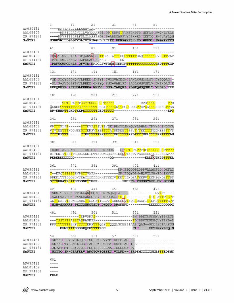
domains between the CBDs (regions 97–111 and 255–298; Fig. 1).
In addition, two N-glycosylation sites were predicted in SsPTP1 at
position 90 and 283 of the amino acid sequence, while two regions
with putative strong O-glycosylation were found (amino acids 97–
116 and 185–312) indicated by the presence of repetitive serine
and threonine residues in these regions (Fig. 1). The three aligned
insect peritrophins were also analyzed for N- and O-glycosylation.
The prediction showed that all four proteins may be hyper-
glycosylated over the full length of their sequences with the
exception of their chitin binding domains (Fig. 1).
E. coli expression and purification of full length recombinant
SsPTP1 was without success due to the high repetitiveness of the
sequence. The first chitin binding domain of SsPTP1 (CBD1)
expressed in E. coli was insoluble, but could be purified in large
amounts under denaturing conditions. Dialyzed and resuspended
CBD1 was used to raise polyclonal antibodies in mice. In order to
produce soluble proteins, CBD1 was additionally fused to the C-
terminus of the schistosomiasis vaccine antigen Sm-TSP-2. Both
proteins contained a C-terminal hexaHis tag. The fusion protein
TSP-CBD1 was soluble after purification by nickel affinity
chromatography from washed inclusion bodies and refolding,
A Novel Scabies Mite Peritrophin
www.plosntds.org 4 September 2011 | Volume 5 | Issue 9 | e1331
A Novel Scabies Mite Peritrophin
www.plosntds.org 5 September 2011 | Volume 5 | Issue 9 | e1331

presumably because both fusion partners are highly soluble when
expressed in E. coli [30].
CBD1 and TSP-CBD1 had the molecular masses of 12 kDa and
23 kDa, respectively (Fig. 2). After Ni-NTA purification, average
protein yields of 73.9 6 10.0 mg of TSP-CBD1 per 1 L E. coli
culture were achieved. After optimization of the refolding
procedure, up to 15.1% refolded protein was obtained. The
soluble fusion protein was used in a chitin binding assay.
A Recombinant SsPTP1 Chitin Binding Domain BindsChitin in an Affinity Chromatography Assay
A substantial proportion of the fusion protein TSP-CBD1
bound to chitin magnetic beads. In comparison the chitin binding
chromatography elution profiles of the purified tetraspanin protein
2 fragment from Schistosoma mansoni TSP and BSA as negative
controls revealed small amounts of non-specific binding while
wheat germ agglutinin (WGA) as a positive control showed strong
binding (Fig. 3A). The total protein amounts found in all fractions
including flowthrough and wash fractions showed significant
differences suggesting that considerable amounts of TSP-CBD1
and WGA could not be removed from the matrix during elution.
This was confirmed by Western blotting (Fig. 3B). Additional
boiling of the chitin matrix in 10% SDS in elution buffer did not
elute more protein (data not shown).
Localization of SsPTP1 to the Mite Digestive System andFecal Pellets in Human Skin
Polyclonal antibodies were raised in mice against a 16 amino
acid custom-made peptide conjugated to keyhole limpet hemo-
cyanine (c2). The specificity of both antisera to CBD1 and TSP-
CBD1 was confirmed and no cross-reactions with unrelated
scabies mite proteins or BSA were seen in western blot analysis.
Representative data for anti-CBD1 serum from one mouse are
shown in Fig. 2.
The SsPTP1 protein was localized within and outside the
scabies mite using the polyclonal antibodies for immunohisto-
chemical staining of serial sections of human skin infested with
mites (Fig. 4). Each anti-SsPTP1 antibody showed strong staining
of the digestive system of the mite (Fig. 4A- 2 and 6); while
adjacent sections probed with pre-immune serum showed only
minor background staining (Fig. 4A- 4 and 8). The immunode-
tection of human IgG, which was used as a positive control,
confirmed the localization of the mite gut (Fig. 4A- 3 and 7) as
previously documented [24,37]. In addition to its location in the
digestive tract of the mite, SsPTP1 was localized to external
acellular masses, which are scybala (fecal pellets; Fig. 4B- 2 and
6). The scybala were negative for staining with pre-immune
serum (Fig. 4B- 4 and 8) and positive for IgG staining. (Fig. 4B- 3
and 7). Given its presence in the digestive tract of the scabies mite
and in excreted fecal matter, the peritrophin is likely to be
involved in gut functions and would be predicted to be exposed to
epidermal host proteins within the gut and externally in the
epidermal burrow.
Deglycosylation of Native Scabies Mite Peritrophin 1 andBinding to the Human Complement Factor MBL
Fresh scabies mite homogenates were subjected to deglycosylat-
ing enzymes for one (upper panel, Fig. 5) or three days (bottom
panel, Fig. 5) at 37uC to remove N- and O-glycosylation of the
proteins and subsequently separated by SDS-PAGE. A single band
was detected by the anti-CBD1 antibody in untreated homoge-
nates after Western blotting (Fig. 5A). Incubation of mite extract
with glycosidases revealed smaller bands indicating enzymatic
digestion of glycans and thus a strong glycosylation of the detected
native protein (Fig. 5A). The material subjected to O-glycosidases
showed the strongest decrease in molecular mass, especially after
three days. In comparison PNGase F treatment showed a smaller
effect. Hence in accordance with the large number of predicted O-
glycosylation sites in SsPTP1 (Fig. 1), the O-glycosylation seemed
to be predominant. Further membranes were incubated with 50%
normal human serum containing all complement factors including
MBL (Fig. 5B) or buffer without serum (Fig. 5C) followed by
immunodetection of bound MBL using an anti-MBL antibody.
Human MBL bound to a protein with a molecular mass
corresponding to the band detected by the CBD1 antibody in
the untreated mite extracts (Fig. 5B). Negative control experi-
ments, lacking the incubation step in human serum (Fig. 5C)
confirmed that i) the MBL antibody was specific for MBL. A
distinct band (indicated by an arrow in Fig. 5C) corresponding to
the 32 kDa-peptide polypeptide chains of the human MBL
subunits [29] was detected. Furthermore this blot confirmed that
ii) the MBL antibody did not detect putatively existing scabies mite
haemolymph complement factors and iii) MBL was not detectable
in the mite extract due to food intake using this method.
Deglycosylation of the mite homogenate for one and three days
revealed comparable results, however, the degree of visible
deglycosylation was less after one day and protein degradation
Figure 1. Full length amino acid sequence of scabies mite peritrophin 1 (SsPTP1) aligned with other arthropod peritrophins. Thescabies mite peritrophin SsPTP1 is shown in bold in a sequence alignment with Anopheles gambiae peritrophin 1 (AF030431), peritrophin from Aedesagypti (AAL05409) and a peritrophin matrix protein from Tribolium castaneum (XP_974131). The following predictions are shown: signal sequences(underlined); chitin binding domains (grey background); N-glycosylated residues (red box); O-glycosylated residues (yellow background). Cysteineresidues predicted to form disulfide bonds within the chitin binding domains are shown in magenta. A blue and a red bar indicate the sequence ofthe chitin binding domain CBD1 and the peptide c2, respectively.doi:10.1371/journal.pntd.0001331.g001
Figure 2. Specificity of mouse antisera raised against arecombinant chitin binding domain of SsPTP1. Shown is a seriesof purified scabies mite proteins on a coomassie-stained SDS-PAGE (A)and a corresponding western with a peritrophin-specific antibody (B).The gel (A) demonstrates the purity of the recombinant peptide CBD1(lane 2) and the fusion protein TSP-CBD1 (lane 3) and (B) the specificityof the antibody raised against CBD1, confirming no cross-reaction withunrelated scabies mite proteins SMSB3a (lane 1), SMIPP-S D1 (lane 5),SMIPP-S I1 (lane 6) and BSA (lane 4).doi:10.1371/journal.pntd.0001331.g002
A Novel Scabies Mite Peritrophin
www.plosntds.org 6 September 2011 | Volume 5 | Issue 9 | e1331

was more after 3 days. The results of the 24h digest suggest that
the binding of human MBL to the detected protein may be highly
dependent on the presence of carbohydrates.
Immunohistological Detection of ComplementComponents in the Mite Digestive System
Neoepitope-specific antibodies are suitable to discriminate
between complement activation products and native complement
factors. To detect whether in situ complement activation occurs in
the mite gut we applied immunohistochemistry on scabies mites in
human tissue using a neoepitope-specific antibody that recognizes
the terminal surface-bound C5b-9 complex (MAC) after comple-
ment activation. Localization of C9 was detected using the same
methodology. The antibody specificity and titers required were
determined by an ELISA-based complement deposition assay
(data not shown). While the presence of C9 was localised within
the mite gut and in the epidermal tissue, no MAC formation was
detected in the mite gut using this technique (Fig. 6).
Figure 3. Binding of recombinant peritrophin fusion protein TSP-CBD1 by affinity chromatography to chitin magnetic beads. (A)Shown are elution profiles of the peritrophin fusion protein TSP-CBD1, Schistosoma mansoni TSP protein and bovine serum albumin (BSA) (negativecontrols), and wheat germ agglutinin (WGA; positive control). Data are means of n = 4 independent experiments measured in duplicate. 100 mgprotein was loaded in each chromatography run. E1, elution (8M urea); E2, elution (5% SDS); E3, elution (10% SDS); matrix-bound, the amount ofprotein estimated to be still bound to the matrix after E1-E3 elution; n.d., not detectable (B) Western blotting of chitin binding chromatographyfractions from an representative TSP-CBD1 run. Immunodetection was performed using polyclonal mouse antibodies against peritrophin peptideCBD1. Lanes E1-E3 show elution fractions and lane M protein bound to chitin matrix beads.doi:10.1371/journal.pntd.0001331.g003
Figure 4. Immunohistological localisation of SsPTP1 in scabies mite gut and excreted mite feces. Histological sections were probed withantibodies raised against the peritrophin peptide CBD1 (A2, B2), against peptide c2 (A6, B6), with anti-human IgG (A3, A7, B3, B7) and correspondingpre-immune sera (A4, A8, B4, B8). Red staining indicates binding of primary antibody. A schematic diagram in the left panel (A1 and A5; B1 and B5)outlines the features in the histological sections. The mite gut and fecal pellets are shown in red, the mite body in gray, the burrow in white and theepidermis in blue. b, burrow; f, feces; g, gut; m, mite. Scale bars (100 mM) indicate the magnification level.doi:10.1371/journal.pntd.0001331.g004
A Novel Scabies Mite Peritrophin
www.plosntds.org 7 September 2011 | Volume 5 | Issue 9 | e1331

Figure 5. Deglycosylation of native scabies mite peritrophin 1 and binding to the human complement factor MBL. Scabies mitehomogenates were incubated for 24 hours (upper panel) or 3 days (bottom panel) in either buffer alone (lane 1), or with PNGase F (lane 2), four O-glycosidases (lane 3), or PNGase F and four O-glycosidases (lane 4) at 37uC. The preparations were separated by SDS-PAGE in comparison with asample of fresh mite homogenate (lane 5). Western blots were probed for SsPTP1 using anti-CBD1 sera (A), or for MBL using an anti-MBL antibodyafter incubation of the blot with 50% normal human serum (B) or with buffer without serum (C). In blot (C) an additional lane was loaded with 1%normal human serum in PBS (lane S). Arrows point to a single band corresponding to the 32kDa subunits of human MBL. The experiment wasrepeated three times.doi:10.1371/journal.pntd.0001331.g005
Figure 6. Immunohistological localisation detects complement factor C9, but not the activated SC5b-9 complex in scabies miteinfested human skin sections. Histological sections were probed with antibodies raised against human SC5b-9 neoantigen (2) and againstcomplement factor C9 (6), with anti-human IgG (3 and 7) and the respective pre-immune sera from goat (4) and rabbit (8). Red staining indicatesbinding of primary antibody. A schematic diagram in the left panel (1 and 5) outlines the features in the histological sections. The mite gut and fecalpellets are shown in red, the mite body in gray, the burrow in white and the epidermis in blue. b, burrow; f, feces; g, gut; m, mite. Scale bars (100 mM)indicate the magnification level.doi:10.1371/journal.pntd.0001331.g006
A Novel Scabies Mite Peritrophin
www.plosntds.org 8 September 2011 | Volume 5 | Issue 9 | e1331

Discussion
Peritrophins have common structural elements which were
similarly exhibited by SsPTP1 (Fig. 1). They are secretory proteins
containing a signal peptide and between one and nineteen CBDs
[38,39,40,41,42]. Cleavage sites for serine proteases are often
located downstream of these domains, and it was postulated that
processing into smaller units occurs, when the molecules are
integrated into the PM [43]. As another common motif, the CBDs
contain conserved patterns of six, eight or ten cysteine residues
which form intra- and inter-molecular disulfide bridges that are
important for the interaction with chitin microfibrils.
Four CBDs were identified in SsPTP1, each containing six
conserved cysteines which may form three intra-molecular
disulfide bonds, possibly facilitating a tertiary structure that may
assist in binding of aromatic residues to GlcNAc residues in the
chitin fiber as suggested for other peritrophins [28,40]. This
arrangement is thought to be the key element to the strength and
structure of the PM [44,45]. Disulfide bonds in peritrophin-44 for
example were reported to confer a substantial level of resistance to
protein degradation [41]. Resilience to proteolysis is essential for
proteins found in the PM, because digestive proteases traverse the
PM in order to pass through to the gut lumen [46]. Disulfide
bonds in SsPTP1 presumably have a comparable function. Indeed
our experiments pointed towards substantial binding of the
expressed CBD to chitin. Western blotting of chitin binding
chromatography fractions confirmed that large amounts of TSP-
CBD1 remained on the chitin matrix after thorough removal
attempts using several elution buffers. In comparison, elution
profiles of the positive control WGA showed the strongest binding.
The weaker binding of the peritrophin fusion proteins could be
due to i) the presence of only one instead of four chitin binding
domains, ii) interference of the fused TSP protein, or iii) incorrect
folding of the CBD domain. A recombinant peritrophin-like
protein from Fenneropenaeus chinensis containing three CBDs
revealed binding to a chitin matrix under similar conditions, but
could be completely removed from the beads by SDS elution [47].
In contrast, peritrophin-like proteins containing one or several
CBDs from adult cat flea (Ctenocephalides felis) did not show chitin
binding activity suggesting additional functions of these domains,
because adult cat fleas do not produce a PM [48].
The PM in the gut of insects is made up of proteoglycans,
proteins and chitin [27,49]. It plays a role as a discriminating semi-
permeable molecular matrix [50], and often separates the contents
in the lumen from the epithelial cells forming the midgut lining
[27,28,50]. The PM of some insects described so far can form a
sock-like structure to contain the ingested meal [28,51,52]. At light
microscopy magnification and under the fixation conditions
described the scabies mite peritrophin SsPTP1 was found to be
localized in the entire gut lumen (Fig. 4A). This was repeatedly
observed in many individual mites and representative examples
are shown. While no cross-reactions with other scabies mite
proteins were seen by western analysis (Fig. 5A), binding of the
antisera to other putative peritrophins containing similar chitin
binding domains may be possible. Very few studies have examined
the scabies mite gut structure. The foregut, midgut and hindgut
are directly connected with no obvious constriction joining the mid
and hindguts [53], with the midgut lacking a cuticle. No visible
peritrophic matrix lining the gut was described, although it was
queried in these studies. One of the functions of the PM of insects
is to act as a protective barrier against the invasion of parasites and
microorganisms into the gut epithelium [54]. Different forms of
PM varying in solubility may be produced at different life stages by
the same insect [28] and there is considerable variation between
organisms [27]. The apparent distribution of SsPTP1 throughout
the entire gut lumen may indicate an extended function of this
protein within the mite gut.
Given the millennia of co-evolution between parasites and host,
many pathogens have evolved a range of elaborate counterstrat-
egies to evade complement [55]. Among the many mechanisms
observed the capture of complement initiators (such as immuno-
globulins) and the depletion of complement components due to
binding to secreted pathogen molecules have been described for
bacteria, viruses, fungi and parasites [19]. Glycoproteins in herpes
viruses have Fc receptor properties and can deplete antibody
recognition and activation of the classical pathway [56]. There is
increasing evidence that microorganisms developed incredible fine
tuning of activation and inhibition. Viruses ‘voluntarily’ activate
complement through surface glycoproteins to become opsonized
and enter host cells through complement receptors. At the same
time they keep complement activation in check by other
mechanisms [19]. It may be possible that the scabies mite
peritrophin targets MBL in the gut lumen, thereby depleting it and
avoiding MAC formation on the gut epithelial cells, in addition to
the inactivation of complement factors by the SMIPP-Ss [26].
MBL is an oligomeric molecule made up of eighteen 32 kDa
polypeptides that form a hexameric structure [57]. It is a pattern
recognition molecule specific for mannose, fucose and N-acetyl
glucosamine (GlcNAc) [58]. This allows binding to sugar arrays on
the surfaces of microorganisms and invertebrates [59] but not to
most human glycoprotein glycans terminating in galactose or sialic
acid. MBL thereby triggers the lectin pathway in the host serum to
eliminate microbial and parasitic intruders [55]. The abundance
of SsPTP1 within the mite gut and its high degree of predicted
glycosylation led us to test the possible interaction between MBL
and native SsPTP1 in extracts of native total mite protein.
Peritrophins vary greatly in molecular weight with the 12.5 kDa
Ag-Aper1 of A.gambiae being the smallest and the 400 kDa
invertebrate intestinal mucin of Trichoplusia ni being the largest
known to date. On SDS-PAGE native SsPTP1 was detected in
mite extracts as a single band running higher than expected. The
binding of peritrophins within the PM to chitin and other proteins
is unclear and some peritrophins may require strong denaturants
to be solubilized [28]. The conditions used here in the preparation
and the SDS-PAGE may not have completely dissociated SsPTP1
from other matrix components. In addition, the large number of
negatively charged amino acids in the sequence may have reduced
SDS-binding during electrophoresis, and the relatively high
number of prolines may result in extended structures [46].
Repetitive antigens displaying large arrays of hydrophilicity have
shown anomalous migration on SDS-PAGE [60,61,62]. Another
obvious reason for the difference in experimental and theoretical
molecular mass is glycosylation. The full extent of the glycosylation
of SsPTP1 may not be addressed by our experiment as insoluble
matrix structures may have interfered with the enzymatic digestion
of the glycans. Carbohydrates can comprise up to 50% of the
molecular mass of invertebrate intestinal PM associated mucins
[38] with proline residues interspersed among the glycosylated
residues contributing to a rigid conformation that may be
important for resistance to host and pathogen proteases. Besides
providing protection from proteolytic degradation, glycosylation is
also thought to avoid infection through the gut wall, physical
damage and dehydration by lubricating the epithelium [63,64,65].
SsPTP1 was predicted to be strongly glycosylated as the amino
acid sequence outside of the CBDs was covered with putative N-
and O-glycosylation sites. With an average concentration of only
,1.2 mg/ml MBL is relatively scarce in human serum [66].
Nonetheless the experimental removal of glycans affected the
A Novel Scabies Mite Peritrophin
www.plosntds.org 9 September 2011 | Volume 5 | Issue 9 | e1331

binding of MBL drastically (Fig. 5B), indicating that MBL may
have bound to SsPTP1-carbohydrates. In accordance with our
finding, oligosaccharides attached to the native glycoprotein
peritrophin 95 from Lucilia cuprina larvae have been found to play
an essential role in raising parasite inhibitory activity in host sera
[67] and seem to be an important constraint in vaccine
development [68]. Furthermore, vaccination trials against Orni-
thodoros erraticus mid-gut secreted proteins have shown to induce
lethal gut damage, which was thought to be mediated by the
activation of the complement system [69].
Protection of gut epithelium from complement lysis by salivary
proteins released into the bloodmeal of heamtophagous hemiptera
Triatoma brasiliensis was recently demonstrated in situ [70]. Using a
forced feeding procedure individual nymphs were exposed to
concentrated active human complement, while the ingestion of
salivatory complement inhibitors was simultaneously prevented.
The midgut epithelia of these nymphs showed increased levels of
MAC and cell lysis, evidenced by immunoflourescence and
propidium iodide staining respectively. Control animals, that were
either naturally fed, allowing access of salivary complement
inhibitors to the gut, or received inactivated complement showed
minimal MAC deposition and cell death [70]. In the study
presented here we have used the same antibodies specific for the
human SC5b-9 complex to detect any MAC formation in
histological sections of scabies mite infested skin. Interestingly,
the levels of MAC detection did not exceed background staining
while the complement component C9 was strongly detectable in
the mite gut. Evidently the combined anti-complement mecha-
nisms present in the mite gut seem to inhibit MAC formation and
thus may prevent complement mediated gut damage.
It is highly likely that, apart from SsPTP1, other mechanisms
are leading to complement inhibition in the mite gut. Elucidating
in depth the molecular mechanisms that are involved in restricting
complement activation within the mite gut and in the infested
epidermal tissue will be vital for developing novel strategies of
therapeutic intervention against scabies and associated bacterial
infections.
Acknowledgments
We are thankful to Dr. Alex Loukas, Queensland Tropical Health Alliance,
James Cook University, Cairns QLD 4878, Australia for providing the Sm-
TSP-2 vector construct, and the purified tetraspanin protein 2 fragment
from Schistosoma mansoni as control protein. We would like to thank Darren
Pickering and the QIMR Histology Laboratory for excellent technical
assistance.
Author Contributions
Conceived and designed the experiments: AM PG DJK KF. Performed the
experiments: AM PG. Analyzed the data: AM DCH DJK KF. Contributed
reagents/materials/analysis tools: DJK DCH KF. Wrote the paper: AM
PG DJK KF. Sequence analysis: DCH.
References
1. Hengge UR, Currie BJ, Jager G, Lupi O, Schwartz RA (2006) Scabies: a
ubiquitous neglected skin disease. The Lancet Infectious Diseases 6: 769–779.
2. Arlian LG (1989) Biology, host relations and epidemiology of Sarcoptes scabiei.
Annual Review of Entomology 34: 139–161.
3. Mellanby K (1944) The development of symptoms, parasitic infection and
immunity in human scabies. Parasitology 35: 197–206.
4. Walton SF, Holt DC, Currie BJ, Kemp DJ, Baker, et al. (2004) Scabies: New
Future for a Neglected Disease. Advances in Parasitology: Academic Press. pp
309–376.
5. Currie BJ, Carapetis JR (2000) Skin infections and infestations in Aboriginal
communities in northern Australia. Australas J Dermatol 41: 139–143; quiz 144-
135.
6. Clucas DB, Carville KS, Connors C, Currie BJ, Carapetis JR, et al. (2008)
Disease burden and health-care clinic attendances for young children in remote
aboriginal communities of northern Australia. Bull World Health Organ 86:
275–281.
7. McDonald M, Currie BJ, Carapetis JR (2004) Acute rheumatic fever: a chink in
the chain that links the heart to the throat? Lancet Infect Dis 4: 240–245.
8. Mounsey K, Holt D, McCarthy J, Currie B, Walton S (2008) Scabies: molecular
perspectives and therapeutic implications in the face of emerging drug resistance.
Future Microbiol 3: 57–66.
9. Fischer K, Holt DC, Harumal P, Currie BJ, Walton SF, et al. (2003) Generation
and characterization of cDNA clones from Sarcoptes scabiei var. hominis for an
expressed sequence tag library: identification of homologues of house dust mite
allergens. American Journal of Tropical Medicine and Hygiene 68: 61–64.
10. Fischer K, Holt DC, Wilson P, Davis J, Hewitt V, et al. (2003) Normalization of
a cDNA library cloned in lambda ZAP by a long PCR and cDNA reassociation
procedure. BioTechniques 34: 250–252, 254.
11. Harumal P, Morgan M, Walton SF, Holt DC, Rode J, et al. (2003) Identification
of a homologue of a house dust mite allergen in a cDNA library from Sarcoptes
scabiei var. hominis and evaluation of its vaccine potential in a rabbit/S. scabiei var.
canis model. American Journal of Tropical Medicine and Hygiene 68: 54–60.
12. Holt DC, Fischer K, Allen GE, Wilson D, Wilson P, et al. (2003) Mechanisms
for a novel immune evasion strategy in the scabies mite Sarcoptes scabiei: a
multigene family of inactivated serine proteases. J Invest Dermatol 121:
1419–1424.
13. Holt DC, Fischer K, Pizzutto SJ, Currie BJ, Walton SF, et al. (2004) A multigene
family of inactivated cysteine proteases in Sarcoptes scabiei. J Invest Dermatol 123:
240–241.
14. Dougall A, Holt DC, Fischer K, Currie BJ, Kemp DJ, et al. (2005) Identification
and characterization of Sarcoptes scabiei and Dermatophagoides pteronyssinus
glutathione S-transferases: implication as a potential major allergen in crusted
scabies. American Journal of Tropical Medicine and Hygiene 73: 977–984.
15. Mounsey KE, Holt DC, McCarthy J, Walton SF (2006) Identification of ABC
transporters in Sarcoptes scabiei. Parasitology 132: 883–892.
16. Kemp DH, Pearson RD, Gough JM, Willadsen P (1989) Vaccination against
Boophilus microplus: localization of antigens on tick gut cells and their interaction
with the host immune system. Exp Appl Acarol 7: 43–58.
17. Blom AM, Hallstrom T, Riesbeck K (2009) Complement evasion strategies of
pathogens-acquisition of inhibitors and beyond. Mol Immunol 46: 2808–
2817.
18. Botto M, Kirschfink M, Macor P, Pickering MC, Wurzner R, et al. (2009)
Complement in human diseases: Lessons from complement deficiencies. Mol
Immunol 46: 2774–2783.
19. Lambris JD, Ricklin D, Geisbrecht BV (2008) Complement evasion by human
pathogens. Nat Rev Microbiol 6: 132–142.
20. Zipfel PF, Wurzner R, Skerka C (2007) Complement evasion of pathogens:
common strategies are shared by diverse organisms. Mol Immunol 44:
3850–3857.
21. Nunn MA, Sharma A, Paesen GC, Adamson S, Lissina O, et al. (2005)
Complement Inhibitor of C5 Activation from the Soft Tick Ornithodoros moubata.
J Immunol 174: 2084–2091.
22. Ribeiro JC (1987) Ixodes dammini: Salivary anti-complement activity. Experi-
mental Parasitology 64: 347–353.
23. Tyson KR, Elkins C, de Silva AM (2008) A novel mechanism of complement
inhibition unmasked by a tick salivary protein that binds to properdin. J Immunol
180: 3964–3968.
24. Willis C, Fischer K, Walton SF, Currie BJ, Kemp DJ (2006) Scabies mite
inactivated serine protease paralogues are present both internally in the mite gut
and externally in feces. Am J Trop Med Hyg 75: 683–687.
25. Fischer K, Langendorf CG, Irving JA, Reynolds S, Willis C, et al. (2009)
Structural mechanisms of inactivation in scabies mite serine protease paralogues.
J Mol Biol 390: 635–645.
26. Bergstrom FC, Reynolds S, Johnstone M, Pike RN, Buckle AM, et al. (2009)
Scabies mite inactivated serine protease paralogs inhibit the human complement
system. J Immunol 182: 7809–7817.
27. Hegedus D, Erlandson M, Gillott C, Toprak U (2009) New insights into
peritrophic matrix synthesis, architecture, and function. Annu Rev Entomol 54:
285–302.
28. Tellam RL, Wijffels G, Willadsen P (1999) Peritrophic matrix proteins. Insect
Biochem Mol Biol 29: 87–101.
29. Australian Government NHMRC (2004) Australian code of practice for the care
and use of animals for scientific purposes. 7th edition ed. pp 1–84.
30. Pearson MS, Pickering DA, Tribolet L, Cooper L, Mulvenna J, et al. (2010)
Neutralizing antibodies to the hookworm hemoglobinase Na-APR-1: implica-
tions for a multivalent vaccine against hookworm infection and schistosomiasis.
J Infect Dis 201: 1561–1569.
31. Bradford MM (1976) Rapid and Sensitive Method for Quantitation of
Microgram Quantities of Protein Utilizing Principle of Protein-Dye Binding.
Analytical Biochemistry 72: 248–254.
A Novel Scabies Mite Peritrophin
www.plosntds.org 10 September 2011 | Volume 5 | Issue 9 | e1331

32. Mounsey K, Ho MF, Kelly A, Willis C, Pasay C, et al. (2010) A tractable
experimental model for study of human and animal scabies. PLoS Negl TropDis 4: e756.
33. Altschul SF, Gish W, Miller W, Myers EW, Lipman DJ (1990) Basic local
alignment search tool. J Mol Biol 215: 403–410.34. Thompson JD, Higgins DG, Gibson TJ (1994) CLUSTAL W: improving the
sensitivity of progressive multiple sequence alignment through sequenceweighting, position-specific gap penalties and weight matrix choice. Nucleic
Acids Res 22: 4673–4680.
35. Jensen LJ, Gupta R, Blom N, Devos D, Tamames J, et al. (2002) Prediction ofHuman Protein Function from Post-translational Modifications and Localization
Features. Journal of Molecular Biology 319: 1257–1265.36. Julenius K, Molgaard A, Gupta R, Brunak S (2005) Prediction, conservation
analysis, and structural characterization of mammalian mucin-type O-glycosylation sites. Glycobiology 15: 153–164.
37. Rapp CM, Morgan MS, Arlian LG (2006) Presence of host immunoglobulin in
the gut of Sarcoptes scabiei (Acari: Sarcoptidae). J Med Entomol 43: 539–542.38. Shi X, Chamankhah M, Visal-Shah S, Hemmingsen SM, Erlandson M, et al.
(2004) Modeling the structure of the type I peritrophic matrix: characterizationof a Mamestra configurata intestinal mucin and a novel peritrophin containing 19
chitin binding domains. Insect Biochem Mol Biol 34: 1101–1115.
39. Shen Z, Jacobs-Lorena M (1998) A type I peritrophic matrix protein from themalaria vector Anopheles gambiae binds to chitin. Cloning, expression, and
characterization. J Biol Chem 273: 17665–17670.40. Shen Z, Jacobs-Lorena M (1999) Evolution of chitin-binding proteins in
invertebrates. J Mol Evol 48: 341–347.41. Elvin CM, Vuocolo T, Pearson RD, East IJ, Riding GA, et al. (1996)
Characterization of a major peritrophic membrane protein, peritrophin-44,
from the larvae of Lucilia cuprina. cDNA and deduced amino acid sequences.J Biol Chem 271: 8925–8935.
42. Kawabata S, Nagayama R, Hirata M, Shigenaga T, Agarwala KL, et al. (1996)Tachycitin, a small granular component in horseshoe crab hemocytes, is an
antimicrobial protein with chitin-binding activity. J Biochem 120: 1253–1260.
43. Wang P, Li G, Granados RR (2004) Identification of two new peritrophicmembrane proteins from larval Trichoplusia ni: structural characteristics and their
functions in the protease rich insect gut. Insect Biochem Mol Biol 34: 215–227.44. Schorderet S, Pearson RD, Vuocolo T, Eisemann C, Riding GA, et al. (1998)
cDNA and deduced amino acid sequences of a peritrophic membraneglycoprotein, ‘peritrophin-48’, from the larvae of Lucilia cuprina. Insect Biochem
Mol Biol 28: 99–111.
45. Wang P, Granados RR (2001) Molecular structure of the peritrophic membrane(PM): identification of potential PM target sites for insect control. Arch Insect
Biochem Physiol 47: 110–118.46. Wijffels G, Eisemann C, Riding G, Pearson R, Jones A, et al. (2001) A novel
family of chitin-binding proteins from insect type 2 peritrophic matrix. cDNA
sequences, chitin binding activity, and cellular localization. J Biol Chem 276:15527–15536.
47. Du XJ, Wang JX, Liu N, Zhao XF, Li FH, et al. (2006) Identification andmolecular characterization of a peritrophin-like protein from fleshy prawn
(Fenneropenaeus chinensis). Mol Immunol 43: 1633–1644.48. Gaines PJ, Walmsley SJ, Wisnewski N (2003) Cloning and characterization of
five cDNAs encoding peritrophin-A domains from the cat flea, Ctenocephalides
felis. Insect Biochem Mol Biol 33: 1061–1073.49. Peters W (1992) Peritrophic Membranes. Berlin: Springer.
50. Lehane MJ (1997) Peritrophic matrix structure and function. Annu RevEntomol 42: 525–550.
51. Baines DM (1978) Observations on peritrophic membrane of locusta-migratoria-
migratoriodes (R and F) nymphs. Acrida 7: 11–21.52. Ramos A, Mahowald A, Jacobslorena M (1994) Peritrophic matrix of the black
fly simulium- vittatum- formation, structure, and analysis of its protein-
components. Journal of Experimental Zoology 268: 269–281.53. Desch CE, Andrews JRH, Arlian LG, eds (1991) The digestive system of Sarcoptes
scabiei (L.): Light and Electron Microscope Study. Prague: SPB AcademicPublishing bv, The Hague. pp 271–279.
54. Billingsley PF, Rudin W (1992) The Role of the Mosquito Peritrophic
Membrane in Bloodmeal Digestion and Infectivity of Plasmodium Species.Journal of Parasitology 78: 430–440.
55. Ricklin D, Hajishengallis G, Yang K, Lambris JD (2010) Complement: a keysystem for immune surveillance and homeostasis. Nat Immunol 11: 785–797.
56. Favoreel HW, Van de Walle GR, Nauwynck HJ, Pensaert MB (2003) Viruscomplement evasion strategies. J Gen Virol 84: 1–15.
57. Swierzko A, Madalinski K, Cedzynski M (2003) The lectin pathway of
complement activation. The role of complement in pathological processes andpossible strategies of its activity modulation in therapy of some diseases. Central
European Journal of Immunology 28: 67–73.58. Turner MW (1996) Mannose-binding lectin: the pluripotent molecule of the
innate immune system. Immunol Today 17: 532–540.
59. Holmskov U, Malhotra R, Sim RB, Jensenius JC (1994) Collectins: collagenousC-type lectins of the innate immune defense system. Immunol Today 15: 67–74.
60. Anders RF, Coppel RL, Brown GV, Kemp DJ (1988) Antigens with repeatedamino acid sequences from the asexual blood stages of Plasmodium falciparum.
Prog Allergy 41: 148–172.61. Stahl HD, Kemp DJ, Crewther PE, Scanlon DB, Woodrow G, et al. (1985)
Sequence of a cDNA encoding a small polymorphic histidine- and alanine-rich
protein from Plasmodium falciparum. Nucleic Acids Res 13: 7837–7846.62. Dame JB, Williams JL, McCutchan TF, Weber JL, Wirtz RA, et al. (1984)
Structure of the gene encoding the immunodominant surface antigen on thesporozoite of the human malaria parasite Plasmodium falciparum. Science 225:
593–599.
63. Allen A, Flemstrom G, Garner A, Kivilaakso E (1993) Gastroduodenal MucosalProtection. Physiological Reviews 73: 823–857.
64. Shao L, Devenport M, Fujioka H, Ghosh A, Jacobs-Lorena M (2005)Identification and characterization of a novel peritrophic matrix protein, Ae-
Aper50, and the microvillar membrane protein, AEG12, from the mosquito,Aedes aegypti. Insect Biochemistry and Molecular Biology 35: 947–959.
65. Van den Steen P, Rudd PM, Dwek RA, Opdenakker G (1998) Concepts and
principles of O-linked glycosylation. Critical Reviews in Biochemistry andMolecular Biology 33: 151–208.
66. Arnold JN, Dwek RA, Rudd PM, Sim RB (2006) Mannan binding lectin and itsinteraction with immunoglobulins in health and in disease. Immunol Lett 106:
103–110.
67. Tellam RL, Eisemann CH, Vuocolo T, Casu R, Jarmey J, et al. (2001) Role ofoligosaccharides in the immune response of sheep vaccinated with Lucilia cuprina
larval glycoprotein, peritrophin-95. Int J Parasitol 31: 798–809.68. Willadsen P (2006) Vaccination against ectoparasites. Parasitology 133(Suppl):
S9–S25.69. Manzano-Roman R, Encinas-Grandes A, Perez-Sanchez R (2006) Antigens
from the midgut membranes of Ornithodoros erraticus induce lethal anti-tick
immune responses in pigs and mice. Vet Parasitol 135: 65–79.70. Barros VC, Assumpcao JG, Cadete AM, Santos VC, Cavalcante RR, et al.
(2009) The Role of Salivary and Intestinal Complement System Inhibitors in theMidgut Protection of Triatomines and Mosquitoes. PLoS ONE 4: e6047.
A Novel Scabies Mite Peritrophin
www.plosntds.org 11 September 2011 | Volume 5 | Issue 9 | e1331