Embed Size (px)
Citation preview

Chen et al. Molecular Cancer 2013, 12:68http://www.molecular-cancer.com/content/12/1/68
RESEARCH Open Access
Role of SIRT3 in the regulation of redox balanceduring oral carcinogenesisI-Chieh Chen1, Wei-Fan Chiang2,3, Shyun-Yeu Liu2, Pei-Fen Chen1 and Hung-Che Chiang1,4,5*
Abstract
Background: Sirtuins (SIRT1-7) are a family of NAD-dependent deacetylases, which play an important role inregulating cancer tumorigenesis; however, their role in oral cancer has been controversial. SIRT3 is localized in themitochondria, where it deacetylates and activates several enzymes involved in cellular redox balance and defenseagainst oxidative damage.
Results: We found that compared with normal human oral keratinocytes (HOK), SIRT3 is highly expressed in oralsquamous cell carcinoma (OSCC) cell lines, but the enzymatic deacetylation is significantly reduced. We alsosequenced the entire coding region of SIRT3 and found the same mutation in 2 different OSCC cell lines. This pointmutation is located in close proximity to the active site of deacetylase in the SIRT3 protein, and reduces the overallenzymatic efficiency of deacetylation. Furthermore, up-regulation of SIRT3 inhibited the cell growth of OSCCs anddecreased the levels of basal reactive oxygen species (ROS) in both OSCC lines. To verify that the SIRT3 sequencevariation was associated with oral carcinogenesis, we sequenced the SIRT3 gene from 21 OSCC patients, and 5 ofthe 21 patients (23.8%) carried the heterozygous missense mutation, p.Val208Ile. The heterozygous missensemutation in these patients was present in gremlin DNA isolated from both normal and tumor tissues.
Conclusions: Our findings provide a valuable insight into the potential role of SIRT3 in the development of oralsquamous cell carcinoma, by showing that a non-synonymous point mutation in SIRT3 contributes to reducedcatalytic activity of the protein and affects redox balance in OSCCs.
Keywords: Sirtuin 3, Reactive oxygen species, Oral squamous cell carcinoma, Human oral keratinocyte
BackgroundOral cancer is the eighth most common cancer in menworldwide, and its incidence is increasing [1]. Oral squa-mous cell carcinoma (OSCC) accounts for >90% of oralcancers and can develop from oral precancerous lesionssuch as leukoplakias and erythroplakias [1,2]. A signifi-cant risk factor for the development of oral cancer is thechewing of areca nuts, which is a widespread habitthroughout Southeast Asia [3,4]. Previous studies haverevealed that areca nut extract (ANE) and other ingredi-ents induce oxidative stress, elicit the formation of react-ive oxygen species (ROS), and activate a stress responsein cells [5-7]. ROS, such as superoxide (O2
-) or hydrogen
* Correspondence: [email protected] of Environmental Health and Occupational Medicine, NationalHealth, Research Institutes, No. 35, Keyan Road, Zhunan, Miaoli 35053, Taiwan4National Environmental Health Research Center, National Health ResearchInstitutes, Miaoli, TaiwanFull list of author information is available at the end of the article
© 2013 Chen et al.; licensee BioMed Central LCommons Attribution License (http://creativecreproduction in any medium, provided the or
peroxide (H2O2), are constantly produced during meta-bolic processes in all living species. ROS can react withDNA, proteins, and lipids, and play important roles inmany physiological and pathophysiological conditions,such as diabetes, neurodegenerative diseases, cancer, andaging [8]. Some chemical reactions involving ROS haveconsiderable potential to damage genomic integrity [9].The mitochondrial respiratory chain is one of the majorsources of cellular ROS generation (~90% of ROS) [8].In this cycle, electrons can escape the electron transportchain and react with molecular oxygen, leading to thegeneration of superoxides. ROS are involved in both theinitiation and promotion of multistage carcinogenesis.Therefore, an understanding of mitochondrial regulationof ROS generation can provide important insights intothe contribution of ROS to oral carcinogenesis. Recently,it was reported that several members of the sirtuin fam-ily (SIRT1-7), the human homologues of the Sir2 gene inyeast, play an important role in carcinogenesis [10]. The
td. This is an Open Access article distributed under the terms of the Creativeommons.org/licenses/by/2.0), which permits unrestricted use, distribution, andiginal work is properly cited.

Chen et al. Molecular Cancer 2013, 12:68 Page 2 of 12http://www.molecular-cancer.com/content/12/1/68
sirtuins are a family of nicotinamide adenine dinucleo-tide (NAD+)-dependent protein deacetylases [11], andtheir enzymatic activity is regulated by the ratio of NAD+
to NADH; high NAD+ levels activate sirtuins, and con-versely, high NADH levels inhibit sirtuin activity [12].Sirtuins regulate multiple cellular processes and physio-logical states, including the amount of oxidative stress,genomic stability, cell survival, development, metabolism,aging, and the longevity of organisms ranging from yeaststo humans [13-15]. Of the 7 SIRT analogues, SIRT1,SIRT6, and SIRT7 are primarily localized in the nucleus,whereas SIRT2 is in the cytoplasm, and SIRT3, SIRT4,and SIRT5 are localized in the mitochondria [16]. SIRT3 issynthesized as an inactive protein, is activated by matrixpeptidase [17,18], and mediates regulation of oxidativestress [19-21]. In addition, SIRT3-deficient animals exhibita striking global acetylation of mitochondrial proteins,while no significant changes in protein acetylation areobserved in SIRT4−/− and SIRT5−/− mitochondria [22].These findings indicate that SIRT3 is a major mitochon-drial deacetylase. Acetylation by SIRT3 controls theenzymatic activity of mitochondrial proteins such as long-chain acyl-CoA dehydrogenase [19] in the fatty acid oxida-tion pathway, 3-hydroxy-3-methylglutaryl CoA synthase 2in the ketone body synthesis pathway [23], ornithinetranscarbamylase [24] in the urea cycle, isocitrate de-hydrogenase 2 [21], and manganese superoxide dismutase[20,25] in the antioxidant system. Furthermore, the en-zymatic activities of these enzymes are regulated by adap-tive changes in acetylation in response to environmentalstimuli [26]. In addition, a key role of SIRT3 in mitochon-dria is to serve as a mitochondrial localized tumor sup-pressor via its ability to inhibit mitochondrial ROSproduction [27-29]. Specifically, SIRT3 deficiency alone issufficient to stimulate primary mouse embryo fibroblastswithMyc and/or RAS to form tumors in xenograft models.These data indicate that deletion of SIRT3 replaces theneed for the loss of tumor suppressor required for trans-formation of primary cells by an oncogene. Additionally,studies have shown that germline SIRT3−/− mice displayincreased levels of cellular ROS [20,21,27,30,31] andimpaired cellular respiration in different tissues afterprolonged fasting [19]. In contrast, SIRT3 overexpressionin vivo suppresses cellular ROS levels [29]. In addition,these SIRT3−/− mice display higher rates of high fat diet-induced obesity, insulin resistance, hyperlipidemia, andsteatohepatitis [32]. The etiology of such defects may befound in the ability of SIRT3 to enhance cellular levels ofantioxidants [19-21,24,33]. Although ROS levels were in-creased in SIRT3−/− cells, these cells also contain detoxifi-cation enzymes that should scavenge the increased ROS.Thus, in accordance with the studies, cells lacking SIRT3may have dysfunctional coordination of both mitochon-drial respiratory chain and detoxification enzymes, which
can result in aberrant and potentially damaging levels ofROS. Accordingly, this suggests that SIRT3 may regulatethe initiation and progression of cancer by controlling thecellular redox balance. Although some investigators havesuggested a particular role for SIRT3, these reports under-score the complexity of the biologic functions of SIRT3,which may differ according to the tissue of origin or can-cer type. However, to our knowledge, the role of SIRT3 inregulating antioxidant defenses has not been investigatedin oral squamous cell carcinoma. Therefore, the objectiveof the current study was to elucidate the role of SIRT3 inregulating cellular redox balance in OSCC.
ResultsVariable levels of SIRT3 expression and its activityTo examine whether expression of SIRT3 was differentbetween normal primary human oral keratinocytes (HOK)and OSCCs, we examined the mRNA and protein levels in2 OSCC cell lines (HSC-3 and OECM1) and comparedthose cells with HOK cells (Figure 1A). We found thatSIRT3 was slightly overexpressed in both OSCC cell linescompared to expression in HOK, although the SIRT3mRNA levels were lower in OSCC cell lines. To under-stand the function of SIRT3, we examined its enzymaticactivity. For this purpose, we isolated the mitochondriafractions from HOK cells and OSCCs. Subsequently,endogenous SIRT3 was immunoprecipitated from mito-chondrial fractions and tested for deacetylase activity. Sur-prisingly, we found that both OSCC cell lines haddrastically lower levels of SIRT3 activity compared withHOK cells. The enzyme activities of SIRT3 in OECM-1and HSC-3 were decreased by ~65% and 61%, respectively(Figure 1B). Because SIRT3 controls the enzymatic activityof mitochondrial proteins by deacetylation, we wanted toexamine SIRT3-mediated deacetylation of its target pro-teins, such as long-chain acyl coenzyme A dehydrogenase(LCAD) in the fatty acid oxidation pathway, and manga-nese superoxide dismutase (SOD2) in the antioxidantsystem. We first examined the ability of SIRT3 to bind tar-get proteins by co-immunoprecipitation. As shown inFigure 1C, western blotting detected SOD2 and LCAD inSIRT3 immunoprecipitates from mitochondrial extracts ofnormal cells and OSCCs. We next determined the acetyl-ation levels of SOD2 and LCAD by western blotting, andfound that the acetylation level of SOD2 was significantlylower in HOK cells compared with OSCCs. Furthermore,the acetylation level of LCAD was also slightly higher inOSCCs cultured under basal conditions. These results in-dicated that SIRT3 expression was slightly higher inOSCCs than in normal cells. However, the enzyme activityof SIRT3 was significantly reduced in OSCCs, leading todecreased deacetylation of mitochondrial proteins, whichcould be caused by a genetic change.

HOK OECM-1 HSC-3
Rel
ativ
e ac
tivity
of S
IRT
3 (f
old
chan
ge)
0.0
0.2
0.4
0.6
0.8
1.0
1.2
1.4
HOK OECM-1 HSC-3
Rel
ativ
e SI
RT
3 m
RN
A
0
2
4
6
8
10
WB:GAPDH
WB:SIRT3
A B
C
WB:SOD2
WB:Ac-K
HO
K (
10%
)
OE
CM
-1 (
10%
)
HSC
-3 (
10%
)
HO
K
OE
CM
-1
HSC
-3
HO
K
OE
CM
-1
HSC
-3
IP:IgG IP:SIRT3
WB:LCAD
WB:Ac-K
HO
K (
10%
)
OE
CM
- 1 (
10%
)
HSC
- 3 (
10%
)
HO
K
OE
CM
- 1
HSC
-3
HO
K
OE
CM
- 1
HSC
-3
IP:IgG IP:SIRT3
Figure 1 Variable levels of SIRT3 expression and its activity are noted in normal cells (HOK) and OSCCs. (A) RT-PCR and western blottingreveal the expression levels of SIRT3 in HOK and OSCC cell lines. (B) Specific activities of SIRT3 in HOK and OSCC cell lines were determined byenzyme assay. Each data point represents the mean ± SD from at least 3 independent experiments (mean ± SD). (C) Mitochondrial extracts fromHOK, OECM1, and HSC3 cells were immunoprecipitated by using a SIRT3 antibody and analyzed by western blot using antibodies against SOD2,IDH2, and acetylated-lysine (Ac-K).
Chen et al. Molecular Cancer 2013, 12:68 Page 3 of 12http://www.molecular-cancer.com/content/12/1/68
Discovery of SIRT3 sequence variationsBecause the enzyme activity of SIRT3 was decreased inOSCCs, we tested whether sequence variations existedin the SIRT3 gene that could be correlated with de-creased deacetylase activity in OSCCs. We sequencedthe entire coding region of SIRT3 in HOK cells and the2 OSCC cell lines. From alignment of the SIRT3 geneDNA sequence in HOK cells and OSCC cell lines, wefound 5 and 2 positions of genetic variation in the SIRT3coding region in OECM-1 and HSC-3 cells, respectively(Figure 2). As shown in Figure 2, these genetic variationsin SIRT3 were present within the conserved catalyticdeacetylase domain of SIRT3 [34] and could thereforeaffect its enzymatic activity. Furthermore, the non-synonymous point mutation at nucleotide position 622,which led to the change of valine at position 208 to iso-leucine (V208I), was present in both OECM-1 and HSC-
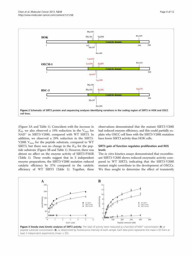
3 cells. This change was located in close proximity tothe deacetylase active site of SIRT3, and might be re-sponsible for the reduced catalytic efficiency. To test forthis possibility, we expressed recombinant WT SIRT3,SIRT3-V208I, SIRT3-P345R, and catalytically inactiveSIRT3-H248A in Escherichia coli and tested theirdeacetylase activity in vitro. A steady-state kinetic ana-lysis of SIRT3 activity was performed, and the initialrates of fluorescence release were measured as a func-tion of NAD+ concentration. The resulting saturationcurves were fitted to the Michaelis-Menten equation,and the Vmax and KM kinetic parameters were comparedamong WT SIRT3, SIRT3-V208I, SIRT3-P345R, andcatalytically inactive SIRT3-H248A. We observed a 20%increase in the KM value for NAD+ in SIRT3-V208I,compared to WT SIRT3, indicating more NAD+ was re-quired for the SIRT3-V208I deacetylation reaction
142
Phe184
Phe185
His187
Pro189
Catalytic domain
Val208 Pro345HOK
1 399
Leu184
Leu185
Asn187
Lys189
Catalytic domain
Ilu208 Pro345OECM-1
1 399
Phe184
Phe185
His187
Pro189
Catalytic domain
Ilu208 Arg345HSC-3
1 399
346
Figure 2 Schematic of SIRT3 protein and sequencing analyses identifying variations in the coding region of SIRT3 in HOK and OSCCcell lines.
Chen et al. Molecular Cancer 2013, 12:68 Page 4 of 12http://www.molecular-cancer.com/content/12/1/68
(Figure 3A and Table 1). Coincident with the increase inKM, we also observed a 19% reduction in the Vmax forNAD+ in SIRT3-V208I, compared with WT SIRT3. Inaddition, we observed a 29% reduction in the SIRT3-V208I Vmax for the peptide substrate, compared to WTSIRT3, but there was no change in the KM for the pep-tide substrate (Figure 3B and Table 1). However, there wasalmost no affect on the enzyme activity of SIRT3-P345R(Table 1). These results suggest that in 3 independentenzyme preparations, the SIRT3-V208I mutation reducedcatalytic efficiency by 37% compared to the catalyticefficiency of WT SIRT3 (Table 1). Together, these
NAD+ (mM)
0 1 2 3 4 5 6
pmol
e/m
in/u
g
0
50
100
150
200
250
WT SIRT3SIRT3-V208ISIRT3-H248A
A
Figure 3 Steady-state kinetic analyses of SIRT3 activity. The rates of acpeptide substrate concentration (B), as determined by fluorescence intensileast 3 independent experiments (mean ± SD).
observations demonstrated that the mutant SIRT3-V208Ihad reduced enzyme efficiency, and this could partially ex-plain why OSCC cell lines with the SIRT3-V208I mutationhave lower SIRT3 activity than HOK cells.
SIRT3 gain of function regulates proliferation and ROSlevelsThe in vitro kinetics assays demonstrated that recombin-ant SIRT3-V208I shows reduced enzymatic activity com-pared to WT SIRT3, indicating that the SIRT3-V208Imutant might contribute to the development of OSCCs.We thus sought to determine the effect of transiently
B
Peptide substrate (nM)
0 100 200 300 400 500
pmol
e/m
in/u
g
0
2
4
6
8
10
12
14
WT SIRT3SIRT3-V208ISIRT3-H248A
tivity were measured as a function of NAD+ concentration (A) orty of each sample. Each data point represents the mean ± SD from at

Table 1 Summary of SIRT3 steady-state kinetic analyses
SIRT3
WT V208I P345R H248Y
NAD+ Vmax (pmole/min/mg) 16.03 ± 0.02 12.14 ± 0.06 15.94 ± 0.14 1.08 ± 0.16
KM (mM) 0.88 ± 0.01 1.07 ± 0.03 0.88 ± 0.07 N.D.
Catalytic efficiency (Vmax/KM) 18.21 11.35 18.11 N.D.
Peptide Vmax (pmole/min/mg) 17.5 ± 0.69 12.5 ± 0.03 17.32 ± 0.45 1.32 ± 0.11
KM (mM) 147.3 ± 12.4 153.4 ± 17.5 146.8 ± 10.8 54.2 ± 14.0
Catalytic efficiency (Vmax/KM) 0.11 0.07 0.12 0.02
Rates of activity were measured as a function of NAD+ concentration or peptide substrate concentration, as measured by fluorescence intensity;n = 3 independent experiments (mean ± SD).
Chen et al. Molecular Cancer 2013, 12:68 Page 5 of 12http://www.molecular-cancer.com/content/12/1/68
expressed SIRT3 in OSCC cell lines by using a flag-tagged SIRT3 expressing vector. We observed a nearly2- to 3-fold induction of flag-tagged SIRT3, relativeto endogenous levels (Figure 4A). Furthermore,overexpression of SIRT3 decreased basal ROS levelsby ~30% and 48% in HSC-3 and OECM-1, respectively(Figure 4A). We also confirmed the enzymatic activityof SIRT3 after transfection with expression vector.OSCC cell lines were transiently transfected with orwithout WT SIRT3, and SIRT3 activity was measured.Both the OECM-1 and HSC-3 overexpressing cell
A
OECM-1 HS
Nor
mal
ized
RO
S le
vel
0.0
0.2
0.4
0.6
0.8
1.0
1.2
1.4
WB:GAPDH
WB:Flag
OECM-1C T
HSC-3C T
C
Time (h)0 20 40 60 80
OE
CM
-1 c
ell n
umbe
r (1
x105 )
0
2
4
6
8
10
12
14
16
18 ParentalpCMV2BSIRT3
0
HSC
-3 c
ell n
umbe
r (1
x105 )
0
2
4
6
8
10
Figure 4 ROS levels and cell proliferation are regulated by SIRT3 in Ooverexpressing flag-tagged SIRT3 and their relative fluorescence after incubatiOSCCs. OECM-1 and HSC-3 cell lines were transiently transfected with or withcell lines. Each data point represents the mean ± SD from at least 3 independdifference (*, p < 0.05) compared to control.
lines showed a mild increase in SIRT3 activity whentransfected with the flag-tagged SIRT3-expressing vec-tor (Figure 4B). Next, we examined whether SIRT3 hada role in cell proliferation by transiently expressingSIRT3 in OSCC cell lines. Exogenously expressed WTSIRT3 significantly decreased cell growth compared tothe growth of vector transfected cells in both cell lines(Figure 4C). These data indicated that SIRT3 could pos-sess tumor suppressor functions in OSCC cell lines, inaddition to its previously described role in inhibitingtransformation of primary cells [27].
C-3
ControlSIRT3
OECM-1 HSC-3Rel
ativ
e ac
tivity
of
SIR
T3
(fol
d ch
ange
)
0.0
0.5
1.0
1.5
2.0
2.5
3.0
3.5ControlSIRT3
B
Time (h)
20 40 60 80
ParentalpCMV2BSIRT3
SCCs. (A) Western blotting of whole cell lysates isolated from cellson with DHE. C: control; T: transfected cells. (B) Relative activity of SIRT3 inout flag-tagged SIRT3. (C) Cell proliferation of SIRT3-overexpressing OSCCent experiments (mean ± SD). The asterisk indicates a significant

Chen et al. Molecular Cancer 2013, 12:68 Page 6 of 12http://www.molecular-cancer.com/content/12/1/68
SIRT3 sequence variations in the OSCC patientsOSCC is a complex disease resulting from interactionsbetween genetic and environmental factors [35,36]. Its de-velopment is a multistep process requiring the accumula-tion of multiple genetic alterations. These alterations areinfluenced by a patient’s genetic predisposition as well asby environmental factors, including tobacco, alcohol,chronic inflammation, and viral infection [35]. Because ex-ogenously expressed WT SIRT3 produced inhibition of cellgrowth in OSCC cell lines, we tested whether variability inthe SIRT3 gene was correlated with increased susceptibilityfor developing oral squamous cell carcinoma. We obtainedreliable forward and reverse DNA sequencing results fromamplified fragments of the SIRT3 gene obtained from 21OSCC patients. We found that 4 of the 21 patients (19%,OSCC_31, 32, 39, 482) had a G to T transition at nucleo-tide 477 in exon 2 of SIRT3; this was a silent mutation thatdid not produce a change in the amino acid sequence. An-other 5 patients (23.8%, OSCC_30, 31, 32, 39, 482) carrieda G to A transition at nucleotide 622 in exon 3; this was amissense mutation resulting in the replacement of valine atresidue 208 with isoleucine (p.Val208Ile). To validate ourresults and discriminate whether the detected SIRT3 muta-tions were present in the germ-line cells or somatic cells,
Figure 5 Sequence chromatograms of DNA isolated from a patient wOSCC_32. Left, heterozygous mutation in normal DNA; Right, heterozygous m(p.Val208Ile) carried by patient OSCC_32.Left, heterozygous mutation in normaRight, predominance of expression of the mutated allele in cDNA synthesized
we performed targeted exon sequencing of DNA isolatedfrom tumor and normal cells of patients. Patients carrieds.477G >T or c.622G >A as a heterozygous mutation inboth normal and tumor cells. This finding indicatedthey had a germ-line genetic alteration of the SIRT3 gene(Figure 5), resulting in a V208I mutation in the matureprotein. Sequence analysis of cDNA synthesized fromtumor RNA revealed that the mutant allele was predomin-antly expressed in the tumor (Figure 5). Together, theseresults indicate that OSCC patients carried a germ-linemutation in the SIRT3 gene, represented by one single-base change, which introduced a non-synonymous pointmutation that resulted in reduced catalytic activity of theSIRT3 protein. This could partially explain how patientswith a V208I mutation in the SIRT3 enzyme may have anenhanced genetic susceptibility for developing OSCC.
SIRT3 activity in OSCC patientsA sequence analysis conducted with OSCC patientsdemonstrated that variability in the SIRT3 gene was cor-related with increased susceptibility for developing oralsquamous cell carcinoma. To obtain additional evidencefor the effect of SIRT3 activity in OSCC patients, we an-alyzed the SIRT3 enzyme activity of normal subjects and
ith OSCC. (A) Region harboring the s.477G > A mutation in patientutation in tumor DNA. (B) Region harboring the c.622G> A mutationl DNA; Middle, heterozygous mutation in tumor DNA;from OSCC_32 tumor RNA.

Chen et al. Molecular Cancer 2013, 12:68 Page 7 of 12http://www.molecular-cancer.com/content/12/1/68
OSCC patients. We collected 5 samples of normal gingivaltissue from healthy individuals after tooth extractions, and10 pairs of matched normal and tumor tissue samples from21 OSCC patients. We then immunopurified endogenousSIRT3 proteins from these tissue samples and assayed forenzymatic activity. Surprisingly, we found that the SIRT3proteins from tumor and normal tissues of patients(OSCC_30, 31, 32, 39, 482) who carried c.622G >A haddrastically lower levels of SIRT3 activity compared tissuesfrom normal individuals (Figure 6). The enzyme activity ofSIRT3 from the 5 OSCC patients (OSCC_24, 26, 28, 490,495) who didn’t carry this missense mutation was higherthan activity in patients with the SIRT3 V208I mutation,but it remained lower than activity in normal individuals.These results indicate that the V208I mutation in theSIRT3 enzyme was capable of reducing the enzyme’s activ-ity, and that decreasing SIRT3 activity may increasesusceptibility to the development of oral squamouscell carcinoma.
DiscussionIn this study, we demonstrated that SIRT3 regulates cellu-lar ROS, and this regulation has important implicationsfor the growth of oral squamous cell carcinoma cells. Ithas been previously shown that SIRT3 contributes to cellsurvival by modulating oxidative stress pathways [37,38].Our study showed that SIRT3 was slightly overexpressedin both OSCC cell lines compared with HOK cells, butthe enzyme activity of SIRT3 was significantly reduced inOSCCs, which could be caused by genetic change. Wefound that increased activity SIRT3 in oral squamous cellcarcinoma contributes to decreased ROS levels and in-creased cell proliferation. In addition, we sequenced the
SIR
T3
activ
ity (
coun
ts/m
in)
0
500
1000
1500
2000
Nor
mal
1
Nor
mal
2
Nor
mal
3
Nor
mal
4
Nor
mal
5
OSC
C_3
0
T N
OSC
C_3
1
T N
OSC
C_3
2
T N
Figure 6 SIRT3 enzyme activity of normal individuals and OSCC patieN, normal tissue and T, tumor tissue. Each data point represents the mean
entire coding region of SIRT3 and found a non-synonymous point mutation at nucleotide position 622 inthe SIRT3 coding region in both OSCC cell lines. Signifi-cantly, mutation of this position in the recombinant pro-tein SIRT3-V208I was responsible for a 37% reduction ofcatalytic efficiency compared to WT SIRT3. This idea wasfurther validated by the finding that in 5 of 21 OSCCpatients, the SIRT3 mutation carried c.622G >A as a het-erozygous mutation in both normal and tumor tissues, in-dicating that these patients had a germ-line geneticalteration of the SIRT3 gene. Additionally, we also deter-mined the SIRT3 activity in 5 normal subjects and 10OSCC patients (with or without c.622G >A) and foundthat all the OSCC patients had lower SIRT3 activity thannormal subjects, and activity was especially lower inpatients who carried the heterozygous missense mutation(c.622G > A). Taken together, these data indicate thatSIRT3 may function as a tumor suppressor in OSCC celllines by regulating cellular ROS levels and decreasingSIRT3 activity, and also may increase susceptibility to thedevelopment of oral squamous cell carcinoma. Our find-ings are consistent with previous work showing thatSIRT3 functions as a tumor suppressor through regulationof ROS [27]. Kim et al. found that elevated ROS in the ab-sence of SIRT3 increased genomic instability, promoting atumor-permissive environment. We propose that SIRT3might play a role in promoting tumorigenesis by alteringcellular ROS levels.In this study, we found that SIRT3 was slightly
overexpressed in both OSCC cell lines compared withHOK cells, but the SIRT3 enzyme activity in OECM-1 andHSC-3 was decreased by ~65% and 61%, respectively. Wesequenced the entire coding region of SIRT3 in HOK cells
OSC
C_3
9
T N
OSC
C_4
82
T N T N
OSC
C_4
90
OSC
C_4
95
T N T N
OSC
C_2
4
T N
OSC
C_2
6
T N
OSC
C_2
8
nts who did or did not carry the c.622G > A mutation of SIRT3.± SD from at least 3 independent experiments (mean ± SD).
Chen et al. Molecular Cancer 2013, 12:68 Page 8 of 12http://www.molecular-cancer.com/content/12/1/68
and the 2 OSCC cell lines, and found that the V208I mu-tation was located in close proximity to the deacetylaseactive site of SIRT3 in OECM-1 and HSC-3 cells. How-ever, the enzyme activity of recombinant protein (SIRT3-V208I) was responsible for a ~37% reduction in catalyticefficiency compared to WT SIRT3. These data showedthat overexpression of SIRT3 did not result in greaterdeacetylase activity in OSCCs, and that SIRT3 activitymight be regulated by mechanisms such as post-translational modifications. Recently, it was discoveredthat SIRT3 is required for PGC-1α-mediated differenti-ation of brown adipose tissue in a manner that isdependent on the level of an estrogen-related receptorα (ERRα) [33,39]. The transcription coactivator PGC-1α could be regulating the SIRT3 gene involved in en-ergy metabolism [40]. Thus, upon cellular stress, e.g.,an increase in ROS or nutrient deprivation, humanSIRT3 transcription might be stimulated [40], and theprotein translocated to the mitochondrial inner mem-brane. There, SIRT3 could deacetylate and therebyactivate the target enzymes and play a key role inmodulating mitochondrial activities and the regulationof many pathways. However, current studies of SIRT3have mainly focused on its cellular localization andidentification of its substrates; the mechanism regulat-ing SIRT3 function and activity remains unclear. Wenow know that the N-terminal extension of SIRT3 con-tains a mitochondria targeting signal peptide. Duringimport of SIRT3 into the mitochondrial matrix, theprotein is proteolytically cleaved at position 101 andthus enzymatically activated [18]. It has been postu-lated that the proteolytically shortened N-terminal re-gion and the C-terminal extension form a module thatmight regulate access of substrate proteins to the activesite [41]. Six phosphorylated serine residues (out of atotal of 8 possible sites) between positions 101 and 118may have been identified in a high-resolution massspectrometry-based phosphoproteome analysis [42].Therefore, it is possible that phosphorylation modu-lates the enzymatic activity of SIRT3 in mitochondriaby regulating interaction of the N-terminal region withthe catalytic domain. However, the biological relevanceor influence on SIRT3 function has not yet been ana-lyzed and more studies will be needed to understandthe mechanism for regulation of SIRT3 activity.In this study, we hypothesized that reduced SIRT3 ac-
tivity might lead to an increase in cellular ROS signaling,thereby enhancing tumorigenesis. We identified the en-tire coding region of SIRT3 in HOK cells and 2 OSCCcell lines, and found a correlation between a geneticchange at nucleotide position 622 in the SIRT3 gene inOECM-1 and HSC-3 cells. We tested for the possibilityof a functional impact of the non-synonymous pointmutation (V208I) in the SIRT3 protein. We used
AcArgGlyLys(Ac)AMC peptide as the substrate and de-termined deacetylase enzyme activity by measuringfluorescence intensity. As shown in Figure 3 and Table 1,the KM value and Vmax of SIRT3-V208I for NAD+ were1.07±0.03 mM and 12.14±0.06 pmole/min/mg, respect-ively. The KM v and Vmax kinetic parameters were com-pared between WT SIRT3 and SIRT3-V208I, indicatinga 20% increase in the KM and a 19% reduction in theVmax for NAD+ in SIRT3-V208I. Moreover, we observeda 29% reduction in the SIRT3-V208I Vmax for the pep-tide substrate compared to WT SIRT3, but there was nochange in the KM for the peptide substrate. These resultsindicate that the V208I lies within the conserved sirtuincatalytic deacetylase domain, and the mutation from val-ine to isoleucine reduced SIRT3 enzyme efficiency byboth increasing the KM for NAD+ and reducing theVmax. Our data were consistent with a model in whichreduction in SIRT3 enzymatic activity associated withthe V208I mutation plays a pathogenic role in oral squa-mous cell carcinoma. Interestingly, it has been reportedthat genetic polymorphism in the SIRT3 gene is linkedto longevity [43,44] and metabolic syndrome [32,45-48].Similar results of V208I mutation had reported by EricVerdin’s group [32] using different assay procedures.Verdin et al. used [3H-histone H4 peptide] as a substrateand measured rates of activity by using an organic-soluble radioactive signal. They found an 18% increasein the KM and a 19% reduction in the Vmax for NAD+ inSIRT3-V208I, compared to WT SIRT3, respectively.Their observations demonstrate that the SIRT3-V208Imutation reduced 34% catalytic efficiency and could in-crease susceptibility to developing the metabolic syn-drome. However, less is currently known about thegenetic polymorphism of SIRT3 in tumor cells. In thepresent study with 21 OSCC patients, we found that 4(19%) had s.477G > T of SIRT3, a silent mutation produ-cing no change in the amino acid sequence. Additionally,5 patients (23.8%) carried c.622G > A of SIRT3, a mis-sense mutation resulting in the replacement of valine atresidue 208 with isoleucine (p.Val208Ile). As shown inFigure 5, the OSCC_32 patient carried either s.477G > Tor c.622G > A as a heterozygous mutation in both nor-mal and tumor tissues, indicating there was a germ-linegenetic alteration of the SirT3 gene. The findings indi-cate that the SIRT3 +622A allele is positively associatedwith development of OSCC. This led to the reduction ofcatalytic efficiency in OSCCs, and promoted tumorigen-esis by altering cellular ROS levels.The exact role of mutant SIRT3 in tumorigenesis is
poorly understood. Recently, several groups have pro-vided evidence that SIRT3 is required to suppresstumorigenesis, and to induce stress-mediated cell deathin tumors, including breast cancer, colorectal carcinoma,osteosarcoma, and leukemia [27-29,49,50]. Furthermore,

Chen et al. Molecular Cancer 2013, 12:68 Page 9 of 12http://www.molecular-cancer.com/content/12/1/68
Kim et al. showed that genetic deletion of SIRT3 pushesmouse embryonic fibroblasts in the direction of oncogenictransformation, and that SIRT3−/− mouse embryonic fibro-blasts exhibit stress-induced genomic instability [27].While the activation of both oncogenes, Myc and Ras, isneeded to transform an immortalized fibroblast into atumor-forming cell, genetic deletion of SIRT3 reduced thatrequirement to only the activation of either Myc or Ras.Thus, SIRT3 functions as a tumor suppressor [51]. Inaddition, overexpression of SIRT3 is shown to decreasetumorigenesis in xenografts, even when induction of SIRT3occurs after tumor ignition [29]. In our study, we deter-mined the endogenous SIRT3 activity from 10 pairs ofmatched normal and tumor tissue samples obtained fromOSCC patients. We found that the SIRT3 proteins fromOSCC patients had significantly lower deacetylase activitythan SIRT3 proteins from normal subjects, thus suggestingthat a decrease of SIRT3 activity may increase susceptibilityto tumor development. However, current studies of SIRT3have mainly focused on its cellular localization and identifi-cation of its substrates; the molecular mechanism regulat-ing SIRT3 activity remains unclear.In this study, we began to investigate the potential role
of SIRT3 in regulating cellular redox balance in oralsquamous cell carcinoma and studied how SIRT3 is in-volved in oral carcinogenesis. However, the sample sizeof this study was small, and a larger number of OSCCpatients must be evaluated for SIRT3 mutation to reachdefinitive conclusions about the role of this gene in thedevelopment of oral squamous cell carcinoma.
ConclusionsIn this study, we found that SIRT3 gene mutations werepresent in both OECM-1 and HSC-3 cell lines, and inOSCC patients, supporting the hypothesis that SIRT3may act as a tumor suppressor and regulate the initi-ation and progression of cancer by controlling cellularredox balance. Our studies indicate that the germlinemutation of the SIRT3 gene potentially plays a criticalrole in tumorigenesis and may contribute to increasedsusceptibility for the development of OSCC. Futurestudies screening a larger number of OSCC patients forSIRT3 mutations to highlight important genetic alter-ations may help identify useful targets for personalizedcancer therapy and more successful cancer treatment.
MethodsCell culture and reagentsHOK cells used in this study were cultured in oral kera-tinocyte growth medium (ScienCell, Carlsbad, CA, USA).Cells were cultured in a 37°C incubator filled with 5% CO2
and routinely passaged at 90% confluence. Two humanOSCC cell lines, HSC-3 (tongue carcinoma), and OECM-1 (gingival carcinoma), were also used in this study. HSC-
3 cells were cultured in Dulbecco’s modified Eagle’smedium with 2 mM glutamine; OECM-1 cells weremaintained in RPMI 1640 medium. Each culture mediumwas supplemented with 10% fetal bovine serum and100 units/mL of penicillin and streptomycin (Invitrogen,Camarillo, CA, USA). All OSCC cells were maintained at37°C in a humidified atmosphere of 5% CO2.
Genomic DNA, RNA isolation, and quantitativereal-time PCRFor gene expression analysis, pairs of tumor and normalmarginal tissues were obtained from 21 OSCCs. Frozentissues were stored in liquid nitrogen at −196°C until use.Genomic DNA obtained from cultured cells and humantissue was subjected to bisulfite treatment using theEpiTect Bisulfite kit (Qiagen, Hilden, Germany) accordingto the manufacturer’s instructions. Total RNA obtainedfrom cultured cells and human tissue was extracted usingTRIzol reagent (Invitrogen). cDNA was then reverse-transcribed and amplified by PCR using a TranscriptorFirst Strand cDNA Synthesis kit (Roche Diagnostics,Mannheim, Germany). Quantitative RT-PCR was carriedout using the FastStart Universal SYBR Green Master(Roche) on an Applied Biosystems ABI 7900 RealTimePCR System (Applied Biosystems, Foster City, CA,USA). The oligonucleotide primers for human SIRT3and glyceraldehyde-3-phosphate dehydrogenase (GAPDH)were as follows: SIRT3-F 5′-ACCCAGTGGCATTCCAGAC-3′; SIRT3-R 5′-GGCTTGGGGTTGTGAAAGAAG-3′; GAPDH-F 5′-GAGTCAACGGATTTGGTCGT-3′;GAPDH-R 5′-GACAAGCTTCCCGTTCTCAG-3′. Thegene expression level was normalized using GAPDH as aninternal reference gene, and the average relative changewas calculated using 3–5 determinations by relative quan-tification, applying the delta-delta cycle threshold method.This study was approved by the Institutional Review Board(IRB) of the Department of Oral and Maxillofacial Surgeryof Chi-Mei Medical, Liouying, Taiwan (EC-1000202-R1).
Plasmid construction and infectionThe human SIRT3 coding region (GeneBank: NM_012239)was amplified by polymerase chain reaction (PCR) usingthe forward primer 5′-TTCGAACCATGGCGTTCTGGGGTTGG-3′, which introduced a Nsp V site, and 5′-CTCGAGCTATTTGTCTGGTCCATCAAGCTTCCC-3′,which introduced a XhoI site, under the following condi-tions: denaturing for 30 sec at 94°C, annealing for 30 sec at62°C, and elongation for 1 min at 72°C for 35 cycles. Thefull-length of SIRT3 was subcloned into the constitutivemammalian expression vector, pCMV-Tag 2B (Stratagene,Amsterdam, Netherlands), and verified by DNA sequen-cing. Transfected cells were seeded in 6-cm cell dishes at5 × 105 cells/dish and transfected with pCMV2B-SIRT3 orempty vector using Lipofectamine reagent (Invitrogen),

Chen et al. Molecular Cancer 2013, 12:68 Page 10 of 12http://www.molecular-cancer.com/content/12/1/68
according to the manufacturer’s protocol. Transfected cellswere used for further cell proliferation assays.
Cell proliferation assayTransfected cells were seeded in 96-well culture plates at1 × 104 cells/well and incubated for 72 h. Cell proliferationwas assessed using the MTT assay according to the manu-facturer’s recommendations (Roche).
Mitochondria isolation and immunoprecipitationMitochondria were isolated from cultured cells by using amitochondria isolation kit (Pierce Biotechnology, Inc.,Rockford, IL, USA) following the manufacturer’s protocol.Isolated mitochondria were lysed with RIPA buffer, andthen underwent direct western blot analysis or immunopre-cipitation. Then, 2 mg of protein from samples (total lysatesor mitochondrial extracts) was used for immunoprecipita-tion with a Pierce® Crosslink IP Kit (Pierce) following themanufacturer’s protocol, and analyzed by western blot.
Western blot analysisCells were lysed directly in RIPA buffer and adjusted forprotein concentration using the BCA protein assay kit(Bio-Rad, Hercules, CA, USA). Lysates were resolved by10% SDS-PAGE and then transferred to PVDF mem-branes. The membranes were blocked and incubatedwith specific antibodies against SIRT3 (Cell Signaling),actin (Sigma-Aldrich, St. Louis, MO, USA), GAPDH(Santa Cruz Biotechnology, Santa Cruz, CA, USA),SOD2 (Epitomics, San Diego, CA, USA), LCAD (Pierce),and acetylated-lysine (Cell Signaling) antibodies. Proteinswere visualized by enhanced chemiluminescence usingan ECL-Plus detection system (Perkin Elmer-NEN,Courtaboeuf, France).
ROS measurementCellular ROS was detected using the fluorescentdye dihydroethidium (DHE) obtained from Vigorous(Vigorous, Beijing, China) according to a previous study[52]. Cells were transiently transfected with or withoutflag-tagged SIRT3 and cultured in 6-well plates for 24 h.The cells were then washed with PBS and labeled withDHE (5 μmol/L dissolved in 1% DMSO) in the cultureplates at 37°C for 30 minutes in PBS. Culture plateswere placed on ice to stop the labeling, trypsinized, andresuspended in ice-cold PBS. Samples were analyzedusing a flow cytometer (BD FACS Calibur, BD Biosci-ences, San Jose, CA, USA). The mean fluorescenceintensity (MFI) of 10,000 cells was analyzed in eachsample and corrected for auto-fluorescence fromunlabeled cells.
Enzyme activity assaySIRT3 proteins from total lysates of cultured cells and hu-man tissue were concentrated using a Pierce® Crosslink IPKit (Pierce), according to the manufacturer’s recommen-dations. Protein concentration was determined using theBio-Rad protein assay kit (Bio-Rad). The enzyme activityassay for SIRT3 was performed in 50 μL of deacetylasebuffer (4 mM, MgCl2, 0.2 mM dithiothreitol, 50 mMTris–HCl, pH 8.5) containing 25 μL of SIRT3 proteins(10 ng/μL), 2 mM NAD+, and 25 μL of 1 mM fluorogenicpeptide substrate AcArgGlyLys(Ac)AMC (R&D systems,Minneapolis, MN, USA). Deacetylation reactions wereconducted at 37°C for 30 minutes, and stopped by adding50 μL of stop solution made by combining recombinantmouse trypsin 3/PRSS3 AMC (R&D systems) and nico-tinamide (Sigma-Aldrich) to final concentrations of0.2 ng/μL and 4 mM, respectively. The assays were thenincubated at room temperature for 15 minutes, and readat excitation and emission wavelengths of 380 nm and460 nm, respectively in endpoint mode. The activitywas measured with a SpectraMax M2 microplate reader(Molecular Devices Corporation, Sunnyvale, CA, USA).
Statistical analysisThe data are reported as the mean ± S.D. of at least 3 inde-pendent experiments. The P values for linear trend ofmRNA expression levels were analyzed using the t test(slope estimate) in simple linear regression models. Thedifference was considered statistically significant at thelevel of P < 0.05 or P < 0.01.
ConsentWritten informed consent was obtained from thepatient for publication of this report and any accom-panying images.
AbbreviationsSIRT: Sirtuin; HOK: Human oral keratinocyte; OSCC: Oral squamous cellcarcinoma; ROS: Reactive oxygen species; H2O2: Hydrogen peroxide;ANE: Areca nut extract; NAD: Nicotinamide adenine dinucleotide;NAM: Nicotinamide; GAPDH: Glyceraldehyde-3-phosphate dehydrogenase;DHE: Dihydroethidium.
Competing interestsNone of the authors of this manuscript had any conflict of interest regardingthe study.
Authors’ contributionsICC carried out the gene expression analyses, DNA sequencing studies,enzyme activity assays, flow cytometry studies, participated in design of thestudy, and helped draft the manuscript. WFC examined the transfectionstudy results and protein expression in the cell lines, and participated indesigning the study. SYL carried out the flow cytometry studies, westernblotting studies, and participated in designing the study. PFC conductedstatistical analysis for the manuscript. HCC conceived the study, participatedin its design and coordination, and helped to draft the manuscript. Allauthors read and approved the final manuscript.

Chen et al. Molecular Cancer 2013, 12:68 Page 11 of 12http://www.molecular-cancer.com/content/12/1/68
AcknowledgmentsThis work was supported by grants from the National Health ResearchInstitutes of Taiwan (Grant numbers: EO-100-PP-11, EO-101-PP-11). We thankAhi Cheun Lee (Postdoctoral Fellow at the National Environmental HealthResearch Center, National Health Research Institutes) for editorial assistance.
Author details1Division of Environmental Health and Occupational Medicine, NationalHealth, Research Institutes, No. 35, Keyan Road, Zhunan, Miaoli 35053,Taiwan. 2Department of Oral & Maxillofacial Surgery, Chi-Mei Medical Center,Liouying, Tainan, Taiwan. 3School of Dentistry, Yang-Ming University, Taipei,Taiwan. 4National Environmental Health Research Center, National HealthResearch Institutes, Miaoli, Taiwan. 5Department of Occupational Medicine,Taipei Medical University-Shuang Ho Hospital, Taipei, Taiwan.
Received: 9 January 2013 Accepted: 10 May 2013Published: 23 June 2013
References1. Bsoul SA, Huber MA, Terezhalmy GT: Squamous cell carcinoma of the oral
tissues: a comprehensive review for oral healthcare providers. J ContempDent Pract 2005, 6:1–16.
2. Chen YJ, Lin SC, Kao T, Chang CS, Hong PS, Shieh TM, Chang KW: Genome-wide profiling of oral squamous cell carcinoma. J Pathol 2004, 204:326–332.
3. Jeng JH, Chang MC, Hahn LJ: Role of areca nut in betel quid-associatedchemical carcinogenesis: current awareness and future perspectives.Oral Oncol 2001, 37:477–492.
4. Sharma DC: Betel quid and areca nut are carcinogenic without tobacco.Lancet Oncol 2003, 4:587–587.
5. Liu TY, Chen CL, Chi CW: Oxidative damage to DNA induced by areca nutextract. Mutat Res-Genet Tox 1996, 367:25–31.
6. Chang MC, Ho YS, Lee PH, Chan CP, Lee JJ, Hahn LJ, Wang YJ, Jeng JH:Areca nut extract and arecoline induced the cell cycle arrest but notapoptosis of cultured oral KB epithelial cells: association of glutathione,reactive oxygen species and mitochondrial membrane potential.Carcinogenesis 2001, 22:1527–1535.
7. Tang DW, Lin SC, Chang KW, Chi CW, Chang CS, Liu TY: Elevatedexpression of cyclooxygenase (COX)-2 in oral squamous cell carcinoma–evidence for COX-2 induction by areca quid ingredients in oralkeratinocytes. J Oral Pathol Med 2003, 32:522–529.
8. Balaban RS, Nemoto S, Finkel T: Mitochondria, oxidants, and aging.Cell 2005, 120:483–495.
9. Valko M, Rhodes CJ, Moncol J, Izakovic M, Mazur M: Free radicals, metalsand antioxidants in oxidative stress-induced cancer. Chem Biol Interact2006, 160:1–40.
10. Taylor DM, Maxwell MM, Luthi-Carter R, Kazantsev AG: Biological andpotential therapeutic roles of sirtuin deacetylases. Cell Mol Life Sci 2008,65:4000–4018.
11. Imai S, Armstrong CM, Kaeberlein M, Guarente L: Transcriptional silencingand longevity protein Sir2 is an NAD-dependent histone deacetylase.Nature 2000, 403:795–800.
12. Lin SJ, Guarente L: Nicotinamide adenine dinucleotide, a metabolicregulator of transcription, longevity and disease. Curr Opin Cell Biol 2003,15:241–246.
13. Saunders LR, Verdin E: Sirtuins: critical regulators at the crossroadsbetween cancer and aging. Oncogene 2007, 26:5489–5504.
14. Haigis MC, Guarente LP: Mammalian sirtuins–emerging roles inphysiology, aging, and calorie restriction. Genes Dev 2006, 20:2913–2921.
15. Michan S, Sinclair D: Sirtuins in mammals: insights into their biologicalfunction. Biochem J 2007, 404:1–13.
16. Michishita E, Park JY, Burneskis JM, Barrett JC, Horikawa I: Evolutionarilyconserved and nonconserved cellular localizations and functions ofhuman SIRT proteins. Mol Biol Cell 2005, 16:4623–4635.
17. Onyango P, Celic I, McCaffery JM, Boeke JD, Feinberg AP: SIRT3, a humanSIR2 homologue, is an NAD-dependent deacetylase localized tomitochondria. Proc Natl Acad Sci USA 2002, 99:13653–13658.
18. Schwer B, North BJ, Frye RA, Ott M, Verdin E: The human silent informationregulator (Sir)2 homologue hSIRT3 is a mitochondrial nicotinamide adeninedinucleotide-dependent deacetylase. J Cell Biol 2002, 158:647–657.
19. Hirschey MD, Shimazu T, Goetzman E, Jing E, Schwer B, Lombard DB,Grueter CA, Harris C, Biddinger S, Ilkayeva OR, et al: SIRT3 regulates
mitochondrial fatty-acid oxidation by reversible enzyme deacetylation.Nature 2010, 464:121–125.
20. Qiu X, Brown K, Hirschey MD, Verdin E, Chen D: Calorie restriction reducesoxidative stress by SIRT3-mediated SOD2 activation. Cell Metab 2010,12:662–667.
21. Someya S, Yu W, Hallows WC, Xu J, Vann JM, Leeuwenburgh C, Tanokura M,Denu JM, Prolla TA: Sirt3 mediates reduction of oxidative damage andprevention of age-related hearing loss under caloric restriction. Cell 2010,143:802–812.
22. Lombard DB, Alt FW, Cheng HL, Bunkenborg J, Streeper RS, Mostoslavsky R,Kim J, Yancopoulos G, Valenzuela D, Murphy A, et al: Mammalian Sir2homolog SIRT3 regulates global mitochondrial lysine acetylation. Mol CellBiol 2007, 27:8807–8814.
23. Shimazu T, Hirschey MD, Hua L, Dittenhafer-Reed KE, Schwer B, LombardDB, Li Y, Bunkenborg J, Alt FW, Denu JM, et al: SIRT3 deacetylatesmitochondrial 3-hydroxy-3-methylglutaryl CoA synthase 2 and regulatesketone body production. Cell Metab 2010, 12:654–661.
24. Hallows WC, Yu W, Smith BC, Devries MK, Ellinger JJ, Someya S, ShortreedMR, Prolla T, Markley JL, Smith LM, et al: Sirt3 promotes the urea cycle andfatty acid oxidation during dietary restriction. Mol Cell 2011, 41:139–149.
25. Tao R, Coleman MC, Pennington JD, Ozden O, Park SH, Jiang H, Kim HS,Flynn CR, Hill S, Hayes McDonald W, et al: Sirt3-mediated deacetylation ofevolutionarily conserved lysine 122 regulates MnSOD activity inresponse to stress. Mol Cell 2010, 40:893–904.
26. Guan KL, Xiong Y: Regulation of intermediary metabolism by proteinacetylation. Trends Biochem Sci 2011, 36:108–116.
27. Kim HS, Patel K, Muldoon-Jacobs K, Bisht KS, Aykin-Burns N, Pennington JD, vander Meer R, Nguyen P, Savage J, Owens KM, et al: SIRT3 Is a mitochondria-localized tumor suppressor required for maintenance of mitochondrialintegrity and metabolism during stress. Cancer Cell 2010, 17:41–52.
28. Finley LW, Carracedo A, Lee J, Souza A, Egia A, Zhang J, Teruya-Feldstein J,Moreira PI, Cardoso SM, Clish CB, et al: SIRT3 opposes reprogramming ofcancer cell metabolism through HIF1alpha destabilization. Cancer Cell2011, 19:416–428.
29. Bell EL, Emerling BM, Ricoult SJ, Guarente L: SirT3 suppresses hypoxiainducible factor 1alpha and tumor growth by inhibiting mitochondrialROS production. Oncogene 2011, 30:2986–2996.
30. Jing E, Emanuelli B, Hirschey MD, Boucher J, Lee KY, Lombard D, Verdin EM,Kahn CR: Sirtuin-3 (Sirt3) regulates skeletal muscle metabolism andinsulin signaling via altered mitochondrial oxidation and reactive oxygenspecies production. Proc Natl Acad Sci USA 2011, 108:14608–14613.
31. Sundaresan NR, Gupta M, Kim G, Rajamohan SB, Isbatan A, Gupta MP: Sirt3blocks the cardiac hypertrophic response by augmenting Foxo3a-dependent antioxidant defense mechanisms in mice. J Clin Invest 2009,119:2758–2771.
32. Hirschey MD, Shimazu T, Jing E, Grueter CA, Collins AM, Aouizerat B,Stancakova A, Goetzman E, Lam MM, Schwer B, et al: SIRT3 deficiency andmitochondrial protein hyperacetylation accelerate the development ofthe metabolic syndrome. Mol Cell 2011, 44:177–190.
33. Kong X, Wang R, Xue Y, Liu X, Zhang H, Chen Y, Fang F, Chang Y: Sirtuin 3,a new target of PGC-1alpha, plays an important role in the suppressionof ROS and mitochondrial biogenesis. PLoS One 2010, 5:e11707.
34. Frye RA: Phylogenetic classification of prokaryotic and eukaryotic Sir2-like proteins. Biochem Biophys Res Commun 2000, 273:793–798.
35. Choi S, Myers JN: Molecular pathogenesis of oral squamous cellcarcinoma: implications for therapy. J Dent Res 2008, 87:14–32.
36. Hillbertz NS, Hirsch JM, Jalouli J, Jalouli MM, Sand L: Viral and molecularaspects of oral cancer. Anticancer Res 2012, 32:4201–4212.
37. Benigni A, Corna D, Zoja C, Sonzogni A, Latini R, Salio M, Conti S, Rottoli D,Longaretti L, Cassis P, et al: Disruption of the Ang II type 1 receptorpromotes longevity in mice. J Clin Invest 2009, 119:524–530.
38. Finkel T, Deng CX, Mostoslavsky R: Recent progress in the biology andphysiology of sirtuins. Nature 2009, 460:587–591.
39. Giralt A, Hondares E, Villena JA, Ribas F, Diaz-Delfin J, Giralt M, Iglesias R,Villarroya F: Peroxisome proliferator-activated receptor-gammacoactivator-1alpha controls transcription of the Sirt3 gene, an essentialcomponent of the thermogenic brown adipocyte phenotype. J Biol Chem2011, 286:16958–16966.
40. Shi T, Wang F, Stieren E, Tong Q: SIRT3, a mitochondrial sirtuindeacetylase, regulates mitochondrial function and thermogenesis inbrown adipocytes. J Biol Chem 2005, 280:13560–13567.
Chen et al. Molecular Cancer 2013, 12:68 Page 12 of 12http://www.molecular-cancer.com/content/12/1/68
41. Schlicker C, Gertz M, Papatheodorou P, Kachholz B, Becker CF, Steegborn C:Substrates and regulation mechanisms for the human mitochondrialsirtuins Sirt3 and Sirt5. J Mol Biol 2008, 382:790–801.
42. Olsen JV, Vermeulen M, Santamaria A, Kumar C, Miller ML, Jensen LJ, GnadF, Cox J, Jensen TS, Nigg EA, et al: Quantitative phosphoproteomicsreveals widespread full phosphorylation site occupancy during mitosis.Sci Signal 2010, 3:ra3.
43. Rose G, Dato S, Altomare K, Bellizzi D, Garasto S, Greco V, Passarino G,Feraco E, Mari V, Barbi C, et al: Variability of the SIRT3 gene, human silentinformation regulator Sir2 homologue, and survivorship in the elderly.Exp Gerontol 2003, 38:1065–1070.
44. Bellizzi D, Rose G, Cavalcante P, Covello G, Dato S, De Rango F, Greco V,Maggiolini M, Feraco E, Mari V, et al: A novel VNTR enhancer within theSIRT3 gene, a human homologue of SIR2, is associated with survival atoldest ages. Genomics 2005, 85:258–263.
45. Heid IM, Jackson AU, Randall JC, Winkler TW, Qi L, Steinthorsdottir V,Thorleifsson G, Zillikens MC, Speliotes EK, Magi R, et al: Meta-analysisidentifies 13 new loci associated with waist-hip ratio and reveals sexualdimorphism in the genetic basis of fat distribution. Nat Genet 2010,42:949–960.
46. Dupuis J, Langenberg C, Prokopenko I, Saxena R, Soranzo N, Jackson AU,Wheeler E, Glazer NL, Bouatia-Naji N, Gloyn AL, et al: New genetic lociimplicated in fasting glucose homeostasis and their impact on type 2diabetes risk (vol 42, pg 105, 2010). Nat Genet 2010, 42:464–464.
47. Teslovich TM, Musunuru K, Smith AV, Edmondson AC, Stylianou IM, KosekiM, Pirruccello JP, Ripatti S, Chasman DI, Willer CJ, et al: Biological, clinicaland population relevance of 95 loci for blood lipids. Nature 2010,466:707–713.
48. Musunuru K, Strong A, Frank-Kamenetsky M, Lee NE, Ahfeldt T, Sachs KV, LiXY, Li H, Kuperwasser N, Ruda VM, et al: From noncoding variant tophenotype via SORT1 at the 1p13 cholesterol locus. Nature 2010,466:714–U712.
49. Marfe G, Tafani M, Indelicato M, Sinibaldi-Salimei P, Reali V, Pucci B, Fini M,Russo MA: Kaempferol induces apoptosis in Two different cell lines ViaAkt inactivation, Bax and SIRT3 activation, and mitochondrialdysfunction. J Cell Biochem 2009, 106:643–650.
50. Allison SJ, Milner J: SIRT3 is pro-apoptotic and participates in distinctbasal apoptotic pathways. Cell Cycle 2007, 6:2669–2677.
51. Schumacker PT: A tumor suppressor SIRTainty. Cancer Cell 2010, 17:5–6.52. Slane BG, Aykin-Burns N, Smith BJ, Kalen AL, Goswami PC, Domann FE, Spitz
DR: Mutation of succinate dehydrogenase subunit C results in increasedO2.-, oxidative stress, and genomic instability. Cancer Res 2006, 66:7615–7620.
doi:10.1186/1476-4598-12-68Cite this article as: Chen et al.: Role of SIRT3 in the regulation of redoxbalance during oral carcinogenesis. Molecular Cancer 2013 12:68.
Submit your next manuscript to BioMed Centraland take full advantage of:
• Convenient online submission
• Thorough peer review
• No space constraints or color figure charges
• Immediate publication on acceptance
• Inclusion in PubMed, CAS, Scopus and Google Scholar
• Research which is freely available for redistribution
Submit your manuscript at www.biomedcentral.com/submit