Embed Size (px)
Citation preview
“fnagi-05-00048” — 2013/9/4 — 15:02 — page 1 — #1
REVIEW ARTICLEpublished: 06 September 2013doi: 10.3389/fnagi.2013.00048
Forever young: SIRT3 a shield against mitochondrialmeltdown, aging, and neurodegenerationBrad Kincaid and Ella Bossy-Wetzel*
Burnett School of Biomedical Sciences, College of Medicine, University of Central Florida, Orlando, FL, USA
Edited by:
Gizem Donmez, Tufts UniversitySchool of Medicine, USA
Reviewed by:
Carsten Culmsee, Philipps Universityof Marburg, GermanyUwe-Karsten Hanisch, University ofGöttingen, Germany
*Correspondence:
Ella Bossy-Wetzel, Burnett School ofBiomedical Sciences, College ofMedicine, University of CentralFlorida, 4000 Central FloridaBoulevard, Orlando, FL 32816, USAe-mail: [email protected]
Caloric restriction (CR), fasting, and exercise have long been recognized for their neu-roprotective and lifespan-extending properties; however, the underlying mechanisms ofthese phenomena remain elusive. Such extraordinary benefits might be linked to theactivation of sirtuins. In mammals, the sirtuin family has seven members (SIRT1–7), whichdiverge in tissue distribution, subcellular localization, enzymatic activity, and targets. SIRT1,SIRT2, and SIRT3 have deacetylase activity. Their dependence on NAD+ directly linkstheir activity to the metabolic status of the cell. High NAD+ levels convey neuroprotectiveeffects, possibly via activation of sirtuin family members. Mitochondrial sirtuin 3 (SIRT3)has received much attention for its role in metabolism and aging. Specific small nucleotidepolymorphisms in Sirt3 are linked to increased human lifespan. SIRT3 mediates theadaptation of increased energy demand during CR, fasting, and exercise to increasedproduction of energy equivalents. SIRT3 deacetylates and activates mitochondrial enzymesinvolved in fatty acid β-oxidation, amino acid metabolism, the electron transport chain,and antioxidant defenses. As a result, the mitochondrial energy metabolism increases.In addition, SIRT3 prevents apoptosis by lowering reactive oxygen species and inhibitingcomponents of the mitochondrial permeability transition pore. Mitochondrial deficitsassociated with aging and neurodegeneration might therefore be slowed or even preventedby SIRT3 activation. In addition, upregulating SIRT3 activity by dietary supplementation ofsirtuin activating compounds might promote the beneficial effects of this enzyme.The goalof this review is to summarize emerging data supporting a neuroprotective action of SIRT3against Alzheimer’s disease, Huntington’s disease, Parkinson’s disease, and amyotrophiclateral sclerosis.
Keywords: SIRT3, neuroprotection, caloric restriction, aging, neurodegeneration, antioxidants, mitochondria
INTRODUCTIONCaloric restriction (CR), fasting, and exercise promote neuropro-tection and extend healthy lifespan in mammals. Reducing foodconsumption without malnutrition extends the lifespan of rodentsby up to 50% (Weindruch et al., 1986; McCay et al., 1989). Recentstudies suggested these extraordinary benefits may be linked toupregulation of sirtuins.
Sirtuins were first described as NAD+-dependent type III his-tone deacetylases with yeast Sir2 as its founding member, silencinggene expression by histone deacetylation (Guarente and Kenyon,2000). However, mammalian sirtuins target not only histones inthe nucleus but also other proteins in the cytoplasm and mito-chondria. In mammals, the sirtuin family has seven members(SIRT1–7), which differ in tissue distribution, subcellular local-ization, enzymatic activity, and target proteins. SIRT1, SIRT6,and SIRT7 are present in the nucleus; SIRT2 in the cytoplasm;and SIRT3, SIRT4, and SIRT5 in mitochondria (Frye, 2000).Based on sequence homology, SIRT1, SIRT2, and SIRT3 belongto class I sirtuins and exhibit deacetylase activity. SIRT4 belongsto class II and has ADP-ribosylation activity; SIRT5 to class IIIand has demalonylation and desuccinylation activity; and SIRT6and SIRT7 to class IV (He et al., 2012). SIRT6 has deacetylase andADP-ribosylase activity, while SIRT7 has no reported activity.
Sirtuin enzymatic activity requires NAD+ as a cofactor whoselevels increase by energy stress which occurs, e.g., during fasting,CR, and exercise. Thus, NAD+ mediates the adaptive response tolow energy by activating sirtuins and their downstream targets.Sirtuins transform NAD+ to nicotinamide, which acts as a com-petitive inhibitor of sirtuins by a negative feedback mechanism.The other breakdown product of NAD+ is O-acetyl-ADP-ribose.
The average lifespan of humans has steadily increased as a resultof advances in medicine and improved living conditions. Despitethis progress, the maximum human lifespan remains constant,for reasons not entirely clear. Among the aging theories is the“free radical theory of aging” proposed by Denham Harman inthe 1950s, which attempts to explain the limit on human lifespanas the result of accumulated damage to proteins, nucleic acids,lipids, and organelles by free radicals (Harman, 1956; Weissmanet al., 2007; Wong and Cuervo, 2010). Mitochondria are both thesource and target of reactive oxygen species (ROS) in cells includ-ing superoxide anions, hydrogen peroxide, hydroxyl radicals, andreactive nitrogen species such as peroxynitrite. Oxidative andnitrosative stress can evoke irreversible damage to proteins, lipids,and DNA. As a result, mitochondria have been linked to agingand age-related diseases (Hall et al., 2001; Singh, 2006). SIRT3emerged as a protein of particular interest to the aging field due
Frontiers in Aging Neuroscience www.frontiersin.org September 2013 | Volume 5 | Article 48 | 1
“fnagi-05-00048” — 2013/9/4 — 15:02 — page 2 — #2
Kincaid and Bossy-Wetzel SIRT3 in neuroprotection
to its mitochondrial localization and association with exceptionallong lifespan in humans (Hurst et al., 2002). SIRT3 deacetylatesand activates many mitochondrial enzymes involved in fatty acidβ-oxidation, amino acid metabolism, the electron transport chain(ETC), and antioxidant defenses. Neurons are especially sensi-tive to insults that result in energy depletion and oxidative stress(Du et al., 2003). Here, we will review the well-documented rolesof SIRT3 in metabolism and antioxidant defenses, and the newevidence linking SIRT3 to neuroprotection.
SIRT3 SUBCELLULAR LOCALIZATIONAlthough most reports indicated an exclusive mitochondrial local-ization of SIRT3 (Onyango et al., 2002; Shi et al., 2005; Schweret al., 2006; Cooper and Spelbrink, 2008; Gurd et al., 2012), othershave argued that SIRT3 is also present in the nucleus and cytoplasm(Scher et al., 2007; Sundaresan et al., 2008; Shulga et al., 2010;Iwahara et al., 2012). SIRT3 target proteins were identified in allthree compartments: nucleus, cytoplasm, and mitochondrion (Heet al., 2012). Thus, the localization and role of SIRT3 in differentcellular compartments is a matter of considerable debate.
Within mitochondria, SIRT3 appears to be localized to theinner mitochondrial membrane cristae and the matrix (Shi et al.,2005; Schwer et al., 2006). Additional investigations are needed todetermine whether SIRT3 also targets to the outer mitochondrialmembrane or the intermembrane space.
SIRT3 ISOFORMSInitial reports indicated that human SIRT3 is present in a full-length form and short form. Full-length SIRT3 translocates fromthe cytoplasm to mitochondria. Once imported into mitochon-dria, SIRT3 is processed to the short form (Schwer et al., 2006).Initially only the short, mitochondrial form of human SIRT3 wasthought to exhibit NAD+-dependent deacetylase activity (Schweret al., 2006). However, several reports challenged this view anddemonstrated that both the full-length and short form of SIRT3exhibit deacetylase activity. In agreement, full-length SIRT3 accu-mulates in the nucleus and deacetylates histones (Lombard et al.,2007). In cardiomyocytes, full-length mouse SIRT3 is present inthe nucleus and cytoplasm, while the short form is only foundin mitochondria (Sundaresan et al., 2008). SIRT3 deacetylates Ku-70 in the cytoplasm and thereby prevents pro-apoptotic BAX totranslocate to mitochondria. In addition, three different splicevariants of mouse SIRT3 were discovered (Jin et al., 2009). Twoof these splice variants contain an amino-terminal mitochondrialtargeting sequence (MTS); however, the third form lacks the MTS(Cooper et al., 2009; Jin et al., 2009). Upon translocation to mito-chondria, the MTS is cleaved by a matrix processing peptidase toproduce a short form of SIRT3. Without the MTS, SIRT3 fails totranslocate to mitochondria (Schwer et al., 2002).
Sirt3 POLYMORPHISMSInitial interest in SIRT3 and its role in human aging was sparked bythe discovery of unique single nucleotide polymorphisms (SNPs)linked to centenarians (Hurst et al., 2002; Rose et al., 2003; Bellizziet al., 2005). Males carrying the G477T transversion in exon 3 ofSirt3 are healthy and live beyond the average lifespan. However,this nucleotide transition is silent and does not alter the amino
acid sequence (Rose et al., 2003). Therefore, the mechanism bywhich this SNP prolongs life is unknown.
The second SNP involves the variable number tandem repeat(VNTR) region within intron 5 of Sirt3 (Bellizzi et al., 2005). Thespecific VNTR polymorphism exhibits improved enhancer activityand increases Sirt3 expression (Bellizzi et al., 2005). Interestingly,a strong linkage disequilibrium has been observed between theVNTR and G477T polymorphism (Bellizzi et al., 2005). Thus, onecan speculate that the two polymorphisms might exhibit coop-erative effects. Of additional note, Sirt3 maps to a region withinthe short arm of chromosome 11 shared by a cluster of longevitygenes, including insulin-like growth factor 2 (IGF2), proinsulin(INS), and tyrosine hydroxylase (TH). Therefore, it is possiblethat Sirt3 shares common regulatory elements with these genes(Hurst et al., 2002).
Finally, a third SNP in the coding sequence of Sirt3 has beendiscovered. Unlike the first two SNPs that are linked to increasedlifespan, the third SNP increases the risk for age-related metabolicsyndrome. The particular SNP involves an amino acid substitutionin the conserved deacetylase catalytic region, resulting in lowerSIRT3 deacetylase activity (Hirschey et al., 2011). Taken together,additional studies are required to investigate how the various SNPsmodify human health and lifespan.
SIRT3 TISSUE-SPECIFIC EXPRESSIONSIRT3 expression is highest in metabolically active tissues includ-ing the brain, heart, liver, brown adipose tissue (BAT), andskeletal muscle (Lombard et al., 2007; Ahn et al., 2008; Gurdet al., 2012). SIRT3 knockout (KO) mice show no obvious phe-notypic changes, but are prone to age-linked diseases includingmetabolic syndrome, cancer, and cardiac failure (Hirschey et al.,2011). Whether SIRT3 KO mice exhibit neurological defects hasyet to be elucidated. SIRT3 deletion in mice causes hyperacetyla-tion of mitochondrial proteins and major metabolic defects. Massspectrometry revealed that 65% of all proteins in mitochondriahave at least one lysine acetylated in liver tissue of SIRT3 KO mice(Hebert et al., 2013). By contrast, SIRT4 KO and SIRT5 KO micedo not exhibit global hyperacetylation of mitochondrial proteins(Lombard et al., 2007). In fact, SIRT4 and SIRT5 have either noor only very weak NAD+-dependent deacetylase activity (Verdinet al., 2010). SIRT4 has ADP-ribosylase activity and SIRT5 hasdemalonylase and desuccinylase activities (Haigis et al., 2006; Duet al., 2011). Therefore, SIRT3 is the key deacetylase in mitochon-dria. Future studies using conditional, tissue-specific knock-in orknockout mice will establish the functions of SIRT3 in tissues suchas the brain.
MITOCHONDRIAL DYNAMICSEnergy homeostasis is a delicate balance between energy supply,use, and storage. Mitochondria are highly dynamic organelles andundergo constant cycles of fission and fusion. This dynamic pro-cess is modified by nutrient supply, linking cellular energy produc-tion with energy demands. For example, nutrient excess stimulatesmitochondrial fission, fragmented mitochondrial morphology,and an arrest in oxygen consumption, oxidative phosphoryla-tion, and ATP synthesis. By contrast, nutrient deficiency triggersmitochondrial fusion, elongated mitochondrial morphology, and
Frontiers in Aging Neuroscience www.frontiersin.org September 2013 | Volume 5 | Article 48 | 2

“fnagi-05-00048” — 2013/9/4 — 15:02 — page 3 — #3
Kincaid and Bossy-Wetzel SIRT3 in neuroprotection
an acceleration of mitochondrial respiration and ATP produc-tion (Molina et al., 2009; Gomes and Scorrano, 2011; Ramboldet al., 2011; Liesa and Shirihai, 2013). Thus, mitochondrial fis-sion and fusion is a fine tuned process that controls the switchesof energy production with energy demand, thereby maintaininghomeostasis.
Mitochondrial fission and fusion is regulated by conservedlarge GTPases of the Dynamin family, which exert opposite func-tions. Fission is mediated by dynamin related protein 1 (DRP1)while fusion is controlled by optic atrophy 1 (OPA1), mitofusin1 (MFN1), and mitofusin 2 (MFN2; Arnoult et al., 2005; Miskoet al., 2010). It can be speculated whether increased SIRT1 andSIRT3 activity during CR, fasting, and exercise enhance energymetabolism by activating fusion and inhibiting fission of mito-chondria. In support, sirtuin activating compounds, e.g., NAD+and resveratrol, seem to promote a fused mitochondrial mor-phology. In addition, exercise promotes DRP1 phosphorylationat serine 637 by protein kinase A (PKA), a process thought toinhibit DRP1 function and decrease mitochondrial fission. Futurestudies are needed to investigate how mitochondrial fission/fusionGTPases are regulated by dietary changes and sirtuins.
Mitochondrial dynamics is also implicated in aging. For exam-ple, reducing mitochondrial fission by DRP1 deletion slows agingand increases lifespan of yeast (Scheckhuber et al., 2007). Simi-larly, DRP1 inactivation in worm enhances the positive effects ofreduced insulin signaling on lifespan extension (Yang et al., 2011).Thus, promoting mitochondrial fusion correlates with longevityof these model organisms. Whether mitochondrial dynamicsregulates the aging process in mammals remains to be investigated.
A balance between mitochondrial fission and fusion plays acritical role in neuronal function and survival (Knott et al., 2008).In neurons, mitochondrial dynamics is required for bioenergeticand synaptic function, Ca2+ buffering, transport of mitochondriaacross neurites, removal of damaged mitochondria by autophagy,and neuronal cell survival. Future studies are needed to investigatewhether SIRT3 enhances neuronal survival by regulating processessuch as mitochondrial fission and fusion.
SIRT3 IN CR AND EXERCISEMore than 70 years ago the lifespan-extending properties of a CRdiet were demonstrated in rats (McCay et al., 1989). The detailsof this observation were not further explored until almost half acentury later when Walford and Weindruch discovered that reduc-ing food consumption 25–60% without malnutrition extends thelifespan of mice up to 50% (Weindruch et al., 1986). More recently,CR has been shown to inhibit neurodegeneration in animal mod-els of both Alzheimer’s disease (AD) and Parkinson’s disease (PD;Zhu et al., 1999; Mattson, 2000). A deeper look into the metabolicand cellular changes that occur during metabolic stresses such asCR may reveal the unique mechanisms by which these benefits areconferred.
During the transition from the fed to fasted state, blood glu-cose levels decline causing carbohydrate utilization and fatty acidsynthesis to cease in the liver, while fatty acid oxidation and keto-genesis are induced (McGarry and Foster, 1980). Furthermore,prolonged starvation results in increased amino acid catabolism ascells search for a new source of energy. Autophagy is also increased
during nutrient stress (Kuma et al., 2004). Blocking autophagyaccelerates cell death, aging, and neurodegeneration (Kuma et al.,2004; Hara et al., 2006; Komatsu et al., 2006). Interestingly, NAD+levels greatly increase during CR in muscle and white adiposetissue (WAT), thus suggesting potential upregulation of Sirtuinactivity (Chen et al., 2008).
Remarkably, SIRT3 expression levels fluctuate with diet.Numerous studies have reported CR-induced increased SIRT3expression (Palacios et al., 2009; Tauriainen et al., 2011). For exam-ple, mice on a 12-month CR diet exhibited increased SIRT3 levelsin skeletal muscle compared to mice fed ad libitum (Palacioset al., 2009). In addition, Sirt3 mRNA expression was increasedin liver, skeletal muscle, and adipose tissue of mice on a 30% CRdiet (Tauriainen et al., 2011). By contrast, a chronic high fat diet(HFD) resulted in suppression of SIRT3 and elevated global mito-chondrial protein acetylation (Hirschey et al., 2011). Akin to CR,exercise training in mice increased SIRT3 protein expression incardiac and triceps muscles (Palacios et al., 2009).
SIRT3 expression is also modulated during aging. Notably,elderly individuals exhibited a 50% reduction in peroxisome-proliferator activated receptor gamma coactivator 1-alpha (PGC-1α) and SIRT3 protein levels in skeletal muscle compared toyoung subjects, regardless of activity level (Joseph et al., 2012).By contrast, another study suggested exercise training could sus-tain equivalent levels of SIRT3 in skeletal muscle of both youngand elderly individuals (Lanza et al., 2008). Additional studies arerequired to elucidate the mechanisms of these observations. Onepossible explanation for reduced SIRT3 levels during aging may beepigenetic regulation of the Sirt3 gene promoter. Further investi-gation is required to determine the cause of SIRT3 downregulationduring aging.
SIRT3 IN MITOCHONDRIAL BIOGENESISCaloric restriction or exercise induce PGC-1α, a master regulatorof genes involved in mitochondrial biogenesis, metabolism, sup-pression of ROS, and stress responses (Herzig et al., 2001; Yoonet al., 2001; Palacios et al., 2009). The importance of PGC-1α inthese processes is underscored by observations of PGC-1α KOmice, which exhibit many defects including obesity, neurodegen-eration, cardiomyopathy, and heightened sensitivity to ROS (Linet al., 2004; St-Pierre et al., 2006).
Interestingly, PGC-1α may play a role in controlling Sirt3 geneexpression. A sequence motif in the Sirt3 promoter is recognizedby the estrogen related receptor-alpha (ERRα), an orphan nuclearreceptor upregulated in CR (Ranhotra, 2009). PGC-1α mediatesERRα binding to this sequence motif in the Sirt3 promoter andpromotes Sirt3 gene expression. Additionally, siRNA knockdownof PGC-1α reduces SIRT3 expression (Kong et al., 2010).
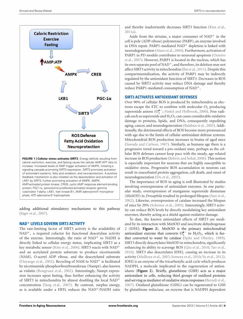
SIRT3 may also upregulate PGC-1α through a positive feed-back mechanism (Figure 1). SIRT3 deacetylates and activatesliver kinase B1 (LKB1) in cardiomyocytes (Pillai et al., 2010).Active LKB1 phosphorylates and stimulates AMP-activated pro-tein kinase (AMPK; Woods et al., 2003). As a result, activatedAMPK phosphorylates cyclic AMP response element binding pro-tein (CREB) leading to increased PGC-1α expression (Bergeronet al., 2001; Zong et al., 2002; Thomson et al., 2008). More-over, AMPK can directly phosphorylate and activate PGC-1α,
Frontiers in Aging Neuroscience www.frontiersin.org September 2013 | Volume 5 | Article 48 | 3
“fnagi-05-00048” — 2013/9/4 — 15:02 — page 4 — #4
Kincaid and Bossy-Wetzel SIRT3 in neuroprotection
FIGURE 1 | Cellular stress activates SIRT3. Energy deficits resulting fromcalorie restriction, exercise, and fasting cause the cellular AMP:ATP ratio toincrease. Increased levels of AMP trigger activation of AMPK, initiating asignaling cascade promoting SIRT3 expression. SIRT3 promotes activationof antioxidant systems, fatty acid oxidation, and neuroprotection. A positivefeedback mechanism is also initiated via the deacetylation and activation ofLKB1 by SIRT3, further promoting activation of AMPK. AMPK,AMP-activated protein kinase; CREB, cyclic AMP response element-bindingprotein; PGC-1α, peroxisome proliferator-activated receptor gammacoactivator 1-alpha; LKB1, liver kinase B1; AMP, adenosine-5′-monophos-phate; ATP, adenosine-5′-triphosphate.
adding additional stimulatory mechanisms to this pathway(Jäger et al., 2007).
NAD+ LEVELS GOVERN SIRT3 ACTIVITYThe rate-limiting factor of SIRT3 activity is the availability ofNAD+, a required cofactor for functional deacetylase activityof the enzyme. Interestingly, the ratio of NAD+ to NADH isdirectly linked to cellular energy status, implicating SIRT3 as akey metabolic sensor (Kim et al., 2006). SIRT3 reacts with NAD+and an acetylated protein substrate to produce nicotinamide(NAM), O-acetyl ADP ribose, and the deacetylated substrate(Onyango et al., 2002). Recycling of NAM to NAD+ is facilitatedby nicotinamide phosphoribosyltransferase (Nampt), also knownas visfatin (Rongvaux et al., 2002). Interestingly, Nampt expres-sion increases upon fasting, thus further enhancing the activityof SIRT3 in mitochondria by almost doubling the local NAD+concentration (Yang et al., 2007). By contrast, surplus energy,as is available under a HFD, reduces the NAD+/NADH ratio
and thereby inadvertently decreases SIRT3 function (Kim et al.,2011a).
Aside from the sirtuins, a major consumer of NAD+ in thecell is poly (ADP-ribose) polymerase (PARP), an enzyme involvedin DNA repair. PARP1-mediated NAD+ depletion is linked withneurodegeneration (Alano et al., 2004). Furthermore, activation ofPARP1 in PD models contributes to neuronal apoptosis (Outeiroet al., 2007). However, PARP1 is located in the nucleus, which hasits own separate pool of NAD+, and therefore, its deletion may notaffect SIRT3 activity in mitochondria (Bai et al., 2011). Despite thiscompartmentalization, the activity of PARP1 may be indirectlyregulated by the antioxidant function of SIRT3. Decreases in ROScaused by SIRT3 activity may reduce DNA damage and therebyreduce PARP1-mediated consumption of NAD+.
SIRT3 ACTIVATES ANTIOXIDANT DEFENSESOver 90% of cellular ROS is produced by mitochondria as elec-trons escape the ETC to combine with molecular O2 producingsuperoxide anions (O•−
2 ; Finkel and Holbrook, 2000). Free radi-cals such as superoxide and H2O2 can cause considerable oxidativedamage to proteins, lipids, and DNA, consequently expeditingaging, cancer, and neurodegeneration (Balaban et al., 2005). Addi-tionally, the detrimental effects of ROS become more pronouncedwith age due to the limits of cellular antioxidant defense systems.Mitochondrial ROS production increases in brains of aged mice(Sawada and Carlson, 1987). Similarly, as humans age there is aprogressive trend toward a pro-oxidant state, perhaps as the cel-lular ROS defenses cannot keep pace with the steady, age-relatedincrease in ROS production (Rebrin and Sohal, 2008). This notionis especially important for neurons that are highly susceptible tooxidative stress. Progressive ROS accumulation in neurons canresult in exacerbated protein aggregation, cell death, and onset ofneurodegeneration (Du et al., 2003).
The importance of ROS in aging is well illustrated by studiesinvolving overexpression of antioxidant enzymes. In one partic-ular study, overexpression of manganese superoxide dismutase(MnSOD) in Drosophila resulted in prolonged lifespan (Sun et al.,2002). Likewise, overexpression of catalase increased the lifespanof mice by 20% (Schriner et al., 2005). Interestingly, SIRT3 activ-ity can reduce ROS levels by directly modulating key antioxidantenzymes, thereby acting as a shield against oxidative damage.
To date, the known antioxidant effects of SIRT3 are medi-ated by its interaction with MnSOD and isocitrate dehydrogenase2 (IDH2; Figure 2). MnSOD is the primary mitochondrialantioxidant enzyme that converts O•−
2 to H2O2, which is fur-ther converted to water by catalase (Spitz and Oberley, 1989).SIRT3 directly deacetylates MnSOD in mitochondria, significantlyenhancing its ability to scavenge ROS (Qiu et al., 2010; Tao et al.,2010). SIRT3 also deacetylates IDH2, causing an increase in itsactivity (Mailloux et al., 2007; Someya et al., 2010; Yu et al., 2012).IDH2 is an enzyme of the tricarboxylic acid cycle which producesNADPH, a molecule implicated in the regeneration of antiox-idants (Figure 2). Briefly, glutathione (GSH) acts as a majorantioxidant in cells, reducing thiol groups of oxidized proteinsand serving as mediator of oxidative stress responses (Berndt et al.,2007). Oxidized glutathione (GSSG) can be regenerated to GSHby glutathione reductase, an enzyme that is NADPH dependent
Frontiers in Aging Neuroscience www.frontiersin.org September 2013 | Volume 5 | Article 48 | 4

“fnagi-05-00048” — 2013/9/4 — 15:02 — page 5 — #5
Kincaid and Bossy-Wetzel SIRT3 in neuroprotection
FIGURE 2 | SIRT3 promotes ROS defense systems. SIRT3 deacety-lates and activates MnSOD and IDH2, increasing their activity. MnSODscavenges superoxide produced by the respiratory complexes, convertingit to hydrogen peroxide which is further converted to water. Activationof IDH2 by SIRT3 increases its activity, thus producing more NADPH foruse by glutathione reductase. GR converts oxidized glutathione to itsreduced form, which is further used by GPX to convert the reactive
hydrogen peroxide into water. O2, molecular oxygen; O•−2 , super-
oxide; MnSOD, manganese superoxide dismutase; H2O2, hydrogenperoxide; CAT, catalase; GPX, glutathione peroxidase; GSH, reducedglutathione; GSSG, oxidized glutathione; GR, glutathione reductase;NADP+, nicotinamide adenine dinucleotide phosphate; NADPH, reducednicotinamide adenine dinucleotide phosphate; IDH2, isocitratedehydrogenase 2.
(Anderson, 1998). Enhanced activity of IDH2 by SIRT3-mediateddeacetylation produces increased levels of NADPH, which in turncan increase the activity of glutathione reductase to further facili-tate regeneration of GSH from GSSG (Figure 2). During aging,oxidized glutathione accumulates and alters the mitochondrialGSH to GSSG ratio. Therefore, the ratio of GSH to GSSG can beused as marker for both cellular oxidative stress and aging (Rebrinet al., 2003).
The relevance of SIRT3 in antioxidant defense systems is evi-dent in studies of mice under CR conditions. Accordingly, CRprevented age-related hearing loss in mice by increasing the mito-chondrial GSH:GSSG ratio in wild-type mice, but not in SIRT3 KOmice (Someya et al., 2010). In another study, CR led to decreasedROS and increased cell survival by SIRT3-mediated deacetylationand activation of MnSOD (Qiu et al., 2010).
Aging and neurodegeneration are linked with increased oxida-tive stress and mitochondrial DNA (mtDNA) mutations. Trans-genic mice expressing a proofreading-deficient mitochondrialpolymerase gamma (Polγ), also known as mutator mice, exhibitincreased mtDNA mutation rates, mitochondrial dysfunction,multisystem degeneration, and premature aging (Trifunovic et al.,2004). Remarkably, endurance exercise of the mice was ableto entirely abolish the accumulation of mtDNA mutations andthe premature aging phenotype (Safdar et al., 2011). The under-lying mechanisms how exercise eradicates the accumulation ofmtDNA mutations in these mice remain unknown. It is possi-ble that exercise activates PGC1-α and SIRT3. Increased SIRT3
then lowers ROS-mediated mtDNA damage. Consistent withthis idea, SIRT3 was recently found to directly deacetylate 8-oxoguanine-DNA glycosylase 1 (OGG1), a base excision repairenzyme located in both the nuclear and mitochondrial com-partments. SIRT3 deacetylates and stabilizes OGG1, therebypromoting its capacity to repair mtDNA (Cheng et al., 2013).Furthermore, aged SIRT3 null mice exhibit increased oxida-tive stress and loss of mtDNA copies (Kim et al., 2010; Someyaet al., 2010). It would be interesting to test in future experi-ments whether SIRT3 deletion in Polγ mutant mice abolishesthe exercise-mediated protective effects against mtDNA mutationsand premature aging.
In addition to increasing the activities of antioxidant systemssuch as MnSOD and IDH2, SIRT3 might also promote the tran-scription of oxidative stress response genes. Members of the forkhead box subgroup O (FOXO) transcription factors regulate cellmetabolism and the response to oxidative stress (Burgering andKops, 2002; Accili and Arden, 2004). SIRT3 binds FoxO3a andpromotes the transcription of catalase and MnSOD (Jacobs et al.,2008). In mouse cardiomyocytes SIRT3 overexpression elevatesthe mRNA levels of both MnSOD and catalase (Sundaresan et al.,2009). Additionally, in response to glucose restriction, FoxO3atranslocates to mitochondria via an AMPK-dependent pathway.Within the mitochondrial matrix, SIRT3 deacetylates FoxO3a,allowing it to bind to mtDNA. Together, SIRT3 and FoxO3aare recruited with RNA polymerase to mtDNA and promote theupregulation of all 13 mitochondrial-encoded genes. As a result,
Frontiers in Aging Neuroscience www.frontiersin.org September 2013 | Volume 5 | Article 48 | 5
“fnagi-05-00048” — 2013/9/4 — 15:02 — page 6 — #6
Kincaid and Bossy-Wetzel SIRT3 in neuroprotection
an increase in mitochondrial respiration is observed, thus link-ing the AMPK–FoxO3a–SIRT3 pathway to the beneficial effects ofCR in mammals (Peserico et al., 2013). Interestingly, the Foxo3alocus has previously been shown to be associated with longevity,underscoring its potential relevance in human aging (Willcox et al.,2008).
MITOHORMESISNumerous studies have documented the deleterious effects ofoxidative stress on nucleic acids, proteins, and lipids; however, ben-eficial effects of ROS have also been reported. Therefore, loweringROS with antioxidants may not always be advantageous and may,contrary to the general belief, accelerate as opposed to slow aging.For example, administration of antioxidant vitamin C and vita-min E severely impairs the insulin-sensitizing and beneficial effectsof exercise in humans (Ristow et al., 2009). The exercise-inducedexpression of SOD1, MnSOD, and glutathione peroxidase 1 wereprevented by these antioxidants (Gomez-Cabrera et al., 2008; Ris-tow et al., 2009). Thus, ROS can have both positive and negativeeffects on human health. How can these seemingly contractingobservations be reconciled? An explanation might be found instudies of the mitohormesis.
Hormesis refers to the generally beneficial biological responsesactivated upon exposure to low levels of toxins or cellular stres-sors. Hormesis-stimulating compounds act by eliciting an adaptivestress response, which in turn conveys resistance to subsequenthigher doses of the same agent (Kirkland, 2010). In one study, tran-sient exposure of neurons to low levels of ceramide were protectiveagainst subsequent exposure to high levels of oxidative stress thatwould otherwise have induced cell death (Goodman and Mattson,1996). Likewise, it can be speculated that the benefits of CR andexercise involve induction of a hormetic response (Kouda and Iki,2010). Furthermore, protective stress response genes such as SIRT3may be part of this hormetic response. However, the amount ofagent required to produce a beneficial hormetic response may varyfor each person depending on their susceptibility to that particularagent and might depend on genetic and epigenetic factors as well
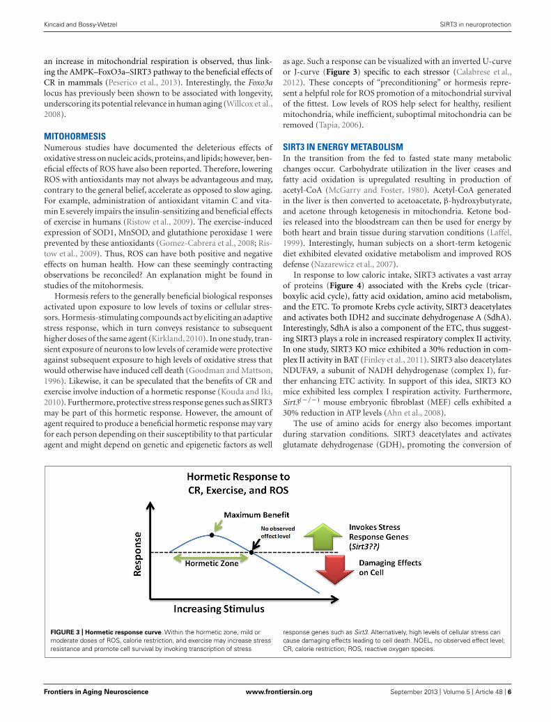
as age. Such a response can be visualized with an inverted U-curveor J-curve (Figure 3) specific to each stressor (Calabrese et al.,2012). These concepts of “preconditioning” or hormesis repre-sent a helpful role for ROS promotion of a mitochondrial survivalof the fittest. Low levels of ROS help select for healthy, resilientmitochondria, while inefficient, suboptimal mitochondria can beremoved (Tapia, 2006).
SIRT3 IN ENERGY METABOLISMIn the transition from the fed to fasted state many metabolicchanges occur. Carbohydrate utilization in the liver ceases andfatty acid oxidation is upregulated resulting in production ofacetyl-CoA (McGarry and Foster, 1980). Acetyl-CoA generatedin the liver is then converted to acetoacetate, β-hydroxybutyrate,and acetone through ketogenesis in mitochondria. Ketone bod-ies released into the bloodstream can then be used for energy byboth heart and brain tissue during starvation conditions (Laffel,1999). Interestingly, human subjects on a short-term ketogenicdiet exhibited elevated oxidative metabolism and improved ROSdefense (Nazarewicz et al., 2007).
In response to low caloric intake, SIRT3 activates a vast arrayof proteins (Figure 4) associated with the Krebs cycle (tricar-boxylic acid cycle), fatty acid oxidation, amino acid metabolism,and the ETC. To promote Krebs cycle activity, SIRT3 deacetylatesand activates both IDH2 and succinate dehydrogenase A (SdhA).Interestingly, SdhA is also a component of the ETC, thus suggest-ing SIRT3 plays a role in increased respiratory complex II activity.In one study, SIRT3 KO mice exhibited a 30% reduction in com-plex II activity in BAT (Finley et al., 2011). SIRT3 also deacetylatesNDUFA9, a subunit of NADH dehydrogenase (complex I), fur-ther enhancing ETC activity. In support of this idea, SIRT3 KOmice exhibited less complex I respiration activity. Furthermore,Sirt3(−/−) mouse embryonic fibroblast (MEF) cells exhibited a30% reduction in ATP levels (Ahn et al., 2008).
The use of amino acids for energy also becomes importantduring starvation conditions. SIRT3 deacetylates and activatesglutamate dehydrogenase (GDH), promoting the conversion of
FIGURE 3 | Hormetic response curve. Within the hormetic zone, mild ormoderate doses of ROS, calorie restriction, and exercise may increase stressresistance and promote cell survival by invoking transcription of stress
response genes such as Sirt3. Alternatively, high levels of cellular stress cancause damaging effects leading to cell death. NOEL, no observed effect level;CR, calorie restriction; ROS, reactive oxygen species.
Frontiers in Aging Neuroscience www.frontiersin.org September 2013 | Volume 5 | Article 48 | 6

“fnagi-05-00048” — 2013/9/4 — 15:02 — page 7 — #7
Kincaid and Bossy-Wetzel SIRT3 in neuroprotection
FIGURE 4 | Bona fide and suggested SIRT3 substrates.
glutamate to α-ketoglutarate, which can enter the Krebs cycle(Schlicker et al., 2008; Verdin et al., 2010; Dong et al., 2012).Additionally, SIRT3 deacetylates and activates ornithine transcar-bamoylase (OTC), a critical enzyme in the urea cycle. Activationof the urea cycle could aid in amino acid catabolism during fastingconditions (Hallows et al., 2011). Remarkably, Sirt3(−/−) MEFsexhibit accumulation of many amino acids, indicating a defect inamino acid catabolism (Hebert et al., 2013).
The large amount of acetate released into the blood streamby liver cells under fasting conditions is utilized mainly by theheart and skeletal muscle. These tissues express acetyl-CoA syn-thetase 2 (ACS2), an enzyme which catalyzes the ligation ofacetate and CoA to form acetyl-CoA for use in energy produc-tion (Fujino et al., 2001). SIRT3 directly deacetylates and activatesACS2 in mitochondria, thereby promoting this process (Hallowset al., 2006; Schwer et al., 2006). Ketone body formation is alsoregulated by SIRT3 during fasting. SIRT3 deacetylates 3-hydroxy-3-methylglutaryl CoA synthase 2 (HMGCS2), elevating its activityand enhancing β-hydroxybutyrate production (Shimazu et al.,2010). Additionally, SIRT3 promotes the use of triacylglycerolsin the liver by deacetylating and activating long-chain acyl co-enzyme A dehydrogenase (LCAD). Interestingly, mice lacking Sirt3exhibit accumulation of fatty acid oxidation intermediates andtriacylglycerols in the liver (Hirschey et al., 2010).
The metabolic role of SIRT3 has also been proposed to extendto processes such as regulation of mitochondrial protein synthesis,
steroidogenesis, and ATP synthesis. One report found that SIRT3may associate with the mitochondrial ribosomal subunit MRPL10,thus implicating SIRT3 in regulation of mitochondrial proteinsynthesis (Yang et al., 2010). An interaction between SIRT3 andP450 cholesterol side chain cleavage monooxygenase (P450scc)has also been suggested. Specifically, overexpression of SIRT3 maystabilize P450scc by deacetylation, potentially connecting SIRT3with steroidogenesis (Li et al., 2012). Additionally, potential reg-ulation of the F1F0ATPase by SIRT3 has been proposed. A recentstudy demonstrated that SIRT3 binds to the oligomycin sensitivityconferring protein (OSCP), a subunit of the mitochondrial ATPsynthase (Wu et al., 2013).
SIRT3 may also play a role in the response to consumption ofethanol. The oxidative metabolism of acetaldehyde (derived fromethanol) is facilitated by mitochondrial aldehyde dehydrogenase 2(ALDH2), which is also an NAD+-dependent enzyme (Marchittiet al., 2008). Interestingly, significant acetylation of mitochon-drial proteins is observed in liver tissues of mice fed high ethanoldiets (Fritz et al., 2012). Remarkably, 30 min after acute ethanoltreatment of human aortic endothelial cells (HAECs), the mito-chondrial NAD+/NADH ratio decreased by 65%, thus potentiallylimiting the activities of NAD+-dependent enzymes such as SIRT3.Additionally, a decrease in the acetylation state of ALDH2 uponoverexpression of SIRT3 suggests ALDH2 is a target of SIRT3;however, additional studies must be performed to confirm thisinteraction (Xue et al., 2012).
Frontiers in Aging Neuroscience www.frontiersin.org September 2013 | Volume 5 | Article 48 | 7
“fnagi-05-00048” — 2013/9/4 — 15:02 — page 8 — #8
Kincaid and Bossy-Wetzel SIRT3 in neuroprotection
Numerous reports suggest that SIRT3 may act to preventmetabolic maladies such as insulin resistance, metabolic syn-drome, and obesity. Mice fed a chronic HFD exhibit elevated globalmitochondrial protein acetylation as a result of suppression ofSIRT3 (Hirschey et al., 2011). This global acetylation of the mito-chondrial proteome may play a role in HFD-induced liver injury(Choudhury et al., 2011). Moreover, SIRT3 KO mice exhibitedimpaired insulin action due to increased ROS accumulation (Jinget al., 2011). Finally, PGC-1α KO mice exhibited many metabolicdefects including obesity, cardiomyopathy, and neurodegenera-tion (Lin et al., 2004). PGC-1α-mediated upregulation of SIRT3may play a role in preventing such illnesses.
SIRT3 IN NEUROPROTECTIONThe large ATP requirements of neurons predispose them to insultsthat result in energy depletion, including DNA damage, exci-totoxicity, and oxidative stress (Sokoloff, 1981; Du et al., 2003).Within neurons, mitochondria are the main sources of ROS andenergy production, suggesting these specialized organelles are crit-ical mediators of age-related diseases such as neurodegeneration(Singh, 2006). Surprisingly, CR reduces neurodegeneration in ani-mal models of both PD and AD, possibly via upregulation of SIRT3(Zhu et al., 1999; Mattson, 2000). Furthermore, overexpression ofSIRT3 has been shown to significantly increase neuronal lifespan(Weir et al., 2012). The antioxidant and metabolic effects medi-ated by SIRT3 suggest a potential neuroprotective role throughimproved mitochondrial function, which subsequently results inincreased neuronal survival and reduced aging effects.
The direct causes of many forms of neurodegeneration remainunknown, though insults or agents resulting in neuronal celldeath are likely to play a key role. Studies in aged rat brain haverevealed an age-dependent increase in both mitochondrial ROSproduction and cytosolic Ca2+ levels (Sawada and Carlson, 1987;Xiong et al., 2002; Toescu and Vreugdenhil, 2010). Interestingly,increased levels of ROS and Ca2+ trigger mitochondrial perme-ability transition pore (mtPTP) formation, an event which canlead to apoptosis and trigger neurodegeneration (Du and Yan,2010). Briefly, the mtPTP includes the voltage-dependent anionchannel (VDAC), adenine nucleotide translocator (ANT), andcyclophilin D (CypD). In response to ROS and increased Ca2+,binding of CypD to ANT initiates formation of a tunnel-likestructure, which connects the mitochondrial matrix to the cytosolresulting in the rapid flow of NAD+ from the mitochondria tothe cytosol (Lemasters et al., 2009). Within the cytosol, NAD+is quickly hydrolyzed by multiple NADases to yield ADP-riboseand NAM (Zhang et al., 1995; Bernardi, 1999). The frequencyof mtPTP formation may result in the destruction of defectivemitochondria by autophagy or possibly even cell death via apop-tosis (Kim et al., 2007). Recent studies suggest SIRT3 may be ableto suppress mtPTP formation during aging. In response to CR,SIRT3 is upregulated and directly deacetylates CypD, preventingits association with ANT and therefore blocking mtPTP formation(Hafner et al., 2010; Shulga et al., 2010). Additionally, interactionbetween CypD and amyloid-β in mitochondria of AD patientshas been reported. Such an interaction caused increased oxidativestress and increased mtPTP opening, triggering neurodegenera-tion (Du and Yan, 2010). Upregulation of SIRT3 may be able to
prevent or delay this process, conveying a neuroprotective effectin AD.
Recent observations also hint at additional neuroprotectiveeffects of SIRT3 involving regulation of mitochondrial dynamics.A defective mitochondrial fission and fusion balance affects mito-chondrial transport and function, potentially leading to synapticdysfunction and neurodegeneration (Knott et al., 2008). In fact,mutations in the mitochondrial fusion protein OPA1 cause domi-nant optic atrophy, thereby linking mitochondrial dynamics withneuronal functionality (Seo et al., 2010). In a recent study, SIRT3was able to rescue the mitochondrial fragmentation associatedwith a model of amyotrophic lateral sclerosis. Specifically, spinalcord motor neurons transfected with SOD1G93A displayed anincrease in round fragmented mitochondria in addition to defectsin bi-directional axonal transport and increased cell death. How-ever, co-expression with either SIRT3 or PGC-1α was able to rescueSOD1G93A-induced mitochondrial fragmentation and improvecell survival (Song et al., 2012). Interestingly, PGC-1α directly reg-ulates mitochondrial dynamics by increasing MFN2 expression(Soriano et al., 2006). It may be that by inducing PGC-1α, ROS isdecreased by the antioxidant stimulating abilities of SIRT3, whilemitochondrial fusion is increased by MFN2. Additionally, PGC-1α null mice are more sensitive to the neurodegenerative effects ofROS, further identifying itself and its target genes such as Sirt3 aspotential mediators of neuroprotection (St-Pierre et al., 2006).
In neurons, increased SIRT3 expression has also been reportedin response to oxidative stress (Kim et al., 2011b). Furthermore,oxidative stress has been shown to upregulate β-secretase activity,an enzyme associated with AD (Tamagno et al., 2002). In a studyutilizing a mouse model of AD,Sirt3 mRNA upregulation mirroredspatiotemporal amyloid-β deposition. Additionally, Sirt3 mRNAwas found to be increased in human post-mortem cortex samplesof AD patients (Weir et al., 2012). In this case, it may be thatupregulation of SIRT3 is a compensatory mechanism in neuronsto attempt to protect against the increased oxidative stress thataccompanies AD development and progression.
The ability of CR to induce SIRT3 expression has been welldocumented. Additional stimulators of the Sirtuins have also beenproposed, such as resveratrol, a polyphenol found in red wine.However, the effect of resveratrol on Sirtuin expression remainscontroversial. In one study, mice on a 30% CR diet exhibitedincreased Sirt3 mRNA levels, while treatment with resveratrol didnot affect Sirt3 expression levels. In light of this finding, resvera-trol may be ineffective in mimicking CR-mediated health benefits(Tauriainen et al., 2011). However, not all hope is lost for thepolyphenol compounds. A recent report found that a resvera-trol derivative, trans-(−)-ε-viniferin, is able to increase SIRT3expression and provide protection in cell models of Huntington’sdisease (HD). Specifically, viniferin treatment of striatal precursorcells overexpressing mutant huntingtin resulted in increased SIRT3expression, increased the NAD+/NADH ratio, reduced intracellu-lar ROS accumulation, and decreased acetylated MnSOD levels.Additionally, treatment with viniferin increased levels of acti-vated AMPK and decreased acetylated LKB1, effects which wereshown to require the presence of SIRT3. Thus, Sirt3 is requiredfor viniferin-mediated neuroprotection in HD models (Fu et al.,2012).
Frontiers in Aging Neuroscience www.frontiersin.org September 2013 | Volume 5 | Article 48 | 8
“fnagi-05-00048” — 2013/9/4 — 15:02 — page 9 — #9
Kincaid and Bossy-Wetzel SIRT3 in neuroprotection
CONCLUSIONS AND FUTURE PERSPECTIVESThe beneficial effects of SIRT3 in regulating metabolism and acti-vating antioxidant defense systems in response to CR and exerciseis apparent. Additionally, while mechanisms by which SIRT3 canprovide neuroprotection are better understood, there are still somediscrepancies among studies that have not been accounted for.To date, the majority of studies acknowledge the strict local-ization of active SIRT3 to mitochondria. Despite this, SIRT3activity has been reported in the nucleus where it plays a rolein associating with Ku-70 to help promote cell survival (Sundare-san et al., 2008). The discovery of a splice variant in mice thatlacks a MTS might help explain such observations, but furtherexperiments could help settle any disputes over SIRT3 subcel-lular localization. Additionally, short-term and long-term CRappear to have varying effects on SIRT3 expression (Jing et al.,2011). What should be the recommended level of CR to max-imally induce SIRT3 expression? Furthermore, the decrease inSIRT3 expression with age cannot be explained. What transcrip-tional regulators affect expression of SIRT3 during aging and howdo these factors get turned on or off over time? These questionsremain unanswered and require further investigation. Finally,recent global mitochondrial protein acetylome studies have been
performed, potentially identifying a myriad of exciting SIRT3substrates that are not yet known (Hebert et al., 2013). Identi-fication of additional bona fide SIRT3 targets will help solidifyour understanding of the role of SIRT3 in neuroprotection andlongevity.
It has been proposed that excessive energy intake can exposehumans to oxidative consequences during the fourth and fifthdecade of life, potentially leading to cognitive decline during lateryears (Debette et al., 2011). Conversely, CR can promote healthyaging and neuroprotection via the actions of SIRT3. However, theeating habits of the majority of Western civilization are far fromconducive for induction of optimal SIRT3 expression. For thisreason, molecular mimetics of CR are needed to substitute for anactual decrease in food intake. In addition, novel agents that caninduce a beneficial hormetic response to promote the upregulationof stress response genes such as Sirt3 may also be of great thera-peutic value, providing a means for enhancing neuroprotectionand healthy aging.
ACKNOWLEDGMENTSWe thank A. Knott for editorial assistance. This work wassupported by NIH grant R01 NS055193 to Ella Bossy-Wetzel.
REFERENCESAccili, D., and Arden, K. C. (2004).
FoxOs at the crossroads of cel-lular metabolism, differentiation,and transformation. Cell 117, 421–426. doi: 10.1016/S0092-8674(04)00452-0
Ahn, B.-H., Kim, H.-S., Song, S., Lee,I. H., Liu, J., Vassilopoulos, A., et al.(2008). A role for the mitochon-drial deacetylase Sirt3 in regulatingenergy homeostasis. Proc. Natl. Acad.Sci. U.S.A. 105, 14447–14452. doi:10.1073/pnas.0803790105
Alano, C. C., Ying, W., and Swanson,R. A. (2004). Poly(ADP-ribose)polymerase-1-mediated cell deathin astrocytes requires NAD+ deple-tion and mitochondrial permeabilitytransition. J. Biol. Chem. 279, 18895–18902. doi: 10.1074/jbc.M313329200
Anderson, M. E. (1998). Glutathione:an overview of biosynthesis andmodulation. Chem. Biol. Interact.111–112, 1–14. doi: 10.1016/S0009-2797(97)00146-4
Arnoult, D., Grodet, A., Lee, Y.-J., Estaquier, J., and Blackstone,C. (2005). Release of OPA1 dur-ing apoptosis participates in therapid and complete release ofcytochrome c and subsequentmitochondrial fragmentation.J. Biol. Chem. 280, 35742–35750. doi: 10.1074/jbc.M505970200
Bai, P., Cantó, C., Oudart, H., Brun-yánszki, A., Cen, Y., Thomas,C., et al. (2011). PARP-1 inhi-bition increases mitochondrial
metabolism through SIRT1 acti-vation. Cell Metab. 13, 461–468. doi:10.1016/j.cmet.2011.03.004
Balaban, R. S., Nemoto, S., and Finkel,T. (2005). Mitochondria, oxidants,and aging. Cell 120, 483–495. doi:10.1016/j.cell.2005.02.001
Bellizzi, D., Rose, G., Cavalcante, P.,Covello, G., Dato, S., De Rango, F.,et al. (2005). A novel VNTR enhancerwithin the SIRT3 gene, a humanhomologue of SIR2, is associated withsurvival at oldest ages. Genomics 85,258–263. doi: 10.1016/j.ygeno.2004.11.003
Bergeron, R., Ren, J. M., Cadman, K.S., Moore, I. K., Perret, P., Pypaert,M., et al. (2001). Chronic activationof AMP kinase results in NRF-1 acti-vation and mitochondrial biogenesis.Am. J. Physiol. Endocrinol. Metab.281, E1340–E1346.
Bernardi, P. (1999). Mitochondrialtransport of cations: channels,exchangers, and permeability transi-tion. Physiol. Rev. 79, 1127–1155.
Berndt, C., Lillig, C. H., and Holmgren,A. (2007). Thiol-based mechanismsof the thioredoxin and glutaredoxinsystems: implications for diseases inthe cardiovascular system. Am. J.Physiol. Heart Circ. Physiol. 292,H1227–H1236. doi: 10.1152/ajp-heart.01162.2006
Burgering, B. M. T., and Kops, G. J. P. L.(2002). Cell cycle and death control:long live Forkheads. Trends Biochem.Sci. 27, 352–360. doi: 10.1016/S0968-0004(02)02113-8
Calabrese, V., Cornelius, C., Dinkova-Kostova, A. T., Iavicoli, I., Di
Paola, R., Koverech, A., et al. (2012).Cellular stress responses, hormeticphytochemicals and vitagenes inaging and longevity. Biochim. Bio-phys. Acta 1822, 753–783. doi:10.1016/j.bbadis.2011.11.002
Chen, D., Bruno, J., Easlon, E., Lin,S.-J., Cheng, H.-L., Alt, F. W.,et al. (2008). Tissue-specific regu-lation of SIRT1 by calorie restric-tion. Genes Dev. 22, 1753–1757. doi:10.1101/gad.1650608
Cheng, Y., Ren, X., Gowda, A. S.,Shan, Y., Zhang, L., Yuan, Y.-S., et al. (2013). Interaction ofSirt3 with OGG1 contributes torepair of mitochondrial DNA andprotects from apoptotic cell deathunder oxidative stress. Cell DeathDis. 4, e731. doi: 10.1038/cddis.2013.254
Choudhury, M., Jonscher, K. R., andFriedman, J. E. (2011). Reducedmitochondrial function in obesity-associated fatty liver: SIRT3 takeson the fat. Aging (Albany NY) 3,175–178.
Cooper, H. M., Huang, J.-Y., Verdin,E., and Spelbrink, J. N. (2009). Anew splice variant of the mouseSIRT3 gene encodes the mitochon-drial precursor protein. PLoS ONE4:e4986. doi: 10.1371/journal.pone.0004986
Cooper, H. M., and Spelbrink, J. N.(2008). The human SIRT3 proteindeacetylase is exclusively mitochon-drial. Biochem. J. 411, 279–285. doi:10.1042/BJ20071624
Debette, S., Seshadri, S., Beiser, A.,Au, R., Himali, J. J., Palumbo,
C., et al. (2011). Midlife vascu-lar risk factor exposure acceleratesstructural brain aging and cogni-tive decline. Neurology 77, 461–468. doi: 10.1212/WNL.0b013e318227b227
Dong, K., Pelle, E., Yarosh, D. B.,and Pernodet, N. (2012). Sirtuin 4identification in normal human epi-dermal keratinocytes and its relationto sirtuin 3 and energy metabolismunder normal conditions and UVB-induced stress. Exp. Dermatol.21, 231–233. doi: 10.1111/j.1600-0625.2011.01439.x
Du, H., and Yan, S. S. (2010).Mitochondrial permeability transi-tion pore in Alzheimer’s disease:cyclophilin D and amyloid beta.Biochim. Biophys. Acta 1802, 198–204. doi: 10.1016/j.bbadis.2009.07.005
Du, J., Zhou, Y., Su, X., Yu, J. J.,Khan, S., Jiang, H., et al. (2011). Sirt5is a NAD-dependent protein lysinedemalonylase and desuccinylase. Sci-ence 334, 806–809. doi: 10.1126/sci-ence.1207861
Du, L., Zhang, X., Han, Y. Y., Burke, N.A., Kochanek, P. M., Watkins, S. C.,et al. (2003). Intra-mitochondrialpoly(ADP-ribosylation) con-tributes to NAD+ depletion andcell death induced by oxidativestress. J. Biol. Chem. 278, 18426–18433. doi: 10.1074/jbc.M301295200
Finkel, T., and Holbrook, N. J. (2000).Oxidants, oxidative stress and thebiology of ageing. Nature 408, 239–247. doi: 10.1038/35041687
Frontiers in Aging Neuroscience www.frontiersin.org September 2013 | Volume 5 | Article 48 | 9
“fnagi-05-00048” — 2013/9/4 — 15:02 — page 10 — #10
Kincaid and Bossy-Wetzel SIRT3 in neuroprotection
Finley, L. W. S., Haas, W., Desquiret-Dumas, V., Wallace, D. C., Procaccio,V., Gygi, S. P., et al. (2011). Succi-nate dehydrogenase is a direct targetof sirtuin 3 deacetylase activity. PLoSONE 6:e23295. doi: 10.1371/jour-nal.pone.0023295
Fritz, K. S., Galligan, J. J., Hirschey,M. D., Verdin, E., and Petersen, D.R. (2012). Mitochondrial acetylomeanalysis in a mouse model of alcohol-induced liver injury utilizing SIRT3knockout mice. J. Proteome Res.11, 1633–1643. doi: 10.1021/pr2008384
Frye, R. A. (2000). Phylogenetic classifi-cation of prokaryotic and eukaryoticSir2-like proteins. Biochem. Biophys.Res. Commun. 273, 793–798. doi:10.1006/bbrc.2000.3000
Fu, J., Jin, J., Cichewicz, R. H., Hage-man, S. A., Ellis, T. K., Xiang, L.,et al. (2012). trans-(−)-ε-Viniferinincreases mitochondrial sirtuin 3(SIRT3), activates AMP-activatedprotein kinase (AMPK), and pro-tects cells in models of Huntingtondisease. J. Biol. Chem. 287, 24460–24472. doi: 10.1074/jbc.M112.382226
Fujino, T., Kondo, J., Ishikawa, M.,Morikawa, K., and Yamamoto, T.T. (2001). Acetyl-CoA synthetase2, a mitochondrial matrix enzymeinvolved in the oxidation of acetate.J. Biol. Chem. 276, 11420–11426. doi:10.1074/jbc.M008782200
Gomes, L. C., and Scorrano, L.(2011). Mitochondrial elongationduring autophagy: a stereotypi-cal response to survive in difficulttimes. Autophagy 7, 1251–1253. doi:10.4161/auto.7.10.16771
Gomez-Cabrera, M.-C., Domenech, E.,Romagnoli, M., Arduini, A., Bor-ras, C., Pallardo, F. V., et al. (2008).Oral administration of vitaminC decreases muscle mitochondrialbiogenesis and hampers training-induced adaptations in enduranceperformance. Am. J. Clin. Nutr. 87,142–149.
Goodman, Y., and Mattson, M.P. (1996). Ceramide protects hip-pocampal neurons against excitotoxicand oxidative insults, and amyloidbeta-peptide toxicity. J. Neurochem.66, 869–872. doi: 10.1046/j.1471-4159.1996.66020869.x
Guarente, L., and Kenyon, C. (2000).Genetic pathways that regulate age-ing in model organisms. Nature408, 255–262. doi: 10.1038/35041700
Gurd, B. J., Holloway, G. P., Yoshida,Y., and Bonen, A. (2012). In mam-malian muscle, SIRT3 is presentin mitochondria and not in the
nucleus; and SIRT3 is upregu-lated by chronic muscle contractionin an adenosine monophosphate-activated protein kinase-independentmanner. Metabolism 61, 733–741. doi: 10.1016/j.metabol.2011.09.016
Hafner, A. V., Dai, J., Gomes, A. P.,Xiao, C.-Y., Palmeira, C. M., Rosen-zweig, A., et al. (2010). Regulationof the mPTP by SIRT3-mediateddeacetylation of CypD at lysine166 suppresses age-related cardiachypertrophy. Aging (Albany NY) 2,914–923.
Haigis, M. C., Mostoslavsky, R., Haigis,K. M., Fahie, K., Christodoulou,D. C., Murphy, A. J., et al.(2006). SIRT4 inhibits glutamatedehydrogenase and opposes theeffects of calorie restriction inpancreatic beta cells. Cell 126,941–954. doi: 10.1016/j.cell.2006.06.057
Hall, D. M., Sattler, G. L., Sattler,C. A., Zhang, H. J., Oberley, L.W., Pitot, H. C., et al. (2001).Aging lowers steady-state antioxidantenzyme and stress protein expressionin primary hepatocytes. J. Geron-tol. A Biol. Sci. Med. Sci. 56,B259–B267. doi: 10.1093/gerona/56.6.B259
Hallows, W. C., Lee, S., andDenu, J. M. (2006). Sirtuinsdeacetylate and activate mammalianacetyl-CoA synthetases. Proc. Natl.Acad. Sci. U.S.A. 103, 10230–10235. doi: 10.1073/pnas.0604392103
Hallows, W. C., Yu, W., Smith, B.C., Devries, M. K., Devires, M. K.,Ellinger, J. J., et al. (2011). Sirt3promotes the urea cycle and fattyacid oxidation during dietary restric-tion. Mol. Cell 41, 139–149. doi:10.1016/j.molcel.2011.01.002
Hara, T., Nakamura, K., Matsui,M., Yamamoto, A., Nakahara, Y.,Suzuki-Migishima, R., et al. (2006).Suppression of basal autophagyin neural cells causes neurodegen-erative disease in mice. Nature441, 885–889. doi: 10.1038/nature04724
Harman, D. (1956). Aging: a theorybased on free radical and radiationchemistry. J. Gerontol. 11, 298–300.doi: 10.1093/geronj/11.3.298
He, W., Newman, J. C., Wang, M.Z., Ho, L., and Verdin, E. (2012).Mitochondrial sirtuins: regulators ofprotein acylation and metabolism.Trends Endocrinol. Metab. 23,467–476. doi: 10.1016/j.tem.2012.07.004
Hebert, A. S., Dittenhafer-Reed, K. E.,Yu, W., Bailey, D. J., Selen, E. S.,
Boersma, M. D., et al. (2013). Calorierestriction and SIRT3 trigger globalreprogramming of the mitochondrialprotein acetylome. Mol. Cell 49,186–199. doi: 10.1016/j.molcel.2012.10.024
Herzig, S., Long, F., Jhala, U. S.,Hedrick, S., Quinn, R., Bauer, A.,et al. (2001). CREB regulates hepaticgluconeogenesis through the coacti-vator PGC-1. Nature 413, 179–183.doi: 10.1038/35093131
Hirschey, M. D., Shimazu, T., Goet-zman, E., Jing, E., Schwer, B.,Lombard, D. B., et al. (2010).SIRT3 regulates mitochondrialfatty-acid oxidation by reversibleenzyme deacetylation. Nature464, 121–125. doi: 10.1038/nature08778
Hirschey, M. D., Shimazu, T., Jing,E., Grueter, C. A., Collins, A. M.,Aouizerat, B., et al. (2011). SIRT3deficiency and mitochondrial pro-tein hyperacetylation accelerate thedevelopment of the metabolic syn-drome. Mol. Cell 44, 177–190. doi:10.1016/j.molcel.2011.07.019
Hurst, L. D., Williams, E. J. B., andPál, C. (2002). Natural selection pro-motes the conservation of linkageof co-expressed genes. Trends Genet.18, 604–606. doi: 10.1016/S0168-9525(02)02813-5
Iwahara, T., Bonasio, R., Narendra,V., and Reinberg, D. (2012). SIRT3functions in the nucleus in the con-trol of stress-related gene expression.Mol. Cell. Biol. 32, 5022–5034. doi:10.1128/MCB.00822-12
Jacobs, K. M., Pennington, J. D., Bisht,K. S., Aykin-Burns, N., Kim, H.-S., Mishra, M., et al. (2008). SIRT3interacts with the daf-16 homologFOXO3a in the mitochondria, aswell as increases FOXO3a depen-dent gene expression. Int. J. Biol.Sci. 4, 291–299. doi: 10.7150/ijbs.4.291
Jäger, S., Handschin, C., St-Pierre,J., and Spiegelman, B. M. (2007).AMP-activated protein kinase(AMPK) action in skeletal mus-cle via direct phosphorylation ofPGC-1alpha. Proc. Natl. Acad.Sci. U.S.A. 104, 12017–12022. doi:10.1073/pnas.0705070104
Jin, L., Galonek, H., Israelian, K.,Choy, W., Morrison, M., Xia, Y.,et al. (2009). Biochemical characteri-zation, localization, and tissue distri-bution of the longer form of mouseSIRT3. Protein Sci. 18, 514–525. doi:10.1002/pro.50
Jing, E., Emanuelli, B., Hirschey, M.D., Boucher, J., Lee, K. Y., Lombard,D., et al. (2011). Sirtuin-3 (Sirt3)regulates skeletal muscle metabolism
and insulin signaling via alteredmitochondrial oxidation and reactiveoxygen species production. Proc.Natl. Acad. Sci. U.S.A. 108, 14608–14613. doi: 10.1073/pnas.1111308108
Joseph, A.-M., Adhihetty, P. J., Buford,T. W., Wohlgemuth, S. E., Lees, H.A., Nguyen, L. M.-D., et al. (2012).The impact of aging on mitochon-drial function and biogenesis path-ways in skeletal muscle of sedentaryhigh- and low-functioning elderlyindividuals. Aging Cell 11, 801–809. doi: 10.1111/j.1474-9726.2012.00844.x
Kim, H.-J., Kim, J. H., Noh, S., Hur,H. J., Sung, M. J., Hwang, J.-T.,et al. (2011a). Metabolomic analy-sis of livers and serum from high-fatdiet induced obese mice. J. ProteomeRes. 10, 722–731. doi: 10.1021/pr100892r
Kim, S. H., Lu, H. F., and Alano,C. C. (2011b). Neuronal Sirt3 pro-tects against excitotoxic injury inmouse cortical neuron culture. PLoSONE 6:e14731. doi: 10.1371/jour-nal.pone.0014731
Kim, H.-S., Patel, K., Muldoon-Jacobs, K., Bisht, K. S., Aykin-Burns, N., Pennington, J. D., et al.(2010). SIRT3 is a mitochondria-localized tumor suppressor requiredfor maintenance of mitochondrialintegrity and metabolism duringstress. Cancer Cell 17, 41–52. doi:10.1016/j.ccr.2009.11.023
Kim, I., Rodriguez-Enriquez, S., andLemasters, J. J. (2007). Selec-tive degradation of mitochondriaby mitophagy. Arch. Biochem.Biophys. 462, 245–253. doi:10.1016/j.abb.2007.03.034
Kim, S. C., Sprung, R., Chen, Y., Xu,Y., Ball, H., Pei, J., et al. (2006).Substrate and functional diversityof lysine acetylation revealed by aproteomics survey. Mol. Cell 23,607–618. doi: 10.1016/j.molcel.2006.06.026
Kirkland, J. L. (2010). Perspectives oncellular senescence and short termdietary restriction in adults. Aging(Albany NY) 2, 894–896.
Knott, A. B., Perkins, G., Schwarzen-bacher, R., and Bossy-Wetzel, E.(2008). Mitochondrial fragmenta-tion in neurodegeneration. Nat.Rev. Neurosci. 9, 505–518. doi:10.1038/nrn2417
Komatsu, M., Waguri, S., Chiba, T.,Murata, S., Iwata, J., Tanida, I.,et al. (2006). Loss of autophagy inthe central nervous system causesneurodegeneration in mice. Nature441, 880–884. doi: 10.1038/nature04723
Frontiers in Aging Neuroscience www.frontiersin.org September 2013 | Volume 5 | Article 48 | 10
“fnagi-05-00048” — 2013/9/4 — 15:02 — page 11 — #11
Kincaid and Bossy-Wetzel SIRT3 in neuroprotection
Kong, X., Wang, R., Xue, Y., Liu,X., Zhang, H., Chen, Y., et al.(2010). Sirtuin 3, a new targetof PGC-1alpha, plays an importantrole in the suppression of ROS andmitochondrial biogenesis. PLoS ONE5:e11707. doi: 10.1371/journal.pone.0011707
Kouda, K., and Iki, M. (2010).Beneficial effects of mild stress(hormetic effects): dietary restrictionand health. J. Physiol. Anthropol. 29,127–132. doi: 10.2114/jpa2.29.127
Kuma, A., Hatano, M., Matsui, M.,Yamamoto, A., Nakaya, H., Yoshi-mori, T., et al. (2004). The role ofautophagy during the early neonatalstarvation period. Nature 432, 1032–1036. doi: 10.1038/nature03029
Laffel, L. (1999). Ketone bodies: areview of physiology, pathophysiol-ogy and application of monitoring todiabetes. Diabetes Metab. Res. Rev. 15,412–426. doi: 10.1002/(SICI)1520-7560(199911/12)15:6<412::AID-DMRR72>3.0.CO;2-8
Lanza, I. R., Short, D. K., Short, K.R., Raghavakaimal, S., Basu, R.,Joyner, M. J., et al. (2008). Enduranceexercise as a countermeasure foraging. Diabetes 57, 2933–2942. doi:10.2337/db08-0349
Lemasters, J. J., Theruvath, T. P.,Zhong, Z., and Nieminen, A.-L.(2009). Mitochondrial calciumand the permeability transitionin cell death. Biochim. Bio-phys. Acta 1787, 1395–1401. doi:10.1016/j.bbabio.2009.06.009
Li, D., Dammer, E. B., and Sewer, M.B. (2012). Resveratrol stimulates cor-tisol biosynthesis by activating SIRT-dependent deacetylation of P450scc.Endocrinology 153, 3258–3268. doi:10.1210/en.2011-2088
Liesa, M., and Shirihai, O. S.(2013). Mitochondrial dynamicsin the regulation of nutrient uti-lization and energy expenditure.Cell Metab. 17, 491–506. doi:10.1016/j.cmet.2013.03.002
Lin, J., Wu, P.-H., Tarr, P. T., Linden-berg, K. S., St-Pierre, J., and Zhang,C.-Y., et al. (2004). Defects in adap-tive energy metabolism with CNS-linked hyperactivity in PGC-1alphanull mice. Cell 119, 121–135. doi:10.1016/j.cell.2004.09.013
Lombard, D. B., Alt, F. W., Cheng,H.-L., Bunkenborg, J., Streeper, R.S., Mostoslavsky, R., et al. (2007).Mammalian Sir2 homolog SIRT3 reg-ulates global mitochondrial lysineacetylation. Mol. Cell. Biol. 27, 8807–8814. doi: 10.1128/MCB.01636-07
Mailloux, R. J., Bériault, R., Lemire,J., Singh, R., Chénier, D. R.,Hamel, R. D., et al. (2007). The
tricarboxylic acid cycle, an ancientmetabolic network with a novel twist.PLoS ONE 2:e690. doi: 10.1371/jour-nal.pone.0000690
Marchitti, S. A., Brocker, C., Sta-gos, D., and Vasiliou, V. (2008).Non-P450 aldehyde oxidizingenzymes: the aldehyde dehydro-genase superfamily. Expert Opin.Drug Metab. Toxicol. 4, 697–720. doi:10.1517/17425255.4.6.697
Mattson, M. P. (2000). Apoptosis inneurodegenerative disorders. Nat.Rev. Mol. Cell Biol. 1, 120–129. doi:10.1038/35040009
McCay, C. M., Crowell, M. F., andMaynard, L. A. (1989). The effectof retarded growth upon the lengthof life span and upon the ultimatebody size. 1935. Nutrition 5, 155–171;discussion 172.
McGarry, J. D., and Foster, D. W.(1980). Regulation of hepatic fattyacid oxidation and ketone bodyproduction. Annu. Rev. Biochem.49, 395–420. doi: 10.1146/annurev.bi.49.070180.002143
Misko, A., Jiang, S., Wegorzewska,I., Milbrandt, J., and Baloh, R.H. (2010). Mitofusin 2 is necessaryfor transport of axonal mitochondriaand interacts with the Miro/Miltoncomplex. J. Neurosci. 30, 4232–4240. doi: 10.1523/JNEUROSCI.6248-09.2010
Molina, A. J. A., Wikstrom, J. D., Stiles,L., Las, G., Mohamed, H., Elorza,A., et al. (2009). Mitochondrialnetworking protects beta-cells fromnutrient-induced apoptosis. Dia-betes 58, 2303–2315. doi: 10.2337/db07-1781
Nazarewicz, R. R., Ziolkowski, W., Vac-caro, P. S., and Ghafourifar, P. (2007).Effect of short-term ketogenic dieton redox status of human blood.Rejuvenation Res. 10, 435–440. doi:10.1089/rej.2007.0540
Onyango, P., Celic, I., McCaffery, J.M., Boeke, J. D., and Feinberg, A.P. (2002). SIRT3, a human SIR2homologue, is an NAD-dependentdeacetylase localized to mitochon-dria. Proc. Natl. Acad. Sci. U.S.A.99, 13653–13658. doi: 10.1073/pnas.222538099
Outeiro, T. F., Grammatopoulos, T.N., Altmann, S., Amore, A., Stan-daert, D. G., Hyman, B. T., et al.(2007). Pharmacological inhibitionof PARP-1 reduces alpha-synuclein-and MPP+-induced cytotoxicity inParkinson’s disease in vitro models.Biochem. Biophys. Res. Commun. 357,596–602. doi: 10.1016/j.bbrc.2007.03.163
Palacios, O. M., Carmona, J. J., Michan,S., Chen, K. Y., Manabe, Y., Ward, J.
L. III, et al. (2009). Diet and exercisesignals regulate SIRT3 and activateAMPK and PGC-1alpha in skeletalmuscle. Aging (Albany NY) 1, 771–783.
Peserico, A., Chiacchiera, F., Grossi, V.,Matrone, A., Latorre, D., Simonatto,M., et al. (2013). A novel AMPK-dependent FoxO3A-SIRT3 intrami-tochondrial complex sensing glucoselevels. Cell. Mol. Life Sci. 70, 2015–2029. doi: 10.1007/s00018-012-1244-6
Pillai, V. B., Sundaresan, N. R., Kim, G.,Gupta, M., Rajamohan, S. B., Pillai,J. B., et al. (2010). Exogenous NADblocks cardiac hypertrophic responsevia activation of the SIRT3-LKB1-AMP-activated kinase pathway. J.Biol. Chem. 285, 3133–3144. doi:10.1074/jbc.M109.077271
Qiu, X., Brown, K., Hirschey, M. D.,Verdin, E., and Chen, D. (2010).Calorie restriction reduces oxidativestress by SIRT3-mediated SOD2 acti-vation. Cell Metab. 12, 662–667. doi:10.1016/j.cmet.2010.11.015
Rambold, A. S., Kostelecky, B., andLippincott-Schwartz, J. (2011). Fuseor die: shaping mitochondrial fateduring starvation. Commun. Integr.Biol. 4, 752–754.
Ranhotra, H. S. (2009). Up-regulationof orphan nuclear estrogen-relatedreceptor alpha expression duringlong-term caloric restriction inmice. Mol. Cell. Biochem. 332,59–65. doi: 10.1007/s11010-009-0174-6
Rebrin, I., Kamzalov, S., and Sohal, R.S. (2003). Effects of age and caloricrestriction on glutathione redox statein mice. Free Radic. Biol. Med.35, 626–635. doi: 10.1016/S0891-5849(03)00388-5
Rebrin, I., and Sohal, R. S. (2008). Pro-oxidant shift in glutathione redoxstate during aging. Adv. DrugDeliv. Rev. 60, 1545–1552. doi:10.1016/j.addr.2008.06.001
Ristow, M., Zarse, K., Oberbach, A.,Klöting, N., Birringer, M., Kiehn-topf, M., et al. (2009). Antioxidantsprevent health-promoting effects ofphysical exercise in humans. Proc.Natl. Acad. Sci. U.S.A. 106, 8665–8670. doi: 10.1073/pnas.0903485106
Rongvaux, A., Shea, R. J., Mulks, M. H.,Gigot, D., Urbain, J., Leo, O., et al.(2002). Pre-B-cell colony-enhancingfactor, whose expression is up-regulated in activated lymphocytes,is a nicotinamide phosphoribo-syltransferase, a cytosolic enzymeinvolved in NAD biosynthesis.Eur. J. Immunol. 32, 3225–3234.doi: 10.1002/1521-4141(200211)32:11
Rose, G., Dato, S., Altomare, K., Bel-lizzi, D., Garasto, S., Greco, V.,et al. (2003). Variability of the SIRT3gene, human silent information reg-ulator Sir2 homologue, and survivor-ship in the elderly. Exp. Gerontol.38, 1065–1070. doi: 10.1016/S0531-5565(03)00209-2
Safdar, A., Bourgeois, J. M., Ogborn,D. I., Little, J. P., Hettinga, B. P.,Akhtar, M., et al. (2011). Enduranceexercise rescues progeroid aging andinduces systemic mitochondrial reju-venation in mtDNA mutator mice.Proc. Natl. Acad. Sci. U.S.A. 108,4135–4140. doi: 10.1073/pnas.1019581108
Sawada, M., and Carlson, J. C. (1987).Changes in superoxide radical andlipid peroxide formation in the brain,heart and liver during the life-time of the rat. Mech. Ageing Dev.41, 125–137. doi: 10.1016/0047-6374(87)90057-1
Scheckhuber, C. Q., Erjavec, N., Tina-zli, A., Hamann, A., Nyström, T.,and Osiewacz, H. D. (2007). Reduc-ing mitochondrial fission results inincreased life span and fitness oftwo fungal ageing models. Nat.Cell Biol. 9, 99–105. doi: 10.1038/ncb1524
Scher, M. B., Vaquero, A., and Rein-berg, D. (2007). SirT3 is a nuclearNAD+-dependent histone deacety-lase that translocates to the mito-chondria upon cellular stress. GenesDev. 21, 920–928. doi: 10.1101/gad.1527307
Schlicker, C., Gertz, M., Papatheodorou,P., Kachholz, B., Becker, C. F. W.,and Steegborn, C. (2008). Sub-strates and regulation mechanismsfor the human mitochondrial sirtu-ins Sirt3 and Sirt5. J. Mol. Biol. 382,790–801. doi: 10.1016/j.jmb.2008.07.048
Schriner, S. E., Linford, N. J., Mar-tin, G. M., Treuting, P., Ogburn,C. E., Emond, M., et al. (2005).Extension of murine life spanby overexpression of catalase tar-geted to mitochondria. Science 308,1909–1911. doi: 10.1126/science.1106653
Schwer, B., Bunkenborg, J., Verdin,R. O., Andersen, J. S., and Verdin,E. (2006). Reversible lysine acety-lation controls the activity of themitochondrial enzyme acetyl-CoAsynthetase 2. Proc. Natl. Acad.Sci. U.S.A. 103, 10224–10229. doi:10.1073/pnas.0603968103
Schwer, B., North, B. J., Frye, R.A., Ott, M., and Verdin, E. (2002).The human silent information regu-lator (Sir)2 homologue hSIRT3 is amitochondrial nicotinamide adenine
Frontiers in Aging Neuroscience www.frontiersin.org September 2013 | Volume 5 | Article 48 | 11
“fnagi-05-00048” — 2013/9/4 — 15:02 — page 12 — #12
Kincaid and Bossy-Wetzel SIRT3 in neuroprotection
dinucleotide-dependent deacetylase.J. Cell Biol. 158, 647–657. doi:10.1083/jcb.200205057
Seo, A. Y., Joseph, A.-M., Dutta,D., Hwang, J. C. Y., Aris, J.P., and Leeuwenburgh, C. (2010).New insights into the role of mito-chondria in aging: mitochondrialdynamics and more. J. Cell Sci.123, 2533–2542. doi: 10.1242/jcs.070490
Shi, T., Wang, F., Stieren, E., andTong, Q. (2005). SIRT3, a mitochon-drial sirtuin deacetylase, regulatesmitochondrial function and ther-mogenesis in brown adipocytes.J. Biol. Chem. 280, 13560–13567. doi: 10.1074/jbc.M414670200
Shimazu, T., Hirschey, M. D., Hua,L., Dittenhafer-Reed, K. E., Schwer,B., Lombard, D. B., et al. (2010).SIRT3 deacetylates mitochondrial3-hydroxy-3-methylglutaryl CoAsynthase 2 and regulates ketonebody production. Cell Metab. 12,654–661. doi: 10.1016/j.cmet.2010.11.003
Shulga, N., Wilson-Smith, R., andPastorino, J. G. (2010). Sirtuin-3 deacetylation of cyclophilin Dinduces dissociation of hexokinaseII from the mitochondria. J. CellSci. 123, 894–902. doi: 10.1242/jcs.061846
Singh, K. K. (2006). Mitochondria dam-age checkpoint, aging, and cancer.Ann. N.Y. Acad. Sci. 1067, 182–190.doi: 10.1196/annals.1354.022
Sokoloff, L. (1981). Relationshipsamong local functional activity,energy metabolism, and blood flow inthe central nervous system. Fed. Proc.40, 2311–2316.
Someya, S., Yu, W., Hallows, W. C., Xu,J., Vann, J. M., Leeuwenburgh, C.,et al. (2010). Sirt3 mediates reduc-tion of oxidative damage and pre-vention of age-related hearing lossunder caloric restriction. Cell 143,802–812. doi: 10.1016/j.cell.2010.10.002
Song, W., Song, Y., Kincaid, B., Bossy, B.,and Bossy-Wetzel, E. (2012). MutantSOD1(G93A) triggers mitochondrialfragmentation in spinal cord motorneurons: neuroprotection by SIRT3and PGC-1α. Neurobiol. Dis. 51,72–81. doi: 10.1016/j.nbd.2012.07.004
Soriano, F. X., Liesa, M., Bach, D., Chan,D. C., Palacín, M., and Zorzano, A.(2006). Evidence for a mitochon-drial regulatory pathway definedby peroxisome proliferator-acti-vated receptor-gamma coactivator-1alpha, estrogen-related receptor-alpha, and mitofusin 2. Diabetes
55, 1783–1791. doi: 10.2337/db05-0509
Spitz, D. R., and Oberley, L. W.(1989). An assay for superoxide dis-mutase activity in mammalian tis-sue homogenates. Anal. Biochem.179, 8–18. doi: 10.1016/0003-2697(89)90192-9
St-Pierre, J., Drori, S., Uldry, M.,Silvaggi, J. M., Rhee, J., Jäger,S., et al. (2006). Suppression ofreactive oxygen species and neu-rodegeneration by the PGC-1 tran-scriptional coactivators. Cell 127,397–408. doi: 10.1016/j.cell.2006.09.024
Sun, J., Folk, D., Bradley, T. J., and Tower,J. (2002). Induced overexpressionof mitochondrial Mn-superoxide dis-mutase extends the life span of adultDrosophila melanogaster. Genetics161, 661–672.
Sundaresan, N. R., Gupta, M.,Kim, G., Rajamohan, S. B.,Isbatan, A., and Gupta, M. P.(2009). Sirt3 blocks the cardiachypertrophic response by augment-ing Foxo3a-dependent antioxidantdefense mechanisms in mice. J. Clin.Invest. 119, 2758–2771. doi: 10.1172/JCI39162
Sundaresan, N. R., Samant, S. A., Pil-lai, V. B., Rajamohan, S. B., andGupta, M. P. (2008). SIRT3 is astress-responsive deacetylase in car-diomyocytes that protects cells fromstress-mediated cell death by deacety-lation of Ku70. Mol. Cell. Biol.28, 6384–6401. doi: 10.1128/MCB.00426-08
Tamagno, E., Bardini, P., Obbili,A., Vitali, A., Borghi, R., Zac-cheo, D., et al. (2002). Oxida-tive stress increases expression andactivity of BACE in NT2 neurons.Neurobiol. Dis. 10, 279–288. doi:10.1006/nbdi.2002.0515
Tao, R., Coleman, M. C., Pennington, J.D., Ozden, O., Park, S.-H., Jiang, H.,et al. (2010). Sirt3-mediated deacety-lation of evolutionarily conservedlysine 122 regulates MnSOD activityin response to stress. Mol. Cell 40,893–904. doi: 10.1016/j.molcel.2010.12.013
Tapia, P. C. (2006). Sublethal mito-chondrial stress with an attendantstoichiometric augmentation of reac-tive oxygen species may precipitatemany of the beneficial alterationsin cellular physiology produced bycaloric restriction, intermittent fast-ing, exercise and dietary phytonutri-ents: “mitohormesis” for health andvitality. Med. Hypotheses 66, 832–843.doi: 10.1016/j.mehy.2005.09.009
Tauriainen, E., Luostarinen, M.,Martonen, E., Finckenberg, P.,
Kovalainen, M., Huotari, A., et al.(2011). Distinct effects of calo-rie restriction and resveratrol ondiet-induced obesity and fattyliver formation. J. Nutr. Metab.2011, 525094. doi: 10.1155/2011/525094
Thomson, D. M., Herway, S. T., Fill-more, N., Kim, H., Brown, J. D.,Barrow, J. R., et al. (2008). AMP-activated protein kinase phospho-rylates transcription factors of theCREB family. J. Appl. Physiol. 104,429–438. doi: 10.1152/japplphys-iol.00900.2007
Toescu, E. C., and Vreugdenhil,M. (2010). Calcium and normalbrain ageing. Cell Calcium 47,158–164. doi: 10.1016/j.ceca.2009.11.013
Trifunovic, A., Wredenberg, A.,Falkenberg, M., Spelbrink, J. N.,Rovio, A. T., Bruder, C. E.,et al. (2004). Premature ageingin mice expressing defective mito-chondrial DNA polymerase. Nature429, 417–423. doi: 10.1038/nature02517
Verdin, E., Hirschey, M. D., Finley, L.W. S., and Haigis, M. C. (2010).Sirtuin regulation of mitochondria:energy production, apoptosis, andsignaling. Trends Biochem. Sci. 35,669–675. doi: 10.1016/j.tibs.2010.07.003
Weindruch, R., Walford, R. L., Fligiel,S., and Guthrie, D. (1986). The retar-dation of aging in mice by dietaryrestriction: longevity, cancer, immu-nity and lifetime energy intake. J.Nutr. 116, 641–654.
Weir, H. J. M., Murray, T. K.,Kehoe, P. G., Love, S., Verdin, E.M., O’Neill, M. J., et al. (2012).CNS SIRT3 expression is alteredby reactive oxygen species andin Alzheimer’s disease. PLoS ONE7:e48225. doi: 10.1371/journal.pone.0048225
Weissman, L., Jo, D.-G., Sørensen, M.M., de Souza-Pinto, N. C., Markes-bery, W. R., Mattson, M. P., et al.(2007). Defective DNA base exci-sion repair in brain from individ-uals with Alzheimer’s disease andamnestic mild cognitive impairment.Nucleic Acids Res. 35, 5545–5555. doi:10.1093/nar/gkm605
Willcox, B. J., Donlon, T. A., He,Q., Chen, R., Grove, J. S., Yano,K., et al. (2008). FOXO3A geno-type is strongly associated withhuman longevity. Proc. Natl. Acad.Sci. U.S.A. 105, 13987–13992. doi:10.1073/pnas.0801030105
Wong, E., and Cuervo, A. M. (2010).Autophagy gone awry in neurode-generative diseases. Nat. Neurosci.
13, 805–811. doi: 10.1038/nn.2575
Woods, A., Johnstone, S. R., Dick-erson, K., Leiper, F. C., Fryer,L. G. D., Neumann, D., et al.(2003). LKB1 is the upstreamkinase in the AMP-activated pro-tein kinase cascade. Curr. Biol. 13,2004–2008. doi: 10.1016/j.cub.2003.10.031
Wu, Y.-T., Lee, H.-C., Liao, C.-C.,and Wei, Y.-H. (2013). Regulationof mitochondrial F(o)F(1)ATPaseactivity by Sirt3-catalyzed deacetyla-tion and its deficiency in humancells harboring 4977bp dele-tion of mitochondrial DNA.Biochim. Biophys. Acta 1832, 216–227. doi: 10.1016/j.bbadis.2012.10.002
Xiong, J., Verkhratsky, A., and Toescu,E. C. (2002). Changes in mitochon-drial status associated with alteredCa2+ homeostasis in aged cere-bellar granule neurons in brainslices. J. Neurosci. 22, 10761–10771.
Xue, L., Xu, F., Meng, L., Wei, S.,Wang, J., Hao, P., et al. (2012).Acetylation-dependent regula-tion of mitochondrial ALDH2activation by SIRT3 mediatesacute ethanol-induced eNOSactivation. FEBS Lett. 586, 137–142. doi: 10.1016/j.febslet.2011.11.031
Yang, C. C., Chen, D., Lee, S.S., and Walter, L. (2011). Thedynamin-related protein DRP-1 andthe insulin signaling pathway coop-erate to modulate Caenorhabditis ele-gans longevity. Aging Cell 10, 724–728. doi: 10.1111/j.1474-9726.2011.00711.x
Yang, H., Yang, T., Baur, J. A.,Perez, E., Matsui, T., Carmona,J. J., et al. (2007). Nutrient-sensitive mitochondrial NAD+ lev-els dictate cell survival. Cell 130,1095–1107. doi: 10.1016/j.cell.2007.07.035
Yang, Y., Cimen, H., Han, M.-J.,Shi, T., Deng, J.-H., Koc, H., et al.(2010). NAD+-dependent deacety-lase SIRT3 regulates mitochon-drial protein synthesis by deacety-lation of the ribosomal proteinMRPL10. J. Biol. Chem. 285,7417–7429. doi: 10.1074/jbc.M109.053421
Yoon, J. C., Puigserver, P., Chen, G.,Donovan, J., Wu, Z., Rhee, J., et al.(2001). Control of hepatic gluconeo-genesis through the transcriptionalcoactivator PGC-1. Nature 413, 131–138. doi: 10.1038/35093050
Yu, W., Dittenhafer-Reed, K. E.,and Denu, J. M. (2012). SIRT3
Frontiers in Aging Neuroscience www.frontiersin.org September 2013 | Volume 5 | Article 48 | 12
“fnagi-05-00048” — 2013/9/4 — 15:02 — page 13 — #13
Kincaid and Bossy-Wetzel SIRT3 in neuroprotection
protein deacetylates isocitrate dehy-drogenase 2 (IDH2) and regulatesmitochondrial redox status. J. Biol.Chem. 287, 14078–14086. doi:10.1074/jbc.M112.355206
Zhang, J., Ziegler, M., Schneider, R.,Klocker, H., Auer, B., and Schweiger,M. (1995). Identification and purifi-cation of a bovine liver mitochondrialNAD(+)-glycohydrolase. FEBS Lett.377, 530–534. doi: 10.1016/0014-5793(95)01366-0
Zhu, H., Guo, Q., and Mattson, M.P. (1999). Dietary restriction pro-tects hippocampal neurons against
the death-promoting action of apresenilin-1 mutation. Brain Res.842, 224–229. doi: 10.1016/S0006-8993(99)01827-2
Zong, H., Ren, J. M., Young, L. H.,Pypaert, M., Mu, J., Birnbaum, M. J.,et al. (2002). AMP kinase is requiredfor mitochondrial biogenesis in skele-tal muscle in response to chronicenergy deprivation. Proc. Natl. Acad.Sci. U.S.A. 99, 15983–15987. doi:10.1073/pnas.252625599
Conflict of Interest Statement: Theauthors declare that the research was
conducted in the absence of any com-mercial or financial relationships thatcould be construed as a potential con-flict of interest.
Received: 04 June 2013; paper pend-ing published: 30 June 2013; accepted:21 August 2013; published online: 06September 2013.Citation: Kincaid B and Bossy-WetzelE (2013) Forever young: SIRT3 ashield against mitochondrial meltdown,aging, and neurodegeneration. Front.Aging Neurosci. 5:48. doi: 10.3389/fnagi.2013.00048
This article was submitted to the journalFrontiers in Aging Neuroscience.Copyright © 2013 Kincaid and Bossy-Wetzel. This is an open-access article dis-tributed under the terms of the CreativeCommons Attribution License (CC BY).The use, distribution or reproduction inother forums is permitted, provided theoriginal author(s) or licensor are cred-ited and that the original publicationin this journal is cited, in accordancewith accepted academic practice. No use,distribution or reproduction is permit-ted which does not comply with theseterms.
Frontiers in Aging Neuroscience www.frontiersin.org September 2013 | Volume 5 | Article 48 | 13