Embed Size (px)
Citation preview

ROLE OF CARBOHYDRATE METABOLISM IN
HORMONAL REGULATION OF SPERMATOGENESIS


ROLE OF CARBOHYDRATE METABOLISM IN
HORMONAL REGULATION OF SPERMATOGENESIS
Pl,l.OEFSCHRIFT
TER VERKRIJGINGVAN DE GRAAD VAN DOCTOR IN DE
GENEESKUNDE
AAN DE ERASMUSUNIVERSITEIT ROTTERDAM
OP GEZAGVAN DE RECTOR MAGNIFICUS
PROF. DR. J. SPERNA WEILAND
EN VOLGENS BESLUITVAN HET COLLEGEVAN DEKANEN.
DE OPENBAREVERDEDIGING ZAL PLAATSVINDEN OP
WOENSDAG 15 DECEMBER I982 DES NAMIDDAGS
TE3.45 UUR
DOOR
NICOLETTA HENDRIKA PETRONELLA MARIA JUTTE
geboren te Schiedam

PROMOTOR PROF. DR. H.J. VAN DER MOLEN CO-REFERENTEN ' PROF. DR. V. HANSSON
PROF. DR. J.F. JONGKIND
Dit proefschrift werd bewerkt in het instituut Biochemie Il
(Chemische Endocrinologie) van de Faculteit der Geneeskunde,
Erasmus Universiteit te Rotterdam.
Het onderzoek werd mede mogelijk gemaakt door steun van de
Stichting voor Medisch Wetenschappelijk Onderzoek FUNGO.

I
I
I
I

CONTENTS
page
Chapter I. Introduetion 11
l.I. Some aspectsof spermategenesis 11
1 . 2. Aim and scope of the present thesis IS
Chapter 2. Materials and Methods 17
Chapter 3. Protein synthesis in germ cells 23
3.1. Differences in protein synthesis in different germ cell 23
types
3.2. Maintenance of qualitative protein synthesis in isolated 2S
pachytene spermatocytes
Chapter 4. Carbohydrate metabolism of germ cells and Sertoli cells 27
4 . 1. Carbohydrate metabolism of isolated germ cells 28
4 . 1.1. Effect of carbohydrates on synthetic activities 29
of germ cells
4 .1.2. Non- energy generating pathways of carbohydrate 32
metabolism in germ éells
4.1.3. Possible other energy sourees for spermatocytes 34
and spermatids
4.1.4 . Presence of germ cell- specific enzymes 3S
4 . l.S. Effect of temperature on isolated germ cells 36
4.2. Carbohydrate metabolism of isolated Sertoli cells 37
4 . 2 .1 . Glucose metabolism of Sertoli cells 37
4.2.2. Non-carbohydrate substrates and energy metabolism 39
of Sertoli cells
4 . 2.3. Direct effects of hormones on' Sertoli cells 39
4.2,4. Effect of incubation conditions on Sertoli cells 42
4.3. Metabolic dependenee of germ cells on Sertoli cells 43
4.3 . 1 . Effect of glucose depletion on the activity and 43
·Survival of germ cells
4.3 . 2; Effect of secretion products from Sertoli cel ls 4S
on germ cells
4.3.3. Indirect effects of horrnanes on germ cells via 4S
Sertoli . cells

Chapter 5. General Discussion
5.1. Changes in carbohydrate metabolism during development
of germ cells
5.2. Influence of germ cells on Sertoli cells
5.3. Effects of interference with carbohydrate metabolism
on germ cell development in vivo
page
49
49
52
55
5.3.1. Diabetes and spermategenesis 55
5.3.2. Possible relationship between the effect of some 56
chemieals on spermategenesis and the carbohydrate
metabolism of Sertoli cells and developing germ
cells
5.4. Rêles of FSH and testesterene in relation to age
5.5. Regulation of spermategenesis
References
Summary
Samenvatting
List of non-standard abbreviations
List of trivial narnes
Nawoord
Curriculum vitae
Appendix Papers
I. N.H.P.M. Jutte, R~ Jansen, J.A. Grootegoed, F.F.G. Rornmerts
& H.J. van der Molen: 11Maintenance of the pattern of
protein synthesis in isolated pachytene spermatocytes 11;
(submitted to Int. J. Androl.).
II. N.H.P.M. Jutte, J.A. Grootegoed, F.F.G·. Rormnerts & H.J. van
der Molen: "Exogenous lactate is essential for roetabolie
activities in isolated rat spermatocytes and spermatids";
J. Reprod. Fert. 62 (1981) 399-405.
59
62
65
73
75
77
79
81
83
85

Appendix Papers (continued)
III. N.H.P.M. Jutte~ R. Jansen, J.A. Grootegoed, F.F.G. Rommerts,
O.P.F. Clausen & H.J. van der Molen: "Regulation of survival
of rat pachytene spermatocytes by lactate supply from
Sertoli cells"; J. Repnod. Fert. 65 (1982) 431-438.
IV. N.H.P.M. Jutte, R. Jansen, J.A. Grootegoed, F.F.G. Rommerts
& H.J. van der Molen: "FSH stimulation of the secretion of
pyruvate and lactate by Sertoli cells may be involved in
hormonal regulation of spermatogenesis"; (submitted to
J. Reprod. Fert.).


Chapter I
INTRODUCTION
In mammals the testis, the site of male germ cell development, can be
divided morphologically in two cellular compartments, viz. the seminiferous
tubules and the interstitial tiSsue between the tubules. The seminiferous
tubules contain developing germ cells and Sertoli cells and are surrounded
by a boundary layer of myoid cells. The interstitial tissue contains Leydig
cells, blood.and lymph vessels, nerves and fibroblasts. In intact animals
transfer between the two compartments is restricted (Setchell & Waites,
1975). A harrier to substances of widely varying molecular size is formed,
because the Sertoli cells which line the seminiferous tubules are closely
connected by elaborate tight junctions between the basal parts of the cells
(blood-testis harrier). Insome species a second, less effective harrier is
formed by myoid cells which surround the seminiferous tubules (Fawcett,
1975) (Figure I. 1).
Development of gerrn cells starts with the spermatogonial stem cells
which are located at the basis of the Sertoli cells in the seminiferous
tubules but outside the blood-testis barrier (basal compartment). After
several mitotic divisions of the spermatogonia, preleptotene spermatocytes
develop which synthesize DNA) so that they finally contain twice the
amount of DNA present in non-dividing somatic cells. In the propbase fol
lowing the preleptotene stage, rearrangement of the chromosomal material
takes place as a preparatien for the first meiotic division. The meiotic
propbase is subdivided in the leptotene, zygotene, pachytene and diplo
tene. During the leptotene and zygotene tight junctions are formed between
the Sertoli cells at the basal side of the germ cells and subsequently the
tight junctions at the luminal side are dissolved (Russell, 1980). In this
way the germ cells are transported through the blood-testis harrier to the
lumen of the seminiferous tubules (adluminal compartment) without disrupt
ion of the blood-testis harrier. The propbase is foliowed by the first mei
otic di vis ion, and the generated secondary spermatocytes_ go quickly through
the Second meiotic division, without synthesis of DNA. The resulting
11

A
B L
Figure l.I. a) Transverse sectien of a small fragment of testicular tis-sue from a 32-day-old (immature) rat (x1400).
Spermategenesis is still incomplete at this age. In the fragments of seminiferous tubules shown, the most advanced germ cells, which are located at the luminal side of the tubules, are late pachytene spermatocytes (tubules A and B) or round spermatids (tubule C). Between the tubules the intersti~ial tissue can be observed (~) which contains Leydig cells, blood and lymph vessels, and fibroblasts.
b) Sclieme of. the position of the cells in the germinal epi-thelium and the adjacent cell layer.
Sertoli dells (S) and myoid cells (M) line the basal cernpartment (B), the location of spermatogonia and preleptotene spermatocytes. During germ cell development leptotene and zygotene s'permatocytes pass through the tight junctions (T) between the Sertoli cells, and the more mature germ cells are located in the adluminal cernpartment (A), surrounded ·by Sertoli cells . Finally, the spermatozoa are transported from the testis via the lumen (L) of the seminiferous tubules.
I 2

hapleid spermatids, which contain half the amount of DNA present in ·
non- dividing somatic cells, differentiate from round cells to spermatozoa
invalving nucl~ar and cytoplasmic elangation and reorganization . The close
contact between the developing germ cells and the Sertoli cells is then
lost and the spermatozoa are released into the lumen of the tubule and are
transporte~ from the testis. During spermategenesis the germ cells remain
~n~erconnected by cytoplasmatic bridges, which remain after the mitotic
divisions of the spermatogonia and which are broken only during spermatid
elonga.tion.
The entry of spermatogonia into meiosis occurs at fixed intervals,
lasting approximately 13 days in .the rat. Total developmént. fróm spermate
gooiurn to spermatezoon also has a fixed. length which is approximately 50
days for rats. Therefore in a given tubule sectien different generations
of germ cells are present which occur in cellular associations of a fixed
composition. For the rat 14 associations have been distinguished, basedon
the morphology of the differentiating spermatids (Leblond & Clermont,
1952).
Sertoli cells enclose almost alt developing germ cells within the
blood- testis barrier. In the immature animal, differentiation beyond the
spermatocyte stage is temporally correlated wi th the development of
Sertoli cell-tight junctions (Fawcett, 1974). This may indicate that .
the barrier is important for spermatogenesis. It is thought that Sertoli
cells contribute to the composition of the tubular fluid by active
secretion (Fritz, 1978; Waites & Gl.adwell, 1982). Extensions of Sertoli
cell cytoplasm surround all germ cells and cytoplasmic and membrane
specializations were shown on the boundary of Sertoli cells and the
adjacent germ cells (review: Russell , 1980). Based mainly on such morphol
ogical evidence it is generally thought that ' the presence of Sertoli cells
is obligatory for spermatogenesis. As ·possible explanations for ti'\e action
of Sertoli cells upon germ cells it has been ·suggested, that Sertoii cells
may render mechanic.tl support, nutrition or control of differentiation.
Spermategenesis is dependent on the pituitary hormones follitropin
(FSH) arid lutropin (LH). LH acts upon spermategenesis via its stimulation
of testosterone synthesis by Leydig cells. In adult hypophysectomized
rats, the absence of FSH and testosterone results in an increased
degeneration of advanced germ cells, viz. mid-pachytene spe~atocytes ,
round spermatids step 7 and elongating spermatids step 19 (Russell &
13
Clermont, 1977). Ample research (mainly on rats) has been performed to
learn which part of germ cell development is influenced by the different
hormones. The literature on this subject bas been reviewed extensively by
Steinberger (1971) and Fritz (1978). In summary, it is thought now that
the presence of FSH is mainly required duririg init~ation of spermategen
esis in immature rats and during rest~ration of spertnatogenesis in adult
animals. Testasterene is thought to be most important for maintenance of
spermategenesis in mature anirnals. However, in addition testasterene
possibly facilitates initiatien of spermategenesis in immature rats when
FSH is present,and FSH may cooperate with testasterene to maintain sperm
atogenesis in mature rats. With regard to the hormonal regulation of sperm
atogenesis it is important to distinguish ~~~~~:~:~~= regulation, i.e.
the influence of horrnanes on the spermategenie process per se~ and quant
~!~!~~~ regulation, i.e. the effect on the number of germ cells which
develop.
Secretion of andregen binding protein by Sertoli cells is correlated
with the state of spermategenesis under different hormonal conditions,
suggesting that spermategenesis requires functioning Sertoli cells
(review: Purvis & Hansson, 1981). FSH exerts many effects on Sertoli cell
activities and these effects have been reviewed recently (Means et al., 1980;
Davies, i981; Purvis&Hansson, 1981; Ritzénetal., 1981; Waites&Gladwell,
1982). No direct effects of FSH on germ cells have been reported, although
FSH binding to spermatogonia was suggested (Orth & Christensen, 1978).
Androgen receptars were shown to be present in Sertoli cells (Nulder et
al., 1976), but were absent in spermatocytes and spermatids (Grootegoed
et al., 1977b). Several observations, which support the absence of a direct
andregen effect on germ cells, were recently reviewed (Fritz, 1978).
Iherefore, at the moment it is generally assumed that the effects of FSH
and testasterene on spermategenesis are mediated by Sertoli cells
(Grootegoed et al., 1977b;Fritz, 1978; Ritzén et al., 1981). Still, the
mechanism by which Sertoli cells act upon germ cells remains to be
elucidated.
14

The assumption that the effect of hormones on spermategenesis is me
diated by Sertoli cells is based on rather indirect evidence because it
remains unclear in which way Sertoli cells may influence germ cells and
which biochemica!· events in germ cells may be influenced by Sertoli cells.
To gain more insight into these questions, investiga.tions with isolated
Sertoli celland germ cell preparations were performed (Chapter 2). We
have investigated in particular pachytene spermatocytes and round sperma
tids,_ two germ cell stages which are sensitive to the absence of FSH and
testasterene and to other conditions that disturb spermatogenesis, like
hypoglycemia and heat (Clermont & Morgentaler, 1955; Mancini et al., 1960;
review: Setchell, 1978). Moreover, these two cell types can be isolated
from rat testes.
Specific changes in cells will aften be expressed in a change of
proteins synthesized. The development of advanced tWo-dimensional electra
pharetic techniques (O'Farrell, 1975) has made it possible to visualize
small changes in protein synthesis. We investigated whether
protein synthesis in germ cells, incubated in the absence of Sertoli
cells, changed, to gain insight into the dependenee of germ cells on
Sertoli cells (Chapter 3). The rate of protein synthesis in adult teaticu
lar tissue was previously shown to be extremely dependent on glucose
supply when compared toether tissues (Davis, 1969). It was concluded from
experiments with total testicular tissue, that pachytene s-permatocytes and
round spermatids required glucose (Means & Hall, 1968; Davis, 1969). Pre
liminary investigations on isolated germ cells, however, indicated that
synthetic processes in pachytene spermatocytes and round spermatids were
hardly stimulated by glucose, but were markedly stimulated by catabolites
of glucose (Chapter 4. 1). It seemed that in total testicular tissue the
effect of glucose on germ cells could be mediated by the glucose catabol
ism of Sertoli cells (Figure 1.2). This phenomenon was the first known
reaction of germ cells on a controllable and possibly physiologic
stimulus that might be regulated by Sertoli cells. In this regard we have
stuclied some aspects of carbohydrate metabo-lism in isolated gem cells and
Sertoli cells (Chapter 4.1, 4.2),and we have investigated a possible car
bohydrate-mediated relationship between Sertoli cells and germ cells
(Chapter 4.3). Finally, a possible rele of carbohydrate metabolism in the
effect of horrnanes on gerrn cells was investigated (Chapter 4.2, 4.3).
15

FSH, tes tos terone
glucose
+
---------·· • pyruvate
lactate
SERTOLI CELLS
glücose
SPERMATOCYTES SPERMATlOS
Figure 1.2. Hypothetical model for the effect of gl~cose on syn- . thetiç processes· in germ cells.
Glucose was found to stimulate protein synthesis of germ ce lls present in testicular tissue. However, protein synthesis in isolated germ cells was hardly increased by -glucose and stimulation by pyruvate and lactate was far· more effec tive. Possibly, Sertoli cells exert a pósitive effect on germ cells via a (hormonedependent) . supply of pyruvate and lactate.
16
Chapter 2
MATERIALS AND METHODS
A summary of some general aspects of the methods is presented in
this chapter. Details of the materials and methods used in the presene
experiments have been described in detail in the appendix papers.
Animals
Pachytene spermatocytes and round spermatids were isolated fro_m
testes of immature rats (30-35 days old). At ·t1,1is age spermategenesis bas
progressed up to and including development of round sperrnatids and no
elongated spermatids or spermatozoa have been formed, which during purif
ication could sediment with pachytene spermatocytes and round spermatids.
Sertoli cells were isolated from testes of rats of different ages,
which were depleted of germ cells after irradiation in utero on day 19 of
gestation.(B~aumont, 1960). At this age the primordial germ cells are
very sensitive to irradiation and degenerate when they attempt to divide.
Therefore, this treatrnent results in a sterile testis. It bas been shown
that in these Sertoli cell-enriched testes from rats below 20 days of age,
FSH binding,adenylate cyclase and protein kinase activation, and ABP
production take place like in testes of norma{ rats. However, formation
of the blood testis barrier appears to be delayed for approximately 10
days (reviews: Means et al., 1976; Means et al., 1978). It was ohserved
in our experiments that after a few mantbs some gonocytes started to
divide and spermategenesis could take place in several tubules. This
indicates that after irradiation Sertoli cells remain capable to support
spermatogenesis.
Seminiferous tubules were generally abtairred from testes of 30-35
day old rats. Insome experiments, testes of 25 day old rats were used.
At this age nat many round spermatids have formed in the testes and spermate
genesis bas progressed up to and including pachytene spermatocytes.
For one_ experiment rats of 25 days of age were hypophysectomized via
the auditory canal.
17

.. The isolation of germ cells and .tubular fragments
· An outline of the isolation procedure is given in t he following
scheme.
Intact rats
l decëipsula ted testes
r- - ~ -- --fragments of
seminiferous tubules
prenatally irradiated rats
- l decapsulated testes
collo~':'~e _=- .=- :- __ ~ . 1 Sertoli cell aggregates I . I
mechanica I
trypsin, DNAase
- fragmentation 1d'91 incubati6n - -
<l,sd vcose •. '1- 9/I..J 0 fJd
Co se cell suspension layer of Sertoli cells
. I ~~~~city ~mentation at 1 x g
pachytene round
sp••mfytes -Pe<eoll~:·r·;ds
pachytene ro~Ad spermatocytes spermatids
(increased purity)
The distribution of cells over the fractions obtained after sedimentation
of the cell suspension at . J x g is shown in Figure 2.1 . Details· of the
procedures for the iso l ation of germ cells and Serto l i cells have been
presented in Appendix Paper tii. The additional purificat i on of the iso
lated germ cells by centrifugation in a 'Percoll g:r:adient has been used
fo~ the experiments described in Chapter 3 and Appendix Paper I. The
procedure has.been descr ibed in Appendix Paper I •
.18

bottorn top
lil
Figure 2.1. Dis tribution of cells eluted from a sedimentation column used for enrichment of germ cells.
A germ cell suspension was obtaihed from testicular tissue of 32 day old rats as described in Appendix Paper III. After sedimentation of the cells in a non-linear alburnin gradient (l-3.2%) (Grootegoed et al., 1977a) for 75 min at room temperature, the column was eluted from the bottorn at a ra te of IS ml· p_er min. The eluat~ was lead through a cuvette which was placed in a spectrophotometer and the transmission, measured at 360 nm, was monitored cohtinuously with a recorder. Simultaneously, fractions of JO rnl were collected and paoled as indicated in the figure. The peäks cantairred predorninantly pachytene spermatocytes (I), round spermatids (II) or sOmatic cells and cellular debris (III) (see. Appendix Paper IV).
Media used for isolation and incubation of cells
The isolation and incubation medium wer·e respectively essentially
Hank' s Balanced Sal t Solution. (Hanks & Wallace, 1949) and Eagle 1 s Minimal
Essential Medium (Eagle, 1959). Bath media were rnodified with an increased
amount of KCl ·(56.9 mM) and the osmolarity was adjusted by lowering the
NaCl concentratien (Grootegoed et al., 1977a). The isolation medi~ was
supplemented with 6 mM sodium DL-lactate and 2 mM sodium pyruvate. The
incubation medium contained 3.3 mM glucose, and no pyruvate and lact~te,
unless described otherwise at the individual experiments. For estima-
tion of oxygen consumption,_ cells have been incubated in phosphate-buffered
saline (Dulbecco' &'Vogt, 1954), supplemented with vitamins and amino acids
as present in the incubation medium.
19

Incubation conditions
Incubations were performed at 32°C under a humidified atmosphere of
5 % co2 in air. The measurements of germ cell activities were generally
started within I h after completion of the isolation procedure and the
incubation period was i h, unless described otherwise. The activities of
Sertoli cells were generally estimated during 24-48 h after completion of
the isolation procedure. After the first period of 24 h the medium was
renewed.
Experiments were performed by incubation of isolated cell types or
combinations of different cell types according to the following scheme.
isolated cells
germ cells
Sertol-i cells
germ cells in spent medium from Sertoli cells (conditioned medium)
Analysis of cellular activities
combination of cells
seminiferous tubules
isolated Sertoli cells + isolated germ cells (recombination)
Qualitative analysis of cellolar protein synthesis was performed,
after incubation of the cells for 2 h in the preserree of C35s)methionine,
by means of one- and twodimensional electrophoresis as descr~bed in
Appendix Paper II.
Oxygen consumption of intact cells was measured in a Warburg apparatus
(J..lmbreit et al., 1964).
Quantitative analysis of cellular RNA and/or p~otein synthesis was
p2rformed by estimating the amount of radioactively labelled precursors,
incorporated during 2 h -in acid-precipitable material, as described in
Appendix Papers II and III.
Glucose, pyruvate and lactate were estimated in media from germ cells
incubated from 0-24 h and in media from Sertoli cells incubated from 24-
48 h after isolation, unless described otherwise. Glucose was estimated
using enzymic conversion with hexokinase and glucose-6-phosphate dehydro
genase (Schmidt, 1961). Pyruvate and lactate were estimated enzymically,
using lactate dehydrogenase (Czok & Lamprecht, 1970; Hohorst~ 1970).
20
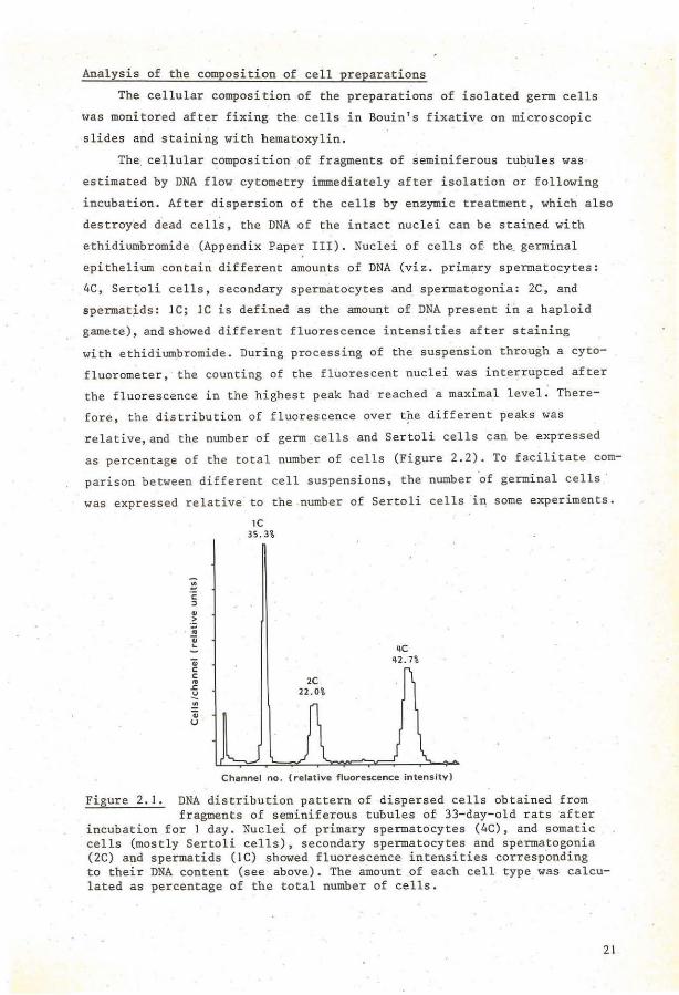
Analysis of the composition of cell preparations
The cellular composition of the preparations of isolated germ cells
was monitored after fixing the cells in Bouin's fixative on microscopie
. slides and staini~g with hematoxylin.
The cellular ~omposition .of fragmentsof seminiferous tubules was
estimated by DNA flow cytometry immediately after isolation or following
incubation. After dispersion of the cells by enzymic treatment~ which also
destroyed dead cells, the DNA of the intact nuclei can be stained with
ethidiumbromide (Appendix Paper III). Nuclei of cells of the. germinal
epithelium contain different amounts of DNA (viz. primary spermatocytes:
4C, Ser~oli cells, secondary spermatocytes and spermatogonia: 2C, and
spermat~ds: JC; JC is defined as the amount of DNA present in a haploid
gamete), andshowed different fluorescence intensities after staining
with ethidiumbromide. During processing of the suspension through a cyto
fluorometer, the counting of the fluorescent nuclei was interrupted after
the fluorescence in the highest peak had reached a maximal level. There
fore, the distribution of fluorescence over t?e different peaks was
relative,and the number of germ cells and Sertoli cells can be expressed
as percentage of the total number of cells (Figure 2.2). To facilitate com
parison between different cell suspensions, the number ·of germinal cells ·
was expressed relative· to the number of Sertoli cells in some experiments.
-~ c ::l .. ·~ .. "ii ... "ii c c ..
.s:: ~ "' "ii u
lC 35.3%
4C 42.7%
Channel no. ( relative fluorescence intensity)
Figure 2.1. DNA distribution pattern of dispersed cells obtained from fragments of seminiferous tubules of 33-day-old rats after
incubation for I day. Nuclei of primary spermatocytes (4C), and somatic cells (mostly Sertoli cells), secondary spermatocytes and spermatogonia (2C) and spermatids (IC) showed fluorescence intensities corresponding to their DNA content (see above). The amount of each cell type was calculated as percentage of the total number of cells.
21.


Chapter 3
PROTEIN SYNTHESIS IN GERM CELLS
Description of the mor~hology of germ cell development was foliowed
by investigation of several biochemical parameters of this complicated
series of events (reviews: Bellvé, 1979; Ericksonet al., 1981). With
autoradiography it was shown that protein synthesis occurs at all germ
cell stages (rat: Davis, 1969; mouse: Monesi, 1971; ram: Loir, 1972).,...,
Nuclear basic proteins are thought to play a role in regulation of
nuclear events and were studi~d extensively, which bas resulted in the
discovery of testis-specific histones in germ cells in addition to the
regular somatic histones. During spermatid elangation the histones are
replaced by testis-specific non-histone basic proteins, which are subse- ·
quently replaced by arginine- and cysteine-rich protamine. The role of
the testis-specific histones in germ cell development is not yet clear,
despite recent studies on histones concerning site of synthesis (Broek
et al., 1980; Meistrich et al., 1981), isolation, characterization and
quantification (Chiu & Irvin, 1980; Kumaroo & Irvin, 1980; Seyedin &
Kistler, 1980; Trostle-Weige et al., 1982). and organ specificity (Seyedin
& Kistler, 1979). The non-histone basic proteins are possibly involved in
stabilization and condensation of the nuclear material (review: Bellvé,
1979; Radman et al., 1979). Nuclear non-histone acidic proteins have
notbeen intensively investigated (Bellvé, 1979).
The abundance of some specific proteins in spermatozoa initiated
studies concerning their preserree and site of synthesis during spermato
genesis. These studies were performed mainly by correlating the appearance
of proteins in total testicular tissue with the appearance of particular
germ cell stages ar by histochemical detection. A special group of
proteins are the testis-specific isozymes, which emerge in germ cells
during spermategenesis (reviews: Goldberg, 1977; Bellvé, 1979; Blanco,
1980; Ericksonet al., 1981). The occurrence of specific,protein synthesis
in cells be~ore or after meiosis was aften used to investigate whether
postmeiotic gene expression ·occurred. However, definite proof for hapleid
23

gene exp.ression is still lacking (Erickson et al., . 1981).
Biochemica! analysis of germ cells has progressed through the in
creasing use of cell separation techniques. With respect to proteins,
recent studies, performed on different germ cell preparations enriched
with one stage of development, involved characterization of total cellular
proteins (Boitani et al., 1980;' Geremia et al., 1981; Kramer & Erickson,
1982), mitochondria! proteins (Hecht & Bradley, 1981), membrane prot-eins
(Millette & Moulding, 1981~,b), glycoproteins (Grooteg~ed et al., 1982c} or
specific proteins, such as specific membrane antigens (Tung & Pritz, 1978;
Millette & Bellvé, 1980; O'Rand & Romrell, 1981; Gaunt, 1982; Romreil et
al., 1982), a~enylate cyclase (Adamo et al . , 1980; Gordeladze et al.,
1981), pro te in kinase (Conti .'et al., 1979), DNA polymerase · (Grippo et al.,
1978; Hecht et al . , 1979), protein carboxymethylase (Gagnon et al., 1979),
phosphoglycerate kinase (Kramer & Erickson, 1981) and glycosyltransferases
{Letts et al., 197-8).
In our studies we were interested in a bï'ochemical "finger'print" of
different germ celi -stages as · a potential parameter for the investigation
of the action of Sertol i cells upon germ cells. Wi th two-dimensional elec
trophoresis several hundreds ·of proteins could be separated.
With this tnethod we investigated _protidn synthesis in ce11 preparations
highly enriched with pachytene spermatocytes and round spermatids and the
results, shown in Appendix Paper I, demonstrated c!'ear differences between
the two cell types . Some proteins were synthesized predominantly in pachy
tene spermatocytes, some ,predominantly in round spermatids and a large
group of proteins ·was synthesized at the same rate in spermatocytes and
spermatids. The synthesis of total cellular proteins in these germ cell
stages has also been analyzed by others (Boitani et al., 1980; Kramer &
Erickson, 1981). All studies demonstrated synthesis of stage-specific
proteins, 'although the number varied in the different ce.lls 1 possib1y as a
result of differences in exp,erimental conditions. Because we obtained with
two-dimensional electrophoresis a specific patte~n of proteins newly syn
thesized by pachytene spermatocytes, this technique was used to inves'tigate
the protein synthesis in spermatocytes under different conditions.
24

3 • 2 • ~~i!!!~!!~!!~~-~Lg~~!i!~Ei:::c~_EE~!~i!!_ê'l!!El:!"~i.~-i!!_i~~!!!!~~
E~.S:h2!.ê~~-~E~E!!!!!!~S:2!.~~
The presence of Sertoli cells appears obligatory for spermatogenesis.
It is largely unknown, however, which biochemica! activities of germ cells
are primarily dependent on activities of Sertoli cells. We have investiga
ted whether changes were induced in protein synthesis of pachytene sperm
ato~ytes during incubation of 24 hours in the absence of Sertoli cells, in
medium containing pyruvate (2 mM) and DL-lactate (6 mM). As shown in
Appendix Paper I, the results demonstrated that the pattern of proteins,
newly synthesized by isolated pachytene spermatocytes innnediately after
isolation, and analyzed with two-dimensional electrophoresis, was main
tained during 24 h. Moreover, the amount of amino acids incorporated into
acid-precipitable material was camparabie for spermatocytes incubated
from 0-2 h or from 24-26 h in the preserree of pyruvate and lactate. In
contrast, the pattern of proteins synthesized in Sertoli cells was shown
to change during one day of incubation.
Previously it was reported, that isolation of mouse pachytene sperm
atocytes and spermatids from seminiferous tubules did nat result in an
immediate change of the protein synthesis pattern (Boitani et al., 1980).
Moreover, it was shown that qualitative RNA synthesis by isolated sperm
atocytes and glycoprotein fucosylation by isolated spermatids were main
tained after respectively 12 h and 20 h of incubation in chemically defined
medium (Grootegoed et al., 1977a; Grootegoed et al., 1982a). These observa
tions demonstrate that pachytene spermatocytes and round spermatids incu
bated in a chemically defined medium, can maintain a specific pattern of
synthetic activities in the absence of Sertoli cells. Many activities of
Sertoli cells in vitro are stimulated by FSH and therefore it seems like'ly
that activities of Sertoli cells in vivo are decreased after hypophysectomy .
. In rats, no changes were observed in qualitative RNA synthesis of pachytene
spermatocytes at 64 h after hypophysectomy (Grootegoed et al., 1979).
Therefore, spermatocytes appear to maintain their qualitative synthetic
activities in vitro in the absence of Sertoli cells, and also in vivo in
the preserree of Sertoli cells with a reduced activity.
25


Chapter 4
CARBOHYDRATE METABOLISM OF GERM CELLS AND SERTOLI CELLS
In intact animals, the development, composition and normal function
of the testis are dependent on an adequate supply of several nutrients
(review: Leathem, 1975). It is difficult .to establish, however, whether
the effects of nutrients on the testis are exerted directly on Leydig
cells and/or germinal epithelium or occur via an intermediate action of
nutrients on the pituitary (Leathem, 1975; Setchell, 1978), This problem
can be partly circumvented by investigation of testicular tissue in vitro.
The first in vitro studies on energy metabolism of rat testis already
showed that bath lipids and carbohydrates were oxidized with the latter
predominating. These and following investigations on carbohydrate metab
olism of total testicular tissue were extensively reviewed by Free (1970).
Utiiization of glucose by testicular tissue was demonstrated in different
species, and it was shown that intact testicular tissue from adult rats is
extremely dependent on glucose supply as cornpared to 16 ether tissues.
From studies with testicular tissue containing different germ cell popula
tions, Free (1970) suggested that blood glucose is mainly used by germ
cells, while lipids provide the main souree of energy for non-germinal
testicular cells. The rele of amino acids and lipids as sourees of energy for
testicular cells was nat extens-ively investigated. In contrast, pathways of
carbohydrate metabolism in the testis have been amply investigated (review:
Free, 1970). However, these investigations have been performed with total
testicular tissue which contains cell types with various functions which
require different roetabolie pathways. Therefore it is difficult to ascribe
pathways to individual cell types.
Activities of individual cells can be investigated with autoradio
grapbic studies. It was shown with autoradiography that protein synthesis
in pachytene spermatocytes and round spermatids can be stimulated by
addition of glucose to testis slices in vitro (Davis, 1969). Basedon
these studies and the studies mentioned previously in this section, it was
suggested that glucose directly stimulated the synthetic activity of
pachytene spermatocytes and round spermatids. Preliminary investigations
in our laboratory, however, showed only a minor effect of glucose and a
27

major effect of its metabolites (pyruvate and l actate) on synthetic
aetivities (RNA - and protein synthesis) of isolated spermatocytes and
spermatids . Henee, some aspeets of earbohydrate metabolism in isolated
germ cells (seetion 4.1) and Sertoli cells (section 4.2) were investiga
ted. The possible dependenee of germ cells on carbohydrate metabolism of
Sertoli cells induced studies on the effect of hormones on the car bo
hydrate metabolism of Ser toli eel l s (section 4.2), and the possible inter
action between Sertoli cells and germ eells via intermediates of carbo
hydrate metabolism (seetion 4.3).
For studies on the effects of substrate supply on germ cells, measure
ments of energy- requiring acti vities Üke RNA - and protein synthesis
seemed an appr opriate endpoint. A scheme for the pathways of carboh~drate
me~abolism in germ cells, basedon data presenred in sections 4.1.1 and
4 . 1. 2, i~ presen red in Figure 4.1.
glucose
pyruvate
lactate
lactate
Figure . 4.1.
the preserree
28
SPERMATOCYTE
I acta te
,. NADH
pyruvate
[
protein &J ATP RNA
synthesis
electrbn transport chain
Pathways of carbohydrate metabol ism in pachytene spermatocytes incubated in the preserree of'glueose and pyruvate (J) or in of glucose and l actate (2). See section 4.1.

Medium,composed in our laboratory for the incubation of isolated germ
cells, which cantairred glucose, pyruvate and lactate, was successfully
used to investigate RNA - and protein synthesis in these cells (Grootegoed
et al., 1977a,1982c; Appendix Paper I). An indication that glucose was nat
the most important substrate to support isolated germ cells, was obtained
by the remaval of pyruvate and lactate from the medium. When observed by
phase-contrast microscopy, isolated spermatocytes showed signs of degenera
tion af ter incubation for 24 h in med,ium containing only glucose, a lso
when insulin (134 mU/ml) was added. When pyruvate and/or lactate were
present in the medium, the morphology of spermatocytes after 24 h was
still very similar to that of freshly isolated cells. Therefore, the
effects of pyruvate, lactate and glucose on the synthetic activities of
isolated germ cells were further investigated.
Addition of glucose to spermatocytes and round spermatids did only
have a small effect on uridine and leucine incorporation, and no effect on
oxygen consumption. Addition of lactate in the absence or preserree of
glucose, however, increased bath uridine and leucine incorporation at
least five-fold and oxygen consumption two-fold (Appendix Paper II). In
cell preparations enriched with pachytene spermatocytes or round sperm
atids, which were incubated in the preserree of glucose, the effect of lac
tate and pyruvate on protein synthesis was shown to be close-dependent
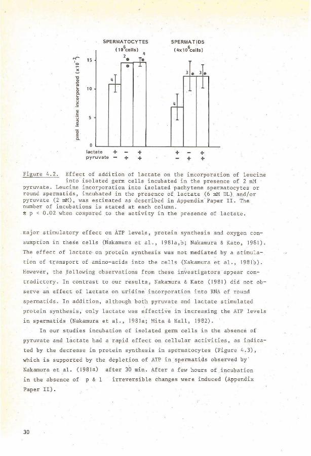
(Appendix Paper IV). Pyruvate stimulated the leucine incorporation into
spermatocytes and spermatids at lower concentrations than lactate. Maximal
leucine incorporation was reached at 0.2 mM pyruvate or 2-3 mM lactate
(Appendix Paper IV). Addition of lactate tagether with a high close of
pyruvate did nat further increase protein synthesis. above the level ob
tained with pyruvate alone, indicating that bath suhstrates stimulate
activities via the sarne routes (Figure 4.2). These observations strongly
suggest that pyruvate and lactate can supply more energy for rnainterrance
of synthetic activities in spermatocytes and spermatids than glucose.
Other investigators partly confirmed and extended our observations.
Glucose, fructose, galactose, mannose, ribose, glycerol and acetate had
only a small stimulatory effect on ATP levels and protein synthesis in
round spermatids (Nakamura & Hall, 1976,1977; Nakarnura et al., 1978·,
198la,b; Nakamura &Kata, 1981; Mita & Hall, 1982), whereas lactate had a
29
""' I 0
~ ""0 ~ "' ... 0 c. ... 0 u c QJ
c u :J .!!!
ö E c.
SPERMATOCYTES
( t06ce ll s)
2 15 •
•
10 ~ 5
0
lactate + pyr uvate +
4
~· .L
+ +
SPERMATl OS 6 .
( 4x 10 c e l Is )
4 ~ r
+
3 • 3.
+ + +
Figure 4.2. Eff~ct of addition of lactat e on the incorporation of leucine into isolated germ cells incubated in the presence of 2 mM ·
pyruvate. Leucine incorporation into ~solated pachytene sperrnatocytes or round sperrnatids, incubated in the presence of lactate (6 rnM DL). and/or pyruvate (2 rnM), was estirnated as des cribed in Appendix Paper II . The number of incubations is stated at each col umn. :1: p < 0 . 02 when cornpared to the' activity in the presen ce of lactate.
major sti rnulatory effect on ATP level.s, protein syn thes i s and oxygen con
suroption in these cells (N.akarnura et al., 198 1a,b; Nakarnura & Kata, 1981).
The effect of lactate· on protein synthesis was not rnediated by a stirnula
tion of ~ansport of arnino- acids into the cells (Nakamura et al., 1981b).
However, the ,following observations frprn these in\restigators appear con
tradi ctory. In contrast to our results, Nakarnura & Kato ( 1981) did nat ob
serve an ef fect of lactate on uridine incorporation into RNA of round
sperrnatids . In addition, although bath pyruvate and lactate stirnulated
protein synthesis, only lactate \vas effective in increasing the ATP level s
~n sperrnatids (Nakarnura et al . , 198Ja; Mita & Hall, 1982) .
In our studies incubation of isolated germ cells in the absence of
pyruvate and lac t ate had a rap i d effect on cel l ular activi ti es, as indica
ted by the decreasein protein synthesis in sperrnatocytes (Figure 4.3),
which is supported by the depletion of ATP in .spermatids observed by
Nakamura et al. (198Ja) after 30 min. Aftera few hours of incubation
in the absence of p & 1
Paper II).
30
irreversible changes were induced (Appendix

facta te Jenghth of prein~ cubation
+ 30mifl
+ 70min
Figure 4. 3. Rapid effect of p & 1 dep.letion on- the leucine incorporation into pachytene spermatocytes.
Cell fractions enriched with pachytene sperm8.tOcytes were incubated in incubat.ion medium without additions or in the preserree of 6 mM DL-lactate. After 30 ar 70 min 0.6 vCi (14c)leucine was added, foliowed by an incubation _of 2 h. Subsequently, cold 0.9% NaCl _(w/v), co:Otaining 3 mM leucine, was added and the cells were wasbed thrice in this salution by eentrifugatien for 7 min at 200 g at 4oc. Next, the cells were collected- by eentrifugatien at 600 g and lysed in 0'.5% SDS during 10 min in ice. Macromolecules were precipitated in cold 10% trichloroacetic acid, foliowed by three washings in 10% TCA by centrifugation for 5 min at 700 gat 4°C. Finally, the p~llets wère wasbed with 96% ethanol, centrifuged for 10 min at 1500 g at 4oc, dried and solubilized in 1 N NaOH .. Part of this salution was counted for radioactivity and part was used for protein estimation (Lowry è.t al., 1951)". Mean values and range of duplicate incubations are given. The results indicate that after 30 min of incubation in the absence of p & 1 but in the presence of glucose, no substrate remained in the cell which could maintain protein synthesis during the subsequent period.
Combined morphological and biochemica! studies of iSolated germ cell
mitochondria indicate that the -typical condensed mitochondria, present in
late pachytene spermatocytes and round spermatids, hàve a high oxida
tive capacity (Setchell, 1'978; De Martino et al., 1979.). Moreover, in
early round spermatids, mitochondria are localized close to the cell sur
face (Fawcett, 1974}. These observations suggest that pachytene spermato
cytes and round spermatids are equipped to oXidize exogenous p & 1 at a
high rate.
In conclusion, the preserree of p & 1 appears to be essential for
the rnainterrance of synthetic activities in isolated germ cells. The com-
31.

bined biochemica! and morphological observations indicated that decreased
synthetic activities are correlated with a decrease in survival time of
the germ cells.
4. 1.2. ~~~=~~~Egy_g~~~E~!i~g_Eë!~~~Y~-~f-"~E~~~Y~Eë!~-~~!ë~~li~~-i~-g~~ cells
Glucose was shown to be inadequate in maintaining synthetic activities
in isolated spermatocytes and spermatids in short-term experiments. However,
the preserree of glucose might be required in long-term incubations, as a
substrate for the hexose monophosphate shunt, for the supply of inter
mediates, such as ribose, for RNA synthesis, and to keep a proper NADPH/
NADP+ balance, or glucose may be used for glycoprotein or glycolipid syn
thesis. It bas been shown that RNA synthesis (review: Bellvê, 1979) and
fucosylation and glycosylation of proteins take place in late pachytene
spermatocytes and spermatids (Grootegoed et al., 1982c; Letts et al.,
1978).
After incubation of isolated germ cells in the preserree of pyruvate,
lactate was found in the incubation medium, and the amount of lactate in
the medium was increased even in the preserree of added lactate and irres-.
pective of the preserree of glucose (Table 4. 1). At least 50% of the con
sumed pyruvate was converted to lactate. After incubation of germ cells in
the preserree of lactate, na pyruvate was found in the incubation medium in
our experiments (Table 4.1), which is in contrast to the results of Mita &
Hall (.1982), who observed secretion of pyruvate by round spermatids.
The conversion of pyruvate to lactate reduces the amount of NADH
available for ATP generation in the electron transport chain, firstly by
diverting pyruvate from oxidation in the Krebs cycle, which would generate
~ADH, and secondly because NADH is oxidized concomitant with pyruvate
reduction. Therefore, the conversion of pyruvate to lactate appears to be
a waste for the cell in terros of energy supply, but it could play a role
to maintain a proper ratio between NAD+ and NADH in the cells. In spite of
the apparent waste of energy-yielding substrates when pyruvate is added,
germ cells acquire maximal synthetic activity at a pyruvate concentration
which is 10-fold lower than the lactate concentratien required (Appendix
Paper IV). Therefore, the effect of pyruvate on isolated germ cells might be
32

w w
Table4.1. p & 1 consumption and secretion by isolated gerrn cells.
6 wmol p & 1/10 cells/24 h
incubation conditions pyruvate consurned lactate secreted lactate consurned pyruvate secreted
glucose + pyruvate 3.52 + 0.42 (3) 2.08 + 0.40 (3)
pyruvate 3.80 + 0.26 (2) 2.40 + 0.04 (2)
glucose + pyruvate + lactate 3.24 + 0.26 (3) 1.90 + 0.32 (3)
glucose + lactate 0.70 + 0.28 (4) 0 (2)
Pachytene spermatocytes (0. 5 x 10⁶ cells) were incubat.ed in 2 rnl incubation medium in the preserree ·of pyruvate
(2 mM), lactate (6 mM DL) and/or glucose (3.3 mM) or combinations of these subStrates.After24hthe amounts of
pyruvate and L-lactate in the media were estimated. Results are presented as means + S.D. The numbers between
brackets indicate the nurnber of' cell preparations used.
The results indicate that the consurnption of pyruvate and the amount of pyruvate converted to and secreted as
lactatewere similar in the preserree ar absence of .added glucose or lactate. No secretion of pyruvate was
observed after incubation of sperrnatocytes in the preserree of lactate.

two-fold, viz. firstly the effect of pyruvate as a souree of energy, and
secondly an effect on the redox balance in the cells by pyruvate reduction.
No depots of energy-rich substrates, like glycogen or lipids, have
been reported for pachytene spermatocytes or round spermatids. The apparent
absence of energy stores is confirmed by the rapid effect of triose deple
tion on the synthetic activities of these germ cells (section 4. I .·1;
Appendix Paper II).
In vivo, exogenous substrates for germ cells could be supplied via
the fluid which surrounds the germ cells, but the chemical cornposition of
this fluid is largely unknown . More is known about the fluid in the rete
testis, where the ends of the tubules meet. It must be kept in mind that
the composition of the latter fluid may differ from the composition of
the fluid which is closer to the germ cells, as has already been shown for
the ionic composition (Setchell, 1978). Moreover, the meaning of high con
centrations· of substrates in re te testis fluid is by no_ means unequivocal;
it may reflect e ither that these substrates have to be available to gerrn
cells or that they are not used by the germ cells and are being removed as
waste .
In many species the concentratien of myoinositol in the rete testis
fluid is very high in co ntrast to the concentratien of glucose and fruc
tose, which are hardly detectable (Setchell, . 1978). Myoinositol has been
shown to be an important nutrient for marnmalian cells in culture (Eagle et
al., 1957). However, myoinositol did not increase the ATP level in round
spermatid·s (Mita & Hall, 1982). Rete testis fluid from rats contains high
amounts of the amino- acids: aspartic acid, ala~ine, glycine, proline and
lysine, as compared to plasma . In some other species glutamic acid is in
creased insteadof proline and lysine (Setchell, 1978). These amino- acids
might be used by the germ cells as ~n energy souree or as substrates for
purine and pyrimidine synthesis. Hmvever, the amino- acids present in the
incubation medium were not sufficient to maintain synthetic activities in
isolated germ cells (section 4.1. 1) .
Little is known· about lipids as substrates ~or germ cells. The
specific acti~ity of carnitine acyltransferase, an enzyme involved in the
translocation of f.atty acid .across the mitochondria! membrane, was low in

spermatogonia and high in pachytene and diplotene spermatocytes (Vernon
et al., 1971). However-, this e·nzyme is requiied in fatty acid synthesis as
well as fatty acid oxidation and it was shown recently that purified mouse
pachytene spermatocytes and round spermatids synthesize cholesterol and do
lichol (Potter et al., 1981). From experiments with testicular cell sus
pensions from rats of different ages, it was concluded that spermatogonia
were active in palmitate oxidation, but the activity of spermatocytes and
spermatids appeared to be less (Lin & Fritz, 1972). In conclusion, up to
now there are no indications that other substrates than p & 1 are
important energy sourees for pachytene. spermatocytes and round spermatids.
It is striking that several isozyme-s of enzymes which catalyze path
ways involved in carbohydrate metabolism, are locatedingerm cells and nat
in any other cell type. Same of these enzymes are involved ïn lactate and/
ar pyruvate oxidation, viz. lactate dehydrogenase-c 4 (LDH-X) and cyto
chrome- ct. These testis-specific isozymes emerge in mid-late pachyten·e
spermatocy"tes and remain present in more mature germ cells (Hennig, 1975;
Goldberg et al., 1977; Hintz & Goldberg, 1977; Meistrich et al., 1977;
Wheat et al., 1977). LDH-c 4 was shown to catalyze preferentially lactate
oxidation (review: Goldberg, 1977). It has been suggested that LDH-c4
is
involved in a shuttle for transport of reducing equivalents across the
mi toehondrial membrane (review: Blanco, 1980) .. Nothing is known about a
specific kinetic property of cytochrome ct' although the amino-acid
sequence has been elucidated (Hennig, 1975), In addition, several testis
specific glycolytic isozymes, hexokinase (reviews: Gornes & VandeMark,
1974; Goldberg, 1977), phosphoglycerate kinase (PGK-2) (Van de Berg et
al., 1973,1976,1981; Kramer, 1981), enolase (Edwards et al., 1981) and
glucose phosphate isomerase (Buehr & McLaren, 1981) are known, which are
more abundant in spermatozoa than indeveloping ger~ cells.
Another germ cell-specific property is the inactivation of the sex
chromosomesthroughout the meiotic prophase, while the autosarnes are
transcriptionally active (Monesi, 1965). Two enzymes involved in glucose
metabolism, viz. glucose 6-phosphate dehydrogenase, which catalyzes the
first reaction of the hexose rnonophosphate shunt, and an isozyme of the
glycolytic enzyme phosphoglycerate kinase (PGK-1), are coded for by the
35

X- chromosome. This may be t he cause of the absence or low activity of these
enzymes in e longating _spermatids and spermatozoa (Van de Berg et al., 1973 ,
1976,1981; Erickson; 1976; Broek , 1977; Kramer, 1981). It must be kept in
mind, however, . that germ cells also may contain non-testis- specific iso
zymic forms or autosomally linked forms of the enzymes mentioned in this
section. Hence, the understanding of the importance of the testis-specific
isozymes awaits èlucidation of their specific kinetic properties .
Increase of the temperat ure_of the testis from 32° to 37°C is known to
induce degeneration of pachytene spermatocytes and more mature germ cells.
This may be caused by disturbances in carbohydrate metabolism, because addition . .
of glucose during the incubation of testicular tissue prevented the decrease
~n protein synthesis, ,.;hich was observed at temperatures a"Qove 32°C (Davis, .
1969).
Isolated, partly purified pachytene spermatocytes and spermatids ~n
cuba ted at 32° or 37°C showed increased membrane damage at the higher tem
perature (Lee, 1974). In isolated round spermatids increase in temp·erature
caused a decrease of protein synthesis (possibly by an effect on the initia
tien of protein synthesis or by a change· in availability· of messenger-RNA)
and a decrease of transport of hexoses and amino-acids. The effect of tern
perature on this transport was reversible, "'hiçh was interpreted as an in
dication that .cell death was not causing the phenomena ob~erved (Nakamura
et al., 1978; Nakamura & Hall, 1978; Hall et al., 1981 ; Hall & Nakamura,
1981) .. None of these effec"ts we re found · on spermatocytes, al though i t "as
shown that these cells are also affected by cryptorchidism in mature animals
(Davis, 1969; Setchell, 1978; Rornme rts et al., 1980) .
These effects of temperature increase on isolated germ cells were ob
served during incubations in the absence of pyruvate and lactate and it
remains to be demonstrared "'hether sirnilar changes are induced in the
presence of proper energy y i elding substrates.
36

We have investigated whether Sertoli cells could supply the p & 1
required by pachytene spermatocytes and round spermatids. A scheme for the
pathways of carbohydrate metabolism in Sertol i cells, based on data pre
sented in this section, is shown in Figure 4.4.
glucose
pyruvate
lactate •• llllactate
SERTOLI CELL
02 electron transport chain
Figure 4.4. Pathways of carbohydrate metabolism in Sertoli cells incubated for 24 h in the preserree of glucose. See:Section 4.2.1.
Bath pyruvate and 1actate were secreted by Sertoli cells from 4-week-old
rats (Appendix Papers III, IV). Approximately .4 times more lactate than
pyruvate accumula·ted in the medium. The amoun-t of lactate, present in the
medium of Sertoli cells from prenatally irrad.iated rats ar from intact
rats, increased with increasing exogenous concéntrations of glucose (Ap
pendixPaper III), indicating that exogenous glucose is converted to lactate.
37

Sertoli cells f r om 20 day old intact rats were reported to convert
maximally .2.9% of the glucose utilized t o co 2 , while 95.8% was converted
to anionic compounds,mainl y to l actate,during incubation for 60 min
(Robinson & Fritz, 1981). In our studies, during incubation of Ser to li
cells f rom 4-week-old r ats for 24 h approximately 50% of the glucose
utilized accumulated as l actate (Appendix Paper I II). These observations
may indicate that, when the p & 1 concentration in the medium incr eases
during prolonged incubation, Sertoli cells utilize glucose in other path
ways not involved in p & l s ecretion .
It was shown previous ly that Sertoli cel ls in vitro can convert g l u
cose to rnyoinositol (Robinson & Fritz, 1979) which i s present in high con
centrat i ons in rete testi s fluid (Setchell, 1978). However, the quantita
tive importance of this pathway in the total rate of glucose utilization
by Serto! i cells seerns smal! (Robinson & Fritz , 1981). Another minor glu
cose- consurning pathway in Sertoli cel l s might be the conversion of glucose
to sorbitol catalyzed by aldose reductase, which was recentTy shown to be
presen t in Sertoli cel l s and not in gerrn cells (Ludvigson et al., 1982) .
Together with sorbitol dehydr ogena'se, aldose r eductase constitut es the
polyol pat hway, which has a net producti on of NADP and NADH, and of fruc
tose when glucose is the substrate (Metzier, 1977; Ludvigson & Sorenson,
1980). Intravenous infusion of radioactively labelled glucose in rams
results in t he appearance qf radioactive ly labelled .amino- acids in rete
· testis f luid (Setchell, 1975). I t is not known whe t her this conversion
takes pl ace in Sertoli cells.
In conclusion, Sertoli cells f r om intact as \vell as f r om prenat al l y
irradiated rats appear to have a large capacity to convert g l ucose to
· p & 1 , which are secre ted by the cells, and li ttle p & '1 appear to be
used as energy souree by Sertoli cel l s. However, in vivo a high extra
cel lular p & 1 concentration or a change in t he rati o between NAD and
NADH may influence the rate of secretion of pyruvate and l actate by
Sertoli cells,and other pathways of glucose metabolism may be come more
important .
38

It was suggested previously, basedon investigations with total
testicular tissue, that rion-germinal cells in the testis are dependent on
lipid oxidation for energy supply (Free, 1970). Lipid diaplets are aften
observed in the cytoplasm of Sertoli cells. In vitro as well as in vivo
the amOunt of lipid stored in Sertoli cells increa_ses parallel to germ
cell degeneration (Faw·cett, 1975), suggesting that germ cells which have
been phagocytized are an energy souree for Sertoli cells. However, increase
in lipid material was also shown to occur in Sertoli cells in prenatally
irradiated (germ cell-depleied) testes- as a result of cryptorchidism
(Bergh, 1981). When isolated Sertoli cells were incubated for one day in
the absence of glucose and p & 1 foliowed by incubation in the presence
of glucose, the cells retained the property to migrate on the bottorn of
the Petri dishes and retained their p & l-secr€.ting capacity. Sertoli
cells may have used lipids as energy substrates to survive one day of glu
cose depletion.
A possible role for amino ·acids as additional energy substrates for
Sertoli cells is suggested by a high rate of branched-chain amino acid
oxidation by Sertoli cells in vitro (data not- shown) (Grootegoed et al.).
In vivo Sertoli cells may have access to all substrates present in
the interstitial lymph fluid and the amount of substrates available to
Se-rtoli cells can be regul.ited by tbe testicular blood flow .9.nd the per
meability of the testicular capillaiies. In conclusion, non-~arbohydrate
substrates may be a major souree of energy for Sertoli cells, but to
support this conclusion, fur·ther investigations are required.
FSH
It is well established now that follicle-stimulating hormone (FSH)
can stimulate many Sertoli cell activities in vi·tro and in vivo (recent
reviews: 1'1eans et al., 1976,1978,1980; Fritz, 1978; Davies, 1981; Purvis
& Hansso~, 1981). This FSH stimulation is age-dependent and stimulation of
intracellular events decreases in rats above 20 days of age (Fritz, 1978;
Steinberger et al., 1978; Means et al., 1980), whereas stimulation of oes
tradiol secretion decreases above 5 days of age (Dorrington et al.,
39
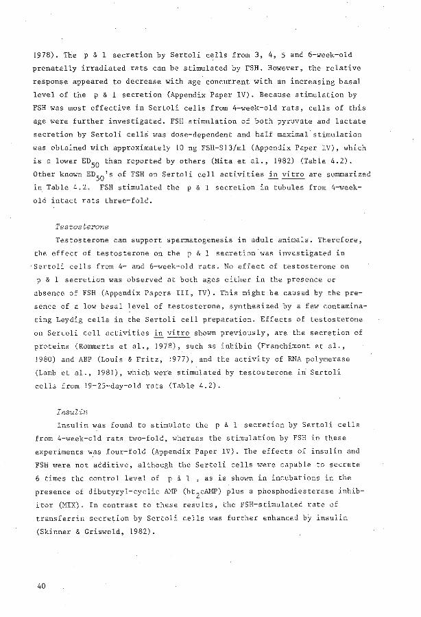
1978). The p & 1 secretion by Sertol i cells from 3, 4, 5 and 6-week-old
prenatally irradiated rats can be stimulated by FSH. However, the relative
response appeared to decrease with age concurrent with an increasing basal
level of the p & 1 secretion (Appendix Paper IV). Because stimulation by
FSH was most effective in Sertoli cells from 4-week-old rats, cells of this
age were further investigated. FSH stimulation of bath pyruvate and lactate
secretion by Sertoli cellS was close-dependent and half maximal stimulation
was obtained with approximately 10 ng FSH-813/ml (Appendix Paper IV), which
is a lower ED50
than reported by atbers (Mita et al., 1982) (Table 4.2).
Other known ED50
's of FSH on Sertoli cell activities in vitro are summarized
in Table 4.20 FSH stimulated the p & 1 secretion in tubules from 4-week
old intact rats three-fold.
Testosterone
Testasterene can support spermategenesis in adult animals. Therefore,
the effect of testasterene on the p & l secretion was investigated in
Sertoli cells from 4- and 6-week-old rats. No effect of testasterene on
p & 1 secretion was observed at both ages either in the preserree or
absence of FSH (Appendix Papers III, IV). This might be caused by the pre
senee of a low basal level of testosterone, synthesized by a few contamina
ting Leydig cells in the Sertoli cell preparation. Effects of testasterene
on Sertoli cell activities in vitro shown previously, are the secretion of
proteins (Rommerts et al., 1978), such as inhibin (Franchimont et al.,
1980) and ABP (Louis & Fritz, 1977), and the activity of RNA polymerase
(Lamb et al., 1981), which wer·e stimulated by testosterone in Sertoli
cells from 19-25-day-old rats (Table 4.2).
Insulin
Insulin was found to stimulate the p & 1 secretion by Sertoli cells
from 4-week-old rats two-fold, whereas the stimulation by FSH in these
experiments was four-fold (Appendix Paper IV). The effects of insulin and
FSH were nat additive, although the Sertoli cells were capable to secrete
6 times the control level of p & 1 , as is shovm in incubations in che
preserree of dibutyryl-cyclic AMP (bt2
cAMP) plus a phosphodiesterase inhib
itor (MIX). In contrast tothese results. the FSH-stimulated rate of
transferrin secretion by Sertoli cells was further enhanced bj insulin
(Skinner & Griswold, 1982).
40

...
Table 4:2. Comparison of the doses of FSH or testosterone. necessary for stimulation of different Sertoli
cell activities in vitro.
Sertoli cell activity
FSH
ABP secretion
Protein kinase illhibitor
Pyruvate/lactate secretión
Sulfoprotein secretion
Plasminogen activator sectetion
DNA synthesis
Testasterene aromatization
Phosphodiesterase
Lactate secretion
Intracellular cAMP
Intracéllular cAMP
Phosphodiesterase
Testosterone
ABP se ere ti on
Inhibin secretion
RNA po~ymerase
E05ö (ng/ml)
age (days)
3.5 20
100 16 sterile
150 28 sterile
162 (MIX) 20
180/460 20
219 20
288 18-20
300 19
635 16
815 20
1150-3450 18-24-33
200 15 rat FSH-Bl
1.2
144
6056
20
19
25
incubation period (hours)
72-144"
0- 72 ...
24- 48"
48-120
24-?
??.- 96
24- 48
168-192
48- 54
48- 49
96- 96.5
120-144
72-168"
144-168
120-120.25
Reference
Louis & Fritz, 1979
Tashet al., 1980
Appendix Paper IV
Elkington & Fritz, 1980
Lacroix & Fritz, 1982
Fritz et al., 1978
Dorrington et al., 1978
Verhoeven et al., 1981
Mita et al., 1982
Fritz et al., 1978
Steinberger et al., 1978
Conti et al., 1981
Louis & Fritz, 1979
Franchimont et al., 1980
Lambet al., 1981
The different ovine FSH preparations used were converted to FSH-Sl Units. I ng Sairam FSH-81554 = 45 ng F8H-811 (Louis & Fritz, 1979); F8H-811 • I. 15 F8H-81; F8H-812 • I .25 F8H-81; F8H 813 • 15 F8H-81 . X Hormone present from the time of plating.

Recently, thyroid- stimulating hormone (TSH) was shown to stimulate the
activity of Sertoli cells from -1 6- day- old rats, as expressed in an
increased secretion of ABP and plasminogen activator (Hutsen & Stocco,
1981).
In summary, Sertoli cells in vitro can be stimulated by FSH, testoste
rone, insu1in and TSH. The p & 1 secretion by Serto1i ce11s was increased
by FSH and insu1in, but not by testosterone.
4.2.4. Effect of incubation conditions on Serto1i cel1s ------------------------------------------------
We have observed that the p & 1 secretion by Sertoli cells from 4- and
S-week-o1d rats was higher during the first 24 hof . incubatie~ than during
the fo11owing 24 h (24-48 h) (Figure 4.5). The FSH- stimu1ated p & 1
secretion by these ce11s decr eased a1so during incubation, but at a 1ower
rate. Possib1y the effect of FSH stimu1ation i n vivo is temporari1y retained I
in vitro. Hence, the ratio stimu1ated/unstimu1ated ~ & 1 secretion
.!: QJ
0 ... a. Ol E -QJ ... "' ... u 2 0 E :J..
10
5
0
LACTATE
~· \
\ \
\ \
\
'+
PYRUVATE
~\ \ \
\
' \ \
\ \ \ \ •
0- 24h
incubation period
24-48h 0- 24h 24- 48h
.r. ~ N 3c ëjj
0 ... a. Ol
2 ~ !
0
"' > :J ... >a. 0 E ::t
Figure 4 . 5 . Decrease of the amount of p & 1 secreted by Sertoli cells during incubation.
Sertoli cells from prenatally irradiated rats were isolated at 4 weeks of age. The cel1s were isolated and .incubated as described in Material s and Methods in the absence (t---1) or presence of NIH- FSH- Sl3 (0.5 ~g/ml) and testesterene (200 ng/ml) <•---•) . The media of the first and the second · day of incubation were collected separately, thè ·amounts of pyruvate and lactate were estimated and expressed per mg pro t ein present at the end of the incubati on. Mean ·and range of duplicate i ncubations are given.
42

increased during incubation. Other activities óf Sertoli cells have been
shown to change during incubation, viz. ABP - and transferrin secreti~n by
Sertoli cells from 20 day old rats decreased (Louis & Fritz, 1977; Rommerts
et al., 1978; Gianetto & Griswold, 1979; Karl & Griswold, 1980; Skinner &
Griswold, 198.2), basal cyclicAMP levels in Sertoli cells from 33 day old
rats and FSH-stimulated oestradiol secretion by Sertoli cells from 20 day
old rats increased (Rommerts et al., 1978; Steinberger et al., !978) and
protein synthesis changed (Chapter 3, Appendix Paper I). Therefore, care
must be taken in extrapolating in vitro results directly to the in vivo
situation.
In the previous sections some aspects of carbohydrate metabolism b.y
isolated germ cells and isolated Sertoli cells have been reported. In this
sectien it will be considered whether and to which extent the activities
of germ cells are influenced by Sertoli cells.
4.3. I. ~~~~"E-~~-g!~"~~~-~~E!~!b~g-~g_!~~-~"!b~!!z_~g~-·~E~b~~!-~f_g~E~
E~l!~
Isolated germ cells were shown to require p & 1 as substrates for
rnainterrance of synthetic activities (secfion 4.1.1). We observed also, that
glucose supply to Sertoli cells is correlated with the p & 1 secretion by
Sertoli cells (sections 4.2. 1). Therefore, we have investigated the effect
of glucose on the activity and survival of germ cells incubated in the
preserree of Sertoli cells.
Fragments of seminiferous tubules from 4!-week-old rats, containing
germ cells up to and including round spermatids, were incubated without
glucose for 24 h followed by incubation in the preserree of glucose. After
spreading of the tubule fragments, pachytene spermatocytes and round
spermatids were virtually absent from the cultures, whereas in control
tubules incubated in the continuous preserree of glucose pachytene sperm
atocytes and round spermatids were still present. Many early spermatocytes
and spermatogonia appeared to remain present under bath conditions. The
latter observation would agree with autoradiograpbic studies of slices of
testis tissue incubated in the absence ar in the preserree of glucose. The
43

early spermatocytes and spermatogonia A in these slices retained a high
level of leucine incorporation in the absence of glucqse (Davis, !969) and
therefore, these cells appear less dependent on supply of glucose or its
metabolites than the more mature germ cells. The degenera~ion of the
maj ori ty of the germ cells in fragments of seminiferous t 'ubules · incubated
in the absence of glucose was confirmed by quantitative cytofluorometric
measurement of the cell types present in the incubations (Appendix Paper
III) • .
For measurement of the synthetic Activity of germ cells in the
presence of Sertoli cells, a homogeneaus population of germ cells was co
incubated with a monolayer of Sertoli cells. The incorporation of uridine
into pachytene spermatocytes co- incubated with Sertoli cells in increasing
glucose concentrations increased concomitant with the lactate secretion by
Sertoli cells (Appendix Paper III). Spermatocytes, inoubated · under
comparable conditions in the absence of Sertoli cells but in the presence
of lactate and glucose, shm•ed the same uridine incorporations as sperm
atocytes co-incubated with Sertoli cells in the presence of glucose.
Because it was demonstrated that glucose has only a small effect on sperm
atocytes (section 4.1.1), the results suggest that glucose influenced germ
cells via p & 1 secretion from Sertoli cells. Only lactate was measured,
but pyruvate was probably also involved .
In conclusion, survival of pachytene spermatocytes and round sperm
atids, a nd RNA synthesis in pachytene spermatocytes, could be regulated by ·
glucose in the presence of Sertoli cells. This regulation was accompanied
by changes in the p & 1 secretion by Sertoli cells, sugges ting that germ
cell activity can be regulated by p & 1 secretion from Sertoli cells.
Possibly, glucose depletion in testicular ti.ssue is also involved in
the effects of cryptorchidism on germ cell activities. The blood flow in
the cryptorchid testis is either unchanged or slightly increased as com
pa red to the scrotal. testis (Setchell, 1978). Therefore, the amount of
glucos e available to the Sertóli cells in .the cryptarcbid testis would not
be increased and the amourit of p & 1 secreted would remain rather
~onstant during temperature e levation . Hence, an increased demand .for
energy- rich substrates by germ cells caused by the higher temperature
may not be compensated by an increased supply and this may result in degen
eration of pachytene spermatocytes and round spermatids (see also ' section
4.1.5).
44

tt cannot be excluded from the experiments described in the previous
section, that the stimulation of germ cell activities was mediated by
membrane cantacts between the germ cells and the Sertoli cells. Therefore,
germ cells were incubated in medium which was conditioned by Sertoli cells
-(Appendix Paper IV). Isolated pachytene- spermatocytes and round spermatids
incubated in the conditioned medium incorporated significantly higher
amounts of leucine than cells incubated in fresh medium. The total leucine
incorporation into cells incubated in the conditioned medium was similar
to the incorporation into cells, which ha~ been incubated in fresh medium
containing the same concentratien of pyruvate and lactate as the conditioned
medium. These observations confirm that secretion products from Sertoli
cells can stimulate activities of germ cells and ,p & 1 seem to be the
major secretion products from·hormone-stimulated Sertoli cells which
affect the synthetic activity of germ cells.
With cytofluorometric measurements we have found, that more primary
spermatocytes appeared to survive in fragm~nts of seminiferous tubules
incubated in the absence of horrnanes than in tubules of hypophysectomized
rats (Figure 4.6). The relative number of spermatocytes in these incuba
tions of tubule fragments was virtually equal to the number in tubules of
intact rats (Figure 4.6). Therefore, incubation of tubule fragments under
these conditions appears close to optimal for the survival of spermato
cytes fora few days, This may explain why na effect of FSH and/or testa
sterene was observed on the synthetic activities of spermatocytes co
incubated with Sertoli cells, ar on survival of spermatocytes in incuba
tions of fragments of seminiferous tubules (results Uot shown). It may be
possible that some nutrients (glucose, amino acids etc.) are available at
a higher concentratien in vitro than in vivo. Under these conditions un
stimulated Sertoli cells may produce sufficient p & l for germ cells to
survive. To demonstrate an effect of hormones on germ cells in vitro,
imitation of the effect of hypophysectomy may require a change in incuba
tion conditions of seminiferous tubules, so that germ cells degenerate in
vitro in the absence of hormones.
45

A
B
c
" " .~ .. ~
!) 'ë " " .~ -.. 'ii ...
2C 36.1%
qc 62.7%
Channel no. (relative fluorescence i,ntensity)
2C '38.6%
Chan~el no . (re lative fluorescence intensity)
Figure 4.6. Effect of incubation on the composition of the cell population 'in seminiferous tubules isolated from 25-day- old rats and com
parison with the effect of hypophysectomy . Cell preparations were obtained from semin iferous t ubules of 25-day- old intact rats immediately after isolation (A) or following i ncubation of the tubules for 4 days in the absence of hormones (B), of 29-day-o l d rats hypophysectomized at 25 days of age (C) and of 29- day-old intact rats (D). The number of cells and their DNA content were measured by DNA flow cytometry. The quantity of cells containing IC, 2C or 4C DNA (see Fi g. ·2.2) was calculated as percentage of the total number of cells. For comparison, the number of germ cells i s expressed relative to the number of Sertoli cells . · The re l ative number of primary spermatocytes remained vi rtua l ly constant in intact rats of 25 and 29 days of age (4C/2C = 1.7) and in tubule frag-
46

c
D
.. c c • ~ ~ . .. u
. c 0
• . ~ ;; ~ .. c c • ~ ~ . .. u
lC 2. 7%
2C 50.8%
•c 46.5%
Channel no. (relative ffuorescence intensity)
lC 7. 5%
2C 34.0%
I
•c 58. 6%
\ Channel no. (relative fluorescence intensity)
ments incubated for 4 days (4C/2C = 1 .6). In the hypophysectornized rats the relative number of spermatocytes was decreased after 4 days (4C/2C = 0.9). Some spermatids had developed in intact rats of 29 days old (IC/2C = 0.2 compared to < 0. I in intact 25 day old rats). No spermatid development was observed in the hypophysectomized rats (IC/2C < 0. 1). These results indicate that germ cell development stops and degeneration of pachytene spermatocytes occurs after hypophysectorny of immature rats. In contrast, na pronounced reduction of pachytene spermatocytes occurred in fragments of seminiferous tubules ·incubated in the absence of horrnanes. Hence, the present incubation conditions appear close to optimal lvith respect to survival of spermatocytes for a few days, and more favourable for surviv"al of these germ cells than the conditions in vivo in seminiferous tubules of hypophysectomized rats.
47


Chapter 5
GENERAL DISCUSSION
The studies on carbohydrate metabolism of germ cells described in
this thesis concentrated on pachytene spermatocytes and round spermatids.
In this sectien some information about the effects of trioses and hexoses
on ether stages of germ cell development will be conside.red, starting with
the earliest stage (Figure 5.1).
It bas been shown that primordial germ cells, taken from the gonadal
ridges of 15 day-old mouse fetuses, are 10 times more active in pyruvate
oxidation than in glucose oxidation (Brinster & Harstad, 1977). This
pattern of substrate preferenee appears to be similar to that of pachytene
spermatocytes and round spermatids. However, spermatogonia and early
spermatocytes, stages interposed between the ·primordial germ cell and the
pachytene spermatocyte stage, appear less dependent on carbohydrate supply
than the more mature germ cells, because spermatogonia and early spermato
cytes survived in seminiferous tubules incubated in the absence of glucose,
whereas pachytene spermatocytes and round spermatids degenerated (section
4. 3--.1). Spermatogonia may oxidize palmitate and acetoacetate at a higher
rate than pachytene spermatocytes and spermatids, whereas in these
experiments spermatogonia appeared to be less active in pyruvate oxidation
than the more mature germ cells and thus lipids may be the substrates pre
terred by spermatogonia (Lin & _Fritz, 1972).
Preliminary results indicate that elongated spermatids, in contrast
to the preceding stage of round spermatids, maintain their ATP level with
glucose as well as· with lactate as substrates (Le Gac et al., 1982). Elon
gated spermatids develop into testicular spermatozoa, which, during their
passage through the epididymis, acquire the capacity to be motile and to
fertilize. Although development from testicular to ejaculated spermatozoa
is accompanied by roetabolie changes (Voglmayr, 1975), little is known
about the effects of carbohydrates on energy-consuming prócesses in this
stage. In rats, spermatozoa from the cauda epididymis maintained their
motility and energy charge for 60 min in the preserree of pyruvate and lac-
49

primordial germ cells
spermatogonia, early spermatocytes
mid-late pachytene spermatocytes
round "d / spermat1 s
"-elongated
~ testicular
spermatozoa
~from cauda epididymis
PL ({fjj)
GPL
PL ~
PL (Q) SERTOLI CELL
GPL~ ?
PL
Figure 5.1. Carbohydrates as energy-yielding substrates in different stages of germ cell development.
Preferred substrates: PL - pyruvate and/or lactate G - glucose GPL - glucose and/or pyruvate and/or lactate
50

tate, but not in the preserree of glucose (Ford & Harrison, 1981). However,
it bas been reported that in several species, including man, the motility
of ejaculated spermatozoa can be enhanced by hexoses (Mann & Lutwak-Mann,
1981). Many ether substrates, e.g. lactate, pyruvate and amine
acids, also can stimulate motility, but marked species differences exist
(Mann & Lutwak-Mann, 1981). Similar to rat spermatocytes and spermatids
(section 4.1.2), spermatozoa fram baar and bull metabolize 50% ar more of
the pyruvate taken up to lactate concomitant with pyruvate oxidation (Mann
& Lutwak-Mann, 1981). It is apparent from this information that some dif
ferences in carbohydrate dependenee occur in the different stages of germ
cell development.
The substrate supply to spermatogonia, which are located outside the
blood-testis barrier, may be less controlled by Sertoli cells than the
supply to pachytene spermatocytes and round spermatids at the luminal side
of this harrier, and spermatogonia rnay have the ability to use a variety
of substrates. However, it is unlikely that a dramatic developmental shift
in roetabolie pattern occurs during the transformation of spermatogonia to
pachytene spermatocytes, because spermatocytes resembie primordial germ
cells with respect to a high rate of pyruvate oxidation. In 15 day-old
fetuses, the male primordial germ cells are surrounded by supporting cells
(precursors of Sertoli cells), although noblood-testis harrier exists at
this age (Kluin & de Rooij, 1981) and possibly these supporting cells
supply p & 1 to the primordial germ cells, like Sertoli cells to the
differentiating germ cells. A decrease in p & 1 dependenee and an in
crease in glucose utilization appears to occur when round spermatids
develop into ejaculated spermatozoa. Concomitantly, the isozymic pattern
of some glycolytic enzymes changed (section 4.1.4). The effect of pyruvate
and lactate -on rat spermatozoa from the cauda epididymis may indicate that
elongated spermatids do nat loose their dependenee on pyruvate and lactate.
On the other hand, the relative independenee of elongated spermatids on
p & 1 could suggest that an increase in glucose utilization occurs
during spermatogenesis.
When the dependenee of germ cells on p & 1 or other substrates
changes during spermatogenesis, possible effects of horrnanes on the avail
ability of substrates may have different effects at different stages of
development. The present knowledge of the roetabolie requirements
of spermatogonia, early spermatocytes and elongated spermatids
51

suggests that these cells would be less affected by a decrease in p ·& 1
secretion by Sertoli cells than pachytene spermatocytes and round sperm
atids . Therefore, an effect of hormones on the p & 1 secretie~ by Sertoli
cells would mainly influence. the ~atter cells. H0wever, also elongated i
spermatids (step 19) were affected by hypophysectomy (Russell & Clermont,
1977). Further investigations of the metabolism at different stages of
germ cell development are required to clarify the precise rele of metabolic
interactions between germ cells. and Sertoli cells in the effect of hormones
on spermatogenesis.
The effect of many agents on spermategenesis is thought to be mediated
by Sertoli cells (Chapter 1). In this thesis (section 4.3.) it was shown
that Sertoli cells can influence synthetic activities of pachytene sperm
atocytes and round spermatids. However, the relationship between Sertoli
cells and germ cells is probably net a one-way influence and several · ob
servations support an effect of germ cells on Sertoli cells.
Morphological studies demonstrated in Sertoli cells intracellular and
membranal specializations which are associated with certain stages of
developing germ cells (review.: Russell, 1980). Tubules from rats, treated
with busulphan or X-irradiated .in utero, do net contain germ cells and the
tubular fluid, the composition of which is probably regulated by Sertoli
cells, contains a low potassium concentration, in contrast to t he
high potassium concentratien in tubular fluid from intact rats (Levine &
Marsh, 1975; Setchell et al., 1978). Preliminary experiments demonstrated
an effect of co-incubation of germ cells and Sertoli cells a glycoprotein
in the membranes of Sertoli cells (Galdieri et .al., 1982). All these ob
servations s~ggest an influence of germ cells on activities of Sertoli
cells.
Co- inçubation of isolated Sertoli cells and isolated . germ cells can
be of use in further investigations on the interaction between these two
cell types (Stefanini et al.,- 1980; Galdieriet al., 1981; Grootegoed et
al., 1982a;b). Another metbod which has been used to inves~igate the
effect of germ cells on Sertoli cells uses pieces of seminiferous tubules
of definite stages of the cycle of the germinal epithelium, which can be
dissected with help of a "transillumination technique" (Parvinen .& Vanha-
52

Perttula, 1972). In these tubular fractions, containing Sertoli cells with
different germ cell associations, different activities, ascribed to
Sertoli cells, were recently investigated (Figure 5.2). Secretory activ
ities of Sertoli cells appear to be maximal'at stages VII and VIII of the
cycle in rats (Figure 5.2A). The secretion of plasminogen activator
appears to vary most clearly at the different stages, but the camparisen
with other activities is influenced by the expression per mg cellular
protein in contrast to per mm tubule length. The apparent parallelism
between the different secretory activities of Sertoli cells in different
stages may indicate that the cells show a general high activity in stage
VII-VIII. Recent experiments indicated that phagocytosis of residual
bodies, shed by late spennatids prior to spermiation, may be one of the
stimuli which increase the production of plasminogen activator by Sertoli
cells at stages VII and VIII of the cycle (Lacroix et al., 1982). An in
crease in activities of Sertoli cells could also be caused by a high
demand from the germ cells present during this stage. The carbohydrate
metabolism by Sertoli cells might be stimulated to some extent by the
number of germ cells, which consume the p & 1 secreted by Sertoli cells.
In contrast to the secretory activity of Sertoli cells, the binding of FSH
and the FSH-stimulated cyclic AMP production (in the preserree of MIX) are
minimal in stages VII-VIII (Fig. 5.2B). These studies were all performed
with adult (rat) testes, because complete spermategenesis is required to
recognize the stages to be dissected. The response of Sertoli cells to FSH
and the effect of FSH on spermategenesis in intact rats decreases with age
(sections 4.2.3, 5.4). This may explain why, in spite of the stage-dependent
FSH binding and intracellular cAMP increase after stimulation with FSH (in
the preserree of MIX), only a small stage-independent increase in ABP
secretion was observed after stimulation with FSH in the preserree or ab
sence of MIX (Ritzén et al., 1982). Therefore, a possible role of the
stage dependency of FSH binding and cAMP response yet remains to be
clarified.
Camparisou of the activities of Sertoli cells incubated either in the
preserree of stage-specific associations of germ cells ar in the presence
of isolated germ cells could throw some light on the influence of germ
cells on Sertoli cells.
53

A
B
/.ol\ / '.
11'
/i \
Q. \
', ~ ' . ' I
VIl
0"
'\. \', __ ....o. __
I o-- --'-o I · A ~·....6--·~· - ·br-·- · -Ä- ·-·«''-·
I I I 11 11 I I IX-XI X III 111 V
VIII XII XIV 11 IV VI
/
o" _,-''
/
/
/ /
/
/
I I
I I
I
I I
I
p I
I I
I I
I I I 11 I I VIl IX-X I XIII 111 V
VII I XII XIV 11 IV VI .
Figure 5.2. Characteristics of Sertoli cells from differ ent stages of the spermatogenic cyc1e in rats.
Fragments of seminiferous tubules containi ng differen t stages of the cycle of the germin~l epithelium were dissected, according to Parvinen & Vanha-Perttula (1972). Insome experiments different stages were
· pooled. abscissa - s t ages of the cycle ordinate - relative units
A. Secretion products from Sertoli cells ·present in' the medium af ter approximately 24 h of incubati on : · e---t ABP (Ritzén et al., 1982) 0---0 · aromatase inhibitor (Bo i tani & Ritzén, 1981) i . .. i somatomedin-like activity (Johnsonbaugh et al., 1982) 6.. - .6. plasminogen activator (Lacroix et al., 198J.)
B. e---t 0---0
•54
binding of FSH FSH-stimul ated ~AMP production from 0-4 h
(Parvinen et al., Ï980)

5.3. ~~~~~!~-~~-i~!~E~~E~~E~-~i!g_~~E2~hY~E~!~-~~!~2~!i~~-~g_g~~-~~l! ~~~~l~E~~~!_ig_~i~~
The roetabolie interrelationship of carbohydrate metabolism of germ
cells and Sertoli cells as described in this thesis (section 4.3) could
imply that in vivo disturbances of carbohydrate metaboÜsm, whiç.h occur
in diabetes mellitus, affect spermategenesis directly. A review of the
literature concerning the effect of diabetes on male reproduetion in man
gives cOntradictory information about the severity of the disturbances
observed in different studies, mainly as aresult of the variatien in the
severity of diabetes between the different patients (Rodriguez-Rigau,
1980). More controlled studiescan be performed on laboratory animals
where diabetes can be induced by injection of alloxan ar streptozotocin
which destray the insulin-producing pancreatie islet tissue. In recent
studies using the experimentally induced diabetes model, streptozotocin
was preferred as diabetogen, because alloxan was thought to cause con
siderable damage unrelated to its diabetegenie action on beta islet cells
(Oksanen, 1975; Paz et al., 1978a). In adult rats, the effect of strep
tozotocin-diabetes resulted, after at least 6 weeks, in a decreased dia
meter of the seminiferous tubules (Oksanen, 1975). In addition, a reduced
production of spermatozoa and/or a decrease in fertility was ·observed (Paz
et al., 1978a,b; Paz & Homonnai, 1979). However, the effect on testicular
histology was nat uniform in all animals. One group of rats showed a
sloughing of the germinal epithelium at the early spermatocyte stage,
whereas another group showed all steps of spermatoge·nesis, but an overall
decrease of germ cell density in the epithelium (Oksanen, 1975). In rats,
injected with streptozotocin befare spermatogenesis was fully developed,
viz. at 30 days of age, development of germ cells was completed but with
a very low yield, judged by the number of spermatozoa observed in the
epididyrnis (Paz et al., 1978a). These observations suggest a quantitative
rather than a qualitative effect of streptozotocin-induced diabetes on
germ cell development.
The effect of diabetes on spermategenesis may be a direct or an
indirect effect on the seminiferous tubules. The observation that insulin
stimulates the secretory activity of Sertoli cells in vitro (section
55

4 . 2.3) mqy suggest that at least part of the effect of diabetes .on sperm
atogenesis is exerted directly on the Sertoli cells. The increase of
testicular ABP in 4 week streptozoto'cin- diabetic rats was thought to
reflect an in .vivo effect of diabetes on Sertoli cells (Murray et al . ,
1981). However, a concomitant decreasein epididymal ABP may indicate
that not ABP secretion, but ABP transport is diminished . Indirect inter
ference with spermatogenesis by an effect of insulin on cells o~tside 'the
seminiferous tubules is supported by a decrease in FSH and testosterone
levels in serum of streptozotocin- diabetic rats (Paz et al., 1978a,b; .
Murray et al., '1981). Direct effects of insulin or streptozotocin
diabetes on the testosterone production by Leydig cells appear small
(Murray et al . , 1981; Adashi et al., 1.982) and tberef ore the reduced
· testosterone levels in the serum are probably caused óy the decreased LH
levels observed in strepto«otocin- diabetic rats (Paz et al., 1978a).
Possibly FSH - as well as LH secretion by the pituitary is .reduced. In
this respect it has been suggested that the stimulation of gonadotropin
secretion by LHRH was defective in streptozotocin~diabetic rats (Paz et
al., 1978a).
In summary, streptozotocin- induced diabetes appears to affect. the
levels of FSH and testosterone, hormones which are required for spermato
genesis to occur . When this· experimentally induced diabetes is a proper
model for spontaneous -diabetes,' these hormonal changes may play a role in
the observed disturbances of spermat~genesis in diabetic males. In
addi tion, a direct action of , insulin upor:t Sertoli cells may influence
spermatogenesis, possibly by an effect on die p & 1 secretion.
5.3.2 .. ·~~22i2l~-E~l~!i~~2~iE_2~!~~~~-!~~-~fÉ~E!_of_2~~~-~~~~i~~l2_2~
2E~~~!~g~~~2i2_~~~L!~~-~~E2~~:t~E~E~-~~!~2~li2~-2L~~E!~E-~~l!2
~~~-~~~~l2Ei~g_g~~-~~ll2
Impairment of spermatogenesis does not only resul t {rom hormonal dis
turbances, but can be caused by many other influences, such as the adminis
tration of some chemicals. In this section agents, which possibly affect
spermatogenesis through interference with the carbohydrate me.tabolism of
Sertoli cells and germ cells, will be considered (Fig. 5.3). Degeneration
of ·all geirn cells, except spermatogonia, was observed af ter feeding of the
drugs 5- thio- D- glucose (Zysk et al., 1975; Homm ' et al. , 1977; 'Lobl &
56

glucose 1 CELL
g lucose-6-phospha te __ _.,myoinositol
1 4 pyruvate :;:====~lactate
acetylcoA fatty acid
4 3
acid cycle
rnalate ;;=:::'4=~oxaloacetate
Figure 5.3. Theoretica! sites of interference with carbohydrate metabolism by drugs discussed in sectien 4.3.
1 - 5-thio-D-glucose 2 - nitrofurans 3 - fluoroacetate 4 - gossypol
57

Porteus, 1978; Majumdar & Udelsman, 1979; Majurodar et al., 1979), nitro
furazones (Gomes, 1970; Hagenäs et al., 1"978; Setchell, 1978), fluoro
acetate or fluoroacetamide (Gomes, 1970; Setchell, 1978; Sullivan et al.,
1979) and gossypol or gossypol acetic acid (National Coordinating Group on
Male Antifertility Agents, China, 1979; review : Prasad & Diczfalusy, 1982).
After discontinuatien of the drugs spermategenesis was restored in most cases.
5-Th~o-D-glucose is an analog of D-glucose and inhibits the transport
of D- g.lucose across plasma membranes (Whistlet & Lake, 1972). However,
Davies & Meanock (1981) did not cbserve a decrease in uptake of (3H)deoxy
glucose into testes of thio- glucose-treated adult mice. Thio- glucose was
. reported to decrease protein synthesis in isolated round and elongated
spermatids to 75- 55% of the controls, but the drug had no effect on sperm
atocytes (Nakamura &_Hall, 1976, 1977). However, these experiments have been
performed in the absence of pyruvate and lactate. 5- Thio- glucose- 6- phosphate,
which is formed inside the cell (Whistler & Lake, 1972) gave competitive
inhibition with testicular myo- 1- phosphate synthase, the enzyme that cata
lyzes myo- inositol formation from glucose-6-phosphate (Burton & Wells,
1977) . Hence, s~thio-D-glucose may influence spermategenesis at different
sites, which are interdependent. Firstly, the drug may exert a direct
effect on spermatids. Although the effect in vitro appeared small , this
may in vivo eventually cause degeneration o.f the cells. However, although
spermatocytes _do degenerate after treatment i n vivo, these cells were not
affected by the drug in vi tro . Secondly, thio-glucos e may interfere
specifically with myo- inositol synthesis . Secretion of this polyol was
shown to occur in Sertoli cells (Robinson & Fritz, 1979). -However, a role
of myo- inositol in spermategenesis is still unknown and administration of
a high dose of 5-thio- D-glucose for 3 days caused no decrease, but an in
crease of the testicular myo-inositol levels (Burton & Wells, 1977).
Thirdly, inhibition of glucose uptake by Sertoli cells will reduce p & 1
secretion ·by Sertoli cells , which could cause the degeneration of pachy
tene spermatocytes and round spermatids observed in the testes of the
thio- glucose- treated animals. The survival of Sertoli cells and spermato
gonia after treatment may result from the difference in carbohydrate
dependenee between these cells and pachytene spermatocytes and round
spermatids, which was described 1n sections 4 . 3 . .1 and 5.1.
It has been suggested that nitrofurans inhibit the conversion of
pyruvate to acetyl- CoA (Gomes, 1970). Fluoracetamide is converted in vivo
58

to fluoracetate (Matsamura & O'Brien, 1963) and further to fluorocitrate,
which inhibits the tricarboxylic acid cycle enzyme cis-aconitase (Peters,
1952). Hence, nitrofurans and fluoroacetate may inhibit oxidation of
P & 1 by pachytene spermatocytes and round spermatids and in this way cause
degeneration of the cells. Cells from the seminiferous tubules which are
able to use glucose and/or fatty acids as energy yielding substrates
(Sertoli cells and spermatogonia?) remain viable.
Gossypol can affect developing germ cells, but also epididymal sperm
atozoa (Prasad & Diczfalusy, 1982). Among the biochemica! effects reported
of gossypol, the inhibition of lactate dehydrogenase-c4 in rodeuts and man
and the inhibition of rnalate dehydrogenase in rodeuts appear particularly
interesting (Lee & Malling, 1981). Inhibition of these enzymes may impair
pyruvate and/or lactate oxidation by germ cells. In addition, the redox
state of the cells may be affected, because the conversion of pyruvate to
lactate, which was shown to occur in spermatocytes and spermatids (section
4. 1.2), may be required to maintain a proper NAD level.in the cells. In
addition, impairment of transport of reducing equivalents over the mito
chondria! membrane, which may be mediated by the malate-aspartate shuttle
or by a lactate dehydrogenase-c 4-mediated shuttle (Storey & Kayne, 1977;
Calvin & Tubbs, 1978; Blanco, 1980), would affect the redox state of germ
cells.
The rapidity and severity of the reported effects of the drugs on
spermatogenesis was dependent on the close. The doses necessary tO obtain
an effect on spermategenesis did nat or hardly affect other tissues, and
Sertoli cells and spermatogonia always remairred present during treatment.
The actions of these drugs in vivo appear to support the in vitro studies,
which show the exceptional dependenee of pachytene spermatocytes and round
spennatids on p & 1 oxidation.
Both FSH and testasterarre are required for completion of spermatogen
esis, but it is thought that the relative importance of these horrnanes
changes with age. Hence, for the evaluation of the hormone effects stuclied
in vitro it is important to assembie more information about the rêles of
FSH and testasterene at different ages in vivo. The studies on this
subject, which were performed mainly with rats, can be divided in two
59

groups, viz. : l) the effect of hormones during i nitiatien of spermategen
esis in the immature testis and during restoration of spermategenesis in
the regressed testis, and 2) the effect of .hormones on the maintenance of
spermategenesis in the adult ' testis. In this section only results obtained
with rats will be .discussed .
Initiatien and restoration of spermategenesis
Hypophysectomy of 24-29- day- ol d rats (i.e. during the first wave of sperm
atogenesis) resulted, after 4- 7 days, in a stop of spermategenesis beyond
the pachytene stage (Collins et al., 1981; section 4.3 .3) and sperrnato
cytes had disappeared after 2 months following hypophysectorny (Lostroh,
1969) . Daily inje_ction of FSH starting directly after hypophysectorny of
28 day oid ~ats · supported spermiegenesis quanti tatively, judged by adenyl
cyclase as a marker ·for late spermatids, whereas testosterone only sup
ported developrnent to step 8- 12 spermatids qualitatively (Collins et al.,
1981), Either FSH or testosterone can be selectively suppressed prior to or
during the first wave of spermatogenesis ~ by treatrnent with antisera against
either FSH or LH from birth (Chemes et al., 1979) or from 20 days of age
(Madhwa Raj et ·al., 1976),or by treatrnent. with a gonadotropin-suppressing
dose of testosterone propionate from birth (Steinberger & Ducket, 1965) .
Under such conditions spermategenesis proceeded at least up to and
including pachytene spermatocytes, but the yield of sp~rmatocytes was
·below the yield of intact controls (Madhwa Raj et al., 1976; Chemes et al.,
1979). Some round spermatids and occasionally some elongated spermatids
(step IS) were dbserved (Steinberger & Duckett, 1965). It must be kept in
mind, however, that suppression of FSH or testosterone levels in these
experiments may have been incomplete. '
When both FSH and testosterone had been abs ent for 1-6 months aft er
hypophysectomy of immature · (28-42 day old) or adult rat.s, complete or
partial regression of spermategenesis occurred . In these rats
histology, progression of (3H)thymidine (which is incorporated in sperm
atogonia and preleptotene spermatocytes) or testicular l evels of carnitine
acetyl transferase (as a marker for pachytene spermatocytes) indicated
that FSH - , LH - or testosterone tr.eatment supported development of pachy
tene spermatocytes qualitatively, and no or only very few spermatids and
spermatozoa were present (Lostroh, 1963a,b, 1969; Go et al., 1971). In
60

one study a similar effect of FSH, .but no effect of LH was observed
(Sivelle et al., 1978). In these regressed testes, FSH and LH ar testaste
rene tagether improved the ~ield of all germ cells and at least the number
of pachytene spermatocytes was restored to the normal level (Lostroh,
1969; Go et al., 1971).
In summary, the preserree of either FSH or testasterarre appeared to
support initiation and restoration _of spermategenesis qualitatively up to
and including formation of pachytene spermatocytes. When both FSH and tes
tasterene were present ?uring development of the first wave of pachytene
spermatocytes, spermiogenesis appeared to be supported by FSH, but nat by
testosterone. Quantitative restoration of spermategenesis is supported
most effectively by FSH and LH or testasterene together.
Maintenance of spermategenesis
For adult rats it is generally accepted that testasterene is required to
maintain complete spermategenesis qualitatively (review: Steinberger,
1971). However, two aspects of hormorral regulation of adult spermatogen
esis are still a matter of debate, viz. :·does testasterene alone rnaintain
spermategenesis quantitatively and what is the possible role of FSH in
adult rats? Sirree the review by Steinberger (1971) additional quantitative
studies have been performed. Quantitative rnainterranc-e of spermategenesis
was observed when testasterarre propionate ar dihydrotestosterone inject-
ions were started on the day of hypophysectomy ar the following day (Matsuyama
et al., 1971; Ahmad et al., 1975). However, other investigators observed
na complete quantitative rnainterrance of spermategenesis when injections of
teatasterene propionate, dihydrotestosterone or LH were started within
24 h after hypophysectomy (Russell & Clermont, 1977; Chowdhury, 1979)
or 2-5 days after hypophysectorny (Clermont & Harvey, 1967; ·Elkington &
Blackshaw, 1974; Chowdhury & Tcholakian, 1979)~ Adrninistration of FSH and
LH or testasterene propionate tagether appeared to maintain spermatogenesis
quantitatively when treaoment started 12 h after hypophysectomy (Russ~ll &
Clermont, 1977), but nat when horrnanes were injected starting 2 days after
hypophysectomy (Elkington & Blackshaw, 1974).
In summary, most studies indicate that testosterope alone cannot com
pletely maintain spermategenesis quantitatively and it appears likely that
FSH is required i~ adult rats to supplement the effect of testosterone.
61

Conclusion
Both FSH and testesterene appear to influence development of germ cells in
vivo in immature as well as in adult rats. However, in .vitro only FSH
. s timulated the p & 1. secretion by Sertoli cells f.rom itmnature rats, ·
whereas testostero~e did not (section 4.2.3). These Dbservations may
reflect that under the present in vitro conditions testesterene canrrot
stimulate Sertoli cells, o~ that the mechanism of action of testesterene
on spermategenesis does not involve an . effect of testesterene ·on the carbo
h~drate metabolism -of Sertoli cells.
The literature SUtmnarized in the previous sectien indicated that in
the absence of hormones few germ cells develop, whereas in the presence
of FSH and/or testesterene a larger or maximal number of germ cells go
through spermatogenesis. No effect of hormones on the duration of the
spermategenie cycle was observed (Clermont & Harvey, 1965). This effect
of hormones on germ cell numbers rather than on germ cell differentiation
was confïrmed by measurements of synthetic ac~ivities in pachytene sperm
atocytes. After hypophysectomy the number of cells which were inactive in
RNA synthesis was increased, whereas the pattern of .qualitative RNA syn
thesis did notchange (Grootegoed et al., 1979). Such ~ quantitative
regulation of the synthetic activities and survival of germ cells may
reflect an effect of hormones upon the supply of energy yielding sub
strates to the cells. If the amount of energy-rich substrates is limiting
in the micro milieu around the germ cells, an increase of the substrate
supply by stimulation of Sertoli cells may influence the nu~ber of devel
oping germ cells. In addition to hormones, many ether agents or conditions
have an effect on the number of germ ' cells in vivo, such as some drugs
(section 5.4.2), diabetes (section 5.4.1) and heat (Setchell, 1978) .
In vitro, isolated pachytene spermatocytes or round spermatids rnain
taio a specific pattern of RNA - and protein synthesis or glycoprotein
fucosylation for 12, 24 and 20 h respectively when incubated in the
presence of p & 1· (Grootegoed et al., 1977a; 1982c;' sectien 3 . 2), .
whereas they are virtually inactive and degenerate within 24 h in
the absence of p & 1 (section 4.1.1). Because Sertoli cells are capable
to supply p & 1 to germ cells (section 4.2.1), these observations
'62

imply that the presence of Sertoli cells, at least during short-term incu
bations, is primarily important for the energy supply, via p & 1 , to
pachytene spermatocytes. This appears to be confirmed by the reduction in
the number of germ cells in seminiferous tubules in vitro after P & 1
depletion by remaval of glucose from the medium (section 4.3.1). As the
p & 1 secretion by Serto1i cells can be stimu1ated by FSH in vitro
(section 4.2.3) 7 it can be suggested that .p & 1 may be involved in the
quantitative regulation of spermategenesis by horrnanes in vivo. Attempts
to induce such an FSH effect on gerrn cells in vitro have failed, probably
because the incubation conditions in vitro in the absence of horrnanes are ----more favourable for survival of pachytene sperrnatocytes and round sperm
atids than the conditions in vivo after hypophysectomy (section 4.3.3).
In actdition to p & 1 , other factors from Sertoli cells probably
affect the actlvities of germ cells. This is most clearly suggested by the
observation that isolated pachytene sperrnatocytes and round sperrnatids in
cubated in the presence of p & 1 start degenerating after maximally 48 h,
whereas these cells survive for at least 4 days in iso1ated fragments of
seminiferous tubules (section 4.3.1). Yet, p & 1 are the most important
secretion products from Sertoli cells, which maintain actlvities of pachy
tene spermatocytes and round spermatids during short-term incubations
( sectien 4.3.2). Under conditions that energy supply to ge~·· cells is nat
limiting, ether faCtors from Sertoli cells which are essential for germ
cell development may exert an effect.
In conclusion, the quantitative nature of the regulation of spermate
genesis in vivo by horrnanes and some ether agents suggests, that energy
supply to germ cells may be involved. The FSH-stimulated secretion of
p & 1 by Sertoli cells and the effect of these substrates on pachytene
spermatocytes and round spermatids in vitro indicate that p & 1 may be
involved in the effect of horrnanes on spermatogenesis.
63


REPERENCES
Adamo, A., Conti, M., Geremia, R. & Monesi, V. (1980) Biochem. Biophys.
Res. Commun. 22• 607-613.
Adashi, E.Y., Fabics, C. & Hsueh, A.J.W. (1982) Biol. Reprod, ~. 270-280.
Ahmad, N., Haltmeyer, G.C. & Eik-Nes, K.B. (1975) J. Reprod. Fert. 44,
103-107.
Beaumont, H.H. (1960) Int. J. Radiat. Biol. I• 247-256.
Bellvê, A.R. (1979) In: Oxford Reviews of Reproductive Biology, Vol. I;
C.A. Finn (ed.). Oxford University Press, Oxford, pp. l59-26J.
Bergh, A. (1981) Biol. Reprod. 24, 145-152.
Blanco, A. (1980) The John Hopkins Med. J. ~. 231-235.
Boitani, C., Geremia, R., Rossi, R. & Monesi, V. (1980) Cell Differentia
tien 2._, 41-49.
Boitani, C., Ritzên, E.M. & Parvinen, M. (1981) Molec, Cell. Endocr. ~.
11-22.
Brinster, R.L. & Harstad, H. (1977) Exp. Cell Res. 109, 111-117.
Broek, W.A. (1977) J. Exp. Zool. 202, 68-80.
Broek, H.A., Trostle, P.K. & Meistrich, M.L. (19~0) Proc. Nat. Acad. Sci.
USA]}_, 371-375.
Buehr, M. & McLaren, A. (1981) J. Reprod. Fert. ~. 169-173.
Burton, L.E. & Wells, W.W. (1977) Arch. Biochern. Biophys. ~. 384-392.
Calvin, J. & Tubbs, P.K. (1978) Eur. J. Biochern. ~. 315-320.
Chernes, H.E., Dyrn, M. & Raj, H.G.M. (1979) Biol. Reprod. ~. 241-249.
Chiu, M.L. & Irvin, J.L. (1980) Anal. Biochern. 109, 102-108.
Chowdhury, A.K. (1979) J. Endocr-. g, 331-340.
Chowdhury, A.K. & Tcholakian, R.K. (1979) Steraids ~. 151-162.
Clerrnont, Y. & Harvey, S.C. (1965) Endocrinology ~. 80-89.
Clerrnont, Y. & Harvey, S.C. (1967) Ciba Fdn. Colloq. Endocr. ~. 173-196.
Clerrnont, Y. & Morgentaler, H. (1955) Endocrinology 57, 369-382.
Collins, J.A., Sepsenwol, s. & Braun, T. (1981) Am. J. Phys. ~. E233-
E237.
Conti, M., Geremia, R., Adamo, S. & Stefanini, M. (1981) Biochem. ·Biophys.
Res. Commun. ~. 1044-1050.
Conti, M., Geremia, R. & Monesi, V. (1979) Molec. Cell. Endocr. _1_2, 137-148.
Czok, R. & Lamprecht, W. (1970) In: Methoden der enzymatische Analyse, 2nd
ed., part II; H.U. Bergmeyer (ed.). Verlag Chemie GmbH, Weinheim, pp.
1407-1411.
65
Davies, A.G. (1981) Arch. Androl • . I> 97- 108.
Davies, A.G. & Meanock, S . J. (1981) Arch. Androl. I• 153- 158.
Davis, J.R. (1969) Biel. Reprod . !• 93- 118.
De Martino, C., Floridi, A., Marcante, M.L., Malorni, W., Scorza Barcellona,
P., Bellocci, M. & Silvestrini, B. (1979) Cell Tissue Res. 196, 1- 22.
Dorrington, J.H., Fritz, I.B. & Armstrong, D.T. (1978) Biel. Reprod. ~.
55- 64.
Dulbecco, R. & Vogt, M. (1954) J. Exp. Med. ~. 167-182.
Eagle, H. (1959) Science 130, 432-437.
Eagle, H., Oyama, V.J., Levy, M. & Freeman, A.E. (1957) J. Biel. Chem. 226,
191- 206.
Edward, Y., Lacki, C. & Hopkinson, D.A . (1981) Biochem. Soc. Transactions
2_, 257p.
Elkington, J.S.H. & _Blackshaw, A.W . (1974) Austr. J. Biel. Sci. ~. 47-57.
Elkington, J.S.H. & Fritz, I.B. (1980) Endocrinology 107, 970- 976.
Erickson, R.P. (1976) Developm. Biel.~. 134- 137.
Erickson, R.P . , Lewi_s, S.E. & Butley, M. (1981) J. Reprod. Immun. 1_, 195-
. 252 .
Fawcett, D. W. (1974) In: Male Fertility and Sterility; R.E. Mancini &
L. Martini (eds) . Academie Press, Londen, p'p . 13-36.
Fawcett, D.W . (1975) In: Handbock of Physiology, sectien 7: Endocrinology,
Vol. V; R.O. Greep & E.B. Astwood (eds). Am. Phys. Soc., I.Jashington
D.C., pp. 21-57.
Ford, W. C.L. & Harrison, A. (1981) J. Reprod. Fert. ~. 75-79.
Franchimont, P., Croze, F. & Verhoeve, G. (1980) Comptes Rendus Soc. Biel.
174, 893-897.
Free, M.J. (1970) In: The Tes.tis, Vol. II: Biochemistry; A.D. Johnson,
W.R. Gornes & N.L. Vandemark (eds). Academie Press, Ne\v York, pp. 125-192.
Fritz, I.B. (1978) In: Biochemica! Actions of Hormones, Vol . 5; G. Litwack
(ed.). Academie Press, New York, pp . 249- 281 .
Fritz, I.B., Griswold, M.D., Louis, B. G. & Dorrington, J.H. (1978) Can. J~
Biochem. 56, 875-879 :
Gagnon, C., Axelrod, J., Musto, N., Dym, M. & Bardin, C.W. (1979)
Endocrinology 105, 1440- 1445.
Galdieri, M., Zani, B. & Monaco~ L. (1982) Ann. N.Y. Acad. Sci . 383, 449-
450.
Ga'ldieri, M., Zani, B. & Stefanini, M. (1981) In: Oligozoöspermia;
66
G. Frajese, E.S •. E. Hafez, A. Fabbrini & C. Conti (eds). Raven Press,
New York, in press .

Gaunt, S.J. (1982) Developm. Biol. ~' 92-100.
Geremia, R., Boitani, C., Conti, M., Mocini, D. & Monesi, V. (1981) Cell
Biol. Int. Rep.~. 1071.
Gianetto, B.O. & Griswold, M.D. (1979) Horm. Metab. 'Res. ll• 80-81.
Go, V.L., Vernon, R.G. & Fritz, I.B. (1971) Can. J. Biochem. 49, 768-775.
Goldberg~ E. (1977) Curr. Topics Biel. Med. Res. !• 79-124.
Goldberg, E., Sberna, D., Wheat, T.E., Urbanski, G.J. & Margoliash, E.
(1977) Science 196, 1010-1012.
Games, W.R. (1970) In: The Testis, Vol. III; A.D. Johnson, W.R. Gornes &
N·.L. Vandemark (eds). Academie Press, New York, pp. 483-554.
Games, W.R. & Vandemark, N~L. (1974) Ann. Rev. Physiol. 36, 307-330.
Gordeladze, J.O., Purvis, K., Clausen, O.P.F., Rommerts, F.F.G. & Hansson,
V. (1981) Int. J. Androl. ~, 172-182.
Grippo, P., Geremia, R., Locorotondo, G. & Monesi, V. (1978) Cell Differ
entiation I• 237-248.
Grootegoed, J.A., Grollé-Hey, A.H., Rommerts, F.F.G. & van der Molen, H.J.
(1977a) Biochem. J. 168, 23-31,
Grootegoed, J.A., Jutte, N.H.P.M., Jansen, R., Heusdens, F.A.-, Rommerts,
F.F.G. & van der Molen, H.J. (1982a) In: Hormo~al Factors in Fertility,
Infertility and Contraception; H.J. van der Molen, ·A. Klopper,
B. Lunenfeld, M. Neves-e-Castro, F. Sciarra & A. Vermeulen (eds).
Excerpta Medica, Amsterdam, pp. 169-183.
Grootegoed, J.A., Jutte, N.H.P.M., Rommerts, F.F.G. & van der Molen, H.J.
(1982b) Exp. Cell Res. 139, 472-475.
Grootegoed, J.A., KrÜger-Sewnarain, È.C., Jutte, N.H.P.M., Rommerts,
F.F.G. & van der Molen, H.J. (I982c) Gamete Res. ~. 303-315.
Grootegoed, J.A., Peters, M.J., Mulder, E., Rommerts, F.F.G. & van der
Molen, H.J. (1977b) Molec. Cell. Endocr. ~. 159-167.
Grootegoed, J.A., van Meerkerk, L.M., Rommerts, F.F.G. & van der Molen,
H.J. (1979) Int. J. Androl. I, 330-342.
Hagenäs, L., PlÖen, L. & Ritzén, E.M. (1978) Andrologia IQ, 107-126.
Hall, P.F. & Nakarnura, M. (I 98 I) J. Reprod. Fert. §1_, 373-379.
Hall, P.F., Mita, M. & Nakamura, M. (I 98 I) Biol. Reprod. _e, I JO-l 14.
Hanks, J.H. & Wallace, R.E. (1949) Proc. Soc. ·Exp. Biel. Med. 2_!_, 196-200.
Hecht, N.B. & Bradley, F.M. (I 981) Game te Res. ~. 433-449.
Hecht, N.B., Farrell, _D. & Williams, J.L. (1979) Biochirn. Biophys. Acta
561, 358-368.
Hennig, B. (1975) Eur. J. Biochem. ~. 167-183.
Hintz, M. & Goldberg, E. (1977) Developrn. Biol. ~' 375-384.
67

Hohorst, H.J. (1970) In: Methoden der enzymatische Analyse, 2nd ed., part
II; H.U. Bergmeyer (ed.). Verlag Chemie GmbH, Weinheim, pp. 1425-1429.
Horrun, R.E., Rusticus, C. & Hahn, D.W. (1977) Biol. Reprod . ..!..2_, 697-700.
Hutson, J.C. & Stocco, D.M. (1981) Biol. Reprod. 25, 303-306.
Johnsonbaugh, R.E., Ritzén, E.M., Hall, K., Parvinen, M. & Wright, W.W.
(1982) 2nd Eur. Workshop on Molec. and Cell. Endocr. of the Testis,
11-14 May 1982 (abstract).
Karl, A.F. & Griswold, M.D. (1980) Biochem. J. 186, 1001-1003.
Kluin, Ph.M. & de Rooij, D.G. (1981) Int. J. Androl. i• 475-493.
Kramer, J.M. (1981) Developm. Biol. ~. 30-36.
Kramer, J.M. & Erickson, R.P. (1981) Developm. Biol. ~. 37-45.
Kramer, J.M. & Erickson, R.P. (1982) J. Reprod. Fert. ~. 139-144.
Kumaroo, K.K. & Irvin, J.L. (1980) Biochem. Biophys. Res. Commun. 94, 49-
54.
Lacroix, M. & Fritz, I.B. (1982.) Molec. Cell. Endocr. ~. 247-258.
Lacroix, M., Parvinen, M. & Fritz, I.B. (1981) Biol. Reprod. ~. 143-146.
Lacroix, M., Smith, F.E. & Fritz, I.B. (1982) Molec. Cell. Endocr. ~.
259-267.
Lamb, D.J., Tsai, Y., Steinberger, A. & Sanborn, E.M. (1981) Endocrinology
108, 1020-1026.
Leathem, J.H. (1975) In: Handhook of Physiology, sectien 7, ·vol. V;
R.O. Greep & E.B. Astwood (eds). Am. Phys. Soc., Washington D.C.,
pp. 225-232.
Leblond, C.P. & Clermont, Y. (1952) Ann. N.Y. Acad. Sci, ~. 548-573.
Lee, C.Y. & Malling, H.V. (1"981) Fed. Proc. 40, 718.
Lee, L.P.K. (1974) Can. J. Bioèhem. ~. 586-593.
Le Gac, E., Attramadal, H., Horn, R., Tvermyr, M., FrÖysa, A. & Hansson,
V. (1982) 2nd Eur. Workshop on Molec. and Cell. Endocr. of the Testis,
11-14 May 1982 (abstract).
Letts, P.J., Runt, R.C., Shirley, M.A., Pinteric, L.. & Schachter, H. (1978)
Biochim. Biophys. Acta~. 59-75.
Levine, N. & Marsh, D.J. (1975) J. Reprod. Fert. 43, 547-549.
Lin, C.H. & Fritz, I.E. (1972) Can. J. Biochem. 2Q, 963-968.
Lobl, T.J. & Porteus, S.E. (1978) Contraception ~. 123-130.
Lostroh, A.J. (1963) Acta Endocr. ~. 592-600.
Lostroh, A.J. (1969) Endocrinology ~. 438-445.
Lostroh, A.J., Johnson, R. & Jordan Jr., C.W. (1963) Acta Endocr. 44,
536-544.
68

Loir, M. ( 1972) Ann. Biol. anim. Biochim. Biophys. ~. 411-429.
Louis, B.G. & Fritz, I.E. ( 1977) Molec. Cell. Endocr. 7_, 9-16.
Louis, B.G. & Fritz, I.B. (1979) Endocrinology 104, 454-461.
Lowry, O.H., Rosebrough, N.H., Farr, A.L. & Randall, R.J. (1951) J. Biol.
Chem. 193, 265-275 •.
Ludvigson, M.A. & Sorenson, R.L. (1980) Diabetes 29, 438-449.
Ludvigson, M.A., Waites, G.M.H. & Hamilton, D.W. (1982) Biol. Reprod. ~.
311-317.
Madhwa Raj, H.G. & Dym, M. (1976) Biol. Reprod, ~. 489-494.
Majumdar, S.K., Brady, K.D., Ringer, L.D., Natoli, J., Killian, C.M.,
Portnoy, J.A. & Koury, P. (1979) J. Heredity 2Q, 142-145.
Majumdar, S.K. & Udelsman, R. (1979) J. Heredity ZQ, 194-198.
Mancini, R.E., Penhos, J.C., Izquierdo, I.A. & Heinrich, J.J. (1960)
Proc. Soc. Exp. Biol. Med. ~. 699-702.
Mann, T. & Lutwak-Mann, C. (1981) Male Reproductive Function ànd Semen.
Thernes and Trends in Physiology, Biochemistry and Investigative
Andrology. Springer-Verlag, Berlin, Heidelberg.
Matsamura, F. & O'Brien, R.D. (1963) Biochem. Pharmacol. 1201-1205.
Matsuyama, S., Yokoki, Y. & Ogasa, A. (1971) Nat. Inst. Anim. Hlth.
Quart. l!• 46-52.
Means, A.R., Dedman, J.R., Fakunding, J.L. & Tindall, D.J. (1978) In:
Receptars and Hormone A_ction, Vol. III; L. Birribaumer & B.W. O'Malley
(eds). Academie Press, New York, pp. 363-392.
Means, A.R., Dedman, J.R., Tash, J.S., Tindall, D.J., van Sickle, M. &
Welsh, M.J. (1980) Ann. Rev. Physiol. ~. 59-70.
Means, A.R., Fakunding, J.L., Huckins, C., Tindall, D.J. & Vitale, R.
(1976) Rec. Progr. Horrn. Res. ~. 477-527.
Means, A.R. & Hall, P_.F. (1968) Endoerinology 82, 597-602.
Meistrich, M.L., Longtin, J., Broek, W.A., Grimes, S.R. & Mace, M.L.
( 1981) Biol. Reprod. ~. I 065-1077.
Meistrich, M.L., Trostle, P.K., Frapart, M. & Erickson, R.P. (1977)
Developm. Biol. 60, 428-441.
Metzler, D.E. (1977) Bioehemistry. The Chemical Reactions of Living Cells.
Academie Pre ss, New York.
Millet te, C.F. & Bellvé, A.R. ( 19 80) Dev'elopm. Biol. J2., 309-324.
Millette, C.F. & Moulding, C.T. (198Ia) J. Cell Sci. ~. 367-382.
Millette, C.F. & Moulding, C.T. (198Ib) Gamete Res. ~. 317-331.
69

Mita, M. &·Hall , P.F. (1982) Biol. Reprod. ~' 445- 455.
Mita, M., Price, J.M. & Hall, P .F. (1982) Endocrinology ~. 1535- ~541.
Monesi, V. (1965) Exp. Cell Res. 39, 197- 224.
Monesi, V. (1971) J . Reprod. Fert. suppl. Il• 1-14.
Mulder, . E., Peters, M.J., van Beurden, W.M.O ., Galdieri, M., Rornmerts,
F.F .G. , Janszen, F.H.A . & van der Molen, H.J. (1976) J. Endocr. IQ, 331-332.
Murray, F.T., Orth, J., Gunsalus, G., \~eisz, J., Li, J.B., Jefferson, S.,
Musto, N.A. & Bardin, C.W~ (1981) Int. J. Androl. ~' 265-280.
Nakarnura, M. & Hall, P .F . (1976) Biochim. Biophys. Acta 447, 474- 483,
Nakamura, M. & Hall, P.F . (1977) J. Reprod. Fert. ~. 395-397.
Nakamura, M. & Hall., P.F. (1978) Biochem. Biophys. Res. Commun. 85, 756-761.
Nakàmura, M. & Hall, P.F. (1980) J. Biol. Chem. 255, 2907-2913 .
Nakamura, M., Hino, A, & Kato, J . (1981a) J. Biochem. 90, 933- 940.
Nakamura, M., Hino, A., Yasumasu, I. & Kato, J. (1981b) J. Biochem. 89,
1309-1315.
Nakamura, M. & Kato, J. (1981) Developm. Growth Diff. Q, 255-261.
Nakamura, M., Romrell, L.J. & Hall, P .F . (1978) J. Cell Biol. J.J.., 1- 9 .
National Coordinating Group on Male Antifertility Agents (China) (1979)
Gynec. Obstet. Invest. IQ, 163-176 .
O'Farrell, P.R. (1975) J. Biol. Chern. 250 , 4007-4021.
Oksanen, A. (1975) Hormone Res. ~. 138- 144.
O'Rand, M.G. & Romrell, L.J. (1981) Deve lopm . Biol. ~. 322-331.
Orth, J. & Christensen, A.K. (1978) Endocrinólogy 103, 1944-1951.
Parvinen, M., Marana, n.., Robertson, O.M., Hansson, V. & Ritzén, E.M.
(1980) In: Testicular Development, Structure and Function;
A. Steinberger & E. Steinberger (eds). Raven Press, New York, pp . 425.
Parvinen, M. & Vanha-Perttula, T. (1972) Anat. Rec. 174, A35-449.
Paz, G. & Homonnai, Z.T. (1979) Int. J. Androl. l• 353- 360.
Paz, G., Homonnai, Z.T., Drasnin, N., Sofer, A. , Kaplan, R. & Kraicer, .
P.F. ( 1978a) Andrologia IQ, 127-136.
Paz, G., Homonnai, Z.T., Hare l l, A. & Kraicer, P.F. ( 1978b) Isr. J. Med.
Sci. ~. 1073-1 078.
Peters, R.A. (1952) Proc. R. Soc . B. 139, 143-167.
Potter, J.E.R., Hillette, C. F . , Jarnes, M.J. & Kandutsch, A.A. (1981)
J . Biol. Chem. 256, 7150- 7154.
Prasad, M.R. N. & Diczfalusy, E. (1982) Int. J. Androl. suppl. ~. 53- 70 .
70

Purvis, K. & Hansson, V. (1981) Int. J. Androl. Suppl. 1_, 81-143.
Ritzén, E.M"., Boitahi, C., Parvinen, M., French, F.S. & Feldman, M. (1982)
25-33.
Ritzên, E.M., Hansson, V. & French, F.S. (1981) In: The Testis; H. Burger
& D. de Kretse-r (eds). Raven Press, pp. 171-194.
Robins on, R-. & Fritz, I.B. (1979) Can. J. Biochem. 1Z_, 962-:967.
Robinson, R. & Fritz, I.B. (1981) Biol. Reprod. ~' I032-I04L
Rodman, T.C., Litwin, S.D., Romani, M. & Vidali, G. (1979) J. Cell Biel.
80; 605-620.
Rodriguez-Rigau, L.J. (1980) J. Androl. _!_, 105-110-.
Rommerts, F.F.G., de Jong,· F.H., Grootegoed, J.A. & van der Molen, H.J.
(1980) J. Endocr. 85, 471-479.
Rommerts, F .F .G., KrÜger-Sewnarain, B.C., van Weerkom-Blik, A., Grootegoed,
J.A. & van der ·Molen, H.J. (1978) Molec. Cell. Endocr. IQ, 39-55.
Romrell, L.J., O'Rand, M., Sandow, P.R. & Porter, J.P. (1982) Gamete Res.
2_, 35-48.
Russell, L.D. (1980) Gamete Res. ~. 179-202.
Rus$ell, L.D. & Clermont, Y. (1977) Anat. Rec. 187, 347-366.
Schmidt, F.H. (1961) Klin. Wchschr. 39, 1244-1247.
Setchell, B.P. (1978) The Mammalian Testis, Reproductive Biology Hand
hooks; C.A. Finn (ed.). Elek Books Ltd., Londen.
Setchell, B.P., Davies, R.V., Gladwell, R.T., Hinten, B.T., Main, S.J.,
Pilsw~rth, L. & Waites, G.M.H. (1978) Ann. Biol. anim. "Biochim·.
Biophys. ~. 623~632.
Setchell, B.P. & Waites, G.M.H. (1975) In: Handhook of Physiology, sectien
7: Endocrinology, V?l· V; R.O. Greep & E.B. Astwood (eds). Am. Phys.
Soc., Washington D.C., pp. 143-173.
Seyedin, S.M. & Kistler, W.S. (1979) Biochemistry .!!!.• 1371-1375.
Sey_edin, S.M. & Kistler, W.S. (1980) J. Biol. Chem. 255, 5949-'5954.
SiVelle, P.C., McNeilly, A.S. & Collins, P.M. (1978) Biol. Reprod. 12• 878-885.
Skinner, M.K. & Griswold, M.D. (1982) 'Biol. Reprod. 27, 211-221.
Stefanini, M., Ziparo, E., Palombi, F., Galdieri, M., Geremia, R. &
Russo, M.A. (1980) In: Animal Models in Human Reproduction; M. Serie
& L. Martini (eds). Raven Press, New York, pp. 149-157.
Steinberger, E. (1971) Physiol. Rey. 1l• 1-22.
Steinberger, E. & Duckett, G.E. (1965) Endod:inology !.!:._, 1184-1189.

Steinberger, A., Hintz, M. & Heind~l, J.J. (1978) Biel. Reprod. ~. 566~
572.
Storey, B.T. & Kane, F.J. (1977) Biol. Reprod • .!.§_, 549-556.
Sullivan, J.L., Smith, F.A. & Garman, R.H. (1979) J. Reprod. Fert. ~.
201-207.
Tash, J.S., Welsh, M.J. & Means, A.R. (1980) In: Testicular Development,
Structure and Function; A. ·Steinberger & E. Steinberger (eds), Raven
Press, New York, pp. 159-167.
Trostle-Weige, P.K., Meistrich, M.L., Broek, W.A., Nishioka, K & Bremer,
J.W. (1981) J. Biol. Chem. 257, 5560-5567.
Tung, P.S. & Fritz, I.E. (1978) Developm. Biel. 64, 297-315.
Umbreit, W.W., Burris, R.H. & Stauffer, J.F. (1964) Manametrie tecbniques,
4th ed. Burgess Publ. Co., Minnesota.
VandeBerg, J.L., Cooper, D.W. & Close, P.J. (1973) Nature New Biel. 243,
48-50.
VandeBerg, J.L., Cooper, D.W. & Close, P.J. (1976) J. Exp. Zool. 198, 231-
240.
VandeBerg, J.L., Lee~ C.Y. & Goldberg, E. (1981) J. Exp. Zool. 217, 435-
441.
Verhoeven, G., Cailleau, J. & de Moor, P. (1981) Molec. Cell. Endocr.
~. 41-51.
Vernon, R.G., Go, V.L.W. & Fritz, I.B. (1971) Can. J. Biochem. 49, 761-767.
Voglmayr, J .. K. (1975) In: Handbock of Physiology, sectien 7: Endocrinology,
Vol. V; R.O. Greep & E.B~ Astwood (eds). Am. Phys. Soc., Wasbinton
D.C., pp. 437-453.
Waites, G.M.H. & Gladwell, R.T. (1982) Physiol. Rev. 62, 624-671.
Wheat, T.E., Hintz, M., Goldberg, E. & Margoliash, E. (1977) Differentiation
2_, 37-41.
Whistler, R.L. & Lake, W.C. (1972) Biochem. J. 130, 919-925.
Zysk, J.R., Bushway, A.A., Wbistler, R.L. & Carlton, W.W. (1975) J. Reprod.
Fert. ~. 69-72.
72

SUMMARY
Development of male germ cells in mammalian testes takes place within
seminiferous tubules in the close proximity o~ Sertoli cells. Initiatien of
spermategenesis in immature rats and maintenance of spermategenesis in
adult animals requires the presence of FSH and testosterone, and it is
generally thought that the effect of these horrnanes on developing germ
cells is mediated by Sertoli cells. Yet, the biochemica! background of the
action of Sertoli cells upon germ cells is far from clear. The aim of the
experiments presented in this thesis was to contribute to the elucidation
of the interaction between Sertoli cells and germ cells, in particular
between Sertoli cells and pachytene spermatocytes and round spermatids,
two stages of germ cell development which are sensitive to the presence of
horrnanes in vivo. Some aspects of spermategenesis and the scope of this
thesis have been described in Chapter I.
Chapter· 2 gives a short summary of the materials and methods used to
analyze activities of germ cells and Sertoli cells.
Experiments described in Chapter 3 and Appendix Paper I indicate that
pachytene spermatocytes do not require the preserree of Sertoli cells to
maintain a cell-specific pattern of protein synthesis (analyzed with two
dimensional ele.ctrophoresis) during incubation for at least 24 hours.
The dependenee of testicular tissue on glucose ·as a substrate has
been recognized for some time and it was concluded from experiments with
testicular tissue that pachytene spermatocytes and round spermatids are
very dependent on glucose. Experiments described in Chapter 4, sectien 1
and Appendix Paper II indicate- that after isolation of these germ cells,
glucose could not maintain synthetic activities in pachytene spermatocytes
and round spermatids, whereas the catabolitès pyruvate and lactate were
more effective in this respect~ When incubated in a chemically defined
medium in the absence of pyruvate and lactate, isolated pachytene spermato
cytes degenerated within 12 hours, whereas these cells survived for at least
24 hours during incubation in the preserree of pyruvate and lactate. It was
shown (Chapter 4, sectien 2, Appendix Papers III, IV) that Sertoli cells can
secrete pyruvate and lactate, and that the rate of secretion can be changed
by the concentratien of exogenaus glucose, the age of the rats from which
testes were abtairred and by the horrnanes FSH and insulin. Changes in the
conce_ntration of exogenous glucose also influenced synthetic activities
73

. and/or survival of pachytenè spermatocytes and round spermatids when the
cells were incubated in the presence of Sertoli cells (Chapter 4, section
3, Appendix Paper lil). Correlation of tbe effect of spent medium of
Sertoli cells on ·synthetic activities of isolated germ cells, and the
amount of pyruvate and lactate in this medium, suggests that pyruvate and
lactate a~e major secretion products from Sertoli cells which maintain
synthetic activities in germ cells during short- term incubations (Chapter
~. section 3, Appendix Paper IV). The present results suggest that Sertoli
cells can influence activities and survival of pachytene spermatocytes and
round spermatids by supply of pyruvate and lactate to the cells.
Experiments described in Chapter 4, section 3 indicate that the sur
vival of pachytene spermatocy~es in seminiferous tubules incubated in the
absence of hormones is virtually optimal, in contrast to the poor survival
of these cells in testes of hypophysectomized rats. Therefore, no effect
of FSH on the survival of pachytene spermatocytes could be observed in
vitro under the incubation conditions used. The amount of substrates
present in vitro may be enough for unstimulated Sertoli cells to produce
sufficient pyruvate and lactate for the energy supply of these germ cells.
In conclusion, the presence of Sertoli cells appears to be required
primarily to maintain the overall rate of synthetic activities in pachy
tene spermatocytes and round spermatids, rather than to support synthesis
of specific proteins in germ cells. Regulation of the yield of germ cell
development is an important aspect of hormonal regulation of spermatogen
esis. From the results in Chapter 4 it is suggested that secretion of
pyruvate and lactate by Sertoli cells is involved in hormonal regulation
of the .number of developing germ cells.
74

SAMENVATTING
De ontwikkeling van mannelijke zaadcellen in de zaadballen van zoog
dieren vindt plaats binnen zaaddragende buisjes (de tubuli seminiferi) in
nauw kontakt met Seitoli-cellen. Voor de aanzet tot de ontwikkeling
van zaadcellen in de jonge rat en het in stand houden van de zaadcel
ontwikkeling in de volwassen rat, is de aanwezigheid van de hormonen folli
tropine (FSH) en testosteron noodzakelijk. Er. zijn diverse aanwijzingen dat
de Sertoli-cellen een belangrijke rol spelen bij het effekt van hormonen op
de zich ontwikkelende zaadcellen. De biocheillische achtergrond van de
invloed van Sertoli-cellen op zich ontwikkelende zaadcellen is echter nog
niet duidelijk. Via de in dit proefschrift' beschreven experimenten is ge
probeerd een beter inzicht te krijgen in de mogelijke interaktie tussen
Sertoli-cellen en zaadcellen, en wel die tussen Sertoli-cellen en pachy
teenspermatocyten en ronde- spermatiden, twee stadia uit de Ontwikkeling
van zaadcellen die gevoelig zijn voor de aanwezigheid van hormonen in het
levende dier.
De ontwikkeling van zaadcellen en de regulatie hiervan door hormonen
is in het kort beschreven in Hoofdstuk 1, evenals het doel van dit proef
schrift.
In Hoofdstuk 2 is een kort overZicht gegeven van de materialen en
methodes die gebruikt zijn om enkele aktiviteiten van zaadcellen en
Sertoli-cellen te onderzoeken.'
De in Hoofdstuk 3 en Aanhangsel I beschreVen experimenten wijzen erop,
dat pachytene spermatocyten, in afwezigheid van Sertoli-cellen, gedurende
minstens 24 uur een aantal eiwitten kunnen synthetiseren die specifiek
zijn voor deze cellen.
Het was al bekend dat het weefsel in de zaadbal afhankelijk is van
suikers, zoals glucose, als bron van energie, en uit experimenten met de
gehele zaadbal was gekonkludeerd, dat pachyteenspermatocyten en ronde
spermaticten glucose nodig hebben. De in Hoofdstuk 4, deel 1 en Aanhangsel
II beschreven experimenten wijzen erop, dat na isolatie van deze cel
types, de opbouw van eiwitten en ribonucleïnezuren in pachyteenspermato
cyten en ronde spermaticten niet in stand gehouden werd in aanwe.zigheid
van glucose. De afbraakprodukten van glucose, pyruvaat en lactaat daaren
tegen waren effektiever in dit opzicht. Tijdens kweek zonder pyruvaat en
l~ctaat beginnen pachyteenspermatocyten binnen 12 uur te degenereren,
75

terwijl de cellen minstens 24 uur in goede konditie lijken te zijn wanneer
ze gekweekt worden in aanwezigheid van pyruvaat en lactaat. Het is aange
toond (Hoofdstuk 4, deel 2 en Aanhangsels III, IV), dat Sertoli-cellen
pyruvaat en lactaat- kunnen uitscheiden en dat d'e hoeveelheid die uitge
scheiden wordt, kan veranderen onder invloed van de 'koncentratie glucose,
de leeftijd van de ratten waaruit de zaadballen werden verkregen, en de
hormonen follitropine en insuline. Verandering van de koncentratie glucose
buiten de cellen beÏnvloedt ook de opbouw van ribonucleÏnezuren en/of de
overleving van pachyteenspermatocyten en ronde spermaticten wanneer deze
cellen gekweekt worden in aanwezigheid van Sertoli-cellen (Hoofdstuk 4,
deel 3 en Aanhangsel III). Vergelijking van het effekt van gebruikt kweek
medium van Sertoli-cellen op de aanmaak van eiwitten in geïsoleerde sperma
tocyten en spermatiden met het effekt van de hoeveelheid pyruvaat en
lactaat in dit medium, suggereert dat pyruvaat en lactaat relatief de be
langrijkste uitscheidingsprodukten van Sertoli-cellen zijn, welke, op
korte termijn, de synthetische aktiviteiten in deze zaadcellen kunnen hand
haven (Hoofdstuk 4, deel 3 en Aanhangsel IV). De verkregen resultaten sug
gereren, dat Sertoli cellen een invloed kunnen hebben op aktiviteiten en
overleving van pachyteenspermatocyten en ronde spermaticten door deze
cellen te voorzien van pyruvaat en lactaat. De in Hoofdstuk 4, deel 3 be
schreven experimenten wijzen erop, dat in zaaddragende buisjes, gekweekt
zonder toevoeging van hormonen, de overleving van pachyteenspermatocyten
bijna optimaal is, dit in tegenstelling tot de sterfte van deze cellen in
zaadballen van ratten zonder hypofyse. Onder deze kweekomstandigheden kon
geen effekt van FSH op de overleving van spermatocyten worden waargenomen.
Mogelijk zijn er in kweek zoveel substraten aanwezig, dat engestimuleerde
Sertoli-cellen voldoende pyruvaat en lactaat kunnen produceren voor de
energievoorziening van deze zaadcellen.
Op grond van de beschreven resultaten is gekonkludeerd, dat de aanwe
zigheid van Sertoli cellen belangrijker is om aktiviteiten van pachyteen
spermatocyten en ronde spermaticten te handhaven dan om de aanmaak van be
paalde eiwitten te bevorderen. Regulatie van het aantal zich ontwikkelende
zaadcellen is een belangrijk effekt van hormonale regulatie van de sperma
togenese. De gevonden resultaten tonen aan, dat uitscheiding van pyruvaat
en lactaat door Sertoli cellen mogelijk betrokken is bij de hormonale
regulatie van de overleving van zich ontwikkelende zaadcellen.
76

LIST OF NON-STANDARD ABBREVIATIONS
ABP
bt2cAMP
IC
1D
2D
EDSO
FSH
LDH
LH
MIX
p & l
SI
andregen binding protein
- dibutyryl cyclic AMP;
N6-2'-0-dibutyryl-adenosine-3' :5'-monophosphate
- amount of DNA ~resent in a hapleid gamete
- one-dimensional
- two-dimensional
- dose required for half-maximal stimulation
- follicle-stimulating hormone (follitropin)
- lactate dehydrogenase
- luteinizing hormone (lutropin)
- 3-isobutyl-1-ffiethylxanthine
- pyruvate and lactate
unit for characterization of FSH preparations
activity in one mg of ovine NIH-FSH-SI
77


LIST OF TRIVIAL NAMES
Trivia! name used in this thesis
alloxan
gossypol
nitrofurazone
oestradiol
streptozotocin
testasterene
thioglucose
Systematic name
- 2, 4-,5, 6 ( IH,3H)-pyrimidinetetrone
- l,l' ,6,6' ,7,7'-hexahydroxy-3,3'-dimethyl,
5,5'-bis-isopropy1[2,2'-bi-naphtalene]-
8,8'-dicarboxaldehyde
- 5-nitro~Z-furaldehyde-semicarbazone
- 1 ,3,5{10)-oestratriene-3, 17S-diól
Z,deoxy-2[] (methylnit~osoamino)-carbonyl)
amino]-glucopyranose
- l7S-hydroxy-4-androsten-3-one
- 5-thio-a-D-glucopyranose
79


NAWOORD
Graag wil ik iedereen bedanken die bijgedragen heeft aan het ontstaan van
dit proefschrift. Met name:
-mijn ouders, die de basis hebben gelegd door mij tot studeren te
stimuleren.
- Henk van der Molen, mijn promotor, die het onderzoek en het schrijven
van dit proefschrift kritisch en met interesse begeleidde. Ik ben hem
erkentelijk voor de snelheid waannee hij de verschillende versies van
het proefschrift heeft doorgelezen.
- Focko Romrnerts, mijn werkleider, en Aht'on Grootegoed, die respektievelijk
als "aartsvader11 en "vader" van de- spermatogenese-groep door hun kennis
van zaken en stimulerende kritiek een grote bijdrage hebben geleverd aan
het boekje dat voor U ligt. Hoewel het aantal versies van de artikelen
mij soms wat teveel werd, heb ik veel van hen geleerd.
- Ruud Jansen, die een groot deel van de experimenten heeft uitgevoerd en
waarmee ik met plezier heb samengewerkt. Van zijn bereidheid om 's avonds
door te werken, heb ik vaak geprofiteerd.
- Loes van Meerkerk, die een gedeelte van de experimenten heeft uitgevoerd.
- Willie Bakhuizen, die op onnavolgbare wijze het proefschrift en de
artikelen typte.
- Pim Clotscher, die altijd bereid was zijn kennis over technische zaken
toe te passen en toe te lichten.
- alle andere medewerkers van Biochemie II waarmee ik prettig heb samen
gewerkt.
-dr. O.P.F. Clausen from Oslo, to whom I am thankful for his assistance
with DNA flow cytometry.
- prof.dr. V. Hanssen from Oslo, to whom I am grateful for his quick
reading of the manuscript.
- prof.dr. J.F. Jongkind, die ik erkentelijk ben voor het snel en kritisch
doorlezen van het manuskript.
- Rien Blankenstein, voor de laatste kritische blik.
- tenslotte mijn zus Ineke, voor het ontwerpen en uitvoeren van de omslag.
Nicolet Jutte.
81


CURRICULUM VITAE
Ik ben geboren in Schiedam op 19 december 1952 maar getogen in Rotterdam,
waar ik in 1970 het diploma gymriasium-S behaalde op lyceum nMaria Virgo 11•
In hetzelfde jaar begon ik ·met de biologiestudie aan de Rijksuniversiteit
Leiden. Na het kandidaatsexamen in december 1973, met als tweede hoofdvak
scheikunde (B4), behaalde ik in december 1977 het doktoraaldiploma, met
·de vakken chemische endocrinologie, celbiologie en dierfysiologie. Tijdens
de postkandidaats.fase assisteerde ik verschillende malen bij praktika in
mikrotechniek en histologie. Via FUNGO ben ik vanaf maart 1978 tot en met
december 1981 werkzaam geweest op de afdeling Biochemie II (Chemische
Endocrinologie) van de Erasmus Universiteit Rotterdam.
83

I
I
I
I
Appendix Paper I
(submitted to Int.' J. Androl.)
85


MAINTENANCE OF THE PATTERN OF PROTEIN SYNTHESIS IN ISOLATED
PACHYTENE SPERMATOCYTES
N-.H.P.M . Jutte, R. Jansen, J.A. Grootegoed, F.F.G . Romrnerts
and H. J. van der Molen
Department of Biochemistry (Division of Chemical
Endocrinology), Medical Faculty, Erasmus University
Rotterdam, Rotterdam, The Netherlands
SUMMARY
Protein synthesis in isolated pachytene s9ermatocytes and
round spermatids was investigated by means of one- and two
dimensiorial polyacrylamide gel electrophoresis . The patterns
of newly- synthesized proteins of spermatocytes and spermatids
showed, next to many proteins comrnon to both snermatogenic
cell types, several cell specific proteins. El ectrophoretic
patterns of radioactively labe l led proteins of pachytene
spermatocytes were virtually the same after incor9oration of
( 35 s) rnethionine from 0- 2 h or from 24 - 26 h after isolation.
In contrast, one-dimensional electrophoretic analysis of
proteins newly-synthesized by Sertoli cells showed marked
changes of the pattern wi thin 26 h of incubation.
It can be concluded that, although Sertoli cells may support
the rate of prote~n synthesis in developing gerrn cells, the
rresence of Sertóli cells is nat required for the maintenance
of a specific pattern of ~rotein synthesis by spermatocytes
during incubation for at least 26 h .
INTRODUCTION
Close association of developing germ cells and active Sertoli
cells in the germinal epithelium is thought to be essential
for spermatogenesis. This is supported by morphologi~al
evidence showing the contact between Sertoli cells and germ
cells, which is extended by cytoplasmic and membranal
specializations on the boundary of the two cell types (review:
1

Russell, 1980). Moreover, the greater part of spermatogenesis
takes place within a tubular campartment which is separated
from the interstitium by Sertoli cells. The regulation of
spermategenesis by FSH and testasterene is thought to be
rnediated by Sertoli cells (Fritz, 1978). Mid-pachytene
spermatocytes and round and elongated sperrnatids may be
dependent on Sertoli cell activities, in particular at stages
VII-VIII of the cycle of the seminiferous e~itheliurn in rats,
because at these stages germ cells rapidly degenerate after
hyDophysectomy (Russell & Clermont, 1977). It is largely
unknown \Vhich biochernical activi ties of germ cells are
primarily dependent on activities of Sertoli cells. Moreover,
it is still uncertain whether Sertoli cells direct the
developrnent of gerrn cells, and/or support processes which are
cornpletely progranuned within the germ cells.
Cellular protein synthesis can be studied in detail by
qualitative analysis of proteins using two-dimensional
polyacrylamide electrophoresis. 'i'Ji th this methad we
investigated protein synthesis of isola-ted pachytene
sperrnatocytes under conditions where support by Sertoli cells
was absent.
MATERIAL AND METHOOS
Preparatiens enriched with rach:ytene sperTnatocytes or round
spermatids were obtained by fractionation of a testicular cell
suspension, isolated frorn testes of inunature rats ( age 30-35
days 1 Vhstar 1 substrain R-A.-rnsterdarn), by sedimentation at unit
gravity (Jutte et al., 1982). These fractions were further
purified on Percoll gradients by the following rnodification of
the methad of Meistrich et al., (1981). Percoll gradients were
formed by c~ntrifugation of 10 ml 30% Percoll (w/v)
(Pharrnacia) in calcium-free phosphate-buffered saline (PBS) ,
containing 1 mM !1gCl2
, 100 1-1g DNA ase/rnl (DN25, Sigma), 2 rnM
sodiurn pyruvate and 6 mM sodiurn DL-lactate 1 during 20 min at
4°C at 12000 g in an angle rotor (SS 34) in a Servall RC-2B
centrifuge. The cells collected frorn the sedimentation column
were washed twice in calcium- and magnesium-free PBS 1
2

containing pyruvate and lactate and once in calcium- and
magn~sium-free PBS, containing pyruvate, lactate, 20% Percoll
and 20 v~ DNA ase/ml. Maximally 10 8 pachytene spermatocytes
or ~.10 7 round spermatids were suspended in 1 ml of the last
washing fluid, layered upon the Percoll gradients and
centrifugated for 10 min at 650 g at 4°C. The cell bands
which had bèen formed were collected with a syringe and
diluted l: 3 (v /v) wi th PBS. Subsequen.tly the cells we re
collected by centrifugat·ion for 7. 5 min at 150 g at room
temperature-. The preparations contained the following cells:
A) mià-late pachytene spermatocytes (95.6 + 1.5%)·, early
primary spermatocytes (1.0 ~ 0.9%), round spermatids (0.7 ~
0.6%1, ether types of germ cell3 and unidentified cells
(2.4 ± 0.9%) and somatic cells (0.2 ::t 0.2%) (m€an ::t S.D.,
n = 3); B) round sperrnatids (88.0 ::t 0.9%), early primary
sperrnatocytes (7 .6 ~ 2.5%), other types_ of germ cells and
unidentified cells (4.2 ::t 2.1%) and somatic cells (0.2 + 0. 3 %) (mean ::t range, n 2) .
Sertoli cell aggregates (tubular fragments from ster-ile rats)
were isolated as described pre"Jiously. (Jutte et al., 1962)
from immature (28-day-old) rats, which had been irradiated in
utero at day 19 of gestation (Beaurriont, 1960). Followi:r:g iso
lation, the cells were washed thrice in the medium ~sed for
incubation.
Incubation conditions
Freshly isolated pachytene spermatocytes (0.5 x 10 6 cells),
round spermatids (10 6 cells) or Sertoli cells (::t 200 ~g protein) were- incubated for 2 h in 200 vl incubation medium
containing 2 mM sodium pyruvate and 6 mM DL-lactate, in the
presence of 10 ~Ci t 35 s)methionine .. Medium without unlabelled
rnethionine was used to obtain a high specific radioactivity
of (35s)methionihe (final specific radioactivity 200 Ci/mmol)
The incubations were performed at 32°C under a humidified
atmosphere of 5% co2 in air in Plastic tubes (10 ml) in a
shaking water bath (120 cycles/min) . The .incorporation of
(35 s)méthionine into germ cells was linear for at least 3 h.
3

Spermatocytes which had been incubated in Petri dishes {32°C
5% co2 in air) for 24 h in 2 ml of incubation medium
containing methionine (0.6mM) were collected by
centrifugation and after three washes in incubation medium
without rnethionine the cells were incubated with
c35
s)rnethionine as described above.
After 24 h of incubation (~ 200 ug protein/2 ml; 32°c; 5%
co2 in air) Sertoli cells were attached to the bottorn of
plastic Petri dishes. The attached cells were washed thriCe
and incubated in 600 ul incubation medium, containing 30 uCi
c35
s)rnethionine (final specific radioactivity 200 Ci/rnrnol).
After incubation the Sertoli cells were detached frorn the
plastic surface with a rubber policernan. All incubations with
(35
s)methionine were terminated by addition of 2 volumes of
0.9% (w/v) NaCl (4°C), containing 7 mM rnethionine. The cells 0 were collected by centrifugation for 10 min at 1500 g at 4 ~
and subsequently frezen at -80°C and stared at -20°C.
Electrophoresis
For one-dimensional (10) electrophoresis ( 35s)rnethionine
labelled cells were suspended in buffer (approxirnately 100 wg
of protein/50 ul), containing sodiurn dodecyl sulpha·te and
2-mercapto-ethanol (Laemrnli, 1970), and heated for 5 min at
100°C. Thci solubilized proteins were separated by electropho
resis {~ 10 wg/slot) as described previously (Grootegoed et
al., l982b).
For two-dimensional (20) electrophoresis c35s)methionine
labelled whole cells were suspended in buffer (approximately
100 ug of protein/30 wl), containing Non-idet P40, ureuam and
2-mercapto-ethanol (O'Farrell, 1975) and were frozen and thawn
three times. Iso-electric focussing gels with a diameter of
0.2 mrn and a length of 10 cm containing 2% Ampholines (w/v)
(LKB; ratio pH-range 5-7 to pH-range 3-10 4 to 1) were prepared.
After pre-electrophoresis of the gels, the solubilized proteins
were applied and were separated in the buffer system, as des
cribed by O'Farrell (1,975). Electrophoresis was carried out
under continuous cooling at 20°C at a fixed voltage of 400 V
4

for 12-16 h, followed by 1 h at 800 v. After the iso-electric focussing, the gels were equilibrated
during 2 h in buffer containing SDS and 2-rnercapto-ethanol
(Laemmli, 1970). Subsequently, the buffer was renewed and the
gels were quickly frezen at -80°C and stared at -20°C. The
iso-electric focussing gels were loaded onto slab gels in a
solutio.n of 1% (w/v)- agarose in buffer cont.aining SDS and
2-mercapto-ethanol_ (O"Farrell, 1975). The stacking gel, separa
ting gel and the conditions of electrophoresis used were as
described for one-dirnensional electrophoresis (Grootegoed et
al., 1982b).
After electrophoresis, the separated proteins were fixed in
the slab gels (1D and 2D) with 10% (w/v) trichloro-acetic acid.
For visualization of labelled proteiris after separation in two
dimensions, the gels were treated with 2,5-diphenyl-oxazole
(PPO), to perfarm fluorography (Bonner & Laskey, 1974). Proteins
separated in one dirnension only, were made visible by using
autoradiography. Unlabelled proteins were detected by staining
of the proteins w-i th PA.GE Blue 23 {BDH) or wi th a silver
staining metho? {Sarnmons et al., 1981). The slab gels were
dried under vacuum and heat in a Bic-Rad model 224 gel slab
dryer and subsequently exposed to Kodak XR-1 X-Offiat film
(Eastman Kodak Company, Roehester, New Yo:èk) at -80°c·.
RESULTS
Isolated pachytene spermatocytes were incubated in the presence
of {35s)methionine either from 0-2 h after completion of the
isolation procedure or from 24-26 h after isolation. Cellular
proteins within a molecular weight range of approximately
20,000-120,000 were separated with SDS-electrophoresis and
analyzed with radioautography. The pattern of proteins newly
synthesized af ter preincubation of the isolated cells for -24 h
at 32°C in chemi-cally defined. medium, was· virtually identical
to the pattern of proteins ne_wly-synthesized irnmediately after
isolat_ion (Fig. 1). In contrast, quantitative differences were
observed when the pattern of proteins newly-synthesized by
freshly isolated Sertoli cells _was compared with the pattern
5

of proteins of Sertoli cells labelled from 24-26 h after iso
lation. At least 8 protein bands ftom Sertoli cells showed
more incorporation of radioactivity after labelling from 0-2 h,
and at least 3 protein bands were more intensely labelled
from 24-26 h. At 24 h after isolation, the rnorphology (examined
by phase-contrast rnicroscopy) of more than 90% of the sperrnato
cytes was virtually indistinguishable from that of freshly iso
lated cells. Moreover, the incorporation of ( 3H)leucine into
acid precipitable material from spermatocytes (expressed per
number of cells) was comparable when rneasured during 0-2 h or
24-26 h after isolation. At 48 h after isolation, however, most
spermatocytes showed morphological signs of degeneration. Ser
toli cells can be maintained in incubation for more than one
week (Grootegoed et al .. , 1978).
Only a small number of proteins can be analyzed with one
dimensional electrophoresis, and the effect of a 24 h incuba
tion period on protein sYnthesis in isolated spermatocytes was
therefore also studied by using two-dimensional electrophore
sis. Proteins with iso-electric points between 4.5 and 7.0 and
molecular weights between 20,000 and 120,000 were analyzed
with the gel system used. Approximately 300-400 spots could be
observed after staining of the separated proteins with a very
sensitive silver-stain technique, or in fluorograrns of proteins
labelled with (35s)methionine. Fluorograrns of radioactively
labelled proteins, newly-synthesized by pachytene spermato
cyteS or round sperrnatids during 2 h, are shown in Fig. 2.
Also with this powerful methad virtually no effect of preincu
nation for 24 h was observed on the pattern of protein synthe
sis by pachytene sperrnatocytes (Figs. 2a and 2b). However, the
protein synthesis patterns of pachytene sperrnatocytes and
round spermatids were markedly different (Figs. 2a and 2c).
Several differences between the patterns of proteins, newly
synthesized- by pachytene spermatocytes and by round spermatids
are indicated in the figure. At least 8 spots represent
proteins which were synthesized at a much higher rate in sperm
atocytes than in spermatids, and at least 10 spots represent
proteins which were synthesized preferentially in spermatids.
These results, observed in at least three different experiments,
6
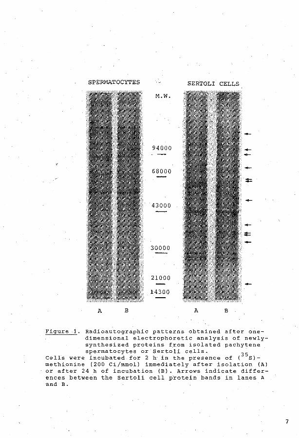
SPERMATOCYTES SERTOLI CELLS
M.W.
94000
68000
43000
30000
21000
14300
A B A B
Figure -1. Radioautographic patterns obtained after onedimensional electrophoretic analysis of newlysynthesized proteins from isolated pachytene spermatocytes ar Sertoli cells.
35 Cells were incubated for 2 h in the presence of ( S)methi9nine {200 Ci/mmol) immediately after isolation (A) or after 24 h of incubation (B). Arrows indicate differences between the Sertoli cell protein bands _in lanes A and B.
7

basic acidic A
M.\'1.
94000
68000
43000
30000 ~
-21000
14300
basic acidic
B
M.\'1.
94000
68000
43000
30000
-21000
14300
8

basic acidic
c
94000
68000
30000
14300
Figure 2. Fluorographic patterns abtairred after two-dimensional electrophoretic analysis of ne~ly-synthesized proteins from:
freshly ~solated pachytene spermatocytes (A), pachytene spermatocytes after 24 h of incubation (B) , or freshly isolated rou_nd sp·ermatids (C). Cells were incubated for 2 h in the preserree of (35s)methionine. Referènce spots which were present in bath cell types were interconnected to campose a frame for mutual camparisen of the fluorograms. Arrows indicate -qualttative differences between the two cell types. Two very dense spots, which .are exclusively present in spermatids, are numbered.
9

indicate that a specific pattern of proteins is synthesized
in sperrnatocytes a~ cornpared to sperrnatids. Moreover, the
pattern of protein synthesis in pachytene sperrnatocytes was
rnainta:ined during incubation for 24 h in the absence of
Sertoli cells.
DISCUSSION
The present results -show rnarked differences between the pat
terns of proteins, newly-synthesized by pachytene spermato
cytes or by round spermatids. We have trie? to purify the
gerrn cell preparations extensively by subjecting preparations
of pachytene spermatocytes and round spermatids, collected
frorn the sedimentation column, to a subsequent purification
on Percoll gradients. A similar procedure was first described
by Meistrich et al. (1981) and resulted in a significant im
provement of purity. Despite this extensive purification,
cross-contamination of the preparations of sperrnatocytes and
sperrnatids does not allow definite conclusions with respect
to absolute qualitative differences between the protein pat
terns of spermatocytes and spermatids. However, some very
dense spots (rnarked 1 and 2 in Fig. 2c) present in the pattern
of spermatids, are completely absent in the pattern of sperrn
atocytes, indicating that contaminating spermatids do not con
tribute to the pattern of prqtein synthesis of spermatocytes.
Changes in the patterns of protein- or RNA synthesis, conco
mitant with development from pachytene spermatocytes to round
spermatids, have also been shown by other investigators
(Grootegoed et al., 1977; Boitani et al., ~980; Kramer &
Erickson, 1982). The occurrence and timing of biochemical
changes during developrnent may rep·resent a series of events
programmed within germ cells, and the execution of this pro
gram mày be supported and/or directed by Sertoli cells. A
change in the pattern of newly synthesized proteins, resulting
from development of pachytene spermatocytes during 24 hours
of incubation, is unlikely to be observed. The reason for
this is that development through the pachytene sperrnatocyte
stage takes about 13 days in rats, and the preparations of
rnid-late pachytene spermatocytes used in our experirnents may
10

contain cells which dif·fer up to 8-10 days in developrnent.
The present results show that in Sertoli cell-s the pattern of
newly synthesized·proteins was changed after 24 hof incuba
tion •. Previously, it has been Shciwn that after isolation and
incubation of Sertoli cells. changes in Stercid and protein
secretion occur (Louis & Fritz, 1977; Grootegoed et al., 1978;
Rornmerts et al., 1980; Skinper & Griswold, 1982). Hence,
activities of Sertoli cells can be influenced, and these
·changes are not related te cell death, because Sertoli cells
can be maintained in incubation for more than ene week. In
contrast, the present results show that the pattern of proteins
newly-synthesized by pachytene spermatocytes did net change
after incubatio.n of the cells for 24 h, although many cells
die during a subs€quent incubation period. Previousiy, it was
reported th~t isolation of mouse pachytene spermatocytes and
spermatids from seminiferous tubules did not result in an im
rnediate change of the protein synthesis pattern (Boitani et
al., 1980). MoreOver,· the electrophoretic pattern of glyco
pi-otein fucosylation by isolated round spermatids was main
tained for at least 20 hof incubation (Grootegoed et al.,
1982b). These observations indicate that developing germ cells
rnaintain a specific pattern of synthètic activities in vitro
in the absence of Sertoli cells.
In vivo activi ties of Sertoli cells will be changed af·ter
hypophysectomy, because Sertoli cells are target cells for FSH
and testasterene. It could be suggested that acti vi.ties of
germ cells are also directly regulated by hormones, because
pachytene sperrnatocytes and round spermatids contain adenyl
Cyclase, cAMP-dependent protein kinase and phosphodies.terase,
enzyrnes involved in cAMP-medi·ated modulation of céllular
activities (Adamo et al., 1980a,b; Gordeladze et al., 1981;
Rossi et al., 1982). However, in, rats no changes were observed
in the pattern of RNA synthesis and RNA processing by pachy
tene spermatocytes at 64 h after hypophysèctomy (Grootegoed
et al., 1979). Moreover, it has been shown that after hypophy
sectomy spermatogenesis was carried out at a normal rate
(Clermont & Harvey, 1965). These· observations indicate that
the absence of FSH and testosterone, and impairment of Sertoli
11

cell function following hypophysectorny does not induce qualit
ative changes in sperrnatocytes. In contrast, after hypophy
sectorny of rats the nurnber of RNA-synthesizing sperrnatocytes
decreased (Grootegoed et al., 1979) and the number of degen
erating cells increased (Russell & Clerrnont, 1977).
In vitro a positive quantitative effect of pyruvate and lactate
on RNA - and protein synthesis of isolated pachytene spermato
cytes was observed (Jutte et al., 1981) and it was postulated
that synthetic activities of pachytene spermatocytes and round
spermatids are supported by supply of pyruvate and lactate
frorn Sertoli cells (Jutte et al., 1982). The metabolism of
glucose and the secretion of pyruvate and lactate by Sertoli
cells was stimulated by FSH in vitr6 (Jutte et .al., 1982). The
arnount of proper energy substrates available to germ cells in
the spermategenie micro-environment in vivo may be reduced
after hypophysectorny, leading to an inhibition of energy
requiring processes in sperrnatocytes and sperrnatids. This may
explain why synthetic activities of developing germ cells show
rapid quantitative changes, but no qualitative changes after
hypophysectomy. In addition to pyruvate and lactate, other
factors frorn Sertoli cells may bè involved in the action of
Sertoli cells upon gerrn cells, but a proper supply of pyruvate
and lactate to pachytene sperrnatocytes and round spermatids is
probably a prerequisite for other interactions between Sertoli
cells and germ cells to occur.
In summary, isolated pachytene sperrnatocytes maintain a
specific pattern of protein synthesis during incubation for
24 h in a chemically defined medium, fortified with pyruvate
and lactate. Therefore, within this period, the presence of
Sertoli cells may be required to support an optimal rate of
synthetic activities in sperrnatocytes, rather than to modulate
these activities.
ACKNOWLEDGEMENTS
This work was supported in part by the Dutch Foundation for
~edical Research (FUNGO) . We thank Miss W. Bakhuizen for her
help in the preparation of this manuscript.
12

REPERENCES
Adamo, S., Conti, M., Gemeria, R., Boitani, C. and·Monesi, V.
(1980) The cyclic nucleotide systern in isolated male germ
cells. In: Animals Models in Human Reproduetion (M. Serie
& L. Martini, eds). Raven Press, New York, pp. 159-168.
Adamo, S., Conti, M., Geremia, R. and Monesï, V. (1980) Partic
ulate and soluble adenyla:te cyclase activities of mouse
male germ cells. Biochem. Bio.phys. Res. Cornmun. 2]_, 607-613.
Beaumont, H.M. (1960) Changes în the rad.iosensitivity of the
testis during feetal development. Int. J. Radiat. Biel.~~
247-256.
Boitani, c., Geremia, R., Rossi, R. and Monesi, V. (1980)
E~ectrophoretic p~ttern of polypeptide synthesis' in spenn
atocytes and spermatids of the mouse.· Cell. Differentiation
2_, 41-49.
Bonner, W.M. and Laskey, R.A. (l974) A f-ilm detection methad
for tritium-labelled proteins and nucleic acids in poly
acrylamide gels. Eur. J. Biochem. !§_, ..83-88.
Clermont, Y. and Harvey, S.C. (1965) Duration of the cycle ·of
the Seminiferous epithelium of norrnal, hypophysectomized
and hypophysectomized-hormone treated albino rats. Endo
crinology ~, 80-89.
Fritz, I.B. {1978) Sites of actions of androgens and follicle
stimulating hormone on cells of the seminiferous tubule.
In: Biochernical Actions of Horrnanes (G. Litwack, ed.).
Academie Press, New York, pp. 249-28~.
Gordeladze, J.O., Purvis, K., Clàusen, O.P.F., Rommerts, F.F.G.
and Hansson, V. (1981) Cellular lOcalization of the Mn 2+
dependent .adenylcyclase {AC) in rat t·estis. I rit. J. Androl.
_i, 172-182.
Grootegoed, J.A., Grollé-Hey, A.H., Rommerts, F.F.G. and van
der Molen, H.J. (1977) Ribonucleic acid synthesis in vitro
·in primary spermatocytes isolated from rat testis. Biochem.
J. 168' 23-31.
Grootegoed, J.A., Jutte, N.H.P.M., Rommerts, F.F.G. and-van
der Molen, H.J. (1982a) Concanavalin A-induced a_ttachment
of spermategenie cells to Sertoli cells in vitro. Exp. Cell
Res. 139, 472-475.
13

Grootegoed, J.A., Krüger-Sewnarain, E.C., Jutte, N.H.P.M.,
Rornmerts, F.F.G. and van der Molen, H.J. (1982b) Fucosy
lation of glycoproteins in rat spermatocytes and sperm
atids. Garnete Res. ~' 303-315.
Grootegoed, J.A., van Meerkerk, L.M., Rommerts, F.F.G. and
van der Molen, H.J. (1979) Effect of hypophysectomy on
ribonucleic acid synthesis in prirnary spermatocytes isola
ted from rat testis. Int. J. Androl. ~' 330-342.
Grootegoed, J.A., Rornmerts, F.F.G. and van der Molen, H.J.
{1978) Hormonal regulation of spermatogenesis. Studies
with isolated germinal cells and Sertoli cells. In:
Horrnanes and Cell Regulation {J. Dumont & J. Nunez, eds),
vol. 2. Elsevier/North-Holland Biomedical Press,
Amsterdam, pp. 55-69.
Jutte, N.H.P.M., Grootegoed, J.A., Rornmerts, F.F.G. & van der
Molen, H.J. (1981) Exogenous lactate is essential for meta
bolie activities in isolated rat spermatocytes and sperm
atids. J. Reprod. Fert. ~' 399-405.
Jutte, N.H.P.M., Jansen, R., Grootegoed, J.A., Rommerts, F.F.G.,
Clausen, O.P.F. and van der Molen, H.J. (1982) Regulation
of survival of rat pachytene spermatocytes by lactate
supply from Sertoli cells. J. Reprod. Fert. ~, 431-438.
Kramer, J.M. and Erickson, R.P. (1982) Analysis of stage
specific protein synthesis during spermatogenesis of the
mouse by two-dimensional gel electrophoresis. J. Reprod.
Fert. fi, 139-144.
Laernmli, U.K. (1970) Cleavage of structural proteins during
the assernbly of the head of bacteriophage T4. Nature 227,
680-685.
Louis, E.G. and Fritz, I.B. (1977} Stimulation by androgens
of the production of andregen Pinding protein by cultured
Sertoli cells. Molec. Cell. Endocr. 2, ·9-16.
Meistrich, M.L., Longtin, J., Broek, W.A., Grimes, S.R. and
Mace, M.L. (1981} Purification of rat spermategenie cells
and preliminary biochemica! analysis of these cells. Biol.
Reprod. l2' 1065-1077.
O'Farrell, P.H. (1975) High resolution two-dimensional elec
trophoresis of proteins. J. Biol. Chem. 250, 4007-4021.
14

Rossi, R., Conti, M., Pezzotti, R. and Gerernia, R. (1982)
Developrnental changes of cyclic nucleotide phosphodiesterase
activity in somatic cells and germ cells of the rat testis.
2nd Eur. Workshop on Molec. Cell. Endocr. of the Testis,
Boekelo, The Netherlands.
Rornmerts, F.F.G., de Jong, F.H., Grootegoed, J.A. and van der
Molen, H.J. (1980) Metabolic changes in testicular cells
frorn rats after long-term exposure to 37°C in vivo or in
vitro. J. Endocr. 85, 471-479.
Russell, L.D. (1980) Sertoli-gerrn cell intèrrelations: a review.
Gamete Res. }, 179-202.
Russell, L.D. and Clerrnont, Y. {1977) Degeneration of gerrn cells
in normal, hypophysectornized and horrnone-treated hypophy
sectornized rats. Anat. Rec. 187, 347-366.
Sarnmons, o.w., Adams, L.D. and Nishizawa, E.E. (1981) Ultra
sensitive silver-based color staining of polypeptides in
polyacrylarnide gels. Electrophoresis ~, 135-141.
Skinner, M.K. and Griswold, M.D. (1982) Regulation of testic
ular transferrin secretion by Sertoli cells cultured in
serum-free medium. J. Biol. Chern. (in press).
15


Appendix Paper II
(J. Reprod. Fert. 62 (1981) 399-405)


J. Reprod. Fert. (1981) 62, 399-405 Printed in Great Brirain
Exogen.ous lactate is essential for roetabolie activities in isolated rat spermatocytes and spermatids
Nicolet H. P.M. Jutte, J. A. Grootegoed, F. F. G. Rommerts and H. J. van der Molen
Department of Biochemistry (Division of Chemica/ Endocrinology), Medica/ Facu/ty, Erasmus Universiiy, Rotterdam, The Netherlands
Summary. Spermatocytes and round spermatids wete isolated from rat testis and the effects of actdition of 3 · 3 mM-glucose and 6 mM-DL-lactate to the incubation medium on the morphology, oxygen consumption and incorporation of uridine and amine acids of these cells were investigated. Actdition of lactate to isolated germ cells increased 0 2 consumption 1·8-fold and incorporation of precursors of RNA and protein by at least 5-fold. The amine acid incorporation into spermatocytes and spermatids was irreversibly decreased after a preincubation for at least 5 h or 70 min respectively in the absence of lactate, indicating degeneration of the germ cells. In the presence of Iactate, however, spermatocyfes rnaintained their morphological integrity for at least 24 h. Actdition of glucose to isolated gerrn cells had no effect on any ofthe p.ararneters investigated. It is concluded that isolated sperrnatocytes and round spermatids rnay utilize lactate, perhaps secreted by Sertoli cells, as the main souree of energy.
Introduetion
Germ Cells in mammalian testes develop in a tubular environment which is controlled to a high degree by Sertoli cells. It is still unknown, however, which Sertoli cell products are important for germ cells. 'Glucose is known to be very important for the survival of germ cells in the testis. Inhibition of glucose transport by injections of 5-thio-n-glucose (Zysk, Bushway, Whistter & Carlton, 1975;· Lob! & Porteus, 1978; Basu, Rarnakrishnan, Prasannan, Rama Sarrna & Sundaresan, 1979; Majumdar et al., 1979; Majumdar & Udelsman, 1979) and induction of acute hypoglycaemia (Mancini, Penhos, Izquierdo & Heinrich, 1960) result in degeneration of germ cells. In isolated testicular tissue from adult rats, the actdition of glucose to the incubation medium resulted in increased RNA synthesis (Hollinger & Hwang, 1972), protein synthesis (Means & Hall, 1968b; Davis, 1969; Hollinger & Hwang, 1972), ATP levels (Means & Hall, 1968a) and oxygen consumption (Tepperman, Tepperman & Dick, 1949; Serfaty & Boyer, 1956; Games, 1971). Testicular protein synthesis was increased 7-fold by gluèose while in 16 other tissues from adult rats the stimulation was less than 1· 5 times (Davis, 1969). Radioautographic analysis of testicular tissue incubated with [3H]lysine has shown that actdition of glucose increases especially incorporation of this precursor into protein of pachytene spermatocytes and sperrnatids (Davis, 1969). No effe"cts of glucose on roetabolie activities in testicular tissue have been observed, however, when the number of gerrn cells was low, as in immature rats (Means & Hall, 1968a, b), following hypophysectomy (Means & Hall, 1968b) or after induction of experimental cryptorchidism (Davis, 1969; Games, 1971). Hence~ it was concluded that pachytene spermatocytes and spermatids are most dependent on a proper supply of glucose. On the other hand, the effect of glucose on isolated sperrnatocytes
0022-4251/81/040399-07$02.00/0 © 1981 Joumals of Reproduetion & Fertility Ltd

400 Nicolet H. P. M. Jutte et al
and spermatids is small, i.e. approximately 1· 5-fold stimulation of amino acid incorporation (Nakamura & Hall, 1976, 1977; Nakamura, Romreil & Hall, 1978): These results suggest a discrepancy between the effects of glucose on germ cells in vivo and in vitro. In our experiments we observed a pronounced positive effect of lactate and pyruvate on isolated germ cells in media éontaining _ glucose. Therefore, we have studied the effects of glucose and lactate on different roetabolie activities in isolated germ cells. ·
Materials and Methods Cell isolation
Germ cell suspensions were prepared enzymically from testicular tissue of immature rats (Wistar strain, substrain R-Amsterdam, age 3(}-35 days, body weight 7(}-80 g) (Romrell, Bellvé & Fawcett, 1976; Bellvé et al., 1977a; Bellve, Millette, Bhatnagar & O'Brien, 1977b). Pachytene spermatocytes and round spermatids were isolated by velocity sedimentation of the cell suspensions in non-linear albumin gradients (1·{}-3-2%) at unit grávity during 70 min at
· room temperature (Grootegoed, Grollé-Hey, Rommerts &· van der Molen, 1977). Cells were counted in a haemocytometer. Isolation medium and incubation medium were essentially Hanks' Balance.d Salt Solution (Hanks & Wallace, 1949) and Eagle's Minimal Essential Medium (Flow Laboratodes Ltd, Irvine, Ayrshire, Scotland, U.K.). Both media were moèli.fied with an increased amount of KC! (56·9 mM) and the osmolarity was adjusted by lowering the NaCI concentration (Grootegoed ·et al., 1977). The isolation. medium was supplemented with 6 mMsodium-DL-lactate (Sigma; 50% L-lactate). When indicated in the text, lactate and/or glucose were added to the incubation medium to a fin al con centration of 6 mM-DL-lactate and 3 · 3 mMglucose.
Sertoli cells were obtained from 26-day-old rats which had been irradiated in utero on Day 19 of gestation with 150 rad (Beaumont, 1960). As a result of this tniatment the testes were depleted of germ cells. Testes of these irraoiated rats were treated with collagenase (0·5 mg/ml) during 20 min at 32°C to obtain tu.bules free from myoid .and interstitial cells. These tubules were fragmented with a Dounce homoge~izer (Fritz, Rommerts, Louis & Dorrington, 1976). All cells were incubated at 32°C. ·
RNA and protein synthesis
Isolated germ cells were incubated in the incubation medium described above, containing labelled precursors for RNA and protein, during 2 h at 32°C under a humidified atmosph.ere of 5% col in air. ·on several occasions incubation with radioactively labelled precursors was preceded by a preincubation in the absence· of this radioactivity. The incul;lations were stopped by cooling t<;> 4 ° C and actdition of cold 0· 9% (w/v) Na Cl with ·either 0· 5 mM-uridine and 7 mMleucine or 6 · 7 mM-methionine. Cells were filtered over 0· 2 1.1.m filters (Sartorius) and washed with 0·9% (w/v) NaCJ. Subsequently the cells were lysed and macromolecules were precipitated with 10% (w/v) trichloroacetic acid for 10 min. The precipitate was washed with 10% (w/v) trichloroacetic acid, to remove non-incorporated precursor, and then with 70% (v/v) ethanol. The precipitate was dissolved in 500 JJ.l I M-NaOH and radioactivity in the samples was counted after addition of 8 mi Picofluor (Packard).
Oxygen consumption
Isolated germ cells were incubated in phosphate-buffered saline (Dulbecco & Vogt, 1954), supplemented with vitamins and amino acids as present in the incubation medium and, when indicated in tJte.text, 6 mM-DL-lactate and/or 3·3 mM-glucose were added. Oxygen consumption was measured in a Warburg apparatus (Umbreit, Burris & Stauffer, 1964). ·

Lactate effects on r:at spermatocytes and spermatids 401
Results
The morphology (examined with phase-contrast microscopy) of isolated spermatocytes incubated for 24 h in a medium containing 3 · 3 roM-glucose and 6 mM-DL-lactate or 1 mMpyruvate, was· very similar to that of freshly isolated germ cells. In contrast, the cells cultured without added la~tate were· degenerate, with translucent cytbplasm and clumped chromatîn. Isolated spermatids cultured without 1actate became degerlerate after several hours. Isolated spermatocytes and spermatids therefore appear to require lactate more than glucose.
The effect of lactate was also demonsti'ated in short-term experiments. Lactate stimulated oxygen éonsumption of freshly isolated spermatocytes and spermatids both in the presence and absence of glucose (Text-fig. 1). Lactate· had no significant effect, however, on the oxygen consurnption of Sertoli cells. Uridine and leucine incorporation into isolated spermatocytes and spermatids was increased at least 57fold by the actdition of lactate, whereas glucose had no significant effect (Table 1)._ This effect was demonstrated after 60 min preincubation with lactate. In Sertoli cells, actdition of lactate with or without glucose caused a 2-fold maximal stimulation of leucine incorporation.
9 e + lactate 0 -lactate
8 ---+glucose --~glucose
7 c •• ö
" 6
"' E 5
" • E 0 4 " c 0 u
o" 3 ' ~
2
0 15 30
' ' ' '
45
Minutes
' ' '
60
/ ~}'/
/ *
75
' •• / .
90
Text-fig. I. The· oxygen consumption by isolated germ cells (3 7% spermatocytes, 53% round spermatids, 4% other germ cells and 6% somatic cells) from rat testes incubated with (---) or without (--) glucose and with (8) or without (0) lactate. The values are the means t?btained with 4 different cell preparations, and vertical bars indicate the s.d. * Significantly different from valm!s without lactate, P < 0·0 I (Student's t test).
Incubation of germ cells in the absence of Jactate for short periods did not cause an irreversible change of the capacity to incorporate amine acids. However, after a preincubation period without Jactate of at least 5 h and 70 min for spermatocytes and spermatids respectively, the incorporation_ of methionipe was ':lot restored after additiori of lactate (Table 2).

402 Nicolet H. P.M. Jutte et al.
Table 1. Incorporation of [ 3H]uridine and [ 14Clleucine into isolated germ cells from rat testes
Substrate in incubation medium
3-3 mM-glucose
+
+
6 mM-DL-.
lactate
+ +
Incorporation
[3H]Uridine
(nCi/106 cells)
5·5 ± 3-6 21-8 ± 1-5-5 91-8 ± 24-5*
125-5 ± 50-9*
[ 14C]Leucine (x I0-1 nCi/106 cells)
1·3 ± 1-0 2-0 ± 1·6
17-4 ± 7-2* 20-3 ± 6-7*
The 5 x lOj celis (55-88% spennatocytes, 1-23% round spermatids, 9-15% other germ cells and 2--9% somatic cells) were incubated for 2 h in I mi incubation medium containing 5 ).lCi [5- 3H]uridine (fl.nal sp. act. 5 mCi/mmol) and 0-6 !J.Ci L-[U- 14C]leucine (final sp. act. 12 mCi/mmol) after preincubation for 60 min. Radioactivity in trichloroacetic addprecipitable material was estimated. Values are mean ± s.d. obtained with 4 different ce!l preparations.
* Significantly different from incorporationin the absence of glucose and lactate, P < 0-01 (paired Stuctent's t test).
Table 2. Effect of preincubation without lactate on incorporation of methionine into isolated germ cells from rat testes
Lactate in medium
Preincubation• Incubation
+ + +
[ 35S]Methionine incorporation (nCi/106 cells)
Spermatocytes
5-7 ± 4·0 18-8 ± 6·6 31·6±10-7
Spermatids
0·8 ± 0-3 2·8 ± 1-0 4-9 ± 2-1
The 5 x 10⁵ eens of a spermatocyte fraction ( 80% spermato· cytes, 6-8% round spermatids, 8-12% other germ. eens and 4--5% somatic cells) or 106 eens from a spermatid fraction (15-20% spermatocytes, 70--76% round spermatids, 6-9% other germ cells and 1-4% somatic eens) were incubated for 2 h in 200 j.d incubation medium containing 10 IJ,Ci L-[ 35S]methionine (final sp. act. 20 Ci/mmol) in tubes shaken at 120 oscinations/min. The incubation medium contained 3·3 mM-glucose and, as indicated, 6 mM-DL-lactate. Values are meao± s.d. for 3 different cell preparations.
• 5-7 h for spermatocytes and 70--90 min for spermatids.
Discussion
The present results show that in germ cells isolated from rat testes exogenous lactate is neerled for roetabolie activities, as shown by oxygen consumptiOn. and RNA and protein synthesis. The effects of the absence of lactate were immediate and were measurable in short-term incubations. When the germ cells were incubated in the absence of lactate for several hours, the decreased roetabolie activity was nat completely restored after actdition of lactate and therefore the cells appear to have degenerated. Degeneration of isolated spermatocytes and round spermatids was readily apparent with phase-contrast microscopy after an incubation without lactate for 24 h. A small percentage of Sertoli cells was always present in our germ cell preparations, but the effects of actdition of lactate to germ cell cultures could nat be ascribed to contamînating Sertoli cells.

Lactate effects on rat spermatocyteS and spermatids 403
The present observations are very relevant for investigations with male germ cells in vitro. The isolation and incubation of these cells have been performed ·by many investigators using media that did not contain lactate or pyruvate. For example, protein synthesis in isolated germ ceUs (Nakamura et al. 1978) and permeability of the plasma. membrane of iSolated gerrn eens (Lee, 1974) have been measured at 32°C and J7°C in media which contained no other substrate than glucose. It is very likely that the observations made in theSe experirnents were.markedly infiuenced by degeneration of the germ cells due to the absence of a proper substrate.
The fact that glucose cannot support roetabolie activities of gerrn cells may be due either to a block in glucose transport or to a block in glycblysis and the hexose rnonophosphate shunt. It has been demonstrated that the X chromosome is inactive throughout meiotic prophase (Monesi, 1965) and therefore the specific activity of the X-liriked iso-enzyme of the glycolytic enzyme phosphoglycerate kinase (PGK) in germ cells may be rather low. It -is not possible to conclude, however, that a block in glycolysis in gerrn cells is caused by the fact that PGK is X~linked becimse an autosome-linked PGK iso-enzyme, present in testicular tisSue, is probab1y active in spermatocytes and spermatids (VandeBerg, Cooper & Close, 1973, 1976). Similarly, the enzyme glucose-6~phosphate dehydrogenase is X-linked, but a block of the hexose monophosphate shunt in germ cells may be prevented by the activity in these cells of an autosomally coded iso-enzyme demonstrated in testicular tissue (Broek, 1977). There is no definite proof, however, that the enzymes mentioned above or other enzymes involved in glucose metabolism are fully active in spermatocytes and spermatids. It is, therefore, still possibie that glucose metabolism is blocked in these cells by inactivity of these enzymes. However, protein synthesis in isolated spermatocyte!? is not inhibited by the preserree of 5-thio~ o-glucose (N akamura & Hall, 19 77), an inhil:>itor of transport of o-glucose (Whist1er & Lake, 1972). This observation supports the idea that spermatocytes do not depend on an active glucose transport mechanism.
In pachytene sperrnatocytes and in spermatids a specific îso-enzyrne of lactate dehydrogenase (LDH-X) is present (Meislrich, Trost1e, Frapart & Erickson, 1977). lt has been shown that testicular LDH-X catalyses preferentially lactate oxidation and is localized in cytosol and mitochondria (Blanco, Burgos, Gerez de Burgos & Montamat, 1976; Montamat & Blanco, 1976). This, as well as the localization of mitochondria in early round spermatids close· to the cel! surface (Clermont & Rambourg, 1978), may indicate that germ cells are specialized to use exogenous lactate efficiently. Spermatocytes in contact with Sertoli ceHs in cultures of seminiferous tubule fragments can survive for more than 5 days in a medium with glucose and without lactate (Palombi et al., 1979). The survival of germ cells in this. system may be expHüned by the secretîon of lactate -.by Sertoli cells, because we have observed lactate production by Sertoli cells in culture (2·2 ± 0·8 ~mol/mg protein per day). The effects of glucose on testicular tissue (see 'Introduction') may therefore largely reftect a stimulation of germ cells by Jaclate produced by Sertoli cells.
The dependenee of male germ cells on Sertoli cells is not unique, because· a similar relation ex.ists between female germ cells and .follicular cells. Granulosa cells have been shown to produce pyruvate (Donahue & Stern, 1968). Isolated rnouse and rat oocytes use pyruvate or lactate as an energy souree (Biggers, Whittingham & Donahue, 1967; Zeilmaker & Verhamme, 1974; Hillensjö, Hamberger & Ahrén, 1975; Eppig, 1976), but growing oocytes can survive in the absence of pyruvate when cultured in the presente of follicular granulosa cells (Baran & Bachvarova, 1977; Eppig, 1977; Bachvarova, Baran & Tejblum, 1980). lt remains tó be demonstrated whether lactate and/or pyruvate represent an important intermediate for the interaction between Sertoli cells or granulosa cells and germ cells in vivo.
We thank Mrs L. M. Koolen-van Meerkerk and Mr R. Jansen for expert technica! assistance and Miss W. Bakhuizen for the help in the preparatien of this manuscript. This work was supported in part by the Dutch Foundation for Medica! Research (FUNGO).

404 Nico/et H. P. M. Jutte et al.
References
Bachvarova, R., Baran, M.M. & Tejblum, A. (1980) Development of naked growing mouse oocytes in vitro. J. exp. Zool. 211, 159-169.
Baran, M.M. & Bachvarova, R. (1977) In vitro culture of growing mouse oocytes. J. exp Zool. 202, 283-289.
Basu, S.L., Ramakrishnan, S., Prasannan, K.G., Rama Sarma, P.R. & Sundaresan, R. (1979) Reversible sterility due to diminished glucose metabolism in male mice treated with 5 thio-oglucose.lnd. J. exp. Bio!. 17, 632-636.
Beaumont, H.M. ( 1960) Changes in the radiosensitivity of the testis during foetal development. !nr. J. Radia't. Biol. 2, 247-256.
Bellvé, A.R., Cavicchia, J.C., Millette, C.F., O'Brien, O.A .. Bhatnagar, Y.M. & Dym, M. (1977a) Spermatogenic cells of the prepuberal mouse. Isolation and morphological characterization. J. Ce!! Bio!. 74. 68-85.
Bellvé, A.R., Millette, C.F., Bbatnagar, Y.M. & O'Brien, O.A. (1977b) Dissociation of the mouse testis and characterization of isolated spermategenie cel!s. J. Histochem. Cyrochem. 257, 480-494.
Biggers, J.D .• Wbittingham, O.G. & Donahue, R.P. (196 7) The pattern of energy metabolism in the mouse oocyte and zygote. Proc. narn. Acad. Sci. L".S.A. 58. 560-567.
Blanco. A .. Burgos, C., Gerez de Burgos, N.M. & Mantam at, E.E. ( 1976) Properties of the testicular lactate dehydrogenase isoenzyme. Biochem. J. 153. 165-172.
Broek, W.A. (1977) Evidence against gene expression after meiosis in the male mouse. J. exp. Zool. 202. 69-80.
Clermont, Y. & Rambourg, A. (1978) Evólution of the endoplasmatic reticulum during rat spermiogenesis. Am.J.Anar. 151.191-212.
Davis, J.R. (1969) Metabolic aspects of spermatogenesis. Biol. Reprod. I. 93-118.
Oonahue, R.P. & Stern, S. (1968) Follicular cell support of oocyte maturation: production of pyruvate in dtro. J. Reproc!. Fert. 17.395-398.
Dulbecco. R. & Vogt, M. (1954} Plaque formation and isolation of pure lines with poliomyelitis viruses. J. exp. Jfed. 99, 167-182.
Eppig, JJ. (1976) Analysis of mouse oogenesis in vitro. Oocyte isolation and the utilization of exogenous energy sourees by growing oocytes. J. exp. Zool. 198. 375-382.
Eppig, JJ. (1977) Mouse oocyte development in vitro with various culture systems. Devl Bio/. 60, 37!-388.
Fritz, I.B., Rommerts, F.F.G., Louis, B.G. & Oorrington, J.H. (1976) Regulation by FSH and dibutyryl cyclicAMP of the formation of androgenbinding protein in Sertoli cell-enriched cultures. J. Reprod. Fert. 46, 17-24.
Games, W.R. 11971) Oxygen consumption by seminiferous tubule5 and interstitial tissues of normal and cryptorchid rat testes. J. Reprod. Fert. 26, 427-429.
Grootegoed, J.A., Grollé·Hey, A.H., Rommerts, F.F.G. & van der Molen, H.J. (1977) Ribonucleic acid
synthesis in virro in primary spermatocytes isolated from rat testis. Biochem. J. 168, 23-31.
Hanks, J,H. & Wallace, R.E. (1949) Relation ofoxygen and temperature in the preservation of tissues by refrigeration. Proc. Soc. exp. Biol. Med. 71, 196-200.
Hillensjö, T., Hamberger, L. & Abren, K. {1975) Respiratory activity of oocytes isolated from ovarian follicles of the rat. Acta endocr., Copenh. 78, 751-759.
Hollinger, M.A. & Hwang, F. (1972) Effect of puro· mycin and actinomycin Don glucose-stimulated protein and RNA labelling, in vitro, in rat testis. Biochim. biophys.Acta 262,336-343.
Lee, L.P.K. (1974) Temperature effect on the perme· ability of plasma membranes of advanced germinal cells of the rat testis. Can. J. Biochem. 52, 586-593.
Lobl, T.J. & Porteus, S.E. (1978) Antifertility activities of 5-thio-0-glucose in mice and rats. Contraception 17. 123-130.
Majumdar, S.K. & Udelsman, R. ( 1979) Fine structure of mouse testes following intraperitoneal treatment with 5-thio-D-glucose. J. Hered. 70. 194-198.
Majumdar, S.K., Brady, K.O., Ringer, L.O., Natoli, J., Killian, C.M., Portnoy, J.A. & Koury, P. (1979) Reproduetion and teratogenic studies of 5-thio-Dglucosein mice.J. Hered. 70,142-145.
Mancini, R.E., Penhos, J.C., Izquierdo, I.A. & Heinricb, J.J. (1960) Effects of acute hypoglycemia on rat testis. Proc. Soc. exp. Biol. Med. 104, 699-702.
Means, A.R. & Hall, P.F. (1968a) Protein biosynthesis in the testis: II. Role of adenosine triphosphate (ATP) in stimulation by glucose. Endocrinology 83, 86-96.
Means, A.R. & Hall, P.F. {1968b) Protein biosynthesis in the testis. I. Comparison between stimulation by FSH and glucose. Endocrinology 82,597-602.
Meistricb, M.L., Trostle, P.K., Frapart, M. & Erickson, R.P. (1977) Biosynthesis and localization of.lactate dehydrogenase X in pachytene spermatocytes ·and spermatids of mouse testes. Devl Biol. 60, 428-441.
Monesi, V. (1965) Synthetic actlvities during spermatogenesis in the mouse. Expl Cel! Res. 39, 197-224.
Montamat, E.E. & Blanco, A. (1976) Subcellular distribution of the lactate dehydrogenase isozyme specific for testis and sperm. Expl Cel! Res. 103, 241-245.
Nakamura, M. & Hall, P.F. (1976) Inhibition by 5-thio-o-glucopyranose of protein biosynthesis in vitro in sp.ermatids from rat testis. Biochim. biophys. Acta 447,474-483.
Nakamura, M. & Hall, P.F. (1977) Effect of 5-thio-Dglucose on protein synthesis in vitro by various types of cells from rat testes. J. Reprod. Fert. 49, 395-397.
Nakamura, M., Romrell, L.J. & Hall, P.F. ( 1978) The effects .of temperature and glucose on protein biosynthesis by immature (round) spermatids from rat testes. J. Cel! Biol. 79, l-9.
Palombi, F., Ziparo, E., Rommerts, F.F.G., Grootegoed, J.A., Antonini, M. & Stefanini, M. (1979) Morpho·
Lactate effects on rat spermatocytes and spermatids 405
logica! characteristics of male germ cells of rats in contact with Sertoli cells in vitro. J. ReprOd. Fert. S1, 325-330.
Romrell, LJ., Bellvé, A.R. & Fawcett, D.W. (1976) Separation of mouse spermatogenic cells by -sedimentation velocity. A morphological characterization. Devl Biol. 19, li9-I3l.
Serfaty, A. & Bofer, J. (l956) l'Influence de divers métabolîtes glucidiques sur l'intensité respiratoire du testicu\e de rat blanc. Experienlia 12, 386-387.
Tepperman, J., Tepperman, H.M. & Dick, HJ. (1949) A study of the metabolism of rat testis in vitro. Elidocrinology 45, 491-503.
Umbreit, W.W., Burris, R.H. & Stauffer, J.F. (1964) Manometric Techniques, 4th edn. Burgess Pub!. Co., Minnesota.
VandeBerg, J.L., Cooper, D.W. & Close, PJ. (1973)
Mammalian testis phosphoglycerate kinase. Nature, New Biol. 243, 48-50.
VandeBerg, J.L., Cooper, D.W. & Close, PJ. (1976) Testis specifk phosphoglycerate kinase B in mouse. J. exp. Zool. 198, 231-240.
Whistler, R.L. & Lake, W.C. (1972) lnhibition of c_ellular transport by 5-thio-D-glycopyranose. Biochem. J. 130, 919-925.
Zeilmaker, G.H. & Verhamme, C.M.P.M. ( 1974) Observations on rat oöcyte maturation in vitro: morphology and energy requirements. Biol. Reprod. 11, 145-152.
Zysk, J.R., Bushway, A.A., Whistler, R.L. & Carlton, W.W. (1975) Temporary sterility produced in male mice_ by 5-thio-o-glucose. J. Reprod. Fert. 45, 69-72.
Received 28 July 1980


Appendix Paper III
(J. Reprod. Fert. 65 (1982) 431-438)


J. Reprod. Fert. (1982) 65, 431-438 Printed in Great Brirain
Regulation of survival of rat pachytene spermatocytes by lactate supply from Sertoli cells
Nicolet H. P.M. Jutte, R. Jansen, J. A. Grootegoed, F. F. G. Rommerts, 0. P.F. Clausen* and H. J. van der Molen
Department of Biochemistry (Division of Chemica! Endocrinology), Medica! Faculty, Erasmus University, Rotterdam, The Netherlands and *Institute of Forensic Medicine, Rikshospitalet,
University of Oslo, Oslo, Norway
Summary. During incubation of fragments of seminiferous tubules in the absence of glucose, pachytene sperrnatocytes and round spermatids died within 24 h, while. Sertoli cells were still viable. The germ cells survived for at least 72 h in seminiferous tubule fragments which were incubated in the presence of glucose. Lactate rather than glucose is essential for [3H]uridine incorporation and su-rvival of isolated pachytene spermatocytes. However, if the spermatocytes were încubated in the présence of Sertoli cells, glucose maintained the incorporation of [3H]uridine into the germ cells. Sertoli cells secreted lactate in the presence of glucose and the lactate secretion was stimulated 2-4-fold by FSH. It is concluded that the activity and survival of pachytene spermatocytes in vitro can be regulated by the supply of lactate from Sertoli cells.
Introduetion
In mammalian testes the walls of the seminiferous tubules are lined by Sertoli cells which are connected by tight junctions and thus form a barrier between the intratubular (adluminal) cernpartment and the extratubular environment (blood-testis barrier). The composition of the tubular ftuid differs from the composition of lymph ftuid and blood plasma (Setchell & Waites, 1975). Early primary spermatocytes move across the blood-testis barrier and further germ cell development occurs in the adluminal cernpartment of the seminiferous tubules.
In the adluminal cernpartment Sertoli cells may control the composition of the fluid which surrounds the germ cells during development. Sertoli cells are target cells for FSH and testosterone (Fritz, 1978) and these hormones may therefore have an effect on the tubular micro-environment and thus on developing germ cells. Several compounds are known to be secreted by Sertoli cells, e.g. andregen binding protein (Fritz, Rommerts, Louis & Dorrington, 1976), transferrio (Skinner & Griswold, 1980), plasminogen activator (Lacroix & Fritz, 1980), glycoproteins (Galdieri, Zani & Stefanini, 1981), sulphoproteins (Elkington & Fritz, 1980) and myo-inositol (Robinson & Fritz, 1979). However, none of these compounds has been shown to affect germ cells. We have reported previously that RNA and protein synthesis and oxygen consumption of isolated spermatocytes and spermatids were stimulated by exogenous lactate (Jutte et al., l981a; Jutte, Grootegoed, Rommerts & van der Molen, 198lb). These results have been confirmed by the observation that protein synthesis in round spermatids was stimulated by lactate (Nakamura, Hino, Yasumasu & Kato, 1981). The capacity of isolated germ cells to use glucose as an_ energy souree appears to be very low. Glucose, however, is essential for maintenance of spermategenesis in vivo (Mancini, Penhos, Izquierdo & Heinrich, 1960; Zysk,
0022-4251/82/040431·08$02.00/0 © 1982 Journals of Reproduetion & Fertility Ltd

432 Nicolet H. P. M. Jutte et al.
Bushway, Whistier & Carlton, 1975). It is possible that the Sertoli cells convert glucose to a substrate which is used by germ cells, and we have investigated whether Sertoli cells can ·influence the activity and survival of germ cells via the secretion of lactate.
Materials and Methods
lsolation ofseminiferous tubules
Testes from immature rats (Wistar, substrain R-Amsterdam, age 30-35 days, body weight 70-80 g) were decapsulated and treated with collageoase (CLS-1; Worthington, Freehold, New Jersey, U.S.A.; 10 mg in 20 mi) in isolation medium (Jutte et al., 198lb) in a 100 mi Erlenmeyer flask during 20 min at 32°C in a shaking waterbath (120 cycles per min). Tubules released by the enzyme treatment were wasbed three times by sedimentation at unit gravity in isolation medium. When used for culture, the tubules were also washed three times in incubation medium.
Isolation ojpachytene spermatocytes
Isolated seminiferous tubules were treated for 15 min with isolation medium containing trypsin (TRL; Worthington; 10 mg in 20 mi) and deoxyribonuclease (DN-CI; Sigma, St Louis, Missouri, U.S.A.; 20 ~g in 20 mi), using the conditions described above, foliowed by actdition of trypsin inhibitor (Sigma; 10 mg in 20 mi) and bovine serum àlbumin (Sigma, fraction V; fin al concentration 0·5% w/v). Subsequently, the tubules were dispersed during 3 min with a I ml measuring pipette (i.d. 2·5 mm) and the cell suspension was filtered through a nylon filter (60 )JI11
pore size) to remove tubular fragments. A fraction of middle-late pachytene spermatocytes (72% middle-late pachytene spermatocytes, 10% early primary spermatocytes, 14-17% other germ cells and unidentified cel1s, 1-4% somatic cells) was obtained by sedimentation of the ce\1 suspension at unit gravity for 70 min at 22°C in a non-linear albumin gradient ( 1-3·2% w/v) (Grootegoed, Grollé-Hey, Rommerts & van der Molen, 1977). The cells were washed three times in incubation medium without glucose befere they were used for incubation.
lsolation of Sertoli cells
Sertoii cells were obtained as fellows. Isolated seminiferous tubules were fragmented with a Dounce homogenizer (Fritz et ai., 1976) and washed three times in incubation medium without glucose. The fragments were incubated without glucose for 1 day, foliowed by an incubation period of 4.....:5 days in the presence of 3·3 mM-glucose and I% (v/v) feta! calf serum (Gibco, Glasgow. Scotland). Pachytene spermatocytes and more mature germ ce\ls were kilied during incubation in the absence of glucose and could easily be aspirated from these cultures.
In one series of experiments, as indicated in the text, Sertol i cells we re isolated from ·testes· of rats irradiated in utero with 150 rad on Day 19 of gestation (Beaumont, 1960). The germ cell-depleted testes of these rats at 24-29 days of age were treated with collagenase, as d~scribed above, to obtain Sertoli cell aggregates which were fragmented withaDounce homogenizer.
!ncubation conditlans
Cells or tubules were incubated in incubation medium (Jutte et al., 198lb) at 32°C under a humidified atmosphere of 5% C02 in air.. Glucose and Jactate concentrations are given in the 'Results'.
Co-culture of Sertoli celis and pachytene spermatocytes
Sertoli cell monolayers, cultured in 24-we\1 tissue culture plates for 4-5 days, were washed once with medium without glucose and twice with medium containing the required glucose

Sertoli cells and suroival of rat spermatocytes 433
concentration. Subsequently, 0· 25 x 106 pachytene spermatocytes were added to each well (2 cm2). After 30 min, 2 ~Ci [5-'Hiuridine/ml were added (final sp.act. 5 Ci/mmol). After another 2 h the labelling was stopped with cold 0·9% (w/v) NaCl containing 0·57 mM-uridine. The germ cells were, removed from the Sertoli cell monolayer by gentie pipetting with-a Pasteur pipette and the amount of [3H]uridine incorporated into RNA of the germ cells was determined by trichloroacetic acid precipitation of RNA on filters (Jutte et al., 1981b). Filters and precipitate were dissolved in 8 ml Filtercount (Packard). Duplicate or triplicate incubations were performed for all conditions. Parallel incubations of Sertoli cells without germ cells were performed to estimate the concentratien of lactate in the medium and the amount of cellular protein.
Es tirnation of lactate and glucose Seminiferous tubules or Sertoli cells were preincubated for 1 day in 2 mi incubation medium,
unless stated otherwise. This medium contained 5 ~g FSH/ml (NIH-FSH-812, 1·25 U/mg, was a gift from the Endocrinology Study Section, NIH, Bethesda, U.S.A.) and/or 200 ng testosterone/ml or the solvents. Concentrated (x 100) stock solutions of FSB and testosterone· were prepared in 0·9% (w/v) NaCl containing 0·1% albumin (Povite, Amsterdam, Holland) and in 10% (v/v) ethanol respectively.
The medium was removed and the cells were wasbed once with incubation medium. Subsequently, the cells were incubated for 24 h in 2 mi fresh incubation medium containing FSH and/or testoSterone and glucose (0-6·6 mM). The medium from the 2nd day of culture was collected, centrifuged to remove unattached cells and kept frezen at -20°C for notmore than 1 week until lactate and/or glucose were determined. The attached and unattached cells were collected and lysed in l M-NaOH and proteîn was estimated (Lowry, Rosebrough, Farr & Randall, 1951). Lactate was estimated enzymically, using lactate dehydrogenase (Hohorst, 1970). Glucose was estimated using enzymic conversion with hexokinase and glucose-6-phosphate dehydrogenase (Schmidt, 1961). In all experiments single estimations oflactate and glucose were performed for duplicate incubations of Sertoli cells or seminiferous tubules.
DNAflow çytometry
Seminiferous tubules cultured for 1 day without or with glucose (3 · 3 mM), foliowed by 2 days of c_ulture in the presence of glucose, were treated with 0· 25% trypsin (Difco, Detroit, Michigan, U.S.A.; 1:250) in phosphate·buffered saline (Dulbecco & Vogt, 1954) for 20 min at 37°C. After dispersion by pipetting with a Pasteur pipette, a few drops of fetal calf serum (Gibco, Glasgow, Scotland) were added and the cells were wasbed in 0·9% (w/v) NaCI. The cells were fixed under continuous mixing by slow actdition of cold ethanol (96%, v/v) toa final concentratien of 70% (v/v) ~thanol. After 30 min at room temperature the cells were stored for several weeks at 4.°C. Then.the suspensions were wasbed in 0·9% (w/v) NaCl and incubated in 0·4% pepsin in 0·02 M-HCI at 37°C .for 15 min. The cells were cocled in ice, centrifuged and incubated for 30 min at room temperature in a mixture of ribonuclease A (Sigma type l-A; 10 mg/1), ethidiumbromide (BDH, Poole, England: 10 mg/1) and Nonidet P-40 (BDH; 300 !!1/1) (Vindel0v, 1977; Clausen & Ábyholm, 1980). The suspension was filtered through a nylon filter (70 ~m pore ~ize) and procéssed through an Ortho Cytofluorograph 50 H (Ortho lnstruments, Westwood, U.S.A.). The counting ofthe fluorescent nuclei was interrupted after the fiuorescence in the highest peak had reached a maximalleveL Therefore, the distribution of fluorescence over the different peaks was relative.
Results
Seminiferous tubules .in culture produced lactate and the lactate production was dependent on tb.e concentratien of glucose in the medium (Text-fig. 1). No detectable change in glucose

434 Nicolet H. P. M. Jutte et al .
.. 0'--,----2;:--;:3---.--,--6--7
Glucose (mM)
Text~fig.l. Effect of glucose on the lactate production by rat seminiferous tubules. Fragmented tubules were incubated for 48 h in 7-5 mi medium containing different concentrations of glucose. Lactate was measured in the medium from the 2nd day of incubation (24-48 h). Each point represents the meao of duplicate incubations. Two different cell preparations were used.
~
"' '2 ·a; ë ~
~
4; 0 E "-
1 0 1
5
Lactate production
Glucose consumption
o~-------------FSH
Testasterene -~-'
+ + + +
+ +
Text-fig. 2. Effect of FSH and testosterone on the Jactate production and the glucose consumption of rat Sertoli cells. Sertoli cells obtained from irradiated rats were incubated in medium containing 3·3 mM-glucose. FSH (5 )lg/ml) and/or testosterone (200 ng/ml) were present during the incubation from 0 to 48 h. Glucose and !acta te concentrations were measured in media collected during 24 h culture (24-48 h). Results obtained with the same cell preparatien are connected.
concentration occurred during incubation under these conditions. The lactate secretion by seminiferous tubule fragments from 27-day-old rats was stimulated 3-fold by FSH. Because germ cells may metabolize part of the lactate secreted in seminiferous tubule cultures, we have also studied pure Sertoli cells isolated from irradiated rats. Like seminiferous tubule fragments from intact rats these pure Sertoli cells were able to convert glucose to lactate and the lactate secretion and glucose consumption were stimulated 2~4-fold by a mixture of FSH and testosterone (Text-fig. 2). This stimulation appeared to be caused by FSH under these conditions (Text-fig. 2).

Sertoli cells and survival of rat spermatocytes
1C (a) 1C (b) (c)
2C
4C 4C
Channel no. (relative fluorescencè intensity)
Text~fig. 3. DNA flow cytometric analysis of rat seminiferous tubules cultured for 1 day with or without glucose. Isolated seminiferous tubû.les were processed for DNA flow cytomettic measurements as described in 'Materials and Methods' section. (a) Freshly isolated; (b) incubated for 3 days in the presence of glucose (3· 3 mM); (c) incubated for 1 day in the absence of glucose foliowed by 2 days in the presence of glucose (3·3 mM). Nuclei of primaty spermatocytes (4C), spermatids (IC), somatic cells, secóndary spermatocytes and spermatögonia (2C) showed different fluoreScence after staining aftheir DNA with ethidium bromide.
"0 40 ~ ·-"~ ON
""' 30 c:C 0-
J~] 1·5 0~ ·.;:::; . • ~~ •• cO "0 ·-:g:; e • "-U 1 ·0 0. ë ::> c .'l 0. l'- ~ "' "-- 0 5 0 E ~ ." ~ 0 .. E
-"' 00-05 0 5 1 0 1 5
Glucose {mM)
435
Text-fig. 4. The effect of glucose on the incorporation of f 3H]uridine into· rat spermatocytes, · co-cultured with Sertoli cells. Pachytene spermatocytes (0-25 x 106) were labelled- with l5-3Hiuridine during 2 hof co-culture with Sertöli cells. The radioactivity incorporations into the spermatocytes are given as means ± s.d., obtained with 3 different germ cell and Sertoli cell preparations. The amount of lactate estimated in parallel incubations of Sertoli cells is given for 2 different cell preparations.
It was shown by phase-contrast microscopy and by cytofluorometric measurements that spermatids and pachytene spermatq_cytes survived when fragmentsof seminiferous tubules were incubated for 3 days in the presence of glucose (3-3 mM) (Text-figs 3a, b). However. when seminifetous tubules were incubated without glucose for 1 day, spermatids and spermatocytes were absent (Text~fig. 3c). The Sertoli cells in these tubule cultures survived 1 day of glucose depletion, because after actdition of glucose (3 · 3 mM) on the 2nd day of incubatioh, the Sertoli cells migrated 'normally, incorporated leucine (as judged by radioautography) and produced 0·96 ± 0·55 MffiOI lactate/24 h (compared to controls incubated with glucose. which produced 0·85 ± 0·37 Jili10I lactate/24 h). Previously we observed that isolated pachytene spermatocytes, incubated in the presence of glucose. did not incorporate sigrtifica:nt amounts of !3H !uridine and !l 4Cileucine (Jutte et al., 198lb). However, the incorporation of PHiuridine into RNA of isolated spermatoCyteS incubated in contact. with Sertoli cells increásed with increasing glucose Concentratiens (Text-fig. 4). In the absence of glucose. the activity of spermatocytes in

436 Nicolet H. P.M. Jutte et al.
these co-cultures was minimal; maximal activity was reached only in the presence of glucose concentrations greater than 0·5 mM. Lactate production by the Sertoli cells under these conditions was also dependent on the glucose concentratien (Text-fig. 4).
Isolated spermatocytes incubated in the presence of 6 mM-DL-lactate and 1· 5 ruM-glucose incorporated 22·7 ± 4·2 nCi [~Hluridine {106 cells/2 h). Spermatocytes incubated on top of Sertoli cells in the presence of 1· 5 mM-glucose incorporated 23 ·1 ± 8 · 3 nCi [3H ]uridine ( 106
cells/2 h) (means ± s.d. of 3 different cell preparations). Therefore, the [3H]uridine incorporation in spermatocytes appeared to be maintained both in the presence of Sertoli cells plus glucose and in the presence of lactate.
Discussion
Isolated pachytene spermatocytes do not survive in the presence of glucose (Jutte et al., 1981b). The present results, however, indicate that RNA synthesis and the integrity of pachytene spermatocytes in cultures of seminiferous tubules and in co-cultures of isolated germ cells and Sertoli cells were maintained by exogenous glucose (Text-figs 3 and 4). When glucose was omitted from these incubations, the pachytene spermatocytes did not survive (Text-fig. 3c). Lactate is known to be essential for survival of germ cells (Jutte et al., 1981 b) and the present results indicate that this substrate maintained isolated pachytene spermatocytes as well as did Sertoli cells. Lactate was secreted by the Sertoli cells and this secretion was stîmulated by horrnanes and- glucose (Text-figs 1 and 2). These observations support the idea that the activity and survival of pachytene spermatocytes is regulated by Sertoli cells via conversion of glucose to I acta te.
Most studies on metabolic pathways in the testis have been performed with mixed cell populations ·and little is known about metabolic activities in different cell types. It has been suggested that somatic cells in the testis are mainly dependent on lipids for the provision of energy (Free, 1970). We observed that Sertoli ce11s survived an incubation of 24 h without glucose; Sertoli cells may use lipids under these conditions. Study of the metabo\ism of glucose by cuitures of pure Sertoli cells showed that maximaUy 2-9% of the glucose utilized was converted to carbon dioxide and 95-8% was converted to anionic compounds, mostly to lactate (R(_)binson & Fritz, 1981). These observations demonstrate the enormous capacity of Sertoli cells to convert glucose into lactate. In contrast to our observations, Robinson & Fritz ( 1981) did not ob serve a stimulatory effect of hormones on I act a te production by Sertoli cells. Th is may be caused by ditTerences in the preparatien of the Sertoli cells. The concentratien of lactate in tubular fluid in vivo is not known, but might be camparabie to the concentration of lactate in rete testis ftuid (0-65 mM in rams and 0-74 mM in bulls), which is half the concentration present in blood plasma (Setchell, Scott, Voglmayr & Waites, 1969). Glucose appears to be available in .the testis because its con centration in the testicular lymph of the rat is as high as the concentratien in blood plasma(± 3 mM) (Setchell & Waites, 1975). This does nol imply, however, that the Sertoli cells are directly exposed to these concentrations, because at least one other cel! type (myoid cells) is interposed between the Sertoli cells and the lymph.
The glucose uptake and/or the glycolysis in isolated germ cells appears to be defective. Lactate oxidation via the Krebs' cycle could supply energy for metabolic activîties. Germ cells may be able to maintain an aerobic metabolism, because it has been shown that the oxygen tension in seminiferous tubules is as high as the tension in testis interstitial tissue (Free, Schluntz & Jaffe, 1976). An extreme dependenee of germ cells on the activity of the Krebs' cycle is illustrated by in-vivo experiments with rats which were fed inhibitors of the Krebs' cycle. While other tissues were nat atTected, spermatocytes and spermatids died. This degeneration is probably not caused by a direct effect of the inhibitors on Sertoli cells, because these cells remained in the tubules and were able to support a restored spermategenesis (Paul, Paul,

Sertoli cells and survival of rat spermatocytes 437
Kopko, Bender & Everett, 1953; Novi, 1968; Steinberger & Sud, 1970; Sullivan, Smith & Garman, 1979).
In different stages of germ cell development, a different dependenee of gerrn cells on carbohydrate substrates appears to exist. Round sperrnatids and pachytene spermatocytes are dependent on a supply -of lactate (Jutte et al., 1981 b), whereas ejaculated spermatozoa can use glucose or fructose as substrates (Voglmayr, 1975). Therefore, following a stage of dependenee on lactate, germ cells may become independent of lactate during or after spermatid elongation.
The present results indicate thát lactate from Sertoli cells inftuences the survival of pachytene sperrnatocytes and that the lactate production is increased by hormones. Lactate could therefore be an important intermedia te for. the regulation of the survival of pachytene spermatócytes and round spermatiets by hormones.
We thank D~; B. G. Elijenberg and the Central Clinical-Chemical Laberatory of the Dijkzigt Hospita! for assistance with the estimations of glucose and Miss W. Bakhuizen for help in the preparatien of this manuscript. This work was supported in part by the Dutch Foundation for Medica! Research (FUNGO) and NATO·grant 180.80.
References
Beaumont, H.M. ( 1960) Changes in the radiosensitivity ofthe testis during foetal development. Int. J. Radial. Biol. 2, 247-256.
Clausen, O.P.F. & Ábyholm, T. (1980) Deoxyribonucleic acid flow cytometry of gerrri cells in the investigation of male infertility. Fert. Steril. 34, 369-374.
Dulbeceo, R. & Vogl, M. (1954) Plaque formation and isolation of pure lines with poliomyelitis viruses. J. exp. Med. 99, 167-182.
Elkington, J.S.H. & Fritz, I.B. (1980) Regulation of sulfoprotein synthesis by rat Sertoli ce11s in culture. Endocrino/ogy 107, 970-976.
Free, M.J. (1970) Carbohydrate metabolism in the testis. In The Testis, Vol. 11, pp. 125-192. Eds A. D. Johnson, W._ R. Gornes & N. L VanDemark. Academie Press, New York.
Free, MJ., Schluntz, G.A. & Jatfe, R.A. (1976) Respiratory gas tensions in tissues and fluids of the male reproductive tract. Biol. Reprod. 14, 481--:488.
Fritz, I.B. (1978) Sites of actions .of androgenS and follicle-stimulating hormone on cells of the seminiferous tubule. In Biochemica! Actions of Hormones, Vol. 5, pp. 249-281. Ed. G. Litwack. Academie Press, New York.
Fritz, I.B., Rommerts, F.F.G., Louis~ B.G. & Dorrington, J.H. (1976) Regulation by FSH and dibutyryl cyc\ic AMP of the formation of andregenbinding protein in Sertoli ce11-enriched cultures. J. Reprod. Ferr. 46, 17-24.
Galdieri, M., Zani, 8. & Stefanini, M~ (1981) Effect of the association with germ ce\ls on the secretory activities of Sertoli ce\ls in in-vitro cultures. In 0/igozoó'spermia. Eds G. Frajese, E. S. E. Hafez, A. Fabbrini & C. Conti. Raven Press. New York (In press).
Grootegoed, J.A., Grollé-Hey, A.H., Rommer1.s, F.F.G. · .& van der Molen, HJ. (1977) -Ribonucleic acid
synthesis in vitro in primary spermatocytes isolated from rat testis. Biochem. J. 168; 23-31.
H'ohorst, HJ. (1970) L-(+)-Lactat, Bestimmung mit Lactat-Dehydrogenase und NAD. In Methoden der Enzymatische Analyse, 2nd edn, part 11, pp. 1'425-1429. Ed. H. U. Bergmeyer. Verlag Chemie GmbH, Weinheim.
Jutte, N.H.P.M., Kooien, L.M., lansen, R., Grootegoed, J.A., Rommerts, F.F.G. & van der Molen, HJ. ( 1981 a) Lactate essential for biochemical activities in isolated germinal cells. Int. J. Androl., Suppl. 3, 59, Abstr.
Jutte, N.H.P.M., Grootegoed, J.A., _Rommerts, F.F.G. & van der Molen, HJ. (198Ib) Exogenous lactate is essential for metabolic activities. in isolated rat · spermatocytes and spermatids. J. Reprod. Fert. 62., · 399-405.
Laeroix, M. & Frjtz, I.B. (1980) Characterization of a plasminogen a!.i::tivàtor secreted by rat Sertoli cells in culture. J. Cell Biol. 87, 152a.
Lowry, O.H., Rosebrough, N.H., Farr, A.L. & Randall, RJ. (1951) Protein measurement with the 'Folin phenol reagent. J. bio/. Chem. 193, 265-275.
Mancini, R.E., Penhos, J.C., lzquierdo, I.A. & Heinrieh, J.J. (1960) Effects of acute hypog\ycemia on rat testis. Proc. Soc. exp. Biol. Med. 104,699-702.
Nakamur8, M., Hino, A., Yasumasu, I. & Kato, J. (1981) Stimulation of protein synthesis in round sperrriatids from rat testes by lactate. J. Biochem. 89, 1309-1315.
Novi, A.M. (1968) Injury of primary spermatocytes during the meiotic prophase and temporary arrest of sperritatogenesis indoeed by ftuoroacetamide in the rat. Virchows Archiv. Abt. B. Ze/lpath. I, 346-364.
Paul, H.E., Paul, M.F., Kopko, F., Dender, R.C. & Everett, G. (1953) Carbohydrate metabolism studies on the testiS ·of rats fed eertaio nitrofurans. Endocrinology 53, 585-592.
Robinson, R. & Fritz, I.B. (1979) Myoinositol biosynthesis by Sertoli cells, and levels of-myoinositol biosynthetic enzymes in testis and epididymis. Can. J. Biochem. 57,962-967.

438 Nico/et H. P.M. Jutte et al.
Robinson, R. & Fritz, I.B. (1981) Metabolism of glucose by Serto!i ceils in culture. Biol. Reprod. 24, 1032-1041.
Schmidt, F.H. (1961) Die enzymatische Bestimmung von Glucose und Fructose nebeneinander. Klin. Wschr. 39, 1244-1247.
Setchell, B.P. & Waites, G.M.H. (1975) The blood-testis harrier. In Handhook of Physio!ogy, sectien 7, vol. V, chapter 7, pp. 143-172. Eds R. 0. Greep, E. 8. Astwood, D. W. Hamilton & S. R. Geiger. Am. Physiol. Soc" Washington, D.C.
Setchell, B.P., Scott, T.W., Voglmayr, J.K. & Waites, G.M.H. ( 1969) Characteristics of testicular spermatozoa and the Auid which transports them into the epididymis. Bio!. Reprod. i, 40-66.
Skinner, M.K. & Griswold, M.D. ( J 980) Sertoli cells synthesize and secrete transferrin-like protein. J. biol. Chem. 255, 9523-9525.
Steinberger. E. & Sud, B.N. ( 1970) Specific effect of
fiuoroacetamide on spermiogenesis. Biol. Reprod. 2, 369-375.
Sullivan, J.L., Smith, F.A. & Garman, R.H. (1979) Effects of fiuoroacetate on the testis of the rat. 1. Reprod. Fert. 56, 201-207.
Vindelev, L.L. (1977) Flow microfiuorometric analysis of nuclear DNA in cells from solid tumours and cel\ suspensions. Vitrchows Arch, B Cel! Path. 24, 227-242.
Voglmayr, J.K. (1975) Metabolic changes in spermatozoa during epididymal transit. In Handhook of Physîology, section 7, vol. V, chapter 21, pp. 437-451. Eds R. 0. Greep, E. B. Astwood, D. W. Haruilton & S. R. Geiger. Am. Physiol. Soc. Washington. O.C.
Zysk, J.R., Bushway, A.A., Whistler, R.L. & Carlton, W.W. (l975) Temporary sterility produced in male mice by 5-thio-o-glucose. J. Reprod. Fert. 45, 69-72.
Received 6 October 1981
Appendix Paper IV
(submitted to J. Reprod. F.ert.)

FSH STIMULATION OF THE SECRETION OF PYRUVATE AND LACTATE BY
SERTOLI CELLS MAY BE INVOLVED IN HORMONAL REGULATION OF
SPERMATOGENESIS
N.H.P.M. Jutte, R. Jansen, J.A. Grootegoed, F.F.G. Rornmerts
and H.J. van der Molen
Department of Biochemistry (Division of Chemical
Endocrinology), Medical Faculty, Erasmus University
Rotterdam, Rotterdam, The Netherlands
SUMMARY
Pyruvate and lactate secretion by Sertoli cells, and the
effect of p & 1 on isolated pachytene sperrnatocytes and round
spermatids were studied. Pure Sertoli cells were obtained frorn
3-6 week-old rats, which were sterile after prenatal irradiation.
The lactate secretion by these Sertoli cells, rneasured from 24-
48 hours after isolation during incubation in the absence of
horrnones, increased with age of the rats frorn 3-6 weeks. At all
ages investigated, the lactate secretion was enhanced in the
presence of FSHplus testosterone, but the stimulation was most
pronounced at 4 weeks of age. The increase of the lactate
secretion was caused by follicle-stimulating hormone (FSH) 1
whereas testosterone did not exert an effect. Sertoli cells
from. 4-week-old rats secreted both pyruvate and lactate, which
accurnulated in the incubation medium in a ratio 1:4. The stimUla
tion of the pyruvate and lactate secretion by FSH was dose
dependent (ED50
at approxirnately 10 ng NIH-FSH-Sl3/ml). Tbe o & l
l

secretion was stimulated two- fold by insul in, four - fold by FSH
and more than six- fo l d by bt2
cAMP (in the presence of
3- isobutyl - 1 - methylxanthine). The effects of FSH a nd insulin·
were not additive.
The leucine incorporation into isolated pachytene sperrnatocytes
and round spermatids was stimulated by both exogenous pyruvate
and lactate in a dose- depende nt way. A maximal rate of leucine
incorporation was obtained with 0.2 rnM pyruvate or 2 rnM L- lactate.
Spent medium from incubated Sertoli cells (from 4'- week- old rats)
stimulated the leucine incorporation into isolated pachytene
s~ermatocytes and round spermatids 4- 8- fold. This effect could
be explained by the arnounts of pyruvate and lac tate present in
the spent medium . I t is concluded that pyruvate and lactate are
major secretion products from Sertoli cells, which can support
synthetic activities in germ cells, and the present results
indicate p~ruvate and ~actate may olay a role in the hormonal
regulation of spermatogenesis .
INTRODUCTION
The initiatien and rnainterrance of spermategenesis in marnrnalian
tes tes i s dependent on the presence of follic l e - stimulating
hormone (FSH) and testosterone. It has been shown that these
hormones exert direct effects on Sertoli cells , whereas it can
be concluded from several observations that spermatocytes and
sperrnatids are no target cells for FSH and testosterone
(Grootegoed, Peters, Mulder, Rornrnerts & van der Molen, 1977;
Fritz, 1978). Hence, hormonal regulation of spermategenesis
.2

appears to be rnediated by Sertoli cells.
Support for a possible nutritive rêle of Sertoli cells in germ
cell development are the recent observations that Ser.toli cells
secrete p & 1 at a high rate (Jutte, Grootegoed, Rommerts
& van der Molen, 1981; Robinson & Fritz, 1981) and that pachytene
sperrnatocytes and round spermatids require exogenous py~uvate
and lactate to carry out energy requiring processes (Jutte et al.,
~981a; Jutte, Koelen, Jansen, Grootegoed, Rommerts & van der
Molen, 1981; Mita & Hall, 1982). Moreover, the rate of aerobic
glUcose utilization and p & 1 production by Sertoli cells from
immature rats was found to be increased during incubation iri
the -presence· of FSH (Jutte, Jansen, G.i-ootegoed, Rorrunerts, Clausen
& va-n der Moleri, 1982; Jutte, Jansen, Grootegoed, Ronnerts &
van der Molen, 1982). The present. experiments were perforrne'd to
study the p & 1 secretion by Sertol·i cells in more detail wi th
emphasis on the rêle of FSH-during prepubertal testis
deve'lopment, and effects of insulin.
Many unknown componenents secreted by Sertoli cells may be
involved in germ cell deve1opmeTI.t. Therefore 1 we studied also
whether p & 1 are the most important factors, present in
spent mediUm from Sertoli cells, which maintain activities of
germ cells during short-term incubations.
MATERIALS AND METHOOS
Ç~b!_;h§Q!èti:Qg
Germ cells were iso_lated from immature rats (Wistar, substrain
R-Amsterdam, age 30-35 days, body weight 70-80 g) by sedimenta-
3

tion at unit gravity as described previously (Jutte et al.,
1982b). Two germ cell fractions were obtained: a pachytene
sperrnatocyte fraction, which contained rnid-late pachytene
sperrnatocyteS (80 2:. 9%), round sperrnatids (4 2:. 3%), somatic
cells (1 ~ 1%), other germ cells and unidentified cells
(7.± 4%); a round sperrnatid fraction, which contained round sperma
tids (81 2:. 9%), early sperrnatocytes (7 2:. 4%), mid pachytene
spermatocytes (6 2:. 4%), somatic cells (1 2:. 1%), other germ cells
and unidentified cells (4 2:. 2%) (rnean _-.!:. S.D., n = 13).
Sertoli cells were isolated frorn 3-6-week-old rats as decribed
previously (Jutte et al., 198la). The rats were irradiated in
utero on day 19 of gestation to obtain germ cell depleted testes,
for the isolation of Sertoli cells not contaminated with germ
cells (Beaumont, 1960). Following isolation, the gerrn cells and
Sertoli cells were washed thrice in incubation medium containing
3.3 ITi..L\1 gluCose (Jutte et al., 1981a).
Incubation conditions
Sertoli cells (approximately 300 ~g of protein) were incubated
for 24 h in 2 rol incubation medium (no pyruvate and lactate
added) at 32°C under a humidified atmosphere of 5% C0 2 in air,
ln the absence or presence of FSH (5 ~g NIH-FSH-512, 1.25 U/mg
or 0.5 ~g/ml NIH-FSH-513, 15 U/mg, gifts frorn the Endocrinology
Study 'Section, National Institute of Health, Bethesda, MS,
U.S.A.), testasterene (200 ng/rnl), dibutyryl cyclic Ar·1P (0.5 mM-),
3-isobutyl-1-methylxanthine (MIX, 25 ~M) and insulin (5 ~g/rnl,
Sigma, 26.8 U/mg). The medium and additions were renewed after
24 h of incubation and the spent medium from the 24-48 h
4

incubation period was collected, centrifUged at 1500 g for
10 min' to remave a few unattached cells, and stared at 4°C.
For sorne experiments twice the arn_ount of Sertol i cells {::ipproximately
600~g of protein) was incubated as described above, to obtain
spent medium (Sertoli cell-conditioned medium).
6 Pachytene sperrnatocytes (0.5 x 10 cells) or round spermatids
(10 6 cells) were incUbated either in 0.5 ml incubation medium
plus 0.5 rol Sertoli cell-conditioned medium or in 1 rnl incubation
medium containing 0-2 mM sodium pyruvate (Boehringer) or 0-4 mM
sodium DL-lactate (Sigma, appioxirnately 50% L-lactate).
Incorporation of (3H)leucine into cellular protein was estirnated
by incubation of germ cells for 2 h in the presenèe of 5 ~Ci
of (4,5- 3H)leucine (final specific radioactivity 20.7 rnCi/mmol)
following a preincubation of 30 min. The incubations were
terrninated by addition of cold 0.9% (w/v) NaCl (containing
7.6 rnH-leucine) and subsequently the arnount.of radioactivity
incorporated into protein was estimated by precipitation of
macromolecules with trichloroacetic acid (Jutte et al., 1981a,
l982b).
The arnounts of lactate and pyruvate in the media were estimated
enzyrnically according to Hohorst (1970) and Czok & Lamprecht
(1970). The data were sta~istically.analyzed with the Student 1 s
t-test.
5

-" ... 8 "' "-.. c
2 6 0 a_
"' E "-.. 4 2 0
û -" 2 0 E ::!.
0 3
(6)
( 4)
* ( 15)
* ( 15)
(4)
"* (4)
• FSH + testosterone
o control
4 5 age ( weeks)
~igure 1. Lactate secretion by Sertoli cells from 3-6-week-old rats.
Sertoli cells obtained from prenatally irradiated rats of different ages were incubated for 24 h in chemically defined medium without further additions (o) or in the preserree of FSH (7. 5 NIH-FSH-Sl U/ml) and testasterene (200 ng/ml) (e). Af ter 24 h of incubation, the medium was renewed and incubation was continued for another period of 24 h in the presencè of the hormones. The amount of lactate present in the soent medium from 24-48 h of incubation was estimated and expressed per mg of cell protein. Results represent means + S.D. for 4-15 different cell preparations (the number of cel! preparations is given between brackets). Each cel! preparatien was tested in duplicate incubations. At all ages the amount of lactate in the spent medium was significantly higher after incubation in the presence of horrnanes (p < 0.01). % Significantly different from the lactate secretion by
Sertoli cells from 3-week-old rats (p < 0.05). ~~ Significantly different from the lactate secretion by
Sertoli cells from 4-week-old rats (p < 0.05).
RESULTS
Secretion of p & l by Sertoli cells
From 3 to 6 weeks of age the testes of intact rats become
populated with the more advanced germ cell types (pachytene
spermatocytes, spermatids) which appear to require p & l
Therefore, we have studied the ? & 1 secretion by Sertoli cells
6

isolated from 3-6-week -old rats. A preparatien of Sertoli cells
was obtained from sterile prenatally irradiated rats. The
Sertoli cells were incubated iri chemically defined med,ium and
lactate was estimated ·in the spent medium. The following results
were obtained:
1) the lactate production in the absence of horrnanes (basal
rroduction, expressed per mg of cell protein) was increased
four-fold from 3-6 weeksof age (Fig, l); 2) the lactate
secretion by Sertoli cells frorn 3-6-week-old rats was increased
during incubation in the presence of FSH plus testosterone
(Fig. !'), Dut the ratio stimulated to unstirn,ul~ted secretion
decreased with age as a result of the increased basal production;
3) testasterene did not increase the basal er FSH-stirnulated
lactate secretion by Sertoli cells from 4-week-old rats (Table 1).
The effects of FSH were most pronounced in Sertoli cells frorn
Table 1. Effects of testosterone and FSH on the lactate secretion by Sertoli cells from 6-week-old rats.
Treatment
No additions
testosterone
FSH
FSH plus testosterone
lactate secretion (~mol/mg protein/24 h)
4.98 + 0.12
5.19 + 0.09
8.05 + 0.25*
8.06 + 0.71%
The lactate secretion by Sertoli cells from 6-week-old rats was estimated as described in the legend to figure 1. Results represent means + S.D. for 3 different cell preoarations. Each cell prepar;tion was tested in duplicat~ i~cubations. * Significantly different from control and testosterone
treated sertoli cells (p < 0.01).
7

~ 2.5 pyruvate • w~
N "<t
-....... N . -....... ·= 2.0 8 c cv
0 cv -... 0 c. ... C> 1.5 6 c. E C>
-....... E
cv -....... 0 1.0· 4 cv -> 0 ::;) . ->:.. · U
0 c. 0.5 ED
50 =± 10 ng 2 .=
0 ol 0
E E :1. :1.
I I I I 0 510 50100 1000
NI H-FSH- S 13· (ng/ml) 10000
Figure 2. PSH dose-response curve of pyruvate and 1actate secre ti on by Serto1i ce11s.
Serto1i ce11s obtained from 4-week- o1d rats were incubated for 48 h in the presence of different amounts of .NIH-FSH-513. The medium was renewed after 24 h and the amounts of pyruvate and 1actate present in the spent medium from 24 - 48 h of incubation were estimated and expressed per mg ce11 protein . Resu1ts re?resent means ~ range of dup1icate incubations.
4-week- old rats and therefore further experiments were performed
us ing these Sertoli cells. The response to FSH of the secretion
of both pyruvate and lactate by Sertoli cells from 4-week-old
rats was dose- dependent (Fig. 2). Pyruvate and lactate were
found in the spent incubation medium in a ratio 1:4. This ratiq
.-,,as the same in the absence or presence of FSH. Half-maximal
sti:nulation of the secretion: of both p & 1 was obtained at
a??roximate ly 10 ng of. NIH-FSH- S13/ml.
Because insulin is involved in g lucose metabolism, its effect
on the triose secretion by Sertoli cells was investigated. The
secretion of pyruvate and lactate by Sertoli cells from 4- week-
old rats was stimulated two-fold after 24-48 h of incubation in
the presence of insulin, whereas a four- fold stimulation was
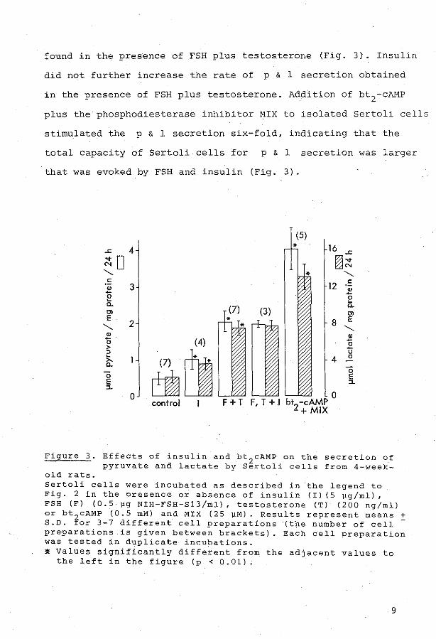
found in the presence of FSH plus testosterone (Fig. 3). Insulin
did not further increase the rate of p & 1 secretion obtained
in the presence of FSH plus testosterone. Addition of bt2-cAMP
plus the phosphodiesterase inhibitor MIX to isolated Sertoli cells
stirnulated the p & 1 secretion six-fold, indicating that the
total capacity of Sertoli cells for p & 1 secretion was larger
that was evoked by FSH and insulin (Fig. 3).
(5)
.J:: 4 16 .J::
~o ril~ "-. "-. .!:
3 12 .!: " 2 -e e a. a. 0> 0> E
2 8 E "-. "-.
" " - (4) 0 0 > -"
cill u
~
4 0 >-a. ö ö E
E ::1. ::1.
0 0 control F+T F, T +.I bt
2-cAMP +MIX
Figure 3. Effects of insulin and bt2
cAMP on the secration of pyruvate and lactate by Sertoli cells from 4-week-
old rats. Sertoli cells were inctibated as described in the l~gend to Fig. 2 in the preserree or absence of insulin {I) (5 llg/ml), FSH (F) (0.5 pg NIH-FSH-813/ml), testasterene (T) (200 ng/ml) ar bt 2 cAMP {0.5 mM) and MIX (25 ]..IM). Results reprasent means :!: S.D. Ior 3~7 different cell preparations (the number of cell preparations is given· between brackets). Each cell preparatien was tested in duplicate incubations. ~ Values significantly different from the adjacent values to
the left in the figure (p < 0.01).
9

Effect of p & 1 : on germ cells
The stimulation of ( 3H)leucine incorporation into isolated
pachytene spermatocytes and round spermatids by exogenous
lactate and pyruvate was dose-dependent (Fig. 4). Pyruvate
was effective at 10-fold lower concentrations than lactate to
support a high rate of leucine incorporation, since maximal
leucine incorporation was reached at approximately 2 mM L-lactate
or 0.2 mM pyruvate for both pachytene spermatocytes and round
spermatids during an incubation for 2 hours in the presence
of 3.3 mH glucose. Moreover, addition of both L-lactate (3 or
6 mM) and pyruvate (2 mM) to spermatocytes and spermatids did
not further increase the leucine incorporation above the level
obtained by addition of pyruvate alone (results not shown).
This indicates that pyruvate, irrespective of the. presence
of lactate, can support a high rate of protein synthesis
during short-term incubations.
Effect of Sertoli cell-conditioned medium on gerrn cells
To define the relative importance of p & 1 to gerrn cells, as
cornpared to other products secreted by Sertoli cells, we have
investigated the effect of spent medium from incubated Sertoli
cells on germ cells. Isolated pachytene spermatocytes and round
spermatids were incubated for 2 h in Sertoli cell-conditioned
medium (1:1 diluted with fresh medium) and (3H)leucine incorpora-
tion into the cells was measured. The pmol amounts of incorporated
leucine were calculated from the incorporated radioactivity,
after correction for an increase of the specific radioactivity
of leucine in the spent media, caused by leucine consumption by
10

A 1.5
""0 m ö 0 a. 1.0 " 0 0 .s m .s 0 ~
~ 0.5 ëi E 0
0
B 1.0
""0 m ö 0 a. 0 0 0
m 0.5 .s 0 ~
~
ëi E 0
0
0
• •
6 spermatocytes ( 10 )
spermotids (4 x 10⁶
)
2 3 pyruvote (mM)
6 0 spermatocytes ( l 0 )
• 0
spermotids (4 x 10⁶)
2 3 I octale (mM)
0
4
4
Figure 4. Effect of lactate and pyruvate concentrations on the incorporation of leucine into isolated spermatocytes
and spermatids. Isolated germ cells were incubated for 2 h in incubation medium cantairring different concentra:Jions of pyruvate (A) or lactate (B) in the preserree of 5 11Ci ( H) leucine (final spec. act. 20.7 mCi/mmol). Radioactivity incorporated into acidprecipitable material was estimated as described in Material and Methods. The total amount of leucine incorporated was calculated.
ll

Sertoli cells. In the 1:1 diluted conditioned medium this leucine
consumption may account for maximally a two-fold increase of the
specific radioactivity of leucine and this could result in
maxima.lly a two- fold increase in incorporation of (3H) leucine
into spermatocytes and spermatids incubated in this medium. The
incorporation of leucine into isolated pachytene spermatocytes
and round spermatids incubated in conditioned medium was
respectively 8- and 4.5-fold (uncorrected 16- and 9-fold) higher
than into germ cells incubated in fresh medium (Figure 5). The
concentrations of pyruvate and lactate in the Sertoli cell
conditioned media (1:1 diluted with fresh medium) were
respectively: 0.16 ± 0.04 mM and 0.70 + 0.19 mM (mean ± S.D.
for 3 different cell preparations) at the start of the incubation
with germ cells. _From the dose - response curves (Fig. 4) it
appears that these p & 1 concentrations may account for the
effect of Sertoli cell- conditioned media on protein synthesis in
spermatocytes and spermatids.
DISCUSSION
The results presented indicate that, among many components
presumably present in spent medium from Sertoli cells, pyruvate
and lactate are major secretion pro"ducts, which act to maintain
a high rate of protein synthesis in isolated spermatocytes and
spermatids during short- term incubations. Moreover, pyruvate and
lactate pro~uction by Sertoli cells were markedly stimulated
by FSH, and it is therefore tempting to suggest that p & l may
be involved in the effects of FSH on spermatogenesis.
FSH and testesterene are involved in initiatien of sp~rmatogen~sis
12

-o • 1.0 ë -0 10-0 u c
• ·" u 0 0.5 .2
0 E c
0
spermatocytes
(106)
•
0 A B
spermotids
(4xl06)
•
D 'A B
Figure 5. Effect of Sertoli cell-conditioned medium on the incorporation of leucine into spermatocytes and
soermatids. Sertoli cells from 4-week-old rats were incubated in the presence of NIH-FSH-S12 (5 ~g/ml) and testasterene (200 ng/ml) Soent media from 24-48 h of incubation were collected (conditioned medium). Isolated germ cells were incubated during 2 h in fresh medium (A), ~or in a mixture of 0. 5 rnl fresh m3dium plus 0.5 mi Sertoli cell-conditioned medium (B). ( H)Leucine incorporation was measured as described in the legend to Fig. 4. The total amount of leucine incorporated into germ cells incubated in conditioned medium was calculated after correction for utilization of leucine by Sertoli cells (see Results). Conditioned media from three different Sertoli cell preparations were tested on six different germ cell preparations (each germ cell preparatien was tested in duplic-ate incubations) and the results are presentedas means ! S.D. (n ~ 3). Incorporation in fresh medium was significantly different from the incorporation in conditioned mediu~ (* p < 0.01).
in immature rats or in restoration of a regressed germinal
epithelium after long-term hypophysectomy in adult animals,
whereas sperrnatogenesis in adult rats largely can be maintained
by testasterene (Steinberger, 1971; Fritz, 1978). Stimulation
of Sertoli cell activities by FSH was previously reported to
decrease after 20 days of age (Dorrington, Fritz & Armstrong,
1978; Fritz, 1978; Means, Dedman, Tash, Tindall,van Sïckle
13

& Welsh, 1980). In our experiments, th~ effect of FSH on
the p & 1. secretion by Sertoli cells was most pronounced using
Sertoli cells from 4-week-old rats and decreased at higher ages.
This may be related to the above mentioned age- dependent pattern
of hormonal stimulation of spermatogenesis. The absence of an
effect of testesterene on the p & 1 secretion by Sertoli cells
from 4- and 6-week- old rats may indicate that glucos·e
metabolism by Sertoli cells is not involved in the effect of
testester ene on spermatogenesis. The results of the present
short- term experiments, however, do not exclude that testesterene
exerts a long-term effect .on glucose metabolism by Sertoli cells
in vivo.
If p & 1. are involved in the effects of FSH on spermatogenesis ,
the production of p & 1 in the spermategenie micro-environment
should be a limi ting factor for germ cell development, so tha.t
production of p & l . in the absence of FSH is too low to support
development of an optimal number of germ ce11s. The .fo11owing
variables may inf1uence the amount of p & 1 present in the
germina1 epithelium in the absence of FSH.
1) In vivo , transport of p & 1 from the blood into the
seminiferous tubules may overshadow the contribution of
Sertoli ce11s to the amount of p & l in the spermategenie
micro-~nvironment. As far as we know, no data are avai1able
on transport of . p & l across the blood-testis harrier.
2) At low levels of FSH, the production of p & 1 in vivo may
be stimulated by insulin. It has previously been shown that
insulin stimulates transferrin and ABP secretion by Sertoli
cells both in the absence and in the presence of FSH (Karl &
14

Griswold, 1980; Skinner & Griswold, 1982). We observed that
insulin did not further enhance p & 1 secretion, when Sertoli
cells were incubated in the presence of FSH. It will be of
interest to study the possible long-term effects in vivo of
FSH and insulin on carbohydrate metabolism by Sertoli cells.
3) The p & 1 secretion by isolated Sertoli cells, incubated
in the absence of FSH, was increased with age from 3-6 weeks.
This may reflect a higher basal rate of glucose metabolism
in Sertoli cells from the older animals. On the other hand
FSH stimulation of Sertoli cells in vivo may be maintained
during incubation, and the basal level of p & 1 secretion
measured in vitro may actually represent a stimulated level.
The production of p & l by Sertoli cells, and the ratio pyruvate
to lactate, may be dependent on many conditions which have an
effect on cellular NAD+/NADH ratios through rnodulation of glucose
utilization, citric acid cycle activity etc. To rnaintain protein
synthesis of germ cells, pyruvate was effective at a 10-fold
lower concentratien than lactate and it seerns that secretion of
pyruvate by Sertoli cells is rate-lirniting for activities of
gerrn cells. However, it is not possible to predict the relative
rates of pyruvate and lactate utilization under conditions in vivo.
In the spermategenie micro-environment, lactate rnay become the
limiting substrate for gerrn cells.
After hypophysectomy rnany sperrnatocytes and spermatids die
(Clerrnont & Morgentaler, 1955). It has been reported that after
hypophysectomy the rate of development of a srnall nurnber of
surviving germ cells was not changed (Clermont & Harvey, 1965).
Moreover, RNA synthesis and processing was unaltered in a part
15

of the popu l ati on of advanced germ cells which survived
2- 4 days hypophysectomy in immature rats (Grootegoed,
van Meerkerk, Rommerts & van der Molen, 1979) . Such a
quantitative effect on the number of developing germ cells
may be related to a lack of substrate required for energy _supply.
In vivo, stimulation of Sertoli cells by FSH may maintain a high
rate of substrate supply, so that development of a maximal
number of germ ce l ls can be supported. From t he results presented,
we conclude that the rate of pyruvate and lactate production
by Sertoli cells may be involved in the hormonál regulation of
spermatogenesis .
ACKNO\vLEDGEMENT S
This wor k was supported in part by the Dutch Foundation for
Hedical Research (FUNGO). We thank Miss W. Bakhuizen for her
help in the preparatien bf the manuscript.
REF.ERENCES
Beaumont, H.M. (1960) Changes in the radiosensitivity of the
testis during feetal development. Int. J . Radiat. Biol. ~'
247 - 256.
Clermont, Y. and Harvey, S.C . (1965) Duration of the cycle of
the seminiferous epithelium of normal, hypophysectomized
a nd hypophysectomized, hormone- t reated albino rats.
Endocrinology ~' 80- 89.
Clermont, Y. and Morgentaler, H. (1955) Quant itative study of
spermategenesis in the hypophysectomized rat. Endocrinology
~. 369- 382.
16
Czok , R. and Lamprecht, W. (1970) Pyruvat, Phosphoenolpyruvat
und D-Glycerat-2-Phosphat. In: Methoden der enzymatische
Analyse, 2nd ed., part II (H.U. Bergmeyer, editor). Verlag
Chemie GmbH, Weinheim, pp. 1407-1411.
Dorrington, J.H. Fritz, I.B. and Armstrong, D.T. (1978) Control
of testicular estragen synthesis. Biol. Reprod. ~' 55-64.
Fritz, I.B. (1978) Sites of actions of androgens and follicle
stirnulating horrnone on cells of the seminiferous tubule. In:
Biochemical Actions of Horrnones, Vol. 5 (G. Litwack, editor).
Academie Pre'ss, New York, pp. 249-281.
Grootegoed, J.A., van Meerkerk, L.M., Rornmerts, F.F.G. and van
der Molen, H.J. (1979) Effect of hypophysectorny on ribonucleic
acid synthesis in prirnary spermatocytes isolated frorn rat
testes. Int. J. Androl. ~, 330-342.
Grootegoed, J.A., Peters, M.J., Mulder, E., Rommerts, F.F.G.
and van der Molen, H.J. (1977) Absence of a nuclear androgen
receptor in isolated gerrninal cells of rat testis. Molec. and
Cell. Endocr. ~. 159-167.
Hohorst, H.J. (1970) L-(+)-Lactat, Bestimmung mit Lactat
Dehydrogenase und NAD. In: Methoden der enzymatische. Analyse,
2nd ed., part II (H.U. Bergmeyer, editor). Verlag Chemie
GrnbH, Weinheim, pp. 1425-1429.
Jutte, N.H.P.M., Grootegoed, J.A., Rommerts, F.F.G. and van der
Molen, H.J. (198la) Exogenous lactate is essential for meta
bolie activities in isolated rat spermatocytes and spermatids.
J. Reprod. Fert. il• 399-405.
17

Jutte, N.H . P.M., Jansen, R., Grootegoed, J.A., Romrnerts, F.F.G.
and van der Molen, H.J. (l982a) Regulation of survival of
gerrn cells by lactat e supply frorn Sertoli cells. Ann. New York
Acad. Sc i . 3~, 469. Abstract.
Jutte, ·N.H.P.M., Jansen, R., Grootegoed, J.A., Romrnerts, F.F.G. ,
Clausen, O.P.F. and van der Molen, H.J . (1982b) Regulation of
sur vival of rat pachytene sperrnatocytes by lactate supply
frorn Sertoli cells . J. Reprod. Fert . ~, 431 - 43?.
Jutte, N.H.P.M., Koelen, L.M., Jansen, R . , Grootegoed, J.A.,
Rornrnerts , F.F.G. and van der Mol en, H.J . (1981b) Lactate
e ssential for biochernical activiti es in isolated gerrninal
cells~ Int. J . Androl., suppl. 3, 59. Abstract.
Karl, A.F. and Griswold, M. D. (1980) Actions of insulin and
vitamin A on Sertoli cells. Biochern. J. 186, 1001-1003.
Means, A. R., Dedrnan, J.R., Tash, J . S ., Tindall, D.J., van
Sickle, M. and Welsh , M.J • . (1980) Regulation of the .tes tis
Sertol i cell by follicle- stirnulating horrnone. Ann . Rev .
Physiol. ~, 59- 70 .
Mita, M. and Hall, P . F . (1982) Metabolisrn of round spermatids
from rats: Lactate as the preferred substrate. Biel. Reprod.
26, 445-455.
Robinson, R. a nd Fritz, I.B . (1981) Metabolism of glucose by
Sertoli cells in culture . Biol. Reprod. ~, 1032-10 41 .
Skinner, M. K. and Griswold, M.D . (1982) Regulation of testicular
tran'S'ferrin secretion by Sertol i cells cul tured .in .serum- free
medium . J. Biol. Chern. (in press) .
Steinberger, E. (1971) Hormonal control of rnarnrnalian sperma
togenesi.s . Physiol. :ESev . ~, 1-22.
18