Embed Size (px)
Citation preview

Bioscience Reports (2017) 37 BSR20160447DOI: 10.1042/BSR20160447
Received: 10 November 2016Revised: 07 April 2017Accepted: 21 April 2017
Accepted Manuscript Online:21 April 2017Version of Record published:11 May 2017
Research Article
Knockdown of PRDX2 sensitizes colon cancer cellsto 5-FU by suppressing the PI3K/AKT signalingpathwayJun Xu1, Shouru Zhang2, Rong Wang1, Xingye Wu1, Li Zeng3 and Zhongxue Fu1
1Department of Gastrointestinal Surgery, The First Affiliated Hospital of Chongqing Medical University, Chongqing 400016, China; 2Department of Gastrointestinal Surgery, WuweiTumor Hospital, Gansu 733000, China; 3Department of Traditional Chinese Medicine, The First Affiliated Hospital of Chongqing Medical University, Chongqing 400016, China
Correspondence: Zhongxue Fu ([email protected])
Although, 5-Fluorouracil (5-FU) remains widely used in adjuvant therapy in patients withcolon cancer, resistance to 5-FU-based chemotherapy is an important reason for treat-ment failure. Recent studies have reported that an enhanced reactive oxygen species (ROS)scavenging system shows drug resistance to 5-FU. Peroxiredoxin-2 (PRDX2), is an impor-tant member of the ROS scavenging system, and may be a potential target that promoteschemosensitivity to 5-FU in colon cancer. Here, we depleted PRDX2 by PRDX2-shRNA-LVtransduction in two colon cancer cell lines and found that in vitro PRDX2 knockdown fa-cilitates cell death, and apoptosis in 5-FU-treated colon cancer cells. In addition, we foundthat PRDX2 depletion in mice treated with 5-FU resulted in, inhibition of tumor growth, com-pared with mice treated with 5-FU alone. Our data also suggested that the PI3K/AKT signal-ing pathway links PRDX2 with 5-FU-induced apoptosis in colon cancer. Furthermore, whenPRDX2 was overexpressed in colon cancer cells, we found increased p-AKT protein expres-sion and reduced Bcl-2/Bax protein expression. PRDX2 and p-AKT protein expression wereanalyzed by immunohistochemistry technology in human colon carcinoma tissues. Pearsoncorrelation coefficient is 0.873 and P<0.05. PRDX2 depletion led to reduced p-AKT ex-pression and PI3K/AKT pathway inhibition promoted cell apoptosis in HT29 cell line. Takentogether, our study suggests that decreasing the expression of PRDX2 could be a promisingstrategy for increasing the sensitivity of colon cancer cells to 5-FU.
IntroductionColorectal cancer is the fifth most common cancer in China and one of the leading causes of cancer-relateddeaths worldwide [1]. Although, chemotherapy is very effective after surgery, resistance to drugs con-tributes to therapy failure [2]. Therefore, understanding the molecular mechanisms involved in drug re-sistance and identifying better targets to promote sensitivity to chemotherapeutics is of great importance.
5-Fluorouracil (5-FU) is one of the most widely used anticancer agents, which inhibits thymidylate syn-thetase and incorporates into both RNA and DNA. 5-FU induces tumor cell apoptosis by increasing theintracellular concentration of reactive oxygen species (ROS); however, an enhanced ROS scavenging sys-tem also shows drug resistance to 5-FU [3,4,5]. Peroxiredoxins (PRDX), belong to a family of antioxidantenzymes that protect cells from oxidative stress. PRDX2 is overexpressed in colon cancer and depletionof PRDX2 expression inhibits colon cancer cell growth [6,7]. These findings indicate that PRDX2 couldbe a potential target to address the problem of 5-FU resistance in colon cancer.
In our study, we used PRDX2-shRNA-LV to deplete PRDX2 expression in colon cancer cells. We showedthat knocking down PRDX2 in vitro facilitates cell death and apoptosis in colon cancer cells treated with
c© 2017 The Author(s). This is an open access article published by Portland Press Limited on behalf of the Biochemical Society and distributed under the Creative Commons AttributionLicence 4.0 (CC BY).
1
Dow
nloaded from https://portlandpress.com
/HTTPH
andlers/ArticlePdfHandler.ashx?partialdoi=BSR
20160447&journal=bioscirep by guest on 11 January 2020

Bioscience Reports (2017) 37 BSR20160447DOI: 10.1042/BSR20160447
5-FU. In addition, PRDX2 depletion in vivo in combination with 5-FU treatment, markedly inhibited tumor growthcompared with treatment with 5-FU alone. Furthermore, we found that the PI3K/AKT signaling pathway plays a rolein 5-FU-induced apoptosis in colon cancer. Therefore, decreasing the expression of PRDX2 could be a promisingstrategy for increasing the sensitivity of colon cancer cells to 5-FU.
Materials and methodsCells and reagents
HT-29 and HCT116 human colon cancer cell lines were obtained from the Culture Collection of the Chinese Academyof Sciences (Shanghai, China). The cell lines were cultured in an RPMI 1640 medium (Gibco, U.S.A.) containing10% FBS (PAN, Germany) and antibiotics (100 U/ml of penicillin and 100 μg/ml of streptomycin), in a humidifiedincubator maintained at 5% CO2 at 37◦C.
Antibody against PRDX2 was purchased from Abcam plc (U.K.). Antibodies against Cleaved PARP were purchasedfrom Cell Signaling Technology (Danvers, MA, U.S.A.). Antibodies against AKT1 were purchased from the Protein-tech Group (Chicago, U.S.A.). Antibodies against p-AKT (Ser473) were purchased from Signalway Antibody LLC(College Park, MD, U.S.A.). MK-2206 2HCl was purchased from Selleck Chemicals (Houston, TX, U.S.A.).
Transfection and stable cell line constructionLentiviral constructs expressing PRDX2 shRNA (PRDX2-shRNA-LV) were purchased from Genechem (Shanghai,China). The PRDX2 shRNA vector sequence was as follows: forward: 5′-TCC TCT TTA TCA TCG ATG GCA ACTCGA GTT GCC ATC GAT GAT AAA GAG GTT TTT TC-3′; reverse: 3′-TCG AGA AAA AAC CTC TTT ATCATC GAT GGC AAC TCG AGT TGC CAT CGA TGA TAA AGA GGA-5′. The positive experimental group (sh-PRDX2) consisted of PRDX2-shRNA-LV transduced into cells at a multiplicity of infection (MOI) of 60 using poly-brene (10 μg/ml) and enhanced infection solution (Genechem, China). The negative control group (shCont) con-sisted of NC-shRNA-LV transduced into cells. After 72 h, cells that were transduced with the lentivirus containingshPRDX2 or shCont were selected with medium containing 5 μg/ml of puromycin. qRT-PCR and Western blottingwere used to detect the inhibition rates of lentivirus-mediated shRNA targetting PRDX2.
RNA extraction and qRT-PCR analysisCellular RNA was extracted from cells using the RNAiso plus reagent (Takara). RNA (1 μg) was reverse transcribedinto cDNA with the PrimeScriptTM RT Reagent Kit and gDNA Eraser (Takara). qRT-PCR was performed in an ABIQ6 qRT-PCR system (Applied Biosystems Inc., U.S.A.), according to the manufacturer’s instructions. The primers forPRDX2 were 5′-GCTGGGCTGTGAAGTGCTGG-3′ (forward) and 5′-ACGCCGTAATCCTCAGACAAGC-3′ (re-verse), and B2M was used as an internal control. The primers for B2M were 5′-CTCTTTCTGGCCTGGAGGCTAT-3′
(forward) and 5′-AGTCAACTTCAATGTCGGATGGAT-3′ (reverse). All the reactions were performed in triplicates.The relative quantitation of gene expression was analyzed according to the ��Ctmethod.
In vitro cytotoxicity assayCells were seeded in 96-well plates at 1 × 10⁴ cells per well in RPMI 1640 medium with 10% FBS and treated with 0,2.5, 5, 10, 20, 40 and 80 μg/ml of 5-FU (Cayman Chemical, Ann Arbor, Michigan, U.S.A.) for 48 h. The viability ofcells was evaluated using the Cell Counting Kit-8 (CCK-8) assay (Dojindo Laboratories, Minato-ku, Tokyo, Japan),according to the manufacturer’s instructions.
Cell apoptosis assayColon cancer cells were seeded in a culture flask at a density of 1 × 10⁶ cells and treated with 5-FU for 48 h. Then, thecells were stained with PE–conjugated Annexin V and 7-amino-actinomycin D (7-AAD) and apoptosis was detectedusing the Annexin V-PE Apoptosis Detection Kit (Beyotime, China). After staining and incubating, according to themanufacturer’s protocol, apoptosis was measured by flow cytometry (BD Biosciences, San Jose, CA, U.S.A.).
Protein extraction and Western blot assayCells were lysed in RIPA lysis buffer (Beyotime, China) supplemented with PMSF (Beyotime, China). Cell lysates werecleared by centrifugation at 12000 rpm at 4◦C for 20 min and collected. Protein concentration was determined by theBCA protein assay (Beyotime, China). Proteins were separated by SDS/PAGE (10% gel) and transferred on to PVDFmembranes (Immobilon-P, Millipore, Germany). Membranes were blotted with the appropriate primary antibodies
2 c© 2017 The Author(s). This is an open access article published by Portland Press Limited on behalf of the Biochemical Society and distributed under the Creative Commons AttributionLicence 4.0 (CC BY).
Dow
nloaded from https://portlandpress.com
/HTTPH
andlers/ArticlePdfHandler.ashx?partialdoi=BSR
20160447&journal=bioscirep by guest on 11 January 2020

Bioscience Reports (2017) 37 BSR20160447DOI: 10.1042/BSR20160447
overnight at 4◦C and then secondary antibodies for 1 h at room temperature. The antigen–antibody complexes weredetected by ECL substrate (Advansta, California, U.S.A.).
Immunohistochemical stainingAll human colon carcinoma tissues were collected from colon cancer patients during surgery at the First AffiliatedHospital of Chongqing Medical University (Chongqing, China). The study was approved by the Ethics Committee ofthe First Affiliated Hospital of Chongqing Medical University (Chongqing, China). Informed consent was obtainedfor experiments with human subjects. Tissue samples were fixed in 4% paraformaldehyde, embedded in paraffin andprocessed as 5-μm thick sections. Sections were deparaffinized in xylene and rehydrated in graded ethanol. Antigenretrieval was performed by boiling in sodium citrate buffer (0.01 mmol/l). Endogenous peroxidases were inactivatedwith 3% H2O2, followed by incubation with goat serum for 20 min at 37◦C, and the primary antibody overnightat 4◦C. Finally, sections were incubated with the secondary antibody for 20 min at 37◦C in a humidified chamber.Peroxidases bound to the antibody complex were visualized by treatment with 3,3′-diaminobenzidine chromogenicsubstrate solution. Immunolabeled sections were dehydrated in a series of graded ethanol and defatted in xylenes. Thesections were then examined with an Olympus BX51 microscope (Olympus, Japan) under bright field illumination,and images were acquired with an Olympus DP70 camera (Olympus, Japan).
Experiments with 5-FU treatment in nude miceAll studies involving animals were approved by the Ethics Committee of Chongqing Medical University. To establisha colon cancer mouse model, HCT116-shCont and HCT116-shPRDX2 cells (5 × 10⁶) in 0.2 ml PBS were injectedintraperitoneally into the flanks of 4-week-old female BALB/c-nu mice (Animal Center of Chongqing Medical Uni-versity, China). Tumors were allowed to grow for 5 days and then the animals were divided into four groups: shCont+ PBS, shPRDX2 + PBS, shCont + 5-FU and shPRDX2 + 5-FU (each group: n=5). Five mice per group were injectedintraperitoneally with either PBS or 5-FU (50 mg/kg per day, every 3 days, respectively). The survival of nude micewas measured on a regular basis.
Statistical analysisStatistical analyses were performed by using the SPSS software, version 21.0 (SPSS, Chicago, IL, U.S.A.) and GraphPadPrism 6. Correlation analyses were performed using the Pearson method. Data are presented as the mean +− S.D. fromat least three independent experiments. Data were analyzed by Student’s t tests and the value of P<0.05 was consideredsignificant.
ResultsLentivirus-mediated shRNA inhibition of PRDX2 in colon cancer cellsTo investigate the role of PRDX2 in colon cancer cells, we transfected PRDX2-shRNA-LV and NC-shRNA-LV into theHT-29 and HCT116 cell lines. The mRNA and protein expressions of PRDX2 were significantly decreased in the sh-PRDX2 group compared with the shCont group (Figure 1A,B). These results demonstrate that the lentivirus-mediatedshRNA targetted PRDX2 effectively and knocked down PRDX2 expression in colon cancer cells.
PRDX2 depletion promotes cell death in colon cancer cellsTo determine the role of PRDX2 in colon cancer cells treated with 5-FU, we used the CCK-8 assay to detect the survivalrate of colon cancer cells that were stably transfected with NC-shRNA-LV and PRDX2-shRNA-LV and treated with5-FU at different concentrations for 48 h. We observed a lower survival rate in the shPRDX2 group, compared withthe shCont group (Figure 2A,B). These results indicated that knocking down the expression of PRDX2 increased thechemosensitivity of colon cancer cells to 5-FU, in a dose-dependent manner.
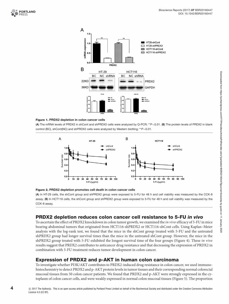
PRDX2 depletion promotes cell apoptosis in colon cancer cellsWe analyzed the cell apoptosis by flow cytometry and found that knocking down PRDX2 expression in FU-treatedcolon cancer cells markedly increased apoptosis and protein levels of cleaved PARP and caspase-3 (Figure 3A,B).Collectively, our results suggest that PRDX2 depletion, combined with 5-FU, promotes cell apoptosis in colon cancercells.
c© 2017 The Author(s). This is an open access article published by Portland Press Limited on behalf of the Biochemical Society and distributed under the Creative Commons AttributionLicence 4.0 (CC BY).
3
Dow
nloaded from https://portlandpress.com
/HTTPH
andlers/ArticlePdfHandler.ashx?partialdoi=BSR
20160447&journal=bioscirep by guest on 11 January 2020
Bioscience Reports (2017) 37 BSR20160447DOI: 10.1042/BSR20160447
Figure 1. PRDX2 depletion in colon cancer cells
(A) The mRNA levels of PRDX2 in shCont and shPRDX2 cells were analyzed by Q-PCR; **P<0.01. (B) The protein levels of PRDX2 in blank
control (BC), shCont(NC) and shPRDX2 cells were analyzed by Western blotting; **P<0.01.
Figure 2. PRDX2 depletion promotes cell death in colon cancer cells
(A) In HT-29 cells, the shCont group and shPRDX2 group were exposed to 5-FU for 48 h and cell viability was measured by the CCK-8
assay. (B) In HCT116 cells, the shCont group and shPRDX2 group were exposed to 5-FU for 48 h and cell viability was measured by the
CCK-8 assay.
PRDX2 depletion reduces colon cancer cell resistance to 5-FU in vivoTo ascertain the effect of PRDX2 knockdown in colon tumor growth, we examined the in vivo efficacy of 5-FU in micebearing abdominal tumors that originated from HCT116-shPRDX2 or HCT116-shCont cells. Using Kaplan–Meieranalysis with the log-rank test, we found that the mice in the shCont group treated with 5-FU and the untreatedshPRDX2 group had longer survival times than the mice in the untreated shCont group. However, the mice in theshPRDX2 group treated with 5-FU exhibited the longest survival time of the four groups (Figure 4). These in vivoresults suggest that PRDX2 contributes to anticancer drug resistance and that decreasing the expression of PRDX2 incombination with 5-FU treatment reduces tumor development in colon cancer.
Expression of PRDX2 and p-AKT in human colon carcinomaTo investigate whether PI3K/AKT contributes to PRDX2-induced drug resistance in colon cancer, we used immuno-histochemistry to detect PRDX2 and p-AKT protein levels in tumor tissues and their corresponding normal colorectalmucosal tissues from 50 colon cancer patients. We found that PRDX2 and p-AKT were strongly expressed in the cy-toplasm of colon cancer cells, and were weakly expressed in normal colon mucosal tissues (Figure 5). The proportion
4 c© 2017 The Author(s). This is an open access article published by Portland Press Limited on behalf of the Biochemical Society and distributed under the Creative Commons AttributionLicence 4.0 (CC BY).
Dow
nloaded from https://portlandpress.com
/HTTPH
andlers/ArticlePdfHandler.ashx?partialdoi=BSR
20160447&journal=bioscirep by guest on 11 January 2020
Bioscience Reports (2017) 37 BSR20160447DOI: 10.1042/BSR20160447
Figure 3. PRDX2 depletion promotes cell apoptosis in colon cancer cells
(A) Cell apoptosis levels were evaluated by flow cytometry in HT-29 and HCT116 cells treated with 5-FU for 48 h; *P<0.05. (B) The protein
levels of C-PARP and caspase-3 in HT-29 and HCT116 cells treated with 5-FU for 48 h were analyzed by Western blotting; *P<0.05,
**P<0.01.
Figure 4. PRDX2 depletion reduces the resistance of colon cancer cells to 5-FU in vivo
Five female BALB/c-nu mice were placed in each group. The shCont group treated with 5-FU and shPRDX2 group with PBS injection had
longer survival times than nude mice in the shCont group with PBS injection. However, nude mice in the shPRDX2 group with 5-FU treatment
had the longest survival time compared with the other three groups; *P<0.05.
of PRDX2-positive cells and p-AKT-positive cells were 86% (43/50) and 88% (44/50), respectively. On further analy-sis, we found that PRDX2 and p-AKT showed a significant positive correlation (r =0.738, P<0.05) through Pearsoncorrelation (Table 1). These results demonstrate that PI3K/AKT contributes to PRDX2-mediated 5-FU resistance.
c© 2017 The Author(s). This is an open access article published by Portland Press Limited on behalf of the Biochemical Society and distributed under the Creative Commons AttributionLicence 4.0 (CC BY).
5
Dow
nloaded from https://portlandpress.com
/HTTPH
andlers/ArticlePdfHandler.ashx?partialdoi=BSR
20160447&journal=bioscirep by guest on 11 January 2020

Bioscience Reports (2017) 37 BSR20160447DOI: 10.1042/BSR20160447
Figure 5. The expression of PRDX2 and p-AKT in human colon carcinoma
Representative images of PRDX2 and p-AKT expression in human colon carcinoma samples and normal adjacent tissues are shown.
Table 1 Correlation of PRDX2 and p-AKT expression in colon cancer tissue samples
p-AKT PRDX2Pearson correlationcoefficient P-value
Positive (n=44) Negative (n=6)
Positive (n=43) 42 1 0.738 <0.05
Negative (n=7) 2 5
PI3K/AKT is crucial for PRDX2-mediated 5-FU resistance in colon cancerThe protein expressions of AKT, p-AKT and p-PI3K were analyzed by Western blotting in HT-29-shCont orHT-29-shPRDX2 cells. We found that depleting PRDX2 resulted in reduced p-AKT and p-PI3K expressions (Figure6A). Furthermore, as shown in Figure 6B, 5-FU-treated HT29 and HCT116 cells were infected with overexpressedPRDX2, LV-PRDX2, and protein expressions of p-AKT and Bcl-2/Bax protein were observed. To verify the role ofPI3K/AKT in PRDX2-induced drug resistance, we treated HT-29 cells with the AKT inhibitor, MK-2206 2HCl andthen observed the resistance of HT-29 cells to 5-FU. MK-2206 inhibited AKT/PI3K protein expression. However, el-evated expressions of caspase-3 and Bax were detected, along with suppressed Bcl-2 expression post MK-2206 treat-ment (Figure 6C). These results were validated by measuring apoptosis by using flow cytometry after cells were treated5-FU in the presence or absence of MK-2206 HCl . We observed higher C-PARP and Bax protein expression and ahigher rate of apoptosis in colon cancer cells treated with MK-2206 2HCl (Figure 6C,D). Together, these data suggestthat 5-FU targets PRDX2 and down-regulates its expression through the AKT/PI3K pathway.
DiscussionIn general, higher levels of ROS contribute to tumor progression and development in most tumors compared withnormal tissues [8]. Moreover, oxidative stress is associated with the efficacy of cancer treatments, including chemosen-sitivity and apoptosis [9]. However, extremely high ROS concentrations are deadly to tumor cells [10]. 5-FU inhibitsthymidylate synthetase and incorporates into both RNA and DNA, inducing intracellular increases in O2− levels incolon cancer [5,11]. This results in a new redox balance with a higher ROS level and an enhanced ROS scavengingsystem, that is resistant to 5-FU [12]. For these tumors, combining ROS generating drugs and restraining the cellularantioxidant capacity is effective. For example, combining arsenic trioxide and ascorbic acid, induces GSH depletion,and is effective in treating relapsed or refractory multiple myeloma [13]. In fact, PRDX3 knockdown combined with5-FU increased cell death and tumor suppression in colon cancer [14].
PRDX2, is an important member of the ROS scavenging system, and exerts an anti-apoptotic effect on colon cancercells. Lu et al. [15] showed that PRDX2 is up-regulated and protects cells from oxidative stress in colorectal cancer. Inour study, we found that knockdown of PRDX2 expression promotes sensitivity to 5-FU in colon cancer cells. Theseresults suggest that depleting PRDX2 combined with 5-FU treatment could be a promising strategy for anticancerdrug resistance.
6 c© 2017 The Author(s). This is an open access article published by Portland Press Limited on behalf of the Biochemical Society and distributed under the Creative Commons AttributionLicence 4.0 (CC BY).
Dow
nloaded from https://portlandpress.com
/HTTPH
andlers/ArticlePdfHandler.ashx?partialdoi=BSR
20160447&journal=bioscirep by guest on 11 January 2020

Bioscience Reports (2017) 37 BSR20160447DOI: 10.1042/BSR20160447
Figure 6. PI3K/AKT is crucial for PRDX2-mediated 5-FU resistance in colon cancer
(A) The protein expressions of PRDX2 AKT/PI3K, Bax/Bcl-2 were analyzed by Western blotting in HT-29-shCont or HT-29-shPRDX2 cells;
*P<0.05. (B) HT29 and HT116 cells were treated with LV-PRDX2 and the protein expressions of AKT, p-AKT, Bax and Bcl-2 were detected
by Western blotting. (C) HT-29 cells were treated with MK-2206 2HCl for 48 h and then the protein expressions of PRDX2, AKT/PI3K,
Bax/Bcl-2 were detected by Western blotting; *P<0.05. (D) Cell apoptosis levels were evaluated by flow cytometry in HT-29 cells treated
with and without MK-2206 2HCl; *P<0.05.
Hwang et al. [4] found that 5-FU treatment induced ROS generation in human lung carcinoma cells, leading to ahigher level of antioxidant enzymes and resistance to 5-FU. PRDX proteins protect MCF-7 breast cancer cells fromdoxorubicin-induced toxicity [16]. PRDX6 overexpression attenuated cisplatin-induced apoptosis in human ovariancancer cells [17]. Kalinina et al. [18] found that cisplatin resistance in cancer cells was accompanied with a signif-icantly increased expression of PRDX2 gene. In pancreatic cancer, a proteomic study using gemcitabine-sensitiveKLM1 and -resistant KLM1-R cells showed that PRDX2 was significantly up-regulated in KLM1-R cells [19]. Theexpression of PRDX2 was higher in osteosarcoma patients that were poor responders to induction chemotherapy.In addition, PRDX2 depletion contributed to increased sensitivity to chemotherapeutic drugs in osteosarcoma cells
c© 2017 The Author(s). This is an open access article published by Portland Press Limited on behalf of the Biochemical Society and distributed under the Creative Commons AttributionLicence 4.0 (CC BY).
7
Dow
nloaded from https://portlandpress.com
/HTTPH
andlers/ArticlePdfHandler.ashx?partialdoi=BSR
20160447&journal=bioscirep by guest on 11 January 2020

Bioscience Reports (2017) 37 BSR20160447DOI: 10.1042/BSR20160447
[20,21]. Furthermore, PRDX2 localized to the nucleus and PRDX2 depletion markedly promoted cell death in cancercells treated with DNA damaging agents [22]. These observations are consistent with data from our study. We treatedshCont and shPRDX2 transfected colon cancer cells with 5-FU and found that apoptotic cells, C-PARP and caspase-3protein expression increased markedly when PRDX2 was depleted. These results indicated that PRDX2 depletionpromotes sensitivity to 5-FU in colon cancer cells.
Recent studies revealed that the PI3K/AKT pathway plays an important role in chemoresistance [23]. NPC (na-sopharyngeal carcinoma) cell lines stably overexpressing miR-3188 exhibited significantly increased sensitivity to5-FU by inactivating the (PI3K)/AKT pathway [24]. NECTIN-4 increased the resistance of colon cancer cells to 5-FUby inducing the PI3K/AKT cascade [25]. Activation of the PI3K/AKT pathway contributes to resistance to multiplecancer therapies, and is deemed a poor prognostic factor for cancers [26]. In our study, we found that p-AKT proteinlevels were lower in PRDX2-depleted colon cancer cells. Moreover, when the AKT inhibitor, MK-2206 was added to5-FU treated colon cancer cells, we oserved higher C-PARP protein levels and increased cell apoptosis. These resultsindicated that PI3K/AKT is a key pathway that contributes to PRDX2-mediated 5-FU-induced apoptosis.
Taken together, our results demonstrate that inhibiting PRDX2 expression promotes 5-FU-induced apoptosis incolon cancer cells via the PI3K/AKT signal pathway.
Competing interestsThe authors declare that there are no competing interests associated with the manuscript.
FundingThis work was supported by the National Natural Science Foundation of China [grant number 81572319].
Author contributionGuarantor of integrity of entire study and manuscript final version approval: Zhongxue Fu; Study design and experimental studies:Jun Xu; Data analysis and statistical analysis: Shouru Zhang; Manuscript editing: Rong Wang; Manuscript revision: Xingye Wu andLi Zeng
AbbreviationsCCK-8, cell counting kit-8; PRDX2, peroxiredoxin-2; ROS, reactive oxygen species; 5-FU, 5-Fluorouracil; LV, lentivirus.
References1 Chen, W., Zheng, R., Zeng, H., Zhang, S. and He, J. (2015) Annual report on status of cancer in China, 2011. Chin. J. Cancer Res. 27, 2–122 Holohan, C., Van Schaeybroeck, S., Longley, D.B. and Johnston, P.G. (2013) Cancer drug resistance: an evolving paradigm. Nat. Rev. Cancer 13,
714–7263 Alexandre, J., Nicco, C., Chereau, C., Laurent, A., Weill, B., Goldwasser, F. et al. (2006) Improvement of the therapeutic index of anticancer drugs by the
superoxide dismutase mimic mangafodipir. J. Natl. Cancer Inst. 98, 236–2444 Hwang, I.T., Chung, Y.M., Kim, J.J., Chung, J.S., Kim, B.S., Kim, H.J. et al. (2007) Drug resistance to 5-FU linked to reactive oxygen species modulator
1. Biochem. Biophys. Res. Commun. 359, 304–3105 Hwang, P.M., Bunz, F., Yu, J., Rago, C., Chan, T.A., Murphy, M.P. et al. (2001) Ferredoxin reductase affects p53-dependent, 5-fluorouracil-induced
apoptosis in colorectal cancer cells. Nat. Med. 7, 1111–11176 Lu, W., Fu, Z., Wang, H., Feng, J., Wei, J. and Guo, J. (2014) Peroxiredoxin 2 knockdown by RNA interference inhibits the growth of colorectal cancer
cells by downregulating Wnt/beta-catenin signaling. Cancer Lett. 343, 190–1997 Wu, X.Y., Fu, Z.X. and Wang, X.H. (2010) Peroxiredoxins in colorectal neoplasms. Histol. Histopathol. 25, 1297–13038 Gorrini, C., Harris, I.S. and Mak, T.W. (2013) Modulation of oxidative stress as an anticancer strategy. Nat. Rev. Drug Discov. 12, 931–9479 Nathan, C. and Cunningham-Bussel, A. (2013) Beyond oxidative stress: an immunologist’s guide to reactive oxygen species. Nat. Rev. Immunol. 13,
349–36110 Trachootham, D., Alexandre, J. and Huang, P. (2009) Targeting cancer cells by ROS-mediated mechanisms: a radical therapeutic approach? Nat. Rev.
Drug Discov. 8, 579–59111 Longley, D.B., Harkin, D.P. and Johnston, P.G. (2003) 5-fluorouracil: mechanisms of action and clinical strategies. Nat. Rev. Cancer 3, 330–33812 Liu, Y., Li, Q., Zhou, L., Xie, N., Nice, E.C., Zhang, H. et al. (2016) Cancer drug resistance: redox resetting renders a way. Oncotarget 7, 42740–4276113 Bahlis, N.J., McCafferty-Grad, J., Jordan-McMurry, I., Neil, J., Reis, I., Kharfan-Dabaja, M. et al. (2002) Feasibility and correlates of arsenic trioxide
combined with ascorbic acid-mediated depletion of intracellular glutathione for the treatment of relapsed/refractory multiple myeloma. Clin. CancerRes. 8, 3658–3668
14 Song, I.S., Jeong, Y.J., Jeong, S.H., Heo, H.J., Kim, H.K., Bae, K.B. et al. (2015) FOXM1-induced PRX3 regulates stemness and survival of colon cancercells via maintenance of mitochondrial function. Gastroenterology 149, 1006–1016.
15 Lu, W., Fu, Z., Wang, H., Feng, J., Wei, J. and Guo, J. (2014) Peroxiredoxin 2 is upregulated in colorectal cancer and contributes to colorectal cancercells’ survival by protecting cells from oxidative stress. Mol. Cell Biochem. 387, 261–270
8 c© 2017 The Author(s). This is an open access article published by Portland Press Limited on behalf of the Biochemical Society and distributed under the Creative CommonsAttribution Licence 4.0 (CC BY).
Dow
nloaded from https://portlandpress.com
/HTTPH
andlers/ArticlePdfHandler.ashx?partialdoi=BSR
20160447&journal=bioscirep by guest on 11 January 2020

Bioscience Reports (2017) 37 BSR20160447DOI: 10.1042/BSR20160447
16 McDonald, C., Muhlbauer, J., Perlmutter, G., Taparra, K. and Phelan, S. (2014) Peroxiredoxin proteins protect MCF-7 breast cancer cells fromdoxorubicin-induced toxicity. Int. J. Oncol. 45, 219–226
17 Pak, J.H., Choi, W.H., Lee, H.M., Joo, W.D., Kim, J.H., Kim, Y.T. et al. (2011) Peroxiredoxin 6 overexpression attenuates cisplatin-induced apoptosis inhuman ovarian cancer cells. Cancer Invest. 29, 21–28
18 Kalinina, E.V., Berezov, T.T., Shtil, A.A., Chernov, N.N., Glazunova, V.A., Novichkova, M.D. et al. (2012) Expression of peroxiredoxin 1, 2, 3, and 6 genesin cancer cells during drug resistance formation. Bull. Exp. Biol. Med. 153, 878–881
19 Suenaga, S., Kuramitsu, Y., Wang, Y., Baron, B., Kitagawa, T., Akada, J. et al. (2013) Human pancreatic cancer cells with acquired gemcitabineresistance exhibit significant up-regulation of peroxiredoxin-2 compared to sensitive parental cells. Anticancer Res. 33, 4821–4826
20 Kikuta, K., Tochigi, N., Saito, S., Shimoda, T., Morioka, H., Toyama, Y. et al. (2010) Peroxiredoxin 2 as a chemotherapy responsiveness biomarkercandidate in osteosarcoma revealed by proteomics. Proteomics Clin. Appl. 4, 560–567
21 Kubota, D., Mukaihara, K., Yoshida, A., Tsuda, H., Kawai, A. and Kondo, T. (2013) Proteomics study of open biopsy samples identifies peroxiredoxin 2 asa predictive biomarker of response to induction chemotherapy in osteosarcoma. J. Proteomics 91, 393–404
22 Lee, K.W., Lee, D.J., Lee, J.Y., Kang, D.H., Kwon, J. and Kang, S.W. (2011) Peroxiredoxin II restrains DNA damage-induced death in cancer cells bypositively regulating JNK-dependent DNA repair. J. Biol. Chem. 286, 8394–8404
23 Hafsi, S., Pezzino, FM., Candido, S., Ligresti, G., Spandidos, D.A., Soua, Z., McCubrey, J.A., Travali, S. and Libra, M. (2012) Gene alterations in thePI3K/PTEN/AKT pathway as a mechanism of drug-resistance (review).. Int. J. Oncol. 40, 639–44, doi:22200790
24 Zhao, M., Luo, R., Liu, Y., Gao, L., Fu, Z., Fu, Q. et al. (2016) miR-3188 regulates nasopharyngeal carcinoma proliferation and chemosensitivity througha FOXO1-modulated positive feedback loop with mTOR-p-PI3K/AKT-c-JUN. Nat. Commun. 7, 11309
25 Das, D., Satapathy, S.R., Siddharth, S., Nayak, A. and Kundu, C.N. (2015) NECTIN-4 increased the 5-FU resistance in colon cancer cells by inducing thePI3K-AKT cascade. Cancer Chemother. Pharmacol. 76, 471–479
26 LoPiccolo, J., Blumenthal, G.M., Bernstein, W.B. and Dennis, P.A. (2008) Targeting the PI3K/Akt/mTOR pathway: effective combinations and clinicalconsiderations. Drug Resist. Updat. 11, 32–50
c© 2017 The Author(s). This is an open access article published by Portland Press Limited on behalf of the Biochemical Society and distributed under the Creative Commons AttributionLicence 4.0 (CC BY).
9
Dow
nloaded from https://portlandpress.com
/HTTPH
andlers/ArticlePdfHandler.ashx?partialdoi=BSR
20160447&journal=bioscirep by guest on 11 January 2020

Dow
nloaded from https://portlandpress.com
/HTTPH
andlers/ArticlePdfHandler.ashx?partialdoi=BSR
20160447&journal=bioscirep by guest on 11 January 2020