Embed Size (px)
Citation preview

París et al, 1
GANGLIOSIDE GD3 SENSITIZES HUMAN HEPATOMA CELLS TO CANCER THERAPY.
Raquel París, Albert Morales, Olga Coll, Alberto Sánchez-Reyes#, Carmen García-Ruiz and
José C. Fernández-Checa*.
Liver Unit, Instituto de Malalties Digestives, # Servicio de Radioterapia, Hospital Clinic i Provincial,
Instituto Investigaciones Biomedicas August Pi Suñer, and *Department of Experimental Pathology,
Instituto Investigaciones Biomédicas Barcelona, Consejo Superior de Investigaciones Científicas,
Barcelona, 08036, Spain.
Running title: Ganglioside GD3 and cancer therapy.
Raquel París and Albert Morales have contributed equally to the work.
Carmen García-Ruiz and José C. Fernández-Checa share senior authorship.
Address correspondence to José C. Fernández-Checa, Liver Unit, Hospital Clinic i Provincial
C/Villarroel, 170
08036-Barcelona
Spain
Copyright 2002 by The American Society for Biochemistry and Molecular Biology, Inc.
JBC Papers in Press. Published on September 25, 2002 as Manuscript M208303200 by guest on June 30, 2018
http://ww
w.jbc.org/
Dow
nloaded from

París et al, 2
ABSTRACT
Ganglioside GD3 (GD3) has emerged as a modulator of cell death pathways due to its ability to
interact with mitochondria and disable survival pathways. Since NF-κB activation contributes to
cancer therapy resistance, this study was undertaken to test whether GD3 modulates the response
of human hepatoblastoma HepG2 cells to radio and chemotherapy. NF-κB was activated in HepG2
cells shortly after therapeutic doses of ionizing radiation or daunorubicin treatment that translated in
upregulation of κB-dependent genes. These effects were accompanied by minimal killing of HepG2
cells by either ionizing radiation or daunorubicin. However, GD3 pretreatment blocked the nuclear
translocation of active κB members, without effect on Akt phosphorylation, induced by either
treatment. The suppression of κB-dependent gene induction by GD3 was accompanied by
enhanced apoptotic cell death caused by these therapies. Furthermore, the combination of GD3 plus
ionizing radiation stimulated the formation of reactive species followed by the mitochondrial release
of cytochrome c and Smac/Diablo and caspase 3 activation. Pretreatment with cyclosporin A before
radiotherapy protected HepG2 cells from the therapeutical combination of GD3 plus ionizing
radiation. These findings underscore a key role of mitochondria in the response of tumor cells to
cancer therapy and highlight the potential relevance of GD3 to overcome resistance to cancer
therapy by combining its dual action as a mitochondria-interacting and NF-κB inactivating agent.
by guest on June 30, 2018http://w
ww
.jbc.org/D
ownloaded from

París et al, 3
INTRODUCTION
The resistance of tumor cells to current cancer therapy-induced cell death underlines a major
problem that limits the successful treatment of human cancer (1, 2). Among the different molecular
strategies that contribute to the resistance of tumor cells to radio and chemotherapy, the role played
by transcription factor NF-κB1 has been well defined. Indeed, the prior inactivation of NF-κB
rendered different tumor cell types sensitive to cancer therapy (3-7). Although ionizing radiation
and chemotherapeutic agents activate apoptosis pathways, the impact of these are often
antagonized by the early activation of NF-κB-dependent survival pathways induced by these
treatments (8, 9).
NF-κB is usually kept inactive in the cytoplasm through association with an endogenous inhibitor
protein of the IκB (inhibitor of NF-κB) family. The most common pathway leading to NF-κB
activation by a wide variety of stimuli including cancer therapy involves the phosphorylation of
IκB at specific serine residues that targets its subsequent degradation by the proteasome (3-9). The
released subunits of NF-κB then translocate to the nuclei where they bind to specific sites in the
promoter/enhancer region of target genes. NF-κB is known to induce the expression of many
different genes involved in the regulation of immune or stress response and apoptosis (3). Since its
first recognition as an antiapoptotic factor in mice lacking the p65 component of NF-κB (10),
increasing evidence has further documented the antiapoptotic activity of NF-κB against a variety of
stimuli (11, 12, 13). The central role of NF-κB in the prevention of apoptosis is mediated through
the induction of antiapoptotic genes including Bcl-XL, c-IAP1, c-IAP2, A1/Bfl1 or antioxidant
enzymes such as MnSOD (14-17).
Glycosphingolipids (GSLs), carbohydrate-bearing lipid components of biological membranes,
participate in the regulation of various cellular functions, including cell adhesion and signal
by guest on June 30, 2018http://w
ww
.jbc.org/D
ownloaded from

París et al, 4
transduction (18, 19). In particular, ganglioside GD3, a sialic acid-containing GSLs, has been
identified as a lipid death effector due to its ability to interact with and recruit mitochondria to
apoptotic pathways, contributing to the mitochondrial-dependent apoptosome activation and
subsequent apoptosis triggered by death ligands (20-26). In addition to the direct apoptosis-
promoting activity of GD3 through mitochondrial interaction, this GSLs species counteracts
survival signals by suppressing the NF-κB activation and subsequent κB-dependent gene induction
(27, 28). This dual function of GD3 has been shown to render rat hepatocytes susceptible to TNF-
mediated cell death through the inactivation of NF-κB (27). Yet, despite the available evidence
supporting the proapoptotic function of GD3, the role of glycolipids in apoptosis is controversial.
The inhibition of glycolipid synthesis has been shown to enhance apoptosis, reversing multi-drug
resistance in certain tumor cells (29, 30).
Hence, in light of these findings and due to the pivotal role of NF-κB in mediating the resistance of
tumor cells to cancer therapy, the purpose of the present work was to test the hypothesis that
GD3 pretreatment sensitizes human hepatoblastoma HepG2 cells to ionizing radiation and
chemotherapy by its combined function as a mitochondrial recruiting and NF-κB disabling agent.
by guest on June 30, 2018http://w
ww
.jbc.org/D
ownloaded from

París et al, 5
MATERIALS AND METHODS
Materials- Daunorubicin and CsA were purchased from Sigma (Madrid, Spain). Hoescht 33258
was obtained from Molecular Probes (Eugene, OR). GD3, 99% purity by thin layer
chromatography (TLC), was from Matreya, Inc. (Pleasant Gap, PA). Caspase 3 substrate, Ac-Asp-
Glu-Val-Asp-7-amino-4-trifluoromethyl coumarin (Ac-DEVD-AMC) and anti-Smac/Diablo were
from Calbiochem. Anti-phospho-Akt and anti-Akt antibodies were from Cell Signaling Technology
(Beverly, MA).
Cell culture and treatments- The human hepatoblastoma cell line, HepG2, was obtained from the
European Collection of Animal Cell Cultures (Salisbury, Wilts, UK) and grown at 37 ºC in 5 % CO2
in Dubelcco’s modified Eagle’s medium containing high glucose levels. Culture medium was
supplemented with 10% heat-inactivated fetal bovine serum, 2mM L-glutamine, penicillin (100
units/ml), and streptomycin (100 µg/ml). Subconfluent HepG2 cells were irradiated in a linear
accelerator (KDS Siemens) at room temperature using an electron beam of 18 MeV as described
previously (31). Doses between 2 and 20 Gy were applied at a rate of 3 Gy/min. Estimate errors on
dose have been calculated to be below 1%. Alternatively, cells were treated with daunorubicin (1-10
µM) for 2-72 h. In some cases, cells were preincubated with GD3 (5 µM in ethanol) four hours
before radiation or daunorubicin treatment and examined for cell survival (2-72 hours). The final
concentration of the carrier solvent did not exceed 0.1% nor affected any of the parameters
determined.
NF- B activation. NF-κB DNA binding activity in nuclear extracts was assessed by EMSA using
NF-κB consensus oligonucleotide (5’AGTTGAGGGGACTTTCCCAGGC-3’) as described
previously (32). After radiation or daunorubicin treatment, cells were washed twice with ice cold
phosphate-buffered saline, collected with a rubber policemen and lysed with Nonidet P-40 (10%).
by guest on June 30, 2018http://w
ww
.jbc.org/D
ownloaded from

París et al, 6
The nuclear pellet was recovered by spinning (13,000 g at 4°C for 30s), resuspended in ice-cold
20mM Hepes, pH 7.4, containing 0.4mM NaCl, 1mM EDTA, 1mM EGTA, 1mM dithiothreitol,
1mM PMSF, 10µg/ml leupeptin, 10µg/ml aprotinin, and 10µg/ml TLCK and stored at -80 °C.
Binding reaction contained 7µg of nuclear extracts, 5µl incubation buffer (10 mM Tris-HCL, 40 mM
NaCl, 1mM EDTA and 4 % glycerol) and 1 µg poly (dI-dC). After 15 min on ice, the labeled
oligonucleotide (30.000 cpm) was added and the mixture incubated for 20 min at room temperature.
The mixture was electrophoresed through a 6% polyacrilamide gel for 90 min at 150 V.
Transactivation of NF- B. HepG2 cells were transfected with the plasmid pNF-κB-Luc
containing four tandem copies of the κB enhancer upstream of the herpes simplex virus thymidine
kinase fused to the luciferase reporter using lipofectAMINE reagent as described previously (32). In
each experiment, six 35-mm wells containing 5x10⁶ cells were transfected with 5 µg of a DNA and 5
µg of the pSV-β-galactosidase vector as control to monitor transfection efficiency. After 24-48 h
transfection cells were irradiated (4Gy) or treated with daunorubicin (1-10µM) and 6 hours later,
harvested in a reporter lysis buffer and supernatants were used to determine luciferase activity.
Results were normalized to β-galactosidase activity and protein concentration.
Localization of NF- B p65 by confocal microscopy. To determine the distribution of NF-κB p65
subunit, HepG2 cells were fixed with formaline 10% and incubated with anti-NF-κB p65 antibody
(Santa Cruz Biotechnology, Santa Cruz, CA.) at 300 ng/ml in antibody diluent (Dako, Carpinteria,
CA) for 1 hour. Cells were washed with PBS and incubated with FITC–coupled anti-rabbit Ig G
antibody (Jackson ImmunoResearch, Santa Cruz, CA) and analyzed by a Leica TCS-NT confocal
microscope.
Akt phosphorylation. Total cell proteins were resolved in a 7.5% SDS/PAGE gel electrophoresis.
Phosphorylated Akt was detected using an anti-phospho-Akt (Ser-473) antibody (Cell-signaling
by guest on June 30, 2018http://w
ww
.jbc.org/D
ownloaded from
París et al, 7
Technology, Beverly, MA). Membranes were then stripped and incubated with anti-Akt antibody
(Cell signaling Technology, Beverly, MA) to estimate the levels of Akt.
Cytochrome c and Smac/Diablo release. Cells were permeabilized with digitonin (40µg/ml ) in
0.5 ml of intracellular medium composed of 120 mM KCl, 10 mM NaCl, 1 mM KH2PO4, 20 mM
Hepes-Tris, pH 7.2, supplemented with 1 µg/ml of each of antipain, leupeptin and pepstatin for 15
min. Upon centrifugation at 14.000 x g the supernatant and the mitochondria-containing pellet were
resolved by SDS/PAGE (15% gels). Proteins were transferred to nitrocellulose, and the blots were
incubated with anti-cytochrome c antibody (Pharmingen), and anti-Smac/Diablo antibody
(Calbiochem) followed by ECL-based detection. To monitor specificity of cytochrome c and
Smac/Diablo release parallel aliquots were immunobloted with human monoclonal antibody anti-
cytochrome c oxidase subunit II (Molecular Probes).
Caspase activation. Cytosolic extracts were used to measure caspase 3 activity from the release of
7-amino-4-trifluoromethyl coumarin from Ac-DEVD-AMC and fluorescence was continuously
recorded with excitation at 380 nm and emission at 460 nm as described previously (23).
Reactive species determination. Reactive species formation were determined using chloromethyl-
2´-7´-dichlorodihydrofluorescein diacetate (CM-H2DCFDA, Molecular Probes, Eugene, OR) which
becomes highly fluorescent upon oxidation by peroxides (17, 31) as well as peroxynitrite (33). DCF
formation was continuously recorded in a fluorimeter with excitation at 380 nm and emission at 460
nm. Relative fluorescence units were normalized per mg cellular protein.
Cell viability and apoptosis. Cell death was determined by the measurement of the release of
lactate dehydrogenase (LDH) into the medium and in the remaining cell monolayer after lysis with
5% Triton X-100, which expresses the percentage of LDH release in the medium as the fraction of
LDH (medium plus cells). Results were confirmed by the MTT assay (Roche) following
by guest on June 30, 2018http://w
ww
.jbc.org/D
ownloaded from

París et al, 8
manufacturer’s instructions. Alternatively, cells were stained with Hoescht-33258 and morphologic
features of apoptosis analyzed by fluorescence microscopy. More than 200 cells per condition were
examined for apoptotic features including the presence of chromatin condensation or fragmentation.
Statistical Analyses. Results are expressed as the mean ± SD and averages of three to five
experiments. Statistical analysis of mean values for multiple comparisons were may by one-way
ANOVA
by guest on June 30, 2018http://w
ww
.jbc.org/D
ownloaded from

París et al, 9
RESULTS
Activation of NF- B by cancer therapy and inhibition by GD3.
NF-κB activation has been reported in different tumor cell lines after chemotherapeutic treatments
and ionizing radiation and thought to function as a defense mechanism to counteract the cell death
cascades generated by these cancer therapies (3-6). To study the effect of ionizing radiation or
chemotherapy on the DNA binding activity of NF-κB in human hepatoblastoma cells, EMSA was
performed using nuclear extracts obtained from HepG2 cells after exposure to ionizing radiation (2-
20 Gy). As shown (Figure 1A), ionizing radiation enhanced in a dose-dependent fashion the DNA
binding activity of several complexes of NF-κB. While these complexes were displaced by molar
excess of unlabeled κB oligonucleotide (not shown), indicating their specificity for κB binding sites,
only the two upper bands were identified as RelA/p52 and p52/p50 dimers by supershifts assays
using antibodies against individual NF-κB components (RelA, p52 and p50). The activation of these
dimers by ionizing radiation occurred within 30 minutes post-treatment and lasted for 6-8 hours
(not shown). Treatment of HepG2 cells with daunorubicin (1 µM) resulted in a similar pattern of
NF-κB DNA binding activity (Figure 1B). The level of activation of both RelA/p52 and p52/p50
dimers ranged from 3 to 4 fold respect to untreated control cells (Figure 1B).
Since previous findings in cultured rat hepatocytes revealed that GD3 suppressed the activation of
NF-κB induced by TNF (27), we next examined whether GD3 pretreatment abolished the activation
of NF-κB by ionizing radiation and daunorubicin treatments. As observed (Figure 1B), GD3
preincubation prevented the activation of NF-κB induced by ionizing radiation (4 Gy) or
daunorubicin (1µM), decreasing the intensity of RelA/p50 and p50/p52 complexes to control levels.
Thus, these findings confirm the activation of NF-κB upon cancer therapy treatment of HepG2
cells, which is blocked by pretreatment of cells with GD3.
by guest on June 30, 2018http://w
ww
.jbc.org/D
ownloaded from
París et al, 10
GD3 blocks the nuclear translocation of NF- B.
To confirm if NF-κB active complexes from GD3 pretreated cells remained in the cytosol after
exposure to radiotherapy, we determined the cellular localization of NF-κB p65 subunit. As seen
(Fig 2), compared to control HepG2 cells which display a diffuse fluorescence pattern, ionizing
radiation induced the localization of p65 into the nuclei. This shift in the cellular distribution of p65
induced by ionizing radiation was blunted by GD3 pretreatment (Fig 2). Moreover, EMSA assays
confirmed the presence of DNA binding activity of NF-κB in cytosolic extracts (not shown).
Similar findings were observed when HepG2 cells were exposed to daunorubicin with or without
GD3 pretreatment (not shown).
Since the blocking effect of GD3 on the nuclear translocation of competent DNA-binding NF-κB
members may be translated at the gene level, we next examined the transactivation of NF- κB using a
luciferase reporter gene construct controlled by four κB-binding sites. Ionizing radiation and
daunorubicin treatment enhanced the κB-dependent luciferase expression by 8-10 fold (Fig 2).
However, the preincubation of HepG2 cells with GD3 abolished the inducible expression of
luciferase by ionizing radiation or daunorubicin. Thus, taken collectively, these findings underscore
the impact of GD3 pretreatment in suppressing the inducible expression of κB-regulated genes.
GD3 sensitizes HepG2 cells to cancer therapy.
Since NF-κB is known to induce the expression of antiapoptotic genes that counteract apoptosis
pathways, we next assessed the consequences of the inactivation of NF-κB by GD3 on the survival
of HepG2 cells following cancer therapy. As shown (Fig 3 A), HepG2 cells were relatively resistant
to a therapeutical dose of ionizing radiation or daunorubicin treatment displaying slight signs of cell
injury after 3 days post-treatment (17±7% and 22±6%, respectiviely) (Fig 3B). Of note, when
HepG2 cells were pretreated with GD3 at a sublethal dose, it rendered HepG2 cells vulnerable to
by guest on June 30, 2018http://w
ww
.jbc.org/D
ownloaded from

París et al, 11
both ionizing radiation or daunorubicin treatment (Fig 3). This sensitization was observed as early
as 24 hours after GD3 pretreatment (17±4% and 19±6% cell death after ionizing radiation or
daunorubicin treatment, compared to 5±3% and 7±4%, without GD3, respectively). Similar
findings were observed when cell survival was quantited by the MTT assays (not shown).
To investigate if the cell death caused by these treatments after GD3 preincubation was
accompanied by apoptotic features, HepG2 cells were exposed to the DNA-binding fluorochrome
H-33258 and chromatin morphology was visualized by fluorescence microscopy. As shown (Fig 4)
the combination of GD3 plus ionizing radiation or daunorubicin resulted in increased presence of
cells displaying chromatin disruption, indicative of apoptosis. Indeed, the estimation of apoptotic
cell death indicated a significant sensitization 24 hours after the combination of ionizing radiation or
daunorubicin after GD3 compared with either therapy alone (Fig 4B). Clearly, these findings
indicate that the pretreatment of HepG2 cells with GD3 increases the efficiency of therapies
intended to kill human tumor cells.
GD3 does not interfere with Akt signaling.
Akt, a serine/threonine protein kinase, has been shown to regulate cell survival signals by rendering
key components of the apoptotic cascade inactive (34-36). Therefore, in view of the preceding
findings, we next assessed whether GD3 disabled the Akt signaling pathway thus contributing to the
susceptibility of HepG2 cells towards cancer therapy. In order to study the contribution of this
pathway, we determined the level of Akt and its phosphorilated form (at serine-473) using cell
extracts from ionizing radiation-exposed cells with or without GD3 preincubation. As seen,
compared to the phosphorylation induced by insulin, ionizing radiation did not induce the
phosphorylation of Akt (Fig 5). The phosphorylated form of Akt was determined at different times
(from 15 minutes to 6 hours) after ionizing radiation without observing a significant increase in the
by guest on June 30, 2018http://w
ww
.jbc.org/D
ownloaded from

París et al, 12
active form induced by ionizing radiation (4 Gy) with or without the presence of GD3. Similar
results were obtained with daunorubicin (not shown). Thus, these findings indicate that Akt-
dependent survival signals do not play a role either in the resistance of HepG2 cells to cancer
therapy or in the sensitization by GD3.
The combination of GD3 plus radiotherapy activates the mitochondrial-dependent
apoptosome.
Mitochondria function as a strategic cell death control center (37, 38). In this role the commitment
to cell death is mediated by a regulated release of specific proteins from the intermembrane space
that assist in the assembly of the apoptosome. Since reactive species have been implicated as early
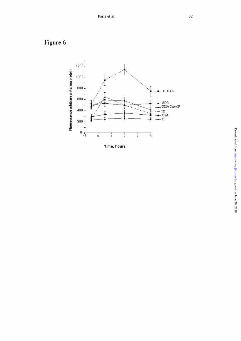
mediators of cancer therapy-induced cell death (31, 39), we next assessed the regulation of ROS
generation by cancer therapy with or without GD3 pretreatment. A significant increase in DCF
fluorescence was observed in HepG2 cells within 30 min after exposure to ionizing radiation that
declined gradually over time (Fig 6). Since this fluorescent probe is sensitive to both peroxides and
peroxynitrite (33), these findings indicate that ionizing radiation generates a modest burst of reactive
oxygen (ROS) and nitrogen (RNS) species insufficient to cause cell death. Treatment of HepG2 cells
with GD3 increased the DCF fluorescence to a level similar to that caused by ionizing radiation that
did not decline as long as GD3 was present. This outcome reflects the ability of GD3 to estimulate
mitochondrial ROS generation (23, 27). However, when HepG2 cells were pretreated with GD3 and
then irradiated there was an over generation of ROS/RNS compared to either of the treatments alone
(Fig 6). To examine whether the mitochondrial permeability transition (MPT) modulates the
generation of reactive species induced by GD3 and ionizing radiation, we investigated the effect of
CsA, an inhibitor of MPT. While CsA by itself did not affect the DCF fluorescence, preincubation
of cells with CsA before exposure to ionizing radiation abolished the ROS/RNS formation (Fig 6).
by guest on June 30, 2018http://w
ww
.jbc.org/D
ownloaded from

París et al, 13
Since MPT allows the release of apoptosis-promoting proteins from mitochondria, we examined the
mitochondrial release of cytochrome c and Smac/DIABLO in HepG2 cells. Consistent with the
findings on ROS/RNS generation, the combination of GD3 plus ionizing radiation resulted in
enhanced release of both cytochrome c and Smac/DIABLO compared to either stimuli alone (Fig 7
A). Furthermore, CsA pretreatment abrogated the release of both factors, further supporting the
involvement of MPT in the release of mitochondrial proapoptotic proteins. Finally, we examined
the activation of effector caspases and the consequences on cell survival and the effect of CsA. As
seen, GD3 plus ionizing radiation stimulated the activation of caspase 3 respect to either treatment
alone (Fig 7B). Pretreatment of GD3-exposed HepG2 cells with CsA blunted the activation of
caspase 3 (Fig 7B) resulting in protection of against GD3 plus ionizing radiation-induced cell death
(55±7% cell death vs. 6±3% at 72 hours post-treatment, p<0.05). Similar findings were observed
when cells were treated with daunorubicin instead of ionizing radiation in terms of ROS/RNS
formation, cytochrome c and Smac/DIABLO release and caspase 3 activation (not shown). by guest on June 30, 2018http://w
ww
.jbc.org/D
ownloaded from

París et al, 14
DISCUSSION
Ganglioside GD3 has emerged as an apoptosis lipid effector through its mitochondrial-interacting
(20-26) and NF-κB suppressing function (27, 28). Based on this dual function exogenous GD3 may
actually modulate cancer resistance, a phenomenon characterized by the activation of pathways that
circumvent the toxicity of current cancer therapies. To test this hypothesis we have used the human
hepatoblastoma cell line HepG2 because of their absence of endogenous GD3 (40) and because
hepatocellular carcinoma is a highly resistance tumor to currently available chemotherapeutics and
radiotherapy (41). The present study shows that exogenous GD3 sensitizes HepG2 cells to both
ionizing radiation and daunorubicin-mediated cell death. Since survival pathways contribute to the
resistance of tumor cells to cancer therapy, we examined whether exogenous GD3 interfered with
survival pathways signaling. Our data show that pretreatment of HepG2 cells with GD3 disrupts
the pathway leading to NF-κB activation induced by ionizing radiation and daunorubicin, resulting
in the suppression of inducible κB-dependent gene expression. These findings are consistent with
previous results reported in cultured rat hepatocytes (27), thus indicating that the anti-NF-κB
function of GD3 is not cell specific. Our findings, in addition, discard the involvement of Akt, a
protein kinase that has been shown to promote cell survival through the inactivation of key elements
of apoptosis pathways (34-36). Thus, rather than inactivating proapoptotic factors, GD3 seems to
interfere with the expression of survival genes controlled by NF-κB. Several κB-regulated
antiapoptotic proteins have been described (14-17), and further work will be required to identify the
specific survival gen(es) responsible for the resistance of HepG2 to cancer therapy.
An important aspect of our findings is the mechanism of NF-κB inactivation by GD3. Unlike other
strategies that target the degradation step of IκB, the inhibitor protein family of NF-κB (4-7), GD3
acts as a late step in the pathway preventing the translocation of active κB members to the nuclei.
by guest on June 30, 2018http://w
ww
.jbc.org/D
ownloaded from

París et al, 15
Although the exact mechanism whereby GD3 prevents this vital step in NF-κB activation is
presently unknown, it offers the advantage of ensuring the inactivation of NF-κB regardless of the
mechanism leading to the release of the active, DNA-binding competent members of NF-κB.
Further work is currently under way to address whether GD3 interferes with the nuclear
localization sequence that directs the translocation of NF-κB members to the nuclei (42).
Our findings define a vital role for the burst of ROS/RNS in the sensitization by GD3 of HepG2
cells to radiotherapy. The level of those potentially harmful species generated by the combination of
GD3 plus ionizing radiation was greater that with either treatment alone, and the deadly cooperation
between both strategies resulted in the additive generation of ROS/RNS. Although ionizing radiation
may signal the stimulation of ROS/RNS as an early event as shown recently (39), it is conceivable
that the induction of antioxidant enzymes such as γ-glutamylcysteine synthetase (43), may
counteract the generation of ROS/RNS below toxic levels. Since NF-κB signal the induction of
antioxidant enzymes, e.g., Mn-SOD or γ-glutamylcysteine synthase (17, 43), the inactivation of this
transcription factor by GD3 would contribute to the enhanced ROS/RNS generation induced by the
combined use of GD3 plus ionizing radiation, resulting in the killing of cells. One of the
consequences of this synergistic ROS/RNS stimulation by GD3 and ionizing radiation is the onset
of MPT that functions as a gateway to apoptosis (44, 45). Consistent with these events, GD3 plus
ionizing radiation estimulate the release of cytochrome c and Smac/DIABLO from mitochondria and
the pretreatment of cells with CsA before exposure to ionizing radiation diminishes the burst of
ROS/RNS induced by GD3 and ionizing radiation. Consequently, the resulting assembly of the
mitochondrial-dependent apoptosome and inactivation of survival genes dependent on NF-κB by
GD3 culminates in an efficient demise of tumor cells following a therapeutical dose of ionizing
radiation.
by guest on June 30, 2018http://w
ww
.jbc.org/D
ownloaded from

París et al, 16
Although the present study contributes to the current evidence supporting a proapoptotic function
of GD3, the role of glycolipids in apoptosis of cancer cells is controversial. Previous studies have
shown that downregulation of glycolipid synthesis enhances apoptosis (29, 30). Much for this
evidence has derived from the inhibition of glucosylceramide synthase, the enzyme responsible for
the glucosylation of ceramide. In particular, the inhibition of this enzyme with a widely-used
inhibitor, PDMP, has been considered as a mechanism for reversing multi-drug resistance of cancer
cells (29, 30, 46) as this strategy is accompanied by enhanced ceramide levels. Ceramide itself has
been shown to cause ROS/RNS from mitochondria (47-49), contributing to the apoptotic cell death
of cancer therapies. However, as recently described the chemosensitizing effect of PDMP appears
to be independent of its role as a inhibitor of ceramide glucosylation (46). In fact, it was shown that
PDMP significantly inhibited the activity of P-glycoprotein, a member of the membrane proteins of
the ATP-binding cassette family, that contributes to multi-drug cancer resistance (50).
Our data, however, are in agreement with recent findings describing that ganglioside GM3
overexpression induces apoptosis and reduces malignant potential in murine bladder cancer (51).
Thus, while the multi-drug resistance in cancer cells is a complex phenomenon involving the
interplay of different molecular mechanisms and multi-step alteration of the sphingolipid
metabolism (52), our present study contributes to the emerging evidence suggesting that gangliosides
may function as sensitizing agents enhancing the anticancer properties of currently used cancer
therapy.
by guest on June 30, 2018http://w
ww
.jbc.org/D
ownloaded from
París et al, 17
ACKNOWLEDGMENTS
The technical assistance of Susana Nuñez in cell culture maintenance and assays is highly
appreciated. The work presented was supported in part by the Research Center for Liver and
Pancreatic Dieases (P50 AA11999) funded by the U.S. National Institute on Alcohol Abuse and
Alcoholism, Plan Nacional de I+D grants SAF 99-0138, SAF2001-2118 and Fondo Investigaciones
Sanitarias, FISS 00-907. Dr. García-Ruiz is an SNS investigator from the Fondo Investigaciones
Sanitarias.
by guest on June 30, 2018http://w
ww
.jbc.org/D
ownloaded from

París et al, 18
REFERENCES
1. Fisher, D.E. (1994) Cell 73, 539-542.
2. Baldini, N. (1997) Nat Med. 3, 378-380.
3. Baldwin A S. (2001) J. Clin. Invest., 107, 241-246.
4. Wang, C.Y., Cusack, J.C., Liu, R. and Baldwin, A.S. (1999) Nat Med., 5, 412-417.
5. Wang C.Y., Mayo M.W. and Baldwin, A.S. (1996) Science 274, 784-787.
6. Russo, S.M., Tepper, J.E., Baldwin, A.S., Liu, R., Adams, J., Elliott, P. and Cusack, J.C.
(2001) Int. J. Radiat. Oncol. Biol. Phys, 50, 183-193.
7. Cusack, J.C., Liu, R. and Baldwin, A.S. (1999) Drug Resist Updat 2, 271-273.
8. Brach, M.A., Hass, R., Sherman, M.L., Gunji, H., Weischselbaum, R., and Kufe, D. (1991)
J. Clin. Invest 88, 691-695.
9. Boland, M. P., Foster, S.J., O´Neill, L.A.J.(1997) J. Biol. Chem., 272, 12952-12960.
10. Beg, A.A., Sha W.C., Bronson, R.T., Ghosh, S. and Baltimore, D. (1995) Nature 376, 167-
170.
11. Van Antwerp, D.J., Martin, S.J., Verman, I.M., and Green. D.R. (1998) Trends Cell Biol 8,
107-111.
12. Hatano, E., Bennett, B.L., Manning, A.M., Qian, T., Lemasters, J.J. and Brenner,
D.A.(2000) Gastroenterology 120, 1251-1262.
13. Colell A., Mari, M., Coll, O., Fernández-Checa, J.C. and García-Ruiz, C. (2002) FEBS Lett,
526, 15-20.
14. Wang, C.Y., Mayo, M.W., Korneluk, R.G., Goeddel, D.V.and Baldwin, A.S. (1998) Science
281, 1680-1683.
by guest on June 30, 2018http://w
ww
.jbc.org/D
ownloaded from

París et al, 19
15. Wang, C.Y., Guttridge, D.C., Mayo, M.W. and Baldwin A.S. (1999) Mol. Cell Biol. 19,
5923-5929.
16. Chen, C. Edelstein, L.C. and Gelinas, C. (2000) Mol. Cell Biol 20, 2687-2695.
17. Tanaka, H., Matsumura I., Ezoe, S., Satoh, Y., Sakamaki, T.l., Albanese, C., Machii, T.,
Pestell, R.G., and Kanakura, Y. (2002) Mol. Cell 9, 1017-1029.
18. Hakomori, S. (2000) Glycoconjug J. 17, 143-151.
19. Riboni, L, Viani, P, Bassi, R., Prinetti, A. and Tettamanti, G. (1997) Prog. Lip. Res. 36,153-
195.
20.DeMaria, R., Lenti, T., Malissan, F., d'Agostino,F., Tomassini, B., Zeuner, A., Rippo, M.R.
and Testi, R. (1997) Science, 277, 1652-1655.
21. Scorrano, L., Petronilli, P., DiLisa, F. and Bernardi, P. (1999) J. Biol. Chem., 274, 22581-
22585.
22.Kristal, B.S. and Brown, A. (1999)J. Biol. Chem., 274, 23169-23175.
23.García-Ruiz, C., Colell, A., París, R. and Fernández-Checa, J.C. (2000) FASEB J., 14, 847-
858.
24. Rippo, M.R., Malisan, F., Ravagnan, L., Tomassini, B., Condo, I., Constantini, P., Susin,
S.A., Ruffini, A., Todaro, M., Kroemer, G. and Testi, R. (2000) FASEB J., 14, 2047-2054.
25. Giammaroli, A.M., Garofalo, T., Sorice, M., Misasi, R., Gambardella, L., Gradini, R, Fais,
S., Pavan, A. and Malorni, W. (2001) FEBS Lett. 506, 45-50.
26. Garcia-Ruiz, C., Colell A., Morales, A., Calvo, M., Enrich, C. and Fernández-Checa, J.C.
(2002) J. Biol. Chem., in press.
27. Colell, A., García-Ruiz, C., Roman, J., Ballesta, A. and Fernández-Checa, J. C. (2001).
FASEB J., 15, 1068-1070.
by guest on June 30, 2018http://w
ww
.jbc.org/D
ownloaded from

París et al, 20
28. Uzzo, R.G., Rayman, P., Kolenko, V., Clark, P.E., Cathcart, M.K., Bloom, T., Novick,
A.C., Bukowski, R.M., Hamilton, T., and Finke, J.H. (1999) J. Clin. Invest., 104, 769-776.
29. Lavie, Y., Cao, H., Bursten, S.L., Giuliano, A.E. and Cabot, M.C. (1996) J. Biol. Chem.,
271,19530-19536.
30. Lavie, Y., Cao, H., Volner, A., Lucci, A., Han, T.Y., Geffen, V., Giuliano, A.E. and Cabot,
M.C. (1997) J. Biol. Chem., 272, 1682-1687.
31. Morales, A., Miranda, M., Sánchez-Reyes, A., Biete, A., and Fernández-Checa, J.C (1998)
Int.J. Radiat. Biol. Phys. 42, 191-203.
32. Roman, J, Gimenez, A., Lluis, J.M., Gasso, M.,Rubio, M, Caballeria, J., Pares, A., Rodes,
J., and Fernandez-Checa, J.C. (2000) J. Biol. Chem., 275, 14684-14690.
33. Crow, J. P. (1997) Nitric Oxide 1, 145-157.
34. Cardone, M.H., Roy, N., Stennicke, H.R., Salvesen, G.S., Franke, T.F., Stanbridge, E.,
Frisch, S. and Reed, J.C. (1998) Science 282, 1318-1321.
35.Datta, S.R., Dudek, H., Tao, X., Masters, S., Fu, H., Gotoh, Y., and Greenberg, M.E. (1997)
Cell 91, 231-241.
36. Brunet, A., Bonni, A., Zigmond, M.J., Lin, M.Z., Juo, P., Hu, L.S., Anderson, M.J., Arden,
K.C., Blenis, J., and Greenberg, M.E. (1999) Cell 96, 857-868.
37. Wang, X. (2001) Genes Dev 15, 2922-2933.
38. Green, D.R. and Reed, J.C. (1998) Science 281, 1309-1312.
39. Leach J.K., Van Tuyle G., Lin, P., Schmidt-Ullrich, R., and Mikkelsen, R.B. (2001) Cancer
Res. 61, 3894-3901.
40. Spitalnik, P.F., Danley, J.M., Burger, S.R. and Spitalnik, S.F. (1989) Arch. Biochem.
Biophys. 273, 578-591.
by guest on June 30, 2018http://w
ww
.jbc.org/D
ownloaded from

París et al, 21
41. Shimada, M., Takenaka, K., Kawahara, N., Yamamoto, K., Shirabe, K., Maehara, Y. and
Sugimachi, K. (1996) Hepatogastroenterology 43, 1159-1164.
42. Beg, A., Ruben, A., Scheinman, S.M., Haskill, R.S., Rosen, S. and Baldwin, A.S. (1992)
Genes Dev 6, 1899-1913.
43. Morales,A., Miranda, M., Sánchez-Reyes, A., Colell A., Biete A., and Fernández-Checa,
J.C. (1998) FEBS Lett 427, 15-20.
44. Lemaster, J. J. (1999) Am.J.Physiol. 276, G1-G6.
45. Brenner C., and Kroemer G. (2000) Science 289, 1150-1151.
46. Bartolomeo, S., and Spinedi, A. (2001) Biochem. Biophys. Res. Commun. 288, 269-274.
47. García-Ruiz, C., Colell, A., Mari, M., Morales, A., and Fernández-Checa, J.C. (1997) J.
Biol. Chem. 272, 11369-11377.
48. Degli Esposti, M. and Mclennan, H. (1998) FEBS Lett 430, 338-342.
49. Quillet-Mary, A., Jaffrezou, J.P., Mansat, V., Boudier, C., Naval, J. and Laurent, G. (1997)
J. Biol. Chem. 272, 21288-21395.
50. Gottesman, M.M. and Pastan, I. (1993) Annu. Rev. Biochem. 62, 385-427.
51. Watanabe, R., Ohyama, R., Aoki, H., Takahashi, T., Satoh, M, Saito, S., Hoshi, S., Ishii, A.,
Saito, M., and Arai, Y. (2002) Cancer Res. 62, 3850-3854.
52. Veldman, R.J., Klappe, K., Hinricks, J., Hummel, I., Van Der Schaaf, G., Sietsma, H. And
Kok, J. W. (2002) FASEB J. 16, 1111-1113.
by guest on June 30, 2018http://w
ww
.jbc.org/D
ownloaded from

París et al, 22
FOOTNOTES
1. Abbreviations used in this paper: Ac-DEVD-AMC, Ac-Asp-Glu-Val-Asp-7-amino-4-
trifluoromethyl coumarin; CsA, cyclosporin A; DCF, dichlorofluorescein; EMSA,
electrophoretic mobility shift assays; GSls, glycosphingolipids; GD3, ganglioside GD3;
LDH, lactate dehydrogenase; MPT, mitochondrial permeability transition; NF-κB,
transcription factor, NF-κB; ROS/RNS, reactive oxygen/nitrogen species
by guest on June 30, 2018http://w
ww
.jbc.org/D
ownloaded from

París et al, 23
FIGURE LEGENDS
Fig 1. NF- B activation by cancer therapy and effect of GD3 treatment.
A, Hep G2 cells were exposed to increasing doses of ionizing radiation (IR) and EMSA were
performed four hours after treatment to assess activation of NF-κB. The arrows denote the
activation of relA/p52 and p52/p50. B, HepG2 cells were preincubated 4 h with GD3 (5 µM)
before ionizing radiation (4 Gy) or daunorubicin (D) treatment (1 µM). Nuclear extracts from 2x10⁶
cells were isolated 2h after treatments and assayed for DNA binding activity. Both are
representiative EMSA gels out of 4-5 individual experiments showing similar results. The right
panel shows the quantitative activation of RelA/p52 (closed bars) and p52/p50 (open bars) by
cancer therapy and the inactivation induced by GD3 pretreatment. Results are the mean±SD of 5-6
independent experiments. # p0.05 vs control, * p 0.05 vs. IR or D alone.
Fig 2. Transactivation of NF- B by cancer therapy and prevention of nuclear translocation
by GD3.
Hep G2 cells were transfected with the plasmid pNF-κB-Luc containing four tandem copies of the
κB enhancer upstream of the herpes simplex virus thymidine kinase fused to the luciferase reporter.
24 hours after transfection, cells were incubated with GD3 (5µM) for four hours before exposure to
ionizing radiation (IR) (4Gy) or daunorubicin (D) (1µM). Supernatants from lysed cells were used
to determine luciferase activity expressed in relative light units and normalized for the protein
content and β-galactosidase activity. Results are the mean±SD of four experiments.*p<0.05 vs. I or
D alone, # p0.05 vs control. The right panel shows the distribution of p65 following ionizing
radiation with or without GD3 pretreatment. Cells were fixed and incubated with antibody anti-p65
labeled with FITC-coupled secondary antibody. In each experiment more than 200 cells were
examined at different fields. Representative confocal microscopy images are shown out of 4-5
individual experiments.
Fig 3. GD3 sensitizes tumor cells to cancer therapy.
Cells were exposed to ionizing radiation (IR) (4Gy) or daunorubicin (D) (1µM) with or without
GD3 preincubation (5µM for 4 hours). The fraction of surviving HepG2 was determined as the
by guest on June 30, 2018http://w
ww
.jbc.org/D
ownloaded from

París et al, 24
LDH release into the medium as described in Materials and Methods. Results are the mean±SD of
five different experiments. *p<0.05 versus control.# p<0.05 versus IR or D alone.
Fig 4. Apoptotic cell death induced by GD3 pretreatment followed by cancer therapy.
HepG2 cells were treated with ionizing radiation (IR) (4Gy) or daunorubicin (D) (1µM) for 24
hours with or without GD3 preincubation (5µM) and then stained with Hoescht-33258 and
examined for chromatin morphology in a fluorescence microscope. Representative images of 5-6
independent experiments performed showing similar results (A). More than 100 cells per condition
were examined at different fields and those showing chromatin fragmentation expressed as the
percentage of total cells examined (B). Results are the mean±SD of 4-5 independent experiments.
*p<0.05 vs. control; #p<0.05 vs. IR or D alone.
Fig. 5. Phosphorilation of Akt by IR and GD3 effects.
Hep G2 cells were preincubated with 5µM GD3 four hours before exposure to ionizing radiation
(IR) 4 Gy. After 2h cells were lysed and P-Akt and total Akt levels examined by western blots
using specific antibodies. Alternatively, cells were treated for 20 min with 0.1µM insulin as a
positive control of Akt activation and processed as above. Representative blots of 5 individual
performed showing similar results.
Fig 6. GD3 enhances ROS/RNS generation after ionizing radiation.
ROS/RNS were determined from the DCF fluorescence at different times after cells were exposed to
ionizing radiation (4Gy) (IR) with or without GD3 (5 µM) preincubation. In some cases, after
preincubation with GD3, cells were treated with CsA (2 µM) before exposure to IR. Cells were
then treated with CM-H2DCFDA and incubated for 30 minutes before cells were harvested. DCF
fluorescence was determined in a fluorimeter. Results are the mean±SD of 5 independent
experiments.
Fig 7. GD3 plus ionizing radiation stimulate the release of mitochondrial pro-apoptotic
factor and caspase 3 activation.
by guest on June 30, 2018http://w
ww
.jbc.org/D
ownloaded from

París et al, 25
A, cells were irradiated (IR, 4Gy) in the presence or absence of GD3 (5 µM) and CysA (2 µM) and
20 hours after cells were fractionated into mitochondria (pel) and both the pellet and supernatants
were analyzed by western blot analysis using anti-cytochrome c antibody and anti-Smac/DIABLO
antibody followed by appropriate secondary antibodies. Specificity of mitochondrial release was
confirmed in parallel aliquots from pellets and supernatants using antibody anti-cytochrome c
oxidase. B, 24 hours after irradiation (4Gy) with or without GD3 or CsA preincubations, Hep G2
cells lysed and caspase-3 activity measured as described in Material and Methods Results are the
mean±SD of three different experiments. *p<0.05 versus control # p<0.05 versus IR or D.
by guest on June 30, 2018http://w
ww
.jbc.org/D
ownloaded from

and Jose C. Fernandez-ChecaRaquel Paris, Albert Morales, Olga Coll, Alberto Sanchez-Reyes, Carmen Garcia-Ruiz
Ganglioside GD3 sensitizes human hepatoma cells to cancer therapy
published online September 25, 2002J. Biol. Chem.
10.1074/jbc.M208303200Access the most updated version of this article at doi:
Alerts:
When a correction for this article is posted•
When this article is cited•
to choose from all of JBC's e-mail alertsClick here
by guest on June 30, 2018http://w
ww
.jbc.org/D
ownloaded from