Embed Size (px)
Citation preview
899www.eymj.org
INTRODUCTION
Hepatocellular carcinoma (HCC) is one of the most common
malignancies across the globe, especially in Asia and south-ern Africa.1 Through a diagnosis followed by systemic exami-nation, HCC can be classified into early, mid, and late (ad-vanced) stages. Surgical resection, liver transplantation,2 or local ablation have generally been used to treat early- and mid-stage HCC, and the 5-year survival rate could be as high as 60–70%.2 However, owing to the lack of effective treatment options and underlying liver disease, patients diagnosed at an advanced stage or with progression experience a much more dismal prog-nosis after locoregional therapy.3 Until sorafenib was used as a first-line therapy, there was no systemic therapy to improve survival in patients with late-stage HCC.4,5
Sorafenib, which has anti-proliferative and anti-angiogenic effects, is an oral multikinase inhibitor.6 It can inhibit several cellular signaling pathways, including those of Raf/mitogen-activated protein kinase (MAPK), extracellular signaling-regu-
Down-Regulation of TGF-β Expression Sensitizes the Resistance of Hepatocellular Carcinoma Cells to Sorafenib
Dongxu Kang1*, Zhezhu Han2,3*, Geun-Hyeok Oh2, Yeonsoo Joo2,3, Hye Jin Choi4, and Jae J. Song2,3
1Department of Oncology, Affiliated Hospital of Yanbian University, Yanji, Jilin Province, P.R. China; 2Institute for Cancer Research, Yonsei University College of Medicine, Seoul; 3Severance Biomedical Science Institute, Yonsei University College of Medicine, Seoul; 4Department of Internal Medicine, Yonsei University College of Medicine, Seoul, Korea.
Purpose: Sorafenib, a multikinase inhibitor, is the standard therapy for patients with advanced-stage hepatocellular carcinoma (HCC). However, resistance develops to the treatment, therefore, we tried to unravel the underlying mechanism in the resistance of HCC cells to sorafenib via the development of more effective therapeutic strategies.Materials and Methods: Various liver cancer cell lines were treated with either sorafenib only or with sorafenib after infection of adenovirus expressing short hairpin RNA (shRNA) against transforming growth factor-β (TGF-β) and p38 activity was examined using western blotting. Results: p38 MAP kinase activity was inhibited by low concentrations of sorafenib, which could potentially lead to sorafenib resis-tance in HCC cell lines. Subsequently, we used constitutive form of MKK3/6 (MKK3/6E) to confirm that massive cell death was induced by the activation of p38, and demonstrated the ability to activate p38 without any stimulation. In addition, sorafenib re-sistance was reduced by the activation of p38. Subsequently, we confirmed that TGF-β shRNA effectively recovered the phos-phorylation of p38 inhibited by sorafenib, and increased the sensitivity of HCC cells to sorafenib, thereby inducing cell death and overcoming the resistance of HCC cells to sorafenib.Conclusion: Our study provides a new therapeutic strategy for HCC that overcomes the resistance of HCC to sorafenib by down-regulation of TGF-β.
Key Words: HCC, sorafenib, resistance, TGF-β, p38, adenovirus
Original Article
pISSN: 0513-5796 · eISSN: 1976-2437
Received: April 24, 2017 Revised: June 5, 2017Accepted: June 5, 2017Co-corresponding authors: Dr. Hye Jin Choi, Department of Internal Medicine, Yonsei University College of Medicine, 50-1 Yonsei-ro, Seodaemun-gu, Seoul 03722, Korea.Tel: 82-2-2228-8133, Fax: 82-2-393-4652, E-mail: [email protected] andDr. Jae J. Song, Severance Biomedical Science Institute, Yonsei University College of Medicine, 50-1 Yonsei-ro, Seodaemun-gu, Seoul 03722, Korea.Tel: 82-2-2228-8037, Fax: 82-2-393-3652, E-mail: [email protected]
*Dongxu Kang and Zhezhu Han contributed equally to this work.•The authors have no financial conflicts of interest.
© Copyright: Yonsei University College of Medicine 2017This is an Open Access article distributed under the terms of the Creative Com-mons Attribution Non-Commercial License (http://creativecommons.org/licenses/by-nc/4.0) which permits unrestricted non-commercial use, distribution, and repro-duction in any medium, provided the original work is properly cited.
Yonsei Med J 2017 Sep;58(5):899-909https://doi.org/10.3349/ymj.2017.58.5.899

900
Down-Regulation of TGF-β to Sorafenib Resistance
https://doi.org/10.3349/ymj.2017.58.5.899
lated kinase (ERK), as well as the receptors of vascular endothe-lial growth factor and platelet-derived growth factor.6,7 As the standard care for patients with advanced HCC, sorafenib has been proven to have the capacity to inhibit HCC cell prolifera-tion, angiogenesis, and so on.8,9 However, the promising treat-ment of HCC with sorafenib has limited benefits to survival and very low rates of tumor response, where some patients with HCC even exhibit no initial response to sorafenib,3,10 in-dicating the existence of both primary and acquired resis-tances of HCC cells to sorafenib.9 The primary resistance of HCC to sorafenib has been shown to be due to genetic hetero-geneity.10 While sorafenib inhibits several cellular signaling pathways as a multikinase inhibitor, it simultaneously or se-quentially activates the addiction switches and compensatory pathways.9 Although several mechanisms have been proposed, as for the acquired resistance of HCC cells to sorafenib, the exact mechanisms remain unclear.11,12
The multifunctional cytokine transforming growth factor-β (TGF-β) orchestrates an intricate signaling network to modu-late tumorigenesis and progression by exerting a dynamic ef-fect on cancer cells.13 Early in the carcinogenesis process, TGF-β1 suppresses tumors and arrests cell growth, whereas in later and more advanced tumor stages, TGF-β1 potentiates epithe-lial-to-mesenchymal transition, angiogenesis, tumor progres-sion, invasion, and metastasis.14,15 Previous studies have shown that TGF-β1 was overexpressed in HCC cells, and clinical stud-ies showed higher blood levels of TGF-β1 in patients with HCC than in patients with chronic hepatitis or cirrhosis.16 Further-more, HCC cells with a higher IC50 in response to sorafenib tended to have higher TGF-β1 mRNA expression,16 and TGF-β2 was overexpressed in some HCC cell lines and patient cohorts and a correlation was found between high TGF-β2 expression and lower survival rates (p<0.01).17 Thus, the TGF-β signaling pathway should be explored as a therapeutic target for pa-tients with advanced HCC.
MAPKs are essential components of intracellular signal tr-ansduction, in which p38 MAPK plays an essential role in the regulation of gene expression and in controlling cellular resp-onses to the environment, cell growth, and apoptosis. These features have previously made p38 MAPK a molecular target for drug development in the treatment of many human diseas-es, most notably in the treatment of a variety tumors.18 The in-volvement of the p38 MAPK cascade in the apoptotic pathway has been demonstrated in two hepatoma cell lines, HepG2 and Huh7, wherein the activation of p38 MAPK inhibited cell gr-owth and induced apoptosis.19 The apoptosis of human hepa-toma cell lines results from increased caspase 3 activity via the activation of p38 MAPK: Bid cleavage and cytochrome c re-lease are modulated by p38 MAPK activation.19 In human HCC patients, the activities of p38 MAPK and MKK6 were signifi-cantly lower than in adjacent uninvolved liver tissue, whereas the activity of ERK1/2 was significantly increased in malignant lesions.19,20 Treatment with sorafenib has been shown to in-
hibit p38α kinase activity in vitro by targeting the DFG-out conformational state of p38α.21 The reduction of p38 activity as a result of sorafenib treatment could be one of the causes of resistance, therefore, it should be a potential target to induce more effective cell death in HCC patients.
Adenovirus serotype 5 is the most commonly used viral vec-tor in cancer gene therapies human serotype due to their high efficiency, broad range of host transduction, easy genome ma-nipulation, and non-integration into the host genome.22 In this research, therefore, we designed adenovirus-delivered TGF-β short hairpin RNAs (shRNAs) to knock down TGF-β expres-sion. The reduction in TGF-β expression resulted in an increase of p38 activity, thus activating the cellular death signal. When TGF-β shRNA was combined with sorafenib, HCC cells be-came highly sensitized to sorafenib, and the efficiency of so-rafenib against advanced HCC was significantly increased.
MATERIALS AND METHODS
Cell culture Hep-3B, Huh7, and SK-Hep-1 cells were cultured in Dulbecco’s modified Eagle’s medium with 10% fetal bovine serum (FBS). SNU-182, SNU-398, and SNU-449 cells were cultured in RPMI with 10% FBS. Cells were maintained in a 37°C humidified at-mosphere containing 5% CO2.
ReagentsAntibodies to GAPDH and sorafenib were purchased from Santa Cruz Biotechnology (Santa Cruz, CA, USA). Antibodies to p38, phospho-p38, phospho-Akt (Ser473), phospho-ERK (Thr202/Tyr204), phospho-p65 (Ser536), phospho-Src (Tyr416), MKK3, and MKK6 were purchased from Cell Signal-ing Technology (Beverly, MA, USA). All other chemicals were purchased from Sigma-Aldrich (St. Louis, MO, USA).
Construction of adenoviral vectorsFor the expression of siRNAs targeting human TGF-β1 or TGF-β2, each annealed sequences for shRNA were cloned into a pSP72ΔE3-U6/H1 vector after BamHI/HindIII digestion. These vectors, designated pSP72ΔE3-U6-shTGF-β1 and pSP72ΔE3-U6-shTGF-β2 (E3 shuttle vector), were linearized by XmnI di-gestion, and co-transformed into E. coli BJ5183 together with the SpeI-digested adenoviral vector (dl324-IX) for homolo-gous recombination. The detailed informations about the construction of TGF-β shRNAs was previously described.23 The recombinant adenoviral vectors with abbreviated names are as follows:
NC: Ad-IX-ΔE1--ΔE3, control virus.shT1: Ad-IX-ΔE1-ΔE3-U6-shTGFβ1, virus expressing shR-
NA of human TGFβ1 (shTGFβ1).shT2: Ad-IX-ΔE1-ΔE3-U6-shTGFβ2, virus expressing shR-
NA of human TGFβ2 (shTGFβ2).
901
Dongxu Kang, et al.
https://doi.org/10.3349/ymj.2017.58.5.899
MTS viability assaysThe CellTiter 96® Aqueous Assay kit (Promega, Madison, WI, USA) comprises solutions of a novel tetrazolium compound [3-(4,5-dimethylthiazol-2-yl)-5-(3-carboxymethoxyphenyl)-2-(4-sulfophenyl)-2H-tetrazolium, inner salt; MTS] and an electron coupling reagent (phenazine ethosulfate). MTS is bioreduced by cells into a formazan product that is soluble in tissue culture media. The conversion of MTS into aqueous, soluble formazan is carried out by dehydrogenase enzymes found in metabolically active cells. Subsequently, HCC cell lines were treated with varying doses of sorafenib (0, 2.5, 5, 10, 15, 20, and 25 μM) in 96-well plates for 24 h. The absorbance of formazan at 490 nm was measured directly from 96-well plates without additional processing, and the quantity of forma-zan product measured at 490 nm is directly proportional to the number of living cells in culture.
Western blot analysesCells were lysed with 1X Laemmli sample buffer (62.5 mM Tris, pH 6.8, 2% sodium dodecyl sulfate, 10% glycerol, 0.002% bromophenol blue) and protein concentration was determined using the BCA Protein Assay Kit (Thermo Scientific, Fremont, CA, USA). Then, protein samples were resolved by sodium do-decyl sulfate-polyacrylamide gel electrophoresis, and gels were electro-transferred onto polyvinylidene difluoride mem-branes (Millipore, Billerica, MA, USA). Immunodetection was performed using anti-phosho-Akt (p-Akt), anti-phosho-Src (p-Src), anti-phosho-p65 (p-p65), anti-phoshoERK (p-ERK), anti-phosho-p38 (p-p38), anti-p38, anti-MKK3, anti-MKK6 and anti-GAPDH primary antibodies, with the chemiluminescent and fluorescent image analysis system (Syngene, Cambridge, UK).
Real-time quantitative polymerase chain reactionsCells were lysed with Trizol reagent (Life Technologies, Carls-bad, CA, USA) and the total RNA was isolated via chloroform extraction. RNA concentration was determined with a Nano-drop 2000 (Thermo Scientific). The real-time polymerase chain reaction (PCR) was performed using a Power SYBR Green RNA-to-CT 1-Step Kit (Life Technologies). The reaction mixture contained the reverse transcriptase enzyme mix, reverse tran-scription PCR mix, forward primer, reverse primer, RNA tem-plate, and nuclease-free water. Human TGF-β1 cDNA was am-plified using the forward primer 5’-CAAGGGCTACCATGCC AACT-3’ and the reverse primer 5’-AGGGCCAGGACCTTG CTG-3’. Human TGF-β2 cDNA was amplified using the for-ward primer 5’-GCTGCCTACGTCCACTTTACAT-3’ and the reverse primer 5’-ATATAAGCTCAGGACCCTGCTG-3’. Human β-actin was amplified using the forward primer 5’-ACTCTTC CAGCCTTCCTT-3’ and the reverse primer 5’-ATCTCCTTCT GCATCCTGTC-3’.
Enzyme-linked immunosorbent assaysCells were plated in the wells of six-well plates at a density of
1×10⁵ cells/well. Supernatants were collected after 48 hr. The levels of TGF-β1 and TGF-β2 expression were determined by an enzyme-linked immunosorbent assays (ELISA) according to the manufacturer’s instructions (R&D Systems, Minneapo-lis, MN, USA).
Clonogenic assaysCells were plated into six-well plates at a density of 1×10⁵ cells/well. Subsequently, HCC cell lines were treated with of vari-ous concentrations (0, 2.5, 5, 10, 15, or 20 μM) sorafenib (24 hr) or treated with 2.5 μM sorafenib (12 hr) followed by a pre-treat-ment with adenoviral vectors (NC, shT1, shT2) for 24 hr. Cells were trypsinized and plated into six-well plates at densities of 5×10³ or 1×10⁴ cells/well. They were then monitored daily by microscopy. When cells exhibited colonies, surviving cells were fixed with 4% paraformaldehyde and stained with 0.5% crystal violet.
Animal studiesTumors were implanted subcutaneously in the abdomen of BALB/c nude mice via the injection of SNU-449 human liver cancer cells (1×10⁷) in 100 μL of Hank’s balanced salt solution (Gibco BRL, Carlsbad, CA, USA). When tumors reached a size range of 70–100 mm3, animals were randomized into 5 groups of 8 animals each per group [phosphate buffered saline (PBS), sorafenib, NC+sorafenib, shT1+sorafenib, shT2+sorafenib]. Animals in adenoviral groups or the control group (PBS) were administered adenoviruses intratumorally [virus; 1×10⁹ PFU (plaque-forming units) per tumor in 50 μL of PBS] on days 1, 3, and 5. Sorafenib (30 mg/kg) was administered once daily by gavage for 10 days. The regression of tumor growth was as-sessed by measuring the length (L) and width (W) of each tumor. Tumor volume was calculated using the following formula: volume=0.52×L×W2.
Statistical analysesThe data are expressed as mean±standard error. Statistical comparisons (students t-test) were made using Graph Pad (Sy-stat Software, Inc., Chicago, IL, USA). p-values less than 0.05 were considered statistically significant (*p<0.05, †p<0.01, ‡p<0.001).
RESULTS
Sorafenib treatment induced changes in several signaling pathways in HCC cell lines To evaluate the impact of sorafenib in vitro, we first identified the IC50 of sorafenib in different HCC cell lines. The MTS as-say showed that the IC50 of sorafenib in HCC cell lines ranged from 7.5 μM to 15 μM (Fig. 1A). Subsequently, we examined various key signaling pathway molecules, including p-p38, p-p65, p-Src, and p-ERK by using western blot analyses, in HCC cell lines after treatment with sorafenib IC50 concentra-
902
Down-Regulation of TGF-β to Sorafenib Resistance
https://doi.org/10.3349/ymj.2017.58.5.899
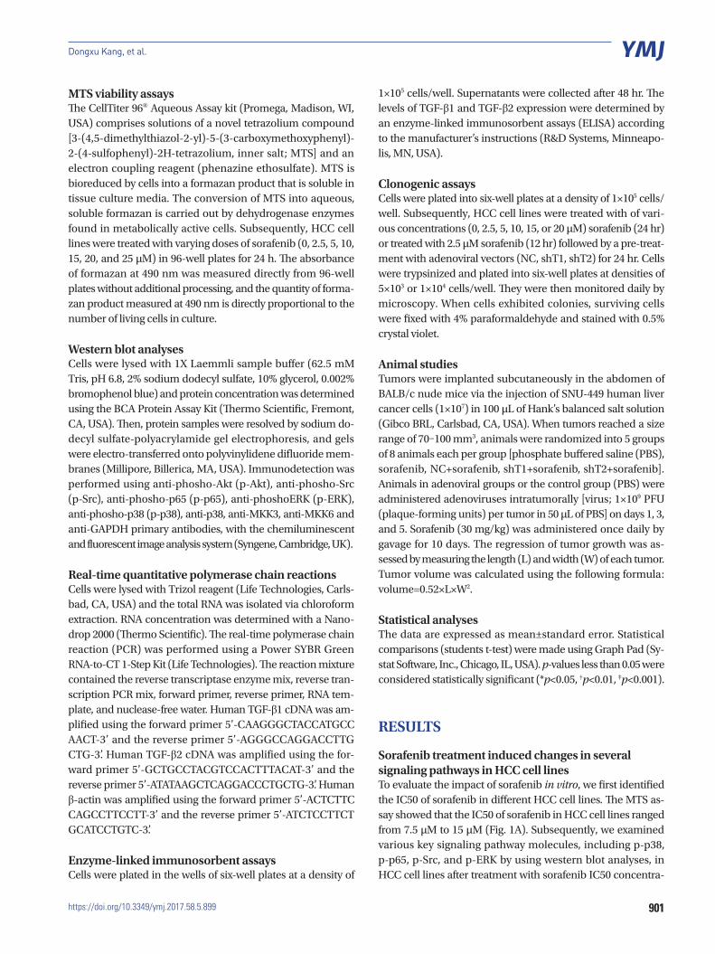
Fig. 1. Effect of sorafenib on different HCC cell lines. (A) Hep-3B, Huh7, SK-Hep-1, SNU-182, SNU-398, and SNU-449 cells were treated with sorafenib in a dose-dependent manner. After 24 h, cell viability was tested via a MTS viability assay. IC50 of each cell line is indicated in each rectangle. Error bars represent the standard error from three independent experiments. (B) HCC cell lines were treated with sorafenib at IC50 concentrations, respec-tively. After 24 h, the expressions of p-p38, p38, p-ERK, p-Akt, p-Src, p-p65, and GAPDH were detected by western blot analysis. (C) HCC cell lines were treated with sorafenib in a dose-dependent manner for 24 h. and incubated for additional 14 days for clonogenic assays. (D) HCC cell lines were treated with a low dose of sorafenib (2.5 μM) for 24 h, and changes in the levels of p-Akt p-p65, p-ERK, and p-p38 expression were then detected by western blot analysis. HCC, hepatocellular carcinoma.
125
100
75
50
25
00 2.5 5 10 15 20 25
Cell
viabi
lity (
%)
Sorafenib (μM)
Hep-3B
15 μM
125
100
75
50
25
00 2.5 5 10 15 20 25
Cell
viabi
lity (
%)
Sorafenib (μM)
Huh7
15 μM
125
100
75
50
25
00 2.5 5 10 15 20 25
Cell
viabi
lity (
%)
Sorafenib (μM)
SNU-449
15 μM
125
100
75
50
25
00 2.5 5 10 15 20 25
Cell
viabi
lity (
%)
Sorafenib (μM)
SNU-182
15 μM
125
100
75
50
25
00 2.5 5 10 15 20 25
Cell
viabi
lity (
%)
Sorafenib (μM)
SK-Hep-1
10 μM
125
100
75
50
25
00 2.5 5 10 15 20 25
Cell
viabi
lity (
%)
Sorafenib (μM)
SNU-398
7.5 μM
Hep-3B Huh7 SK-Hep-1 SNU-182 SNU-398 SNU-449
p-p38
p38
p-ERK
p-Akt
p-p65
p-Src
GAPDH
0 h 0 h 0 h 0 h 0 h 0 h24 h 24 h 24 h 24 h 24 h 24 h
Hep-3B Huh7 SK-Hep-1 SNU-182 SNU-398 SNU-449
p-Aktp-p65
p-ERK
p-p38
GAPDH
0 h 0 h 0 h 0 h 0 h 0 h24 h 24 h 24 h 24 h 24 h 24 h
SNU-449
Hep-3B
Huh7
SNU-182
SK-Hep-1
SNU-398
Cell only 2.5 μM 5 μM 10 μM 15 μM 20 μM
A
D C
B
903
Dongxu Kang, et al.
https://doi.org/10.3349/ymj.2017.58.5.899
tions, and observed that phospho-ERK activity was reduced by sorafenib treatment. The activity of p-p38 was also decreased in many HCC cells, which may inhibit the death-related signal-ing pathway (Fig. 1B). We then performed a clonogenic assay to confirm the cell viability of HCC cells after dose-dependent treatments with sorafenib, and observed that many HCC cells survived treatment, even in the presence of high doses of sora-fenib, indicating low sensitivity of HCC cells to sorafenib and their resistance to this form of treatment (Fig. 1C). As shown in Fig. 1D, p-Akt and p-p65 were increased by incubating cell lines with 2.5 μM sorafenib for 24 h, whereas p-p38 levels were also more clearly reduced during the treatment. The increased activi-ties of p-p65 and p-Akt (Fig. 1D) signify that cell lines were in fact resistant to sorafenib. Likewise, the inhibition of p-p38 was observed under conditions of sorafenib resistance, suggesting that it may play a role in inducing resistance to sorafenib in HCC cell lines.
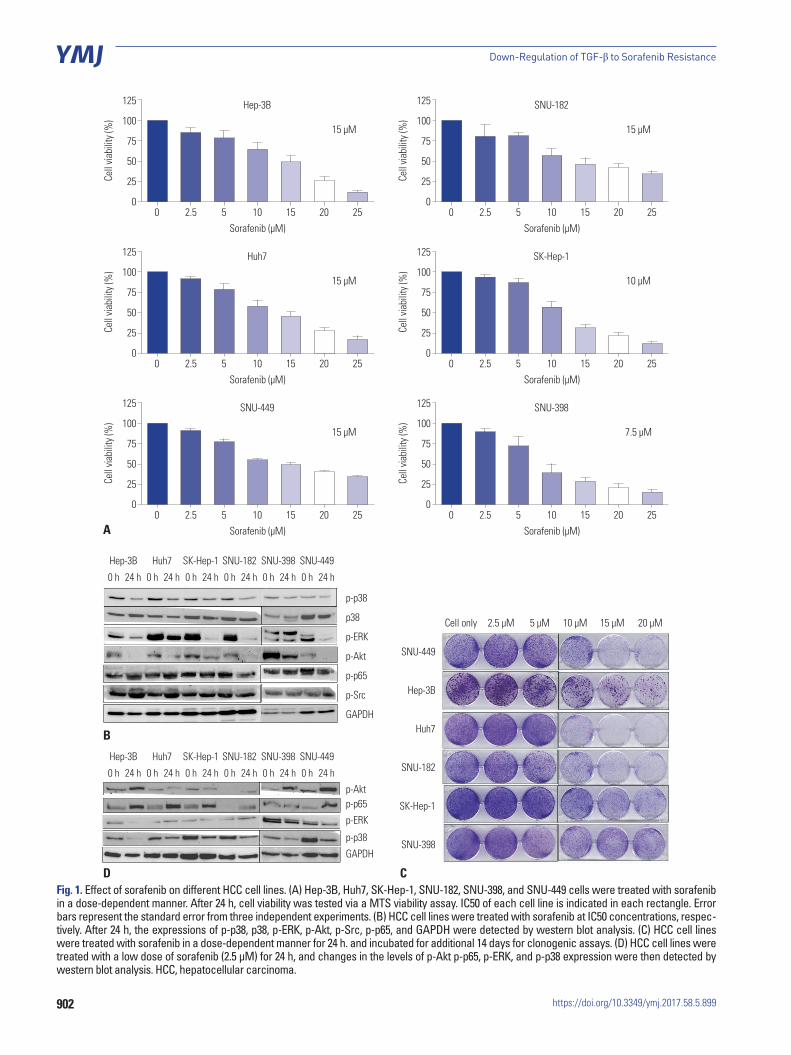
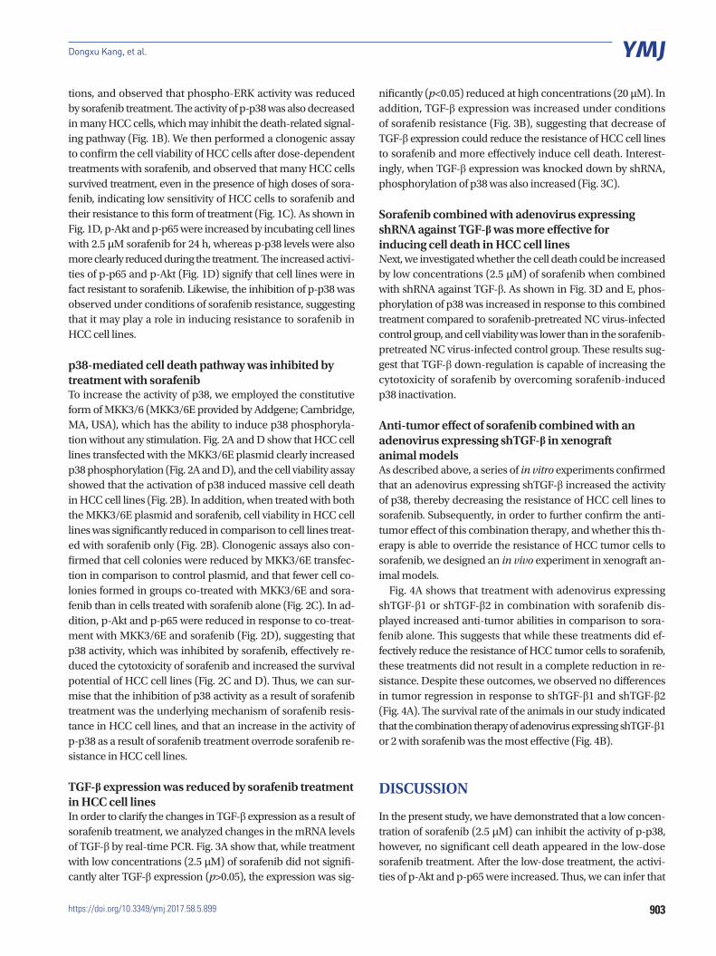
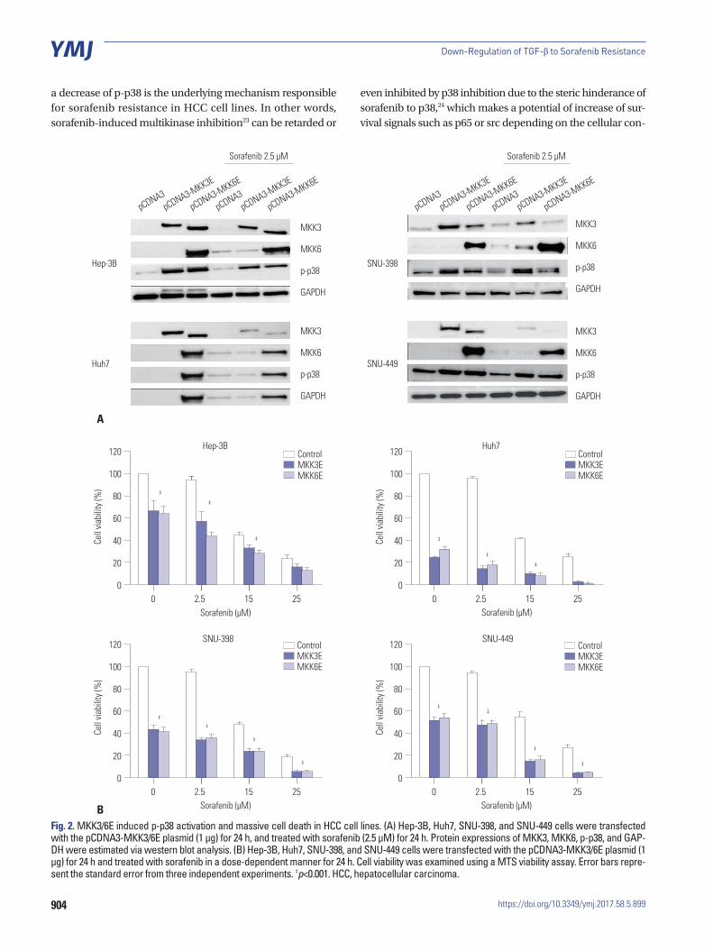
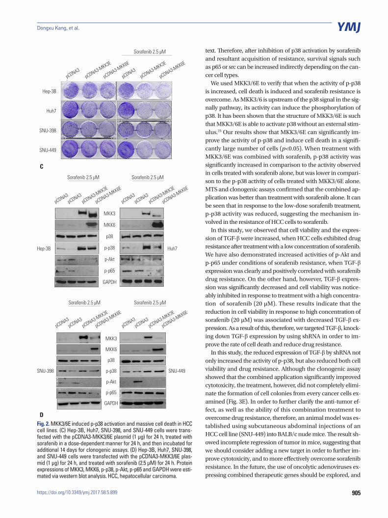
p38-mediated cell death pathway was inhibited by treatment with sorafenibTo increase the activity of p38, we employed the constitutive form of MKK3/6 (MKK3/6E provided by Addgene; Cambridge, MA, USA), which has the ability to induce p38 phosphoryla-tion without any stimulation. Fig. 2A and D show that HCC cell lines transfected with the MKK3/6E plasmid clearly increased p38 phosphorylation (Fig. 2A and D), and the cell viability assay showed that the activation of p38 induced massive cell death in HCC cell lines (Fig. 2B). In addition, when treated with both the MKK3/6E plasmid and sorafenib, cell viability in HCC cell lines was significantly reduced in comparison to cell lines treat-ed with sorafenib only (Fig. 2B). Clonogenic assays also con-firmed that cell colonies were reduced by MKK3/6E transfec-tion in comparison to control plasmid, and that fewer cell co-lonies formed in groups co-treated with MKK3/6E and sora-fenib than in cells treated with sorafenib alone (Fig. 2C). In ad-dition, p-Akt and p-p65 were reduced in response to co-treat-ment with MKK3/6E and sorafenib (Fig. 2D), suggesting that p38 activity, which was inhibited by sorafenib, effectively re-duced the cytotoxicity of sorafenib and increased the survival potential of HCC cell lines (Fig. 2C and D). Thus, we can sur-mise that the inhibition of p38 activity as a result of sorafenib treatment was the underlying mechanism of sorafenib resis-tance in HCC cell lines, and that an increase in the activity of p-p38 as a result of sorafenib treatment overrode sorafenib re-sistance in HCC cell lines.
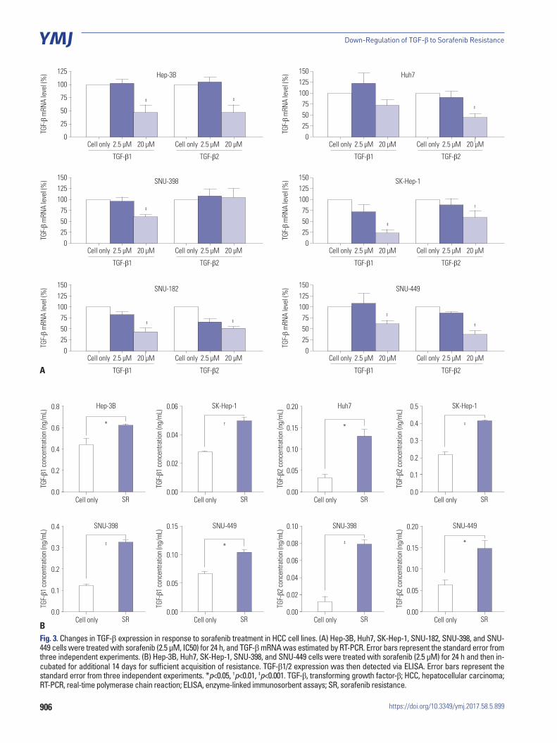
TGF-β expression was reduced by sorafenib treatment in HCC cell linesIn order to clarify the changes in TGF-β expression as a result of sorafenib treatment, we analyzed changes in the mRNA levels of TGF-β by real-time PCR. Fig. 3A show that, while treatment with low concentrations (2.5 μM) of sorafenib did not signifi-cantly alter TGF-β expression (p>0.05), the expression was sig-
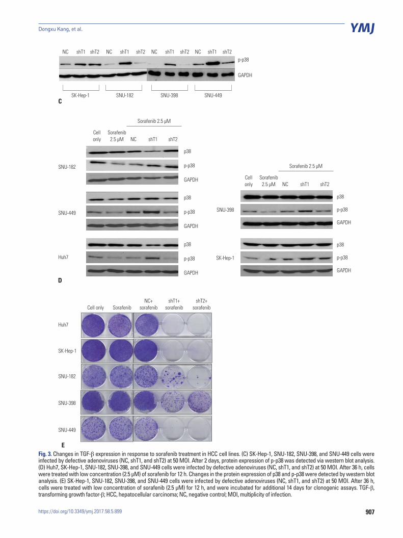
nificantly (p<0.05) reduced at high concentrations (20 μM). In addition, TGF-β expression was increased under conditions of sorafenib resistance (Fig. 3B), suggesting that decrease of TGF-β expression could reduce the resistance of HCC cell lines to sorafenib and more effectively induce cell death. Interest-ingly, when TGF-β expression was knocked down by shRNA, phosphorylation of p38 was also increased (Fig. 3C).
Sorafenib combined with adenovirus expressing shRNA against TGF-β was more effective for inducing cell death in HCC cell linesNext, we investigated whether the cell death could be increased by low concentrations (2.5 μM) of sorafenib when combined with shRNA against TGF-β. As shown in Fig. 3D and E, phos-phorylation of p38 was increased in response to this combined treatment compared to sorafenib-pretreated NC virus-infected control group, and cell viability was lower than in the sorafenib-pretreated NC virus-infected control group. These results sug-gest that TGF-β down-regulation is capable of increasing the cytotoxicity of sorafenib by overcoming sorafenib-induced p38 inactivation.
Anti-tumor effect of sorafenib combined with an adenovirus expressing shTGF-β in xenograft animal modelsAs described above, a series of in vitro experiments confirmed that an adenovirus expressing shTGF-β increased the activity of p38, thereby decreasing the resistance of HCC cell lines to sorafenib. Subsequently, in order to further confirm the anti-tumor effect of this combination therapy, and whether this th-erapy is able to override the resistance of HCC tumor cells to sorafenib, we designed an in vivo experiment in xenograft an-imal models.
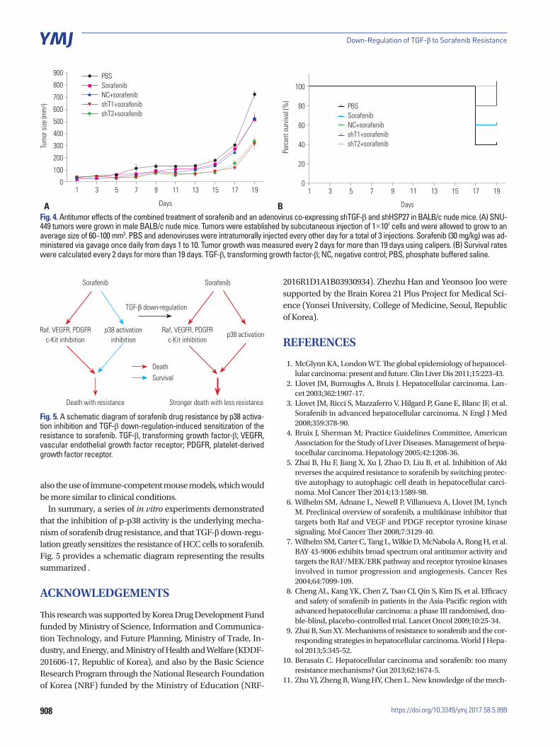
Fig. 4A shows that treatment with adenovirus expressing shTGF-β1 or shTGF-β2 in combination with sorafenib dis-played increased anti-tumor abilities in comparison to sora-fenib alone. This suggests that while these treatments did ef-fectively reduce the resistance of HCC tumor cells to sorafenib, these treatments did not result in a complete reduction in re-sistance. Despite these outcomes, we observed no differences in tumor regression in response to shTGF-β1 and shTGF-β2 (Fig. 4A). The survival rate of the animals in our study indicated that the combination therapy of adenovirus expressing shTGF-β1 or 2 with sorafenib was the most effective (Fig. 4B).
DISCUSSION
In the present study, we have demonstrated that a low concen-tration of sorafenib (2.5 μM) can inhibit the activity of p-p38, however, no significant cell death appeared in the low-dose sorafenib treatment. After the low-dose treatment, the activi-ties of p-Akt and p-p65 were increased. Thus, we can infer that
904
Down-Regulation of TGF-β to Sorafenib Resistance
https://doi.org/10.3349/ymj.2017.58.5.899
Fig. 2. MKK3/6E induced p-p38 activation and massive cell death in HCC cell lines. (A) Hep-3B, Huh7, SNU-398, and SNU-449 cells were transfected with the pCDNA3-MKK3/6E plasmid (1 μg) for 24 h, and treated with sorafenib (2.5 μM) for 24 h. Protein expressions of MKK3, MKK6, p-p38, and GAP-DH were estimated via western blot analysis. (B) Hep-3B, Huh7, SNU-398, and SNU-449 cells were transfected with the pCDNA3-MKK3/6E plasmid (1 μg) for 24 h and treated with sorafenib in a dose-dependent manner for 24 h. Cell viability was examined using a MTS viability assay. Error bars repre-sent the standard error from three independent experiments. ‡p<0.001. HCC, hepatocellular carcinoma.
120
100
80
60
40
20
0
120
100
80
60
40
20
0
120
100
80
60
40
20
0
120
100
80
60
40
20
00
0 0
02.5
2.5 2.5
2.5 15
15 15
15 25
25 25
25
Cell
viabi
lity (
%)
Cell
viabi
lity (
%)
Cell
viabi
lity (
%)
Cell
viabi
lity (
%)
Sorafenib (μM)
Sorafenib (μM) Sorafenib (μM)
Sorafenib (μM)
ControlMKK3EMKK6E
SNU-449
Huh7
SNU-398
Hep-3B
‡
‡
‡ ‡
‡
‡
‡‡
‡
‡‡
‡
‡
‡
ControlMKK3EMKK6E
ControlMKK3EMKK6E
ControlMKK3EMKK6E
MKK3
MKK6
p-p38
GAPDH
MKK3
MKK6
p-p38
GAPDH
Hep-3B
Huh7
pCDNA3-MKK6E
pCDNA3-MKK6E
pCDNA3-MKK3E
pCDNA3-MKK3E
pCDNA3pCDNA3
Sorafenib 2.5 μM
MKK3
MKK6
p-p38
GAPDH
MKK3
MKK6
p-p38
GAPDH
SNU-398
SNU-449
pCDNA3-MKK6E
pCDNA3-MKK6E
pCDNA3-MKK3E
pCDNA3-MKK3E
pCDNA3pCDNA3
Sorafenib 2.5 μM
A
B
a decrease of p-p38 is the underlying mechanism responsible for sorafenib resistance in HCC cell lines. In other words, sorafenib-induced multikinase inhibition23 can be retarded or
even inhibited by p38 inhibition due to the steric hinderance of sorafenib to p38,24 which makes a potential of increase of sur-vival signals such as p65 or src depending on the cellular con-
905
Dongxu Kang, et al.
https://doi.org/10.3349/ymj.2017.58.5.899
MKK3
MKK6
p38
p-p38
p-Akt
p-p65
GAPDH
SNU-398 SNU-449
pCDNA3-MKK6E
pCDNA3pCDNA3-MKK3E
pCDNA3
Sorafenib 2.5 μM
pCDNA3-MKK6E
pCDNA3pCDNA3-MKK3E
pCDNA3
Sorafenib 2.5 μM
Fig. 2. MKK3/6E induced p-p38 activation and massive cell death in HCC cell lines. (C) Hep-3B, Huh7, SNU-398, and SNU-449 cells were trans-fected with the pCDNA3-MKK3/6E plasmid (1 μg) for 24 h, treated with sorafenib in a dose-dependent manner for 24 h, and then incubated for additional 14 days for clonogenic assays. (D) Hep-3B, Huh7, SNU-398, and SNU-449 cells were transfected with the pCDNA3-MKK3/6E plas-mid (1 μg) for 24 h, and treated with sorafenib (2.5 μM) for 24 h. Protein expressions of MKK3, MKK6, p-p38, p-Akt, p-p65 and GAPDH were esti-mated via western blot analysis. HCC, hepatocellular carcinoma.
Hep-3B
Huh7
SNU-398
SNU-449
pCDNA3-MKK6E
pCDNA3-MKK6E
pCDNA3-MKK3E
pCDNA3-MKK3E
pCDNA3pCDNA3
Sorafenib 2.5 μM
MKK3
MKK6
p38
p-p38
p-Akt
p-p65
GAPDH
Hep-3B Huh7
pCDNA3-MKK6E
pCDNA3pCDNA3-MKK3E
pCDNA3pCDNA3-MKK6E
pCDNA3pCDNA3-MKK3E
pCDNA3
Sorafenib 2.5 μM Sorafenib 2.5 μM
C
D
text. Therefore, after inhibition of p38 activation by sorafenib and resultant acquisition of resistance, survival signals such as p65 or src can be increased indirectly depending on the can-cer cell types.
We used MKK3/6E to verify that when the activity of p-p38 is increased, cell death is induced and sorafenib resistance is overcome. As MKK3/6 is upstream of the p38 signal in the sig-nally pathway, its activity can induce the phosphorylation of p38. It has been shown that the structure of MKK3/6E is such that MKK3/6E is able to activate p38 without an external stim-ulus.25 Our results show that MKK3/6E can significantly im-prove the activity of p-p38 and induce cell death in a signifi-cantly large number of cells (p<0.05). When treatment with MKK3/6E was combined with sorafenib, p-p38 activity was significantly increased in comparison to the activity observed in cells treated with sorafenib alone, but was lower in compari-son to the p-p38 activity of cells treated with MKK3/6E alone. MTS and clonogenic assays confirmed that the combined ap-plication was better than treatment with sorafenib alone. It can be seen that in response to the low-dose sorafenib treatment, p-p38 activity was reduced, suggesting the mechanism in-volved in the resistance of HCC cells to sorafenib.
In this study, we observed that cell viability and the expres-sion of TGF-β were increased, when HCC cells exhibited drug resistance after treatment with a low concentration of sorafenib. We have also demonstrated increased activities of p-Akt and p-p65 under conditions of sorafenib resistance, when TGF-β expression was clearly and positively correlated with sorafenib drug resistance. On the other hand, however, TGF-β expres-sion was significantly decreased and cell viability was notice-ably inhibited in response to treatment with a high concentra-tion of sorafenib (20 μM). These results indicate that the reduction in cell viability in response to high concentration of sorafenib (20 μM) was associated with decreased TGF-β ex-pression. As a result of this, therefore, we targeted TGF-β, knock-ing down TGF-β expression by using shRNA in order to im-prove the rate of cell death and reduce drug resistance.
In this study, the reduced expression of TGF-β by shRNA not only increased the activity of p-p38, but also reduced both cell viability and drug resistance. Although the clonogenic assay showed that the combined application significantly improved cytotoxicity, the treatment, however, did not completely elimi-nate the formation of cell colonies from every cancer cells ex-amined (Fig. 3E). In order to further clarify the anti-tumor ef-fect, as well as the ability of this combination treatment to overcome drug resistance, therefore, an animal model was es-tablished using subcutaneous abdominal injections of an HCC cell line (SNU-449) into BALB/c nude mice. The result sh-owed incomplete regression of tumor in mice, suggesting that we should consider adding a new target in order to further im-prove cytotoxicity, and to more effectively overcome sorafenib resistance. In the future, the use of oncolytic adenoviruses ex-pressing combined therapeutic genes should be explored, and
906
Down-Regulation of TGF-β to Sorafenib Resistance
https://doi.org/10.3349/ymj.2017.58.5.899
125
100
75
50
25
0
TGF-
β m
RNA
leve
l (%
)
TGF-β1 TGF-β2
Hep-3B
‡ ‡
‡
Cell only Cell only2.5 μM 2.5 μM20 μM 20 μM
1501251007550250
TGF-
β m
RNA
leve
l (%
)
TGF-β1 TGF-β2
Huh7
Cell only Cell only2.5 μM 2.5 μM20 μM 20 μM
1501251007550250
1501251007550250
TGF-
β m
RNA
leve
l (%
)TG
F-β
mRN
A le
vel (
%)
TGF-β1
TGF-β1
TGF-β2
TGF-β2
SK-Hep-1
SNU-449
Cell only
Cell only
Cell only
Cell only
2.5 μM
2.5 μM
2.5 μM
2.5 μM
20 μM
20 μM
20 μM
20 μM
1501251007550250
1501251007550250
TGF-
β m
RNA
leve
l (%
)TG
F-β
mRN
A le
vel (
%)
TGF-β1
TGF-β1
TGF-β2
TGF-β2
SNU-398
SNU-182
Cell only
Cell only
Cell only
Cell only
2.5 μM
2.5 μM
2.5 μM
2.5 μM
20 μM
20 μM
20 μM
20 μM
Fig. 3. Changes in TGF-β expression in response to sorafenib treatment in HCC cell lines. (A) Hep-3B, Huh7, SK-Hep-1, SNU-182, SNU-398, and SNU-449 cells were treated with sorafenib (2.5 μM, IC50) for 24 h, and TGF-β mRNA was estimated by RT-PCR. Error bars represent the standard error from three independent experiments. (B) Hep-3B, Huh7, SK-Hep-1, SNU-398, and SNU-449 cells were treated with sorafenib (2.5 μM) for 24 h and then in-cubated for additional 14 days for sufficient acquisition of resistance. TGF-β1/2 expression was then detected via ELISA. Error bars represent the standard error from three independent experiments. *p<0.05, †p<0.01, ‡p<0.001. TGF-β, transforming growth factor-β; HCC, hepatocellular carcinoma; RT-PCR, real-time polymerase chain reaction; ELISA, enzyme-linked immunosorbent assays; SR, sorafenib resistance.
0.8
0.6
0.4
0.2
0.0
0.20
0.15
0.10
0.05
0.00
0.06
0.04
0.02
0.00
0.5
0.4
0.3
0.2
0.1
0.0
0.4
0.3
0.2
0.1
0.0
0.10
0.08
0.06
0.04
0.02
0.00
0.15
0.10
0.05
0.00
0.20
0.15
0.10
0.05
0.00
TGF-
β1 co
ncen
tratio
n (n
g/m
L)
TGF-
β2 co
ncen
tratio
n (n
g/m
L)
TGF-
β1 co
ncen
tratio
n (n
g/m
L)
TGF-
β2 co
ncen
tratio
n (n
g/m
L)
TGF-
β1 co
ncen
tratio
n (n
g/m
L)
TGF-
β2 co
ncen
tratio
n (n
g/m
L)
TGF-
β1 co
ncen
tratio
n (n
g/m
L)
TGF-
β2 co
ncen
tratio
n (n
g/m
L)Hep-3B Huh7SK-Hep-1 SK-Hep-1
* *† ‡
SNU-398 SNU-398SNU-449 SNU-449
‡ ‡* *
Cell only Cell onlyCell only Cell only
Cell only Cell onlyCell only Cell only
SR SRSR SR
SR SRSR SR
A
B
‡
‡
†
‡ ‡‡
‡
907
Dongxu Kang, et al.
https://doi.org/10.3349/ymj.2017.58.5.899
Fig. 3. Changes in TGF-β expression in response to sorafenib treatment in HCC cell lines. (C) SK-Hep-1, SNU-182, SNU-398, and SNU-449 cells were infected by defective adenoviruses (NC, shT1, and shT2) at 50 MOI. After 2 days, protein expression of p-p38 was detected via western blot analysis. (D) Huh7, SK-Hep-1, SNU-182, SNU-398, and SNU-449 cells were infected by defective adenoviruses (NC, shT1, and shT2) at 50 MOI. After 36 h, cells were treated with low concentration (2.5 μM) of sorafenib for 12 h. Changes in the protein expression of p38 and p-p38 were detected by western blot analysis. (E) SK-Hep-1, SNU-182, SNU-398, and SNU-449 cells were infected by defective adenoviruses (NC, shT1, and shT2) at 50 MOI. After 36 h, cells were treated with low concentration of sorafenib (2.5 μM) for 12 h, and were incubated for additional 14 days for clonogenic assays. TGF-β, transforming growth factor-β; HCC, hepatocellular carcinoma; NC, negative control; MOI, multiplicity of infection.
p-p38
GAPDH
SK-Hep-1 SNU-182
NC shT1 shT2 NC shT1 shT2
SNU-398 SNU-449
NC shT1 shT2 NC shT1 shT2
Cell onlyNC+
sorafenibSorafenibshT1+
sorafenibshT2+
sorafenib
Huh7
SNU-182
SK-Hep-1
SNU-398
SNU-449
p38
p-p38
GAPDH
p38
p-p38
GAPDH
p38
p-p38
GAPDH
p38
p-p38
GAPDH
p38
p-p38
GAPDH
SNU-182
SNU-449
Huh7
Cellonly NC
Sorafenib2.5 μM shT1 shT2
Sorafenib 2.5 μM
SNU-398
SK-Hep-1
C
D
E
Cellonly NC
Sorafenib2.5 μM shT1 shT2
Sorafenib 2.5 μM
908
Down-Regulation of TGF-β to Sorafenib Resistance
https://doi.org/10.3349/ymj.2017.58.5.899
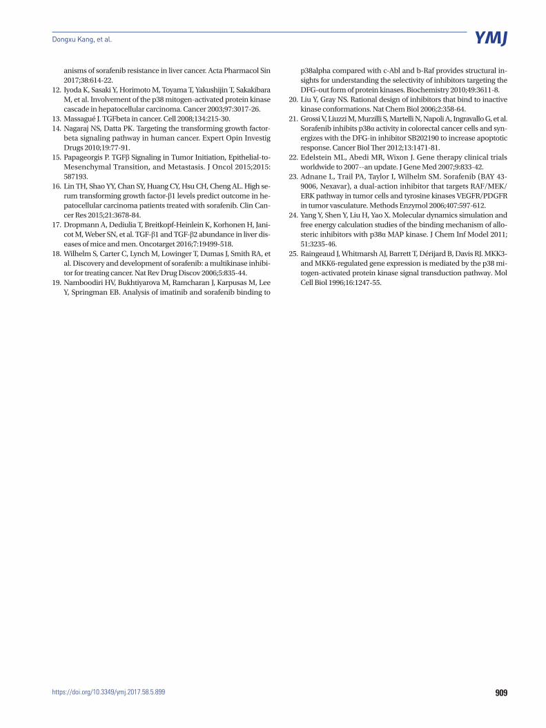
Fig. 5. A schematic diagram of sorafenib drug resistance by p38 activa-tion inhibition and TGF-β down-regulation-induced sensitization of the resistance to sorafenib. TGF-β, transforming growth factor-β; VEGFR, vascular endothelial growth factor receptor; PDGFR, platelet-derived growth factor receptor.
Sorafenib Sorafenib
TGF-β down-regulation
Death with resistance Stronger death with less resistance
Raf, VEGFR, PDGFRc-Kit inhibition
Raf, VEGFR, PDGFRc-Kit inhibition
p38 activation inhibition
p38 activation
DeathSurvival
Fig. 4. Antitumor effects of the combined treatment of sorafenib and an adenovirus co-expressing shTGF-β and shHSP27 in BALB/c nude mice. (A) SNU-449 tumors were grown in male BALB/c nude mice. Tumors were established by subcutaneous injection of 1×10⁷ cells and were allowed to grow to an average size of 60−100 mm3. PBS and adenoviruses were intratumorally injected every other day for a total of 3 injections. Sorafenib (30 mg/kg) was ad-ministered via gavage once daily from days 1 to 10. Tumor growth was measured every 2 days for more than 19 days using calipers. (B) Survival rates were calculated every 2 days for more than 19 days. TGF-β, transforming growth factor-β; NC, negative control; PBS, phosphate buffered saline.
Tum
or si
ze (m
m3 )
900
800
700
600
500
400
300
200
100
01 3 5 7 9 11 13 15 17 19
Days
PBSSorafenibNC+sorafenibshT1+sorafenibshT2+sorafenib
Perc
ent s
urviv
al (%
)
100
80
60
40
20
0
Days
1 3 5 7 9 11 13 15 17 19
PBSSorafenibNC+sorafenibshT1+sorafenibshT2+sorafenib
A B
also the use of immune-competent mouse models, which would be more similar to clinical conditions.
In summary, a series of in vitro experiments demonstrated that the inhibition of p-p38 activity is the underlying mecha-nism of sorafenib drug resistance, and that TGF-β down-regu-lation greatly sensitizes the resistance of HCC cells to sorafenib. Fig. 5 provides a schematic diagram representing the results summarized .
ACKNOWLEDGEMENTS
This research was supported by Korea Drug Development Fund funded by Ministry of Science, Information and Communica-tion Technology, and Future Planning, Ministry of Trade, In-dustry, and Energy, and Ministry of Health and Welfare (KDDF-201606-17, Republic of Korea), and also by the Basic Science Research Program through the National Research Foundation of Korea (NRF) funded by the Ministry of Education (NRF-
2016R1D1A1B03930934). Zhezhu Han and Yeonsoo Joo were supported by the Brain Korea 21 Plus Project for Medical Sci-ence (Yonsei University, College of Medicine, Seoul, Republic of Korea).
REFERENCES
1. McGlynn KA, London WT. The global epidemiology of hepatocel-lular carcinoma: present and future. Clin Liver Dis 2011;15:223-43.
2. Llovet JM, Burroughs A, Bruix J. Hepatocellular carcinoma. Lan-cet 2003;362:1907-17.
3. Llovet JM, Ricci S, Mazzaferro V, Hilgard P, Gane E, Blanc JF, et al. Sorafenib in advanced hepatocellular carcinoma. N Engl J Med 2008;359:378-90.
4. Bruix J, Sherman M; Practice Guidelines Committee, American Association for the Study of Liver Diseases. Management of hepa-tocellular carcinoma. Hepatology 2005;42:1208-36.
5. Zhai B, Hu F, Jiang X, Xu J, Zhao D, Liu B, et al. Inhibition of Akt reverses the acquired resistance to sorafenib by switching protec-tive autophagy to autophagic cell death in hepatocellular carci-noma. Mol Cancer Ther 2014;13:1589-98.
6. Wilhelm SM, Adnane L, Newell P, Villanueva A, Llovet JM, Lynch M. Preclinical overview of sorafenib, a multikinase inhibitor that targets both Raf and VEGF and PDGF receptor tyrosine kinase signaling. Mol Cancer Ther 2008;7:3129-40.
7. Wilhelm SM, Carter C, Tang L, Wilkie D, McNabola A, Rong H, et al. BAY 43-9006 exhibits broad spectrum oral antitumor activity and targets the RAF/MEK/ERK pathway and receptor tyrosine kinases involved in tumor progression and angiogenesis. Cancer Res 2004;64:7099-109.
8. Cheng AL, Kang YK, Chen Z, Tsao CJ, Qin S, Kim JS, et al. Efficacy and safety of sorafenib in patients in the Asia-Pacific region with advanced hepatocellular carcinoma: a phase III randomised, dou-ble-blind, placebo-controlled trial. Lancet Oncol 2009;10:25-34.
9. Zhai B, Sun XY. Mechanisms of resistance to sorafenib and the cor-responding strategies in hepatocellular carcinoma. World J Hepa-tol 2013;5:345-52.
10. Berasain C. Hepatocellular carcinoma and sorafenib: too many resistance mechanisms? Gut 2013;62:1674-5.
11. Zhu YJ, Zheng B, Wang HY, Chen L. New knowledge of the mech-
909
Dongxu Kang, et al.
https://doi.org/10.3349/ymj.2017.58.5.899
anisms of sorafenib resistance in liver cancer. Acta Pharmacol Sin 2017;38:614-22.
12. Iyoda K, Sasaki Y, Horimoto M, Toyama T, Yakushijin T, Sakakibara M, et al. Involvement of the p38 mitogen-activated protein kinase cascade in hepatocellular carcinoma. Cancer 2003;97:3017-26.
13. Massagué J. TGFbeta in cancer. Cell 2008;134:215-30.14. Nagaraj NS, Datta PK. Targeting the transforming growth factor-
beta signaling pathway in human cancer. Expert Opin Investig Drugs 2010;19:77-91.
15. Papageorgis P. TGFβ Signaling in Tumor Initiation, Epithelial-to-Mesenchymal Transition, and Metastasis. J Oncol 2015;2015: 587193.
16. Lin TH, Shao YY, Chan SY, Huang CY, Hsu CH, Cheng AL. High se-rum transforming growth factor-β1 levels predict outcome in he-patocellular carcinoma patients treated with sorafenib. Clin Can-cer Res 2015;21:3678-84.
17. Dropmann A, Dediulia T, Breitkopf-Heinlein K, Korhonen H, Jani-cot M, Weber SN, et al. TGF-β1 and TGF-β2 abundance in liver dis-eases of mice and men. Oncotarget 2016;7:19499-518.
18. Wilhelm S, Carter C, Lynch M, Lowinger T, Dumas J, Smith RA, et al. Discovery and development of sorafenib: a multikinase inhibi-tor for treating cancer. Nat Rev Drug Discov 2006;5:835-44.
19. Namboodiri HV, Bukhtiyarova M, Ramcharan J, Karpusas M, Lee Y, Springman EB. Analysis of imatinib and sorafenib binding to
p38alpha compared with c-Abl and b-Raf provides structural in-sights for understanding the selectivity of inhibitors targeting the DFG-out form of protein kinases. Biochemistry 2010;49:3611-8.
20. Liu Y, Gray NS. Rational design of inhibitors that bind to inactive kinase conformations. Nat Chem Biol 2006;2:358-64.
21. Grossi V, Liuzzi M, Murzilli S, Martelli N, Napoli A, Ingravallo G, et al. Sorafenib inhibits p38α activity in colorectal cancer cells and syn-ergizes with the DFG-in inhibitor SB202190 to increase apoptotic response. Cancer Biol Ther 2012;13:1471-81.
22. Edelstein ML, Abedi MR, Wixon J. Gene therapy clinical trials worldwide to 2007--an update. J Gene Med 2007;9:833-42.
23. Adnane L, Trail PA, Taylor I, Wilhelm SM. Sorafenib (BAY 43-9006, Nexavar), a dual-action inhibitor that targets RAF/MEK/ERK pathway in tumor cells and tyrosine kinases VEGFR/PDGFR in tumor vasculature. Methods Enzymol 2006;407:597-612.
24. Yang Y, Shen Y, Liu H, Yao X. Molecular dynamics simulation and free energy calculation studies of the binding mechanism of allo-steric inhibitors with p38α MAP kinase. J Chem Inf Model 2011; 51:3235-46.
25. Raingeaud J, Whitmarsh AJ, Barrett T, Dérijard B, Davis RJ. MKK3- and MKK6-regulated gene expression is mediated by the p38 mi-togen-activated protein kinase signal transduction pathway. Mol Cell Biol 1996;16:1247-55.