Embed Size (px)
Citation preview
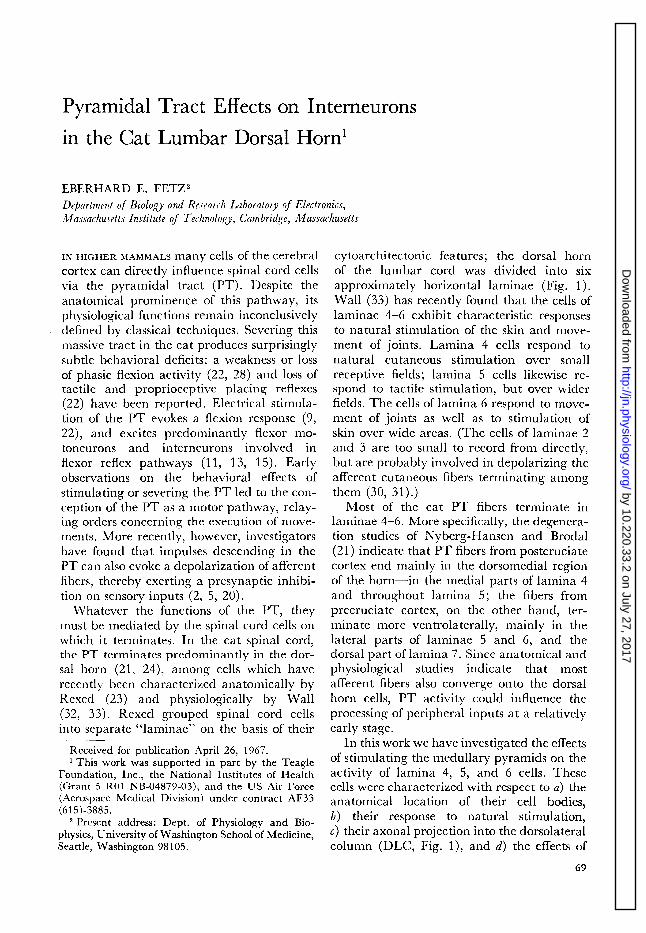
Pyramidal Tract Effects on Interneurons
in the Cat Lumbar Dorsal Horn1
EBERHARD E. FETZ2
Department of Biology and Research Laboratory of Electronics, Illassachusetts Institute of Technology, Cambridge, Massachusetts
IN HIGHER MAMMALS many cells of the cerebral cytoarchitectonic features; the dorsal horn cortex can directly influence spinal cord cells of the lumbar cord was divided into six via the pyramidal tract (PT). Despite the approximately horizontal laminae (Fig. 1). anatomical prominence of this pathway, its Wall (33) h as recently found that the cells of physiological functions remain inconclusively defined by classical techniques. Severing this
laminae 4-6 exhibit characteristic responses to natural stimulation of the skin and move-
massive tract in the cat produces surprisingly subtle behavioral deficits: a weakness or loss of phasic flexion activity (22, 28) and loss of tat tile and propriocep tive placing reflexes (22) have been reported. Electrical stimula- tion of the PT evokes a flexion response (9,
2% and excites predominantly flexor mo- toneurons and interneurons involved in flexor reflex pathways (11, 13, 15). Early observations on the behavioral effects of stimulating or severing the PT led to the con- ception of the PT as a motor pathway, relay- ing orders concerning the execution of move- ments. More recently, however, investigators have found that impulses descending in the PT can also evoke a depolarization of afferent fibers, thereby exerting a presynaptic inhibi- tion on sensory input&%, 5, 20).
Whatever the functions of the PT, they must be mediated by the spinal cord cells on which it terminates. In the cat spinal cord, the PT terminates predominantly in the dor- sal horn (21, 24), among cells which have recently been characterized anatomically by Rexed (23) and physiologically by Wall (32, 33). Rexed grouped spinal cord cells into separate “laminae” on the basis of their
Received for publication April 26, 1767. l This work was supported in part by the Teagle
Foundation, Inc., the National Institutes of Health (Grant 5 R01 NB-04877-03), and the US Air Force (Aerospace Medical Division) under contract AF33 (615)-3885.
2 Present address: Dept. of Physiology and Bio- physics, University of Washington School of Medicine, Seattle, Washington 78105.
ment of joints. Lamina 4 cells respond to natural cutaneous stimulation over small receptive fields; lamina 5 cells likewise re- spond to tactile stimulation, but over wider fields. The cells of lamina 6 respond to move- ment of joints as well as to stimulation of skin over wide areas. (The cells of laminae 2 and 3 are too small to record from directly, but are probably involved in depolarizing the afferent cutaneous fibers terminating among them (30, 31).)
Most of the cat PT fibers terminate in laminae 4-6. More specifically, the degenera- tion studies of Nyberg-Hansen and Brodal (21) indicate that PT fibers from postcruciate cortex end mainly in the dorsomedial region of the horn-in the medial parts of lamina 4 and throughout lamina 5; the fibers from precruciate cortex, on the other hand, ter- minate more ventrolaterally, mainly in the lateral parts of laminae 5 and 6, and the dorsal part of lamina 7. Since anatomical and physiological studies indicate that most afferent fibers also converge onto the dorsal horn cells, PT activity could influence the processing of early stage.
peripheral inputs at a relatively
In this work we have investigated the effects of stimulating the medullary pyramids on the activity of lamina 4, 5, and 6 cells. These cells were characterized with respect to a) the anatomical location of their cell bodies, 6) their response to natural stimulation, c) their axonal projection into the dorsolateral column (DLC, Fig. l), and d) the effects of
by 10.220.33.2 on July 27, 2017http://jn.physiology.org/
Dow
nloaded from

70 E. E. FETZ
- I DC DR
FIG. 1. Rexed’s anatomical subdivision of the dorsal horn into laminae 1-6. Typical cross section of the cat spinal cord at the seventh lumbar level. Numbers label laminae distinguished by Rexed on the basis of cytoarchitectonic criteria. Abbreviations label fiber &acts as follows: DC = dorsal columns, DR = dorsal root, LT = Lissauer’s tract, L)LC = dorsolateral col- umn (location of the spinocervical tract), and PT = pyramidal tract.
stimulating the PT on their spontaneous
activity and responses to peripheral stimula- tion.
METHODS
Experiments were performed on 23 healthy, adult cats each weighing over 2 kg. Under ether anesthesia the carotid arteries were ligated and the trachea cannulated. Sixteen cats were “spinalized” with a special transection at the obex leaving only the PT intact. Following Lloyd’s method (lo), this section was accomplished with a blade shaped to sever all fiber tracts except the ventrally lying PT. After being “guillotined” with this blade, the cats were artificially ventilated. Nervous centers rostra1 to the cut were anemically destroyed by occluding the vertebral arteries for at least 10 min, in addi- tion to the previous ligation of the carotid arteries. Seven cats were decerebrated as described by Wall (33), rather than guillotined.
Ripolar stimulating electrodes were placed in the I?T 6-10 mm rostra1 to the obex, via a dorsal approach. The PT-evoked flexion response proved to be an excellent guide in placing the electrodes: when low-intensity stimulation (less than 0.18 ma or 2 v) evoked flexion of only the contralateral limbs, the electrodes were invariably placed in the appropriate half of the PT, as verified by subse- quen t histologic sections. Standard PT stimulus parameters were 50-msec trains of 0.4-msec rec-
tangular current pulses at 500,‘sec. The various types of responses to PT stimulatioI1--.~flextion, dorsal root potentials, and single-unit activity- generally varied the same way with variation in stimulus parameters. Responses usually decreased for shorter pulses and lower frequencies, but did not increase substantially for longer pulses or higher frequencies.
After placing the electrodes we recorded “flex- ion threshold”---the least intensity of PT stimula- tion which repeatedly evoked a visible twitch of the hindlimb muscles. The animals was then par- alyzed with gallamine triethiodide (Flaxedil) and laminectomy performed from S1 to La. The es- posed spinal cord was covered with warm tnin- era1 oil (Nujol), maintained between 35 and 38 C.
Rectangular stimulating pulses were led to stimulus boxes designed by Dr. Karl Kornacker to deliver positive and negative current pulses whose shapes could be independently con trolled. This control effectively minimized stimulus arti- facts and allowed single-cell responses to be moni- tored during simultaneous stimulation of the PT. The skin was stimulated electrically through two short 30-gauge hypodermic needles placed into the superficial dermal I ayers.
Responses of single cells in the lumbar cord were recorded extracellularly with glass micropipettes filled with 3 M KC1 and having a resistance of about 1 megohm. To record from axons in the DLC, unbroken glass micropipettes with resis- tances of IO-30 megohms were used. Dorsal root potentials (DRPs) were recorded from the smallest convenient caudal rootlet of the Lc dorsal root, with a pair of Ag-AgCl hooks. Signals were ampli- fied and displayed by standard techniques.
Single cells in the rostra1 part of the seventh lumbar segment were characterized bt- two cri- teria: their location in the dorsal horn w&h respect to Rexed’s laminar organization (Fig. 1). and their responses to natural stimulation. The cells’ mi- crometer coordinates with respect to cord dorsum and midline gave a relatively unreliable estimate of location in the dorsal horn, due to the considerable variability of cord geometry; however, the relative depth of successive units encountered in advancing the electrode was often useful in ordering them into layers. In each experiment the electrode tips were cut off in situ, the cord fixed in loo/, formalin and free-hand sections containing the electrode pre- pared for visual inspection. This histological check provided the most accurate estimate of anatotnical location.
Cells were also characterized by their response to natural stimulation. The receptive field was found for each of three categories of skin stimulation: I) brushing-gently moving hairs with a no. 3 camel%-hair brush; 2) touch-resting the brush handle on the skin; and 3) pressure-pinching a fold of skin with flat, blunt forceps. Care was al-
by 10.220.33.2 on July 27, 2017http://jn.physiology.org/
Dow
nloaded from

PT EFFECTS ON SPINAL CORD INTERNEURONS 71
ways taken to avoid moving the limb or pressing the muscle in producing cutaneous stimulation. Cells were also tested for “proprioceptive” re- sponses by moving the joints over small angles. Such movements invariably also stimulated some portions of the skin, but proprioceptive responses could usually be distinguished from cutaneous by being more regular and sustained for maintained joint angles, by being a function of angle only (independent of where the limb was touched in movement), by summing with responses to any deliberate cutaneous stimulation, and usually by decreasing when the direction of joint deflection was reversed.
Wall’s finding (32, 33) that cells in different an- atomical laminae have characteristic responses to natural stimulation was essentially verified, and the combination of anatomical and response charac- teristics was used to group cells into “layers.” Thus, layer 4 cells were located in Rexed’s lamina 4 and responded to skin stimulation over a small receptive field, e.g., a fraction of a toe at rostra1 Ly. Layer 5 cells were found in Rexed’s lamina 5 and also re- sponded only to tactile stimulation; their receptive fields were considerably larger than those of layer 4 cells and usually exhibited a sensitivity gradient. Cells designated as layer 6 were located in lamina 6, responded to skin stimulation over wide regions of the ipsilateral limb only, and often exhibited proprioceptive responses to movements of joints. Cells affected by contralateral stimulation were histologically located ventrally to lamina 6; these deeper cells in general also exhibited more variable and delayed responses to any stimulation.
Most cells were also tested for axonal projection into the dorsolateral column (DLC, Fig. 1) by stimulating the DLC with ball electrodes at Ld. Antidromic responses could be differentiated from orthodromic excitation via dorsal column collaterals by the usual criteria: an all-or-nothing spike response at a short, invariant latency and following of high-frequency stimulus trains.
Pyramidal tract influences on these cells were in- vestigated by stimulating the medullary pyramids with trains of 0.4-msec rectangular pulses at 500/ sec. A standard train length of 50 msec was gen- erally used, since longer trains merely prolonged the response evoked by 50-msec trains. For a given cell, PT stimulation was found to have the same effect on its spontaneous activity and its response to peripheral stimulation, either natural or electrical. However, the threshold intensities at which PT stimulation began to affect these types of activity noticeably were not always the same. A standard test for PT inhibition of responses to peripheral stimulation was a 50-msec conditioning PT train, followed within 10 msec by a test shock of the skin (Fig. 4). With peripheral shocks just strong enough to evoke three to six repetitive cell responses, the “threshold” PT intensity for inhibition was that
minimum strength which repeatedly reduced the number of responses. For excitation of cells, thresh- old was taken to be the minimum PT intensity which repeatedly evoked activity in the cell.
To display the responses of single units in com- pact form we used the so-called dot-raster dis- play (Figs. 3, 4, 6-9). The sequence of action po- tentials of the cell was converted to a horizontal row of dots, preserving only the temporal sequence of firing in the spacing of the dots. Responses to suc- cessive stimuli are displayed in adjacent rows.
RESULTS
Field potentials
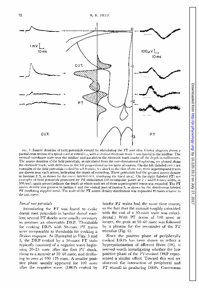
In a preliminary investigation we recorded the field potentials evoked in the spinal cord by electrical stimulation of the skin and PT. Figure 2 shows examples of field potentials evoked by a shock to the skin and by a lo- msec PT train, and recorded at various depths in the cord. The potentials varied relatively little in the rostrocaudal or medio- lateral direction; by far the greatest variation was in the dorsoventral direction. Typically, the height of the PT-evoked field potentials was constant from cord dorsum to around lamina 4; then it increased to peak in lamina 6 or ventral lamina 5 ; then it rapidly de- creased again. In contrast, the peripherally evoked field potentials rose to a sharp maxi- mum in the dorsal region of the horn, often peaking in lamina 4.
As discussed elsewhere (6, 7), the spatial distribution of field potentials themselves is not an accurate index of the distribution of the sources of these potentials, since potentials spread beyond the generating regions. A more accurate index of source density is the common expression for the source density p of any physical scalar field V. namely, the negative Laplacian of the field: p = - v2V. When this expression was used to compute the “source density ” of the spinal cord field potentials in Fig. 2, the sources of the PT- evoked field potentials were found in lamina 6, while the sources of the peripherally evoked field potentials appeared throughout laminae 2-5 (see Fig. 2). Although the exact shapes and widths of the distributions varied from one cat to the next, the sources of the PT-evoked field potentials invariably ap- peared in the ventral portion of the dorsal horn (laminae 5-7), while skin stimulation produced the greatest source density in more dorsal regions (ranging from laminae 2 to 5).
by 10.220.33.2 on July 27, 2017http://jn.physiology.org/
Dow
nloaded from
72 E. E. FETZ
-=- 4
ImVL
IOms
C U T.
IOOpV L IOms
3 PT
FIG. 2. Source densities of field potentials evoked by stimulating the PT and skin. Center diagram shows a
partial cross section of a spinal cord at rostra1 I A7, with a vertical electrode track 1 mm lateral to the midline. The vertical coordinate scale near the midline and parallel to the electrode track marks off the depth in millimeters. The source densities of the field potentials, as calculated from the one-dimensional Laplacian, are plotted along the electrode track, with deflection to the left proportional to intensity of sources. On the left (labeled CUT.) are examples of the field potentials evoked by a 0.4-msec, 4-v shock to the skin of one toe; three superimposed traces are shown near each arrow, indicating the depth of recording. These potentials had the greatest source density in laminae 2-5, as shown by the curve labeled cur. (enclosing the lined area). On the right (labeled PT) are examples of field potentials generated by PT stimulation (10 rectangular pulses at 2 v and 0.4 msec width, at 500/set); again arrows indicate the depth at which each set of three superimposed traces was recorded. The PT source density was greatest in lamina 6 and the ventral part of lamina 5, as shown by the distribution labeled PT (enclosing stippled area). The scale of the PT source density distribution was expanded 10 times relative to
the cut. curve.
Dorsal root potentials
Stimulating the PT was found to evoke dorsal root potentials in lumbar dorsal root- lets; several PT shocks were usually necessary to produce an observable DRP. Thresholds for evoking DRPs with 50-msec PT trains were comparable to thresholds for evoking a flexion response. As illustrated in Figs. 3 and 5, the DRP evoked by a 50-msec ‘PT train typically consisted of a negative wave begin- ning 20-25 msec after the first PT shock, rising to a summit at 50-60 msec, and declin- ing to zero at 100-l 25 msec. A smaller posi- tive phase usually appeared for 100 msec after the negative wave. (DRPs evoked by
briefer PT trains had the same time course, so the fact that the summit roughly coincided with the end of a 50-msec train was coinci- dental.) .With PT trains of 100 msec or longer, the peak at 50-60 msec was followed by a plateau for the remainder of the PT stimulus (Fig. 6).
Since the positive phase of peripherally evoked DRPs has been shown to reflect a hyperpolarization of afferent fibers (1 S), it seemed worth investigating whether the late positive phase of the PT-evoked DRP repre- sented a similar effect. Toward this end we observed the interaction of peripheral and PT stimuli in producing DRPs. Continuous
by 10.220.33.2 on July 27, 2017http://jn.physiology.org/
Dow
nloaded from

PT EFFECTS ON SPINAL CORD INTERNEURONS 73
brushing of the hindlimb evoked a tonic negative DRP of 300-400 pv for duration of stimulus. When a 50-msec PT stimulus train was given during such a tonic DRP, the negative phase of the PT-evoked DRP was occluded, but the positive phase was not en- hanced. This contrasts with the observations of Mendell and Wall (18) on peripherally evoked biphasic DRPs; they found that superimposing the biphasic DRP on a tonic negative DRP led to occlusion of the nega- tive phase and enhancement of the positive.
If the positive phase of the PT-evoked DRP reflected hyperpolarization of dorsal root fibers, any afferent volleys during this phase should be more effective in evoking post- synaptic effects. In particular, they might be more effective in evoking a negative DRP. This possibility was tested by giving a periph- eral skin shock during the positive phase of the PT-evoked DRP; the peripherally evoked negative DRP was consistently reduced, rather than enhanced.
Layer 4 cells
Layer 4 cells responded to cutaneous stimulation over relatively small regions of the skin. Their receptive fields were larger than those of afferent fibers, indicating a con- vergence of input, but smaller than those of layer 5 cells. At the rostra1 L7 level, medial cells had receptive fields on medial toes and lateral cells. responded to stimulation of lateral toes and the lateral side of the foot. Receptive fields generally covered a fraction of a toe or slightly larger areas on the side of the foot. Recording from the axons of these cells, Taub (25) found evidence for weak inhibition from surrounding skin, but such effects were not systematically studied here.
The layer 4 cells typically responded briskly to brushing the hairs and often increased their response to touch and pressure. If main- tained pressure produced a more intense, sustained response than brushing, the cell was characterized as having a “wide dynamic range.” Some cells showed no increase, or a decrease in firing to pressure and were said to have a “narrow dynamic range.”
Table 1 summarizes the effects of PT stimulation on these cells. About two-thirds of the layer 4 cells were found to be inhibited by PT volleys. This inhibition was observed as a reduction in the cells’ spontaneous activity
. 0 0. . . . 0. -- . . . . . . . . _ r.a. . . . . .- . . ” .
. . . . . . . . . PT . . . “m. . . . . *. . : . -. .*’ ‘7 ..U-..-. . - . . . . . . -
.m ..” . .
. . . ..- . . .-- m-B . . . .._“. . . . . . . . .w .-...U . . . l :.-. .:...
. . . . . . . . . .- . . . . :.. : :
. . ; .-.*. -.- m.” .“..““.“‘Y l :“.‘..~:-:-‘:.:
.
..- . .” - . . . . .m . . . . . . l .- . . ..-.. . w .- . . .m
‘. l l l l - : l -: ‘ : * -- n.s.
*.z: : .e.**.: y . ‘= - .* :. .,: l e=‘o--” l *
. . . . . “=.*;; :.. ‘ : : . m t:: . ‘Z. -7.::’ :=. . .
. . m . . . . . . . . . . : - *. - . . . . . . . . . . . . . . . = l . . . . ..* .: .
. .r -0. 0 :: -*’ n.8. + PT . .
. . . - -. . :. *
PT s .
. . . . . . 50m.
FIG. 3. PT inhibition of a layer 4 cell. As described in METHODS, each sequence of extracellularly recorded action potentials was converted to a horizontal row of dots. Successive sequences were recorded at l-set in- tervals and are displayed in adjacent rows from top to bottom. The cell’s spontaneous activity in the absence of intentional stimuli is illustrated at the top (sa.). Dots immediately below this (labeled PT) show the reduced activity when the pyramidal tract was stimu- lated during a 50-msec interval (indicated by PTs). When the skin of the receptive field was continuously brushed, the cell responded with a high maintained rate of firing (n.s.). When the 50-msec PT stimulation was superimposed on the natural stimulation of the skin, the cell’s responses were reduced (n.s.+PT). (The increasing length of the inhibitory period with successive PT trains was not regularly seen.) The 50. msec PT train (PTs) also evoked a dorsal root poten- tial (DRP) recorded from a caudal L6 dorsal rootlet; at the summit of the DRP the proximal end of the rootlet was about 250 pv negative to the distal end. Bottom row of dots gives 50-msec calibration intervals.
and a reduction of their responses to brushing or shocking the skin of the receptive field. Figure 3 illustrates the inhibition of spon-
taneous and naturally evoked activity of a layer 4 cell by a 50-msec PT train, as well as the DRP simultaneously recorded from a caudal Ls dorsal rootlet. Longer PT trains prolonged both inhibition and DRP (Fig. 6). Electric shocks applied to skin evoked a burst of activity in that cells could be reduced by a preceding PT train (Fig. 4).
The time course of inhibition could be de- termined by giving a peripheral test shock at different intervals after the conditioning PT train. Figure 5 illustrates the typical time course of inhibition produced by a 50-msec PT train and also shows the time course of the DRP. Both were maximum at 50-60 msec after the first PT shock, but the reduc- tion of cell responses preceded and outlasted the appearance of the negative DRP. In particular, the late positive phase of the DRP
by 10.220.33.2 on July 27, 2017http://jn.physiology.org/
Dow
nloaded from

74 IL E. FETZ
TABLE 1. PT @ects on dorsal horn interneurons
Layer
4
5
6
- None
10
(9)
(a,
iy,
PT Effects
Exit
(:I 22 (9)
(ii)
Inhib. Mixed
15 03)
10
(4)
Total
(::)
(&
(45)
AKJns in DLC
-
26/60 =43%
10/53 = 19%
O/24=0%
.-
This table gives the total number of cells in each category observed in both “guillotined” and decerebrate cats. Numbers in parentheses indicate cells observed in guillotined cats only. Cells designated “PT Effects- None” showed no change in activity for PT stimulus intensities up to three times “flexion threshold.”
was still accompanied by fewer cell responses hibition resembled that seen in the majority to afferent volleys. of layer 4 cells.
The layer 4 cells designated “mixed” re- sponded to PT stimulation with a brief period of activity preceding a more prolonged inhibition. Typically, they fired once or twice between 10 and 20 msec after the start of PT stimulation, as illustrated in Fig. 4. Following this brief initial firing the PT in-
A few cells showed no response to PT stimu- lation at intensities up to three times flexion threshold, although field potentials evoked by peripheral shock were reduced and adjacent cells were affected by PT stimulation.
To determine how many layer 4 cells had axons projecting up the DLC, this tract was stimulated at the Lq level. Of the 60 cells tested in layer 4, 26 (43%) exhibited anti- dromic responses.
’ . .... . . . . . . ;. . . ;.... . . . . . r.a.
- .:. : . .
PT
e.s.+ Pi
PT I.
. . . . . . . >..> . . . . . . . 10 mr.
FIG. 4. “Mixed” effects of PT stimulation typical of layer 4 cells. Most layer 4 cells with mixed responses to PT stimulation exhibited a brief period of activity followed by a longer period of inhibition. A compari- son of this cell’s spontaneous activity (s.a.) with its response to PT stimulation (PT) shows an initial re- sponse of one to two spikes appearing lo-20 msec after the start of PT stimulation (PTs), followed by a period of inactivity. This figure also illustrates the in- hibition of responses to an electric shock to the skin. In the group of dots labeled es., the first regularly ap- pearing dot (ca. 60 msec after the start of PT stimula- tion) represents the peripheral shock; the succeeding dots represent the cell’s response. When the shock was preceded by a PT train (e.s.+PT) the evoked re- sponses were considerably reduced. This figure illus- trates the standard conditioning-testing sequence used to test for PT inhibition of peripherally evoked re- sponses; however, the intensity of the peripheral shock was usually adjusted to evoke only three to six spikes. The activity shown here was recorded from an axon in the DLC; the unit had a small cutaneous receptive field t.ypical of layer 4 cells.
FIG. 5. Time course of PT inhibition of a peripher- ally evoked response in layer 4 and 5 cells. A shock to the skin was given at various times after the start of a 50-msec PT train, and the average reduction of re- sponses by the PT is plotted in the lower graph. For each point the test shock was given 10 times alone (to determine “control” response), and 10 times with a preceding PT train. The PT stimulation reduced the response to the “percent of control” shown at each latency. In this series the mean number of responses for a single control shock was 4.0 spikes. The vertical line near the beginning of the time axis gives the standard deviation of the means for the control groups. The upper graph gives the time course of the dorsal root potential evoked by a 50-msec PT train. A com- parison of the DRP with inhibition shows that the in- hibition preceded and outlasted the appearance of the negative DRP by a small duration.
by 10.220.33.2 on July 27, 2017http://jn.physiology.org/
Dow
nloaded from

PT EFFECTS ON SPINAL CORD INTERNEURONS 75
Mendell (17) has shown that over half the cells sending axons up the DLC receive excitation from afferent C fibers as well as A fibers. To test for PT effects on these two inputs, we recorded from axons in the DLC and stimulated the sural nerve electrically. Afferent volleys limited to the large A fibers evoked an early burst of high-frequency firing lasting between 4 and 30 msec, followed by a silent period; if the small C fibers were also stimulated additional late bursts of firing from 100 to 600 msec were also produced (17, 19). We observed that the PT could inhibit the responses to both A- and C-fiber volleys. Weak, continuous PT stimulation reduced both responses, but after several seconds the PT stimulation became less effective; the resumption of C responses dur- ing sustained PT stimulation followed the “windup” pattern described by Mendell and Wall (19).
Layer 5 cells
The layer 5 cells responded to skin stimu- lation of the ipsilateral limb, but over wider receptive fields than layer 4 cells. These fields usually included several toes or large areas of the foot and leg. A sensitivity gradient was a common characteristic of these fields; the threshold to natural and electrical stimula- tion usually increased toward the periphery of the receptive fields. In a few cells inhibitory regions could be readily demonstrated.
As shown in Table 1, the PT inhibited about one-third of the layer 5 cells. The time course and average threshold resembled that seen in layer 4 cells. Figure 6 illustrates the inhibition of such a cell by a 200-msec PT train, and shows the accompanying DRP.
In the layer 5 cells designated “mixed,” the PT train generally evoked a long period of activity, followed by a silent period. The activity began lo-20 msec after the first PT shock and sometimes lasted 20-30 msec, but often continued beyond the end of the PT stimulation. During the subsequent silent period, spontaneous and evoked activity were absent or reduced. Figure 7 illustrates the responses of a typical mixed layer 5 cell to PT stimulation and natural stimulation.
About a third of the layer 5 cells were excited by the PT. Typically, their activity began lo-20 msec after the start of PT stimu- lation and terminated 20-50 msec after the
, I . . . . . . . . : . - .
. I . . . ” . . . ” . . ‘
. ...‘. ..“‘ .I’ 1 .
. . . . . . . . . .
s.a.
PT
“.S.
n.s. + PT
PT 5.
DRP
-soIns.
FIG. 6. Layer 5 cell inhibited by prolonged PT stimulation. A 200-msec PT train (shown in PTs) inhibited the spontaneous activity of this layer 5 cell (cf. s.a. and PT), as well as its response to brushing the skin (n.s. vs. n.s.+PT). This PT train also evoked a long DRP with a summit of about 250 pv. The DRPs evoked by long PT stimulation typically peaked at 50-60 msec and continued at some plateau level be- low the peak (6) ; the return of the DRP to peak levels recorded here was not often seen.
end. In these cells no signs of inhibition of response to peripheral stimulation could be observed; in fact, for many such cells the PT facilitated the response to peripheral stimu- lation, i.e., the response to the combined stimuli exceeded the sum of responses to either. Figure 8 illustrates the effect of a PT train on the response to natural and electrical stimulation of the skin for such a cell. I f an “excited” cell had an inhibitory peripheral
” ‘.. ‘.
. ‘,: :
.: ., ‘, .’ . ..“.‘.: ., .’ i- : I . .’ :. ; ; ., n.r.
.’ .: ..I _‘.I.. I‘.. .
:,:;,x;‘.:‘-. :::;.. .,
n.r.+PT . . . . . . .:f::: ,..... :....:: ,. ,.
_. ‘. .’ . ‘.
. . . . . . . . . . . . .
. . . . . . . . . . . . . . . . . . . . . . . . . . . . .
. . . . . . . . . . . . . . .
. . . . . . . . . . . . . . . . . . . . . . . . . . . . .
. . . . . . . . . . . . . . .
. . . . . . . . . . . . . . . . . . . . . . . . . . . :’
. . . . . . . . . . . . . . .
PT
PT s. * . ‘ . . . . . . . 1orm.
FIG. 7. “Mixed” effects of PT stimulation on a layer 5 cell. This figure illustrates the pattern of excitation and inhibition seen in many layer 5 cells and most layer 6 cells characterized as having mixed responses to PT stimulation: a long excitatory period, often lasting for the duration of PT stimulation, followed by an inhibitory period. Here, n.s. shows the responses to brushing the skin, PT gives the responses to a 50- msec PT train, and ns. +PT indicates the responses to both given simultaneously. Although not shown, the responses of these cells to peripheral skin shocks were also reduced by the PT during the inhibitory phase.
by 10.220.33.2 on July 27, 2017http://jn.physiology.org/
Dow
nloaded from

76 E. E. FETZ
. ; . ‘, ,.:’ ,‘!., : , : :‘.,’ ; . \ ” ; , , , : ; , ; , Z,)‘,,‘. : . . , ; :
: i.;,:, .‘:.‘: .:_
: , : , : : : .‘ ‘:, ‘,‘. .::‘.i:.‘. “ : , :’ “ : . : : ; . . : ‘,‘::,;: :,‘“:,‘,‘, : ‘. ” : , . ; , ; , ; ; , ;_’ , ; , , , : . ‘. , , , , ” *
. . : .
: , ,..~ ; ‘,; .,+;: :“‘: ‘, .:.::.‘~I::;. :‘.’ ‘,j,:‘: , . , . , , . . . , , , ; , , , . ‘;::,‘;
. , , , . , ; . . , : : , ; ; ; . , , , . . . . . . . . . . . . . . . ~ . , . . , . ; : , . ; : . , . : : , . ‘ , . . , . , . . . , , , . : :.~ , , , , : j:
‘. .’ ; ;.;..‘. ..-,~‘,.~‘~,~,.~~~~~,.“:.~::~~ : . . ; ::_ , . . , . . , _..,, ! , , . . . , ; . : : . ,
1. . . : : . . . :::::11~; : : . . : . . I : ::v 1:“. .A. ‘.” . :
‘,.:, . ; : ‘Y,
.’ , : ; ,..~.‘.~_,.~ . , . , . , . ~ ; , . .:,.=.. . : . , . , .~.,,, ._.,,. . , . . , n.r. + PT
: :.::. ‘;:. .._.,....,__. .: ‘:::T::.:‘: ‘: .:.,.I: ,:: ,..’ ::
PT
e.s. + PT
PT s. ..‘_.... . . . . . . 10 m.
FIG. 8. Layer 5 cell excited by PT stimulation. This figure illustrates the response pattern of cells categor- ized as “excited” by the PT. As in previous figures, n.s. indicates the response to brushing the skin, and PT the response to a 50-msec PT train. When the two were combined (ns. +PT), there was no evidence of an inhibitory period, as in the mixed cells; in fact, in the interval from 20 to 60 msec after the end of the PT train, the responses to peripheral stimulation were enhanced, i.e., they exceeded the sum of responses to either alone. In es. the skin of the receptive field was stimulated electrically; the first dot in each horizontal sequence marks the skin shock. When preceded by a PT train (es. +PT), the shock evoked no less of a re- sponse; in fact, the late burst (20-25 msec after the shock) was even enhanced. It is noteworthy that these ventral excited cells exhibited no reduction of evoked responses at a time after the PT train when the more dorsal cells were maximally inhibited (Fig. 5).
receptive field, stimulating that region would always inhibit any activity evoked by the PT.
Of the 53 cells tested in layer 5 by DLC stimulation, only 10 (19%) exhibited anti- dromic responses. All these cells with axons ascending in the DLC received inhibition from the PT, although 4 of these also ex- hibited relatively brief periods of excitation. Of all the dorsal horn cells antidromically fired from the DLC, the majority (26/36= 72ojc) were found to be layer 4 cells.
Lqyer 6 cells
Layer 6 cells responded to cutaneous stim- ulation over wide receptive fields on the ipsi- lateral hindlimb only; these fields resembled those found in layer 5, but more often in- cluded an inhibitory region. In addition many layer 6 cells also responded to move- ments of joints. Such cells usually exhibited relatively regular spontaneous activity and responded to maintained joint deflection with a sustained tonic discharge. At the ros- tral Lr level, responses to passive flexion and extension of toes and foot were observed, with more cells responding to flexion than exten- sion (11 to 8 for toes; 12 to 5 for foot).
Many cells located histologically in lamina 6 and characterized as “layer 6” exhibited no demonstrable response to joint movement. The relative proportion of cells with and without proprioceptive responses is difficult
to estimate from our sample, since in some
experiments a preferential search was made
for cells excited by joint movement. The PT was found to affect a given cell’s
response to skin stimulation and joint move- ment in the same way, i.e., the PT either inhibited both or enhanced both. About two- thirds of the layer 6 cells were excited by PT stimulation. The active period generally began lo-20 msec after the PT train and usually lasted 15-30 msec beyond the end, although shorter responses were occasionally seen. Minimal response latencies with high- intensity PT shocks were usually 1 O-l 5 msec. In none of the excited cells tested did the PT reduce the response to peripheral stimulation; the interaction between PT and peripheral skin stimulation resembled that illustrated in Fig. 8. Figure 9 illustrates PT excitation of a layer 6 cell responding to toe flexion. As with layer 5 cells any inhibitory effects from peripheral stimulation could reduce activity evoked by the PT.
The PT had mixed effects on relatively few layer 6 cells; these cells resembled the mixed layer 5 cells in having a long period of ac- tivity followed by a period of no spontaneous activity and reduction of response to periph- eral stimulation (Fig. 7). The PT exerted purely inhibitory effects on a few layer 6 cells. Too few cells were observed to establish any correlation between PT influences and effec- tive peripheral stimulation.
. . I . . ( ’ : .
._ . . . I * . . . ‘,
’ : . . . .*. :
-*.. . . ,* : . I
: : . : : . * . : , : : . fix.
. . ‘.,. . ..f .; .‘: . ‘. . . t
f ‘,.‘. :. . ‘: . .-.: ‘:: y,.. ,. . .--*.. . . . 1.: : *.-_ . . . 9 :,.:~.,. -.. *’ . :
. . . . * :: .; I. . *. : . . ‘. :, : ;. ‘. . ... ‘: .: :, I.
*. ..‘.f .I... -.: : . . . . . I : : : .- . . : ” :. :’ : ‘, . ; ~ fix. + PT I :...: .: :‘ ,.’ f.. ,’
. “ . , ‘ . . . . . . . . . . . . . . . . .
: . ‘ . ) : - . : . . . . .: .
2.:. 1 PT
q.... . ._ . * . . . . :. .
PT I. . . . . . xmr*
FIG. 9. Layer 6 cell excited by PT stimulation. This layer 6 cell responded to tonic flexion of the toes as shown in fix., and to the 50-msec PT train as shown in PT. When the two stimuli were combined (fix. +PT), there was no clear indication that PT stimulation re- duced the proprioceptive responses. The pattern of responses of these cells to skin and PT stimulation re- sembled that illustrated in Fig. 8.
by 10.220.33.2 on July 27, 2017http://jn.physiology.org/
Dow
nloaded from

PT EFFECTS ON SPINAL CORD INTERNEURONS 77
None of the 24 cells tested in laver 6 re- sponded an tidromically to DLC stimulation.
General o bserua t ions
For each layer the relative proportion of cells excited, inhibited, etc., by the PT was the same for spinal (i.e., guillotined) and decerebrate cats. In the decerebrate prepara- tion, extrapyramidal descending influences could conceivably be invoked via collaterals of PT fibers rostra1 to the obex; we found no
evidence that such an extrapyramidal system exerted significant effects. As indicated in Table 1, the distribution of PT effects on each layer population was essentially the same for decerebrate and guillotined preparations. Also, in earlier experiments no difference was found in the distribution and shapes of field potentials evoked by PT stimulation before and after guillotining.
When cells in the medial and lateral third of each layer were compared, no statistically significant difference in PT effects could be found. However, the number of cells in each third of each layer was in the 20’s, too small a sample to justify general conclusions. Ad- ministering barbiturate or cu-chloralose anes- thesia reduced the over-all level of activity of all cells but did not change their response to PT stimulation.
DISCUSSION
Several previous investigators have ex- amined PT effects on spinal cord interneu- rons, but from slightly different perspectives (2, 5, 10, 11, 13-l 5, 33). The classic work of Lloyd (10) first demonstrated that in the cat PT vollevs excite cells in the base of the dorsal horn, and that this excitation facilitates seg- mental reflexes. Recent intracellular record- ings from spinal cord interneurons indicate that cortical stimulation can evoke short latency EPSPs mediated via the PT, but less evidence of IPSPs has so far been pub- lished (2, 11, 13). Cortical stimulation has also been shown to evoke a depolarization in certain afferent fibers and thereby to inhibit sensory input presynaptically (2, 5). In characterizing the dorsal horn cells ac- cording to their responses to natural stim- ulation, Wall (33) also looked at descending influences from PT and brain stem on these cells.
In this study Wall’s scheme was used to group the dorsal horn cells into layers on the basis of their anatomical location and their
responses to natura .l stimu .la tion ; then the effects of PT volleys on the spon tan eous and peripherally evoked activity of cells in these various layers were determined. PT stimula- tion was found to inhibit most of the layer 4 cells and to excite most of the layer 6 cells; it exerted more evenly mixed effects in layer 5.
Pyramidal tract volleys were also found to evoke dorsal root potentials, in agreement with previous observations (2, 5, 20). These DRPs were typically biphasic, with a large, initial negative wave followed by a smaller positive phase (Fig. 5). Although the early negative phase probably reflected a depolari- zation of afferent fibers, we found no evidence to indicate that the late positive phase corre- sponded to a hyperpolarization of afferent fibers reactive to light cutaneous stimuli. The positive phase was not enhanced when super- imposed on a tonic negative DRP, and afferent volleys during this phase did not produce greater postsynap tic effects, as mea- sured by the size of evoked DRPs and post- synaptic cell discharges. More direct tests for hyperpolarization employing excitability measurements (29) or intraaxonal recording (5) were not attempted.
The nature of the inhibition seen in the layer 4 and 5 cells deserves further investiga- tion. Whether this inhibition is entirely presynaptic cannot be determined by our extracellular recordings. It is probably at least partly presynaptic, since PT activity has been shown to depolarize afferent fibers; this depolarization has been measured as a nega- tive DRP, a lower threshold to antidromic firing, and a decreased axonal membrane potential (2, 5). The time course of this depolarization has been found to roughly parallel the reduction of evoked responses in spinal cord cells.
Intracellular recordings are clearly neces- sary to test for postsynaptic inhibition in these cells. Initial investigations of spinal cord interneurons with intracellular recordings emphasized the excitatory postsynaptic poten- tials. In a recent review, Lundberg (11) re- ported that of 31 interneurons investigated, cortical stimulation evoked EPSPs in 27 and IPSPs in only 4; these 4 also received inhibi- tory effects from flexor reflex afferent volleys, presumably via other interneurons. One such cell was illustrated; it received monosynaptic excitation frorn cutaneous afferents, like many layer 4 cells. The cortically evoked IPSP in this cell had a time course similar to
by 10.220.33.2 on July 27, 2017http://jn.physiology.org/
Dow
nloaded from

78 E. E. FETZ
that of the inhibition and DRP illustrated in Fig. 5 (the possibility that this hyperpolariza- tion was the result of presynaptic inhibition of tonic afferent activity was apparently not tested by attempting to reverse its polarity). In a study of cortical effects on ascending spinal pathways, Lundberg, Norrsell, and Voorhoeve (14) recorded intracellularly from 6 units with axons in the spinocervical tract. Of these 6, none was reported to exhibit cortically evoked IPSPs. More recently, how- ever, Lundberg (12) has stated: “ . . . it is possible that the proportion of interneurons that receive IPSP’s from the sensorimotor cortex is larger than found in our first inves- tigation.” Further search for inhibitory post- synaptic effects evoked by PT stimulation in lamina 4 cells seems desirable before the relative importance of pre- and postsynaptic inhibition can be assessed.
Although many layer 4 and 5 cells were inhibited by PT stimulation, there was sur- prisingly Ii ttle inhibition of peripherally evoked responses in the excited layer 5 and 6 cells. The pathway from the skin to these ventral cells may involve relays via the dorsal cells, as suggested by Wall (33), or may come via direct connection from the afferent fibers. In either case one would expect the PT to re- duce the effectiveness of afferent volleys, either by depolarizing the afferent fibers or by inhibiting the relay cells, or both. In fact, little evidence for such inhibitory influences was found (Figs. 8, 9), and in many instances the responses to peripheral stimuli were actually enhanced. The explanation may simply be that the direct excitation by the PT of these ventral cells conjpensated for any reduction of afferent input. The balance be- tween excitatory and inhibitory influences could result in a net excitatory effect. An alternative explanation is that the afferent pathway to these ventral cells escapes inhibi- tory effects. For example, any cutaneous afferent fibers terminating directly on these cells may not be depolarized by PT volleys. A systematic investigation of individual afferent fibers would be necessary to de- termine whether any escape depolarization.
The effects of PT volleys on the dorsal horn in terneurons can be roughly surnmarized in terms of two groups of cells. The PT ex- erted a predominantly inhibitory effect on a ccdorsal” group of cells in laminae 4 and 5. These cells are in direct or close contact with afferent cutaneous fibers and many send
axons up the DLC (presumably the spino- cervical tract). Anatomically, the PT fibers terminating among these dorsal cells are found to arise largely from the postcruciate cortex (21). In contrast, the PT had a pre- dominantly excitatory effect on a more “ven- tral” group of cells in laminae 6 and 5. These cells receive a relatively wide convergence of cutaneous input, and few, if any, project up the DLC. Many of these cells are directly contacted by afferent fibers from muscles and joints, and probably include the cells strongly ‘excited by the flexor reflex afferents. The PT fibers terminating among this ventral group of cells arise largely from the precruciate cortex.
The possibility that PT fibers from pre- and postcruciate cortex may be involved in performing substantially different functions gains support from recent studies in which these PT components were separately stimu- lated. Andersen, Eccles, and Sears (2) evoked spinal cord dorsal root potentials by elec- trically stimulating both pre- and postcru- ciate cortex; however, after ablating these regions, they found that DRPs could only be evoked by stimulating the white matter under the postcruciate cortex. They concluded that precrucia te stimulation evoked DRPs via collaterals to postcruciate cells. Iii a similar experiment, Morrison and Pompeiano (20) ablated precruciate cortex and stimulated the PT after axonal degeneration; volleys in the surviving PT fibers, predominantly from postcruciate cortex, evoked normal D R I’s, but little or no ventral root discharge. In the reverse ablation experiment, Marchiafava and Pornpeiano (16) removed postcruciate cortex and found that stimulating the non-
degenerated PT fibers evoked normal flexion responses. Taken together these experimcn ts would seem to indicate that the component of the PT from postcruciate cortex can evoke a DRP (2, 20) but not a flexion response (20), while the PT fibers from precruciate cortex may evoke flexion (16) but not a DRP (2). Combining these results with ours, we might speculate that PT cells in postcruciate cortex may function predominantly to inhibit the responses of the dorsal cells of layers 4 and 5 to cutaneous stimulation, while PT fibers from precruciate cortex exert predominantly excitatorv influences on the more ventral cells of layers 6 and 5, which could be in- volved in organizing flexor movements. It may be pertinent to note that a similar separa-
by 10.220.33.2 on July 27, 2017http://jn.physiology.org/
Dow
nloaded from

PT EFFECTS ON SPINAL CORD INTERNEURONS 79
tion of function of precentral and postcentral PT fibers in the monkey has been suggested by Kuypers (8) on the basis of anatomical studies.
The difference in response properties of the pre- and postcruciate PT cells argues further for a difference in function. As one moves caudal to the cruciate sulcus, the PT neurons tend to receive a more restricted convergence of sensory input, as-judged by the preponderance of fixed, relatively small cutaneous receptive fields in unanesthetized cats (3), and by the restriction of natural stimuli to somatic modalities (4). The precru- ciate neurons, on the other hand, exhibit a wider convergence of input; they tend to have wide, labile receptive fields (3) and are more likely to respond to diverse sensory modalities (4). Systematic differences in response latencies have also been reported (26); postcruciate PT cells responded on the average 5 msec earlier to a contralateral fore- paw shock than those of the precruciate cortex.
Since the postcruciate PT cells are readily activated by peripheral stimulation, and since this activity seems to inhibit sensory input to the spinal cord, it has been suggested that these cells may be involved in a cortical feed- back loop (2, 27). There is convincing evi- dence that synaptic linkages are sufficiently effective to form such a loop. As early as 1939, Adrian and Moruzzi (1) observed that natural stimulation could evoke reflex activity in the PT of lightly anesthetized cats. A synchronous afferent volley from a shock to a peripheral nerve or skin is even more effective in evoking a reflex PT discharge (22, 27). Conduction of impulses through such a cortical loop might be quite variable, considering the many modulating influences along the way, but for this very reason such a cortical reflex would reflect the states of higher centers.
The concept of a cortical feedback loop assumes that the predominant excitatory influence on the relevant PT cells is due to activity in the afferent sensory pathway. If under normal circumstances the net effect of converging influences from other regions of the nervous system is more important in maintaining the activity of these PT cells, their function might be more accurately pic- tured in terms of sensory “filters.” Clearly, any concept of the role of this inhibition must remain speculative until the sensory functions of the PT are more clearlv defined.
SUMMARY
The pyramidal tract (PT) of “guillotined” and decerebrate cats was stimulated at the medullary level, and the effects on the activity of lumbar dorsal horn cells investigated. These interneurons were characterized according to the laminar organization found anatomically by Rexed (23) and physiologically by Wall
(33) . Field potentials evoked in the lumbar cord
by PT and cutaneous electrical stimulation were analyzed to determine their “source density distribution,” defined as the negative Laplacian of the field potential distribution. The source density of field potentials evoked by PT stimulation was greatest in laminae 6 and 5; peripheral shocks evoked field poten- tials with greatest source density between laminae 2 and 5.
Negative dorsal root potentials evoked by PT stimulation were found to correlate with inhibition of evoked responses in many layer 4 and 5 cells. A late positive phase of the DRP was regularly observed, but no evidence was found to indicate that it correlated with hyperpolarization of the afferen t cutaneous fibers.
Single cells of the dorsal horn were grouped into layers on the basis of their anatomical location and their responses to natural stimu- lation. Layer 4 cells were found in lamina 4 and responded to cutaneous stimulation over small receptive fields; two-thirds of the layer 4 cells were inhibited by PT volleys. Layer 5 cells were located in lamina 5 and responded to skin stimulation over large fields; about one-third were excited and one-third inhibited by PT stimulation; one-fifth responded with mixed excitation and inhibition. Layer 6 cells, in lamina 6, often responded to joint movement as well as cutaneous stimulation on the ipsilateral limb; two-thirds of these cells were excited by PT stimulation.
These results, combined with the findings of others, support the hypothesis that the inhibition seen in layer 4 and 5 cells is at least partly presynaptic and mediated by PT fibers from postcruciate cortex. The excitation of cells in layers 6 and 5 may be largely direct and evoked by PT fibers from precruciate cortex.
ACKNOWLEDGMENTS
The author is grateful to Dr. P. D. Wall for his
guidance and support during this study and thanks Miss Diane Major and Miss Saffron Whitehead for preparing numerous histological sections.
by 10.220.33.2 on July 27, 2017http://jn.physiology.org/
Dow
nloaded from

E. E. FETZ
REFERENCES
1.
2.
3.
4.
5.
6.
7.
8.
9.
10.
11.
12.
13.
14.
15.
16.
ADRIAN, E. D. AND MORUZZI, G. Impulses in the pyramidal tract. J. Physiol., London 97: 153- 199,1939. ANDERSEN, P., ECCLES, J. C., AND SEARS, T. A. Cortically evoked depolarization of primary affer- ent fibers in the spinal cord. J. JVeurophysioI. 27: 63-77, 1964. BROOKS, V. B., RUDOMIN, P., AND SLAE’MAN, C. L. Sensory activation of neurons in the cat’s cerebral cortex; and Peripheral receptive fields of neurons in the cat’s cerebral cortex. J. .iVeuro- physiol. 24: 286-325, 1961. BUSER, P. AND IMBERT, M. Sensory projections to the motor cortex in cats: a microelectrode study. In: Sensory Communication, edited by W. A. Rosenblith. Cambridge, Mass. : Massachusetts
Institute of Technology, 1961, p. 607-626. CARPENTER, II., LUNDBERG, A., AND NORRSELL, U. Primary afferent depolarization evoked from
the sensory motor cortex. Acta I’hysiol. Stand. 59: 126-142,1963. FETZ, IL E. Pyramidal Tract Efects on @inal Cord Interneurons (Ph.D. Thesis). Cambridge, Mass. : Massachusetts Institute of Technology, 1966. HOWLAND, B., LETTVIN, J. Y., MCCULLOCH, W. S., PITTS, W., AND WALL, P. D. Reflex inhibi- tion by dorsal root interaction. J. Neurophysiol. 18: l-17, 1955. KUYPERS, H. G. ,J. M. Central cortical projec- tions to motor and somatosensory cell groups. Brain 83: 161-184, 1960. LANDAU, W. M. Patterns of movement elicited by medullary pyramidal stimulation in the cat. Electroenceph. Clin. jNeurophysio1. 4: 527-546, 1952. LLOYD, D. P. C. The spinal mechanism of the pyramidal system in cats. J. .NeurophysioI. 4: 525- 546,194l. LUNDBERG, A. Supraspinal control of trans- mission in reflex paths to motoneurones and pri- mary afferents. In: Progress in Brain Research: Physiology qf Spinal Neurons, edited by J. C. Eccles and J. P. Schadg. New York: Elsevier, 1964, vol. 12, p. 197-219. LUNDBERG, A. Integration in the reflex path- ways. In: ,Nobel Symposium I: Muscular Afferents and Motor Control, edited by R. Granit. New York: Wiley, 1966, p. 273-305. LUNDBERG, A., NORRSELL, U., AND VOORHO~~:V~S, P. Pyramidal effects on lumbosacral interneu- rons activated by somatic afferents. Acta Physiol. &and. 56: 220-229, 1962. LUNDBERG, A., NORRSELL, U., AND VOORHOEVE, P. Effects from the sensory motor cortex on ascending spinal pathways. Acta Physiol. Stand. 59: 462.-473, 1963. LUNDBERG, A. AND VOORHOEVE, P. Effects from the pyramidal tract on spinal reflex arcs. Acta Physiol. Stand. 56: 201-219, 1962. MARcHIAFAVA, P. 1,. AND POMPEIANO, 0. Pyre- midal influences on spinal cord during desynchro-
17.
18.
19.
20.
21.
22.
23.
24.
25.
26.
27.
28.
29.
30.
31.
32.
33.
nized sleep. Arch. Ital. Biol. 102: 500-529, 1964. MENDELL, L. M. Physiological properties of unmyelinated fiber pro.jection to the spinal cord. Exptl. .Neurol. 16: 316-332, 1966. MENDELL, L. M. AND WALL, P. D. Presynaptic hyperpolarization: a role for fine afferent fibers. J. Physiol., London 172: 274-294, 1964. MENDELL, L. M. AND WALL, P. D. Responses of single dorsal cord cells to peripheral cutaneous un- myelinated fibres. Nature 206: 97-99, 1965. MORRISON, A. R. AND POMPEIANO, 0. Pyramidal discharge from somatosensory cortex and cortical control of primary afferents during sleep. Arch. Ital. Biol. 103: 538-568, 1965. NYBERG-HANSEN, R. AND BRODAL, A. Sites of termination of corticospinal fibers in the cat. J. Camp. Neural. 120: 369-391, 1963. PATTON, H. D. AND AMASSIAN, V. E. The pyra- midal tract: its excitation and functions. In: Handbook qf Physiology. Neurophysiology. Washing- ton, D. C.: Am. Physiol. Sot., 1960, sect. 1, vol. II, chapt. 34, p. 837861. REXED, B. The cytoarchitectonic organization of the spinal cord in the cat. J. Camp. Neural. 96: 415-496, 1952. SCHEIBEL, M. E. AND SCHEIBEL, A. B. Terminal axonal patterns in cat spinal cord. I. The lateral corticospinal tract. Brain Res. 2: 333-350, 1966.
TAUB, A. Local, segmental and supraspinal in- teraction with a dorsolateral spinal cutaneous afferent system. Exptl. JVeurol. 10: 357-374, 1964. TOWE, A. L., PATTON, H. D., AND KENNEDY, T. T. Response properties of neurons in the pericruciate cortex of the cat following electrical stimulation of the appendages. Exptl. JVeuroZ. 10: 325-344, 1964. TOWE, A. L. AND ZIMMERMAN, I. D. Peripher- ally evoked cortical reflex in the cuneate nucleus. Nature 194: 1250-1251, 1962. TOWER, S. S. The dissociation of cortical exci- tation from cortical inhibition by pyramid sec- tion, and the syndrome of that lesion in the cat. Brain 58: 238-255, 1935. WALL, P. D. Excitability changes in afferent fiber transmissions and their relation to slow po- tentials. J. Physiol., London 142: l-21, 1958. WALL, P. D. The origin of a spinal cord slow potential. J. Physiol., London 164: 508-526, 1962. WALL, P. D. Presynaptic control of impulses at the first central synapse in the cutaneous path- ways. In: Progress in Brain Research: Physiology qf Spinal Neurons, edited by J. C. Eccles and J. P. Schade. New York: Elsevier, 1964, vol. 12, p. 92- 115. WALL, P. D. Impulses originating in the region of dendrites. J. Physiol., London 180: 116-l 33, 1965. WALL, P. D. The laminar organization of dorsal horn and effects of descending impulses. J. Physiol., London 188: 403-424, 1967.
by 10.220.33.2 on July 27, 2017http://jn.physiology.org/
Dow
nloaded from