Embed Size (px)
Citation preview

Prokaryotes vs. Eukaryotes

• Growth (increase in size)• Reproduction• Responsiveness• _Metabolism___
Process of Life
• _Metabolism___Both prokaryotes and eukaryotes undergo these processes

• Simple structure• No nucleus• Small; ~1.0 µm in
diameter• One circular
chromosome, not in a membrane
• No histones
Prokaryote Eukaryote
• Complex Structure• Nucleus• Larger; 10-100 µm in diameter• Paired_ chromosomes, in
nuclear membrane• Histones• No histones
• No _membrane___bound organelles
• Peptidoglycan cell walls
• Binary fission • Comprised of bacteria
and archaea
• Histones• Organelles• Polysaccharide cell walls• Mitotic spindle__• Comprised of algae, protozoa,
fungi, animals, and plants

Comparing Prokaryotes and Eukaryotes
Figure 3.2ab

Comparing Prokaryotes and Eukaryotes
Figure 3.2ab

Prokaryotes
• Average size: 0.2 -1.0 µm × 2 - 8 µm• Basic shapes:

Arrangements

• Glycocalyx• Flagella
External Structures of Prokaryotic Cells
• Fimbriae and Pili

• Gelatinous, sticky substance surrounding the outside of the cell
• Composed of polysaccharides, polypeptides, or both
• Two types
Glycocalyx
• Two types– Slime Layer– Capsule

Slime Layer
• Loosely attached to cell surface
• Thinner• Water soluble• Water soluble• Protects cells from
drying out• Sticky layer that allows
prokaryotes to attach to surfaces

Capsule
• Composed of organized repeating units of organic chemicals
• Thicker• Firmly attached to cell
surfacesurface• Protects cells from drying
out• May prevent bacteria from
being recognized and destroyed by host

Flagella• Responsible for
movement• Long, whiplike
structures that extend beyond surface of cell

• Composed of filament, hook, and basal body• Flagellin protein (filament) arranged in chains
and forms helix around hollow core• Base of filament inserts into hook• Basal body anchors filament and hook to cell
Flagella Structure
• Basal body anchors filament and hook to cell wall by a rod and a series of either two or four rings
• Filament capable of _rotating_ 360º

• Rotate flagella to run or tumble• Move toward or away from stimuli (taxis)
– Move toward food (+)– Move away from harmful chemicals (-)
Motile Cells
– Move away from harmful chemicals (-)
• Flagella proteins are H antigens (e.g., E. coli O157:H7)
• Not all bacteria are motile– Only those with a flagellum
• _Cocci_ do not have flagella

Motile Cells
Figure 4.9

Arrangements of Flagella
Monotrichous
Figure 3.6
Lophotrichous

Arrangements of Flagella
Amphitrichous
Figure 3.6
Peritrichous

Axial Filaments
• Endoflagella = flagellum covered in a sheath
• In spirochetes• In spirochetes• Anchored at one end
of a cell• Rotation causes cell
to move
Figure 4.10a
• Nonmotile extensions• Fimbriae
– Sticky, proteinaceous, bristlelike projections– Used by bacteria to adhere to one another, to
hosts, and to substances in environment
Fimbriae and Pili
hosts, and to substances in environment– May be hundreds per cell and are shorter than
flagella– Serve an important function in biofilms

Fimbriae Versus Flagella
Figure 3.9

• Long hollow tubules composed of pilin• Longer than fimbriae but shorter than flagella• Bacteria typically only have one or two per
cell
Pili
• Join two bacterial cells and mediate the transfer of DNA from one cell to another (conjugation)
• Also known as conjugation pili or sex pili

Pilus Versus Fimbriae
Figure 3.10

• Provides structure and shape• Protects cell from osmotic forces• Assists some cells in attaching to other cells
or in eluding antimicrobial drugs
Prokaryotic Cell Walls
• Animal cells do not have cell walls– target cell wall of bacteria with antibiotics
• Bacteria and archaea have different cell wall chemistry

• Most have cell wall composed of peptidoglycan_; a few lack a cell wall entirely
• Peptidoglycan composed of sugars, NAG and NAM
Bacterial Cell Walls
NAM• Chains of NAG and NAM attached to other
chains by tetrapeptide crossbridges– Bridges may be covalently bonded to one another– Bridges may be held together by short connecting
chains of amino acids

• Polymer of disaccharideN-acetylglucosamine (NAG) & N-acetylmuramic acid (NAM)
Peptidoglycan
• Linked by _____________
Figure 4.13a

Structure of Peptidoglycan

• Gram-Positive Cell Walls– Relatively thick layer of peptidoglycan– Contains unique polysaccharides called teichoic
acids
Bacterial Cell Walls
acids• Some covalently linked to lipids, forming lipoteichoic
acids that anchor _peptidoglycan_ to cell membrane
– Retains crystal violet dye in Gram staining procedure; appear purple
– Acid-fast bacteria contain up to 60% mycolic acid; helps cells survive dessication

Bacterial Cell Walls – Gram positive
Figure 3.13a

• Gram-Negative Cell Walls– Have only a thin layer of peptidoglycan– Have a bilayer membrane composed of
phospholipids, channel proteins _(porins)_, and lipopolysaccharide (LPS)
Bacterial cell walls
lipopolysaccharide (LPS) – May be impediment to the treatment of disease– Following Gram staining procedure, cells appear
pink

Bacterial Cell WallsGram Negative
Figure 3.13b

• Union of lipid with sugar• Also known as endotoxin• Lipid portion known as lipid A
– Released from dead cells when cell wall disintegrates
LPS
wall disintegrates– May trigger fever, vasodilation,
inflammation, shock, and blood clotting– Can be released when antimicrobial
drugs kill bacteria

Periplasmic Space• Between outer membrane and cell membrane
– Contains peptidoglycan and periplasm– Contains water, nutrients, and substances secreted
by the cell, such as digestive enzymes and proteins involved in transport

• Do not have peptidoglycan• Cell walls contain variety of specialized
polysaccharides and proteins• Gram-positive archaea stain purple
Archael Cell Walls
• Gram-positive archaea stain purple • Gram-negative archaea stain pink

• Phospholipid bilayer - composed of lipids and associated proteins
• Proteins act as – recognition proteins, enzymes, receptors,
Prokaryotic Cytoplasmic Membrane
– recognition proteins, enzymes, receptors, carriers, or channels
– Integral proteins – Peripheral proteins – Glycoproteins
• Fluid mosaic model describes current understanding of membrane structure

Phospholipid Bilayer of Cytoplasmic Membrane
Figure 3.14

Cytoplasmic Membrane Function
• Controls passage of substances into and out of the cell; selectively permeable
• Functions in energy production• Harvests light energy in photosynthetic
prokaryotesprokaryotes

• Naturally impermeable to most substances• Proteins allow substances to cross
membrane• Occurs by passive or active processes
Control of Substances Across Cytoplasmic Membrane
• Occurs by passive or active processes• Maintains a concentration gradient and
electrical gradient; collectively known as electrochemical gradient– Chemicals concentrated on one side of the
membrane or the other– Voltage exists across the membrane

• Diffusion• Facilitated Diffusion • Osmosis
– Isotonic solution
Passive Processes of Transport
– Isotonic solution – Hypertonic solution – Hypotonic solution
• Active Transport • Group Translocation

Movement Across Membranes• Simple diffusion: Movement of a solute from an
area of high concentration to an area of low concentration.

Movement Across MembranesFacilitated diffusion: Solute combines with a transporter protein in the membrane.
Figure 4.17

Movement Across Membranes
• Osmosis– Movement of water
across a selectively permeable membrane from an area of high water concentration to an area concentration to an area of lower water concentration.
Figure 4.18a

Figure 4.18c-e

Effects of Solutions on Organisms –Osmotic Pressure
Figure 3.18

Movement Across Membranes• Active transport of substances requires a
transporter protein and ATP.• Group translocation of substances requires
a transporter protein and PEP.– Substance chemically modified during transport

• Cytoplasm is the substance inside the plasma membrane– Cytosol is the liquid portion
Cytoplasm of Prokaryotes
Figure 4.6a, b

• Nuclear area (nucleoid)
Nuclear Area
Figure 4.6a, b

Ribosomes
Figure 4.6a

Ribosomes
Figure 4.19
Differ slightly in size from Eukaryotic ribosomal subunits
Streptomycin & gentamycin attach to 30S subunitErythromycin & Chloramphenicol attach to 50S subunit

• Metachromatic granules (volutin)
• Polysaccharide granules• Lipid inclusions
Inclusions
•Phosphate reserves
•Energy reserves•Energy reserves•Energy reserves
• Sulfur granules• Carboxysomes
• Gas vacuoles• Magnetosomes
•Energy reserves
•Ribulose 1,5-diphosphate carboxylase for CO2 fixation
•Protein covered cylinders•Iron oxide (destroys H2O2)

Eukaryotic Cells

• Glycocalyx• Flagella• Cilia
External Structures of Eukaryotic Cells
• Cilia

• Not as organized as prokaryotic capsules• Helps anchor animal cells adhere to each
other• Strengthens cell surface• Provides protection against dehydration
Glycocalyx
• Provides protection against dehydration• Function in cell-to-cell recognition and
communication
• Shaft composed of tubulin arranged from microtubules
• “9 + 2” arrangement of microtubules• Filaments anchored to cell by basal body; no hook• Basal body has “9 + 0” arrangement of
Flagella
• Basal body has “9 + 0” arrangement of microtubules
• Originate inside the cell (not extensions outside the cell)
• May be single or multiple• Generally found at one pole of cell• Do not rotate, but undulate rhythmically

Eukaryotic Flagella and Cilia
Figure 3.23

Flagella & Cilia

Eukaryotic Flagella and Cilia
Figure 3.23

• Shorter and more numerous than flagella
• Composed of tubulin in “9 + 2” and “9 + 0” arrangements
Cilia
0” arrangements• Coordinated beating propels cells
through their environment• Also used to move substances past the
surface of the cell

Eukaryotic Flagella and Cilia
Figure 3.23

• Fungi, algae, and plants have cell walls but no glycocalyx
• Composed of various polysaccharides– Cellulose found in plant cell walls– Fungal cell walls composed of cellulose, chitin,
Eukaryotic Cell Walls
– Fungal cell walls composed of cellulose, chitin, and/or glucomannan
– Algal cell walls composed of cellulose, agar, carrageenan, silicates, algin, calcium carbonate, or combination of these
What is not present in a eukaryotic cell wall?

• All eukaryotic cells have cell membrane• Fluid mosaic of phospholipids and proteins• Contains steroid lipids to help maintain
fluidity• Controls movement into and out of cell
Eukaryotic Cell Membranes
• Controls movement into and out of cell– Use diffusion, facilitated diffusion, osmosis, and
active transport– Endocytosis
• Phagocytosis if solid substance • Pinocytosis if liquid substance
– Exocytosis enables substances to be exported from cell

Phagocytosis

• Ribosomes• Cytoskeleton
Cytoplasm of Eukaryotes –Nonmembranous Organelles
• Centrioles and Centrosome

Ribosomes
• Eukaryotic cells have two types of ribosomes• 80S
– Composed of 60S and 40S subunits– Membrane-bound Attached to ER– Free In cytoplasm
• 70S– In chloroplasts and mitochondria

• Extensive • Functions
– Provides basic shape – Anchor organelles– Cytoplasmic streaming and movement of
organelles
Cytoskeleton
organelles– Cell contraction– Movement during endocytosis – Amoeboid action (pseudopodia)
• Made up of microtubules, microfilaments, and intermediate filaments

• Centrioles play a role in mitosis, cytokinesis, and in formation of flagella and cilia
• Centrioles composed of “9 + 0” arrangement of microtubules
• Centrosome – region of cytoplasm where
Centrioles and Centrosome
• Centrosome – region of cytoplasm where centrioles are found

• Nucleus• Endoplasmic Reticulum• Golgi Body• Lysosomes, Peroxisomes, Vacuoles, and
Cytoplasm of Eukaryotes –Membranous Organelles
• Lysosomes, Peroxisomes, Vacuoles, and Vesicles
• Mitochondria• Chloroplasts

• Often largest organelle in cell• Contains most of the cell’s DNA• Semi-liquid portion called nucleoplasm• One or more nucleoli
Nucleus
– RNA synthesized
• Chromatin – DNA associated with histones• Double membrane bound
– two phospholipid bilayers – nuclear envelope
• Nuclear envelope contains nuclear pores

Nucleus

• Continuous with the nuclear envelope• Netlike arrangement of hollow tubules• Functions
– Protein modification– As transport system
Endoplasmic Reticulum
– As transport system
• Two forms– Smooth endoplasmic reticulum (SER) – plays
role in lipid synthesis– Rough endoplasmic reticulum (RER) –
ribosomes attached to its outer surface; protein modification

Rough and Smooth Endoplasmic Reticulum
Figure 3.32

Golgi Body
• Receives, processes, and packages large molecules for export from cell
• Protein modification and traffickingtrafficking
• Composed of flattened hollow sacs surrounded by phospholipid bilayer
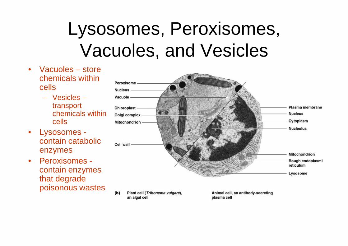
Lysosomes, Peroxisomes, Vacuoles, and Vesicles
• Vacuoles – store chemicals within cells– Vesicles –
transport chemicals within cellscells
• Lysosomes -contain catabolic enzymes
• Peroxisomes -contain enzymes that degrade poisonous wastes

Mitochondria
• Powerhouse of the cell– Produce most of cell’s
ATP
• Double membrane boundbound
• Interior matrix contains– 70S ribosomes – circular molecule of
DNA

Chloroplasts• Used by algae and
plants for photosynthesis
• Double membrane boundbound
• Contain – 70S ribosomes– DNA

• Simple structure• No nucleus• Small; ~1.0 µm in
diameter• One circular
chromosome, not in a
Prokaryote Eukaryote
• Complex Structure• Nucleus• Larger; 10-100 µm in
diameter• Paired chromosomes, in
nuclear membranechromosome, not in a membrane
• No histones• No membrane bound
organelles• Peptidoglycan cell walls• Binary fission • Comprised of bacteria
and archaea
nuclear membrane• Histones• Organelles• Polysaccharide cell walls• Mitotic spindle• Comprised of algae,
protozoa, fungi, animals, and plants

• Eukaryotes formed from phagocytosis of small aerobic prokaryotes– lost ability to exist independently– Retained portion of DNA, ribosomes, and
cytoplasmic membranes
Endosymbiotic Theory
cytoplasmic membranes– Larger cell became dependent on for aerobic
ATP production– Aerobic prokaryotes evolved into mitochondria– Similar scenario for origin of chloroplasts
• Not universally accepted

Endosymbiotic Theory
Figure 10.2

Prokaryotic membranes