Embed Size (px)
Citation preview

Prokaryotes And Eukaryotes
Deepa JohnHarini Chandra
All living organisms are classified into two broad categories, prokaryotes & eukaryote. Prokaryotes are those organisms whose cells lack a cell nucleus while eukaryotes possess a well-defined, membrane bound nucleus.
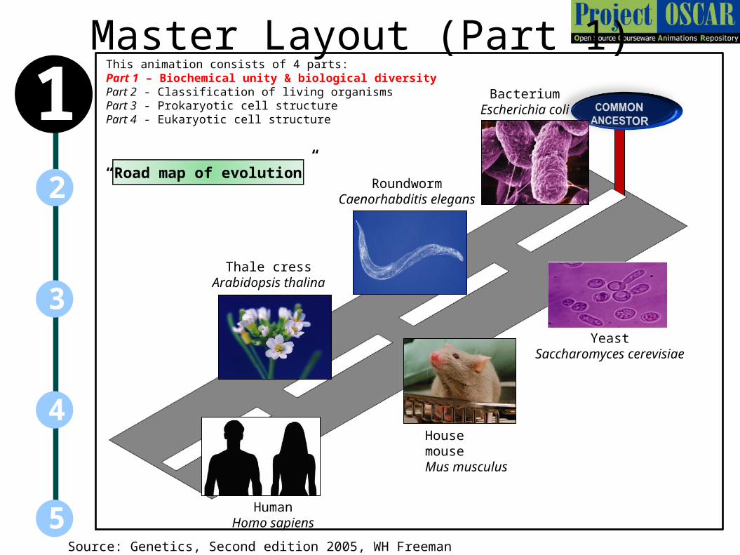
Master Layout (Part 1)This animation consists of 4 parts:Part 1 – Biochemical unity & biological diversityPart 2 - Classification of living organismsPart 3 - Prokaryotic cell structurePart 4 - Eukaryotic cell structure
5
3
2
4
1
Source: Genetics, Second edition 2005, WH Freeman
HumanHomo sapiens
House mouseMus musculus
Thale cressArabidopsis thalina
RoundwormCaenorhabditis elegans
YeastSaccharomyces cerevisiae
BacteriumEscherichia coli
“Road map of evolution”

Definitions of the components:Part 1: Biochemical unity & biological diversity
5
3
2
4
11. Evolution: The process by which various populations of organisms acquire and pass on their novel traits, in addition to other existing traits, from one generation to the next. This explains the origin of new species of organisms and the vast diversity that is observed in the biological world. However, it is believed that the origin of all organisms can be traced back to one common ancestor due to several underlying biochemical similarities.

Part 1,Step 1:
Action Audio Narration
1
5
3
2
4Description of the action
The figures arranged in the circle must move around the central figures.
(Please redraw all figures) The figures arranged in the circle must appear one at a time as shown in animation. Next, the figure in centre “DNA” must be shown followed by “RNA” and finally “amino acids”. While this is happening, the figures arranged in the circle must continuously revolve around the central figures very slowly.
All living organisms from various periods of evolution have been found to exhibit remarkable similarity at the biochemical level. Genetic information is stored in the form of DNA or RNA , the same set of 20 amino acids form the structural elements of proteins, similar metabolic pathways and several proteins with structural similarity have been found to have similar roles in different organisms. All of these point towards the existence of a common ancestor from which various organisms evolved at different points of time.
Source: Genetics, Second edition 2005, WH Freeman; Biochemistry by Lubert Stryer, 6 th edition (ebook)
RNADNA
NC
C+
-
Amino acids
Arabidopsis thalina Homo sapiens
Caenorhabditis elegans
Escherichia coliMus musculus
Saccharomyces cerevisiae
Sulfolobus acidocaldarius
Biochemical uniformity of organisms
Metabolic pathways

Part 1,Step 2:
Action Audio Narration
1
5
3
2
4Description of the action
As shown in animation.
(Please redraw all figures) First show the figure on left top with the sign board below and the grey line followed by the blue figure on left bottom. Next, show the middle panel of figures followed by the right most as depicted in the animation.
Several proteins have been identified that possess similar three dimensional structures and perform very closely related functions in organisms that are separated in evolution over billions of years. One such protein is the TATA-box binding protein, which plays an important role in gene regulation.
Source: Genetics, Second edition 2005, WH Freeman; Biochemistry by Lubert Stryer, 6 th edition (ebook)
Conserved TATA binding protein – Biochemical unity
Evolution timeline
Sulfolobus acidocaldarius Arabidopsis thalina Homo sapiens
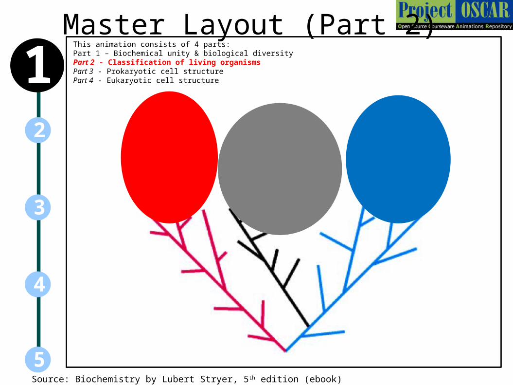
Master Layout (Part 2)This animation consists of 4 parts:Part 1 – Biochemical unity & biological diversityPart 2 - Classification of living organismsPart 3 - Prokaryotic cell structurePart 4 - Eukaryotic cell structure
5
3
2
4
1
Source: Biochemistry by Lubert Stryer, 5th edition (ebook)
Definitions of the components:Part 2: Classification of living organisms
5
3
2
4
11. Bacteria: These are a group of unicellular, prokaryotic microorganisms that are present in every habitat on earth. They are usually a few micrometers in length and have a range of shapes such as rods, spheres, and spirals. Bacteria are one of the most ancient life forms, believed to have been present over 3 billion years ago. These organisms lack a defined cell-nucleus and other cellular organelles, due to which they are classified as prokaryotes.
2. Archae: Archae are a group of single-celled microorganisms that are also classified as prokaryotes due to their lack of cellular organelles and a nucleus. They were originally classified as bacteria but now form their own separate domain due to several genetic and metabolic differences, in which they more closely resemble eukaryotes. Unlike eukaryotes however, they are capable of obtaining their energy from various sources such as organic compounds, sugars, ammonia etc.
3. Eukarya: These are multicellular organisms whose cells contain complex structures including a well-defined, membrane-bound nucleus carrying the genetic material. Eukaryotic cells are typically larger than prokaryotes and contain several other membrane-bound organelles which carry out complex metabolic & cell division processes.

Part 2,Step 1:
Action Audio Narration
1
5
3
2
4Description of the action
As shown in animation.
(Please redraw all figures) First show the figures below with the headings on top. Then show the speech bubble on left appearing followed by the speech bubble from ‘mouse’ . This is followed by the question mark appearing on top of the middle picture which must flicker briefly and then disappear. This is followed by the speech bubble in the centre.
Archaea are a group of prokaryotic organisms that are distantly related to bacteria-like organisms. They are however more similar to eukaryotes than bacteria. Both archaea and eukaryotic genomes encode homologous histone proteins, which are not present in bacteria. The ribosomal RNA and proteins or archaea closely resemble those of eukaryotes. However, archaea are capable of growing in extreme environmental conditions such as high temperatures, salt concentrations etc.
BacteriumEscherichia coli
Sulfolobus acidocaldarius House mouseMus musculus
Prokaryote Archaea Eukaryote
Hey Sulfo, you’re distantly related to us and were originally part of our group!
Well too bad, they’re now similar to us! They have histones, which you guys don’t and their ribosomal RNA and proteins are like ours.
We have membranes that are different from yours and we can live in extreme environments where you guys can’t. We’re going to form our own group!

Part 2,Step 2:
Action Audio Narration
1
5
3
2
4Description of the action
Show the structure growing from bottom to top.
(Please redraw all figures) The figure above must appear from bottom to top like a growing tree. The branches must appear as the lines move upwards. For eg. It must start with the blue & red lines which must expand towards the right & left respectively. When branches appear, they must branch out.
One of the most recent classification of living organisms is the three-domain system consisting of bacteria, eukarya and archae. Although archae were originally considered as bacteria, they were later classified into their own domain due to several differences in their metabolic pathways and genetics. Eukaryotes are believed to have evolved through several endosymbiotic relationships between various bacteria and archae.

Master Layout (Part 3)
5
3
2
4
1
Nucleoid
Cell envelope
Pili
Ribosomes
Flagella
Source: Biochemistry by A.L.Lehninger, 4th edition
This animation consist of 4 parts:Part 1 – Biochemical unity & biological diversityPart 2 - Classification of living organismsPart 3 - Prokaryotic cell structurePart 4 - Eukaryotic cell structure
Definitions of the components:Part 3: Prokaryotic cell structure1. Cell envelope: This encompasses the plasma membrane and layers outside it which confer the cell with rigidity. The composition of the cell envelope varies with organism.
2. Plasma membrane: The bacterial protoplast is bound by a living ultrathin and dynamic plasma membrane. It chemically comprises molecules lipids and proteins which are arranged in fluid mosaic pattern.
3. Cell wall: The plasma membrane is covered with a strong and rigid cell wall.
a) Peptidoglycan: Peptidoglycan is an enormous mesh like polymer composed of many identical subunits, lying outside the plasma membrane of bacteria. The polymer contains two sugar derivative, N- acetylglucoseamine and N- acetlymuramic acid and a chain of three to five amino acids attached to the N- acetlymuramic acid .
b) Periplasmic space: A space observed between the plasma membrane and the outer membrane of Gram negative bacteria or equivalent space between the plasma membrane and wall of Gram positive bacteria.
c) Teichoic acid: They are polymers of glycerol or ribitol joined by phosphate group, covalently linked to either the petidoglycan or plasma membrane of Gram positive bacteria. Amino acids such as D- alanine or sugars like glucose are attached to the glycerol and ribitol groups.
d) Lipoteichoic acid: Teichoic acids covalently connected to plasma membrane of Gram positive bacteria.
e) Integral protein: Proteins attached to the bacterial plasma membrane.
5
3
2
4
1
f) Outer membrane: It is 7 – 8 nm thick membrane lying outside the peptidoglycan layer and is linked to the cell either by a lipoprotein or via many adhesion sites.
g) Porins: Porins are proteins composed of beta sheets , spans the outer membrane of Gram negative bacteria and is more or less tube shaped. Its narrow channels allows passage of molecules smaller than 600 – 700 Daltons.
h) Lipopolysachharide(LPS): LPS is the major component of the outer membrane of Gram-negative bacteria. These large, complex molecules contain both lipid and carbohydrate and consists of three parts:1) lipid A, (2) the core polysachharide and (3) the O side chain or O antigen.
4. Capsule: In some bacteria, the cell wall is surrounded by an additional slime or gel layer called capsule.
5. Nucleoid: In bacteria the nuclear material is not separated from the cytosol by the nuclear membrane. However, the nuclear material is usually concentrated in a specific clear region of the cytoplasm, called nucleoid, Contains a single, simple, long circular DNA molecule.
6. Ribosomes: Ribosomes are tiny spheroidal dense particles that contain approximately equal amount of RNA and proteins. Ribosomes have a sedimentation coefficient of about 70S and are composed of two subunits namely 50S and 30S.
7. Flagella: Many bacteria are motile and one or more flagella for the cellular locomotion that propel cell through its surroundings
5
3
2
4
1Definitions of the components:Part 3: Prokaryotic cell structure
.
5
3
2
4
1Definitions of the components:Part 3: Prokaryotic cell structure
8. Pili: Some bacteria contain non-flagellar, extremely fine, appendages called fimbriae or pilli that provide points of adhesion to surface of other cells.
9. Gram staining: It is a method of differentiating bacterial species into two broad categories, gram-positive and gram-negative, based on the composition of their cell walls.

Part 3, Step 1:
Action Audio Narration
1
5
3
2
4
vv
Description of the actionThe cell structure shown above must be displayed & user should be allowed to click on any of the labels to read definitions.
(Please redraw all figures)The figure above must be displayed with its labels & user must be allowed to click on any of the labels to read definitions as given in the previous slides.
Prokaryotes are simple, unicellular organisms that lack a well-defined nucleus for carrying their genetic material. They are usually a few microns is size and are one of the most ancient life forms known from which eukaryotes are believed to have evolved.<Definitions of components as given in previous slide>
Nucleoid
Cell envelope
Pili
Ribosomes
Flagella
Prokaryotic cell structure
Source: Biochemistry by A.L.Lehninger, 4th edition ; Lodish et al. Mol Cell Biol. Sixth Ed. Page 3
Electron microscopic section of E. coli
Outer membrane
Inner membrane

Part 3, Step 2:
Action Audio Narration
1
5
3
2
4
vv
Description of the action
Lipoteichoic acid
Teichoic acid
Periplasmic space
Peptidoglycan
Show the image on the left and zoom into the cream layer to show image on the right.
(Please redraw all figures) First show the image on the left followed by appearance of the red box. This region must be zoomed into and the image on the right must be shown with its labels.
Bacteria can be divided into two major groups based on the structure of their cell-wall & thereby their response to Gram staining. The cell wall of Gram positive bacteria is composed of mainly polysaccharides and glycosylated molecules. It is made up of a single 20-80 nm thick homogenous layer of peptidoglycan. In addition cell wall usually contains teichoic acid, which is covalently connected to either peptidoglycans itself or to plasma membrane lipids(lipoteichoic acids)Plasma membrane is composed of a bilayer sheet of phospholipid molecules with their polar heads on the surface and their fatty -acyl chains forming the interior.
Source: Microbiology by Michael J.:Pelczar; morayeel.louisiana.edu
Integral protein
Cell envelope (wall) – gram positive bacteria
Plasma membrane
Part 3,Step 3:
Action Audio Narration
1
5
3
2
4Description of the action
Integral protein
Plasma membrane
Periplasmic space
Peptidoglycan
Outer membrane
Lipopolysaccharide
Porins
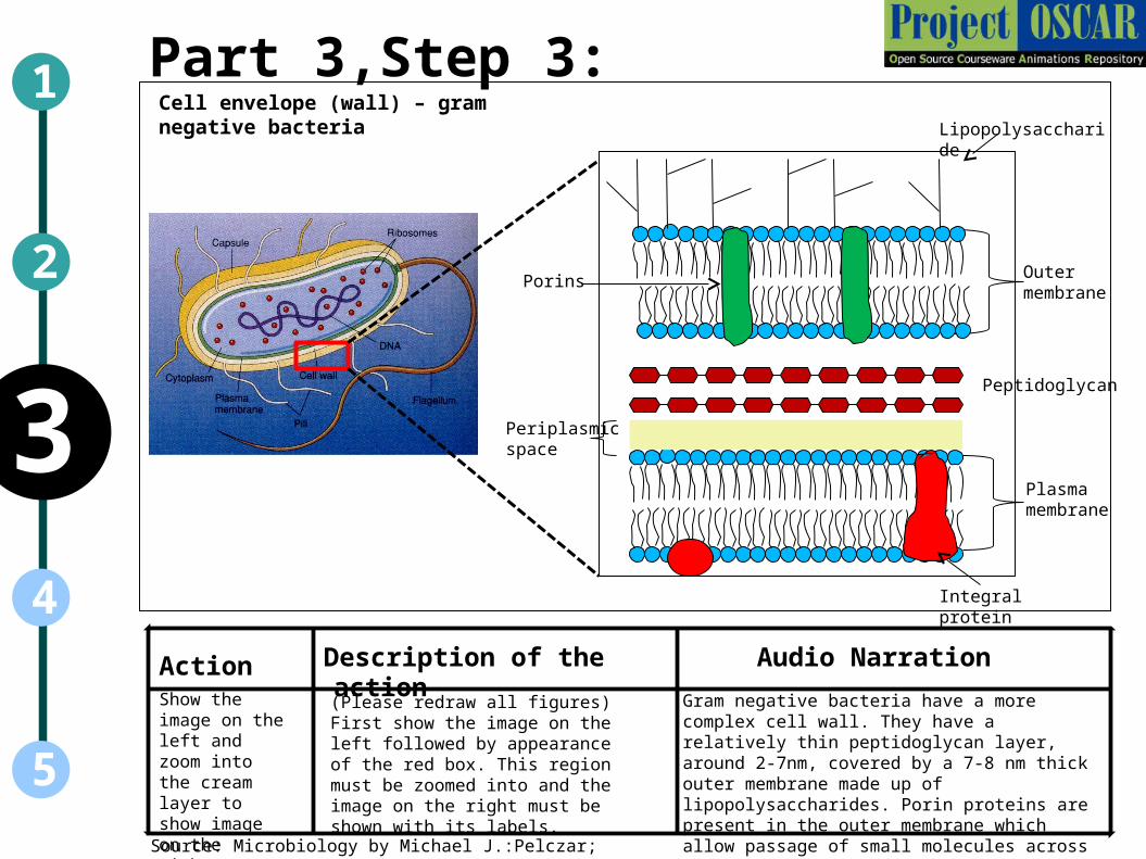
Gram negative bacteria have a more complex cell wall. They have a relatively thin peptidoglycan layer, around 2-7nm, covered by a 7-8 nm thick outer membrane made up of lipopolysaccharides. Porin proteins are present in the outer membrane which allow passage of small molecules across the membrane.
Source: Microbiology by Michael J.:Pelczar; morayeel.louisiana.edu
vv
Cell envelope (wall) – gram negative bacteria
Show the image on the left and zoom into the cream layer to show image on the right.
(Please redraw all figures) First show the image on the left followed by appearance of the red box. This region must be zoomed into and the image on the right must be shown with its labels.

Part 3, Step 4:
Action Audio Narration
1
5
3
2
4Description of the action
Bacterial cell
Nucleoid
Nuclear material in the bacterial cell is not separated from the cytosol by a distinct nuclear membrane. However, it is usually concentrated in a specific clear region of the cytoplasm called the nucleoid. The genetic material usually contains a single, circular DNA molecule.
Source: Biochemistry by A.L.Lehninger, 4th edition
Genetic material of the cell
Show the image on the left and then zoom into the purple region at the centre.
(Please redraw all figures) First show the image on the left followed by appearance of the red box. This region must be zoomed into and the image on the right must be shown with its labels.

Part 3, Step 5:
Action Audio Narration
1
5
3
2
4Description of the action
Bacterial cell
Ribosome
Ribosomes are composed of proteins and ribosomal RNA. The prokaryotic 70S ribosome is made up of a 50S large subunit and a 30S small subunit, where S refers to the Svedberg coefficient, which provides an indication about rate of sedimentation of the particle.
Source: Biochemistry by A.L.Lehninger, 4th edition
Show the image on the left and then zoom into the red dots.
(Please redraw all figures) First show the image on the left followed by appearance of the red box. This region must be zoomed into and the image on the right must be shown with its labels. This is followed by the appearance of the figures on the right end as though emerging from the middle panel.
Ribosomes
70S 30S
16S
Subunits
50S23S
5S
rRNA
Large subunit
Small subunit

Action Audio Narration
1
5
3
2
4 Description of the actionThe cell should move in one direction which should then change.
A motile bacteria propels itself from one place to another within the medium by rotating its flagella. A bacterial flagellum is made up of the protein flagellin. It has a helical structure with a sharp bend called the hook just outside the membrane, and a basal body containing the motor just below the membrane. To “swim” forward, the flagella rotates in counterclockwise direction. However, when flagellar rotation abruptly changes to clockwise direction, the bacterium "tumbles" in its place and seems incapable of moving. It then begins swimming again in another new, random direction.
The first figure on the left must appear & it must be shown to move towards the right. It must then come to a halt & the black thread like structures must move around the red part & rearrange themselves so as to finally appear as in the final right-most image. Once this is done, the cell must then move downwards as shown.
Source: Microbiology by Michael J.:Pelczar
Flagella – motility structures
Part 3, Step 6:
Movement in one directionTumbling of the cell
Change in direction of motion

Master Layout (Part 4)
5
3
2
4
1
Nuclear envelope Nucleolus
Nucleus
chromatin
Ribosomes
PeroxisomeLysosome
Cytoskeleton
Golgi complex
Smooth ERRough ER
Mitochondria
Plasma membrane
Source: Biochemistry by A.L.Lehninger, 4th edition
This animation consist of 4 parts:Part 1 – Biochemical unity & biological diversityPart 2 - Classification of living organismsPart 3 - Prokaryotic cell structurePart 4 - Eukaryotic cell structure

Definitions of the components:Part 4: Eukaryotic cell structure
1. Cell wall: Present only in plant cells, these are responsible for providing the cell with structure and rigidity. They also protect the cell from swelling due to osmosis.
2. Cytoskeleton: Provides the cell with required support and also plays a role in movement of cellular organelles.
3. Plasmodesmata: Helps in interaction between adjacent plant cells by providing a path between them.
4. Plasma membrane: Provides a protective layer around the cell by separating it from the environment and monitoring the movement of molecules into and out of it.
5. Lysosome: These are found exclusively in animal cells for degrading any intracellular debris.
6. Peroxisome: Organelle containing enzymes that are responsible for protecting the cell against free radicals and peroxide. They also play a role in metabolism of very long chain fatty acids.
7. Smooth endoplasmic reticulum (SER): Biosynthesis of lipids and metabolism of several drugs occurs at the SER.
8. Rough endoplasmic reticulum (RER): This is a site where much of the protein synthesis occurs as it is studded with ribosomes.
9. Vacuole: These are essential for storage of metabolites and also for degrading and recycling macromolecules.
10. Ribosomes: The function of ribosomes is common to prokaryotes and eukaryotes. They serve to synthesize proteins from the corresponding RNA.
5
3
2
4
1
11. Golgi complex: It is a cup shaped organelle located near the nucleus. It acts as a downstream processing centre for packaging proteins and then targeting them for distribution to other organelles or for export outside the cell.
12. Mitochondria: Commonly referred to as the “power house of the cell”, this organelle is responsible for energy production in the form of ATP by oxidation of nutrients.
13. Glyoxysome: Site at which the glyoxylate cycle occurs for energy production from acetate precursors in plants and certain bacteria.
14. Nucleus: The defining feature of all eukaryotes, nucleus is the critical organelle housing all genetic information in the form of DNA.
15. Nucleolus: This is present within the nucleus and is responsible for synthesis of ribosomal RNA (rRNA).
16. Nuclear envelope: Surrounds the nucleus and functions to segregate the chromatin i.e. DNA and proteins, from the external cytoplasm.
17. Chloroplasts: Organelles that are responsible for the major difference between plant and animals. They allow the synthesis of carbohydrates and produce energy by harvesting sunlight.
18. Thylakoids: They are stacked organelles present within the chloroplast that are essential for ATP production using light energy.
19. Starch granules: These are also found within the chloroplast and are necessary for temporary storage of carbohydrates.
5
3
2
4
1Definitions of the components:Part 4: Eukaryotic cell structure

Part 4, Step 1:
Action Audio Narration
1
5
3
2
4Description of the action
Nuclear envelope Nucleolus
Nucleus
chromatin
Ribosomes
PeroxisomeLysosome
Cytoskeleton
Golgi complex
Smooth ERRough ER
Mitochondria
Plasma membrane
Source: Biochemistry by A.L.Lehninger, 4th edition
The figure should appear along with its labels.
(Please redraw all figures) Show the figure above with the labels appearing as shown. User should be allowed click on any of the labels to read the definition of that component as given in the previous slide.
Typical animal cell lacks a cell wall and contains several membrane-bound organelles, such as nucleus, mitochondria, endoplasmic reticulum (ER), Golgi apparatus, lysosomes, and peroxisomes. <Definitions of each component as given in previous 2 slides.>
Animal cell structure

Part 4, Step 2:
Action Audio Narration
1
5
3
2
4Description of the action
Source: Lodish et al. Mol Cell Biol. Sixth Ed. Page 3
The figure should appear along with its labels.
(Please redraw all figures) Show the figure above with the labels appearing as shown. User should be allowed click on any of the labels to read the definition of that component as given in the previous slide.
An electron micrograph image of a plasma cell is shown here, clearly depicting the nucleus, golgi material, lysosome and mitochrondrion.
Electron micrograph of plasma cell
Nucleus
Lysosome
Golgi
Endoplasmic reticulum
Mitochondrion

Action Audio Narration
1
5
3
2
4Description of the action
Nucleolus
ChromatinNuclear envelope
Nucleus
Ribosome
Cytoskeleton
Golgi complex
Glyoxysome
PlasmodesmaVacuole
Cell wall
Chloroplast Mitochondria
Plant cells have a rigid cell wall and membrane-bound organelles, such as nucleus, mitochondria, chloroplast, endoplasmic reticulum (ER), Golgi apparatus, lysosomes, vacuoles and peroxisomes. <Definitions of components as given in previous slides>
Source: Biochemistry by A.L.Lehninger, 4th edition
Part 4, Step 3:Plant cell structure
The figure should appear along with its labels.
(Please redraw all figures) Show the figure above with the labels appearing as shown. User should be allowed click on any of the labels to read the definition of that component as given in the previous slide.

Part 4, Step 4:
Action Audio Narration
1
5
3
2
4Description of the action
Source: Biochemistry by A.L.Lehninger, 4th edition (ebook);
The red box must be zoomed into.
(Please redraw all figures) First show the image on the right followed by appearance of the red box. This region must be zoomed into & the figure on the left must appear with its labels.
Mitochondria, commonly referred to as “power house of the cell”, are membrane-bound organelles found in eukaryotic cells. They are responsible for generation of ATP to satisfy the body’s energy requirements and are also involved in other processes such as cell signalling, cell cycle control & cell gorwth. The organelle is made up of several compartments that carry out specialized functions and also contains its own independent genome that codes for mitochondrial proteins.
Cell organelles: Mitochondria
Porin channels
Ribosomes
Outer membraneInner membrane
ATP synthase
Matrix
Cristae

Part 4, Step 5:
Action Audio Narration
1
5
3
2
4Description of the action
Source: Biochemistry by A.L.Lehninger, 4th edition (ebook); www.ncbi.nlm.nih.gov
The red box must be zoomed into.
(Please redraw all figures) First show the image on the right followed by appearance of the red box. This region must be zoomed into & the figure in the middle panel must be shown with labels followed by the two images on the right appearing from this image in the middle panel.
Ribosomes, which are composed of proteins and ribonucleic acids (RNAs), play a central role in protein biosynthesis. They “read” the nucleic acid information from messenger RNA and convert this into the corresponding amino acid code of proteins. Eukaryotic 80S ribosomes are composed of a large 40S subunit, which binds to tRNA and amino acids, and a small 28S subunit which binds to mRNA during protein synthesis. The subunit structure of prokaryotic and eukaryotic ribosomes differ from one another.
Cell organelles: Ribosomes Large subunit
Small subunit
80S
Ribosome
18S
40S
Subunits
5.8S 5S
28S

Part 4, Step 6:
Action Audio Narration
1
5
3
2
4Description of the action
Source: Biochemistry by A.L.Lehninger, 4th edition (ebook); http://employees.csbsju.edu/hjakubowski/classes/ch331/cho/ergolgi.jpeg
The red box must be zoomed into.
(Please redraw all figures) First show the image on the right followed by appearance of the red box. This region must be zoomed into & the figure on the left must appear with its labels.
The endoplasmic reticulum and golgi apparatus are involved in synthesis, packaging & transport of various biomolecules. The ribosome-studded rough ER is a major site for protein synthesis while the smooth ER synthesizes lipids, steroids, metabolizes carbohydrates & steroids and regulates calcium concentration in muscles. The Golgi complex functions to process & package macromolecules such as proteins & lipids for their export to various other cellular organelles or outside the cell.
Cell organelles: Endoplasmic reticulum (ER) & Golgi complex
RER
Ribosomes
SER
ProteinsTransport vesicle
Golgi apparatus
Secretory vesicle
Proteins expelled

Part 4, Step 7:
Action Audio Narration
1
5
3
2
4Description of the action
Source: Biochemistry by A.L.Lehninger, 4th edition (ebook); http://www.biologie.uni-hamburg.de/b-online/library/onlinebio/nucleus_1.gif
The red box must be zoomed into.
(Please redraw all figures) First show the image on the right followed by appearance of the red box. This region must be zoomed into & the figure on the left must appear with its labels.
The nucleus is a membrane-bound organelle found in eukaryotic cells that is often considered as the “control centre” of the cell. It houses the genetic material of the cell in the form of chromosomes containing DNA molecules complexed with proteins known as histones. The nucleus is responsible for maintaining this genetic information by replication and for expression of genes performing various functions. The nucleolus is mainly involved in ribosome assembly, after which the ribosomes are exported to the cytoplasm for protein synthesis.
Cell organelles: Nucleus
Part 4, Step 8:
Action Audio Narration
1
5
3
2
4Description of the action
Source: Biochemistry by A.L.Lehninger, 4th edition (ebook); http://www.biologie.uni-hamburg.de/b-online/library/onlinebio/nucleus_1.gif
The red box must be zoomed into.
(Please redraw all figures) First show the image on the right followed by appearance of the red box. This region must be zoomed into & the figure on the left must appear with its labels.
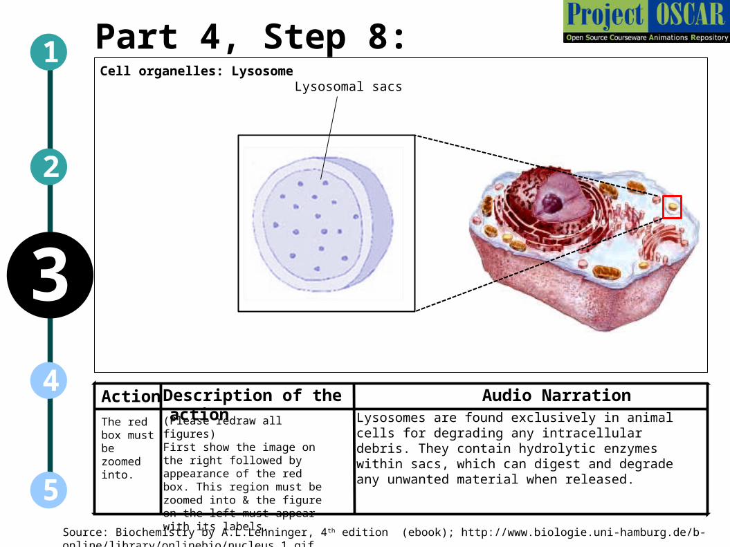
Lysosomes are found exclusively in animal cells for degrading any intracellular debris. They contain hydrolytic enzymes within sacs, which can digest and degrade any unwanted material when released.
Cell organelles: LysosomeLysosomal sacs

Part 4, Step 9:
Action Audio Narration
1
5
3
2
4Description of the action
Source: Biochemistry by A.L.Lehninger, 4th edition (ebook); http://www.biologie.uni-hamburg.de/b-online/library/onlinebio/nucleus_1.gif
The red box must be zoomed into.
(Please redraw all figures) First show the image on the right followed by appearance of the red box. This region must be zoomed into & the figure on the left must appear with its labels.
Peroxisome is an organelle containing enzymes like catalase that are responsible for protecting the cell against free radicals and peroxides. They also play a role in metabolism of very long chain fatty acids. The have a single membrane and no independent genetic system.
Cell organelles: Peroxisome
Plasma membrane
Urate oxidase crystalline core
Interactivity option 1:Step No:1
Boundary/limitsInteracativity Type Options Results
1
2
5
3
4
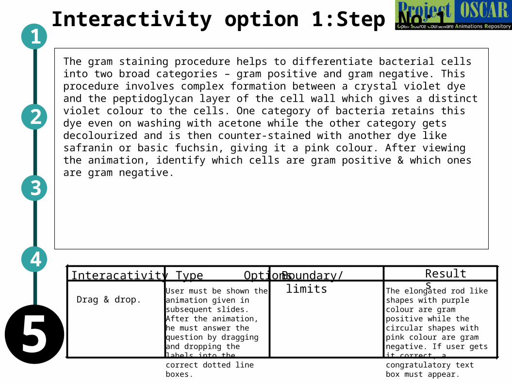
The gram staining procedure helps to differentiate bacterial cells into two broad categories – gram positive and gram negative. This procedure involves complex formation between a crystal violet dye and the peptidoglycan layer of the cell wall which gives a distinct violet colour to the cells. One category of bacteria retains this dye even on washing with acetone while the other category gets decolourized and is then counter-stained with another dye like safranin or basic fuchsin, giving it a pink colour. After viewing the animation, identify which cells are gram positive & which ones are gram negative.
Drag & drop.User must be shown the animation given in subsequent slides. After the animation, he must answer the question by dragging and dropping the labels into the correct dotted line boxes.
The elongated rod like shapes with purple colour are gram positive while the circular shapes with pink colour are gram negative. If user gets it correct, a congratulatory text box must appear.

Interactivity option 1:Step No:2 a 1
2
5
3
4
Smear of culture of gram positive & gram negative cells made.
Slide 1
Slide 2
Decolorize with acetone
Counterstain with Basic fuchsin
Flood with iodine to promote dye retention
Flood with crystal violet
Cells retain crystal violet colour
Cells get decolourizedCells stained pink
Gram staining procedure
Gram positive Gram negative
Now drag & drop the labels below into the corresponding dotted boxes beside each slide.

Interactivity option 2:Step No:1
Boundary/limitsInteracativity Type Options Results
1
2
5
3
4
Drag & drop the statements given below about prokaryotic & eukaryotic cells under the correct column to form a differentiating table.
Drag & drop.The statements shown in the table must be dispersed around the table in a random manner. User must drag & drop the statements into the correct column in the table.
User must drag & drop the statements shown into the correct column in the table. The correct answers are as shown in table above.
Prokaryotic cells Eukaryotic cells
Nuclear material concentrated in a specific region of the cytoplasm.
Nuclear material contained in membrane-bound organelle called nucleus.
Contains golgi complex & mitochrondria.Does not contain golgi complex & mitochrondria.
Majority are unicellular organisms. Mostly multicellular organisms
Can reproduce through sexual & assexual means.
Reproduce through sexual means.

Questionnaire1. Which of the following processes is characteristic to eukaryotic gene expression control?
Answers: a) Alternative splicing b) Alternative use of σ factor c) Transcription initiation
d) Catabolite repression
2. Which of the following correctly matches an organelle with its function
Answers: a) mitochondrion . . . Photosynthesis b) nucleus . . . cellular respiration c)
ribosome . . . manufacture of lipids d) central vacuole . . . storage
3. One can distinguish prokaryotic chromosomes from eukaryotic chromosomes by determining:
Answers: a) Nucleotide sequence b) Chromosome-linked proteins c) Base composition
d)Secondary structure
4. Select the wrong choice: Plasma or cell membrane is ____.
Answers: a) outer covering of each cell b) made of lipids and proteins c) superheated gases
d) a mechanical barrier for the protection of inner cell contents
5. The 'Scavengers' or 'Digestive bags' of a cell are ____.
Answers: a) chromosomes b) centrosomes c) lysosomes d) ribosomes
1
5
2
4
3

Links for further readingBooks:
Biochemistry by A.L.Lehninger, 4th edition
Biochemistry by Stryer et al., 6th edtion
Biochemistry by Voet & Voet, 3rd edition
Microbiology by Michael.J.Pelczar,
Links:
http://www.wellcometreeoflife.org/video/