Embed Size (px)
Citation preview
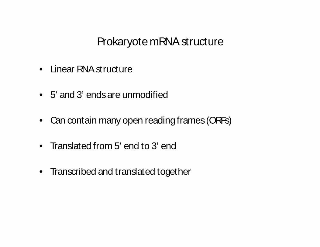
Prokaryote mRNA structure
• Linear RNA structure
• 5’ and 3’ ends are unmodified
• Can contain many open reading frames (ORFs)
• Translated from 5’ end to 3’ end
• Transcribed and translated together
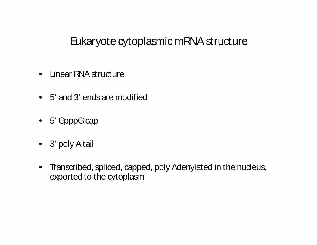
Eukaryote cytoplasmic mRNA structure
• Linear RNA structure
• 5’ and 3’ ends are modified
• 5’ GpppG cap
• 3’ poly A tail
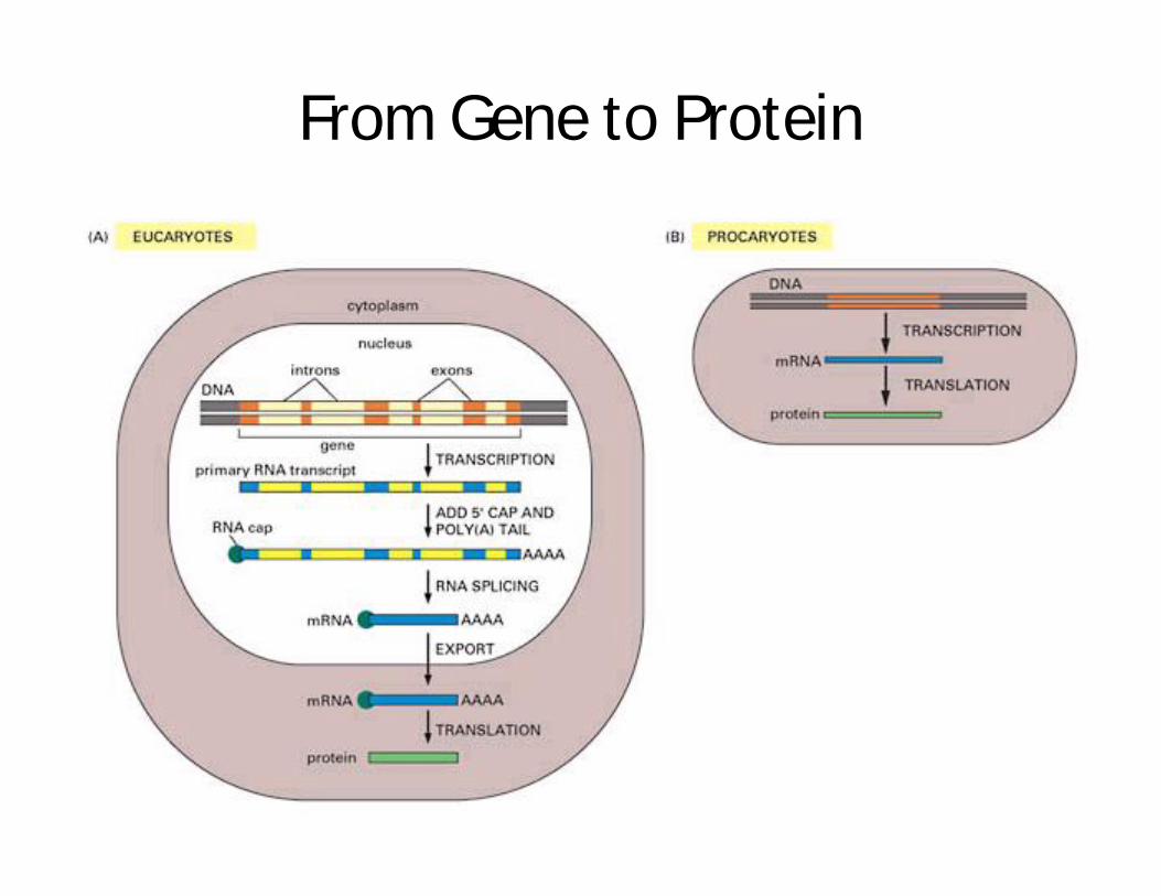
• Transcribed, spliced, capped, poly Adenylated in the nucleus, exported to the cytoplasm
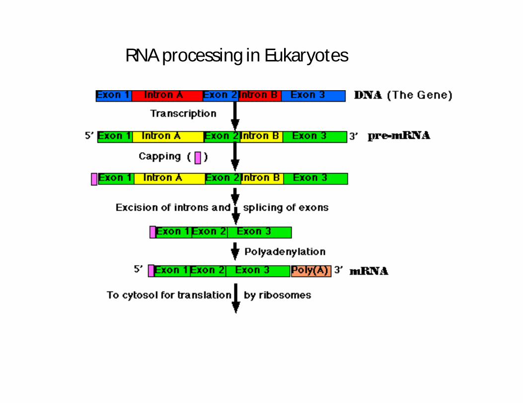
RNA processing in Eukaryotes

Introns and Exons
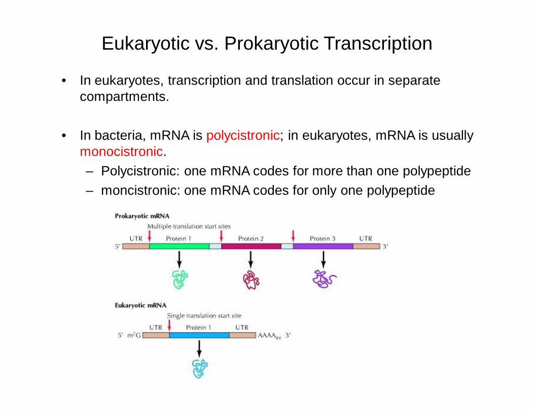
Eukaryotic vs. Prokaryotic Transcription
• In eukaryotes, transcription and translation occur in separate compartments.
• In bacteria, mRNA is polycistronic; in eukaryotes, mRNA is usually monocistronic.– Polycistronic: one mRNA codes for more than one polypeptide– moncistronic: one mRNA codes for only one polypeptide
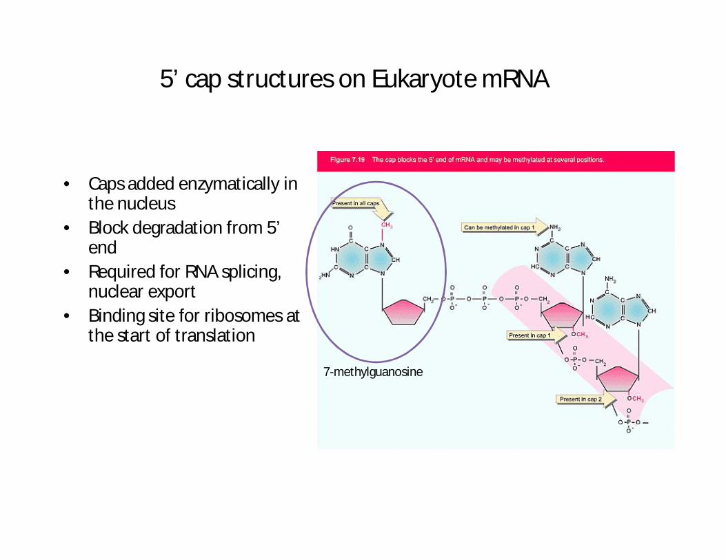
5’ cap structures on Eukaryote mRNA
• Caps added enzymatically in the nucleus
• Block degradation from 5’ end
• Required for RNA splicing, nuclear export
• Binding site for ribosomes at the start of translation
7-methylguanosine
Poly A tails on eukaryote mRNA
• Added to the 3’ end by poly A polymerase• Added in the nucleus• Approximately 200 A residues added in a template
independent fashion• Required for splicing and nuclear export• Bind poly A binding protein in the cytoplasm• Prevent degradation of mRNA• Loss of poly A binding protein results in sudden
degradation of mRNA in cytoplasm• Regulates biological half-life of mRNA in vivo
Eukaryote organelle mRNA structure
• Single stranded
• Polycistronic (many ORFs)
• Unmodified 5’ and 3’ ends
• Transcribed and translated together
• Show similarity to prokaryote genes and transcripts
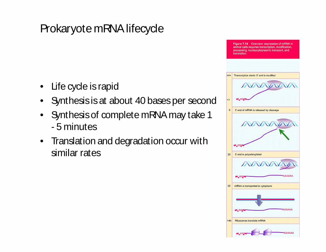
Prokaryote mRNA lifecycle
• Life cycle is rapid
• Synthesis is at about 40 bases per second
• Synthesis of complete mRNA may take 1 - 5 minutes
• Translation and degradation occur with similar rates

Eukaryote mRNA lifecycle
• Transcription, capping, polyA, splicing are nuclear
• Translation is cytoplasmic• mRNA is complete before export to
cytoplasm (20 min to >48 hours)• mRNA half life is 4 to > 24 hours in the
cytoplasm
Translation
• The synthesis of a protein sequence
• Using mRNA as a template
• Using tRNAs to convert codon information into amino acid sequence
• Catalysed by ribosomes
• Process essentially identical between prokaryotes and eukaryotes
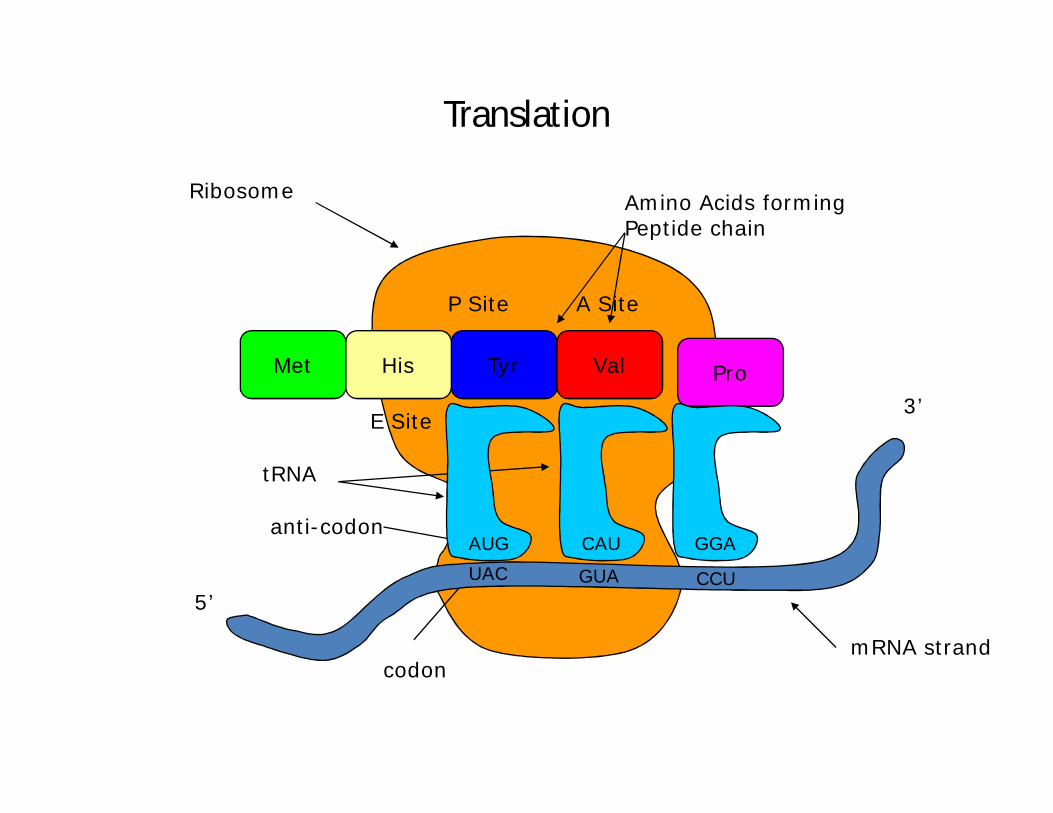
P Site A Site
E Site
Amino Acids forming Peptide chain
Ribosome
tRNA
anti-codon
codon
Translation
UAC
AUG
Tyr
GUA
CAU
Val
mRNA strand
3’
5’
HisMet Pro
GGA
CCU
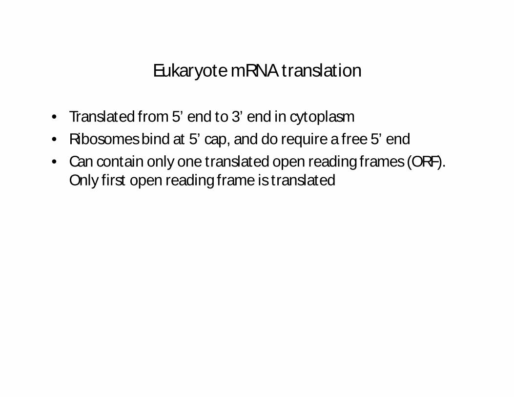
Eukaryote mRNA translation
• Translated from 5’ end to 3’ end in cytoplasm
• Ribosomes bind at 5’ cap, and do require a free 5’ end
• Can contain only one translated open reading frames (ORF). Only first open reading frame is translated
From Gene to Protein

Control of Gene Expression
• Prokaryotic organisms regulate gene expression in response to their environment for the survival.
• Eukaryotic cells regulate gene expression to maintain homeostasis in the organism. The fixed patterns of gene control leading to differentiation serve the needs of the whole organism and not the survival of an individual cell.
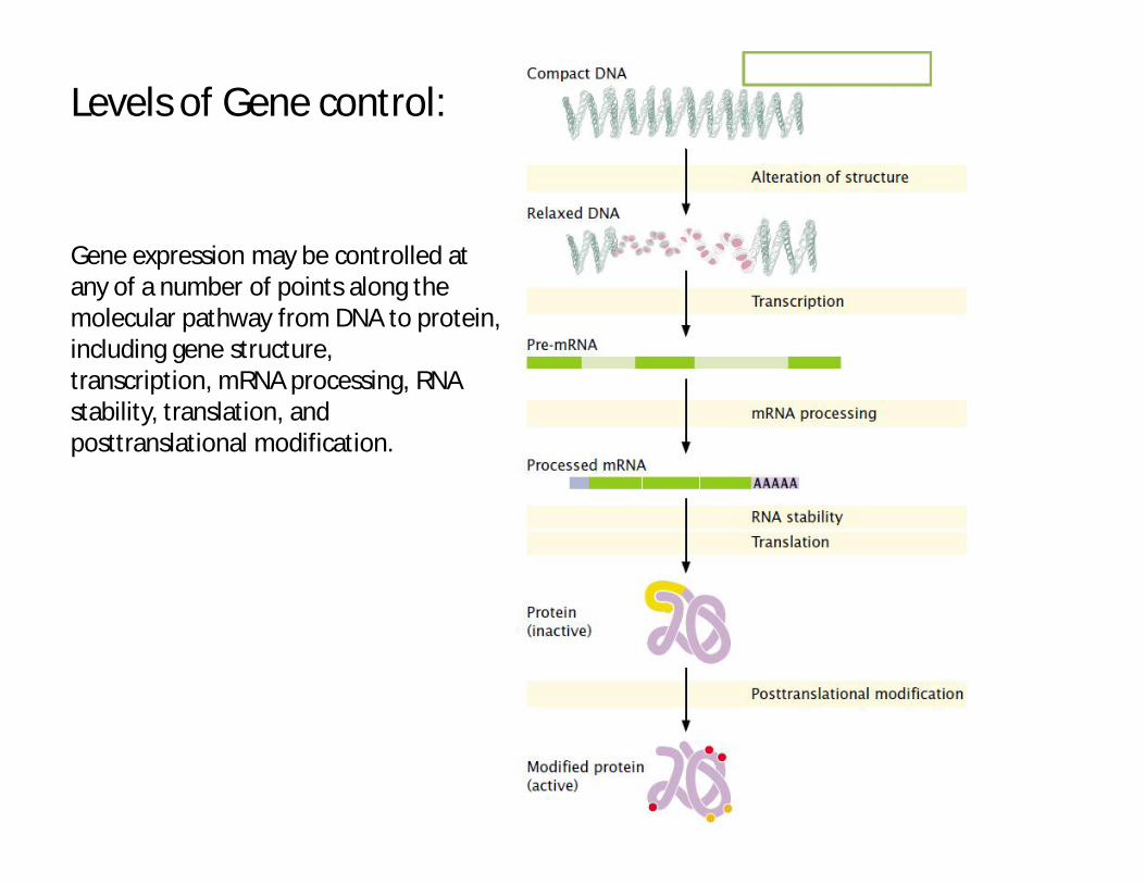
Gene expression may be controlled at any of a number of points along the molecular pathway from DNA to protein, including gene structure,transcription, mRNA processing, RNA stability, translation, and posttranslational modification.
Levels of Gene control:
Regulatory Proteins
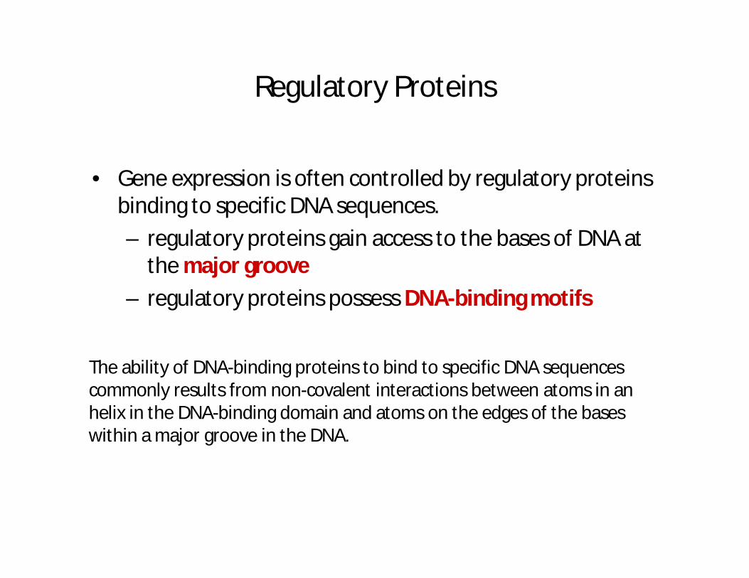
• Gene expression is often controlled by regulatory proteins binding to specific DNA sequences.
– regulatory proteins gain access to the bases of DNA at the major groove
– regulatory proteins possess DNA-binding motifs
The ability of DNA-binding proteins to bind to specific DNA sequences commonly results from non-covalent interactions between atoms in an helix in the DNA-binding domain and atoms on the edges of the bases within a major groove in the DNA.
Regulatory Proteins
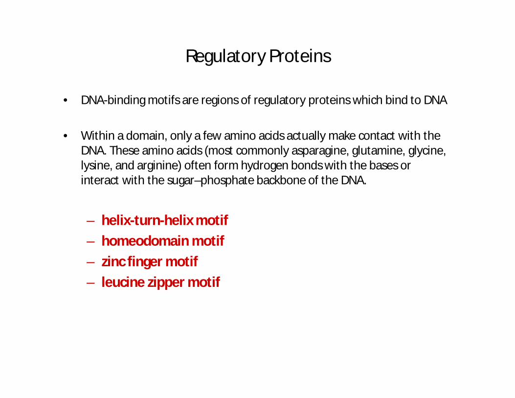
• DNA-binding motifs are regions of regulatory proteins which bind to DNA
• Within a domain, only a few amino acids actually make contact with the DNA. These amino acids (most commonly asparagine, glutamine, glycine, lysine, and arginine) often form hydrogen bonds with the bases or interact with the sugar–phosphate backbone of the DNA.
– helix-turn-helix motif
– homeodomain motif
– zinc finger motif
– leucine zipper motif

20
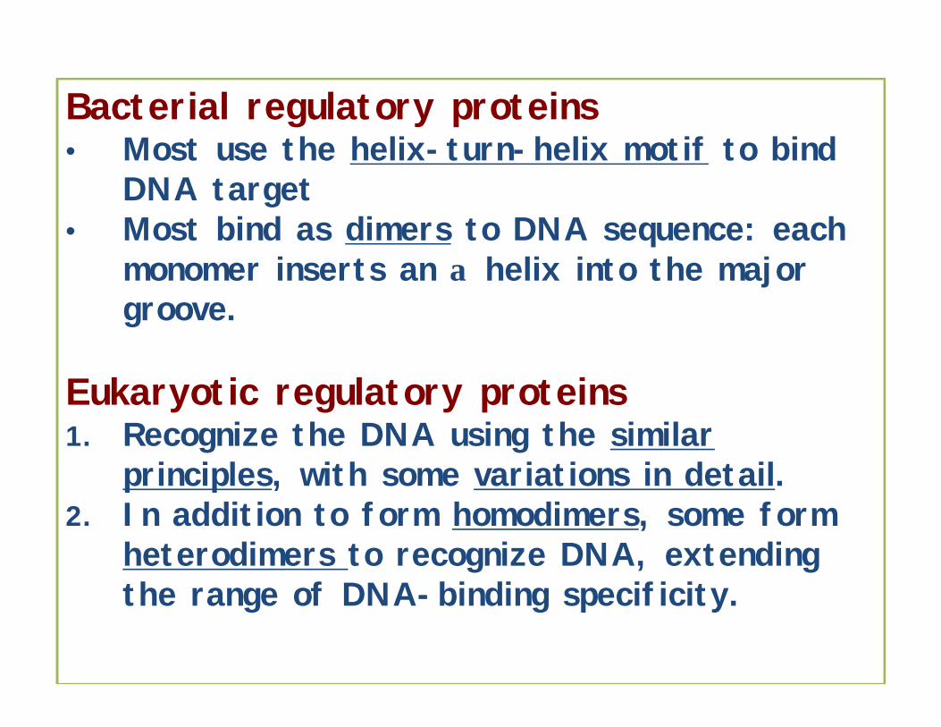
Bacterial regulatory proteins• Most use the helix-turn-helix motif to bind
DNA target• Most bind as dimers to DNA sequence: each
monomer inserts an α helix into the major groove.
Eukaryotic regulatory proteins1. Recognize the DNA using the similar
principles, with some variations in detail.2. In addition to form homodimers, some form
heterodimers to recognize DNA, extending the range of DNA-binding specificity.
21
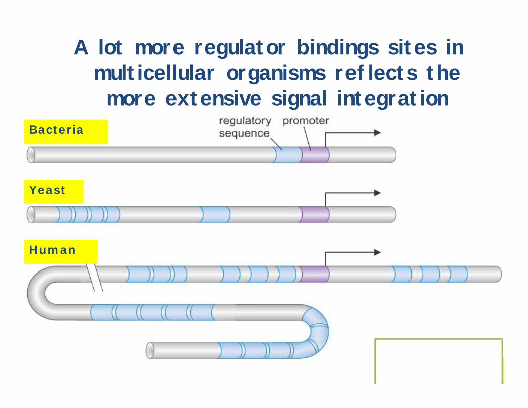
A lot more regulator bindings sites in multicellular organisms reflects the more extensive signal integration
Fig. 17-1
Bacteria
Yeast
Human