Embed Size (px)
Citation preview
J Neurosurg Pediatrics 13:260–272, 2014
260 J Neurosurg: Pediatrics / Volume 13 / March 2014
©AANS, 2014
Tumors of the spine are rare and represent 2%–8% of all musculoskeletal tumors in children. The most common benign vertebral tumors are eosinophilic
granulomas (12%–25%), osteoid osteoma/osteoblastomas (12%), and aneurysmal bone cysts (10%).14,22,27 The most common malignant primary vertebral tumor in children is Ewing’s sarcoma.6,14 With the advent of high-definition imaging of bone tumors, it is natural to extrapolate the staging system used for tumors in long bones to those in the spine.10,21 High-definition imaging is useful for pre-dicting the extent of resection and follow-up needed.
Atlantoaxial tumors in children comprise a distinct entity because of the complexity of the regional anatomy (brain, cervical cord, cranial nerves, CSF spaces, and vas-cular structures); craniocervical stability in a transitional zone; and skeletal growth potential. Thus, tumor resect-ability depends on the location and biological aggressive-ness of the lesion.56 A surgeon considering en bloc tumor resection should take into account the benign or malignant nature of the lesion and the potential involvement of neu-rovascular structures. The atlantoaxial region accounts for more than 25% of primary bone tumors in children.6,22,41,42
Primary atlantoaxial bone tumors in children: management strategies and long-term follow-up
Clinical article
Arnold H. Menezes, M.d., And rAHeel AHMed, M.d., PH.d.Department of Neurosurgery, University of Iowa Hospitals and Clinics, Iowa City, Iowa
Object. Atlantoaxial tumors account for a substantial proportion of primary bone tumors in children. Before resection, surgeons must consider the complex regional anatomy, the potential for neurological compromise, cranio-cervical instability, and the question of tumor resectability in a growing spine. Using current technology, the authors analyzed surgical cases in this light and present outcomes and treatment recommendations after long-term patient follow-up.
Methods: The authors reviewed clinical records for 23 children whose primary atlantoaxial bone tumors were treated from 1996 through 2010.
Results. Pathological lesions among the 23 patients were 4 aneurysmal bone cysts, 2 osteochondromas, 5 chor-domas, 4 osteoblastomas, 3 fibrous dysplasias, 4 eosinophilic granulomas, and 1 Ewing’s sarcoma. Clinical presen-tation consisted of neck pain (n = 23), headaches and occipital pain (n = 16), myelopathy (n = 8), and torticollis (n = 4). Selective angiography and coil embolization were undertaken for all patients with aneurysmal bone cysts and osteoblastomas, 2 patients with chordomas, 1 patient with fibrous dysplasia, and 1 patient with Ewing’s sarcoma. Pri-mary embolization treatment of radiation-induced aneurysmal bone cyst of the atlas showed complete reossification. Results of CT-guided needle biopsy were diagnostic for 1 patient with eosinophilic granuloma and 1 with Ewing’s sarcoma. Needle biopsies performed before referral were associated with extreme blood loss for 1 patient and mis-diagnosis for 2 patients. Surgery involved lateral extrapharyngeal, transoral, posterior, and posterolateral approaches with vertebral artery rerouting. Complete resection was possible for 9 patients (2 with osteochondroma, 3 with fibrous dysplasia, 2 with chordoma, and 2 with osteoblastoma). Decompression and internal fusion were performed for 3 patients with aneurysmal bone cysts. Of the 23 patients, 7 underwent dorsal fusion and 4 underwent ventral fusion of the axis body. Chemotherapy was necessary for the patients with eosinophilic granuloma with multifocal disease and for the patient with Ewing’s sarcoma. There was no morbidity, and there were no deaths. All patients with benign le-sions were free of disease at the time of the follow-up visit (mean ± SD follow-up 8.8 ± 1.1 years; range 2–18 years). Chordomas received proton or LINAC irradiation, and as of 4–15 years of follow-up, no recurrence has been noted.
Conclusions. Because most atlantoaxial tumors in children are benign, an intralesional procedure could suffice. Vascular control and staged resection are critical. Ventral transoral fusion or lateral extrapharyngeal fusion has been successful. Resection with ventral fusion and reconstruction are essential for vertebral body collapse. Management of eosinophilic granulomas must be individualized and might require diagnosis through needle biopsy.(http://thejns.org/doi/abs/10.3171/2013.11.PEDS13245)
Key Words • chordoma • craniocervical fusion • embolization • osteoblastoma • primary spinal tumors • proton-beam radiotherapy • oncology
Unauthenticated | Downloaded 08/06/20 11:46 AM UTC
J Neurosurg: Pediatrics / Volume 13 / March 2014
Atlantoaxial bony tumors in children
261
Careful analysis of this series with long-term outcomes is presented as a balanced, meaningful approach to tumors in this region.
MethodsOur review spans the MRI era: 1996–2010. All re-
search was approved by the University of Iowa Institution-al Review Board. Multiple institutional databases were searched for the following inclusion criteria: age younger than 16 years at diagnosis, histopathological findings con-sistent with origination from vertebral bone, and clinical diagnosis during 1996–2010. These databases searched the institutional tumor registry, records of inpatient and outpatient visits, and pediatric neurosurgery and neuro-radiology databases. Of 87 children with primary spinal tumors at all levels, 23 with primary atlantoaxial tumors were identified. Medical records were reviewed and atten-tion was given to symptoms and signs, imaging findings, disease extent, treatment received, histopathological find-ings, complications, and long-term outcomes.
ResultsFrom 1996 to 2010, a total of 23 children underwent
treatment for atlantoaxial tumors at the Pediatric Neuro-surgery Service at University of Iowa Hospitals and Clin-ics. Of these, 22 were designated as having benign tumors and 1 had a malignant Ewing’s sarcoma. Specifically, pathological examination identified 4 aneurysmal bone cysts, 5 chordomas, 2 osteochondromas, 4 osteoblasto-mas, 3 fibrous dysplasias, 4 eosinophilic granulomas, and 1 Ewing’s sarcoma.
Clinical PresentationOf the 23 patients, 15 were male and 8 were female;
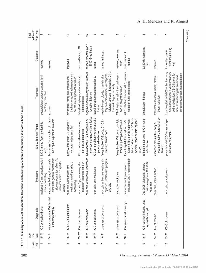
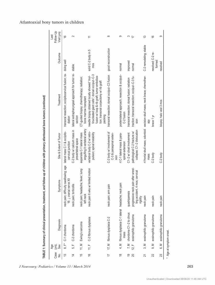
the mean age (± SD) was 10.5 ± 1.0 years (range 3–16 years). The mean (± SD) follow-up duration was 8.8 ± 1.1 years (range 2–18 years). Table 1 summarizes the clini-cal presentation, treatment, and final outcomes for all 23 patients. Cases 7 and 10 have been reported elsewhere.41,43
For nearly all patients, the predominant symptom was neck pain. Neurological deficit was a presenting sign for 9 patients (39%); quadriparesis was noted for 5 patients (22%). Precipitating events included wrestling for 2 pa-tients, induction of general anesthesia for 2 patients with osteochondroma, and cheerleading for 1 patient in whom increasing neck pain with motor weakness developed. One patient with eosinophilic granuloma became symp-tomatic while wrestling at school.
Initial evaluation comprised plain radiography and CT and MRI of the cervical spine. Bone and PET scan-ning were performed when an underlying eosinophilic granuloma was suspected. Either CT angiography or MR angiography was performed for each patient to identify the course of the vertebral artery in relation to the tumor for the planned surgical approach or to determine tumor involvement. Preoperative tumor embolization was per-formed for a patient with a large vascular osteoblastoma,
a 16-year-old patient with an aneurysmal bone cyst, as a definitive procedure for a patient with radiation-induced aneurysmal bone cyst, and before staged excision for a pa-tient with a C-2 chordoma. One patient underwent embo-lization for a vascular lesion identified on MR images that was later determined to be fibrous dysplasia.
The patient in Case 5, who had an osteoblastoma, underwent a CT-guided needle biopsy that was not diag-nostic. One patient with an aneurysmal bone cyst under-went a CT-guided biopsy before referral, during which she experienced substantial (600 ml) blood loss. She later underwent preoperative embolization before resection. A CT-guided biopsy was diagnostic for 2 patients (the pa-tient in case 15 had an underlying Ewing’s sarcoma and the patient in case 23 had an eosinophilic granuloma). For the patient in Case 16, who had fibrous dysplasia, inde-terminate needle biopsy results indicated multinucleated giant cells.
Surgical TreatmentSurgical management was based on a preliminary di-
agnosis determined from imaging. Imaging studies were also used to define the extent of the lesion and its resect-ability. Preoperative angiography involving the vertebral arteries was undertaken to determine tumor vascularity and viability of vessel sacrifice to optimize tumor resec-tion. A ventral transoral resection was always followed by stabilization (with bone graft placed directly into the resection bed) and dorsal fixation. Surgical approaches were staged and customized for each patient. The 2 pa-tients with chordoma received LINAC irradiation. Two other patients with chordoma elected to undergo proton beam treatment.
Six patients (2 with osteochondromas, 2 with osteo-blastomas [Cases 5 and 6], and 2 with aneurysmal bone cysts [Cases 8 and 9]) underwent primary en bloc resec-tion. Among the 5 patients with chordomas, 2 underwent resection by a transoral approach (Cases 11 and 13), and 1 (Case 14) underwent resection by a lateral extrapharyn-geal approach. Complete resection was possible for the 3 patients with fibrous dysplasia. Most patients underwent staged resection followed by spinal stabilization.
Clinical OutcomesOf the 5 patients with chordomas, no recurrence was
seen at a mean (± SD) follow-up time of 8 ± 1.8 years (range 2–16 years). The patient with Ewing’s sarcoma underwent biopsy, chemotherapy, and subsequent bone marrow transplantation and was alive and disease free as of the 8-year follow-up visit. All patients underwent post-operative imaging immediately after the procedure and subsequently underwent serial plain radiography and CT to document re-formation of vertebral bodies or osseous integration into the fusion construct. No infections were detected in any patients, including those who underwent an anterior transoral decompression and fusion. Re-fusion after radiation was needed by the patient in Case 5 (atypi-cal osteoblastoma); at the 16-year follow-up visit, the fu-sion construct was stable. For 1 patient with eosinophilic granuloma within the odontoid process, additional disease
Unauthenticated | Downloaded 08/06/20 11:46 AM UTC
A. H. Menezes and R. Ahmed
262 J Neurosurg: Pediatrics / Volume 13 / March 2014
TABL
E 1:
Sum
mar
y of c
linic
al pr
esen
tatio
n, tr
eatm
ent,
and
follo
w-up
of c
hild
ren
with
prim
ary a
tlant
oaxia
l bon
e tum
ors
Case
No
.
Age
(yrs)
, Se
xDi
agno
sisSy
mptom
sSi
te &
Exten
t of T
umor
Trea
tmen
tOu
tcome
Last
Follo
w-Up
Vi
sit (y
rs) 1
15, M
C-2 o
steoc
hond
roma
my
elopa
thy 2
yrs;
quad
ripar
esis
1
wk af
ter w
restl
inglt C
-2 la
mina
face
t junc
tion i
nto
co
rdpo
stero
later
al ex
posu
re; p
artia
l lami
-
necto
my; r
esec
tion
reso
lved
5
214
, Fos
teoc
hond
roma
; C-2
fami
lial
ch
ondr
odys
troph
y he
adac
hes 3
–4 yr
s; rt
arm
briefl
y
weak
after
gene
ral a
nesth
esia;
rt sid
e wea
k; pla
ntar
refle
x ↑/↑;
Hoffm
an’s
refle
x ↑/↑;
quad
ri-
pa
resis
bony
mas
s fro
m jun
ction
of rt
lami
-
na &
spino
us pr
oces
s into
cord
excis
ionre
solve
d 2
316
, MC-
1, C-
2 oste
oblas
toma
neck
pain
& he
adac
he; a
rm
we
akne
ss; q
uadr
ipare
sis; ↓
swall
owing
bony
& so
ft-tis
sue C
1–2 m
ass;
rt
verte
bral
arter
y eng
ulfed
rt ve
rtebr
al ar
tery c
oil em
boliz
ation
;
trach
eosto
my; e
xtrap
hary
ngea
l dor
-
sal fa
r-late
ral a
ppro
ach &
fusio
n
impr
oved
14
413
, M*
C-2 o
steob
lastom
ane
ck pa
in 1 y
r; wo
rsen
ing af
ter
ibu
profe
n tre
atmen
t for 1
yr
CT: p
ossib
le os
teoid
oste
oma?
;
soft-
tissu
e exp
ansio
nlat
eral
extra
phar
ynge
al re
secti
on at
odon
toid
base
refo
rmed
bone
on C
T 2
59,
MC-
2 oste
oblas
toma
neck
pain
w/ m
otion
& te
nder
ness
lytic
expa
nsile
C-2
bone
lesio
n w/
sc
leroti
c mar
gins;
calci
um fle
cks
nega
tive n
eedle
biop
sy re
sult;
trans
oral
re
secti
on &
fusio
nne
eded
repe
at fu
sion &
3000
-Gy r
adiat
ion16
610
, MC-
2 oste
oblas
toma
neck
pain;
arm
weak
ness
C-2 v
erteb
ral b
ody w
/ extr
adur
al &
pa
rasp
inal e
xtens
ionlat
eral
extra
phar
ynge
al re
secti
on &
fusio
nno
rmal
9
7
6, F
aneu
rysm
al bo
ne cy
stne
ck pa
in wh
ile ch
eerle
ading
; di-
ag
nosis
: C-2
frac
ture;
prog
res-
sive s
ize
“soa
p bub
ble” C
-2 bo
dy; C
1–2 i
n-
sta
bility
; fluid
in bo
nene
edle
biops
y: blo
ody;
rt ve
rtebr
al ar
-
tery c
oil em
boliz
ation
; later
al po
ste-
ro
later
al ap
proa
ch &
rese
ction
; C1–
3
fusio
n & ri
b gra
ft in
cavit
y
heale
d in 4
mos
8
8
6, M
aneu
rysm
al bo
ne cy
sthe
adac
he, n
eck p
ain“s
oap b
ubble
” C-2
bone
; odo
ntoid
fra
cture
; par
aspin
al ex
tensio
nC1
–3 fu
sion d
orsa
lly; tr
anso
ral re
sec-
tion w
/ rib
graf
t & fu
sion
reso
lved;
refo
rmed
bone
2
9
16, F
C-2 a
neur
ysma
l bon
e cys
the
adac
he; n
eck p
ain; p
ain in
shou
lders;
2001
: rec
urre
nt pa
in loc
al ho
spita
l: tra
nsor
al w/
C1–
3
fusio
n & fib
ula gr
aft; h
alo ve
st;
fa
iled f
usion
; tumo
r in C
-2 bo
dy
w/
initia
l “soa
p bub
ble” a
ppea
r-
ance
2001
: C1–
3 rib
fusio
n in 2
001;
trans
oral
re
secti
on &
rib g
raft
pack
ingbo
ne fu
sion i
n 4
mo
nths
11
1016
, FC-
1 rad
iation
-indu
ced a
neu-
rysm
al bo
ne cy
st De
c 200
5: dia
gnos
is: pr
imar
y
brain
stem
gliom
a; Oc
t 200
7:
neck
pain
radia
tion;
destr
oyed
(lt) C
-1 ma
ss
w/
colla
pse
embo
lizati
on &
brac
eJu
l 200
8: he
aled;
no
pa
in 6
11
14, M
C-2 c
hord
oma
neck
pain,
limite
d moti
onex
pand
ed m
ass a
t C-2
body
&
od
onto
id jun
ction
; par
aspin
al ex
-
tensio
n
trans
oral
rese
ction
& fu
sion;
proto
n-
be
am ra
diatio
nre
solve
d 6
12
13, M
C2–3
chor
doma
ne
ck pa
in; qu
adrip
ares
ispr
ever
tebra
l & C
2–3 m
ass w
/ spi-
nal c
anal
exten
sion
local
hosp
ital: i
nitial
C2–
4 lam
inecto
my
&
tumor
rese
ction
; lt ve
rtebr
al ar
tery
co
il emb
oliza
tion;
C2–C
4 late
ral fu
-
sion;
extra
phar
ynge
al ex
cision
w/
co
rpec
tomy;
proto
n-be
am ra
diatio
n
lt sho
ulder
pain
&
weak
ness
; doin
g
well
7
(cont
inued
)
Unauthenticated | Downloaded 08/06/20 11:46 AM UTC
J Neurosurg: Pediatrics / Volume 13 / March 2014
Atlantoaxial bony tumors in children
263
TABL
E 1:
Sum
mar
y of c
linic
al pr
esen
tatio
n, tr
eatm
ent,
and
follo
w-up
of c
hild
ren
with
prim
ary a
tlant
oaxia
l bon
e tum
ors (
cont
inue
d)
Case
No
.
Age
(yrs)
, Se
xDi
agno
sisSy
mptom
sSi
te &
Exten
t of T
umor
Trea
tmen
tOu
tcome
Last
Follo
w-Up
Vi
sit (y
rs)
13
8, F*
C-1 c
hord
oma
neck
pain;
diffic
ulty s
wallo
wing
; age
16: ↓
cran
ial ne
rve X
IIlat
eral
mass
C-1
& oc
cipito
-
cerv
ical ju
nctio
ntra
nsor
al re
secti
on; o
ccipi
toce
rvica
l fusio
n; ra
-
diatio
ndo
ing w
ell16
14
5, F
C-2 c
hord
oma
neck
pain;
torti
collis
C-2 b
ody w
/ calc
ium m
ass i
n
prev
erteb
ral s
pace
later
al ex
traph
aryn
geal
appr
oach
for t
umor
re-
se
ction
stable
2
15
11, M
Ewing
’s sa
rcom
ane
ck pa
in; he
adac
he; fe
ver;
lump
lef
t nec
kex
pans
ile C
-1 lat
eral
mass
engu
lfing l
t ver
tebra
l arte
ry CT
-guid
ed bi
opsy
; che
moth
erap
y; ra
diatio
n;
bone
mar
row
trans
plant
16
11, F
C-2 fi
brou
s dys
plasia
neck
pain
6 wks
w/ li
mited
moti
onve
rtebr
al bo
dy C
-2 w
/ retr
o-
pu
lsion
; spin
al ins
tabil
ityne
edle
biops
y obt
ained
loca
lly sh
owed
“mul-
tinuc
leated
gian
t cell
s”; do
rsal
occip
ut–C
-3
fu
sion w
/ loop
& ri
b gra
ft; tu
mor e
mboli
za-
tio
n; tra
nsor
al co
rpec
tomy w
/ rib
graf
t
pack
ing
solid
C-2
body
in 5
mo
s11
1717
, Mfib
rous
dysp
lasia
C-2
neck
pain;
arm
pain
C-2 b
ody w
/ invo
lveme
nt of
C-
3 & pa
rasp
inal e
xten-
sion
trans
oral
rese
ction
; dor
sal o
ccipu
t–C3
fusio
ngo
od re
cons
tructi
on 8
184,
Mfib
rous
dysp
lasia
C-1 l
atera
l
mass
head
ache
; nec
k pain
rt C-
1 late
ral m
ass &
para
-
spina
l exte
nsion
poste
rolat
eral
appr
oach
, res
ectio
n & oc
ciput
–
C-2 f
usion
norm
al 9
197,
Mch
ordo
ma C
1–2 t
o cliv
usqu
adrip
ares
isC1
–2 w
/ cliv
al inv
olvem
ent
trans
oral
rese
ction
; dor
sal fu
sion;
radia
tion
impr
oved
320
12, F
eosin
ophil
ic gr
anulo
mapr
ogre
ssive
neck
pain
after
wre
st-
lin
g acc
ident,
4 mo
s; ce
rvica
l
rigidi
ty
path
ologic
al C-
2 fra
cture
w/
co
llaps
e; C1
–2 di
sloca
tion
tracti
on; tr
anso
ral re
secti
on; o
ccipu
t–C-
3 fu-
sion
norm
al17
213,
Meo
sinop
hilic
gran
uloma
neck
pain
rt fro
ntal
skull
mas
s; od
onto
id
mass
excis
ion sk
ull m
ass;
neck
brac
e; ch
emoth
er-
ap
yC-
2 rem
odeli
ng; s
table
13
229,
Meo
sinop
hilic
gran
uloma
neck
pain
C-2 b
ody
brac
e 1 yr
reso
lved;
C-2 r
e-
fo
rmed
16
236,
Meo
sinop
hilic
gran
uloma
neck
pain
C-2 b
ody
biops
y; ha
lo ve
st 3 m
osre
solve
d 9
* Ag
e at s
ympto
m on
set.
Unauthenticated | Downloaded 08/06/20 11:46 AM UTC
A. H. Menezes and R. Ahmed
264 J Neurosurg: Pediatrics / Volume 13 / March 2014
developed within the right frontal skull. The skull mass was excised and provided the histopathological diagnosis. This patient later underwent chemotherapy and neck sta-bilization with a brace. Remodeling of the axis body was demonstrable on 2-year follow-up imaging; no disease re-currence was noted during the 13 years of follow-up.
Illustrative CasesCase 10
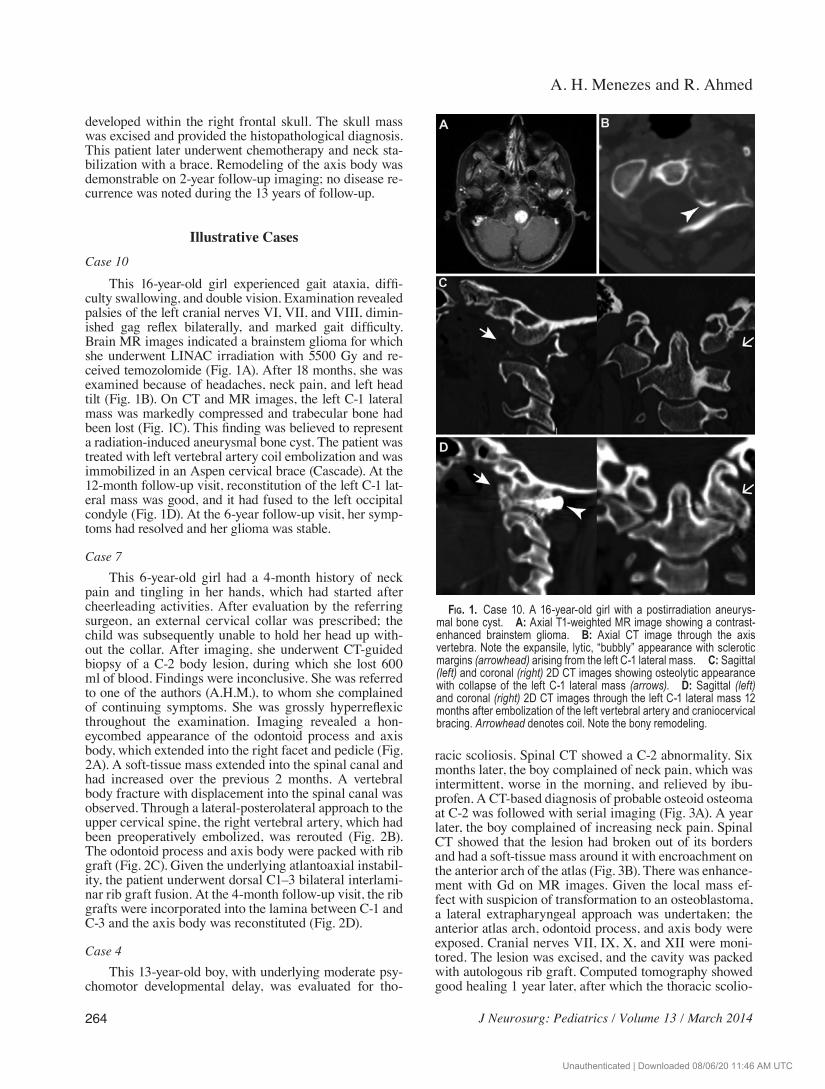
This 16-year-old girl experienced gait ataxia, diffi-culty swallowing, and double vision. Examination revealed palsies of the left cranial nerves VI, VII, and VIII, dimin-ished gag reflex bilaterally, and marked gait difficulty. Brain MR images indicated a brainstem glioma for which she underwent LINAC irradiation with 5500 Gy and re-ceived temozolomide (Fig. 1A). After 18 months, she was examined because of headaches, neck pain, and left head tilt (Fig. 1B). On CT and MR images, the left C-1 lateral mass was markedly compressed and trabecular bone had been lost (Fig. 1C). This finding was believed to represent a radiation-induced aneurysmal bone cyst. The patient was treated with left vertebral artery coil embolization and was immobilized in an Aspen cervical brace (Cascade). At the 12-month follow-up visit, reconstitution of the left C-1 lat-eral mass was good, and it had fused to the left occipital condyle (Fig. 1D). At the 6-year follow-up visit, her symp-toms had resolved and her glioma was stable.
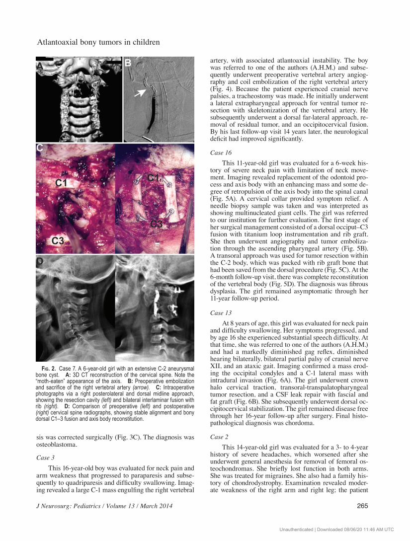
Case 7This 6-year-old girl had a 4-month history of neck
pain and tingling in her hands, which had started after cheerleading activities. After evaluation by the referring surgeon, an external cervical collar was prescribed; the child was subsequently unable to hold her head up with-out the collar. After imaging, she underwent CT-guided biopsy of a C-2 body lesion, during which she lost 600 ml of blood. Findings were inconclusive. She was referred to one of the authors (A.H.M.), to whom she complained of continuing symptoms. She was grossly hyperreflexic throughout the examination. Imaging revealed a hon-eycombed appearance of the odontoid process and axis body, which extended into the right facet and pedicle (Fig. 2A). A soft-tissue mass extended into the spinal canal and had increased over the previous 2 months. A vertebral body fracture with displacement into the spinal canal was observed. Through a lateral-posterolateral approach to the upper cervical spine, the right vertebral artery, which had been preoperatively embolized, was rerouted (Fig. 2B). The odontoid process and axis body were packed with rib graft (Fig. 2C). Given the underlying atlantoaxial instabil-ity, the patient underwent dorsal C1–3 bilateral interlami-nar rib graft fusion. At the 4-month follow-up visit, the rib grafts were incorporated into the lamina between C-1 and C-3 and the axis body was reconstituted (Fig. 2D).
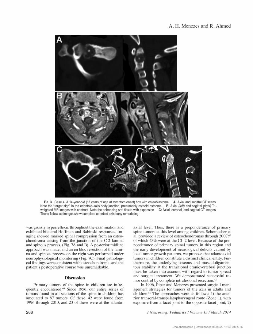
Case 4This 13-year-old boy, with underlying moderate psy-
chomotor developmental delay, was evaluated for tho-
racic scoliosis. Spinal CT showed a C-2 abnormality. Six months later, the boy complained of neck pain, which was intermittent, worse in the morning, and relieved by ibu-profen. A CT-based diagnosis of probable osteoid osteoma at C-2 was followed with serial imaging (Fig. 3A). A year later, the boy complained of increasing neck pain. Spinal CT showed that the lesion had broken out of its borders and had a soft-tissue mass around it with encroachment on the anterior arch of the atlas (Fig. 3B). There was enhance-ment with Gd on MR images. Given the local mass ef-fect with suspicion of transformation to an osteoblastoma, a lateral extrapharyngeal approach was undertaken; the anterior atlas arch, odontoid process, and axis body were exposed. Cranial nerves VII, IX, X, and XII were moni-tored. The lesion was excised, and the cavity was packed with autologous rib graft. Computed tomography showed good healing 1 year later, after which the thoracic scolio-
Fig. 1. Case 10. A 16-year-old girl with a postirradiation aneurys-mal bone cyst. A: Axial T1-weighted MR image showing a contrast-enhanced brainstem glioma. B: Axial CT image through the axis vertebra. Note the expansile, lytic, “bubbly” appearance with sclerotic margins (arrowhead) arising from the left C-1 lateral mass. C: Sagittal (left) and coronal (right) 2D CT images showing osteolytic appearance with collapse of the left C-1 lateral mass (arrows). D: Sagittal (left) and coronal (right) 2D CT images through the left C-1 lateral mass 12 months after embolization of the left vertebral artery and craniocervical bracing. Arrowhead denotes coil. Note the bony remodeling.
Unauthenticated | Downloaded 08/06/20 11:46 AM UTC
J Neurosurg: Pediatrics / Volume 13 / March 2014
Atlantoaxial bony tumors in children
265
sis was corrected surgically (Fig. 3C). The diagnosis was osteoblastoma.
Case 3This 16-year-old boy was evaluated for neck pain and
arm weakness that progressed to paraparesis and subse-quently to quadriparesis and difficulty swallowing. Imag-ing revealed a large C-1 mass engulfing the right vertebral
artery, with associated atlantoaxial instability. The boy was referred to one of the authors (A.H.M.) and subse-quently underwent preoperative vertebral artery angiog-raphy and coil embolization of the right vertebral artery (Fig. 4). Because the patient experienced cranial nerve palsies, a tracheostomy was made. He initially underwent a lateral extrapharyngeal approach for ventral tumor re-section with skeletonization of the vertebral artery. He subsequently underwent a dorsal far-lateral approach, re-moval of residual tumor, and an occipitocervical fusion. By his last follow-up visit 14 years later, the neurological deficit had improved significantly.
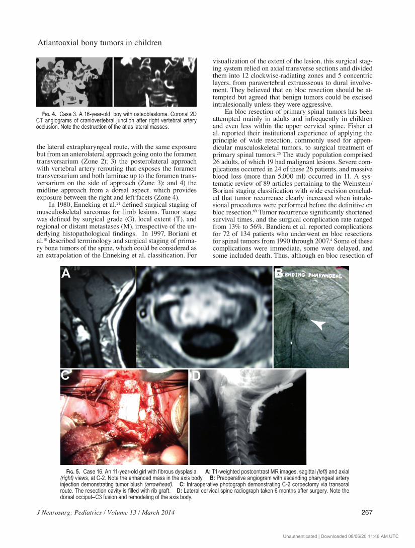
Case 16This 11-year-old girl was evaluated for a 6-week his-
tory of severe neck pain with limitation of neck move-ment. Imaging revealed replacement of the odontoid pro-cess and axis body with an enhancing mass and some de-gree of retropulsion of the axis body into the spinal canal (Fig. 5A). A cervical collar provided symptom relief. A needle biopsy sample was taken and was interpreted as showing multinucleated giant cells. The girl was referred to our institution for further evaluation. The first stage of her surgical management consisted of a dorsal occiput–C3 fusion with titanium loop instrumentation and rib graft. She then underwent angiography and tumor emboliza-tion through the ascending pharyngeal artery (Fig. 5B). A transoral approach was used for tumor resection within the C-2 body, which was packed with rib graft bone that had been saved from the dorsal procedure (Fig. 5C). At the 6-month follow-up visit, there was complete reconstitution of the vertebral body (Fig. 5D). The diagnosis was fibrous dysplasia. The girl remained asymptomatic through her 11-year follow-up period.
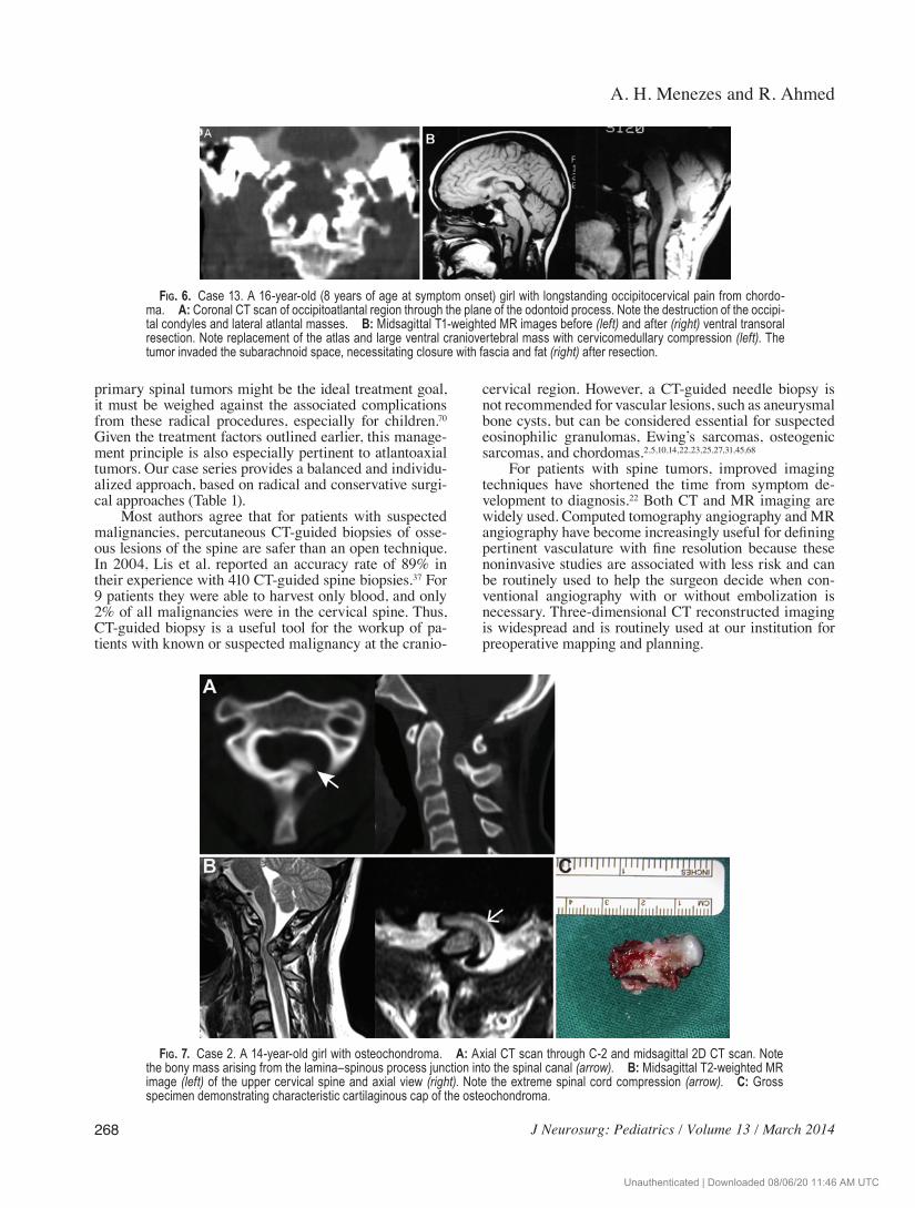
Case 13At 8 years of age, this girl was evaluated for neck pain
and difficulty swallowing. Her symptoms progressed, and by age 16 she experienced substantial speech difficulty. At that time, she was referred to one of the authors (A.H.M.) and had a markedly diminished gag reflex, diminished hearing bilaterally, bilateral partial palsy of cranial nerve XII, and an ataxic gait. Imaging confirmed a mass erod-ing the occipital condyles and a C-1 lateral mass with intradural invasion (Fig. 6A). The girl underwent crown halo cervical traction, transoral-transpalatopharyngeal tumor resection, and a CSF leak repair with fascial and fat graft (Fig. 6B). She subsequently underwent dorsal oc-cipitocervical stabilization. The girl remained disease free through her 16-year follow-up after surgery. Final histo-pathological diagnosis was chordoma.
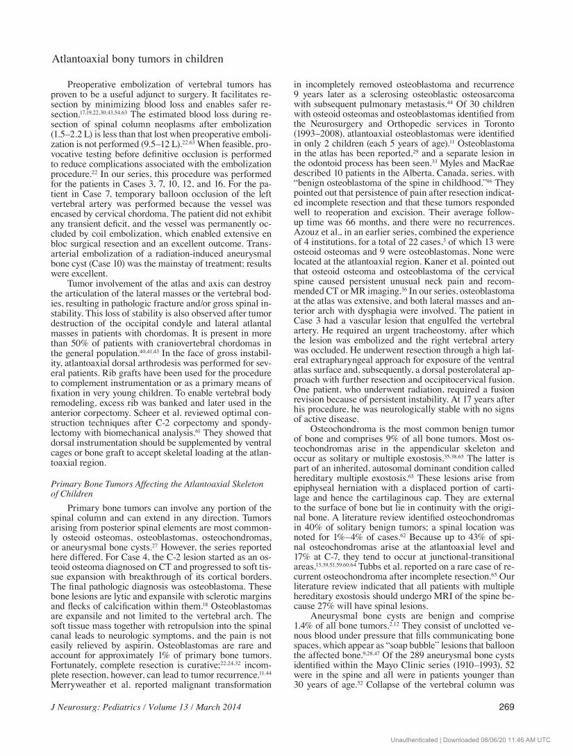
Case 2This 14-year-old girl was evaluated for a 3- to 4-year
history of severe headaches, which worsened after she underwent general anesthesia for removal of femoral os-teochondromas. She briefly lost function in both arms. She was treated for migraines. She also had a family his-tory of chondrodystrophy. Examination revealed moder-ate weakness of the right arm and right leg; the patient
Fig. 2. Case 7. A 6-year-old girl with an extensive C-2 aneurysmal bone cyst. A: 3D CT reconstruction of the cervical spine. Note the “moth-eaten” appearance of the axis. B: Preoperative embolization and sacrifice of the right vertebral artery (arrow). C: Intraoperative photographs via a right posterolateral and dorsal midline approach, showing the resection cavity (left) and bilateral interlaminar fusion with rib (right). D: Comparison of preoperative (left) and postoperative (right) cervical spine radiographs, showing stable alignment and bony dorsal C1–3 fusion and axis body reconstitution.
Unauthenticated | Downloaded 08/06/20 11:46 AM UTC
A. H. Menezes and R. Ahmed
266 J Neurosurg: Pediatrics / Volume 13 / March 2014
was grossly hyperreflexic throughout the examination and exhibited bilateral Hoffman and Babinski responses. Im-aging showed marked spinal compression from an osteo-chondroma arising from the junction of the C-2 lamina and spinous process. (Fig. 7A and B). A posterior midline approach was made, and an en bloc resection of the lami-na and spinous process on the right was performed under neurophysiological monitoring (Fig. 7C). Final pathologi-cal findings were consistent with osteochondroma, and the patient’s postoperative course was unremarkable.
DiscussionPrimary tumors of the spine in children are infre-
quently encountered.66 Since 1956, our entire series of tumors found in all sections of the spine in children has amounted to 87 tumors. Of these, 42 were found from 1996 through 2010, and 23 of these were at the atlanto-
axial level. Thus, there is a preponderance of primary spine tumors at this level among children. Schomacher et al. provided a review of osteochondromas through 2007,62 of which 43% were at the C1–2 level. Because of the pre-ponderance of primary spinal tumors in this region and the early development of neurological deficits caused by local tumor growth patterns, we propose that atlantoaxial tumors in children constitute a distinct clinical entity. Fur-thermore, the underlying osseous and musculoligamen-tous stability at the transitional craniovertebral junction must be taken into account with regard to tumor spread and surgical treatment. We demonstrated successful tu-mor control by complete intralesional resection.42
In 1996, Piper and Menezes presented surgical man-agement strategies for tumors of the axis in adults and children.56 The approaches were as follows: 1) the ante-rior transoral-transpalatopharyngeal route (Zone 1), with exposure from a facet joint to the opposite facet joint; 2)
Fig. 3. Case 4. A 14-year-old (13 years of age at symptom onset) boy with osteoblastoma. A: Axial and sagittal CT scans. Note the “target sign” in the odontoid–axis body junction, presumably osteoid osteoma. B: Axial (left) and sagittal (right) T1-weighted MR images with contrast. Note the enhancing soft tissue with expansion. C: Axial, coronal, and sagittal CT images. These follow-up images show complete odontoid axis bony remodeling.
Unauthenticated | Downloaded 08/06/20 11:46 AM UTC
J Neurosurg: Pediatrics / Volume 13 / March 2014
Atlantoaxial bony tumors in children
267
the lateral extrapharyngeal route, with the same exposure but from an anterolateral approach going onto the foramen transversarium (Zone 2); 3) the posterolateral approach with vertebral artery rerouting that exposes the foramen transversarium and both laminae up to the foramen trans-versarium on the side of approach (Zone 3); and 4) the midline approach from a dorsal aspect, which provides exposure between the right and left facets (Zone 4).
In 1980, Enneking et al.21 defined surgical staging of musculoskeletal sarcomas for limb lesions. Tumor stage was defined by surgical grade (G), local extent (T), and regional or distant metastases (M), irrespective of the un-derlying histopathological findings. In 1997, Boriani et al.10 described terminology and surgical staging of prima-ry bone tumors of the spine, which could be considered as an extrapolation of the Enneking et al. classification. For
visualization of the extent of the lesion, this surgical stag-ing system relied on axial transverse sections and divided them into 12 clockwise-radiating zones and 5 concentric layers, from paravertebral extraosseous to dural involve-ment. They believed that en bloc resection should be at-tempted but agreed that benign tumors could be excised intralesionally unless they were aggressive.
En bloc resection of primary spinal tumors has been attempted mainly in adults and infrequently in children and even less within the upper cervical spine. Fisher et al. reported their institutional experience of applying the principle of wide resection, commonly used for appen-dicular musculoskeletal tumors, to surgical treatment of primary spinal tumors.25 The study population comprised 26 adults, of which 19 had malignant lesions. Severe com-plications occurred in 24 of these 26 patients, and massive blood loss (more than 5,000 ml) occurred in 11. A sys-tematic review of 89 articles pertaining to the Weinstein/Boriani staging classification with wide excision conclud-ed that tumor recurrence clearly increased when intrale-sional procedures were performed before the definitive en bloc resection.69 Tumor recurrence significantly shortened survival times, and the surgical complication rate ranged from 13% to 56%. Bandiera et al. reported complications for 72 of 134 patients who underwent en bloc resections for spinal tumors from 1990 through 2007.4 Some of these complications were immediate, some were delayed, and some included death. Thus, although en bloc resection of
Fig. 4. Case 3. A 16-year-old boy with osteoblastoma. Coronal 2D CT angiograms of craniovertebral junction after right vertebral artery occlusion. Note the destruction of the atlas lateral masses.
Fig. 5. Case 16. An 11-year-old girl with fibrous dysplasia. A: T1-weighted postcontrast MR images, sagittal (left) and axial (right) views, at C-2. Note the enhanced mass in the axis body. B: Preoperative angiogram with ascending pharyngeal artery injection demonstrating tumor blush (arrowhead). C: Intraoperative photograph demonstrating C-2 corpectomy via transoral route. The resection cavity is filled with rib graft. D: Lateral cervical spine radiograph taken 6 months after surgery. Note the dorsal occiput–C3 fusion and remodeling of the axis body.
Unauthenticated | Downloaded 08/06/20 11:46 AM UTC
A. H. Menezes and R. Ahmed
268 J Neurosurg: Pediatrics / Volume 13 / March 2014
primary spinal tumors might be the ideal treatment goal, it must be weighed against the associated complications from these radical procedures, especially for children.70 Given the treatment factors outlined earlier, this manage-ment principle is also especially pertinent to atlantoaxial tumors. Our case series provides a balanced and individu-alized approach, based on radical and conservative surgi-cal approaches (Table 1).
Most authors agree that for patients with suspected malignancies, percutaneous CT-guided biopsies of osse-ous lesions of the spine are safer than an open technique. In 2004, Lis et al. reported an accuracy rate of 89% in their experience with 410 CT-guided spine biopsies.37 For 9 patients they were able to harvest only blood, and only 2% of all malignancies were in the cervical spine. Thus, CT-guided biopsy is a useful tool for the workup of pa-tients with known or suspected malignancy at the cranio-
cervical region. However, a CT-guided needle biopsy is not recommended for vascular lesions, such as aneurysmal bone cysts, but can be considered essential for suspected eosinophilic granulomas, Ewing’s sarcomas, osteogenic sarcomas, and chordomas.2,5,10,14,22,23,25,27,31,45,68
For patients with spine tumors, improved imaging techniques have shortened the time from symptom de-velopment to diagnosis.22 Both CT and MR imaging are widely used. Computed tomography angiography and MR angiography have become increasingly useful for defining pertinent vasculature with fine resolution because these noninvasive studies are associated with less risk and can be routinely used to help the surgeon decide when con-ventional angiography with or without embolization is necessary. Three-dimensional CT reconstructed imaging is widespread and is routinely used at our institution for preoperative mapping and planning.
Fig. 6. Case 13. A 16-year-old (8 years of age at symptom onset) girl with longstanding occipitocervical pain from chordo-ma. A: Coronal CT scan of occipitoatlantal region through the plane of the odontoid process. Note the destruction of the occipi-tal condyles and lateral atlantal masses. B: Midsagittal T1-weighted MR images before (left) and after (right) ventral trans oral resection. Note replacement of the atlas and large ventral craniovertebral mass with cervicomedullary compression (left). The tumor invaded the subarachnoid space, necessitating closure with fascia and fat (right) after resection.
Fig. 7. Case 2. A 14-year-old girl with osteochondroma. A: Axial CT scan through C-2 and midsagittal 2D CT scan. Note the bony mass arising from the lamina–spinous process junction into the spinal canal (arrow). B: Midsagittal T2-weighted MR image (left) of the upper cervical spine and axial view (right). Note the extreme spinal cord compression (arrow). C: Gross specimen demonstrating characteristic cartilaginous cap of the osteochondroma.
Unauthenticated | Downloaded 08/06/20 11:46 AM UTC
J Neurosurg: Pediatrics / Volume 13 / March 2014
Atlantoaxial bony tumors in children
269
Preoperative embolization of vertebral tumors has proven to be a useful adjunct to surgery. It facilitates re-section by minimizing blood loss and enables safer re-section.17,19,22,30,43,54,63 The estimated blood loss during re-section of spinal column neoplasms after embolization (1.5–2.2 L) is less than that lost when preoperative emboli-zation is not performed (9.5–12 L).22,63 When feasible, pro-vocative testing before definitive occlusion is performed to reduce complications associated with the embolization procedure.22 In our series, this procedure was performed for the patients in Cases 3, 7, 10, 12, and 16. For the pa-tient in Case 7, temporary balloon occlusion of the left vertebral artery was performed because the vessel was encased by cervical chordoma. The patient did not exhibit any transient deficit, and the vessel was permanently oc-cluded by coil embolization, which enabled extensive en bloc surgical resection and an excellent outcome. Trans-arterial embolization of a radiation-induced aneurysmal bone cyst (Case 10) was the mainstay of treatment; results were excellent.
Tumor involvement of the atlas and axis can destroy the articulation of the lateral masses or the vertebral bod-ies, resulting in pathologic fracture and/or gross spinal in-stability. This loss of stability is also observed after tumor destruction of the occipital condyle and lateral atlantal masses in patients with chordomas. It is present in more than 50% of patients with craniovertebral chordomas in the general population.40,41,43 In the face of gross instabil-ity, atlantoaxial dorsal arthrodesis was performed for sev-eral patients. Rib grafts have been used for the procedure to complement instrumentation or as a primary means of fixation in very young children. To enable vertebral body remodeling, excess rib was banked and later used in the anterior corpectomy. Scheer et al. reviewed optimal con-struction techniques after C-2 corpectomy and spondy-lectomy with biomechanical analysis.61 They showed that dorsal instrumentation should be supplemented by ventral cages or bone graft to accept skeletal loading at the atlan-toaxial region.
Primary Bone Tumors Affecting the Atlantoaxial Skeleton of Children
Primary bone tumors can involve any portion of the spinal column and can extend in any direction. Tumors arising from posterior spinal elements are most common-ly osteoid osteomas, osteoblastomas, osteochondromas, or aneurysmal bone cysts.27 However, the series reported here differed. For Case 4, the C-2 lesion started as an os-teoid osteoma diagnosed on CT and progressed to soft tis-sue expansion with breakthrough of its cortical borders. The final pathologic diagnosis was osteoblastoma. These bone lesions are lytic and expansile with sclerotic margins and flecks of calcification within them.18 Osteoblastomas are expansile and not limited to the vertebral arch. The soft tissue mass together with retropulsion into the spinal canal leads to neurologic symptoms, and the pain is not easily relieved by aspirin. Osteoblastomas are rare and account for approximately 1% of primary bone tumors. Fortunately, complete resection is curative;22,24,32 incom-plete resection, however, can lead to tumor recurrence.11,44 Merryweather et al. reported malignant transformation
in incompletely removed osteoblastoma and recurrence 9 years later as a sclerosing osteoblastic osteosarcoma with subsequent pulmonary metastasis.44 Of 30 children with osteoid osteomas and osteoblastomas identified from the Neurosurgery and Orthopedic services in Toronto (1993–2008), atlantoaxial osteoblastomas were identified in only 2 children (each 5 years of age).11 Osteoblastoma in the atlas has been reported,29 and a separate lesion in the odontoid process has been seen.33 Myles and MacRae described 10 patients in the Alberta, Canada, series, with “benign osteoblastoma of the spine in childhood.”46 They pointed out that persistence of pain after resection indicat-ed incomplete resection and that these tumors responded well to reoperation and excision. Their average follow-up time was 66 months, and there were no recurrences. Azouz et al., in an earlier series, combined the experience of 4 institutions, for a total of 22 cases,3 of which 13 were osteoid osteomas and 9 were osteoblastomas. None were located at the atlantoaxial region. Kaner et al. pointed out that osteoid osteoma and osteoblastoma of the cervical spine caused persistent unusual neck pain and recom-mended CT or MR imaging.36 In our series, osteoblastoma at the atlas was extensive, and both lateral masses and an-terior arch with dysphagia were involved. The patient in Case 3 had a vascular lesion that engulfed the vertebral artery. He required an urgent tracheostomy, after which the lesion was embolized and the right vertebral artery was occluded. He underwent resection through a high lat-eral extrapharyngeal approach for exposure of the ventral atlas surface and, subsequently, a dorsal posterolateral ap-proach with further resection and occipitocervical fusion. One patient, who underwent radiation, required a fusion revision because of persistent instability. At 17 years after his procedure, he was neurologically stable with no signs of active disease.
Osteochondroma is the most common benign tumor of bone and comprises 9% of all bone tumors. Most os-teochondromas arise in the appendicular skeleton and occur as solitary or multiple exostosis.35,38,65 The latter is part of an inherited, autosomal dominant condition called hereditary multiple exostosis.65 These lesions arise from epiphyseal herniation with a displaced portion of carti-lage and hence the cartilaginous cap. They are external to the surface of bone but lie in continuity with the origi-nal bone. A literature review identified osteochondromas in 40% of solitary benign tumors; a spinal location was noted for 1%–4% of cases.62 Because up to 43% of spi-nal osteochondromas arise at the atlantoaxial level and 17% at C-7, they tend to occur at junctional-transitional areas.13,39,51,59,60,64 Tubbs et al. reported on a rare case of re-current osteochondroma after incomplete resection.65 Our literature review indicated that all patients with multiple hereditary exostosis should undergo MRI of the spine be-cause 27% will have spinal lesions.
Aneurysmal bone cysts are benign and comprise 1.4% of all bone tumors.2,12 They consist of unclotted ve-nous blood under pressure that fills communicating bone spaces, which appear as “soap bubble” lesions that balloon the affected bone.9,28,47 Of the 289 aneurysmal bone cysts identified within the Mayo Clinic series (1910–1993), 52 were in the spine and all were in patients younger than 30 years of age.52 Collapse of the vertebral column was
Unauthenticated | Downloaded 08/06/20 11:46 AM UTC
A. H. Menezes and R. Ahmed
270 J Neurosurg: Pediatrics / Volume 13 / March 2014
common. These cysts commonly arose from the posterior bony elements and were most often treated by internal cu-rettage and bone grafting. In 2001, Boriani et al. reported that of 41 cases of aneurysmal bone cysts,9 only 4 were in the atlas and 1 in the axis. According to their literature review to that date, up to 63% of aneurysmal bone cysts are prevalent among children. Malignant transformation of aneurysmal bone cysts was very rarely described and usually followed irradiation. The incidence of atlantoaxial aneurysmal bone cysts has been reported in the range of 11% to 16%.2,28,47 In our recent experience of 4 aneurysmal bone cyst at the atlas, we more commonly found odontoid fracture, fracture of the vertebral body, and collapse with instability. Our goal was arterial embolization followed by stabilization and tumor resection. Arterial emboliza-tion of the lesion has been accomplished with injection of calcitonin and methylprednisolone.9,17,20,28,43,55,58,63,71
Chordomas are rare, aggressive, destructive tumors presumed to be of notochordal origin; they arise along the vertebral axis, mainly in the foramen magnum, pet-roclival and sphenoclival regions.40,43 Chordomas are ex-tradural as well as intradural; at the craniocervical region, they become intradural after they have either penetrated or circumvented the tectorial membrane to appear in an anterolateral position at the foramen magnum and at C1–2. These lesions, when at the craniocervical region, have been believed to be invariably fatal for patients younger than 5 years of age, regardless of surgical treatment.6,8,16,23 Chordomas are well defined, and CT and MR images are of high resolution.43 Involvement of the lateral articular masses of the atlas and axis and the occipital condyles were indications for stabilization after resection. Relevant surgical techniques include anterior transpalatopharyn-geal and open-door maxillotomy approaches, lateral and posterolateral approaches, and (more recently) endoscopic transoral and transnasal procedures.15,43 En bloc resection has been described as a radical procedure with extensive hardware and substantial long-term sequelae.7,59 A re-view by Borba et al. reported the prognosis to be poor for children younger than 5 years of age and worse for those with atypical chordomas.8 For adult patients with difficult chordomas, en bloc vertebrectomies are associated with significant morbidity, which markedly alters the quality of life.7,26,59 Barrenechea et al. reported that en bloc resec-tions of high cervical chordomas was not feasible.5 Ag-gressive resection followed by adjuvant LINAC or proton-beam radiotherapy provides the best results, as exempli-fied in our series.40,50
Fibrous dysplasia in a spinal location with monostotic disease is extremely rare.14,22,27,45 Until 2006, only 25 cases had been reported among patients of all ages.22 Between 1996 and 2010, a total of 3 such cases were encountered in the axis body. These lesions arise from bone marrow stomal cells, mostly during the patient’s first decade of life, and a few arise later in adulthood. They are lytic with a ground glass appearance, as seen in the cases reported here. Needle biopsy results are usually nondiagnostic, and the treatment goal is resection and stabilization.
Eosinophilic granuloma is the most benign form of disease in the group known as histiocytosis X, which includes Letterer-Siwe and Hand-Schüller-Christian dis-eases.6 Eosinophilic granuloma is considered a benign os-
teolytic disease that primarily involves the skull, ribs, and long bones. Reportedly, 80% of patients are younger than 20 years of age. Of all bone tumors, 6.3%–25% are spinal eosinophilic granulomas.31,57 The 2 major radiographic findings are 1) osteolytic defect with occasional sclerotic reaction and 2) grades of vertebral collapse. Atypical lytic lesions might require biopsy; if neurological compromise is present, decompression and stabilization might be re-quired.48,57 Greenlee et al. reviewed the series reported by one of our authors (A.H.M.) and presented a decision tree for management of patients with suspected eosinophilic granuloma.31 Although eosinophilic granuloma is the most common cause of vertebra plana, diagnosis of ad-ditional entities such as Ewing’s sarcoma, other tumors, or tuberculosis might require CT-guided biopsy. Lytic forms of eosinophilic granuloma need to be differentiated from osteochondritis, tuberculosis, osteomyelitis, and plasma-cytoma. For most children, spinal eosinophilic granulo-ma can be managed nonsurgically. A CT-guided biopsy can aid in diagnosis. In our general series of eosinophilic granulomas, diagnosis for 8 of 12 patients required a tis-sue biopsy. The length of spinal immobilization was de-termined by patient symptoms and radiographic improve-ment.31
Ewing’s sarcoma is a rare, aggressive, primitive neu-roectodermal tumor usually seen in children.68 It may also be a primary spinal epidural lesion.34,49 Only 3.5% of pa-tients have primary Ewing’s sarcoma of the spine; when the sacrum is excluded, this percentage decreases to 1%. Among patients with Ewing’s sarcoma, 58% have neuro-logic deficits. For patients who undergo resection, radia-tion, and chemotherapy, the 5-year survival rate is 33%.1,67 The development of effective chemotherapy has been the main factor for overall improvement and disease-free survival among these patients; the 5-year survival rate is 70%.53 A review of the St. Jude’s Children’s Research Hospital identified 344 patients with Ewing’s sarcoma, of which 33 tumors were primary tumors in the vertebra.67 Only 2 were in the cervical location. The event-free sur-vival rate (± SD) was 48% ± 8.9%.
ConclusionsWe described a large series of primary atlantoaxial
tumors in children for whom current technological ad-vances were used and follow-up duration was long. For most cases, high-definition neurodiagnostic imaging has facilitated diagnosis of primary bone tumors in children. However, when the diagnosis remains in question, a CT-guided biopsy is possible. En bloc resection of tumors at the atlantoaxial region would be ideal, but such resection is unlikely unless the area of pathology is small and focal. Angiography and tumor embolization improve surgical safety and, hence, play a major role in surgical manage-ment. Most primary bone tumors in children, except chor-domas and Ewing’s sarcomas, are benign. Thus, to achieve the same result as an en bloc resection, a staged approach is recommended, except for chordomas, for which a gross resection should be followed with radiation therapy.
Axis corpectomy necessitates bone placement in the C-2 body and dorsal stabilization. Aneurysmal bone cysts can be treated with embolization and stabilization except
Unauthenticated | Downloaded 08/06/20 11:46 AM UTC
J Neurosurg: Pediatrics / Volume 13 / March 2014
Atlantoaxial bony tumors in children
271
for patients with an extensive lesion with collapse and neurologic deficit. For these patients, decompression is necessary.
Diagnosis of eosinophilic granulomas might require biopsy. Fusion and stabilization are essential if skeletal instability is present. Patients with disseminated disease (histiocytosis X) respond well to chemotherapy.
Disclosure
The authors report no conflict of interest concerning the mate-rials or methods used in this study or the findings specified in this paper.
Author contributions to the study and manuscript preparation include the following. Conception and design: Menezes. Acquisi-tion of data: both authors. Analysis and interpretation of data: both authors. Drafting the article: both authors. Critically revising the article: both authors. Reviewed submitted version of manuscript: both authors. Approved the final version of the manuscript on behalf of both authors: Menezes. Administrative/technical/material support: Menezes. Study supervision: Menezes.
References
1. Akeda K, Kasai Y, Kawakita E, Seto M, Kono T, Uchida A: Primary Ewing sarcoma of the spine mimicking a psoas ab-scess secondary to spinal infection. Spine (Phila Pa 1976) 34: E337–E341, 2009
2. Ameli NO, Abbassioun K, Saleh H, Eslamdoost A: Aneurys-mal bone cysts of the spine. Report of 17 cases. J Neurosurg 63:685–690, 1985
3. Azouz EM, Kozlowski K, Marton D, Sprague P, Zerhouni A, Asselah F: Osteoid osteoma and osteoblastoma of the spine in children. Report of 22 cases with brief literature review. Pedi-atr Radiol 16:25–31, 1986
4. Bandiera S, Boriani S, Donthineni R, Amendola L, Cappuccio M, Gasbarrini A: Complications of en bloc resections in the spine. Orthop Clin North Am 40:125–131, vii, 2009
5. Barrenechea IJ, Perin NI, Triana A, Lesser J, Costantino P, Sen C: Surgical management of chordomas of the cervical spine. J Neurosurg Spine 6:398–406, 2007
6. Beer SJ, Menezes AH: Primary tumors of the spine in children. Natural history, management, and long-term follow-up. Spine (Phila Pa 1976) 22:649–659, 1997
7. Biagini R, Casadei R, Boriani S, Erba F, Sturale C, Mascari C, et al: En bloc vertebrectomy and dural resection for chordoma: a case report. Spine (Phila Pa 1976) 28:E368–E372, 2003
8. Borba LAB, Al-Mefty O, Mrak RE, Suen J: Cranial chordomas in children and adolescents. J Neurosurg 84:584–591, 1996
9. Boriani S, De Iure F, Campanacci L, Gasbarrini A, Bandiera S, Biagini R, et al: Aneurysmal bone cyst of the mobile spine: report on 41 cases. Spine (Phila Pa 1976) 26:27–35, 2001
10. Boriani S, Weinstein JN, Biagini R: Primary bone tumors of the spine. Terminology and surgical staging. Spine (Phila Pa 1976) 22:1036–1044, 1997
11. Burn SC, Ansorge O, Zeller R, Drake JM: Management of os-teoblastoma and osteoid osteoma of the spine in childhood. Clinical article. J Neurosurg Pediatr 4:434–438, 2009
12. Capanna R, Bettelli G, Biagini R, Ruggieri P, Bertoni F, Cam-panacci M: Aneurysmal cysts of long bones. Ital J Orthop Trau matol 11:409–417, 1985
13. Chatzidakis E, Lypiridis S, Kazdaglis G, Chatzikonstadinou K, Papatheodorou G: A rare case of solitary osteochondroma of the dens of the C2 vertebra. Acta Neurochir (Wien) 149:637–638, 2007
14. Chi JH, Bydon A, Hsieh P, Witham T, Wolinsky JP, Gokaslan ZL: Epidemiology and demographics for primary vertebral tu-mors. Neurosurg Clin N Am 19:1–4, 2008
15. Choi D, Melcher R, Harms J, Crockard A: Outcome of 132 op-erations in 97 patients with chordomas of the craniocervical junction and upper cervical spine. Neurosurgery 66:59–65, 2010
16. Choi GH, Yang MS, Yoon H, Shin HC, Kim KN, Yi S, et al: Pediatric cervical chordoma: report of two cases and a review of the current literature. Childs Nerv Syst 26:835–840, 2010
17. Cloft HJ, Dion JE: Preoperative and palliative embolization of vertebral tumors. Neuroimaging Clin N Am 10:569–578, 2000
18. Cové JA, Taminiau AH, Obermann WR, Vanderschueren GM: Osteoid osteoma of the spine treated with percutaneous com-puted tomography-guided thermocoagulation. Spine (Phila Pa 1976) 25:1283–1286, 2000
19. De Cristofaro R, Biagini R, Boriani S, Ricci S, Ruggieri P, Ros-si G, et al: Selective arterial embolization in the treatment of aneurysmal bone cyst and angioma of bone. Skeletal Radiol 21:523–527, 1992
20. DeRosa GP, Graziano GP, Scott J: Arterial embolization of an-eurysmal bone cyst of the lumbar spine. A report of two cases. J Bone Joint Surg Am 72:777–780, 1990
21. Enneking WF, Spanier SS, Goodman MA: A system for the surgical staging of musculoskeletal sarcoma. Clin Orthop Relat Res (153):106–120, 1980
22. Fenoy AJ, Greenlee JDW, Menezes AH, Donovan KA, Sato Y, Hitchon PW, et al: Primary bone tumors of the spine in chil-dren. J Neurosurg 105 (4 Suppl):252–260, 2006
23. Ferraresi V, Nuzzo C, Zoccali C, Marandino F, Vidiri A, Sal-ducca N, et al: Chordoma: clinical characteristics, manage-ment and prognosis of a case series of 25 patients. BMC Can-cer 10:22, 2010
24. Figarella-Branger D, Perez-Castillo M, Garbe L, Grisoli F, Gambarelli D, Hassoun J: Malignant transformation of an os-teoblastoma of the skull: an exceptional occurrence. Case re-port. J Neurosurg 75:138–142, 1991
25. Fisher CG, Keynan O, Boyd MC, Dvorak MF: The surgical management of primary tumors of the spine: initial results of an ongoing prospective cohort study. Spine (Phila Pa 1976) 30:1899–1908, 2005
26. Fujita T, Kawahara N, Matsumoto T, Tomita K: Chordoma in the cervical spine managed with en bloc excision. Spine (Phila Pa 1976) 24:1848–1851, 1999
27. Garg S, Dormans JP: Tumors and tumor-like conditions of the spine in children. J Am Acad Orthop Surg 13:372–381, 2005
28. Garg S, Mehta S, Dormans JP: Modern surgical treatment of primary aneurysmal bone cyst of the spine in children and adolescents. J Pediatr Orthop 25:387–392, 2005
29. Gelberman RH, Olson CO: Benign osteoblastoma of the atlas. A case report. J Bone Joint Surg Am 56:809–810, 1974
30. Gellad FE, Sadato N, Numaguchi Y, Levine AM: Vascular metastatic lesions of the spine: preoperative embolization. Ra-diology 176:683–686, 1990
31. Greenlee JDW, Fenoy AJ, Donovan KA, Menezes AH: Eosino-philic granuloma in the pediatric spine. Pediatr Neurosurg 43:285–292, 2007
32. Hadjipavlou AG, Tzermiadianos MN, Kakavelakis KN, Land-er P: Percutaneous core excision and radiofrequency thermo-coagulation for the ablation of osteoid osteoma of the spine. Eur Spine J 18:345–351, 2009
33. Hladky JP, Lejeune JP, Singer B, Lecomte-Houcke M, Herbaux B, Dhellemmes P: Osteoblastoma of the odontoid process. Pe-diatr Neurosurg 21:260–262, 1994
34. Hsieh CT, Chiang YH, Tsai WC, Sheu LF, Liu MY: Primary spinal epidural Ewing sarcoma: a case report and review of the literature. Turk J Pediatr 50:282–286, 2008
35. Julien J, Riemens V, Vital C, Lagueny A, Miet G: Cervical cord compression by solitary osteochondroma of the atlas. J Neurol Neurosurg Psychiatry 41:479–481, 1978
36. Kaner T, Sasani M, Oktenoglu T, Aydin S, Ozer AF: Osteoid
Unauthenticated | Downloaded 08/06/20 11:46 AM UTC
A. H. Menezes and R. Ahmed
272 J Neurosurg: Pediatrics / Volume 13 / March 2014
osteoma and osteoblastoma of the cervical spine: the cause of unusual persistent neck pain. Pain Physician 13:549–554, 2010
37. Lis E, Bilsky MH, Pisinski L, Boland P, Healey JH, O’Malley B, et al: Percutaneous CT-guided biopsy of osseous lesion of the spine in patients with known or suspected malignancy. AJNR Am J Neuroradiol 25:1583–1588, 2004
38. Lotfinia I, Vahedi P, Tubbs RS, Ghavame M, Meshkini A: Neu-rological manifestations, imaging characteristics, and surgical outcome of intraspinal osteochondroma. Clinical article. J Neurosurg Spine 12:474–489, 2010
39. MacGee EE: Osteochondroma of the cervical spine: a cause of transient quadriplegia. Neurosurgery 4:259–260, 1979
40. Menezes AH: Clival and craniovertebral junction chordomas. World Neurosurg [epub ahead of print], 2013
41. Menezes AH: Craniovertebral junction neoplasms in the pedi-atric population. Childs Nerv Syst 24:1173–1186, 2008
42. Menezes AH, Sato Y: Primary tumors of the spine in chil-dren—natural history and management. 1990. Pediatr Neuro-surg 23:101–114, 1995
43. Menezes AH, Traynelis VC: Tumors of the craniovertebral junction, in Winn HR (ed): Youmans Neurological Surgery. Philadelphia: Elsevier, 2011, pp 3114–3130
44. Merryweather R, Middlemiss JH, Sanerkin NG: Malignant transformation of osteoblastoma. J Bone Joint Surg Br 62: 381–384, 1980
45. Mezzadri JJ, Acotto CG, Mautalen C, Basso A: Surgical treat-ment of cervical spine fibrous dysplasia: technical case report and review. Neurosurgery 44:1342–1347, 1999
46. Myles ST, MacRae ME: Benign osteoblastoma of the spine in childhood. J Neurosurg 68:884–888, 1988
47. Novais EN, Rose PS, Yaszemski MJ, Sim FH: Aneurysmal bone cyst of the cervical spine in children. J Bone Joint Surg Am 93:1534–1543, 2011
48. Osenbach RK, Youngblood LA, Menezes AH: Atlanto-axial instability secondary to solitary eosinophilic granuloma of C2 in a 12-year-old girl. J Spinal Disord 3:408–412, 1990
49. Ozturk E, Mutlu H, Sonmez G, Vardar Aker F, Cinar Basekim C, Kizilkaya E: Spinal epidural extraskeletal Ewing sarcoma. J Neuroradiol 34:63–67, 2007
50. Pallini R, Maira G, Pierconti F, Falchetti ML, Alvino E, Cimi-no-Reale G, et al: Chordoma of the skull base: predictors of tumor recurrence. J Neurosurg 98:812–822, 2003
51. Palmer FJ, Blum PW: Osteochondroma with spinal cord com-pression. Report of three cases. J Neurosurg 52:842–845, 1980
52. Papagelopoulos PJ, Currier BL, Shaughnessy WJ, Sim FH, Ebsersold MJ, Bond JR, et al: Aneurysmal bone cyst of the spine. Management and outcome. Spine (Phila Pa 1976) 23: 621–628, 1998
53. Paulino AC, Nguyen TX, Mai WY: An analysis of primary site control and late effects according to local control modality in non-metastatic Ewing sarcoma. Pediatr Blood Cancer 48: 423–429, 2007
54. Pearl MS, Wolinsky JP, Gailloud P: Preoperative embolization of primary spinal aneurysmal bone cysts by direct percutane-ous intralesional injection of n-butyl-2-cyanoacrylate. J Vasc Interv Radiol 23:841–845, 2012
55. Perlmutter DH, Campbell S, Rubery PT, Vates EG, Silberstein HJ: Aneurysmal bone cyst: surgical management in the pedi-atric cervical spine. Spine (Phila Pa 1976) 34:E50–E53, 2009
56. Piper JG, Menezes AH: Management strategies for tumors of the axis vertebra. J Neurosurg 84:543–551, 1996
57. Raab P, Hohmann F, Kühl J, Krauspe R: Vertebral remodeling in eosinophilic granuloma of the spine. A long-term follow-up. Spine (Phila Pa 1976) 23:1351–1354, 1998
58. Rai AT, Collins JJ: Percutaneous treatment of pediatric aneu-rysmal bone cyst at C1: a minimally invasive alternative: a case re port. AJNR Am J Neuroradiol 26:30–33, 2005
59. Rhines LD, Fourney DR, Siadati A, Suk I, Gokaslan ZL: En bloc resection of multilevel cervical chordoma with C-2 in-volvement. Case report and description of operative technique. J Neurosurg Spine 2:199–205, 2005
60. Roach JW, Klatt JWB, Faulkner ND: Involvement of the spine in patients with multiple hereditary exostoses. J Bone Joint Surg Am 91:1942–1948, 2009
61. Scheer JK, Tang J, Eguizabal J, Farin A, Buckley JM, Deviren V, et al: Optimal reconstruction technique after C-2 corpecto-my and spondylectomy: a biomechanical analysis. Laboratory investigation. J Neurosurg Spine 12:517–524, 2010
62. Schomacher M, Suess O, Kombos T: Osteochondromas of the cervical spine in atypical location. Acta Neurochir (Wien) 151:629–633, 2009
63. Smith TP, Gray L, Weinstein JN, Richardson WJ, Payne CS: Preoperative transarterial embolization of spinal column neo-plasms. J Vasc Interv Radiol 6:863–869, 1995
64. Srikantha U, Bhagavatula ID, Satyanarayana S, Somanna S, Chan dramouli BA: Spinal osteochondroma: spectrum of a rare disease. Report of 3 cases. J Neurosurg Spine 8:561–566, 2008
65. Tubbs RS, Maddox GE, Grabb PA, Oakes WJ, Cohen-Gadol AA: Cervical osteochondroma with postoperative recurrence: case report and review of the literature. Childs Nerv Syst 26: 101–104, 2010
66. Unni KK: Dahlin’s Bone Tumors: General Aspects and Da ta on 11,087 Cases, ed 5. Philadelphia: Lippincott-Raven, 1996
67. Venkateswaran L, Rodriguez-Galindo C, Merchant TE, Po-quette CA, Rao BN, Pappo AS: Primary Ewing tumor of the vertebrae: clinical characteristics, prognostic factors, and out-come. Med Pediatr Oncol 37:30–35, 2001
68. Weber KL: Current concepts in the treatment of Ewing’s sar-coma. Expert Rev Anticancer Ther 2:687–694, 2002
69. Yamazaki T, McLoughlin GS, Patel S, Rhines LD, Fourney DR: Feasibility and safety of en bloc resection for primary spine tumors: a systematic review by the Spine Oncology Study Group. Spine (Phila Pa 1976) 34 (22 Suppl):S31–S38, 2009
70. Yang X, Wu Z, Xiao J, Teng H, Feng D, Huang W, et al: Se-quentially staged resection and 2-column reconstruction for C2 tumors through a combined anterior retropharyngeal-pos-terior approach: surgical technique and results in 11 patients. Neurosurgery 69 (2 Suppl Operative):ons184–ons194, 2011
71. Zenonos G, Jamil O, Governale LS, Jernigan S, Hedequist D, Proctor MR: Surgical treatment for primary spinal aneurysmal bone cysts: experience from Children’s Hospital Boston. Clini-cal article. J Neurosurg Pediatr 9:305–315, 2012
Manuscript submitted May 14, 2013.Accepted November 25, 2013.Portions of this work were presented in oral form at the 36th Annual
Meeting of The American Society of Pediatric Neurosurgeons, Feb-ruary 10–15, 2013, Kauai, Hawaii.
Please include this information when citing this paper: published online January 17, 2014; DOI: 10.3171/2013.11.PEDS13245.
Address correspondence to: Arnold H. Menezes, M.D., De -partment of Neurosurgery, University of Iowa Hospitals and Clinics, 200 Hawkins Dr., 1824 JPP, Iowa City, IA 52242. email: [email protected].
Unauthenticated | Downloaded 08/06/20 11:46 AM UTC