Embed Size (px)
Citation preview
Benign Bone Tumors
Dr Anand Ranjan DevDept of Orthopaedics RKCH
LECTURE ON TUMORS CONTINUED...
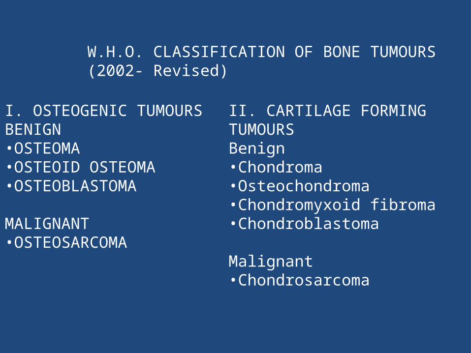
W.H.O. CLASSIFICATION OF BONE TUMOURS (2002- Revised)
I. OSTEOGENIC TUMOURS BENIGN •OSTEOMA •OSTEOID OSTEOMA •OSTEOBLASTOMA
MALIGNANT •OSTEOSARCOMA
II. CARTILAGE FORMING TUMOURS Benign •Chondroma •Osteochondroma •Chondromyxoid fibroma •Chondroblastoma
Malignant •Chondrosarcoma
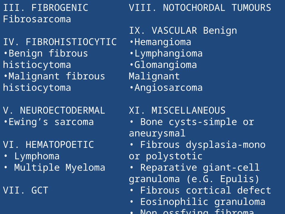
III. FIBROGENIC Fibrosarcoma
IV. FIBROHISTIOCYTIC •Benign fibrous histiocytoma •Malignant fibrous histiocytoma
V. NEUROECTODERMAL •Ewing’s sarcoma
VI. HEMATOPOETIC • Lymphoma • Multiple Myeloma
VII. GCT
VIII. NOTOCHORDAL TUMOURS
IX. VASCULAR Benign •Hemangioma •Lymphangioma •Glomangioma Malignant •Angiosarcoma
XI. MISCELLANEOUS • Bone cysts-simple or aneurysmal • Fibrous dysplasia-mono or polystotic • Reparative giant-cell granuloma (e.G. Epulis) • Fibrous cortical defect • Eosinophilic granuloma • Non ossfying fibroma • Osteitis fibrosa cystica (brown tumour)
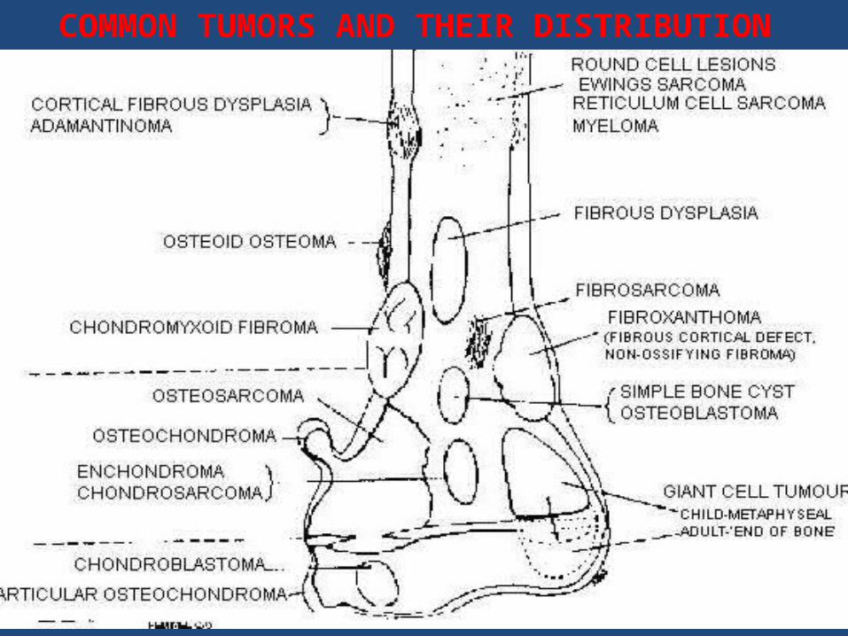
COMMON TUMORS AND THEIR DISTRIBUTION
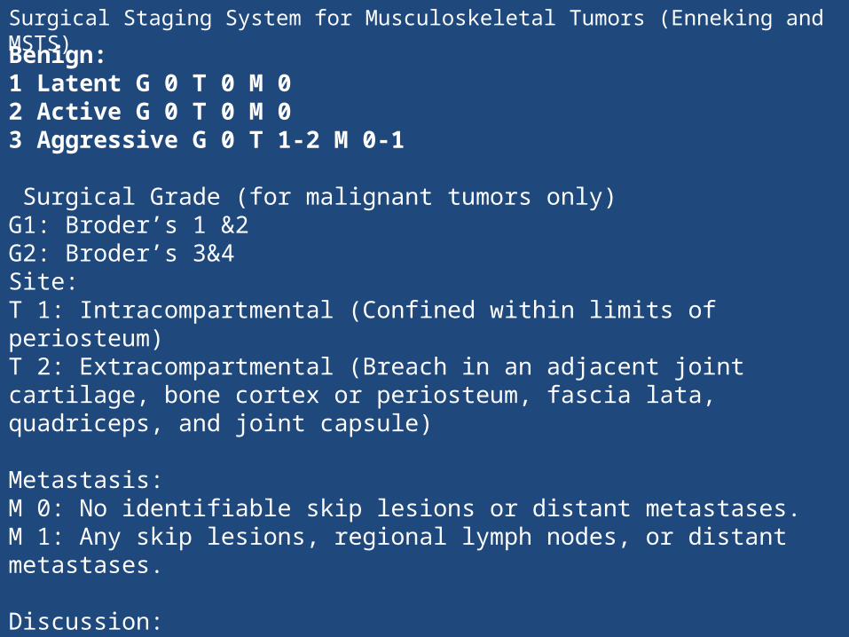
Surgical Staging System for Musculoskeletal Tumors (Enneking and MSTS)
Benign: 1 Latent G 0 T 0 M 0 2 Active G 0 T 0 M 0 3 Aggressive G 0 T 1-2 M 0-1
Surgical Grade (for malignant tumors only)G1: Broder’s 1 &2G2: Broder’s 3&4Site: T 1: Intracompartmental (Confined within limits of periosteum) T 2: Extracompartmental (Breach in an adjacent joint cartilage, bone cortex or periosteum, fascia lata, quadriceps, and joint capsule)
Metastasis: M 0: No identifiable skip lesions or distant metastases. M 1: Any skip lesions, regional lymph nodes, or distant metastases.
Discussion: Benign tumor staging uses Arabic numbers (1,2,3) Malignant tumors identified with Roman numerals and a letter (Ia, Ib, IIa, IIb, IIIa, IIIb)
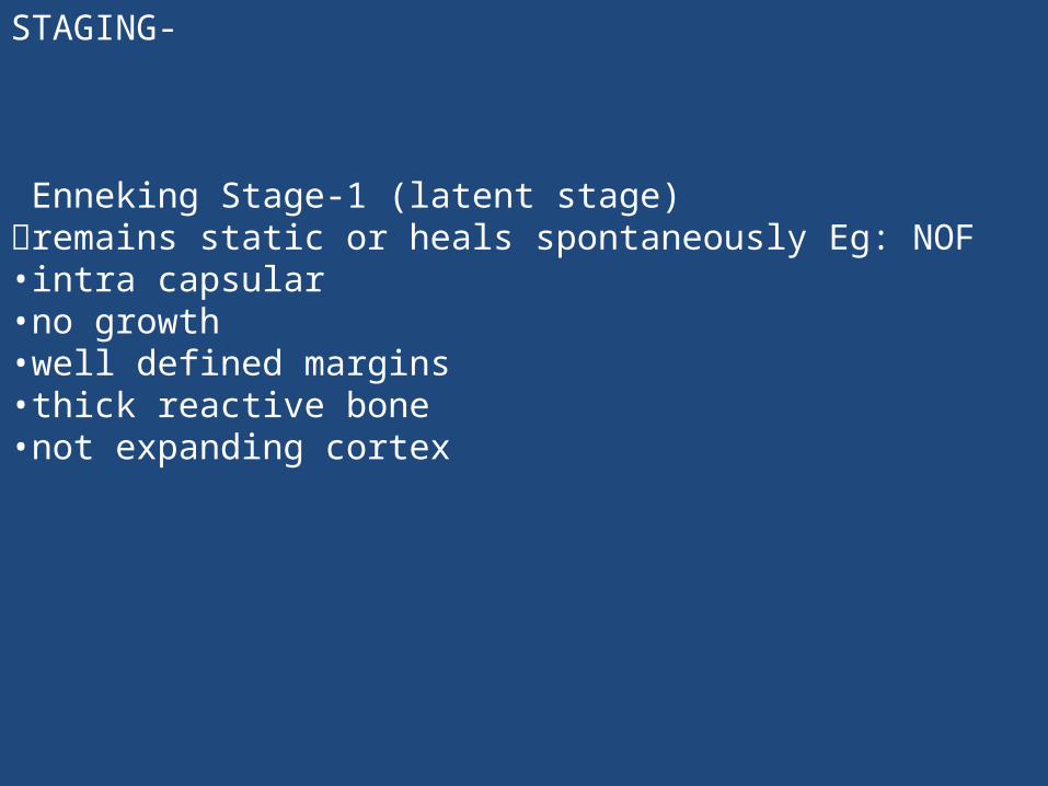
STAGING-
Enneking Stage-1 (latent stage) remains static or heals spontaneously Eg: NOF •intra capsular •no growth •well defined margins •thick reactive bone •not expanding cortex
Stage-2 (active) progressive growth but limited by natural barriers Eg: Simple bone cyst •intra capsular •actively growing •well defined margins •thin rim of reactive bone •cortical expansion with thinning
Stage 3 (aggressive) Progressive growth not limited by natural barriers eg: GCT •Extracapsular •Break through reactive bone/cortex
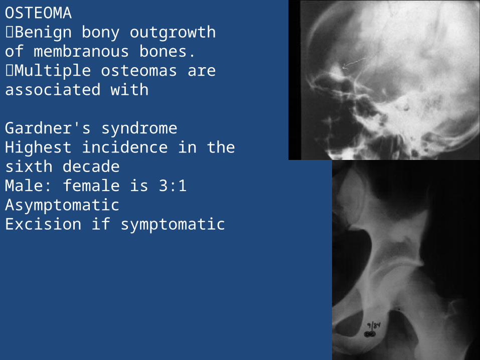
OSTEOMA Benign bony outgrowth of membranous bones. Multiple osteomas are associated with
Gardner's syndrome Highest incidence in the sixth decade Male: female is 3:1 Asymptomatic Excision if symptomatic
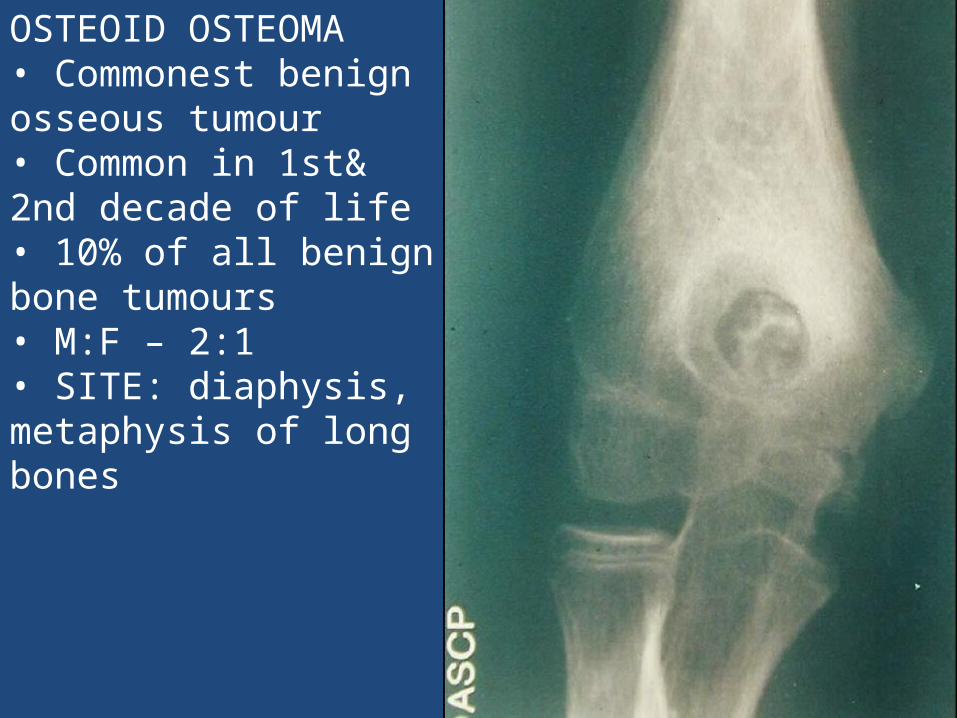
OSTEOID OSTEOMA • Commonest benign osseous tumour • Common in 1st& 2nd decade of life • 10% of all benign bone tumours • M:F – 2:1 • SITE: diaphysis, metaphysis of long bones
CLINICAL FEATURES Dull pain, worse at night (night cries) & responds to salicylates ( aspirin) Swelling uncommon Tenderness
RADIOLOGICAL FEATURES A sharp round or oval lesion. Less than 2 cm in diameter. Radiolucent nidus surrounded by reactive sclerosis Nidus- osteolytic/partially/entirely calcified
Investigations CT SCAN: Nidus is best localized with CT (1 mm cuts)
Bull‟s eye lesion
BONE SCAN: Tc99 Due to intense radioisotope uptake by nidus and decreased uptake by surrounding sclerotic bone, a double density image is created that is typical of osteoid osteoma.
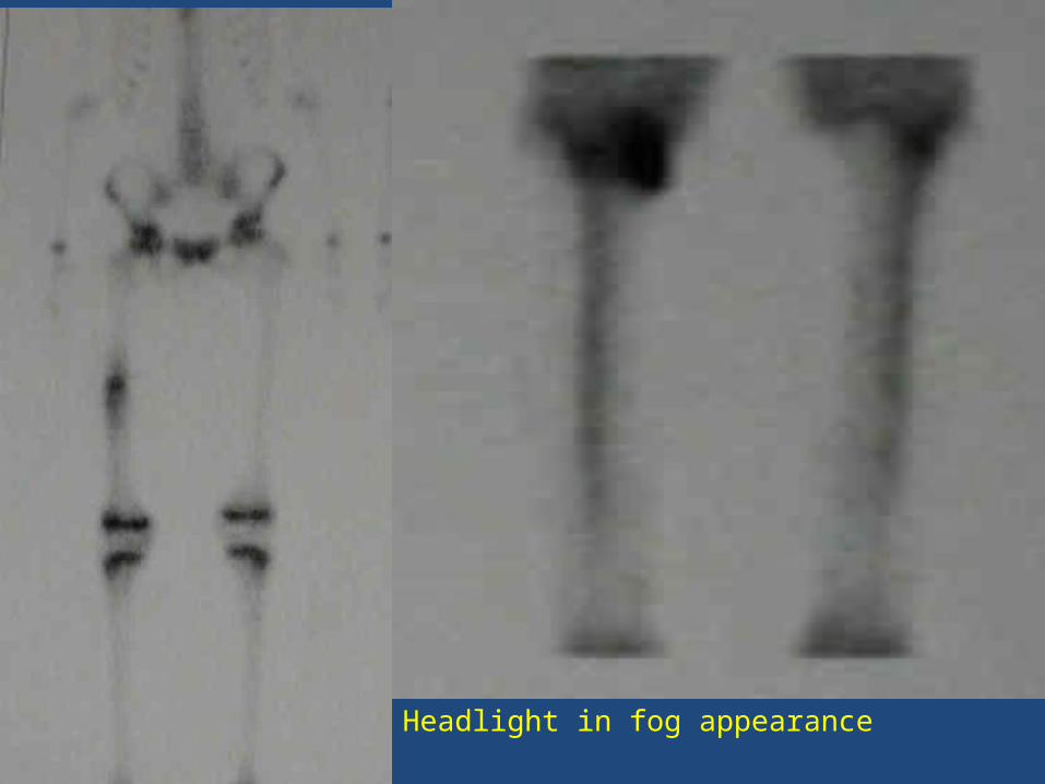
Headlight in fog appearance
Management
Course: Self limiting On maturation, ossify and merge with surrounding bone No reports of malignant transformation till date
Treatment: Conservative - not recommended because of severity of pain Surgical: En Bloc resection, Burr down Percutaneous radiofrequency ablation (PRA)
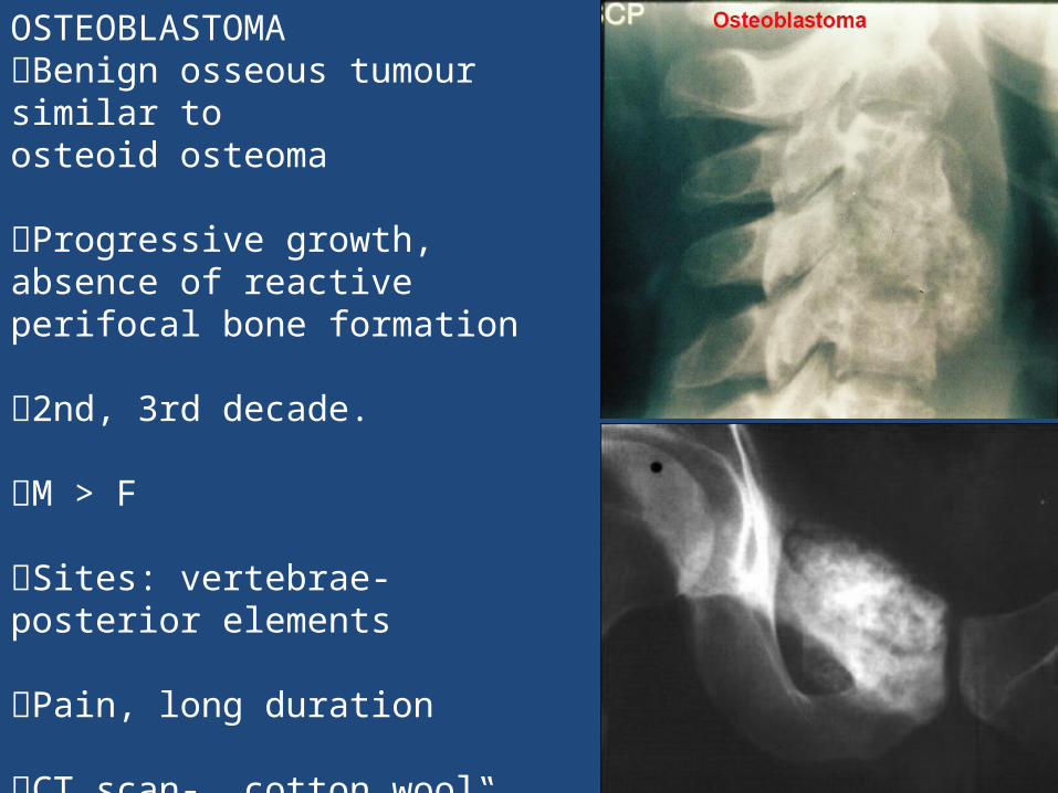
OSTEOBLASTOMA Benign osseous tumour similar toosteoid osteoma
Progressive growth, absence of reactive perifocal bone formation
2nd, 3rd decade.
M > F
Sites: vertebrae- posterior elements
Pain, long duration
CT scan- „cotton wool‟ if calcified
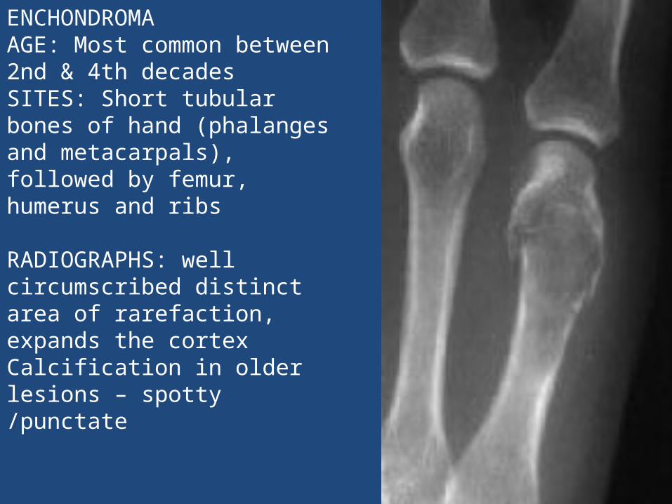
ENCHONDROMA AGE: Most common between 2nd & 4th decades SITES: Short tubular bones of hand (phalanges and metacarpals), followed by femur, humerus and ribs
RADIOGRAPHS: well circumscribed distinct area of rarefaction, expands the cortex Calcification in older lesions – spotty /punctate
Management
Asymptomatic lesions - follow-up with serial radiographs Symptomatic – PET Scan or biopsy to r/o any malignancy Curettage and bone grafting Wide excision to avoid recurrence Pathologic fractures are allowed to heal with closed treatment, curettage and bone grafting is then required after fracture healing.
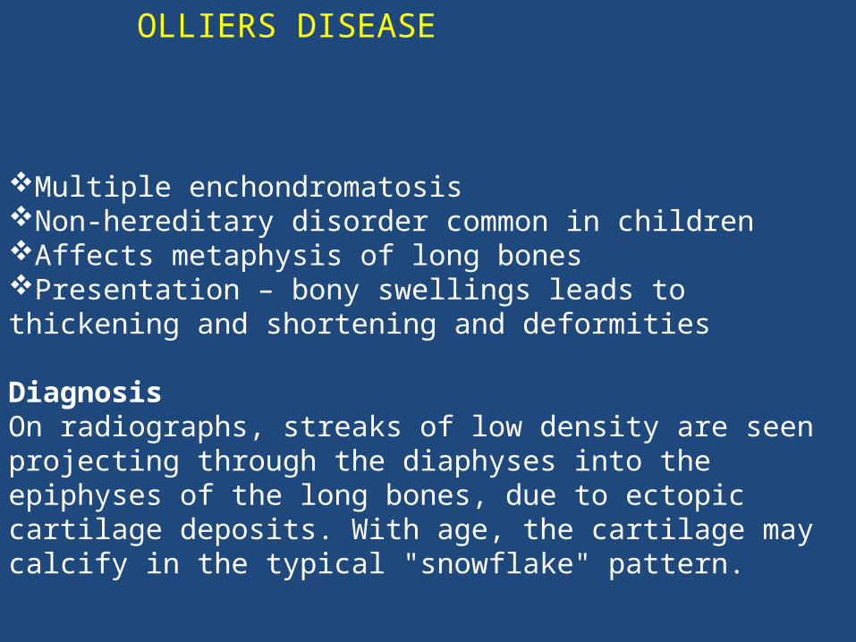
OLLIERS DISEASE
Multiple enchondromatosis Non-hereditary disorder common in children Affects metaphysis of long bones Presentation – bony swellings leads to thickening and shortening and deformities
DiagnosisOn radiographs, streaks of low density are seen projecting through the diaphyses into the epiphyses of the long bones, due to ectopic cartilage deposits. With age, the cartilage may calcify in the typical "snowflake" pattern.
MAFFUCCI SYNDROME • hereditary familial disease • multiple enchondroma and cavernous haemangioma
Treatment:The deformities are managed surgically to preserve the function of the limb.

OsteochondromaAlso known as: Osteocartilaginous Exostosis Cartilage capped bony projection on external surface of bone. Commonest benign tumour of bone. Lesion has its own growth plate, usually stops growing at skeletal maturity.
AGE GROUPS: first two decades
SEX PREDILECTION: M:F-1.5:1
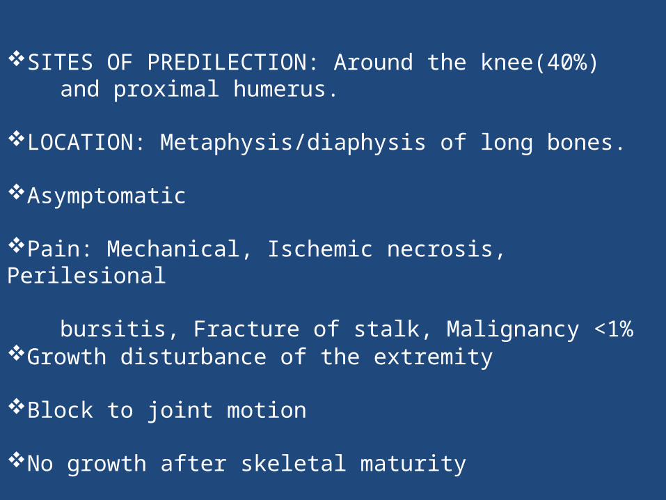
SITES OF PREDILECTION: Around the knee(40%) and proximal humerus.
LOCATION: Metaphysis/diaphysis of long bones.
Asymptomatic
Pain: Mechanical, Ischemic necrosis, Perilesional
bursitis, Fracture of stalk, Malignancy <1%
Growth disturbance of the extremity
Block to joint motion
No growth after skeletal maturity
X RAY Pedunculated / sessile – exophytic metaphysis / diaphysis Marrow and cortices of lesion continuous with bone Directed away from growing end Cartilage cap not seen on x ray
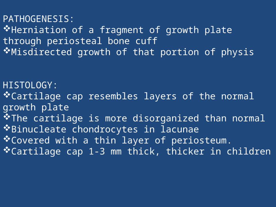
PATHOGENESIS: Herniation of a fragment of growth plate through periosteal bone cuff Misdirected growth of that portion of physis
HISTOLOGY: Cartilage cap resembles layers of the normal growth plate The cartilage is more disorganized than normal Binucleate chondrocytes in lacunae Covered with a thin layer of periosteum. Cartilage cap 1-3 mm thick, thicker in children
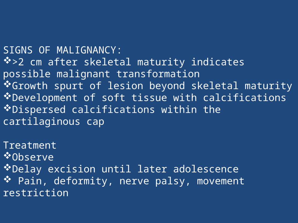
SIGNS OF MALIGNANCY: >2 cm after skeletal maturity indicates possible malignant transformation Growth spurt of lesion beyond skeletal maturity Development of soft tissue with calcifications Dispersed calcifications within the cartilaginous cap
Treatment Observe Delay excision until later adolescence Pain, deformity, nerve palsy, movement restriction
HEREDITARY MULTIPLE EXOSTOSES (H.M.E)
Also known as: Multiple Exostoses, Diaphyseal aclasis Autosomal dominant hereditary disorder, 10% no family history. EXT1,2,3 genes Knees, ankles and shoulders are most frequently affected. Knobby appearance, Short stature Forearm deformity, Tibio-fibular synostosis, Genu valgum, Coxa valga
Rx - Excision of symptomatic exostosis Correction of deformity and limb length discrepancy
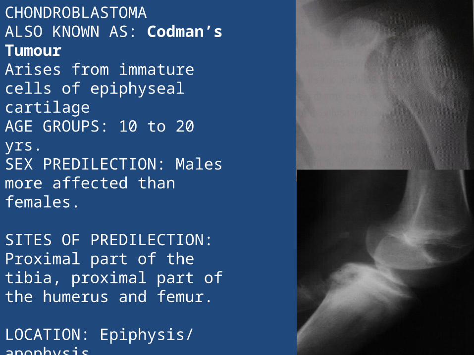
CHONDROBLASTOMA ALSO KNOWN AS: Codman’s Tumour Arises from immature cells of epiphyseal cartilage AGE GROUPS: 10 to 20 yrs. SEX PREDILECTION: Males more affected than females.
SITES OF PREDILECTION: Proximal part of the tibia, proximal part of the humerus and femur.
LOCATION: Epiphysis/ apophysis
SYMPTOMS: Pain and local swelling of joint without h/o trauma
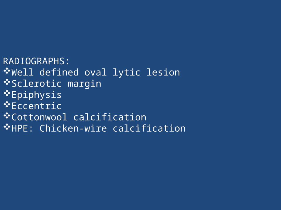
RADIOGRAPHS: Well defined oval lytic lesion Sclerotic margin Epiphysis Eccentric Cottonwool calcification HPE: Chicken-wire calcification
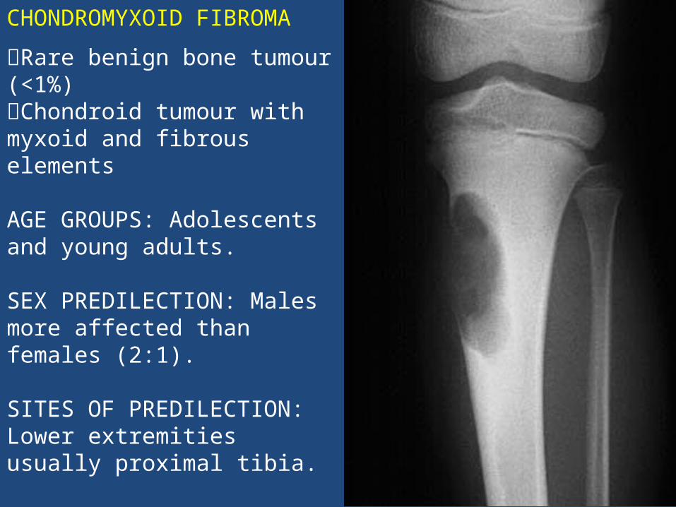
CHONDROMYXOID FIBROMA
Rare benign bone tumour (<1%) Chondroid tumour with myxoid and fibrous elements
AGE GROUPS: Adolescents and young adults.
SEX PREDILECTION: Males more affected than females (2:1).
SITES OF PREDILECTION: Lower extremities usually proximal tibia.
LOCATION: Metaphysis.
SYMPTOMS: Peripherally located mass with local pain and swelling.
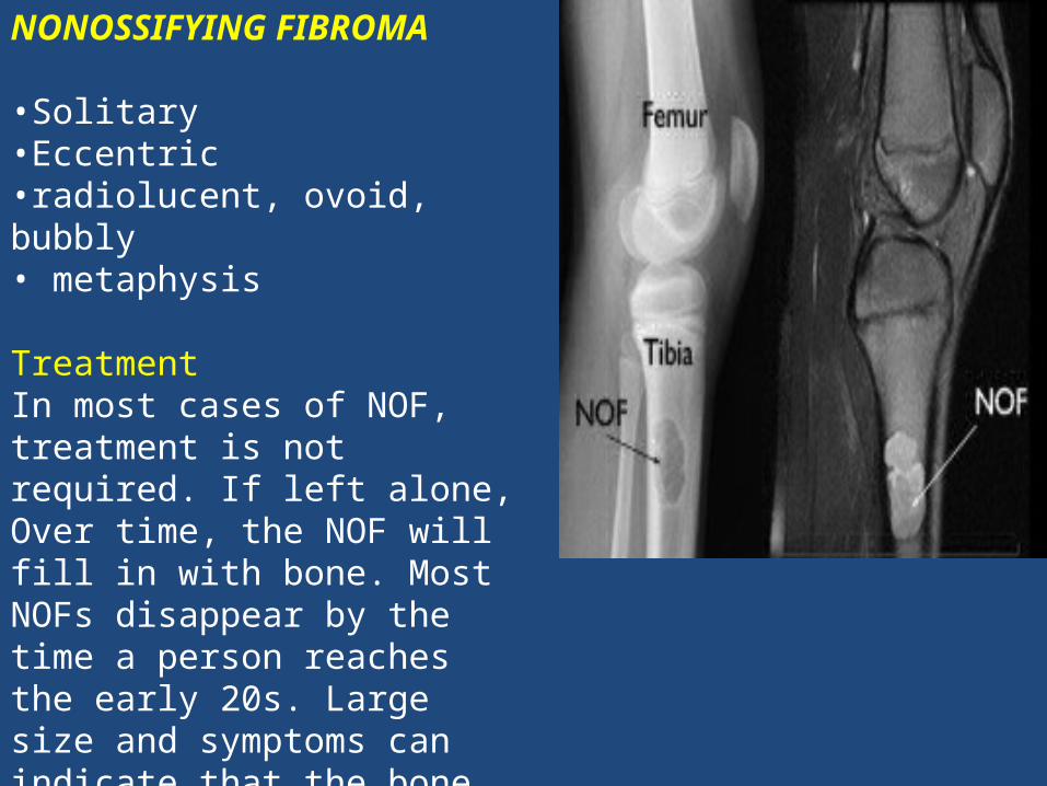
NONOSSIFYING FIBROMA
•Solitary •Eccentric •radiolucent, ovoid, bubbly • metaphysis
TreatmentIn most cases of NOF, treatment is not required. If left alone, Over time, the NOF will fill in with bone. Most NOFs disappear by the time a person reaches the early 20s. Large size and symptoms can indicate that the bone is weak and is likely to fracture, even with very little force.
CurettageThe most common method of treating NOFs is with curettage.
Bone GraftAfter curettage, fill the hole with a bone graft . You may also use a bone cement mixture to fill the hole.
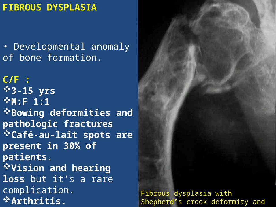
FIBROUS DYSPLASIA
• Developmental anomaly of bone formation.
C/F : 3-15 yrsM:F 1:1Bowing deformities and pathologic fracturesCafé-au-lait spots are present in 30% of patients.Vision and hearing loss but it's a rare complication.Arthritis. Cancer. Fibrous dysplasia with Shepherd‟s crook
deformity and pathological fracture
TYPES:Monostotic fibrous dysplasia (solitary lesion): 70-80% Polyostotic fibrous dysplasia (multiple bones)20-30% McCune Albright syndrome: Polyostotic fibrous dysplasia, Endocrine dysfunction: precocious puberty, hyperthyroidism, Café-au-lait spots (coast of Maine)
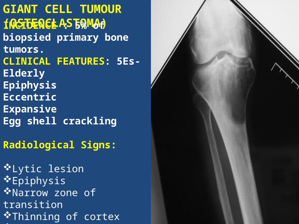
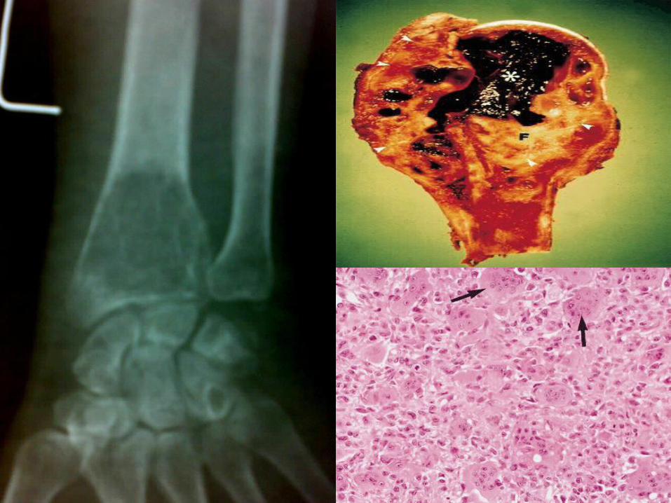
GIANT CELL TUMOUR (OSTEOCLASTOMA) INCIDENCE : 5% of biopsied primary bone tumors. CLINICAL FEATURES: 5Es- Elderly Epiphysis Eccentric Expansive Egg shell crackling
Radiological Signs:
Lytic lesion Epiphysis Narrow zone of transition Thinning of cortex Honey comb appearance Soap bubble appearance
35
Radiological grading(campanacci)
• Grade1(Quiescent): well defined margin with surrounding sclerosis; cortex: slightly thinned
but not deformed• Grade2(Active): well defined margin, lack
sclerosis; cortex-thinned and expanded• Grade3(Aggressive): ill defined margin, often
with cortical destruction and soft tissue extension
36
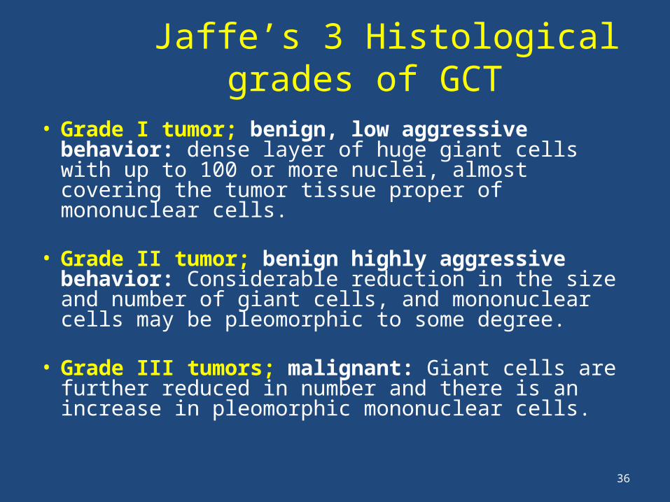
Jaffe’s 3 Histological grades of GCT
• Grade I tumor; benign, low aggressive behavior: dense layer of huge giant cells with up to 100 or more nuclei, almost covering the tumor tissue proper of mononuclear cells.
• Grade II tumor; benign highly aggressive behavior: Considerable reduction in the size and number of giant cells, and mononuclear cells may be pleomorphic to some degree.
• Grade III tumors; malignant: Giant cells are further reduced in number and there is an increase in pleomorphic mononuclear cells.
DIFFERENTIAL DIAGNOSIS:
GCT has to be differentiated from giant cell variants. Unicameral bone cyst Aneurysmal bone cyst Non-ossifying fibroma Chondroblastoma GCT of hyperparathyroidism (Brown tumor)
39
Malignancy in GCT
Histology: high grade spindle cell sarcoma with or without osteoid
Vacular invasion, soft tissue extension & high mitotic activity do not establish malignancy
No residual giant cell tumorPrimary cases: conventional GCT presentPrognosis of primary better than secondary ones
40
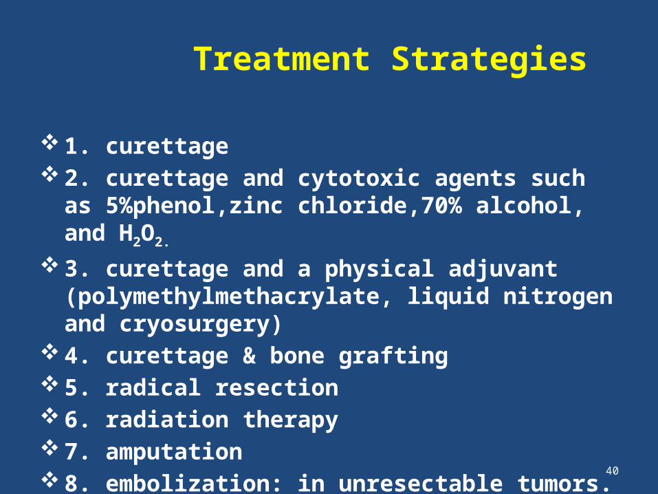
Treatment Strategies
1. curettage 2. curettage and cytotoxic agents such as 5%phenol,zinc
chloride,70% alcohol, and H2O2.
3. curettage and a physical adjuvant (polymethylmethacrylate, liquid nitrogen and cryosurgery)
4. curettage & bone grafting 5. radical resection 6. radiation therapy 7. amputation 8. embolization: in unresectable tumors.
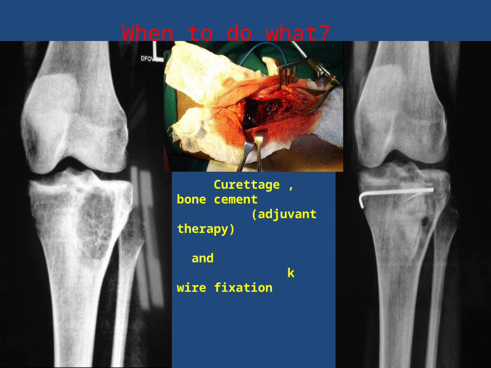
When to do what?
Curettage , bone cement (adjuvant therapy) and k wire fixation
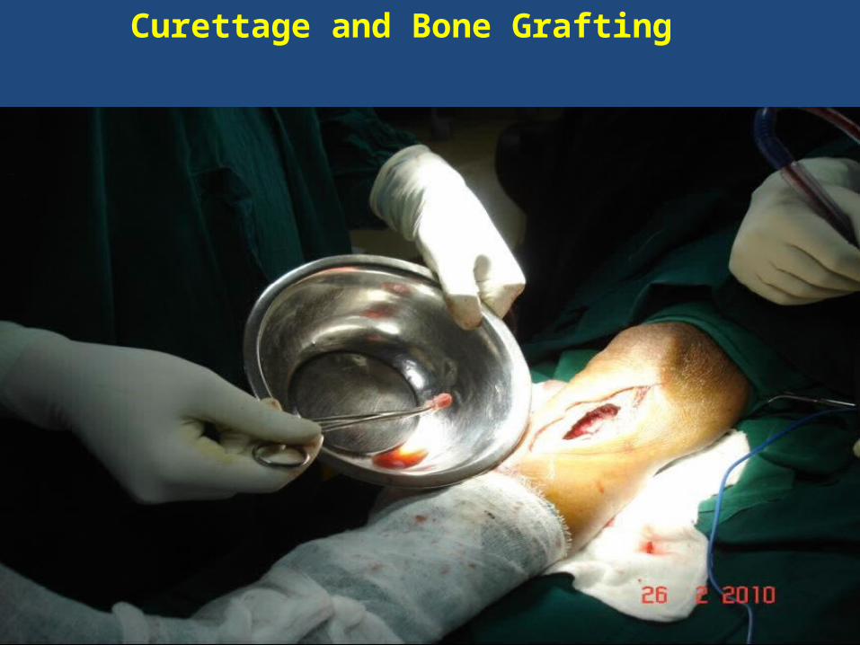
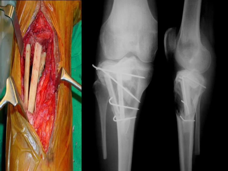
Curettage and Bone Grafting
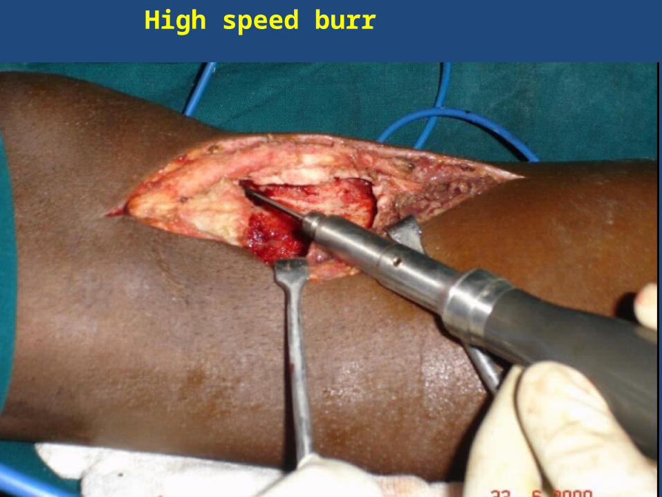
High speed burr
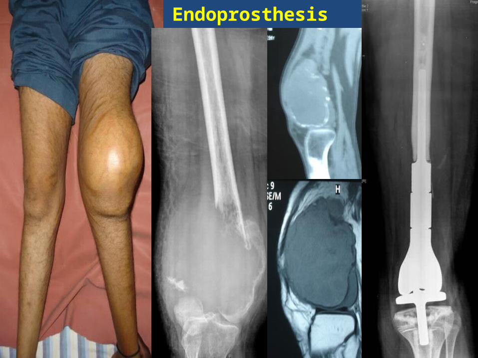
Endoprosthesis
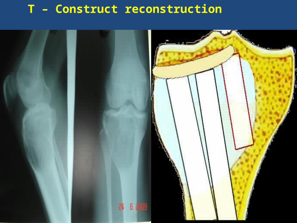
T – Construct reconstruction

47
Thank you !