Embed Size (px)
Citation preview

Pressure sores in Port Harcourt Teaching Hospital Gbeneol and Nwachukwu ISSN -
1597-4292
The Nigerian Health Journal, Volume 21 No 1, January to March 2021 www.tnhjph.com
A Publication of Nigerian Medical Association, Rivers State, Nigeria
A RETROSPECTIVE STUDY OF THE PREVALENCE OF PRESSURE SORES: THE UNIVERSITY OF PORT HARCOURT TEACHING HOSPITAL EXPERIENCE
Gbeneol1*, TJ Nwachukwu, AC2
1Dr Tombari J. Gbeneol. Consultant Plastic Surgeon. Department of Surgery Faculty of Clinical Sciences, College of Health Sciences, University of Port Harcourt, Nigeria. 2Dr Adaeze C. Nwachukwu. Senior Registrar, Plastic Surgery Division, Department of Surgery, University of Port Harcourt Teaching Hospital, Port Harcourt, Nigeria.
*Corresponding Author: Dr Tombari Gbeneol. Plastic Division, Department of SurgeryFaculty of Clinical Sciences, University of Port Harcourt, Nigeria. +234(0)8033426025.Email: [email protected].
ABSTRACT Background: Bed sores occur when there is friction or unrelieved pressure on the soft tissue skin, subcutaneous, muscle against a hard surface or against a bony prominence. It produces as a secondary medical condition; a problematic wound prolonging patients duration of hospitalization with its attendant morbidity and mortality and a huge financial burden. It is a global challenge that this study will aid its early identification, prevention and treatment reducing its morbidity and mortality and financial costs. Aim: The aim of this study is to determine the prevalence and impact of pressure sores in patients admitted to the University of Port Harcourt Teaching Hospital, Nigeria. Methods: The data were obtained retrospectively from 65 patients’ record at the University of Port Harcourt Teaching Hospital, Port Harcourt, spanning from 2014 to 2019. The age ranges between 14 to 95 years. The data was analysed and results presented as frequency tables and pictures. Results: The results showed pressure sore occurrence in University of Port Harcourt
Teaching Hospital, Port Harcourt with more cases in Pott’s Disease of the spine (TB), (27.45%) followed by Cerebrovascular Accident (CVA) (19.61%) and Sickle cell Disease (SCD) (17.65%). Pressure sores occurring at sacral and trochanteric sites were mostly wounds from SCD, Pott’s and CVA. Sacral pressure sore site was the most predominant with percentage frequency of 75%. This was followed by trochanteric pressure sore site with a frequency of 12.5%. Pressure sore development is aggravated by several factors such as poor nutrition, incontinence with persistent soilage and moisture, dementia, paralysis, friction and shear forces. Prolong stay in a particular position and age are real contributory factors. Conclusion: Pressure sores are common and measures should be taken to quickly detect and prevent pressure sore as it has huge financial burden and prolonged hospital stay and its attendant nosocomial infection.
Key words: pressure sore, occurrence, nosocomial infection, University of Port Harcourt Teaching Hospital, Port Harcourt.
34

Pressure sores in Port Harcourt Teaching Hospital Gbeneol and Nwachukwu ISSN -
1597-4292
The Nigerian Health Journal, Volume 21 No 1, January to March 2021 www.tnhjph.com
A Publication of Nigerian Medical Association, Rivers State, Nigeria
INTRODUCTION Pressure sore, which is also called pressure ulcer or bed sores occur when there is unrelieved pressure on the soft tissue skin, subcutaneous, muscle against a hard surface or against a bony prominence. It is generally accepted that pressure sore are causally related to the effects of three tissue forces – pressure, shear and friction. Pressure can result in decreased tissue perfusion and ischemia. Tissue necrosis can be observed where there is unrelieved pressure or ischemia potentiated by host tissue compromise such as chronic medical conditions, protein-energy malnutrition and/or sepsis. Shear forces can cause a change in the angle of the vessels and compromise blood supply, resulting in ischemia, cellular death, and tissue necrosis. Friction is the adherent force resisting shearing movement of the skin, which may result in denuded area of the dermis through repeated epidermal shedding or avulsion of sheets of epidermis. Therefore, prolonged exposure of this tissue injury to moisture from perspiration, urinary or faecal incontinence, or wound exudate will further weaken the intercellular bonds in the epidermal layers, causing maceration and epidermal ulceration.1
When tissue is injured, there is a de-margination and influx of cells responsible for inflammation. For pressure sores to heal, a series of events unfolds including vaso-constriction/vasodilatation, coagulation, influx of pro-inflammatory cells like neutrophils, and macrophages, and finally, matrix formation and maturation.
In chronic wounds, there is breakdown of this sequence due to imbalance of
metalloprotease tissue imbalance metalloprotease. Of specific interest is the ratio of MMP-1 and MMP-9 to TIMP-1 and TIMP-2. In pressure sores the ratio of MMP-9: TIMP-1 is higher in chronic pressure sores, and interestingly decreases as those wounds heal.2
There are common grounds of agreement regarding the basic mechanism of tissue injury in pressure sore formation, each patient is unique and individual differences must be considered when evaluating and treating these wounds. One single mechanism is unlikely to be proven, which is the probable overlap and interplay between the described compromising forces and factors that are both extrinsic and intrinsic that continue to define pressure sore development and deterioration.3
The impact of pressure ulcers is significant both in terms of financial and non-monetary costs. Beckrich and Aronovich4 reported that 1.6 million pressure ulcers develop in hospitals in the United States annually, with an estimated yearly cost of $2.2 to $3.6 billion. Epidemiologically, approximately 9% of all hospital patients develop pressure sores. The occurrence level in acute care is as high as 11%5. Other diseases such as cardiovascular disease, acute neurologic disease and orthopaedic injury have been associated with pressure sore.5 Fisher et al6, reported that using Braden scale for prediction, key risk factors in acute care pressure sore development were identified. Factors like age, male gender, impaired sensory perception, moisture, immobility, poor nutrition, and friction/shear. The aim of this study was to analyze data collected from University of Port Harcourt
35

Pressure sores in Port Harcourt Teaching Hospital Gbeneol and Nwachukwu ISSN -
1597-4292
The Nigerian Health Journal, Volume 21 No 1, January to March 2021 www.tnhjph.com
A Publication of Nigerian Medical Association, Rivers State, Nigeria
Teaching Hospital, on pressure sore occurrence and its impact on wound care in Nigeria. Reconstructive options using flaps depend on the site on principles of enblock wound excision, ostectomy, use of drain and durable supple tissue cover.
MATERIALS AND METHODS The data were obtained retrospectively from 65 patients’ record at the University of Port Harcourt Teaching Hospital, Port Harcourt, spanning from 2014 to 2019. The age ranges between 14 to 95 years. The data was tabulated and analysed using frequency and percentage distribution and expressed in tables. The anatomical site, size or wound dimensions of the pressure sore were noted. Pressure ulcer score or grading as documented (that is, depth, length and breadth of wound) were also recorded. Presence of slough, exudates, microorganisms and by products of microorganisms (toxins, enzymes, membranes, phagocytosed products, biofilms) wound culture was also considered. The age of the ulcer and
underlying medical conditions, method of closure following the principles of pressure sore surgery (Osteotomy and filing of bony prominence, enblock excision of wound, use of drain and skin or myo-cutaneous skin flap) were strictly followed.
RESULTS Table 1 showed pressure sore occurrence in University of Port Harcourt Teaching Hospital, Port Harcourt with more cases with Pott’s Disease of the spine (TB) patients (27.45%), followed by Cerebrovascular Accident (CVA) (19.61%) and Sickle cell Disease (SCDx) (17.65%). Most patients were males and married except cases with breast cancer, ovarian cancer. Pressure sores occurring at sacral and trochanteric sites were mostly wounds from SCDx, Pott’s disease of the spine and CVA as shown in Table 2. Sacral pressure sore site was the most predominant with percentage frequency of 75%. This was followed by trochanteric pressure sore sites with percentage frequency of 12.5%.
Table 1: Pressure sore percentage occurrence amongst Nigerians who received treatment at
the University of Port Harcourt Teaching Hospital
Type of wound Sex Frequency Percentage of
Occurrence
%
Marital Status
males Females Married Single
Sickle Cell Disease
(SCDx)
9 - 9 17.65 3 6
Pott’s Disease of the
Spine
13 1 14 27.45 8 6
36

Pressure sores in Port Harcourt Teaching Hospital Gbeneol and Nwachukwu ISSN -
1597-4292
The Nigerian Health Journal, Volume 21 No 1, January to March 2021 www.tnhjph.com
A Publication of Nigerian Medical Association, Rivers State, Nigeria
Cerebrovascular
Accident
(CVA)
9 1 10 19.61 10 -
Polytraumatised 2 1 3 5.88 2 1
Frailty and Sepsis 1 3 4 7.84 4 -
HIV 1 - 1 1.96 1 -
Burns 1 1 2 3.92 1 1
Multiple Myeloma 1 1 1.96 1
Breast
Cancer/Ovarian/colon
cancer
1 4 5 9.80 5
Other Tuberculosis
Infection
1 1 1.96 - 1
Necrotising Fascities - 1 1 1.96 - 1
Table 2: percentage frequency of pressure sore sites with different associated medical
conditions
FREQUENCIES
SC
Dx
Pot
t’s
CV
A
Poly-
Traumatised
Frailty
and
Sepsis
HI
V
Burns Multipl
e
Myelom
a
Breast
Cancer
/Ovaria
n/colon
cancer
Pott’s
Disea
se
Necro
tising
Fascit
ies
Tota
l
Freq
%
Pressur
e Sore
Sites
37

Pressure sores in Port Harcourt Teaching Hospital Gbeneol and Nwachukwu ISSN -
1597-4292
The Nigerian Health Journal, Volume 21 No 1, January to March 2021 www.tnhjph.com
A Publication of Nigerian Medical Association, Rivers State, Nigeria
Sacral 9 14 10 2 2 1 2 1 5 2 1 48 75
Trocha
nteric
3 2 3 - 1 - - - - - - 8 12.5
Leg - - - 1 - - - - - - - 1 1.56
Foot - - - 1 - - - - - - - 1 1.56
Scalp - 3 - - - - - - - - - 3 4.68
Ear - - - 1 - - - - - - - 1 1.56
Back - 1 - - - - - - - - - 1 1.56
Left
Ischial-
1 - - - - - - - - - - 1 1.56
SCDx- Sickle Cell Disease
Figures 1 and 2 show pressure sores at the sacral site.
Figure1 : Pressure sore at sacrum
38

Pressure sores in Port Harcourt Teaching Hospital Gbeneol and Nwachukwu ISSN -
1597-4292
The Nigerian Health Journal, Volume 21 No 1, January to March 2021 www.tnhjph.com
A Publication of Nigerian Medical Association, Rivers State, Nigeria
Figure 2: Pressure sore treatment at sacral site using gluteal skin rotation flap.
Table 3: Ages of patients, wound types and location of sores
Type of Wound Age (Years) Location of Wound
SCDx 22-63 Sacral, Trochanteric
and Left Ischial
Pott’s Disease 10-70 Sacral, Trochanteric,
Scalp, Back
CVA 41-95 Sacral, Trochanteric
Polytraumatised 27-42 Sacral, Leg, Foot, Ear
Frailty And Sepsis 86-89 Sacral, Trochanteric
HIV 33 Sacral
BURNS 20-37 Sacral
39
Pressure sores in Port Harcourt Teaching Hospital Gbeneol and Nwachukwu ISSN -
1597-4292
The Nigerian Health Journal, Volume 21 No 1, January to March 2021 www.tnhjph.com
A Publication of Nigerian Medical Association, Rivers State, Nigeria
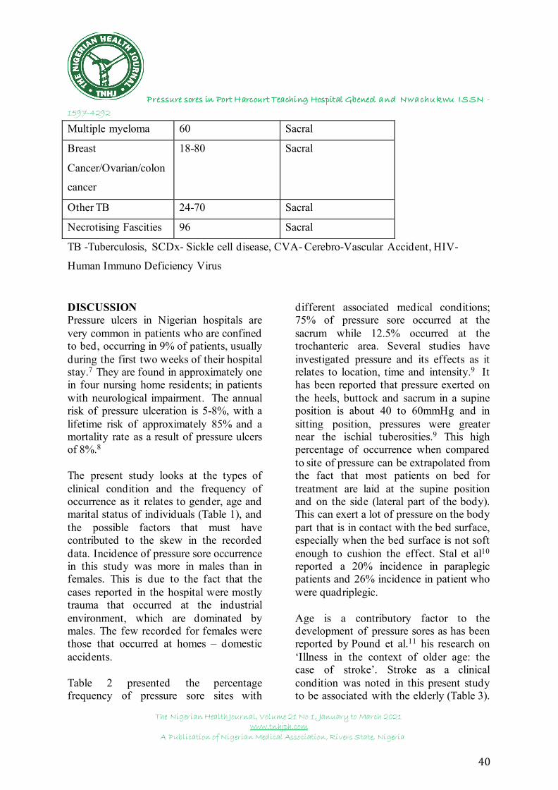
Multiple myeloma 60 Sacral
Breast
Cancer/Ovarian/colon
cancer
18-80 Sacral
Other TB 24-70 Sacral
Necrotising Fascities 96 Sacral
TB -Tuberculosis, SCDx- Sickle cell disease, CVA- Cerebro-Vascular Accident, HIV-
Human Immuno Deficiency Virus
DISCUSSION Pressure ulcers in Nigerian hospitals are very common in patients who are confined to bed, occurring in 9% of patients, usually during the first two weeks of their hospital stay.7 They are found in approximately one in four nursing home residents; in patients with neurological impairment. The annual risk of pressure ulceration is 5-8%, with a lifetime risk of approximately 85% and a mortality rate as a result of pressure ulcers of 8%.8
The present study looks at the types of clinical condition and the frequency of occurrence as it relates to gender, age and marital status of individuals (Table 1), and the possible factors that must have contributed to the skew in the recorded data. Incidence of pressure sore occurrence in this study was more in males than in females. This is due to the fact that the cases reported in the hospital were mostly trauma that occurred at the industrial environment, which are dominated by males. The few recorded for females were those that occurred at homes – domestic accidents.
Table 2 presented the percentage frequency of pressure sore sites with
different associated medical conditions; 75% of pressure sore occurred at the sacrum while 12.5% occurred at the trochanteric area. Several studies have investigated pressure and its effects as it relates to location, time and intensity.9 It has been reported that pressure exerted on the heels, buttock and sacrum in a supine position is about 40 to 60mmHg and in sitting position, pressures were greater near the ischial tuberosities.9 This high percentage of occurrence when compared to site of pressure can be extrapolated from the fact that most patients on bed for treatment are laid at the supine position and on the side (lateral part of the body). This can exert a lot of pressure on the body part that is in contact with the bed surface, especially when the bed surface is not soft enough to cushion the effect. Stal et al10
reported a 20% incidence in paraplegic patients and 26% incidence in patient who were quadriplegic.
Age is a contributory factor to the development of pressure sores as has been reported by Pound et al.11 his research on ‘Illness in the context of older age: the case of stroke’. Stroke as a clinical condition was noted in this present study to be associated with the elderly (Table 3).
40

Pressure sores in Port Harcourt Teaching Hospital Gbeneol and Nwachukwu ISSN -
1597-4292
The Nigerian Health Journal, Volume 21 No 1, January to March 2021 www.tnhjph.com
A Publication of Nigerian Medical Association, Rivers State, Nigeria
In this present study, pressure sore at the sacrum and trochanter were mostly common. Bed ridden patients, especially those with longstanding denervation tend to develop joint contractures. They occur because of tightening of both muscles and joint capsules and are common in hip flexors, contributing to the formation of trochanteric, knee, and ankle ulcers.8
Skin over locations or sites of pressure sore are often affected due to proliferation of bacteria.12 Robson et al13 reported that incision created in areas of applied pressure and innoculation with known concentrations of organism allowed for a 100-fold greater bacterial growth than in areas not subjected to pressure. The increased bacterial count which promoted or necessitated pressure sore development may be due to impaired lymphatic function, ischemia and impaired immune function, which was the cardinal area of the study on molecular basis of chronic wounds.14 Reports have also shown that absence of oxygen for skin cells over several days can cause widespread ischaemic necrosis and ulceration of the skin.15,16
Furthermore, pressure sore occurs when there is a breakdown in the sequence cascades of events that lead to wound healing; and as rightly put the ratio of matrix metalloprotease-9 (MMP) to tissue inhibitors of matrix metalloprotease-1 (TIMMP) is higher in chronic pressure sore. In normal wound healing, MMPs play an indispensable role in cellular migration by cleaving proteins to clear a path for migrating cells, cleaving intracellular binding proteins, and promoting the release of signalling proteins that promote this migration.
TIMMPs, on the other hand, binds to activated proteases to presumably protect uninvolved matrix and protect cell surface adhesion molecules as the wounds mature.17
In some cases, as pressure increases, plasma extravasation occurs, leading to oedema formation due to denervation and compression of the underlying skin. Further denervation causes loss of sympathetic tone of the blood vessel, leading to vasodilatation, creating greater engorgement of the vessels and more oedematous site. At a molecular level oedematous site is brought about by the disruption of the homeostatic balance between prostaglandin F2a (PGF2a) and prostaglandin E2a (PGE2a), in favour of PGE2a which leaks through the cell membrane, leading to increased interstitial fluid accumulation.
Pressure sore incidence in patients with spinal injuries can be reduced by the use of an improvised waterbed as explained.18 In their report, they posited that waterbed could form a very important local alternative in reducing the debilitating morbidity and mortality from pressure ulcers in long-confined hospital patients with spinal injuries. In our study spinal injury formed majority of cases reported in University of Port Harcourt Teaching Hospital, Nigeria (Table 3), hence such a very affordable and much less expensive than the standard waterbed should be put into consideration.
CONCLUSION Pressure sore development is caused by several factors such as poor nutrition, incontinence with persistent soilage and moisture, dementia, paralysis, friction and shear. Prolonged stay in a particular
41

Pressure sores in Port Harcourt Teaching Hospital Gbeneol and Nwachukwu ISSN -
1597-4292
The Nigerian Health Journal, Volume 21 No 1, January to March 2021 www.tnhjph.com
A Publication of Nigerian Medical Association, Rivers State, Nigeria
position and age are all contributory factors. Measures should be taken to quickly detect and prevent pressure sore as it complicates the primary problem and has a huge financial burden.
REFERENCES:
1. Maklebust J. Pressure Ulcers:Etiology and Prevention. NursClinNorth Am., 1987; 22(2): 359-377.
2. Elizabeth A Ayello , CarolineDowsett, Gregory S Schultz, RGary Sibbald, Vincent Falanga,Keith Harding. Time Heals all Wounds. Nursing. 2004;34(4): 36-41. doi:10.1097/00152193-200404000-00040.
3. Niezgoda JA and Mendez-EastmanS. The Effective Management ofPressure Ulcers.Advances in Skin& Wound Care: The Journal forPrevention and Healing. 2006;19(1): 3-15.
4. Beckrich K and Aronovitch S.Hospital-acquired pressure ulcers:A comparison of costs in medicalvs. surgical patients. Nursingeconomics 1999; 17(5):263-71.
5. Bright RA and Brown SL. Medicaldevice epidemiology. In: Medicaldevice epidemiology andsurveillance. Brown SL, BrightRA, Tavris DR, eds. Chichester: John Wiley & Sons, 2007.
6. Premkumar K. Decubitus Ulcers.Pathophysiology and the role ofmassage therapists. Massage andBodywork 2005;100–104.
7. Fisher AR, Well G, Harrison MB.Factors associated with pressureUlcer in adults in acute careHospitals. Adv Skin Wound Care2004; 17: 80.
8. Onigbinde AT, Olafimihan KF,Ojoawo A, Mothabeng J,Ogundiran OO. Management ofdecubitus ulcer using gentamycinsulphateiontophoresis: a casestudy. The Internet Journal ofAllied Health Sciences and Practice2011; 9(1): 1540–80.
9. Dharmarajan TS, Ahmed S. Thegrowing problem of pressureulcers. Evaluation and managementfor an aging population. PostgradMed. 2003; 113(5):77–90.
10. Stal S, Serure A, Donovan W. Theperioperative Management of thepatient with pressure sores. AnnPlast Surg. 1983; 11:347.
11. Pound P, Gompertz P and EbrahimS. Illness in the context of olderage: the case of stroke.Sociology ofHealth and Illness 1998; 20:489–506.
12. Daniel RK, Terzis JK,Cummingham. Sensory Skin Flapsfor Coverings of Pressure Sores inparaplegic patients. A preliminaryReport. Plast Reconstr Surg. 1976;58:317
13. Robson MC, Duke WF and KrizekTJ. Rapid Bacterial Screening inthe Treatment of Civilian Wounds.J Surg Res. 1973; 14: 426-430.
14. Nwomeh BC, Yager DR, ColdenIK. Physiology of the ChronicWound. ClinPlast Surg. 1998; 25:341.
15. Daniel Bluestein, Ashkan Javaheri.Pressure Ulcers: Prevention,Evaluation, and Management. AmFam Physician. 2008 ;78(10):1186-1194.
16. Whittington K, Patrick M, Roberts JL.A national study of pressure ulcerprevalence and incidence in acute
42

Pressure sores in Port Harcourt Teaching Hospital Gbeneol and Nwachukwu ISSN -
1597-4292
The Nigerian Health Journal, Volume 21 No 1, January to March 2021 www.tnhjph.com
A Publication of Nigerian Medical Association, Rivers State, Nigeria
care hospitals. J Wound Ostomy Continence Nurs. 2000;27(4):209–215.
17. Ladwig GP, Robson MC, Liu R(2002). Ratios of Activated MatrixMetalloproteinase-9 to TissueInhibitor of Metalloproteinase-1 inWound Fluids are inverselycorrelated with healing of pressureulcers. Wound Repair Regen.2002; 10: 26.
18. Emejulu Jude-Kennedy C, Nwadi UV, Obiegbu HO. DoesImprovised Waterbed Reduce theIncidence of Pressure Ulcers inPatients with Spinal Injury?Nigerian Journal of Surgery 2015;21(2): 119-123.
43