Embed Size (px)
Citation preview

BRAINA JOURNAL OF NEUROLOGY
Post-surgical inflammatory neuropathyNathan P. Staff,1 JaNean Engelstad,1 Christopher J. Klein,1 Kimberly K. Amrami,2
Robert J. Spinner,3 Peter J. Dyck,1 Mark A. Warner,4 Mary E. Warner4 and P. James B. Dyck1
1 Department of Neurology, Mayo Clinic, Rochester, MN 55905, USA
2 Department of Radiology, Mayo Clinic, Rochester, MN 55905, USA
3 Department of Neurologic Surgery, Mayo Clinic, Rochester, MN 55905, USA
4 Department of Anaesthesiology, Mayo Clinic, Rochester, MN 55905, USA
Correspondence to: P. James B. Dyck, MD,
Department of Neurology,
Mayo Clinic 200 First Street SW,
Rochester, MN 55905, USA
E-mail: [email protected]
Post-surgical neuropathies are usually attributed to mechanical factors, such as compression, stretch, contusion or transection. The
role of inflammatory mechanisms in neuropathies occurring after surgeries is poorly appreciated and not well characterized, and may
provide a rationale for immunotherapy. A total of 23 selected patients with post-surgical neuropathies received nerve biopsies, of
which 21 demonstrated increased inflammation. Here we report the clinical features in these 21 cases of biopsy-confirmed and
12 cases of clinically suspected post-surgical inflammatory neuropathies, in whom no trauma to the nerves was documented. All
neuropathies developed within 30 days of a surgical procedure. Of 33 patients, 20 were male and the median age was 65 years (range
24–83). Surgical procedures were orthopaedic (n = 14), abdominal/pelvic (n = 12), thoracic (n = 5) and dental (n = 2). Patients de-
veloped focal (n = 12), multifocal (n = 14) or diffuse (n = 7) neuropathies. Focal and multifocal neuropathies typically presented with
acute pain and weakness, and focal neuropathies often mimicked mechanical aetiologies. Detailed analyses, including clinical
characteristics, electrophysiology, imaging and peripheral nerve pathology, were performed. Electrophysiology showed axonal
damage. Magnetic resonance imaging of roots, plexuses and peripheral nerves was performed in 22 patients, and all patients had
abnormally increased T2 nerve signal, with 20 exhibiting mild (n = 7), moderate (n = 12) or severe (n = 1) enlargement. A total of
21 patients had abnormal nerve biopsies that showed increased epineurial perivascular lymphocytic inflammation (nine small, five
moderate and seven large), with 15 diagnostic or suggestive of microvasculitis. Evidence of ischaemic nerve injury was seen in
19 biopsies. Seventeen biopsies had increased axonal degeneration suggesting active neuropathy. Seventeen biopsied patients were
treated with immunotherapy. In 13 cases with longitudinal follow-up (median 9 months, range 3–71 months), the median neur-
opathy impairment score improved from 30 to 24 at the time of last evaluation (P = 0.001). In conclusion: (i) not all post-surgical
neuropathies are mechanical, and inflammatory mechanisms can be causative, presenting as pain and weakness in a focal, multifocal
or diffuse pattern; (ii) these inflammatory neuropathies may be recognized by their spatio-temporal separation from the site and time
of surgery and by the characteristic magnetic resonance imaging features; (iii) occasionally post-surgical inflammatory and mech-
anical neuropathies are difficult to distinguish and nerve biopsy may be required to demonstrate an inflammatory mechanism, which
in our cohort often, but not exclusively, exhibited pathological features of microvasculitis and ischaemia; and (iv) recognizing the role
of inflammation in these patients’ neuropathy led to rational immunotherapy, which may have resulted in the subsequent improve-
ment of neurological symptoms and impairments.
Keywords: peripheral neuropathy; inflammation; post-surgical; autoimmunity; microvasculitis
Abbreviations: NIS = Neuropathy Impairment Score
doi:10.1093/brain/awq252 Brain 2010: 133; 2866–2880 | 2866
Received June 17, 2010. Revised August 2, 2010. Accepted August 3, 2010. Advance Access publication September 15, 2010
� The Author (2010). Published by Oxford University Press on behalf of the Guarantors of Brain. All rights reserved.
For Permissions, please email: [email protected]
Dow
nloaded from https://academ
ic.oup.com/brain/article/133/10/2866/325481 by guest on 17 D
ecember 2021

IntroductionPeripheral nerve damage following a surgical procedure is a
well-known clinical problem usually attributed to the mechanical
forces of stretch, compression, contusion or transection. These
neuropathies may result in prolonged patient impairments.
Furthermore, they constitute a significant medico-legal problem
for both anaesthesiologists and surgeons, and are a common con-
sultation for neurologists (Cheney et al., 1999). Conservative
management is customarily recommended in these cases or
often surgical nerve repair is attempted (Dawson and Krarup,
1989).
Occasionally, it is difficult to explain a post-surgical neuropathy
by mechanical forces because it is either spatially or temporally
segregated from the surgery. In these cases, inflammatory or auto-
immune aetiologies need to be considered, which if proven may
provide patients with opportunities for immune-based therapies.
Inflammatory or immune-mediated post-surgical neuropathy is not
a well-described phenomenon. Guillain–Barre syndrome has rarely
been reported following surgeries (Wiederholt et al., 1964;
Arnason and Asbury, 1968), with endoneurial inflammatory infil-
tration on autopsy in some cases. Several series on idiopathic bra-
chial plexopathy have described antecedent surgeries as possible
triggers (Parsonage and Turner, 1948; Malamut et al., 1994; van
Alfen and van Engelen, 2006), but none of these series included
pathological correlation. Furthermore, when a brachial plexopathy
follows a surgery in the vicinity of the brachial plexus (e.g. cardiac
surgery), it is often assumed that mechanical factors are respon-
sible (Lederman et al., 1982).
We describe the clinical, electrophysiological, imaging and
pathological features in a series of patients seen at the Mayo
Clinic who developed post-surgical neuropathies that were usually
difficult to explain based on mechanical factors. We confirmed the
inflammatory nature of the process by performing nerve biopsies
in the involved segment and then treating non-improving patients
with immunotherapy.
Materials and methods
Patient selectionThis study was approved by the Mayo Clinic Institutional Review
Board (IRB#08-006829). Patients were considered for inclusion in the
study if they developed a peripheral neuropathy within 30 days of a
surgical procedure and no documented nerve trauma was recognized.
A total of 27 patients were identified through the routine practice
in the Department of Neurology at the Mayo Clinic, Rochester
(2001–10). The remainder of patients was identified retrospectively
through the Mayo Electronic Record and the Peripheral Nerve
Laboratory database (1999–2009). Subjects were divided into two
groups that either did or did not have a nerve biopsy performed,
which were termed biopsy-confirmed or clinically suspected
post-surgical inflammatory neuropathy, respectively. In order to be
included in the clinically suspected post-surgical inflammatory neur-
opathy group (no biopsy), the patients either clearly developed neur-
opathy outside of the immediate post-operative period or their
neuropathy was spatially remote from the surgical area. In order to
be included in the biopsy-confirmed post-surgical neuropathy group, a
neurologist had to have suspected an inflammatory cause and a nerve
biopsy showing inflammation was obtained. In the process of identify-
ing patients for the study, only two additional patients were identified
who developed post-surgical neuropathy and underwent a nerve
biopsy that did not show prominent inflammation, and these two pa-
tients were not included in this study. The surgical records, including
anaesthesia and blood transfusions, were reviewed by an anaesthesi-
ologist (M.E.W.).
Scoring of neuropathy, severity anddisabilityData were collected on patients via chart review, with 30 of the
33 patients having been seen personally by at least one of the authors
(N.P.S., C.J.K., P.J.D. and P.J.B.D.). Neuropathy severity was quanti-
fied using the Neuropathy Impairment Score (NIS) (Dyck et al., 1980),
which is a linear scale of weakness, sensory loss and hyporeflexia,
and is weighted heavily for weakness (Dyck et al., 2005b).
Neuropathies were categorized into focal, multifocal or diffuse pat-
terns. Focal neuropathies involved single nerves or single limbs.
Multifocal neuropathies involved multiple limbs, often sequentially.
Diffuse neuropathies involved all limbs without focality by history or
examination.
Ancillary testingPatients underwent a comprehensive evaluation that included a com-
bination of serology, CSF, neurophysiology and MRI of spinal cord
and peripheral nerve. The assessment of nerve conduction studies
and needle electromyography used methods standard for the electro-
myography laboratory at the Mayo Clinic with published references
(Daube and Rubin, 2009). Standard nerve conduction studies
for motor nerves are tibial, peroneal, ulnar, median, radial and muscu-
locutaneous. Standard sensory studies are sural, superficial peroneal,
medial plantar, median antidromic, ulnar antidromic, superficial radial,
medial antebrachial and lateral antebrachial. Quantitative sensory
testing using Computerized Assisted Sensory Examination (CASE IV)
was performed (Dyck et al., 1984). Autonomic testing was performed
using the Mayo Clinic Autonomic Reflex Screen, which assesses
cardiovagal, adrenergic and post-ganglionic sympathetic sudomotor
function via the quantitative sudomotor axon reflex test; sudomotor
function was further assessed in some patients via the thermo-
regulatory sweat test (Low, 1993). Nerve biopsy was obtained in
23 patients, 21 of whom were included in this study. The decision
to proceed with nerve biopsy was based on clinical judgement
and was often influenced by neuropathy severity, need for treatment
guidance or imaging abnormalities. In general, distal cutaneous nerve
biopsies were obtained and the nerve was processed using
standard laboratory techniques for teased nerve fibres, paraffin
sections, epoxy sections and immunohistochemistry preparations
(Dyck et al., 2005a). Nerve biopsies were scored by two of the
authors (N.P.S. and P.J.B.D.). Mayo Clinic radiology reports were
reviewed in 24 patients that received spinal cord imaging. MRI of
nerve was obtained in 22 patients and was independently interpreted
by two of the authors (N.P.S. and K.K.A.). Statistical analyses were
performed using Excel software (Microsoft, Inc.) on parametric
data sets.
Post-surgical inflammatory neuropathy Brain 2010: 133; 2866–2880 | 2867
Dow
nloaded from https://academ
ic.oup.com/brain/article/133/10/2866/325481 by guest on 17 D
ecember 2021

Results
Biopsy-confirmed post-surgicalinflammatory neuropathy
General characteristics
Twenty-one patients were identified with biopsy-confirmed post-
surgical inflammatory neuropathy (Tables 1 and 2). The median
age was 65 years (range 24–83). Eleven were female. Seven had
type 2 diabetes mellitus and none had type 1 diabetes mellitus.
Two had a prior history consistent with a mild length-dependent
peripheral neuropathy. None had a history of an autoimmune dis-
order. Four had a concurrent history of cancer (colorectal adeno-
carcinoma, renal cell carcinoma, bladder cancer and prostate
cancer). Five had a concomitant history of infection (genital
herpes, diverticulitis, oral abscess, cellulitis and thoracic osteomye-
litis). One had a family history of neuropathy. Six had more than
one of the above putative risk factors, seven had one and eight
had none. Additionally, nine were current or former smokers. In
the 19 patients where it was documented, 14 reported weight loss
of at least 10 pounds around the time of the development of
neuropathy (median 32.5 pounds; range 10–115 pounds).
A wide spectrum of surgical procedures preceded the neuropa-
thies (Table 2). Six surgeries were performed at the Mayo Clinic,
Rochester, Minnesota, and 15 surgeries were performed elsewhere
with subsequent clinical evaluation at the Mayo Clinic. Nine pa-
tients had orthopaedic procedures, eight had abdominal/pelvic
procedures, two had thoracic procedures and two had dental
procedures. Seventeen patients had prior surgeries without neuro-
logical complications, while four had no prior surgery. Fifteen
patients undergoing thoracic, abdominal/pelvic and orthopaedic
procedures received general anaesthesia consisting primarily of
volatile agent (i.e. isoflurane for 13 and sevoflurane for two pa-
tients). Two patients undergoing lower extremity orthopaedic
procedures received spinal blocks with the local anaesthetic
bupivicaine. Two patients undergoing abdominal procedures
were anaesthetized with isoflurane and received a supplemental
lumbar epidural catheter for postoperative pain management
with a combination of bupivicaine and fentanyl. Two patients
undergoing extensive dental procedures received midazolam and
fentanyl for sedation.
The median time from surgery to onset of neuropathy was two
days (range 0–30 days), with 13 patients developing neuropathy
within three days of surgery (Table 2). All patients reported an
acute or subacute onset of symptoms. All but one patient had a
monophasic illness with stabilization or recovery. A single patient
(Case 12) developed multiple attacks of neuropathy following the
initial post-surgical neuropathy. Pain was present in 18 patients,
which was described as a combination of prickling (n = 11), aching
(n = 7), cramping (n = 5), burning (n = 9), lancinating (n = 10) and
contact allodynia (n = 5). Sensory loss was present in all patients.
Limb weakness was present in 20 patients (two in an upper limb
only, 12 in a lower limb only and six had a combination). Of the
18 patients with lower limb weakness, gait aids were required at
the nadir of neurological deficit in 12 [wheelchair (n = 3), walker
(n = 4), ankle-foot orthoses (n = 5)]. Phrenic neuropathy was seen
in one patient, but no bulbar signs or symptoms were documented
in any patients. Neuropathy severity was quantified using the NIS,
with the median score being 38 (range 6–83.5) at the docu-
mented evaluation and presumed nadir of symptoms and signs.
Neuropathies were further categorized into focal, multifocal or
diffuse patterns. Focal neuropathies involved single nerves or
single limbs (six patients). Multifocal neuropathies involved mul-
tiple limbs (11 patients). Diffuse neuropathies involved all limbs
without focality by history or examination (four patients). The clinical
neuropathy patterns were lumbosacral radiculoplexus neuropathy
(10), brachial plexus neuropathy (5), sciatic mononeuropathy (5),
polyradiculoneuropathy (4), and phrenic mononeuropathy (1); four
patients had a combination of the patterns.
Ancillary testing
Laboratory studies were performed on all patients, which typically
included a broad screen for blood count/chemistries, endocrino-
pathies, inflammatory/autoimmune markers, vitamin/metabolic
deficiencies/toxicities and paraneoplastic processes (Table 3).
With this screen nine patients were found to have abnormalities.
Three patients had an elevated erythrocyte sedimentation rate
(40, 36 and 53 mm/h; normal 529 mm/h), two had an elevated
C-reactive protein (11 and 10.4 mg/dl; normal 58 mg/dl), three
Table 1 Comparisons of demographics and risk factors in biopsy-confirmed and clinically suspected post-surgicalinflammatory neuropathy
Demographics Biopsy-confirmed (n = 21) Clinically suspected (n = 12)
Female, n (%) 11 (52%) 2 (17%)
Median age at onset, years (range) 65 (24–83) 65.5 (27–76)
History of pre-existing peripheral neuropathy, n (%) 2 (9.5%) 1 (8%)
History of diabetes mellitus, n (%) 7 (33%) 2 (17%)
History of concurrent cancer, n (%) 4 (19%) 3 (25%)
History of concurrent infection, n (%) 5 (9.5%) 2 (17%)
History of smoking, n (%) 9 (43%) 5 (42%)
Concomitant weight loss, n (%) 13/19 (74%) 5/8 (63%)
NIS score at nadir (range) 38.0 (6–83.5) 15.75 (0–31)*
*P = 0.001 (two-tailed Student’s t-test); NIS = neuropathy impairment score.
2868 | Brain 2010: 133; 2866–2880 N. P. Staff et al.
Dow
nloaded from https://academ
ic.oup.com/brain/article/133/10/2866/325481 by guest on 17 D
ecember 2021

Tab
le2
Bio
psy
-confi
rmed
post
-surg
ical
infl
amm
atory
neu
ropat
hy:
pat
ient
char
acte
rist
ics,
surg
ery
types
and
clin
ical
feat
ure
s
Pat
ient
Age
atcl
inic
alev
aluat
ion
(yea
rs)
Gen
der
Tim
efr
om
surg
ery
tosy
mpto
ms
(day
s)
Anae
sthes
iaty
pe
Surg
ery
Cli
nic
aldia
gnosi
sN
erve
bio
psi
edFo
cal,
mult
ifoca
lor
dif
fuse
neu
ropat
hy
Neu
ropat
hy
nea
rsi
teof
surg
ery
Pai
nN
ISat
nad
irW
eight
loss
(pounds)
Ris
kfa
ctors
133
F1
Gen
eral
Elec
tive
abort
ion
Bila
tera
lsc
iatic
Sura
lM
uN
Y23
0I
269
M14
Gen
eral
Rig
ht
TH
ALe
ftBPN
and
mild
LSR
PN
Super
fici
alra
dia
lM
uN
Y40.5
27
DM
,PN
349
F5
24
hC
om
bin
edR
ight
radic
alnep
hre
ctom
yBila
tera
lsc
iatic
Sura
lM
uN
Y38.5
45
DM
,Ca
463
M1
Spin
alR
ight
TH
AR
ight
lum
bosa
cral
ple
xopat
hy
Super
fici
alper
onea
lFo
YY
25
20
none
565
F7
Gen
eral
Sple
nec
tom
yPoly
radic
ulo
neu
ropat
hy
Sura
lD
NY
38
ND
I
676
M5
24
hG
ener
alC
ervi
calsp
ine
dec
om
pre
ssio
nBila
tera
lBPN
Late
ralan
tebra
chia
lM
uY
Y24.5
ND
none
775
F3
Sedat
ion
Root
canal
Senso
rypoly
neu
ropat
hy
Sura
lD
NY
615
none
859
M7
Gen
eral
Ileost
om
yta
kedow
nBila
tera
lLS
RPN
with
mild
PN
Sura
lM
uN
Y68
35
DM
,Ca
980
M30
Gen
eral
Bow
elre
anas
tom
osi
sBila
tera
lLS
RPN
Sura
lM
uN
Y59
40
Ca
10
64
F1
Gen
eral
Bila
tera
lTK
ALe
ftsc
iatic
Sura
lFo
YN
30
35
DM
,FH
11
54
F21
Sedat
ion
Root
canal
Left
scia
tic
Scia
tic
fasc
icula
rFo
NN
22.2
50
I
12
78
M21
Gen
eral
CA
BG
aLe
ftBPN
,th
enright
LSR
PN
,th
enle
ftLS
RPN
Sura
lM
uN
Y52.2
50
Ca,
I
13
68
M21
Gen
eral
Ver
tebra
lbio
psy
Bila
tera
lBPN
with
phre
nic
Super
fici
alra
dia
lM
uN
Y60.2
570
I
14
65
M2
Gen
eral
Thora
cic
spin
edec
om
pre
ssio
nPoly
radic
ulo
neu
ropat
hy
Sura
lD
NY
60.2
50
DM
15
24
F1
Gen
eral
Chole
cyst
ecto
my
Bila
tera
lLS
RPN
follo
wed
by
PR
Sura
lM
uN
Y83.5
100
none
16
54
F30
Com
bin
edG
astr
icbyp
ass
Poly
radic
ulo
neu
ropat
hy
Sura
lD
NY
18
115
none
17
56
M5
24
hG
ener
alLu
mbar
spin
efu
sion
Bila
tera
lBPN
,LS
RPN
Sura
lM
uN
Y59.5
30
DM
18
45
F5
24
hSp
inal
Left
TH
ALe
ftLS
RPN
Sura
lFo
YY
910
none
19
74
F5
24
hG
ener
alR
ight
TH
AR
ight
scia
tic
Super
fici
alper
onea
lFo
YY
21
0none
20
83
F5
24
hG
ener
alLe
ftfe
mur
nai
lBila
tera
lLS
RPN
Sura
lM
uN
N51
26
none
21
70
M15
Gen
eral
Circu
mci
sion
Left
LSR
PN
Sura
lFo
NY
11.5
30
DM
,PN
BPN
=bra
chia
lple
xus
neu
ropat
hy;
Ca
=ca
nce
r;C
ABG
=co
ronar
yar
tery
byp
ass
gra
ft;
Com
bin
ed=
gen
eral
and
epid
ura
l;D
=diffu
se;
DM
=dia
bet
esm
ellit
us;
F=
fem
ale;
FH=
fam
ilyhis
tory
of
neu
ropat
hy;
Fo=
foca
l;I=
infe
ctio
n;
LSR
PN
=lu
mbosa
cral
radic
ulo
ple
xus
neu
ropat
hy;
M=
mal
e;M
u=
multifoca
l;N
=no;
ND
=not
docu
men
ted;
NIS
=neu
ropat
hy
impai
rmen
tsc
ore
;PN
=per
ipher
alneu
ropat
hy;
PR
=poly
radic
ulo
pat
hy;
TH
A=
tota
lhip
arth
opla
sty;
TK
A=
tota
lkn
eear
thro
pla
sty;
Y=
yes;
aPoly
phas
icpre
senta
tion.
Post-surgical inflammatory neuropathy Brain 2010: 133; 2866–2880 | 2869
Dow
nloaded from https://academ
ic.oup.com/brain/article/133/10/2866/325481 by guest on 17 D
ecember 2021

Tab
le3
Bio
psy
-confi
rmed
post
-surg
ical
infl
amm
atory
neu
ropat
hy:
anci
llar
yst
udie
s
Pat
ient
Surg
ery
Cli
nic
aldia
gnosi
sLa
bora
tory
studie
sQ
STEl
ectr
ophys
iolo
gy
MR
I
CM
AP
SNA
PEM
GQ
SAR
T"
T2
Enla
rged
ner
ves
Gad
oli
niu
m-
enhan
ced
1El
ective
abort
ion
Bila
tera
lsc
iatic
CSF
pro
tein
V,
C#
#Fb
,N
MU
ND
YM
od
N
2R
ight
TH
ALe
ftBPN
and
mild
LSR
PN
CR
P,
Ach
R,
CSF
pro
tein
C,
HP
##
Fb,
NM
UN
LY
Mild
N
3R
ight
radic
alnep
hre
ctom
yBila
tera
lsc
iatic
ESR
TP,
V,
C,
HP#
#Fb
,N
MU
#Y
Mod
N
4R
ight
TH
AR
ight
lum
bosa
cral
ple
xopat
hy
CSF
pro
tein
C,
HP
##
Fb,
NM
U#
YM
od
N
5Sp
lenec
tom
yPoly
radic
ulo
neu
ropat
hy
CSF
pro
tein
V,
C,
HP
##
Fb,
NM
UN
LY
Mod
N
6C
ervi
calsp
ine
dec
om
pre
ssio
nBila
tera
lBPN
NL
ND
NL
#Fb
,N
MU
ND
YM
ildN
7R
oot
canal
Senso
rypoly
neu
ropat
hy
NL
ND
##
Fb,
NM
UN
DN
DN
DN
D
8Ile
ost
om
yta
kedow
nBila
tera
lLS
RPN
with
mild
PN
CSF
pro
tein
V,
C,
HP
##
Fb,
NM
U#
YM
od
N
9Bow
elre
anas
tom
osi
sBila
tera
lLS
RPN
ESR
,p-A
NC
A,
CSF
pro
tein
ND
##
Fb,
NM
UN
DY
Mild
N
10
Bila
tera
lTK
ALe
ftsc
iatic
p-A
NC
A,
CSF
pro
tein
,C
SFW
BC
sV
,C
,H
P#
#Fb
,N
MU
#Y
Mild
N
11
Root
canal
Left
scia
tic
CSF
pro
tein
V,
C,
HA
NL
NL
NM
UN
DY
Seve
reY
12
CA
BG
aLe
ftBPN
,th
enright
LSR
PN
,th
enle
ftLS
RPN
ESR
,R
F,C
SFpro
tein
TP,
V,
C,
HP#
#Fb
,N
MU
#Y
Mod
N
13
Ver
tebra
lbio
psy
Bila
tera
lBPN
with
phre
nic
Cu,
CSF
pro
tein
V,
C#
#Fb
,N
MU
NL
ND
ND
ND
14
Thora
cic
spin
edec
om
pre
ssio
nPoly
radic
ulo
neu
ropat
hy
NL
C#
#Fb
,N
MU
#N
DN
DN
D
15
Chole
cyst
ecto
my
Bila
tera
lLS
RPN
follo
wed
by
PR
NL
ND
##
Fb,
NM
UN
DY
Mod
N
16
Gas
tric
byp
ass
Poly
radic
ulo
neu
ropat
hy
NL
VN
L#
Fb,
NM
UN
LN
DN
DN
D
17
Lum
bar
spin
efu
sion
Bila
tera
lBPN
,LS
RPN
CSF
pro
tein
TP,
V,
C,
HP#
#Fb
,N
MU
#Y
Mod
N
18
Left
TH
ALe
ftLS
RPN
NL
ND
NL
#Fb
,N
MU
ND
YM
ildN
19
Rig
ht
TH
AR
ight
scia
tic
CSF
pro
tein
TP,
V,
HP
##
Fb,
NM
UN
DY
Mod
N
20
Left
fem
ur
nai
lBila
tera
lLS
RPN
RF
ND
##
Fb,
NM
UN
DY
Mod
N
21
Circu
mci
sion
Left
LSR
PN
CR
PN
D#
#Fb
,N
MU
ND
YM
ildN
AC
hR
=neu
rom
usc
ula
rac
etyl
cholin
ere
cepto
ran
tibody;
BPN
=bra
chia
lple
xus
neu
ropat
hy;
C=
incr
ease
dco
olin
gth
resh
old
;C
ABG
=co
ronar
yar
tery
byp
ass
gra
ft;
CM
AP
=co
mpound
musc
leac
tion
pote
ntial
;C
RP
=C
-rea
ctiv
epro
tein
;C
u=
copper
;EM
G=
elec
trom
yogra
phy;
ESR
=er
ythro
cyte
sedim
enta
tion
rate
;Fb
=fibrilla
tion
pote
ntial
s;H
A=
hyp
eral
ges
ia;
HP
=in
crea
sed
hea
t-pai
nth
resh
old
;LS
RPN
=lu
mbosa
cral
radic
ulo
ple
xus
neu
ropat
hy;
Mod
=m
oder
ate;
N=
no;
ND
=not
docu
men
ted;
NL
=norm
al;
NM
U=
neu
rogen
icm
oto
runit
pote
ntial
s;p-A
NC
A=
per
inucl
ear-
stai
nin
gan
tineu
trophil
cyto
pla
smic
antibodie
s;PR
=poly
radic
ulo
pat
hy;
QSA
RT
=quan
tita
tive
sudo-
moto
rax
on
reflex
test
;Q
ST=
quan
tita
tive
senso
ryte
stin
g;
RF
=rh
eum
atoid
fact
or;
SNA
P=
senso
ryner
veac
tion
pote
ntial
;TH
A=
tota
lhip
arth
ropla
sty;
TK
A=
tota
lkn
eear
thro
pla
sty;
TP
=in
crea
sed
touch
-pre
ssure
thre
shold
;V
=in
crea
sed
vibra
tion
thre
shold
;W
BC
s=
white
blo
od
cells
;Y
=ye
s;D
ow
nw
ard
arro
win
dic
ates
reduce
dor
abse
nt;
upw
ard
arro
win
dic
ates
incr
ease
d.
2870 | Brain 2010: 133; 2866–2880 N. P. Staff et al.
Dow
nloaded from https://academ
ic.oup.com/brain/article/133/10/2866/325481 by guest on 17 D
ecember 2021
had elevated rheumatoid factor (16, 48 and 88 IU/ml; normal
515 IU/ml), one patient had elevated acetylcholine receptor bind-
ing antibodies (0.04 nmol/l; normal 50.02 nmol/l), one had a low
serum copper level (0.64 mg/ml; normal 0.75–1.45 mg/ml) and two
exhibited positive perinuclear-staining antineutrophil cytoplasmic
antibodies. Three patients had multiple abnormalities of the above-
mentioned tests. All elevated C-reactive protein and erythrocyte
sedimentation rate results were drawn at least 2 months after sur-
gery. Data on CSF were obtained in 18 patients. Median CSF protein
was 60 mg/dl (range 23–115 mg/dl), with 12 having an elevated
protein by laboratory standards. One patient had a pleocytosis
(29 cells/ml). Immunoglobulin-G index and oligoclonal bands were
screened in 14 patients, all of which were within normal ranges.
Nerve conduction and electromyography were performed in
all patients (Table 3). In affected limbs, there were reduced
compound muscle action potentials in 17 patients and reduced
or absent sensory nerve action potentials in 20 patients.
Electrophysiological correlates of demyelination were not promi-
nent on any of the nerve conduction studies. Fibrillation potentials
and long duration motor unit potentials in the affected nerve seg-
ment were observed in all but one patient (Case 11) who had
electrodiagnostic study within one week of symptom onset
(before changes are expected to occur in denervated muscles).
Autonomic reflex screen with quantitative sudomotor axon reflex
test of affected limbs was performed in 11 patients, with reduced
or absent quantitative sudomotor axon reflex test responses in
seven patients. Thermoregulatory sweat testing was performed
in five patients, with abnormal sweat responses in the distribution
of signs and symptoms in four patients. Quantitative sensory test-
ing was performed in affected limbs in 14 patients, all of which
showed some abnormality, with a combination of sensory deficits
in touch-pressure (n = 4), vibration (n = 11), cooling (n = 12) and
heat-pain (n = 9) modalities with hyperalgesia in one.
MRI of the roots, plexuses and peripheral nerves were per-
formed in 17 patients (Table 3) and of the spinal cord in 19 pa-
tients (seven cervical, six thoracic and 17 lumbosacral). There was
no spinal cord signal abnormality in any of the studies. Conversely,
all patients had abnormal magnetic resonance signal in the roots,
plexuses or peripheral nerves in the distribution of their clinical
neuropathies. There was increased T2 signal in all studies (Fig.
1), with mild nerve enlargement in six, moderate enlargement in
10 and severe enlargement in one. In the 12 cases where gado-
linium contrast was administered, only one nerve exhibited min-
imal enhancement, which was also the case that demonstrated
severe enlargement (Case 11; Fig. 1D). Case 11 had clinical, ima-
ging and pathological features unlike any of the other cases. This
patient developed a focal sciatic neuropathy in the setting of
dental procedure and had both imaging and biopsy findings that
were suggestive of a focal inflammatory demyelinating neuropathy
(20% segmental demyelination on teased fibres).
Nerve pathology
Nerve biopsies were performed in all 21 patients (Figs 2–4), at
a median of 12 weeks from the onset of symptoms (range
Figure 1 MRI characteristics of post-surgical inflammatory neuropathy. (A) T2 hyperintensity and mild enlargement of bilateral sciatic
nerves, right more than left (arrows) (Case 3); (B) T2 hyperintensity and mild enlargement of left C8 root and lower trunk (arrow) (Case 2);
(C) T2 hyperintensity and moderate enlargement of the bilateral femoral nerves (arrowheads) and mild enlargement of the sciatic nerves
(arrows) (Case 21); (D) T2 hyperintensity and severe enlargement of left sciatic nerve (circled) (Case 11).
Post-surgical inflammatory neuropathy Brain 2010: 133; 2866–2880 | 2871
Dow
nloaded from https://academ
ic.oup.com/brain/article/133/10/2866/325481 by guest on 17 D
ecember 2021

2–100 weeks). Twenty were superficial sensory nerves (15 sural, 2
superficial peroneal, 2 superficial radial and 1 lateral antebrachial)
and one was a targeted fascicular sciatic biopsy. Teased fibre prep-
aration (n = 20) revealed abnormal degrees of axonal degeneration
(�5%) in 16 patients, segmental demyelination (�4%) in seven
and increased numbers of empty strands (�20 strands) in 13
(Table 4). In 18 biopsies there were pathological correlates of is-
chaemic nerve injury, with 11 biopsies revealing focal fibre loss, 13
biopsies with neovascularization, 15 biopsies with perineurial thick-
ening (n = 12) or degeneration (n = 10) and one biopsy with injury
neuroma (Table 4). Eleven biopsies demonstrated hemosiderin de-
position. Nine biopsies revealed small (10–49 cells), five revealed
moderate (50–99 cells) and seven revealed large (�100 cells) epi-
neurial perivascular inflammatory cell collections. The collections
were epineurial perivascular in all 21 patients, with 12 of these
biopsies also demonstrating small collections of endoneurial peri-
vascular inflammation. With immunostaining, the perivascular in-
flammatory collections were predominantly positive for CD-45
(lymphocyte-predominant), with occasional CD-68 (macrophage)
positive cells intermixed. In biopsies with fulminant active axonal
degeneration there was also diffuse endoneurial CD-68 staining
consistent with the scavenging role of macrophages in this pro-
cess. Features of nerve microvasculitis were seen in 15 biopsies,
which were either suggestive (7 vessel wall inflammation) or diag-
nostic (8 vessel wall inflammation and destruction). Biopsies diag-
nostic of microvasculitis were observed in focal (5/6) and
multifocal (3/11) neuropathies, but not in the diffuse (0/4)
post-surgical neuropathies.
Treatment and follow-up
Seventeen patients received immunomodulatory therapy following
nerve biopsy results. Fifteen received intravenous methylpredniso-
lone (typical course of 12 weekly doses of 1 g methylpredniso-
lone), one received oral steroids and one received intravenous
immunoglobulin.
Longitudinal follow-up was obtained in 14 patients (median
10.5 months, range 3–71 months). Thirteen of these patients
had sufficient follow-up documentation for an NIS, which
showed significant improvement from the initial evaluation to
last follow-up (30 versus 24, P = 0.001, paired Student’s t-test;
Fig. 5). Two of these patients (Cases 11 and 19) did not receive
immunomodulatory therapy but also demonstrated significant im-
provement. Of the 14 patients with follow-up, 12 had initially
reported pain that was subsequently improved in 11 (one without
immunotherapy).
Clinically suspected post-surgicalinflammatory neuropathyTwelve additional patients were identified who developed a neur-
opathy within 1 month of a surgical procedure and did not have a
nerve biopsy but were suspected to have an inflammatory
Figure 2 Axonal degeneration and focal fibre loss in post-surgical inflammatory neuropathy. (A) Teased fibre preparation showing
multiple strands with fulminant late axonal degeneration (Case 3). (B) Teased fibre preparation showing multiple closely aligned strands
of fulminant early axonal degeneration (Case 1). (C) Low power methylene blue epoxy section of nerve illustrating multifocal fibre loss
(Case 2). (D) High power methylene blue epoxy sections showing prominent axonal degeneration of large myelinated fibres (Case 1).
2872 | Brain 2010: 133; 2866–2880 N. P. Staff et al.
Dow
nloaded from https://academ
ic.oup.com/brain/article/133/10/2866/325481 by guest on 17 D
ecember 2021

neuropathy on clinical grounds (Tables 1 and 5). The neuropathy
either developed 424 h after surgery or was spatially segregated
from the surgical site. These patients also presented in a focal
(n = 6), multifocal (n = 4) or diffuse (n = 2) fashion characterized
by weakness (n = 10), pain (n = 9) and sensory loss (n = 11).
Details of clinical presentation and ancillary testing in these pa-
tients are provided in Table 5. As a group the clinically suspected
post-surgical inflammatory neuropathy patients had less severe
neuropathy than the biopsy-confirmed patients (NIS median
15.75 versus 38.25, P = 0.001, two-tailed Student’s t-test)
(which was often the reason that a biopsy was deferred).
Additionally, 6 of 12 patients had isolated upper limb signs
and symptoms, similar to idiopathic brachial plexus neuropathies,
which is a well-documented post-surgical phenomenon
(Parsonage and Turner, 1948; Malamut et al., 1994; van Alfen
and van Engelen, 2006).
Representative cases
Case 4 (focal): right lumbosacral plexopathy followingright total hip arthroplasty
A 63-year-old male without significant past medical history re-
ported a 6-year history of right hip and groin pain that was aggra-
vated by exercise and alleviated by rest. Hip radiographs
demonstrated degenerative changes in the right hip, and he sub-
sequently underwent a right total hip arthroplasty with a general
anaesthetic consisting primarily of isoflurane.
On waking from surgery, he did not notice any neuropathic
pain, numbness or weakness. The next morning he had severe
shooting, burning, aching pain in his entire right leg. It was also
noted at that time that he was unable to dorsiflex or plantarflex
his right foot, with additional mild proximal right lower extremity
weakness.
In the intervening month prior to our evaluation, his weakness
remained stable and he required either a walker or wheelchair for
ambulation. His pain symptoms had somewhat improved, but he
still complained of numbness and tingling in his foot and the
lateral aspect of his lower leg with associated allodynia.
Neurological examination of the right lower extremity revealed
severe weakness of peroneal- greater than tibial-innervated mus-
cles, and mild weakness of hip flexors and extensors, knee abduct-
ors, flexors and extensors. Sensory exam revealed sensory loss
(touch, vibration, proprioception, cooling, heat-pain and pinprick)
in the right lower extremity below the knee. Deep tendon reflexes
were reduced at the right knee and absent at the right ankle.
Plantar responses were flexor.
A broad serological survey was negative or normal. CSF evalu-
ation showed a mildly elevated protein (66 mg/dl; normal
15–45 mg/dl) with a normal glucose (53 mg/dl) and cell count
(white blood cells 1 cell/ml, red blood cells 0 cell/ml). Nerve con-
duction studies showed reduced right tibial and absent peroneal
compound muscle action potentials with absent sural and superfi-
cial peroneal sensory nerve action potentials. There were dense
fibrillation potentials and no activation in peroneal division sciatic
nerve-innervated muscles, dense fibrillations with poor activation
in tibial-innervated muscles, and mild fibrillation potentials without
motor unit changes in femoral and obturator-innervated muscles.
Paraspinal muscles showed occasional fibrillation potentials. MRI
of the lumbosacral plexus demonstrated T2 hyperintensity and
moderate enlargement of the right sciatic nerve. There was no
contrast enhancement.
A right superficial peroneal biopsy showed a severe neuropathic
process, which was characterized by multifocal myelinated fibre
loss and axonal degeneration (88% axonal degeneration on
teased fibres). There was neovascularization, perineurial thickening
and hemosiderin deposition (Fig. 3D). There were large epineurial
perivascular inflammatory collections, some of which involved and
Figure 3 Neovascularization, inflammation and hemosiderin
deposition in post-surgical inflammatory neuropathy. (A and B;
Case 3) Serial high power paraffin cross-sections stained
with haematoxylin and eosin (A) and smooth muscle actin
immunostaining (B) showing neovascularization. (C and D;
Case 4) High power haematoxylin and eosin stained (C) and
corresponding Turnbull blue stained (D) longitudinal paraffin
sections showing inflammation and long-standing bleeding
(hemosiderin deposition = blue discoloration) in the vessel wall.
These findings taken together are diagnostic of microvasculitis.
Post-surgical inflammatory neuropathy Brain 2010: 133; 2866–2880 | 2873
Dow
nloaded from https://academ
ic.oup.com/brain/article/133/10/2866/325481 by guest on 17 D
ecember 2021

disrupted the vessel walls (Fig. 3C), and a diagnosis of microvas-
culitis was given.
The patient was treated for 3 days with high-dose intravenous
methylprednisolone (1 g) followed by weekly doses (1 g) for a total
of 12 weeks. In follow-up, after the course of intravenous steroids,
his pain and weakness were improved. He reported rare lancinat-
ing pains and he was walking independently with use of an
ankle-foot orthosis device. His NIS at last follow-up (15 months
after initial evaluation) improved from 25 to 14.5.
Case 3 (multifocal): bilateral sciatic neuropathiesfollowing radical nephrectomy
A 49-year-old female with a medical history of type 2 diabetes
mellitus (diagnosed 4 years earlier), hypertension and restless legs
syndrome was found to have a renal cell carcinoma on a com-
puted tomography of the abdomen. She underwent radical right
nephrectomy with a general anaesthetic consisting primarily of the
volatile agent isoflurane. She also had a lumbar epidural catheter
placed for infusion of bupivicaine and fentanyl to provide intra-
operative and postoperative analgesia. Approximately three days
postoperatively, the patient noticed altered sensation and weak-
ness in her lower limbs. She described severely painful ‘pins and
needles’ sensations in her legs bilaterally up to approximately the
knee. There was bilateral severe leg weakness, worse in the right
leg compared with the left. This combination of pain and weak-
ness initially caused her to be wheelchair bound.
Over the ensuing months, she received opioid and anticon-
vulsant therapy for pain relief, and the painful paraesthesias some-
what improved. She also had mild improvement in the left-sided
weakness, but not on the right. Due to the improvement in pain,
she was able to transition from a wheelchair to a cane; however,
she did not feel that her function had improved significantly since
the onset of her symptoms.
At the time of our evaluation of her case (2.5 months after
surgery), she reported painful prickling paraesthesias in her left
foot and her right heel. There was also a ‘thick feeling’ of the
skin of her right lateral leg. She reported bilateral leg weakness,
much worse in her right leg, with a flail right foot. Neurological
examination was notable for moderate weakness in bilateral ham-
strings, with severe weakness in the right greater than left
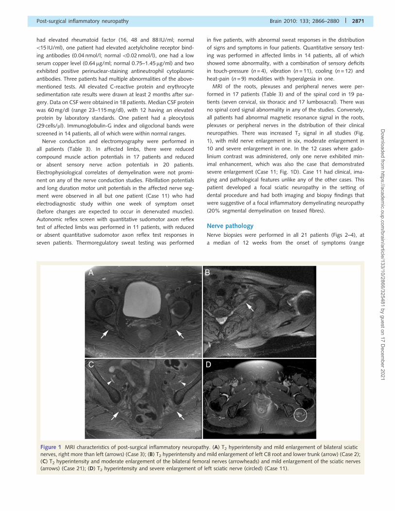
Figure 4 Evidence for focal nature of epineurial microvasculitis in post-surgical inflammatory neuropathy. Serial paraffin sections from a
region of microvasculitis: top row is proximal portion of epineurial microvessel and bottom row is distal portion of same vessel (arrow
denotes vessel involved with microvasculitis in upper panel). Note the separation and destruction of the vessel wall by inflammation in the
top row. There is no intact lumen remaining. (A and D) Haematoxylin and eosin; (B and E) smooth muscle actin immunostaining; (C and F)
CD-45 (leukocyte common antigen) immunostaining (Case 2).
2874 | Brain 2010: 133; 2866–2880 N. P. Staff et al.
Dow
nloaded from https://academ
ic.oup.com/brain/article/133/10/2866/325481 by guest on 17 D
ecember 2021

Tab
le4
Ner
vebio
psy
resu
lts
inbio
psy
-confi
rmed
post
-surg
ical
infl
amm
atory
neu
ropat
hy
Pat
ient
Foca
l,
mult
ifoca
l
or
dif
fuse
neu
ropat
hy
Tim
efr
om
sym
pto
ms
tobio
psy
(wee
ks)
Ner
ve
bio
psi
ed
Tea
sed
fibre
sIn
flam
mat
ion
Isch
aem
icin
jury
Normal:A,B(%)
Demyelination:C,D(%)
Axonal:E,H(%)
Remyelination:F,G(%)
Classifiable:(number)
EmptyStrands:(number)
Per
ivas
cula
r
Infl
amm
atio
n
(sm
all,
moder
ate,
larg
e)a
CD-45positivecollections
Hemosiderindeposition
Mic
rova
sculi
tis
(dia
gnost
ic,
sugges
tive
or
none)
Focalfibreloss
Neovascularization
Perineurialthickening
Perineurialdegeneration
Injuryneuroma
1M
u2
Sura
l26
070
3121
2M
oder
ate
+Su
gges
tive
2M
u24
Super
fici
al
radia
l
59
11
21
991
19
Larg
e+
+D
iagnost
ic+
++
3M
u12
Sura
l8
092
077
29
Larg
e+
+D
iagnost
ic+
++
+
4Fo
6Su
per
fici
al
per
onea
l
12
088
068
26
Larg
e+
+D
iagnost
ic+
++
5D
100
Sura
l72
23
23
96
9Sm
all
+Su
gges
tive
6M
u7
Late
ral
ante
bra
chia
l
85
20
14
117
8Sm
all
+N
one
+
7D
9Su
ral
679
13
284
23
Smal
l+
None
++
++
8M
u20
Sura
l58
726
943
58
Smal
l+
Sugges
tive
++
++
9M
u8
Sura
l68
419
968
37
Smal
l+
Sugges
tive
+
10
Fo36
Sura
l4
096
024
79
Larg
e+
+D
iagnost
ic+
+
11
Fo2
Scia
tic
fasc
icula
r
77
20
1.5
1.5
90
12
Smal
l+
None
++
12
Mu
36
Sura
l83
15
11
80
27
Smal
l+
None
++
+
13
Mu
28
Super
fici
al
radia
l
90
91
077
34
Larg
e+
+Su
gges
tive
+
14
D23
Sura
l90
22
687
20
Moder
ate
+Su
gges
tive
++
15
Mu
2Su
ral
22
078
099
3Sm
all
++
None
16
D16
Sura
lN
DN
DN
DN
D0
ND
Smal
l+
+N
one
++
17
Mu
24
Sura
l60
19
14
874
32
Moder
ate
++
Sugges
tive
++
++
18
Fo7
Sura
l73
023
492
14
Moder
ate
+D
iagnost
ic+
++
+
19
Fo10
Super
fici
al
per
onea
l
50
95
077
35
Larg
e+
+D
iagnost
ic+
20
Mu
9Su
ral
57
134
786
21
Moder
ate
++
Dia
gnost
ic+
21
Fo22
Sura
l54
532
969
30
Larg
e+
+D
iagnost
ic+
++
++
TO
TA
LS:
Med
ian
(Ran
ge)
F:6
M:
11
D:
4
12
(2–1
00)
57.5
(4–9
0)
1.5
(0–7
9)
24.5
(0–9
6)
5(0
–23)
83
(0–1
21)
24.5
(2–7
9)
Smal
l:9
Moder
ate:
5
Larg
e:7
21
11
Dia
gnost
ic:
8
Sugges
tive
:7
None:
6
11
13
12
10
1
D=
diffu
se;
Fo=
foca
l;M
u=
multifoca
l;N
D=
not
done;
+=
pre
sent.
aPer
ivas
cula
rin
flam
mat
ory
colle
ctio
ns:
smal
l=10–4
9ce
lls,
moder
ate
=50–9
9ce
lls,
larg
e=�
100
cells
.C
ateg
ories
Ath
rough
Gre
fer
tosp
ecifi
cte
ased
fiber
clas
sifica
tions
asdes
crib
edin
Ref
eren
ce(D
yck
et
al.,
2005a).
Post-surgical inflammatory neuropathy Brain 2010: 133; 2866–2880 | 2875
Dow
nloaded from https://academ
ic.oup.com/brain/article/133/10/2866/325481 by guest on 17 D
ecember 2021

peroneal- and tibial-innervated muscles. There was loss of sensa-
tion (touch, vibration, proprioception, cooling, heat-pain and pin-
prick) over the lateral part of her right leg and over the dorsum of
the foot. Reflexes were absent at the knees and ankles and the
plantar responses were flexor.
Extensive serological evaluations were notable only for a mild
elevation in erythrocyte sedimentation rate (40 mm/h). Spinal fluid
revealed a normal glucose (107 mg/dl), protein (37 mg/dl), cell
count (white blood cells 1 cell/ml, red blood cells 0 cell/ml),
cytology, oligoclonal bands, immunoglobulin-G index, bacterial
cultures and West Nile Virus serology. Nerve conduction studies
were normal in the upper extremities (median sensory and ulnar
motor), whereas there was no measurable right tibial and peroneal
compound muscle or sural sensory nerve action potential. The left
peroneal compound muscle action potential was reduced in amp-
litude with normal conduction velocity, distal latency and F-wave.
Needle examination revealed prominent fibrillation potentials and
neurogenic motor unit potentials in a pattern consistent with a
right sciatic neuropathy. MRI of the lumbar spine revealed facet
joint arthropathy at L3–5 interspaces but was otherwise normal.
MRI of the lumbosacral plexus showed prominence of both sciatic
nerves, greater on the right, without abnormal enhancement,
which was thought to represent a nonspecific inflammatory pro-
cess (Fig. 1A).
A right sural nerve biopsy revealed evidence for an ongoing
inflammatory neuropathic process (Figs 2A, 3A and B). There
was a severely decreased density of myelinated fibres with actively
degenerating profiles, multifocality, perineurial thickening and
neovascularization. Teased fibre preparation showed a markedly
increased rate of axonal degeneration (92%) and an increased
number of empty strands. Multiple small, moderate and large col-
lections of mononuclear inflammatory cells around epineurial ves-
sels were seen. The inflammation was disrupting the walls of small
epineurial vessels with associated hemosiderin-laden macrophages
and was diagnostic of microvasculitis.
The diagnosis of inflammatory mononeuropathies secondary to
microvasculitis was made. The patient was treated for 3 days with
high-dose intravenous methylprednisolone (1 g), followed by
weekly doses (1 g) for a total of 12 weeks. In follow-up, after
the course of intravenous steroids, the patient reported resolution
of pain and was able to discontinue opioid medications (she re-
mained on anticonvulsant therapy). Her weakness was mildly im-
proved initially after treatment, but the patient reported a marked
increased mobility, which was felt to be due to the combined
effects of improved pain and weakness. Her NIS at last follow-up
(30 months after initial evaluation) improved from 38.5 to 30.
Case 7 (diffuse): sensory-predominant polyneuropathyfollowing root canal
A 75-year-old female with a history of hypertension underwent a
root canal under conscious sedation. Three days later she de-
veloped numbness in the right hand and forearm, followed by
similar symptoms in the left hand the next day. By the next
week she noted bilateral foot numbness and pain had developed
in the arms and feet. The pain was characterized as having a
burning and shooting quality, with sensitivity to light touch. She
lost 15 pounds in weight concurrent with the sensory disturbance.
She had no weakness.
Neurological examination demonstrated sensory loss to vibration
with preservation of touch, proprioception, cooling, heat-pain and
pinprick modalities. Contact allodynia to light touch and hyper-
algesia to pinprick were documented in the hands and feet.
Deep tendon reflexes were reduced at the ankles. Strength was
normal.
A broad serological survey was negative or normal. CSF evalu-
ation showed a normal protein (31 mg/dl), glucose (57 mg/dl) and
cell count (white blood cells 1 cell/ml, red blood cells 0 cell/ml).
Nerve conduction studies demonstrated absent median and ulnar
sensory nerve action potentials. The sural sensory nerve action
potential was normal (7 mV). There was reduced amplitude left
median, ulnar, peroneal and tibial compound muscle action poten-
tials, with mildly prolonged distal latencies in all but the peroneal
nerve. The conduction velocities were relatively preserved. The
median and ulnar F-waves were within estimate, while the tibial
F-wave was absent. Needle electromyography demonstrated
reduced recruitment of long duration motor unit potentials in
distal leg muscles. The study was interpreted as a mixed axonal
and demyelinating sensorimotor peripheral neuropathy.
A whole left sural nerve biopsy was performed, which on teased
fibre preparation exhibited segmental demyelination (79%) and
axonal degeneration (13%). There was mild focal fibre loss,
Figure 5 Longitudinal follow-up of post-surgical inflammatory
neuropathy. NIS improves over time (paired Student’s t-test,
P = 0.001). Solid lines denote treatment period and dashed
lines are during times of no treatment. Every patient showed
improvement or stabilization.
2876 | Brain 2010: 133; 2866–2880 N. P. Staff et al.
Dow
nloaded from https://academ
ic.oup.com/brain/article/133/10/2866/325481 by guest on 17 D
ecember 2021

Tab
le5
Cli
nic
ally
susp
ecte
dpost
-surg
ical
infl
amm
atory
neu
ropat
hy:
pat
ient
char
acte
rist
ics,
surg
ery
types
,cl
inic
alfe
ature
san
dan
cill
ary
studie
s
Pat
ient
Age
at
clin
ical
eval
uat
ion
(yea
rs)
Gen
der
Tim
efr
om
surg
ery
to
sym
pto
ms
(day
s)
Surg
ery
Cli
nic
aldia
gnosi
sFo
cal,
mult
ifoca
l
or
dif
fuse
neu
ropat
hy
Neu
ropat
hy
nea
rsi
teof
surg
ery
Pai
nN
ISat
nad
ir
Wei
ght
loss
(pounds)
Ris
k
fact
ors
Lab
studie
s
QST
Elec
trophys
iolo
gy
MR
I
CMAP
SNAP
EMG
QSART
"T2
Enlargednerves
Gadolinium-enhanced
22
68
M4
Lum
bar
CSF
leak
repai
rLe
ft4
right
LSR
PN
Mu
NY
21.5
25
DM
NL
ND
##
Fb,
NM
UN
DY
Mod
Y
23
52
M21
Cer
vica
lsp
ine
fusi
on
Rig
ht
BPN
FoY
N0
0none
NL
ND
NL
#Fb
,N
MU
NL
YN
N
24
43
M7
Aort
icva
lve
repla
cem
ent
Length
-dep
enden
t
per
ipher
al
neu
ropat
hy
DN
Y2
0none
AN
A,
CSF
pro
tein
ND
##
Fb,
NM
UN
DN
DN
DN
D
25
27
M5
24
h/1
7C
hole
cyst
ecto
my
Left
(524
h)
then
right
(17
day
s)BPN
Mu
NY
26.7
5N
DI
NL
ND
##
Fb,
NM
UN
DY
Mild
N
26
60
F21
Appen
dec
tom
yA
uto
nom
icneu
ropat
hy
DN
Y0
ND
none
NL
ND
NL
NL
NL
#N
DN
DN
D
27
71
M22
Aort
icva
lve
repla
cem
ent
Bila
tera
lLS
RPN
and
left
faci
al
Mu
NY
26.5
20
none
NL
HA
#N
LFb
,N
MU
#N
DN
DN
D
28
67
M1
Rad
ical
pro
stat
ecto
my
Rig
ht
Lum
bosa
cral
ple
xopat
hy
FoN
Y17.5
18
Ca
AC
E,C
SF
pro
tein
TP
##
Fb,
NM
UN
DY
NN
29
64
M5
24
hR
ight
nep
hre
ctom
yR
ight
BPN
FoN
Y2
25
Ca
CSF
pro
tein
C#
#Fb
,N
MU
ND
YM
od
N
30
68
M5
24
hBow
elre
anas
tom
osi
sR
ight
uln
arFo
NY
116
DM
,PN
NL
ND
##
Fb,
NM
UN
DN
DN
DN
D
31
67
M5
24
hC
ABG
Rig
ht
scia
tic
FoN
ND
31
ND
IC
ryo
ND
##
Fb,
NM
UN
DN
DN
DN
D
32
76
M5
24
hR
ight
fem
ur
nai
ling
Rig
ht
BPN
FoN
Y21
ND
Ca
NL
ND
##
Fb,
NM
UN
DN
DN
DN
D
33
44
F9
Rig
ht
bra
chia
lple
xus
schw
annom
a
rese
ctio
n
Left
phre
nic
and
right
med
ian
Mu
NY
14
0none
NL
ND
NL
#Fb
,N
MU
ND
ND
ND
ND
AC
E=
angio
tensi
nco
nve
rtin
gen
zym
e;A
NA
=an
tinucl
ear
antibodie
s;BPN
=bra
chia
lple
xus
neu
ropat
hy;
C=
incr
ease
dco
olin
gth
resh
old
;C
a=
cance
r;C
ABG
=co
ronar
yar
tery
byp
ass
gra
ft;C
MA
P=
com
pound
musc
leac
tion
pote
ntial
;C
ryo
=cr
yoglo
bulin
aem
ia;
D=
diffu
se;
DM
=dia
bet
esm
ellit
us;
F=
fem
ale;
Fb=
fibrilla
tion
pote
ntial
s;Fo
=fo
cal;
HA
=hyp
eral
ges
ia;
I=in
fect
ion;
LSR
PN
=lu
mbosa
cral
radic
ulo
ple
xus
neu
ropat
hy;
M=
mal
e;M
od
=m
oder
ate;
Mu
=
multifoca
l;N
=no;
NIS
=neu
ropat
hy
impai
rmen
tsc
ore
;N
MU
=neu
rogen
icm
oto
runit
pote
ntial
s;N
D=
not
docu
men
ted;
NL
=norm
al;
PN
=per
ipher
alneu
ropat
hy;
QSA
RT
=quan
tita
tive
sudom
oto
rax
on
reflex
test
;Q
ST=
quan
tita
tive
sensa
tion
test
ing;SN
AP
=se
nso
ryner
veac
tion
pote
ntial
;TP
=in
crea
sed
touch
-pre
ssure
thre
shold
;W
BC
s=
white
blo
od
cells
;Y
=ye
s;dow
nw
ard
arro
win
dic
ates
reduce
dor
abse
nt;
upw
ard
arro
win
dic
ates
incr
ease
.
Post-surgical inflammatory neuropathy Brain 2010: 133; 2866–2880 | 2877
Dow
nloaded from https://academ
ic.oup.com/brain/article/133/10/2866/325481 by guest on 17 D
ecember 2021

regenerating clusters and perineurial degeneration. Scattered small
collections of lymphocytes were observed around epineurial and
perineurial venules and microvessels, without vessel disruption.
A diagnosis of a sensory inflammatory demyelinating polyneur-
opathy was made and the patient received a 7-month tapering
course of prednisone. Gabapentin was initiated for pain control.
On last clinical follow-up, 22 months after neuropathy onset, the
patient was improved and left with mild residual neuropathic
symptoms.
DiscussionWe have described the demographics, clinical features, electro-
physiology, imaging and pathology in a series of patients with
post-surgical inflammatory neuropathy. In our cohort, post-
surgical inflammatory neuropathy presented in either a focal,
multifocal or diffuse fashion. Clues to the inflammatory aetiology
of the neuropathy were sometimes available in the spatiotemporal
segregation of the neuropathy from the surgery. Focally increased
T2 signal and enlargement within nerve segments on MRI some-
times provided another clue to the inflammatory aetiology. In
other cases, inflammatory neuropathies were identified on biopsy
in patients who otherwise appeared to have a mechanically
induced neuropathy. Ancillary testing did not usually provide evi-
dence for a systemic inflammatory/immune process; however,
these tests were helpful in ruling out other causes of neuropathy.
In the end, nerve biopsy was often utilized to confirm the diag-
nosis of inflammatory neuropathy and direct treatment for the
patients, most of whom received immunosuppressant medications.
All patients reported some degree of improvement following treat-
ment, although the two patients followed up that were not trea-
ted also showed improvement, which in these patients probably
reflects a monophasic process. In general, pain improved more
rapidly than weakness and all but one patient had some residual
weakness at the last follow-up. Given the retrospective nature of
the study, it is not possible to make strong claims regarding the
efficacy of treatment in this entity.
Our study substantially increases the number of reported cases
of post-surgical inflammatory neuropathy, which to our know-
ledge had previously been limited to Guillain-Barre syndrome
(Wiederholt et al., 1964; Arnason and Asbury, 1968) or idiopathic
brachial plexitis (Parsonage and Turner, 1948; Malamut et al.,
1994; van Alfen and van Engelen, 2006). Conversely, the litera-
ture is replete with studies on the general topic of post-surgical
neuropathies. These post-surgical neuropathies are typically attrib-
uted to mechanical factors (stretch, compression, transection, con-
tusion, suture), neurotoxicities from anaesthesia or ischaemia.
Putative risk factors have also been ascribed for post-surgical neu-
ropathies including surgical positioning, age, gender, body mass
index, hospital length-of-stay, diabetes mellitus, tobacco use and
vascular disease (Barner et al., 2002, 2003).
It is unclear what the incidence of post-surgical inflammatory
neuropathy is, but there are widely variable data on the overall
incidence of post-surgical neuropathy, with the variability often
related to the study population. In a large tertiary academic
centre, Welch et al. (2009) found that centre’s overall frequency
of perioperative (within 48 h) peripheral nerve injuries over a
10-year period to be 0.03%. This frequency is significantly lower
than other large all-inclusive studies that reported incidences of
0.11% (Blitt et al., 1995) and 0.14% (Parks, 1973). While the
frequency of all perioperative neuropathies may have decreased
over time, it is just as likely that these differences are associated
with the use of diverse definitions for peripheral nerve injuries and
variability in identification of cases using retrospective record re-
views. The frequency of post-surgical neuropathies is often higher
when investigated in specific surgical scenarios, ranging from 0.7
to 5.5% (Weber et al., 1976; Lederman et al., 1982; Warner
et al., 2000; Cardosi et al., 2002). For example, lower extremity
neuropathies after procedures performed on patients in lithotomy
positions are far more frequent than those same neuropathies in
patients undergoing abdominal procedures while positioned supine
(Warner et al., 2000). Although ulnar neuropathies following sur-
gery are considered common, a large retrospective epidemiological
study demonstrated a frequency of only 0.036% in patients
undergoing non-cardiac procedures (Warner et al., 1994). A pro-
spective study of ulnar neuropathy in 1502 surgical patients sub-
sequently found a frequency of 0.5% (Warner et al., 1999). What
proportion of these neuropathies is inflammatory in nature is un-
clear given the lack of biopsy material in any of these incidence
studies.
At our tertiary referral centre, it is our impression that post-
surgical inflammatory neuropathy is relatively common. Although
a coincidental (and not causal) association of inflammatory neur-
opathy and surgery is possible, we feel this is unlikely based on
our experience in this study and clinical practice. Our search strat-
egy for identifying patients was not exhaustive and we readily
found 33 patients that had features consistent with a post-surgical
inflammatory neuropathy. Furthermore, in our search for these
cases we found only two patients suspected of having
post-surgical inflammatory neuropathy whose nerve biopsy did
not reveal inflammation (not included in the study). Based on
the ease of identifying patients (30 of 33 cases were personally
seen by one of the authors) and the fact that several biopsy-
confirmed post-surgical neuropathy cases appeared otherwise
mechanical in nature, we suspect that post-surgical inflammatory
neuropathies are a larger problem than we are reporting. The
main difficulty is that the only way to identify a post-surgical in-
flammatory neuropathy with certainty at this time is by doing a
nerve biopsy. We suspect that the vast majority of patients with
post-surgical inflammatory neuropathy are assumed to have a
mechanical cause and so nerve biopsy is rarely considered. For
this reason, even if most post-surgical neuropathies are
mechanical, the incidence of inflammatory causes is vastly
under-appreciated.
Although mechanical forces are the probable cause of the ma-
jority of surgery-related neuropathies, the development of inflam-
matory infiltration into nerve discovered in our cases is not
expected in a purely mechanical insult. Active axonal degener-
ation, as seen in 16 of our biopsies, may result in endoneurial
macrophage (CD-68 positive cells) infiltration but does not cause
significant lymphocyte-mediated (CD-45 positive cells) inflamma-
tion, as is also seen in our cohort. What stimulates the lymphocytic
inflammatory attack on nerve in the post-surgical period is far
2878 | Brain 2010: 133; 2866–2880 N. P. Staff et al.
Dow
nloaded from https://academ
ic.oup.com/brain/article/133/10/2866/325481 by guest on 17 D
ecember 2021

from clear. In all likelihood, there is a complex interplay of inflam-
matory stress responses from surgery, genetic predisposition (Klein
et al., 2002), subclinical pre-existing inflammation or neuropathy
and possibly even mechanical forces, which all combine to trigger
the inflammatory neuropathy. This issue is of particular interest in
the patients (18/33) that developed a neuropathy within three
days of the surgery before a primary learned immune response
would be expected to develop.
It is possible that the surgical process, transfusions and/or an-
aesthetics may also be contributing to post-surgical inflammatory
neuropathies. Fifteen of the biopsied patients received general an-
aesthesia, two received spinal blocks, two received a combination
of general anaesthetics and epidural analgesia, and two were
sedated. Seven patients received transfusion of blood products.
Stress related to surgical procedures and the perioperative period
has been well documented in many types of procedures to reduce
immune function and to be associated with increased generalized
inflammatory responses (Brown et al., 1989). Inhalation anaes-
thetics have been associated with immunosuppression lasting for
weeks after surgical procedures (Hunter, 1999; Myles et al.,
2004). Transfusion of blood products, especially those containing
white blood cells, can cause immunosuppression and promote in-
flammatory responses (Brand, 2002; Vamvakas, 2004). Overall,
patients undergoing surgery and anaesthesia, especially those
with major procedures and concomitant transfusions, have a
high potential to be immunocompromised postoperatively and to
have an increased frequency of inflammatory responses.
Other possible risk factors associated with post-surgical inflam-
matory neuropathies in our study included diabetes mellitus (9/33)
cancer (7/33), infection (7/33) and a history of smoking (14/33).
In our limited retrospective series, the effects of risk factors cannot
be ascertained but raise areas for future study.
The clinical presentation and pathology of our patients were
heterogeneous, with focal, multifocal and diffuse patterns being
found. Given this heterogeneity, it is not clear to the authors
whether the post-surgical inflammatory neuropathies represent
one entity or more than one. Nonetheless, the fact that the ma-
jority (15/21) of biopsies were suggestive or diagnostic of nerve
microvasculitis and ischaemic nerve injury does suggest that there
is one predominant pathology. These ischaemic and microvasculitic
pathological findings were more represented in the patients with
either focal or multifocal neuropathy presentations (none of the
four with symmetrical polyradiculoneuropathy were diagnostic of
microvasculitis). Interestingly, many of these patients had clinical
(subacute onset of pain and weakness with weight loss) and
pathological features (ischaemic nerve injury and microvasculitis)
similar to both the diabetic and non-diabetic lumbosacral radicu-
loplexus neuropathies (Dyck et al., 2000, 2001), and may repre-
sent a form of this entity that is provoked by surgical stress. In the
other cases with diffuse post-surgical inflammatory polyradiculo-
neuropathies (none of which appear to be classic Guillain-Barre
syndrome), it is possible that there are inflammatory mechanisms
other than microvasculitis involved.
Of particular interest are five cases (Cases 4, 10, 18, 19 and 20)
that developed biopsy-confirmed post-surgical inflammatory neu-
ropathies in the same lower limb of an orthopaedic surgical pro-
cedure. All of these cases were diagnostic of nerve microvasculitis
on nerve biopsy of distal cutaneous nerves remote from the ortho-
paedic surgical site. In the past, these post-surgical neuropathies
have been understandably assumed to be mechanical, given the
strong physical forces used in many orthopaedic procedures, and
previous electromyography studies have shown presence of both
clinical and subclinical neuropathies following hip arthroplasty
(Weber et al., 1976). Our data provide strong evidence that in
some cases of lower limb neuropathies following orthopaedic pro-
cedures, an inflammatory component is prominent, if not
causative.
In conclusion, it is important for physicians to recognize that
not all neuropathies that occur in the post-surgical setting are
due to compression, transection or stretch. If a neuropathy
occurs after a delay or in a territory remote from the surgical
site, an inflammatory-immune mechanism should be considered,
and if possible, a nerve biopsy for confirmation should be per-
formed to ensure that the best treatment is given. At this point,
however, it is less clear how extensive a neurological evaluation
(i.e. nerve biopsy) is appropriate in patients that develop neuro-
pathies in the same limb that the surgery was performed on. Our
data suggest that inflammation may be an important mechanism
of nerve injury in these cases, but who should receive immuno-
therapy and how many patients need nerve biopsy are provocative
ideas that warrant future study.
ReferencesArnason BG, Asbury AK. Idiopathic polyneuritis after surgery. Arch
Neurol 1968; 18: 500–7.
Barner KC, Landau ME, Campbell WW. A review of perioperative nerve
injury to the lower extremities: part I. J Clin Neuromuscul Dis 2002; 4:
95–9.Barner KC, Landau ME, Campbell WW. A review of perioperative nerve
injury to the upper extremities. J Clin Neuromuscul Dis 2003; 4:
117–23.
Blitt CD, Kaufer-Bratt C, Ashby J, Caillet JR. QA program reveals safety
issues, promotes development of guidelines: Arizona practice is model.
J Clin Monit 1995; 11: 76–9.
Brand A. Immunological aspects of blood transfusions. Transpl Immunol
2002; 10: 183–90.Brown JM, Grosso MA, Harken AH. Cytokines, sepsis and the surgeon.
Surg Gynecol Obstet 1989; 169: 568–75.
Cardosi RJ, Cox CS, Hoffman MS. Postoperative neuropathies after
major pelvic surgery. Obstet Gynecol 2002; 100: 240–4.
Cheney FW, Domino KB, Caplan RA, Posner KL. Nerve injury associated
with anesthesia: a closed claims analysis. Anesthesiology 1999; 90:
1062–9.Daube JR, Rubin DI. Clinical neurophysiology appendix (CD-ROM). In:
Daube JR, Rubin DI, editors. Clinical neurophysiology. Oxford: Oxford
University Press; 2009.
Dawson DM, Krarup C. Perioperative nerve lesions. Arch Neurol 1989;
46: 1355–60.
Dyck PJ, Dyck PJB, Engelstad JE. Pathologic alterations of nerves. In:
Dyck PJ, Thomas PK, editors. Peripheral neuropathy. Philadelphia:
Elsevier Saunders; 2005a. p. 733–829.Dyck PJ, Karnes J, O’Brien PC, Zimmerman IR. Detection thresholds of
cutaneous sensation in humans. In: Dyck PJ, Thomas PK, Lambert EH,
Bunge R, editors. Peripheral neuropathy. Philadelphia: W.B. Saunders;
1984. p. 1103–1138.
Dyck PJ, Hughes RAC, O’Brien PC. Quantitating overall neuropathic
symptoms, impairments, and outcomes. In: Dyck PJ, Thomas PK,
Post-surgical inflammatory neuropathy Brain 2010: 133; 2866–2880 | 2879
Dow
nloaded from https://academ
ic.oup.com/brain/article/133/10/2866/325481 by guest on 17 D
ecember 2021

editors. Peripheral neuropathy. Philadelphia: Elsevier Saunders; 2005b.p. 1031–52.
Dyck PJ, Sherman WR, Hallcher LM, Service FJ, O’Brien PC, Grina LA,
et al. Human diabetic endoneurial sorbitol, fructose, and myo-inositol
related to sural nerve morphometry. Ann Neurol 1980; 8: 590–6.Dyck PJB, Engelstad J, Norell J, Dyck PJ. Microvasculitis in non-diabetic
lumbosacral radiculoplexus neuropathy (LSRPN): similarity to the dia-
betic variety (DLSRPN). J Neuropathol Exp Neurol 2000; 59: 525–38.
Dyck PJB, Norell JE, Dyck PJ. Non-diabetic lumbosacral radiculoplexusneuropathy: natural history, outcome and comparison with the diabet-
ic variety. Brain 2001; 124: 1197–207.
Hunter JD. Effects of anaesthesia on the human immune system. HospMed 1999; 60: 658–63.
Klein CJ, Dyck PJB, Friedenberg SM, Burns TM, Windebank AJ, Dyck PJ.
Inflammation and neuropathic attacks in hereditary brachial plexus
neuropathy. J Neurol Neurosurg Psychiatry 2002; 73: 45–50.Lederman RJ, Breuer AC, Hanson MR, Furlan AJ, Loop FD,
Cosgrove DM, et al. Peripheral nervous system complications of
coronary artery bypass graft surgery. Ann Neurol 1982; 12: 297–301.
Low PA. Autonomic nervous system function. J Clin Neurophysiol 1993;10: 14–27.
Malamut RI, Marques W, England JD, Sumner AJ. Postsurgical idiopathic
brachial neuritis. Muscle Nerve 1994; 17: 320–4.
Myles PS, Leslie K, Silbert B, Paech MJ, Peyton P. A review of the risksand benefits of nitrous oxide in current anaesthetic practice. Anaesth
Intensive Care 2004; 32: 165–72.
Parks BJ. Postoperative peripheral neuropathies. Surgery 1973; 74:348–57.
Parsonage MJ, Turner JW. Neuralgic amyotrophy; the shoulder-girdle
syndrome. Lancet 1948; 1: 973–8.
Vamvakas EC. White-blood-cell-containing allogeneic blood transfusion,
postoperative infection and mortality: a meta-analysis of observational
’before-and-after’ studies. Vox Sang 2004; 86: 111–9.
van Alfen N, van Engelen BG. The clinical spectrum of neuralgic amyot-
rophy in 246 cases. Brain 2006; 129: 438–50.Warner MA, Warner DO, Harper CM, Schroeder DR, Maxson PM.
Lower extremity neuropathies associated with lithotomy positions.
Anesthesiology 2000; 93: 938–42.Warner MA, Warner DO, Matsumoto JY, Harper CM, Schroeder DR,
Maxson PM. Ulnar neuropathy in surgical patients. Anesthesiology
1999; 90: 54–9.Warner MA, Warner ME, Martin JT. Ulnar neuropathy. Incidence,
outcome, and risk factors in sedated or anesthetized patients.
Anesthesiology 1994; 81: 1332–40.Weber ER, Daube JR, Coventry MB. Peripheral neuropathies
associated with total hip arthroplasty. J Bone Joint Surg Am 1976;
58: 66–9.Welch MB, Brummett CM, Welch TD, Tremper KK, Shanks AM,
Guglani P, et al. Perioperative peripheral nerve injuries: a retrospective
study of 380,680 cases during a 10-year period at a single institution.
Anesthesiology 2009; 111: 490–7.Wiederholt WC, Mulder DW, Lambert EH. The Landry-Guillain-
Barr’e-Strohl syndrome or polyradiculoneuropathy: Historical review,
report on 97 patients, and present concepts. Mayo Clin Proc 1964;
39: 427–51.
2880 | Brain 2010: 133; 2866–2880 N. P. Staff et al.
Dow
nloaded from https://academ
ic.oup.com/brain/article/133/10/2866/325481 by guest on 17 D
ecember 2021