Embed Size (px)
Citation preview

ON NOT Associated with MS•Neuromyelitis optica (NMO, Devic’s disease)•Acute disseminated encephalomyelitis (ADEM)•Chronic relapsing inflammatory optic neuropathy (CRION)•Post-vaccination-associated optic neuritis
•Consider in:–Atypical age (younger, older)–Atypical features/clinical signs–Rapidly progressive disease–Poor recovery with usual treatment

Neuromyelitis Optica• Optic neuritis + transverse myelitis• Simultaneous or sequential• SEVERE inflammation• Marked disability
–at 5 years: 50% blind or walking aid
• Worse prognosis:– # relapses 1st 2 years– severity of 1st attack– associated autoimmune dz

NMO: Diagnosis• Difficult initially; may be monocular presentation• Clinical presentation• Spine MRI• Aquaporin-4 antibodies (anti-NMO-IgG)
–up to 80% of cases• Median age = 40• 9:1 female preponderance• Other autoimmune conditions

NMO: Treatment• Acute attacks:
– high-dose steroids (ONTT protocol)– plasma exchange
• Maintenance:–prednisone + azathioprine (Imuran ®)–mycophenolate mofetil (CellCept ®)–rituximab (Rituxan ®)
• AVOID MS MEDS (especially -interferons)

Infectious Optic Neuropathies• Neuroretinitis• Lyme Optic Neuropathy• West Nile Virus

Neuroretinitis• Young, healthy adults; no sex predilection• 2/3 with viral prodrome• VA loss mild to severe• VF defect usually central/cecocentral• RAPD and dyschromatopsia*• Optic disc edema (usually)• Stellate maculopathy
Bartonella henselae
• Children > adults; M > F• Cats (bite/scratch) >> dogs• Macular star evolves over days to weeks• B. henselae abs in serum• Tx: doxycycline, rifampin, azithromycin• Most often self-limited

Lyme Optic Neuropathy• Borrelia burgdorferi (tick-borne)• Erythema chronicum migrans• CNS involvement = days/weeks p infection• Tertiary stage = months/years

Lyme Disease: Exam• Inflammation of the anterior segment• Exudative retinal detachment• Lyme meningitis:
– Cranial neuropathies (multiple)– Papilledema due to increased ICP
• MRI non-specific +/- meningeal enhancement• ELISA to screen, Western blot to confirm• Intrathecal Lyme Abs: ceftriaxone 2g IV/d x 1 mo
West Nile Virus• Fever, headache, lymphadenopathy, rash• Increasing awareness of meningoencephalitis• Retinitis, optic neuritis, uveitis• Chan 2006: multifocal choroiditis most common• Beware:
– diabetes mellitus– age > 50
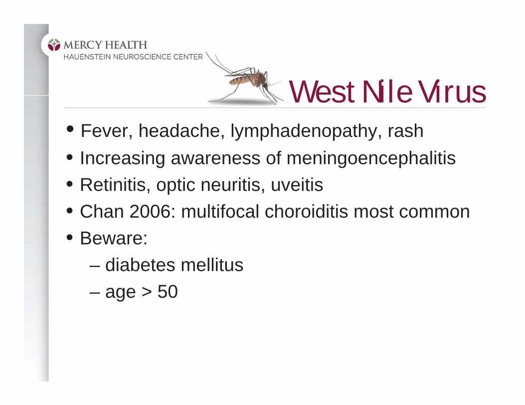
Inflammatory ON in Systemic Disease42-year-old man, painless vision loss OS x weeks

Dx: Optic glioma of L optic nerve/chiasm

• Progressive “loss of detail” OD• BCVA: 20/30 OD, NLP OS• Color (Ishihara): 10/10 OD, 0/10 OS • Fields: Full to confrontation OD• Pupils: 6 mm; Grade 4 APD OD• 2 mm proptosis OS• SLE: Unremarkable• Fundus: dysplastic nerve OD, advanced atrophy OS
2 years later…
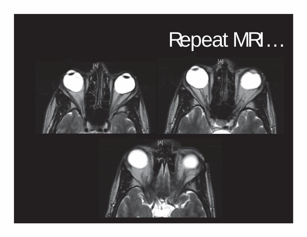
Repeat MRI…
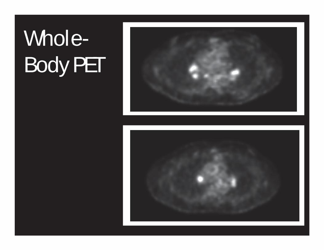
Whole-Body PET
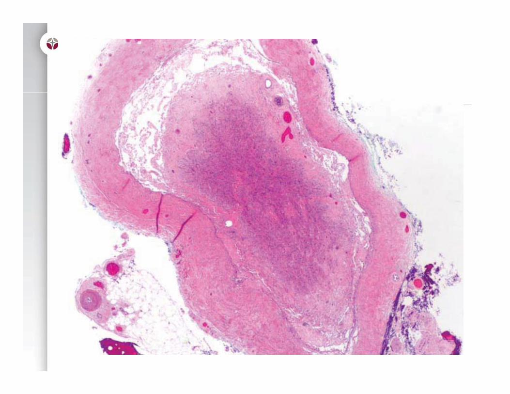
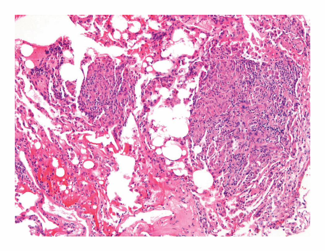
Sarcoidosis• Multisystem granulomatous inflammation• Lymphatic system, lungs• Similar presentation to typical ON• All ethnic groups (> in African Americans in US)• CNS involvement ~5% of cases• Ophthalmic involvement ~25%• Consider if: recurrent vision loss with steroid w/d, conjunctival/iris nodules, uveitis, lacrimal gland enlargement• Dx dependent on TISSUE

Giant Cell (Temporal) Arteritis:• RARE > 60; prevalence increases with age• Acute; vision loss is severe (<20/200)• Pale, “chalky” edema +/- cotton-wool spots
• Risk Factors:• Age (mean age 70)• Associated with PMR
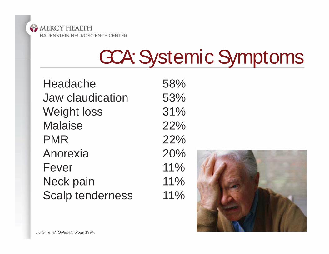
Liu GT et al. Ophthalmology 1994.
Headache 58%Jaw claudication 53%Weight loss 31%Malaise 22%PMR 22%Anorexia 20%Fever 11%Neck pain 11%Scalp tenderness 11%
GCA: Systemic Symptoms

GCA: Diagnosis• Clinical Presentation• Labs: ESR, CRP may be elevated• Temporal Artery Biopsy (**)

Goals:1. Decrease active inflammation2. Improve vision in the affected eye (uncommon)3. Prevent involvement of fellow eye4. Decrease systemic complications of vasculitis
If GCA suspected:1. Treat first; ask questions later…2. Begin steroids immediately (min 80 mg/d)3. Admit to hospital (with internist co-management)4. IV steroids 250 mg every 6 hours x 3-5 days5. Temporal artery biopsy, best if bilateral
GCA: EMERGENCY Treatment

GCA: Myths1. Diagnosis requires systemic symptoms.
- 21% present ONLY with vision loss (occult GCA).
2. Diagnosis requires elevated ESR.
- Normal ESR does not exclude GCA.
3. Steroid therapy can be tapered by protocol.
- Each patient must be treated individually.
- Serial monitoring w/ ESR and CRP most useful.
4. GCA always burns itself out.
- Long-term (9-12 months) is required.
- May need life-long tx to prevent vision loss.

Conclusions:• Optic neuritis is MOST common• May be first presentation of MS; initial MRI + clinical and radiographic monitoring REGULARLY• Other conditions can mimic demyelinating ON• Be suspicious of atypical features and evaluate accordingly