Embed Size (px)
Citation preview
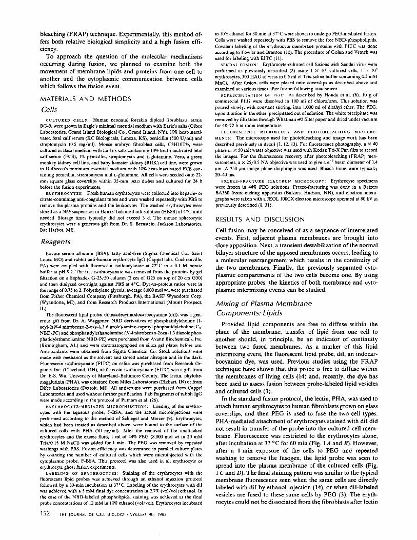
Studies on the Mechanism of
Cell Fusion Using Fluorescent
Probes
Polyethylene Glycol-mediated
Membrane and Cytoplasmic
JOHN W. WOJCIESZYN,* ROBERT A. SCHLEGEL,:J: KATHERINE LUMLEY-SAPANSKI,§ and KENNETH A. JACOBSON¶ Laboratories for Cell Biology, Department of Anatomy *¶, Cancer Research Center, School of Medicine, University of North Carolina at Chapel Hill, Chapel Hill, North Carolina 27514; and Molecular and Cellular Biology Program~.§, The Pennsylvania State University, University Park, Pennsylvania 16802
ABSTRACT The mechanism by which polyethylene glycol (PEG) mediates cell fusion has been studied by examining the movements of membrane lipids and proteins, as well as cytoplasmic markers, from erythrocytes to monolayers of cultured cells to which they have been fused. Fluorescence and freeze-fracture electron microscopy and fluorescence recovery after photo- bleaching have yielded the following results: (a) In the presence of both fusogenic and nonfusogenic PEG 1 membranes are brought together at closely apposed contact regions. (b) Fluorescent lipid probes quickly spread from the membranes of erythrocytes to cultured cells in the presence of both fusogenic and nonfusogenic PEG. (c) Proteins of the erythrocyte membranes were never observed to diffuse into the cultured cell membrane. (d) Water-soluble proteins did not diffuse from the erythrocyte interior into the target cell cytoplasm until the PEG was removed. These data suggest that the coordinate action of two distinct components is necessary for fusion as mediated by PEG. Presumably, the polymer itself promotes close apposition of the adjacent cell membranes but the fusion stimulus is provided by the additives contained in commercial PEG.
Polyethylene glycol (PEG) of high molecular weight is widely used to mediate ceU-ceU fusion in the production of somatic cell hybrids, including hybridomas, and more recently in the fusion injection of macromolecules from erythrocytes (1, 2) or liposomes into cultured cells (3). PEG offers advantages over other fusogens in that it permits fusion of a variety of cell types which may differ in species or even in kingdom and under the proper conditions produces high fusion efficiencies with mini- mal toxicity. Yet little is known of the mechanism by which PEG operates.
PEG causes the redistribution of intramembrane particles (IMPs) of cellular membranes, this ability being attributed to the ordering of water by high concentrations of the polymer (4). When aqueous solutions of PEG exceed 35%, cell aggre-
Dr. J. Lucy has pointed out that the term "nonfusogenic PEG" does not apply to all cell fusion protocols. He and his co-workers (29) have shown that extracted PEG (6) will fuse hen erythrocytes in a protocol requiring a 15-rain incubation in PEG.
gation and fusion are observed, although maximum fusion efficiency occurs at concentrations between 40 and 50%. Since all water is bound to PEG in solutions having concentrations of 35% by weight or greater, dehydration appears to play a role in PEG-mediated fusion (5, 25). However, pure PEG does not appear to be a complete fusogen. Recently, Honda et al. (6) have demonstrated that antioxidants and/or polymerization agents added to commercial PEG are responsible for the fusion ~ctivity since removal of these agents through organic solvent extraction renders the PEG nonfusogenic) Earlier work had shown that membrane active chemicals such as glyceryl mon- ooleate (GMO) are only capable of inducing cell-cell fusion when administered in conjunction with high molecular weight polymers such as dextrans (5).
Our interest in the mechanisms of PEG-induced fusion was prompted by our use of the erythrocyte-mediated microinjec- tion method to introduce fluorescent macromolecules into the cytoplasm of cultured cells (7) and subsequently to measure their diffusion rates by the fluorescence recovery after photo-
THE JOURNAL OF CELL BIOLOGY • VOLUME 96 JANUARY 1983 151-159 © The Rockefeller University Press - 0021-9525/83/01/0151/09 $1.00 1 51
on May 11, 2018jcb.rupress.org Downloaded from http://doi.org/10.1083/jcb.96.1.151Published Online: 1 January, 1983 | Supp Info:
bleaching (FRAP) technique. Experimentally, this method of- fers both relative biological simplicity and a high fusion effi- ciency.
To approach the question of the molecular mechanisms occurring during fusion, we planned to examine both the movement of membrane lipids and proteins from one cell to another and the cytoplasmic communication between cells which follows the fusion event.
MATERIALS AND METHODS
Cells CULTURED CELLS: Human neonatal foreskin diploid fibroblasts, strain
BG-9, were grown in Eagle's minimal essential medium with Eafle's salts (Gibco Laboratories, Grand Island Biological Co., Grand Island, NY), 10% heat-inacti- vated fetal calf serum (KC Biologlcals, Lanexa, KS), penicillin (500 U/ml) and streptomycin (0.5 mg/ml). Mouse embryo flbroblast cells, C3HIOTY~, were cultured in Basal medium with Earle's salts containing 10% heat-inactivated fetal calf serum (FCS), 1% penicillin, streptomycin and L-glutamine. Veto, a green monkey kidney cell line, and baby hamster kidney (BHK) cell line, were grown in Dulbecco's minimum essential medium with 10% heat-inactivated FCS con- taining penicillin, streptomycin and L-glutamine. All cells were seeded onto 22- mm square glass coverslips within 35-mm petri dishes and cultured for 24 h before the fusion experiments.
ERYTHROCYTES: Fresh human erythrocytes were collected into beparin- or citrate-containing anti-coagulant tubes and were washed repeatedly with PBS to remove the plasma proteins and the leukocytes. The washed erythrocytes were stored as a 50% suspension in Hanks' balanced salt solution (HBSS) at 4°C until needed. Storage times typically did not exceed 3 d. The mouse spherocytic erythrocytes were a generous gift from Dr. S. Bernstein, Jackson Laboratories, Bar Harbor, ME.
Reagents Bovine serum albumin (BSA), fatty acid-free (Sigma Chemical Co., Saint
Louis, MO) and rabbit anti-human erythrocyte IgG (Cappel labs, Cochranville, PA) were coupled with fluorescein isothiocyanate at 22°C in a 0.1 M borate buffer at pH 9.2. The free isothiocyanate was removed from the proteins by gel filtration on a Sephadex G-25/50 column (2 cm of G25 on top of 20 cm G50) and then dialyzed overnight against PBS at 4°C. Dye-to-protein ratios were in the range of 0.75 to 2. Polyethylene glycols, average 8,000 mol wt, were purchased from Fisher Chemical Company (Pittsburgh, PA), the BASF Wyandotte Corp. (Wyandotte, MI), and from Research Products International (Mount Prospect, IL).
The fluorescent lipid probe, dihexadecylinodocarbocyanine (dil), was a gen- erous gift from Dr. A. Waggoner. NBD derivatives of phosphatidylcholine (l- acyl-2(N-4 nitrobenzo-2-oxa-1,3 diazole)-amino caproyl phophatidylchollne; C6- NBD-PC) and phosphatidylethanolamine (N-4 nitrobenzo-2oxa- 1,3 diazole phos- phatidylethanolamine; NBD-PE) were purchased from Avanti Biochemicals, Inc. (Birmingham, AL) and were chromatographed on silica gel plates before use. Anti-oxidants were obtained from Sigma Chemical Co. Stock solutions were made with methanol as the solvent and stored under nitrogen and in the dark. Fluorescein isothiocyanate (FITC) on celite was purchased from Research Or- ganics Inc. (Cleveland, OH), while eosin isothiocyanate (EITC) was a gift from Dr. E-S. Wu, University of Maryland-Baltimore County. The lectin, phytohe- magglutinin (PHA), was obtained from Miles Laboratories (Elkhart, IN) or from Difco Laboratories (Detroit, MI). All antiserums were purchased from Cappel Laboratories and used without further purification. Fab fragments of rabbit IgG were made according to the protocol of Putnam et al. (26).
ERYTHROCYTE-MEDIATED MICROINJECTION: Loading of the erythro- cytes with the aqueous probe, F-BSA, and the actual microinjections were performed according to the method of Schlegel and Mercer (9). Erythrocytes, which had been treated as described above, were bound to the surface of the cultured cells with PHA (50 ~g/ml). After the removal of the unattached erythrocytes and the excess fluid, 1 ml of 44% PEG (8,000 tool wt in 20 mM Tris/0.15 M NaC1) was added for I min. The PEG was removed by repeated washings with PBS. Fusion efficiency was determined in parallel culture plates by counting the number of cultured cells which were microinjected with the cytoplasmic probe, F-BSA. This protocol was also used in all erythrocyte or erythrocyte ghost fusion experiments.
LABELING OF ERYTHROCYTES: Staining of the erythrocytes with the fluorescent lipid probes was achieved through an ethanol injection protocol followed by a 30-min incubation at 37°C. Labeling of the erythrocytes with dil was achieved with a 5 mM final dye concentration in 2.7% (vol/vol) ethanol. In the case of the NBD-labeled phosphollpids, staining was achieved at the final probe concentrations of 12 mM in 10% ethanol (vol/vol). Erythrocytes incubated
152 THE JOURNAL OF CELL BIOLOGY - VOLUME 96, 1983
in 10% ethanol for 30 min at 37°C were shown to undergo PEG-mediated fusion. Ceils were washed repeatedly with PBS to remove the free NBD-phospholipids. Covalent labeling of the erythrocyte membrane proteins with FITC was done according to Fowler and Branton (10). The procedure of Golan and Veatch was used for labeling with EITC (11).
SENDAl FUSION: Erythrocyte-cultured cell fusions with Sendal virus were performed as previously described (2) using I x 106 cultured cells, 1 x l08 erythrocytes, 300 HAU of virus in 0.5 ml of Tris-saline buffer containing 0.5 mM MnCI2. After fusion, cells were placed onto coverslips as described above and examined at various times after fusion following attachment.
REPREC1PITATION OF PEG: As described by Honda et al. (6), l0 g of commercial PEG were dissolved in 100 ml of chloroform. This solution was poured slowly, with constant stirring, into 1,000 ml of diethyl ether. The PEG, upon dilution in the ether, precipitated out of solution. The white precipitate was removed by filtration through Whatman # 2 filter paper and dried under vacuum for 48-72 h at room temperature.
FLUORESCENCE MICROSCOPY AND PHOTOBLEACHING MEASURE- MENTS: The microscope used for photobleaching and image work has been described previously in detail (7, 12, 13). For fluorescence photography, a x 40 phase or x 50 salt water objective was used with Kodak Tri-X Pan film to record the images. For the fluorescence recovery after photobleaching (FRAP) mea- surements, a x 25/0.5 NA objective was used to give a e -2 beam diameter of 3.4 /tm. A 350-/~m image plane diaphragm was used. Bleach times were typically 20-40 ms.
FREEZE-FRACTURE ELECTRON MICROSCOPY; Erythrocyte specimens were frozen in 44% PEG solutions. Freeze-fracturing was done in a Bakers BA360 freeze-etching apparatus (Balzers, Hudson, NH), and electron micro- graphs were taken with a JEOL 100CX electron microscope operated at 80 kV as previously described (8, 31).
RESULTS AND DISCUSSION
Cell fusion may be conceived of as a sequence of interrelated events. First, adjacent plasma membranes are brought into close apposition. Next, a transient destabilization of the normal bilayer structure of the apposed membranes occurs, leading to a molecular rearrangement which results in the continuity of the two membranes. Finally, the previously separated cyto- plasmic compartments of the two cells become one. By using appropriate probes, the kinetics of both membrane and cyto- plasmic intermixing events can be studied.
Mixing of Plasma Membrane Components: Lipids
Provided lipid components are free to diffuse within the plane of the membrane, transfer of lipid from one cell to another should, in principle, be an indicator of continuity between two fused membranes. As a marker of this lipid intermixing event, the fluorescent lipid probe, diI, an indocar- bocyanine dye, was used. Previous studies using the FRAP technique have shown that this probe is free to diffuse within the membranes of living cells (14) and, recently, the dye has been used to assess fusion between probe-labeled lipid vesicles and cultured cells (3).
In the standard fusion protocol, the lectin, PHA, was used to attach human erythrocytes to human fibroblasts grow n on glass coverslips, and then PEG is used to fuse the two cell types. PHA-mediated attachment of erythrocytes stained with diI did not result in transfer of the probe into the cultured cell mem- brane. Fluorescence was restricted to the erythrocytes alone, after incubation at 37 °C for 60 min (Fig. 1,4 and B). However, after a l-min exposure of the cells to PEG and repeated washing to remove the fusogen, the lipid probe was seen to spread into the plasma membrane of the cultured cells (Fig. 1 C and D). The final staining pattern was similar to the typical membrane fluorescence seen when the same cells are directly labeled with diI by ethanol injection (14), or when diI-labeled vesicles are fused to these same cells by PEG (3). The eryth- rocytes could not be dissociated from the fibroblasts after lectin
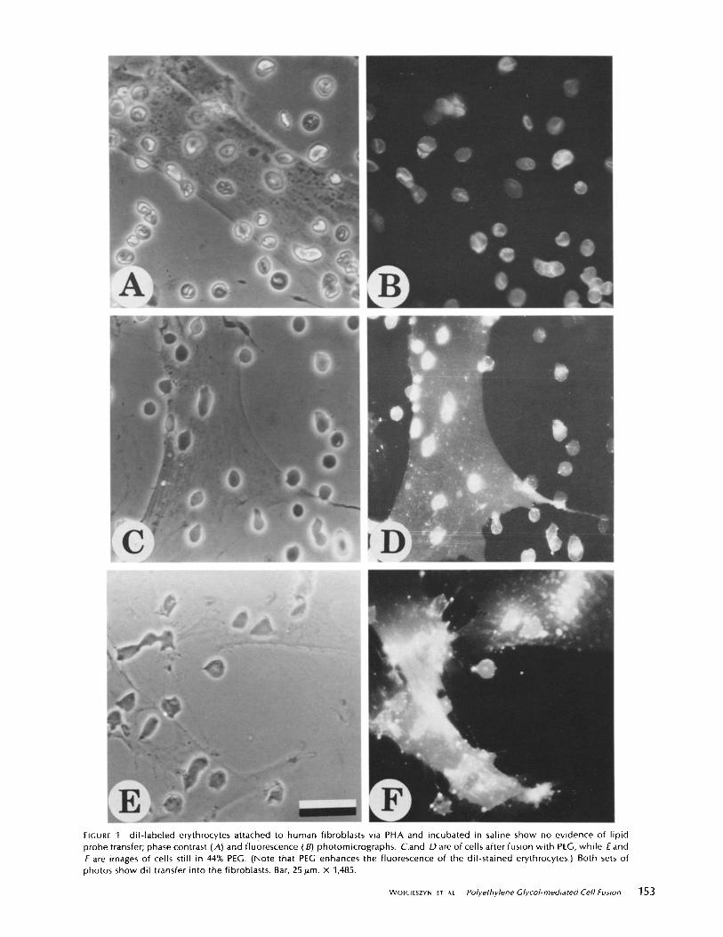
hGURE 1 dil- labeled erythrocytes attached to human fibroblasts via PHA and incubated in saline show no evidence of lipid probe transfer; phase contrast (A) and fluorescence (B) photomicrographs. C.and D are of cells after fusion with PEG, while E and F are images of cells still in 44% PEG. (Note that PEG enhances the fluorescence of the dil-stained erythrocytes.) Both sets of photos show dil transfer into the fibroblasts. Bar, 25 p.m. x 1,485.
WO]CI[SZYN ET AL. Polyethylene Glycol-mediated Cell Fusion 153

treatment with a haptenic sugar, N-acetylgalactosamine, for PHA or after treatment with either PEG or nonfusogenic PEG (see below).
Because cells were examined only after dilution and removal of PEG, it was not possible from these experiments to deter- mine precisely when transfer of the lipid probe had occurred. To answer this question, cultures were examined while PEG was still present. The plasma membranes of the cultured cells were observed to exhibit fluorescence in <30 s after the addition of PEG (Fig. 1 E). After 1-2 min in the presence of undiluted PEG, the stain had spread from the erythrocytes and was distributed over the plasma membranes of the cultured cells (Fig. 1 F).
To demonstrate the generality of these results, we examined the transfer of other lipid probes. Erythrocytes were labeled with a fluorescent derivative of phosphatidylcholine (C6-NBD- PC) or phosphatidylethanolamine (NBD-PE). The results with each of these probes were similar to those with diI, in that label
was transferred only upon addition of PEG and that transfer did not require dilution of PEG. Although these observations were compatible with transfer being accomplished as a result of membrane fusion, it was also possible that transfer was occurring in the absence of fusion. For instance, PEG could have mediated a unique membrane-membrane contact which facilitates an exchange of lipid molecules from one bilayer to another by a mechanism which did not require fusion. Or PEG could have acted as a lipid exchange vehicle, irrespective of its fusion capabilities, by serving as an agent in which the probe was solubilized. In this regard, C6-NBD-PC, but not NBD-PE, is readily exchanged between lipid vesicles or between vesicles and cells (30).
To distinguish between a fusion mechanism or a simple exchange mechanism of lipid probe transfer, we examined transfer in two fusion-inefficient systems. The first system substituted leaky erythrocyte ghosts for intact erythrocytes which do not fuse using Sendai virus as fusogen (15, 16).
FIGURE 2 Fluorescence micrographs show that dil-stained dodge erythrocyte ghosts did not form polyghosts after PEG treatment (see Materials and Methods) (A). Numerous individual ghosts or closed ghost membrane fragments are present (arrow), and most of the remaining membrane material has aggregated (star). Normal erythrocytes treated with PEG did yield poly-erythrocytes (arrows, B). Phase (C) and fluorescence (D) micrographs show that label could be transferred from the ghost membrane into the membrane of the cultured cells in the presence of PEG. Bar, 25/Lm A and B, x 1,850; Cand D, x 1,485.
154 THE IOURNAL OF CELL BIOLOGY. VOLUME 96, 1983
Similarly, PEG addition did not result in large polyghost structures (Fig. 2A) which are indicative of fusion. Note that many individual ghosts or closed ghost fragments remain (ar- row) and that much of the membrane material is aggregated (star). Intact erythrocytes did undergo PEG-mediated fusion (Fig. 2 B, arrows). When these ghosts were labeled with diI and then carried through the standard fusion protocol, fluorescence was again seen to be limited to erythrocyte membranes before addition of PEG (data not shown), but, upon addition of PEG, fluorescence was observed in the cultured cell membranes (Fig. 2 C and D), even though fusion between ghosts and cultured cells was unlikely to have occurred. (Upon dilution of the PEG, images such as seen in Fig. 1 D were observed with this system.)
The second fusion-inefficient system was based on the ob- servations of Honda et al. (6), noted earlier, that solvent- extracted PEG (nonfusogenic PEG) is unable to fuse cells. To confirm this fmding, we first established the fusion efficiency of commercial PEG by determining the percentage of cultured cells into which a cytoplasmic marker, fluorescein conjugated BSA (F-BSA), was transferred after fusion with erythrocytes which had been hypotonically loaded with the marker. In agreement with our previous results, >90% of the cultured cells exhibited cytoplasmic staining following the standard protocol. If, however, nonfusogenic PEG was tested for its ability to promote fusion, < 1% of the cultured cells exhibited the marker within their cytoplasm. Yet, when this nonfusogenic PEG was applied to erythrocytes labeled with diI, the lipid probe was transferred with high efficiency to cultured cell membranes (data not shown, but as in Fig. 1 D).
Thus, both of these fusion-inefficient systems point to a nonfusogenic exchange mechanism of lipid probe transfer. Of the two exchange mechanisms presented above, the one which involved PEG as a vehicle of transfer, as opposed to the one which is based on the dehydrating capacity of PEG, predicts that diI should be solubilized by PEG. Although diI can be dissolved via ethanol injection in 44% PEG and although this PEG/diI solution will stain the membranes of cultured cells, our preliminary experiments were not able to detect spectro- photometrically the solubilization by PEG of diI from the membranes of erythrocytes (data not shown). It is therefore difficult to explain the diI transfer in terms of the extraction of the probe by PEG, at least on the time scale of our experiments where PEG was present for only 1 min.
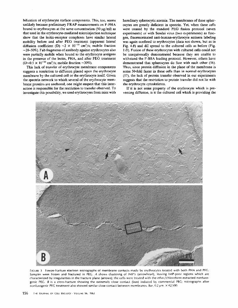
Support for the alternate hypothesis that PEG, by virtue of its dehydrating ability, promotes a unique type of membrane apposition that permits rapid transfer of lipid probes comes from freeze-fracture electron micrographs of erythrocytes which have been treated with both PHA and PEG. In the presence of 44% PEG, clustering of intramembrane particles (IMPs) resulted in the creation of IMP-depleted areas (Fig. 3A), similar to earlier evidence provided by Knutton (4) on erythrocytes and Kdihling (25) on HeLa cells. Note that the fracture plane appears to shift heights in the particle-free areas. This may suggest such close contact between adjacent, particle- free cell membranes that the fracture plane skips from one cell membrane to the other (4). Alternatively, Kdihling (25) has suggested that the loss of a preferred fracture plane represents a marked disorganization of these membrane regions caused by the PEG treatment. At any rate, cross-fracture of the erythrocytes in both fusogenic and nonfusogenic PEG shows that the apposed membranes are brought so close together that a space between them cannot be resolved by this technique (Fig. 3 B). These results are consistent with both types of PEG being able to mediate lipid probe transfer. By transmission
electron microscopy, the membranes of erythrocytes agglutin- ated by PEG are separated by <50 A and in regions appear to touch (4).
Mixing of Plasma Membrane Components: Proteins
Since these data indicate that lipid probes are transferred in the presence of PEG irrespective of fusion, it was of interest to determine whether the proteins of the erythrocyte membrane were similarly transferred. Erythrocyte membrane proteins with moieties accessible from the external medium were di- rectly labeled by covalent linkage of either FITC or EITC, and these ceils displayed a ring fluorescence typical of membrane labeling. In addition, the fluorescence persisted when the ceils were hypotonically lysed, and the hemoglobin and other pro- teins released by this process were not labeled when checked by fluorescence microscopy and gel electrophoresis (data not shown). When these erythrocytes were fused to cultured cells using the standard protocol, fluorescence was limited to the erythrocytes only, 2 even after a 24-h incubation at 37°C (Fig. 4). Fusion was not inhibited by labeling since >75% of the cultured cells were microinjected with F-BSA.
Although others have demonstrated that erythrocyte mem- brane proteins that have been labeled by either of these re- agents are mobile within the plane of the membrane (10, 11), our results might still be explained if the labeling protocol had somehow led to the immobilization of the labeled proteins. To circumvent this possible complication, we labeled erythrocyte membrane components after, rather than before, fusion so that the components would be allowed to diffuse before labeling. This was accomplished by fusing unlabeled human erythro- cytes with mouse cultured cells and then detecting erythrocyte membrane components by antibodies directed to them. When either direct or indirect immunofluorescence protocols were used, staining was again limited to erythrocytes only, even after incubations for 24 h at 37°C before staining (data not shown but as in Fig. 4 B). It might still be argued that the PEG itself had somehow immobilized the membrane components. This seems unlikely because, when Sendai virus was substituted for PEG as the fusogen, similar results were seen, again up to 24 h of incubation (data not shown but as in Fig. 4 B). Further- more, PEG treatment did not inhibit the intermixing of labeled membrane components following fusion (10, 19). Finally, the lectin, PHA, in combination with PEG could result in immo-
2 The detection limit for protein transfer can be roughly estimated as follows: 106 copies of Band 3 in a typical erythrocyte are dispersed over a surface area of 200/ma2; let us estimate the surface of a typical human fibroblast as 50/tm x 100 #m × 2 (for top and bottom) x 2 (for surface roughness) = 2,000 #m 2. (Our SEM photographs show the fibroblasts to be generally quite smooth.) Complete equilibration of Band 3 after one erythrocyte has fused with a fibroblast would result in 9 x 10⁵ copies introduced into the fibroblast. In our experience we can visually detect, by direct immunofluorecence, roughly 5 x 10⁵ copies per cell in fibroblasts. Thus, it is likely that we could detect complete transfer of about 50% of the available Band 3. Furthermore, if more than one erythrocyte transferred its protein to the fibroblast, with our indirect immunofluorescence protocol the detection limit should be reduced by a factor of 10 to about 5 x 10⁴ proteins per fibroblast.
Under these conditions, one would expect to discern the transfer of as little as 10% of the erythrocyte membrane proteins. Finally, it is important to note that no initial spreading of the labeled erythrocyte material to the fibroblast was ever noted; near the cell-cell contact region the concentration of labeled components should be nearly as high as in the erythrocyte itself.
WOIC~ESZYN ET At. Polyethylene Glycol-mediated Cell,Fusion 1 5 5
bilization of erythrocyte surface components. This, too, seems unlikely because preliminary FRAP measurements on F-PHA bound to erythrocytes at the same concentration (50 #g/ml) as that used in the erythrocyte-mediated microinjection technique show that the lectin-receptor complexes have similar lateral mobility before and after PEG treatment (apparent lateral diffusion coefficient (D) - 2 × 10 -1° cm2/s; mobile fraction -20-50%). Fab fragments of antibody against erythrocytes also were partially mobile when bound to the erythrocyte antigens in the presence of the lectin, PHA, and after PEG treatment (D-0.5 x 10 -1° cm2/s; mobile fraction ~30%).
This lack of transfer of erythrocyte membrane components suggests a restriction to diffusion placed upon the erythrocyte membrane by the cultured cell or the erythrocyte itself. Given the spectrin network to which several of the erythrocyte mem- brane proteins are anchored, one might suspect that this inter- action is responsible for the restriction to transfer observed. To investigate this possibility, we used erythrocytes from mice with
L
hereditary spherocytic anemia. The membranes of these spher- ocytes are greatly deficient in spectrin. Yet, when these cells were treated by the standard PEG fusion protocol (seven experiments) or with Sendal virus (two experiments) as fuso- gen, fluoresceinated anti-human-erythrocyte antisera labeling was again confined to erythrocytes (data not shown, but as in Fig. 4 B) and diI spread to the cultured cells as before (Fig. l D). Fusion of these erythrocytes with cultured cells could not be unequivocally demonstrated because they are unable to withstand the F-BSA loading protocol. However, others have demonstrated that spherocytes do fuse with each other (16). Thus, since protein diffusion in the plane of the membrane is some 50-fold faster in these cells than in normal erythrocytes (17), the lack of protein transfer observed in our experiments suggests that the restriction to protein transfer did not lie with the erythrocyte cytoskeleton.
If it is not some property of the erythrocyte which is pre- venting diffusion, is it the cultured cell which is providing the
FIGURE 3 Freeze-fracture electron micrographs of membrane contacts made by erythrocytes treated with both PHA and PEG. Samples were frozen and fractured in PEG. A shows clustering of IMP's (arrowhead), leaving IMP-poor regions which are characterized by irregularities in the fracture plane (arrows); the cells were treated with the ether/chloroform-extracted nonfuso- genic PEG. B is a cross-fracture showing the extremely close contact (box) induced by commercial PEG; micrographs after nonfusogenic PEG treatment also showed similar close contact between membranes. Bar, 0.2 #m. x 62,500.
156 THE JOURNAL OF CELL BIOLOGY-VOLUME 96, 1983

network or a glycocalyx, restrictions to lateral diffusion appear to be attributable to some structural aspect of the recipient cell surface.
FIGURE 4 Erythrocyte membrane proteins labeled wi th F ITCwere fused with cultured fibroblasts and incubated at 37°C for 24 h and then examined for probe location (A, phase; B, fluorescence). The FITC fluorescence is seen to be l imited to the original erythrocyte membranes. Bar, 25 p.m. x 1,485.
restriction? To determine whether the human diploid fibroblast recipient ceils are not a special case, we fused erythrocytes to BHK or Vero cells. In these experiments as well, fluorescence of the cultured cell plasma membranes due to transfer of the labeled erythrocyte components was not observed.
It therefore appears that, although protein intermixing is well-documented in mouse-human (18) and erythrocyte-eryth- rocyte (19) fusion systems, it does not occur in all systems undergoing fusion. Restrictions upon lateral diffusion of mem- brane components introduced into cultured cells by fusion is not unprecedented. In an extensive study, Baumann et al. (20) have reconstituted vesicles from mixtures of deoxycholate-sol- ubilized phospholipids and proteins and then fused the vesicles to mouse L-cells using PEG. The phospholipids of the vesicles mix relatively rapidly with the cell membrane lipids after fusion, as judged by immunofluorescence staining of trinitro- phenylated lipids which had been reconstituted into the vesi- cles. In contrast, proteins transferred to the L-cells showed restricted diffusion using the same immunofluorescence tech- niques. Even after the fused cells had been in culture for 16 h, the majority of labeled glycoproteins was still concentrated in discrete patches. That fusion had indeed occurred was dem- onstrated in that the functions of the serum asialoglycoprotein receptor were conferred upon receptor-deficient L-cells. Since the vesicles presumably do not contain either a cytoskeletal
Mixing of Cytoplasmic Compartments The endpoint of fusion is marked by formation of a pore
through which intercytoplasmic communication can take place. We have previously reported that entry of cytoplasmic markers (375-160,000 daltons) from erythrocytes to cultured cells begins 1 to 3 min after dilution of PEG (7). This result provoked the question of whether the actual endpoint of fusion occurs some- time after dilution of the PEG or during the PEG incubation, but transfer of the cytoplasmic probe is delayed for some reason.
To examine whether the entrapped F-BSA is mobile within the cytoplasm during the fusion protocol, we used the FRAP technique, a method we have recently used to measure the diffusion of cytoplasmic probes within individual, living cells (7). Erythrocytes which were loaded with F-BSA and cultured fibroblasts, previously microinjected with F-BSA, were placed into 44% PEG and the F-BSA diffusion coefficients were determined. In PEG, recovery in both cell types was slow or nonexistent, indicating that the F-BSA was operationally im- mobile (D~ < 10 -12 cm2/s; Fig. 5, + PEG curve). However, if the PEG was removed by extensive washing, F-BSA diffused freely with D=10 -s cm2/s. (Fig. 5, - P E G curve), values consistent with our earlier results for cytoplasmic diffusion in fibroblasts (7). Results were independent of whether the PEG used was fusogenic or nonfusogenic, but, of course, transfer from eryth- rocytes to cultured ceils occurred only when fusogenic PEG was used. Therefore it seems likely that the delay observed between dilution of PEG and the transfer of a cytoplasmic probe may be attributed to probe immobilization which is only relieved by dilution of the polymer. Presumably, this phenom-
-PEG
4
oo
3
z
o
\ //
• * • " " • ,, * • +PEG
TI ME (s)
FIGURE 5 Fluorescence recovery after photobleaching measure- ments of cytoplasmic dif fusion of F-BSA wi th in cultured fibroblasts after PEG addit ion ( . . . . ; + PEG) and after PEG removal (- - -; -PEG). Initial f luorescence is Fi, photobleaching is indicated by arrow labeled pb, and f luorescence after recovery is completed is Foo. The lack of f luorescence recovery in PEG (+ PEG) is interpreted as probe immobi l izat ion (diffusion coeff ic ient =10 -12 cm2/s), whi le after PEG removal (-PEG) the FRAP kinetics are consistent wi th a dif fusion coeff icient of ~10 -8 cm2/s. The same phenomenon was observed wi th F-BSA loaded erythrocyte in the presence and ab- sence of PEG. A x 25 object ive was used, giving an e -2 beam diameter of ~3.4 p.m.
WOICIESZYN [T AL. Polyethylene Glycol-mediated Cell Fusion 157

enon is closely related to the reversible precipitation of proteins induced when sufficient PEG is added to decrease the available volume for proteins; rehydration (dilution) reverses this effect (21). It is clear that pore formation must occur at some time between the beginning of the PEG dilution and the actual observation of probe transfer.
SUMMARY AND HYPOTHESIS
The first requirement for the efficient fusion of erythrocytes with cultured cells by PEG is the attachment of the two cell types to one another by a lectin bridge (9); this procedure enhances the efficiency of the process by binding more of the added erythrocytes with the target cells (2). It is also a prereq- uisite for PEG-mediated fusion ofglycolipid vesicles to various cultured cells (3). However, in neither fusion system does the action of the lectin result in the transfer of membrane or cytoplasmic probes associated with the vesicle or the erythro- cyte, respectively, to the cultured cell.
Once cells have been brought into proximity by the lectin, transfer of lipid probes from erythrocytes to cultured cells occurs only upon addition of PEG. In the presence of PEG, IMPs have been observed to segregate, leaving lipid-rich, IMP- free regions of the membrane (4, 25). In addition, our freeze- fracture experiments reveal that erythrocyte membranes come extremely close to each other in the presence of both fusogenic and nonfusogenic PEG. Since nonfusogenic PEG also mediates lipid probe transfer, this transfer phenomenon is not dependent on fusion. The dehydrating ability of PEG may serve a dual role in mediating these events. First, it may be responsible for IMP clustering, either by dehydrating lipids and thus affecting fluidity or by directly promoting protein aggregation through dehydration. (Alternatively, PEG may simply cross-link prox- imate proteins.) Second, the dehydration of lipid membranes, in general, will force them as close together as 5 A (22). It seems quite plausible that lipid transfer could occur precisely in such regions of ultraclose membrane apposition.
Transfer of a cytoplasmic probe from erythrocytes to cul- tured cells, which is truly indicative of fusion, occurs only with commercial PEG and not with its recrystallized counterpart. Thus, fusion requires, in addition to the pure polymer, some agent contained in commercial PEG which is removed upon recrystallization. Likely candidates are the ingredients added to PEG during production, such as antioxidants and polymer- ization agents, since, upon addition of certain of these com- pounds to purified PEG, fusion competence is restored (6).
Indeed, we have shown that the fusogenic capacity of com- mercial nonfusogenic PEGs and extracted PEG can be restored by addition of several different antioxidants (Wojcieszyn, J., unpublished observations). Thus, if the term fusogen is to be assigned, the designation would most properly fit the second class of ingredients in PEG, keeping in mind that other pre- conditions must be met before the term takes on meaning. This separation of activities in a fusion-competent system is not unprecedented. Lucy et al. (5) have shown that erythrocytes can be fused by the addition of certain lipids but only in the presence of high concentrations of dextran. GMO was later shown to promote an isotropic NMR signal from isolated erythrocyte lipids (23), indicating its potential to destabilize the bilayers. In the present case, the combination of PEG-induced dehydration, favoring a rigid lipid state, together with the fusogenic additives favoring a fluid state may produce unstable phase boundaries which nucleate the fusion process (24). Fu- sion may also be "driven" by membrane tension (stretch)
158 TH[ JOURNAL OF C[tL BIOLOGY. VOLUME 96, 1983
produced by the intracellular volume expansion (32) accom- panying the PEG dilution step in combination with the fixed membrane regions caused by the intercellular lectin cross- bridges; this membrane stress could then be relieved by the formation of pores originating at the fusogen-induced bilayer defects. In any event, after the fusion process, cytoplasmic communication is possible but may be prevented by the im- mobilization of cytoplasmic molecules within both the eryth- rocyte and the cultured cell due to the continued presence of PEG. Once the dehydrant is removed and mobility is restored, actual transfer via diffusion occurs.
In our system, erythrocyte membrane proteins do not diffuse into the membrane of the cultured cell after fusion. This is perhaps surprising since the membrane proteins of two differ- ent cultured ceils are free to mix after fusion. However, when one considers the specialized cytomembrane structure of the erythrocyte, it is not clear that analogies between the two systems are appropriate. At a minimum, our data imply a special, porous erythrocyte-cultured cell junctional region hav- ing channels which permit cytoplasmic transfer. Perhaps what has been produced is an artificial topographic specialization analogous to that which occurs naturally, for example, at gap junctions. Whether further integration is prevented by incom- patible submembranous structures, integral membrane com- ponents, or associated glycocalyx is not clear.
This study strongly suggests that commercial PEG performs two, separable functions: first, it brings the adjacent plasma membranes into very close apposition so that lipid transfer can occur. Second, it must destabilize these close contact areas enough so that fusion events become a predominant route to a more stable state. Similar functions can be identified in both biological and artificial bflayer membrane fusion. For example, chromaffm granules are aggregated by Ca ++ and synexin and can be promoted to fuse by the addition of arachodonic acid (27). In artificial membranes, the ionic fusogen, Ca ++, appears both to aggregate membranes in an anhydrous contact state and to destabilize the apposed membranes leading to fusion (28). Membrane aggregation, close apposition and destabiliza- tion appear to be a recurrent theme and to provide the neces- sary conditions for fusion.
We gratefully acknowledge the helpful discussions and suggestions of Drs. Z. Derzko and E. Evans (University of British Columbia, Van- couver), Drs. R. McDaniel and S. Simon (Duke University, Durham, NC), Dr. S. Nir (The Hebrew University of Jerusalem, Israel), and Dr. J. Lucy (University of London, UK) for making available a preprint of new work on PEG. We thank Dr. M. H6chli (University of North Carolina, Raleigh, NC) for performing the freeze-fracture microscopy and providing advice on the interpretation of the micrographs. We also acknowledge the technical assistance provided by Ms. D. O'Dell.
This work was supported by National Institutes of Health grants GM 29234 (to K. A. Jacobson) and GM 25650 and GM27591 (to R. A. Schlegel) and American Heart Association Grant-In-Aid 81-711 (to J. W. Wojcieszyn). K. A. Jacobson was an Established Investigator of the American Heart Association during the conduct of this project.
Received for publication 13 April 1982, and in revised form 15 September 1982
REFERENCES
1. Davidson, R. L., and P. S. Gerald. 1977. Induction of mammalian somatic cell hybridi- zation by polyethylene glycol. In Methods in Cell Biology. D. Prescott, editor. Academic Press, Inc., London and New York. 325-338.
2. Schlcgel, R. A., and M. C. Rechsteiner. 1978. Red cell-mediated microinjection of macromolecuies into mammalian ceils. In Methods m Ceil Biology. D. Prescott~ editor. Academic Press, Inc., New York. 341 354.
3. Szoka, F., K-E . Magnusson, J. W. Wojcieszyn, Y. Hou, Z. Derzko, and K. Jacobson.

1981. Use of lectins and polyethylene glycol for fusion of glycoilpid-containing liposomes with eukaryotic cells. Proc. Natt Acad. Set U. S. A. 78:1685-1689.
4. Knnttnn, S. 1979. Studies of membrane fusion. III. Fusion of erythrocytes with polyeth- ylene glycol. J. Cell Sci. 36:61-72.
5. Lucy, J. A. 1978. Mechanisms of chemically induced cell fusion. In Membrane Fusion. G. Poste, and G. Nicolson editors. Elsevier/North HoLLand Press. 267-304.
6. Honda, K., Y. Maeda, J. Sasakawa, H. Ohno, and E. Tsuchida. 1981. Activities of ceil fusion and lysis of the hybrid type of chemical fusogens. I. structure and function of the promotor of ceil fusion. Biochem. Biophys. Res. Commun. 100:442-448.
7. Wojcieszyn, J. W., R+ A. Schlegel, E-S. Wu, and K. A. Jacobson. I981. Diffusion of injected macromolecules within the cytoplasm of living cells. Proc. Natt Acad Sci. U. S. A. 78:4407~M. 10.
8. Moor, H., and K. Miihlethator. 1963. Fine structure in frozen-etched yeast cells. J. Cell Blot 17:609-628.
9. Scklegel, R. A., and W. E. Mercer. 1980. Introduction of Macromolecules into Viable Mammalian Ceils. R. Baserga, L. Croce, and G. Rovera, editors. Liss, New York. 145-155.
10. Fowler, V., and D. Branton. 1977. Lateral mobility of human erythrocyte integral membrane proteins. Nature (Lond). 268:23-26.
1 I. Golan, D. E., and W. Veatch. 1980. Lateral mobility of band 3 in the human erythrocyte membrane studied by fluorescence photobleaching recovery: evidence for control by cytoskeletal interactions. Proc. NatL Acad Sci. U. S. A. 77:2537-2541.
12. Jacobson, K., Z Derzko, E-S Wu, Y. Hou, and G. Poste. 1977. Measurement of the lateral mobility of ceil surface components in single living ceils by fluorescence recovery after photobleaching. J. SupramoL Struct. 5:565-576.
13. Szoka, F., K. Jacobson, Z. Derzko, and D. Papahadjopoulos. 1980. Fluorescence studies on the mechanism of liposome-ceil interactions in vitro. Biochim. Biophys. A eta. 600:1-18.
14. Jacobson, K., Y. Hou, Z. Derzko, J. Wojcieszyn, and D. Organisciak. 1981. Lipid lateral diffusion in the surface membrane of cells and in multibilayers formed from I~lasma membrane lipids. Biochemistry 20:5268-5275.
15. Knutton, S., and T. Bachi. 1980. The role of cell swelling and haemolysis in sendal virus- induced cell fusion and in the diffusion of incorporated viral antigens. J. Cell ScL 42:153-167.
16. Sekiguchi, K., and A. Asano. 1978. Participation ofspectrin in sendal virus-induced fusion of human erythrocyte ghosts. Proc. Natl. Acad. Sci. U. S. A. 75:1740-1744.
17. Sheetz, M. P., M. Schlindler, and D. E. Koppel. 1980. Lateral mobility of integral
membrane proteins is increased in spherocytic erythrocytes. Nature (Lond). 285:510-512. 18. Frye, L. D., and M. Edidin. 1970. The rapid mixing of cell surface antigens after formation
of mouse-human heterokaryons. J. Cell Sci. 7:319-335. 19. Koppel, D. E., and M. P+ Sheetz. 1981. Fluorescence photobleaching does not alter the
lateral mobility of erythrocyte membrane glycoproteins. Nature (Lond). 293:159-161. 20. Baumann, H., E Hou, and E. Doyle. 1980. Insertion of biologically active membrane
proteins from rat liver into the plasma membrane of mouse fibroblasts. Z BioL Chem. 255:10001-10012.
2l. Atha, D. H., and K. C. lngham. 1981. Mechanism of precipitation of proteins by polyethylene glycols. J. Biol. Chem. 256:12108-12117.
22. McCown, J. T., E. Evans, S. Diehl, and H. C. Wiles. 1981. Degree of hydration and lateral diffusion in phospholipid multibilayers. Biochemistry. 20:3134-3138.
23. Hope, M. l., and P+ R. Cullis. 1981. The role of nonbilayer lipid structures m the fusion of human erythrocytes induced by lipid fusogens. Biochim. Biophys. Acta. 640:82-90.
24. Boni, L. T., T. P. Stewart, J. L. Alderfer, and S. W. Hui. 1981. Lipid-polyethylene glycol interactions. II. Formation of defects in bilayers. J. Membr. BioL 62:71-77.
25. Kriihling, H. 1981. Investigations on polyethylene glycol-induced cell fusion-- freeze fracture observations. Acta Histochemica (SuppL).-Band X X Ill S. 219-223.
26. Putnam, R. W., M. Tan, T. Lynn, C. W. Easley, and S. Migita. 1962. The cleavage of rabbit gamma globulin by papain. J. BioL Chem. 23"/:717-726.
27. Creutz, C. E. 1981. cis-Unsaturated fatty acids induce the fusion of chromaffin granules aggregated by synexin. J. Cell BioL 91:247-256.
28. Portia, A., C. Newton, W. Pangborn, and D. Papahadjopoulos. 1979. Studies on the mechanism of membrane fusion: evidence for an intermembrane Ca2+-phospholipid complex, synergism with Mg 2+, and inhibition by spectrin. Biochemistry. 18:780-790.
29. Smith, C., Q. Ahkong, D. Fisher, and J. Lucy. 1982. Is purified poly (ethylene glycol) able to induce fusion? Biochim. Biophys. Acta. In press.
30. Pagano, R., O. Martin, A. Schroit, and D. Struck. 1981. Formation of asymmetric phospholipid membranes via spontaneous transfer of fluorescent lipid analogues between vesicle populations. Biochemistry. 20:492~4927.
31. Schneider, H., J. J. Lemasters, M. Hbehli, and C. R. Hackenbrock. 1980. Liposome- mitochondrial inner membrane fusion. Z BioL Chem. 255:3748-3756.
32. Cohen, F., M. Akabas, and A. Finkelstein. 1982. Osmotic swelling of phosphospholipid vesicles causes them to fuse with a planar phosphoilpid bilayer membrane. Science. 217:458-460.
WOIOESZYN ET At. Polyethylene Glycol-mediated Cell Fusion "159