Embed Size (px)
Citation preview

Virology /lecture 3 Department of biology / 4 th stage
Viral Multiplication
•Mechanism used depends on viral structure and genome •Steps are similar
1) Attachment (Adsorption)2) Penetration3) Uncoating4) Gene expression. -Synthesis of viral mRNA (transcription) -Synthesis of viral proteins (translation)
5) Genome replication6) Virion assembly/maturation 7) Release of new infectious virus -lysis : breakdown of cell membrane and release of virus -budding: viruses "bud" through cell membrane and are released without necessarily killing the cell. Viruses acquire envelopes (membranes) during this process.
I: Attachment:
In most cases, specific attachment proteins on the surface of viruses bind to specific receptors on the surface of animal cells. Cellular receptors are usually either glycoproteins or glycolipids, and have other functions for the cell in addition to virus binding. The specific interaction between attachment proteins and cellular receptors is a major determinant of the host-range, or tropism of the virus. Some viruses have a very narrow host range, meaning that they can only infect one or a small number of cell types, while others have broad host ranges, meaning that they can infect a large number of different cell types. This is partially determined by whether the receptor for the virus is expressed on many or a limited number of cell types. Some examples of specific viruses and their known or probable cellular receptors are given in the following table.
Dr. Noor Sami Aboud

Virology /lecture 3 Department of biology / 4 th stage
Virus Viral Attachment Molecule
Likely Cellular Receptor
Target Cell Type
rabies virus (Rhabdoviridae)
glycoprotein Acetylcholine receptor
neuron
FIV
(Retroviridae)
gp120 CD9, leukocyte
differentiation Ag
T cell, macrophage
pseudorabies virus
Herpesviridae
gC Heparin sulfate proteoglycans
many cell types
influenza A virus
Orthomyxoviridae
Hemagglutinin (HA)
Sialic-acid containing glycoproteins
respiratory epithelium
Coronaviridae
Human Coronavirus SARS virus
Fig.1: Envelope spike protein binds to sialic (neuraminic) acid on surface of mammalian cells
OrthomyxoviridaeDr. Noor Sami Aboud

Virology /lecture 3 Department of biology / 4 th stage
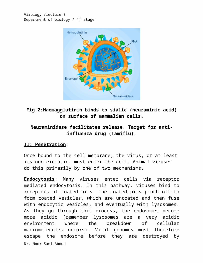
Influenza viruses
Fig.2:Haemagglutinin binds to sialic (neuraminic acid) on surface of mammalian cells.
Neuraminidase facilitates release. Target for anti-influenza drug (Tamiflu).
II: Penetration:
Once bound to the cell membrane, the virus, or at least its nucleic acid, must enter the cell. Animal viruses do this primarily by one of two mechanisms.
Endocytosis: Many viruses enter cells via receptor mediated endocytosis. In this pathway, viruses bind to receptors at coated pits. The coated pits pinch off to form coated vesicles, which are uncoated and then fuse with endocytic vesicles, and eventually with lysosomes. As they go through this process, the endosomes become more acidic (remember lysosomes are a very acidic environment where the breakdown of cellular macromolecules occurs). Viral genomes must therefore escape the endosome before they are destroyed by proteases, nucleases, etc. For enveloped viruses, this usually occurs by membrane fusion mediated by a fusion protein. One example of this is the influenza virus HA protein, which undergoes a conformational change induced by the acidic environment of the endosome. After undergoing this change, it then induces membrane fusion, releasing the nucleocapsid into the cytoplasm. The genomes of non-enveloped viruses must also somehow escape the endosome.
Dr. Noor Sami Aboud
Virology /lecture 3 Department of biology / 4 th stage
Direct Membrane Fusion: Some enveloped viruses directly fuse with the plasma membrane. In these cases the activity of a fusion protein is not dependent on pH change, but rather is induced in response to receptor binding.
III: Uncoating
With some viruses, the genome is completely released from the capsid during or after penetration. This is known as "uncoating". In others, such as retroviruses and reoviruses, the first stages of the viral replication cycle (transcription, replication) actually occur inside the capsid. These capsids have undergone some conformational changes during infection that allow viral gene expression and/or replication to begin, and the resulting structures are sometimes known as partially uncoated particles. Since almost all DNA viruses replicate in the nucleus of infected cells, they must be targeted there. In many cases the entire nucleocapsid enters the nucleus, where uncoating then takes place.
Dr. Noor Sami Aboud

Virology /lecture 3 Department of biology / 4 th stage
HIV Virus Membrane Fusion Influenza Virus
Endocytosis
Fig.3: Penetration of viruses
Dr. Noor Sami Aboud

Virology /lecture 3 Department of biology / 4 th stage
Fig. 4: Entry and Uncoating of viruses
Dr. Noor Sami Aboud

Virology /lecture 3 Department of biology / 4 th stage
IV: Gene Expression and Genome Replication
In order for new virus to be assembled, both new viral genomes and other virion components (proteins) must be produced. Exactly how this occurs varies greatly depending on the family (and Baltimore Class) of virus.
Viruses with DNA Genomes:
Almost all DNA viruses have genomes that are similar to the host cell; that is, they are composed of double stranded DNA, and are therefore able to utilize host enzymes to express viral genes and replicate viral DNA. Most DNA viruses replicate in the cell nucleus, which is where cellular replication and transcription proteins are localized. After infection, the nucleocapsid of DNA viruses is therefore usually delivered to the nucleus where uncoating occurs. An exception is poxviruses, which replicate in the cytoplasm of infected cells.
Viruses with RNA Genomes
As discussed previously, many families of animal viruses have RNA as their genetic material. These RNA genomes can be single stranded (+ sense, - sense, ambisense) or double stranded. Each class of genome has a different replication / gene expression strategy, and there is considerable variation within each class
Some General Points about Viruses with RNA Genomes:
- Most replicate entirely in the cytoplasm of infected cells. Exceptions are orthomyxoviruses and retroviruses.
- They generally have high mutation rates due to high error rates of RNA dependent RNA polymerases, which have no proofreading function.
- Show high levels of "recombination".
- In the case of viruses with segmented genomes, there can be "reassortment" of the segments in cells infected by more than one strain/variant of virus.
V: Virus Assembly and Release
Dr. Noor Sami Aboud

Virology /lecture 3 Department of biology / 4 th stage
Once new viral genomes and proteins have been produced, they are assembled into new virions. This usually occurs in a very specific order. For example, for many viruses, the viral capsid is partially assembled (ie, the newly synthesized capsid proteins associate together into a capsid-like structure). The viral genome is then inserted into the capsid to form a nucleocapsid, which then undergoes some type of maturation that can include proteolytic cleavage of capsid proteins. In the case of non-enveloped viruses, these newly formed virions accumulate in the cell and are released by cell lysis.
In the case of enveloped viruses, the nucleocapsids often assemble on the surface of a cellular membrane (such as the plasma membrane, the nuclear envelope, the ER, etc.) in regions of the membrane where viral envelope proteins are concentrated. Matrix proteins, if present, are underlying this part of the membrane. The virus then "buds" through the membrane to give rise to enveloped viral particles. These particles can then go through additional maturation events to give rise to infectious virus. In the case of viruses that form on the plasma membrane, they can bud from the cell without causing cell lysis. Other enveloped viruses, however, are lytic.
Dr. Noor Sami Aboud

Virology /lecture 3 Department of biology / 4 th stage
Fig. 5: Virus Assembly
Dr. Noor Sami Aboud

Virology /lecture 3 Department of biology / 4 th stage
Fig. 6:Virus Release
Dr. Noor Sami Aboud

Virology /lecture 3 Department of biology / 4 th stage
Fig. 7: Virus Multiplication
Dr. Noor Sami Aboud