Embed Size (px)
Citation preview

APPLIED AND ENVIRONMENTAL MICROBIOLOGY, Mar. 1981, p. 568-5790099-2240/81/030568-12$02.00/0
Vol. 41, No. 3
Physiological Criteria and Mycotoxin Production as Aids inIdentification of Common Asymmetric Penicillia
JENS C. FRISVADFood Technology Laboratory, The Technical University ofDenmark, DK-2800 Lyngby, Denmark
The taxonomy of the asymmetric (predominantly terverticillate) penicillia isbased on morphological differences that leave identification difficult. The appli-cation of physiological criteria facilitated the identification of the common asym-metric penicillia investigated. Changes in the placement of some strains of thesepenicillia made the connection to mycotoxin-producing ability clearer. The clas-sical criterion of conidium color was deemphasized and replaced by the followingcriteria: (i) growth on nitrite-sucrose agar and (ii) growth and acid (and subsequentbase) production on creatine-sucrose agar (containing bromocresol purple). Othercriteria used or developed were: (iii) growth on sorbic acid plus benzoic acid agar(50 + 50 ppm, pH 3.8), (iv) growth on an agar containing 1,000 ppm propionicacid (pH 3.8), (v) growth on an agar containing 0.5% acetic acid, (vi) growth at370C, (vii) growth rate on an agar containing 0.1% pentachloronitrobenzene, (viii)production of extracellular tricaproinase, and (ix) fasciculation on a mediumcontaining 10 ppm botran (2,6-dichloro-4-nitroanilin). The pattern of extracellularmetabolites after thin-layer chromatography was used as a chemotaxonomiccriterion. The species investigated, the number of isolates investigated, and thetoxins which some of these isolates produce were: Penicillium roqueforti (18)(patulin), P. citrinum (11) (citrinin), P. patulum (9) (patulin and griseofulvin), P.expansum (patulin and citrinin), P. hirsutum (13), P. brevicompactum (19), andP. chrysogenum (12). Widespread species of the P. cyclopium, P. viridicatum,and P. expansum series of Raper and Thom (A Manual of the Penicillia, 1949)were subdivided into four new groups: "P. crustosum pA" (29) (penitrem A), "P.melanochlorum" (29), "P. cyclopium p" (119) (penicillic acid and infrequentlypenitrem A), and "P. viridicatum o-c" (43) (ochratoxin A and citrinin). "P.viridicatum o-c" was separated from "P. cyclopium p" due to its ability to growon nitrite as sole nitrogen source. The species and groups investigated wererelated to the new taxonomic classification of the genus Penicillium according toPitt.
The difficulties in identifying the asymmetricpenicillia because ofthe absence of distinct char-acteristics have been pointed out repeatedly (5,6, 28, 33, 35). The colony texture and color usedin Penicillium taxonomy are often artificial andsubjective criteria (20). Raper and Thom (33)were aware of the difficulties and indicated thatthe subgrouping of the non-divaricate asymmet-ric penicillia in velutina, lanata, funiculosa, andfasciculata might separate species that are bio-chemically and physiologically alike. Further-more, most of the common asymmetric penicilliaare difficult to group on the basis of micromor-phological criteria alone. Pitt (30) mentions tran-sition forms between Penicillium brevicompac-tum and P. puberulum, species consideredclearly separable by Raper and Thom (33). Sam-son et al. (35) consider their broad species P.verrucosum as nearly inseparable micromor-phologically from P. roqueforti.
Physiological criteria in Penicillium taxon-omy have not been used extensively becausemolds in general have a sufficiently varied mor-phology to allow identification to the specieslevel (21). Some investigators have, however,used physiological criteria in the subdivision ofthe penicillia, such as the ability to grow on amedium containing 0.5% acetic acid, separatingP. roqueforti from other penicillia (8), or theability to grow on nitrite (1, 8) or creatine (8) assole nitrogen source. Pitt (28, 29, 30) uses colonydiameter at different temperatures and wateractivities to separate sections, series, and speciesin Penicillium.This investigation ainmed to establish physio-
logical criteria for the identification of commonasymmetric penicillia. The mycotoxin-producingability of the species treated was emphasized.The species investigated were the commonasymmetric penicillia in foods and feeds: P. cy-
568
on March 28, 2020 by guest
http://aem.asm
.org/D
ownloaded from

IDENTIFICATION OF COMMON ASYMMETRIC PENICILLIA 569
clopium, P. viridicatum, P. expansum, P. ci-trinum, P. brevicompactum, P. patulum, and P.chrysogenum (19, 20), and species that occurfrequently in Danish products such as P. roque-forti, P. crustosum, P. verrucosum var. melan-ochlorum, and P. hirsutum.
MATERIALS AND METHODSSource and maintenance of cultures. The
strains were obtained from culture collections (Table1) or isolated from Danish and Italian products (Table2). The cultures were maintained on potato dextroseagar (PDA) (Difco) at 0.50C.
All of the isolates were identified by the method ofRaper and Thom (33) with the following exceptions.P. hirsutum was identified by the method of Pitt (30).The members of the P. chrysogenum series (33) wereidentified by the method of Samson et al. (34). P.viridicatum was subdivided in the three subgroupsmentioned by Ciegler et al. (5). P. palitans as de-scribed by Raper and Thom (33) was divided into P.verrucosum var. melanochlorum (called "P. melan-ochlorum") and isolates which could be placed to-gether with P. cyclopium, P. crustosum, or P. viridi-catum (deemphazising the dark colors of such iso-lates). The name P. patulum was used in place of P.urticae (30, 33). As the new manual of the penicilliaby Pitt (30) was published recently, Table 3 shows therelations between the species defined by Pitt (30) andthe species/groups analyzed in this work.Media and growth conditions. The strains were
identified on Czapek agar (33) and Blakeslee malt agar(33). These cultures were incubated at 25°C for 14days. Furthermore the strains were identified by themethod of Pitt (30) using Czapek yeast autolysate agar(CYA), malt extract agar (MEA), and 25% glycerolnitrate agar. These cultures were incubated at 25°Cfor 7 days. Furthermore, the strains were incubated onglucose yeast extract (GY) agar (9, 41) (glucose, 20 g;yeast extract [Difco], 5 g; agar [Difco], 20 g; water, 1liter) at 37°C for 7 days.
Toxin production media used were: yeast extractsucrose (YES) agar (15, 38) (yeast extract, 20 g; su-crose, 150 g; agar, 20 g; water, 1 liter) and GY agar (9).
During preliminary tests on a few typical strainsfrom the different species, it was noted that of manywidely used nitrogen sources (ammonium chloride,ammonium nitrate, creatine, nitrite, cystein, casein,and other N sources), creatine and nitrite were veryuseful. The results concerning different carbon sourceswere not promising. The ability of the penicillia toproduce different extracellular enzymes (proteases, li-pases, and amylases) was tested, and of the lipases,tricaproinase showed the most consistent results. Thefollowing media were developed as selective and indic-ative media.
(i) Media with different N sources. Nitrite-su-crose (N02) agar consisted of the following: NaNO2, 3g; sucrose, 30 g; K2HPO4.3H20, 1.3 g; mineral solution,10 ml; agar, 20 g; water, 1 liter.
Creatine-sucrose (CREA) agar consisted of: crea-tine, lH20 (Merck, p.a.), 3 g; sucrose, 30 g; K2HPO4.3H20, 1.3 g; mineral solution, 10 ml; bromocresol pur-
ple, 0.05 g-, agar, 20 g; water, 1 liter. After autoclaving,the pH of the medium was adjusted to 8.0 with sterile1 N NaOH solution.The mineral solution was made as follows: KCl, 5 g;
MgSO4.7H20, 5 g; and FeSO4.7H20, 0.1 g, were dis-solved in 100 ml of water.
(ii) Lipase medium. Tricaproin agar consisted ofCzapek basal medium (Difco), 35 g; CaCl2.2H20, 0.4g; agar, 20 g; water, 1 liter. After autoclaving, 1 g ofsterile tricaproin (glycerine tricaproate) (Fluka,techn.) per liter of basal medium was added to themolten agar (50°C). The mixture was homogenized inan atomixer (MSE) (12,000 rpm, 1 min).
(iii) Media with different fungicides. Media withdifferent fungicides included PDA (Difco) with 0.1%pentachloronitrobenzene (Fluka, pract.) (PCNB). ThepH was adjusted to 6.5 after mixing the ingredients.After autoclaving, the pH of the medium was read-justed to 6.5. The medium was agitated very thor-oughly before and after autoclaving and before pouringthe medium in the petri dishes.
Another medium with a fungicide was PDA (Difco)containing 10 ppm (10 mg/liter) botran (2,6-dichloro-4-nitro aniline) (the Technical University of Denmark,the botran was recrystallized twice in acetone), whichwas called PDAB. A 10-mg amount of botran dissolvedin 10 ml of acetone was added to 1 liter of molten PDAmedium (500C).GY agar with 50 ppm sorbic acid and 50 ppm
benzoic acid (pH 3.8) (GYBS) was a third medium inthis group. Sodium benzoate, 0.09 g, and potassiumsorbate, 0.067 g, were added to 1 liter of GY agar.After autoclaving, the pH of the medium was loweredto 3.8 with sterile 1 N HCl.GY agar with 1,000 ppm propionic acid (pH 3.8)
(GYP) was another medium in this group. A 1.375-gamount of sodium propionate was added to 1 liter ofGY agar. After autoclaving, the pH of the medium waslowered to 3.8 with sterile 1 N HCl.GY agar with 5,000 ppm acetic acid (GYA) was the
last medium in this group. After autoclaving, glacialacetic acid was added to a final concentration of 0.5%(vol/vol). pH was adjusted to 3.8.These media were three-point inoculated using
spore suspensions of the molds (106 to 107 conidia perml of suspension medium). Suspension medium con-sisted of: NaCl, 8.5 g; Tween 80 (Merck, p.a.), 1 g;water, 1 liter. The cultures were incubated at 20°Cand observed from the day 4 of incubation.
Inoculation of apples. Spore suspensions (as de-scribed) of all the isolates of P. verrucosum var. me-lanochlorum, P. expansum, and P. crustosum, andother penitrem A producers were inoculated into sur-face-sterilized apples (Grasten and Spartan varieties)using a preparation needle. The apples were surfacesterilized by dipping in a 1% solution of NaOCl (1 min)and washed two times in sterile water. The diameterof the rot was recorded after 7 and 14 days of incuba-tion (incubation temperature, 20°C).Mycotoxin analysis. Mycotoxin analyses were
performed the day 4, 5, 7, 14, and 21 of incubationusing the "agar plug method" (9). Agar plugs were cutout at the border of a colony grown on GY and YESagar with a flamed stainless-steel tube (inner diameter,
VOL. 41, 1981
on March 28, 2020 by guest
http://aem.asm
.org/D
ownloaded from

TABLE 1. Molds from culture collections used in this investigation, (subdivided by their physiologicalcharacteristics)
New species/group Received as No. Sourcea Mycotoxins producedb
P. roqueforti P. roqueforti NRRL 849 A PR-toxinc + roquefortinec
P. brevicompactum
"P. viridicatum o-c"
P. citrinum
P. expansum
"P. crustosum pA"
"P. melanochlorum"
"P. cyclopium p"
P. roquefortiP. roquefortiP. roquefortiP. roquefortiP. roquefortiP. roqueforti
P. brevicompactumP. brevicompactumP. brevicompactumP. brevicompactumP. brevicompactumP. puberulum
111143145198Sp. 572Sp. 581
IMI 92044IMI 94149IMI 40225IMI 143520iiIMI 92219IMI 17456
P. viridicatum (II) NRRL 5571P. viridicatum (II) NRRL 5583P. viridicatum (II) NRRL 3712P. viridicatum (III) NRRL A-19179bP. viridicatum (III) NRRL 5573P. verrucosum var. Sp. 134
verrucosumP. verrucosum var. Sp. 991
verrucosumP. cyclopium NRRL 6065
P. citrinum NRRL 1843P. citrinum Sp. 458P. citrinum (V)d 398f
P. expansum CBS 325.48P. expansum CP 554P. expansum Sp. 248P. expansum Sp. 454P. expansum 597P. expansum 599P. expansum 603
P. crustosum NRRL 968P. crustosum NRRL 1983P. crustosum NRRL 5186P. palitans NRRL 3468P. cyclopium NRRL 3476P. cyclopium NRRL 3477P. verrucosum var. cy- Sp. 458clopium
P. verrucosum var. cy- Sp. 1191clopium
P. terreste IMI 89384P. pseudocasei IMI 68235
P. viridicatum IMI 154731P. cyclopium IMI 89374
P. lanosocoeruleum NRRL 888P. aurantiocandidum NRRL 884P. aurantiovirens IMI 34846iiP. aurantiovirens TIM 92199P. cyclopium NRRL 1888P. verrucosum var. cy- Sp. 789clopium
P. verrucosum var. cy- Sp. 948clopium
P. verrucosum var. cy- Sp. 875clopium
A PR-toxin'A PR-toxincAA PR-toxincB PatulinB Patulin
C Mycophenolic acidcC Mycophenolic acid'D Mycophenolic acid'DDD Mycophenolic acidc
E Ochratoxin A + citrininE CitrininE Ochratoxin AE Ochratoxin AEB Ochratoxin A
B Ochratoxin A
E Ochratoxin A
E CitrininB CitrininA Citrinin
FG PatulinB PatulinB Patulin + citrininA Patulin + citrininA PatulinA Patulin + citrinin
E Penitrem AE Penitrem AE Penitrem AE Penitrem AE Penitrem AE Penitrem AB Penitrem A
B Penitrem A
DD
DC
EED Penicillic acidD Penicillic acidE Penicillic acidB Penicillic acid
B Penicillic acid
B Penicillic acid + S-toxinc
0.4 cm). The plugs were placed directly on the pre- The plugs were removed after 10 s (about 7.5 Il wascoated thin-layer chromatography plates (Merck, Sil- transferred to the plate), and the application spotsica Gel G, Art. 5721, activated at 1000C for 20 min). were allowed to dry.
APPL. ENVIRON. MICROBIOL.570 FRISVAD
on March 28, 2020 by guest
http://aem.asm
.org/D
ownloaded from

IDENTIFICATION OF COMMON ASYMMETRIC PENICILLIA 571
TABLE 1 ContinuedNew species/group Received as No. Source' Mycotoxins producedb
P. martensii NRRL 3612 E Penicillic acidP. martensii NRRL 3612 Penicillic acid (white mu-
tant)P. verrucosum var. cy- CBS 744.74 F Pencillic acidclopium
P. viridicatum (I) NRRL 5570 E Penicillic acidP. viridicatum (I) NRRL 5569 EP. viridicatum (I) NRRL A-14307 EP. viridicatum (I) IMI 49096 C Penicillic acidP. puberulum NRRL 3564 E Peniillic acidP. palitans NRRL 2033 EP. olivinoviride NRRL 2028 EP. olivinoviride NRRL 961 EP. olivinoviride NRRL 958 E Penitrem AP. cyclopium NRRL 1899 E Penitrem A (-)' + penicil-
lic acid
P. hirsutum P. corymbiferum NRRL 2032 E
P. patulum P. patulum NRRL 993 EP. patulum ATCC 9260 C Patuhin + griseofulvinP. patulum NRRL 2159-A E Patulin + griseofulvinP. griseofulvum CBS 125.27 F Patulin + griseofulvinP. patulum CSIR 1399 B Patulin + griseofulvin +
cyclopiazonic acid'P. patulum Sp. 242 B Patulin + griseofulvinP. patulum 511 A Patulin + griseofulvinP. patulum 512 A Patuhin + griseofulvinP. patulum 514 A Patulin + griseofulvin
P. chuysogenum P. chrysogenum NRRL 1951 E Penicilhin'P. chrysogenum IMI 178521 D
Transition form between P. citrinum P. citrinum (III) LKF ZL 17 C Citrinin (trace amounts)and P. chrysogenum
a Sources of the microorganisms: A, The Bundesforschungsanstalt fur Lebensmittelfrischhaltung, Karlsruhe, Federal Repub-lic of Germany; B, The Bundesanstalt fur Fleischforschung, Kulmbach, Federal Republic of Germany, C, L0vens KemiskeFabrik, Ballerup, Denmark; D, The Commonwealth Mycological Institute, Kew, England; E, The Agricultural Research ServiceCulture Collection, Peoria, ll.; F, The Centraalbureau voor Schimmelcultures, Baarn, the Netherlands; G, The Royal Veterinaryand Agricultural University, Copenhagen, Denmark.
'As stated by the suppliers and/or confirmed by the agar plug method and extraction analysis.c Not tested.dThis mold was obtained as a sector in P. cyclopium var. album 398, and it is similar to the description of P. citrinum NRRL
783 A given in reference 33, p. 349.'Could not be detected after 3 weeks of incubation (YES agar) by the extraction analysis.
Penitrem A analysis (this toxin is intracellular[32]) and analysis on molds negative in the agar plugmethod (9) were performed using a modification of themethod of L6tzsch et al. (16). After 21 days of incu-bation, the agar-grown cultures (YES agar) and theagar were divided into three pieces (using a sterilescalpel). One piece was used for the agar plug method,and the remaining two pieces were placed in a sterilepolyethylene bag (protected by two additional bags).After addition of 50 ml of chloroform, the contents ofthe bags were treated in a stomacher (Colworth 400)for 2 min. About 30 ml of the chloroform phase wasdried over anhydrous Na2SO4 followed by filtration.After concentration of the solvent (in vacuo, 55°C,flash evaporation) to about 2 ml, portions (10 pl) werespotted on precoated Silica Gel G plates (Merck).
Chloroform solutions of the following toxin stand-ards were spotted on the thin-layer chromatographyplates: patulin and ochratoxin A (Karl Roth, Karls-ruhe, Federal Republic ofGermany), citrnin (Supelco,Bellefonte, Pa.), penicillic acid and griseofulvin (Ald-
rich Chemical Co., Milwaukee, Wis.), and penitrem A(from L. Leistner, Kulmbach, Federal Republic ofGermany). The plates were developed under saturatedconditions in chloroform-ethylacetate-90% formic acid(6:3:1, vol/vol) (25), chloroform-acetic acid-diethyle-ther (17:1:3, vol/vol, vol) (7), and benzene-methanol-acetic acid (24:2:1, vol/vol/vol) (38). Furthermore, amodification of the method of Marti et al. (18; L.Leistner, personal communication) for the detectionof citrinin was used. Unactivated Silica Gel G plateswere dipped in an 8% methanolic solution of oxalicacid for 2 min and air dried overnight. After applica-tion of the samples, the platea were developed inchloroform-methylisobutyl-ketone (4:1, vol/vol) (40).The toxins were observed in light of different wave-lengths (5, 7, 36, 38). Each toxin was visualized in atleast two ways. Citrinin was detected by its fluores-cence without chemical treatment and after FeCl3treatment (5). Ochratoxin A was detected by its fluo-rescence before and after NH3 treatment (5). Griseo-fulvin was detected by its fluorescence before and after
VOL. 41, 1981
on March 28, 2020 by guest
http://aem.asm
.org/D
ownloaded from

TABLE 2. Origin and mycotoxin-producing ability of strains isolated from foods and feedsNo. of No. of strains producing the toxins
Species/group Sourcestanmeiodstrains mentioned
P. roqueforti Air, f.p.p.a 7 1 (patulin)Rye bread 3 0Raspberry 1 0
P. brevicompactum Air, fp.p. 5 0Barley 6 0Tomato 2 0
"P. viridicatum o_c,,b Barley 34 33 (citrinin),
P. citrinum
P. expansum
"P. crustosum pA"
"P. melanochlorum"
"P. cyclopium p"9e
P. hirsutum
P. chrysogenum
Egg powder
Air, f.p.p.Barley
TomatoParsley'BarleyCherryAir, kitchenRaspberryAppleApple, blue rotAir, f.p.p.
Coffee spotWheat breadRyeRaspberryAir, f.p.p.
Air, fp.p.PearBarleyTallowdUnknown
Oregano'Basil'SagecAir, f.p.p.Fodder mixGrain mixWheatBarley
Cinnamon'CorianderAir, kitchenAir, fp.p.BarleyApple
Air, f.p.p.Grain mixEgg powderBarley
1
26
113122343
12 (ochratoxin A)1 (citrinin)
2 (citrinin)6 (citrinin)
1 (citrinin)1 (citrinin), 1 (patulin)3 (citrinin), 2 (patulin)1 (citrinin), 1 (patulin)2 (citrinin), 2 (patulin)1 (citrinin), 2 (patulin)3 (citrinin), 3 (patulin)4 (citrinin), 4 (patulin)3 (citrinin), 3 (patulin)
1 1 (penitrem A)3 3 (penitrem A)1 1 (penitrem A)1 1 (penitrem A)
13 13 (penitrem A)
151281
1118113
So
111171
6211
00000
1 (penicillic acid)1 (penicillic acid)1 (penicillic acid)4 (penicillic acid)1 (penicillic acid)1 (penicillic acid)2 (penicillic acid)
36 (penicillic acid)
000000
0000
a fp.p., Fruit products plants.bAll isolates of "P. viridicatum o-c" (= P. viridicatum II, type III was not found) grew restrictedly and were
green at time of isolation. However, after storage of some of the cultures at 0.5°C for 14 days, 7 of 20 culturesfrom the same barley sample were intensively blue.
'Isolated from Italian products.d Naturally molded garnatalg (fermented minced sheep tallow) from the Faroe Islands.'According to Raper and Thom (33), the 96 fresh isolates of "P. cyclopium p" were: P. cyclopium (44 isolates),
P. martensii (26 isolates), P. viridicatum I (6 isolates), and P. puberulum (20 isolates).572
on March 28, 2020 by guest
http://aem.asm
.org/D
ownloaded from

IDENTIFICATION OF COMMON ASYMMETRIC PENICILLIA 573
TABLE 3. Relations between the species/groupsinvestigated and the new Penicillium taxonomy of
Pitt (30)Name according to Pitt (30) Name in this work
Subgenus Furcatum, series Ci-trina
P. citrinum Thom P. citrinum'Subgenus Penicillium; section
Penicillium, series Ex-pansa
P. expansum Link ex Gray P. expansumP. chrysogenum Thom P. chrysogenumb
Series ViridicataP. viridicatum Westling "P. cyclopium p" pro parte
and "P. viridicatum o-c"pro parte
P. crustosum Thom "P. crustosum pA" and "P.melanochlorum"
P. roquefortii Thom P. roquefortiP. hirsutum Dierckx P. hirsutumP. aurantiogriseum Dierckx "P. cyclopium p" pro parte
Series UrticicolaP. griseofulvum Dierckx P. patulumP. brevicompactum Dierckx P. brevicompactumP. verrucosum Dierckx "P. viridicatum o-c" pro
parte
aIsolates of P. steckii have not been included in the inves-tigation.
Isolates of P. griseoroseum included in P. chrysogenumby Samson et al. (34) is a separate species in the work of Pitt(30). Such isolates have not been included in this investigation.
p-anisaldehyde treatment (7). Penicillic acid was de-tected by its fluorescence after NH3 treatment andafter p-anisaldehyde treatment (5). Patulin was de-tected by its fluorescence after p-anisaldehyde treat-ment (38) and after 3-methylbenzothiazol-2-on-hydra-zon-hydrochloride treatment (36). Penitrem A wasdetected after FeCl3 treatment (6) and after p-anisal-dehyde treatment. After the latter treatment, the pen-itrem A spots became dark blue in daylight (after heattreatment, 10 min, 130°C).
RESULTSThe strains investigated are listed in Tables 1
and 2. These strains were compared mutually intheir physiological, macromorphological, micro-morphological, and "chemotaxonomic" charac-teristics. The strains could be subdivided in the11 species/groups ("taxa") listed in Tables 1 and2 primarily by their physiological characteristics,but also by their morphological characteristicsand by the mycotoxins they produce. Moststrains were typical within the species or groupthey represent, but some of the intergradingstrains listed by Pitt (30) were also included.The strains listed in Table 2 are isolates fromfoods and feeds. Strains similar and dissiil incolony texture, conidium color, and colonygrowth rate from each species/group were inves-tigated. The connection between the species/groups in Tables 1, 2, and 4 and the taxonomyof Penicillium according to Pitt (30) is shown inTable 3.
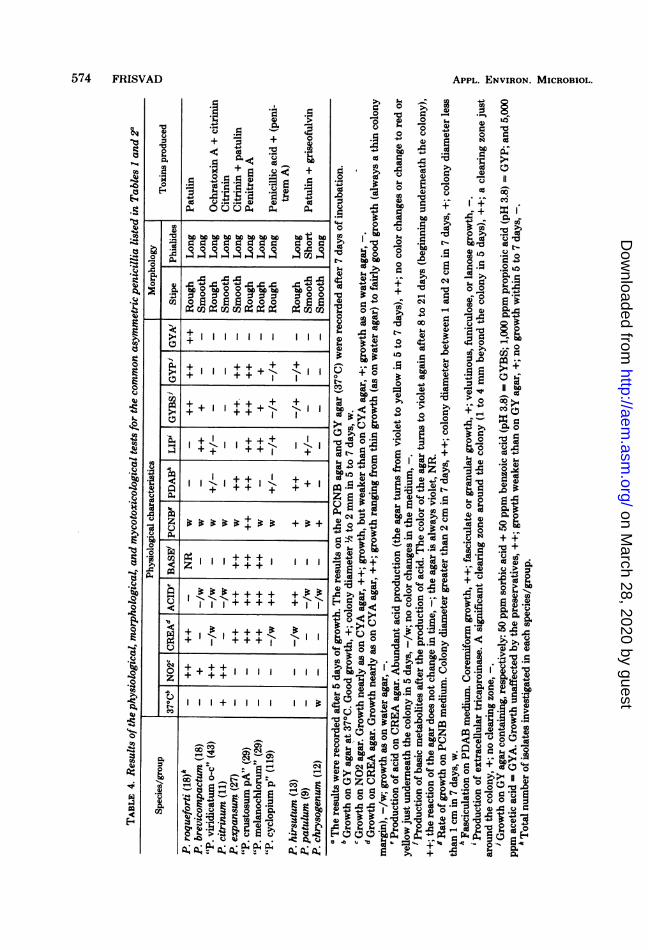
A summary of the results of the physiologicaltests is shown in Table 4. The tests used ordeveloped in this investigation show clear-cutresults and allowed a good subdivision in posi-tive and negative responses. The following ac-cepted and well defined species showed consist-ent patterns of responses in the physiologicaltests: P. roqueforti, P. brevicompactum, P. ci-trinum, P. expansum, P. patulum, P. chryso-genum, and P. hirsutum. Few strains of thelatter species could grow on the GYBS and GYPmedium, however.On the basis of the results it seems relevant to
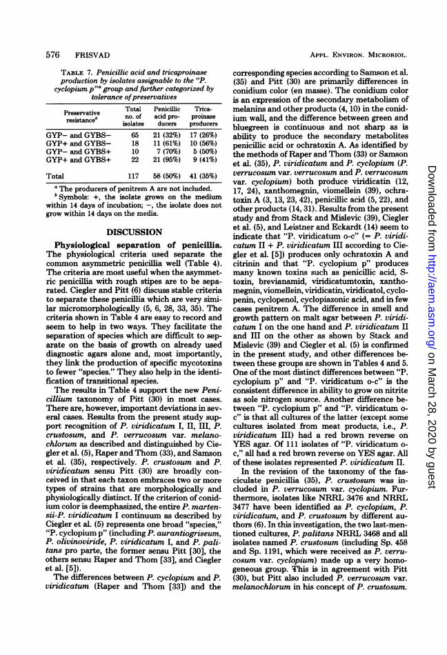
propose four provisional names for groups ofstrains which share several physiological andchemotaxonomic properties: "P. viridicatum o-c" (o-c, ochratoxin-citrinin), "P. cyclopium p"(p, penicillic acid), "P. crustosum pA" (near orequal to P. crustosum sensu Raper and Thom[33]), and "P. melanochlorum" (equal to P. ver-rucosum var. melanochlorum sensu Samson etal. [35]). Definitive characteristics for membersin the four groups, based upon examination of29 to 119 strains in each group, are shown inTables 5 and 6. These four groups also showedconsistent patterns of responses in the physio-logical tests. "P. cyclopium p", however, was avery variable group of strains as was P. cyclo-pium sensu Raper and Thom (33) and corre-sponding species as defined by Pitt (30) or Sam-son et al. (35). Table 7 gives an idea of thisvariability. Preservative resistant isolates of "P.cyclopium p" appeared to be more active bio-chemically than preservative sensitive isolates.Of the lipase producers, 72% were penicillic acidproducers, and of the nonproducers of trica-proinase only 33% produced penicillic acid.The 11 species/groups listed in Table 3 (col-
umn 2) were separable if two micromorphologi-cal criteria (ornamentation of stipe and short orlong phialides) and five physiological tests wereused: growth on N02 agar, growth and acidproduction on CREA agar, growth at 370C, andgrowth rate on PCNB agar (Table 4).
All isolates of P. expansum and "P. crustosumpA" produced the characteristic apple rot (ex-tensive respective restricted) described by Raperand Thom (33). One strain of "P. melanochlo-umm," however, produced a restricted rot in ap-ples.Few strains had special physiological re-
sponses. P. citrinum LKF ZL 17 (P. citrinum IIIsensu Raper and Thom [33]) had a response likestrains of P. chrysogenum. P. olivinovirideNRRL 958 and P. cyclopium NRRL 1899 had a+ reaction on PCNB agar. The latter isolateproduced basic metabolites on CREA agar after10 days of incubation. The type culture of P.expansum received from the Centraalbureau
VOL. 41, 1981
on March 28, 2020 by guest
http://aem.asm
.org/D
ownloaded from
574 FRISVAD APPL. ENVIRON. MICROBIOL.
w .5 2+ "~ + 0
.! oO; vv.
.p +~E C4 +S ... 4
on March 28, 2020 by guest
http://aem.asm
.org/D
ownloaded from

VOL. 41, 1981
Names according to Raper and Thom (33)and Ciegler et al. (5)
Names according to Pitt (30)
Conidium color
Extracellular color producedNO2 as sole N sourceCreatine as sole N source
Acid production on creatine sucrose agarSmell on CYA agarReverse on YES agar
Growth ratePreservative resistanceProduction of penicillic acidProduction of citrinin and/or ochratoxin A
IDENTIFICATION OF COMMON ASYMMETRIC PENICILLIA 575
TABLE 5. Differences between "P. viridicatum o-c" and "P. cyclopium p"Characteristics "P. viridicatum o-c"
P. viridicatum IIP. viridicatum IIISome slow-growing P. cyclopium
P. verrucosumP. viridicatum pro parte
Green, occasionally bluegreen
NoGood growthNo or weak growth
No, or only under the reverseWeak, like spruceRed brown (meat strains have
yellow reverse)SlowNoNoYes/no
"P. cyclopium p"
P. viridicatum IP. olivinovirideP. palitans pro parteP. cyclopium (fast grow-
ing)P. martensiiP. puberulum pro parteP. aurantiovirensP. aurantiocandidumP. lanosocoeruleum
P. aurantiogriseumP. viridicatum pro parte
Bluegreen to green (con-tinuum)
Yes/noNo growthNo, weak, or fairly goodgrowth
AbundantWoody, earthy, moldyYellow to cream colored
Rather fastYes/noYes/noNo
TABLE 6. Differences between "P. crustosum pA" and "P. melanochlorum""P. crustosum pA" "P. melanochlorum"
Names according to Raper and Thom (33) P. crustosum P. palitans pro parte
Names according to Samson et al. (35)
Names according to Pitt (30)
Conidium color
Sterile overgrowthProduction of adherent masses of conidia onmalt agar
Fasciculation in the presence of botranLimited rot produced in applesGrowth rate in the presence of PCNBProduction of penitremsProduction of three metabolites (red on thin-
layer chromaAgraphy plates after anisspray, long-wave ultraviolet light)
Preservative resistance
P. verrucosum var. cyclopium pro P. verrucosum var. me-parte lanochlorum
P. crustosum P. crustosum
Green to greygreen, bluegreen inperipheral zones
NoYes
YesYesVery highYes (two strains no)No
Very good
Dark green (specially onGY agar)
Yes (few strains no)No
NoNo (one strain yes)LowNoYes (few strains no)
Rather good
voor Schimmelcultures (CBS 325.48) seemedrather atypical. It was lanose in texture, sporu-lates late, and does not produce patulin or citri-nin. This isolate had a ++ reaction on the tri-caproin agar unlike all other 'solates of P. ex-pansum. P. terreste IMI 89384 and P. pseudo-casei IMI 68235 had physiological responses like
"P. crustosum pA", but they did not developfascicles on PDAB and they did not producepenitrem A. Furthermore, it was noticed thatfew isolates of P. expansum, "P. crustosum pA"and "P. melanochlorum" developed late on themedia containing preservatives (GYP andGYBS).
Characteristics on March 28, 2020 by guest
http://aem.asm
.org/D
ownloaded from
APPL. ENVIRON. MICROBIOL.
TABLE 7. Penicillic acid and tricaproinaseproduction by isolates assignable to the "P.
cyclopium pfla group and further categorized bytolerance ofpreservatives
Preservative Total Penicillic Trica-resistance' no. of acid pro- promase
isolates ducers producers
GYP- and GYBS- 65 21 (32%) 17 (26%)GYP+ and GYBS- 18 11 (61%) 10 (56%)GYP- and GYBS+ 10 7 (70%) 5 (50%)GYP+ and GYBS+ 22 21 (95%) 9 (41%)
Total 117 58 (50%) 41 (35%)a The producers of penitrem A are not included.bSymbols: +, the isolate grows on the medium
within 14 days of incubation; -, the isolate does notgrow within 14 days on the media.
DISCUSSIONPhysiological separation of penicillia.
The physiological criteria used separate thecommon asymmetric pencillia well (Table 4).The criteria are most useful when the asymmet-ric penicillia with rough stipes are to be sepa-rated. Ciegler and Pitt (6) discuss stable criteriato separate these penicillia which are very simi-lar micromorphologically (5, 6, 28, 33, 35). Thecriteria shown in Table 4 are easy to record andseem to help in two ways. They facilitate theseparation of species which are difficult to sep-arate on the basis of growth on already useddiagnostic agars alone and, most importantly,they link the production of specific mycotoxinsto fewer "species." They also help in the identi-fication of transitional species.The results in Table 4 support the new Peni-
cillium taxonomy of Pitt (30) in most cases.There are, however, important deviations in sev-eral cases. Results from the present study sup-port recognition of P. viridicatum I, II, III, P.crustosum, and P. verrucosum var. melano-chlorum as described and distinguished by Cie-gler et al. (5), Raper and Thom (33), and Samsonet al. (35), respectively. P. crustosum and P.viridicatum sensu Pitt (30) are broadly con-ceived in that each taxon embraces two or moretypes of strains that are morphologically andphysiologically distinct. If the criterion of conid-ium color is deemphasized, the entire P. marten-sii-P. viridicatum I continuum as described byCiegler et al. (5) represents one broad "species,""P. cyclopium p" (including P. aurantiogriseum,P. olivinoviride, P. viridicatum I, and P. pali-tans pro parte, the former sensu Pitt [30], theothers sensu Raper and Thom [33], and Ciegleret al. [5]).The differences between P. cyclopium and P.
viridicatum (Raper and Thom [33]) and the
corresponding species according to Samson et al.(35) and Pitt (30) are primarily differences inconidium color (en masse). The conidium coloris an expression of the secondary metabolism ofmelanins and other products (4, 10) in the conid-ium wall, and the difference between green andbluegreen is continuous and not sharp as isability to produce the secondary metabolitespenicillic acid or ochratoxin A. As identified bythe methods of Raper and Thom (33) or Samsonet al. (35), P. viridicatum and P. cyclopium (P.verrucosum var. verrucosum and P. verrucosumvar. cyclopium) both produce viridicatin (12,17, 24), xanthomegnin, viomellein (39), ochra-toxin A (3, 13, 23, 42), penicillic acid (5, 22), andother products (14, 31). Results from the presentstudy and from Stack and Mislevic (39), Ciegleret al. (5), and Leistner and Eckardt (14) seem toindicate that "P. viridicatum o-c" (= P. viridi-catum II + P. viridicatum III according to Cie-gler et al. [5]) produces only ochratoxin A andcitrinin and that "P. cyclopium p" producesmany known toxins such as penicillic acid, S-toxin, brevianamid, viridicatumtoxin, xantho-megnin, viomellein, viridicatin, viridicatol, cyclo-penin, cyclopenol, cyclopiazonic acid, and in fewcases penitrem A. The difference in smell andgrowth pattern on malt agar between P. viridi-catum I on the one hand and P. viridicatum IIand III on the other as shown by Stack andMislevic (39) and Ciegler et al. (5) is confirmedin the present study, and other differences be-tween these groups are shown in Tables 4 and 5.One of the most distinct differences between "P.cyclopium p" and "P. viridicatum o-c" is theconsistent difference in ability to grow on nitriteas sole nitrogen source. Another difference be-tween "P. cyclopium p" and "P. viridicatum o-c" is that all cultures of the latter (except somecultures isolated from meat products, i.e., P.viridicatum III) had a red brown reverse onYES agar. Of 111 isolates of "P. viridicatum o-c," all had a red brown reverse on YES agar. Allof these isolates represented P. viridicatum II.
In the revision of the taxonomy of the fas-ciculate penicillia (35), P. crustosum was in-cluded in P. verrucosum var. cyclopium. Fur-thermore, isolates like NRRL 3476 and NRRL3477 have been identified as P. cyclopium, P.viridicatum, and P. crustosum by different au-thors (6). In this investigation, the two last-men-tioned cultures, P. palitans NRRL 3468 and allisolates named P. crustosum (including Sp. 458and Sp. 1191, which were received as P. verru-cosum var. cyclopium) made up a very homo-geneous group. This is in agreement with Pitt(30), but Pitt also included P. verrucosum var.melanochlorum in his concept of P. crustosum.
576 FRISVAD
on March 28, 2020 by guest
http://aem.asm
.org/D
ownloaded from

IDENTIFICATION OF COMMON ASYMMETRIC PENICILLIA 577
"P. crustosum pA" and "P. melanochlorum"have many characteristics in common, but theyalso differ in some characteristics (Table 6). "P.melanochlorum" seems to be a distinct fungusand should probably be given species status.This mold has also been found on fermentedtallow used for human consumption on theFaroe Islands (unpublished results). Nearly allof the isolates of "P. melanochlorum" investi-gated produced three characteristic red metab-olites (as seen on thin-layer chromatographyplates after anisaldehyde treatment) irrespectiveof their sources. "P. crustosum pA" is a producerof penitrem A, but P. cyclopium NRRL 1899and P. olivinoviride NRRL 958 also producethis toxin. These latter isolates seem to be inter-mediates between "P. cyclopium p" and "P.crustosum pA". Such strains have never beenencountered in our laboratory, but according toCiegler and Pitt (6) they occur infrequently inP. olivinoviride and P. cyclopium.
Reliability of the physiological criteriaused. If physiological criteria are to be used indeterminative taxonomy, they must be consist-ent and stable. The tests shown in Table 4 werestable as they gave the same results in manyindependent experiments. Furthermore, all ofthe isolates listed in Tables 1 and 2 showedconsistent physiological reactions within eachspecies (on N02 agar, CREA agar, PCNB me-dium, GY agar at 370C, and GYA agar).The ability to grow at 370C has been used as
a taxonomic feature by Pitt (30). However, Pitt(30) mentions that some P. citrinum and P.chrysogenum isolates fail to grow at this tem-perature. It is probable that citrinin producerswithin these species all grow at 370C. However,citrinin production and ability to grow at 370Care not positively correlated in all penicillia (Ta-ble 4).The ability of some species of Penicillium to
grow on N02 agar seems to be a very consistentcriterion. Nitrite media were used earlier by Abe(1) and Engel and Teuber (8) with good results.The ability of the penicillia to grow on CREA
agar is also a consistent criterion (also found forP. roqueforti by Engel and Teuber [8]). How-ever, some strains of "P. cyclopium p" and P.hirsutum first grew insignificantly on CREAagar, but after 8 to 14 days of incubation theygrew rather well. Unlike P. roqueforti, P. expan-sum, "P. crustosum pA," and "P. melano-chlorum," these isolates had thin colony marginson the medium. Acid production on CREA agarwas also a consistent and stable criterion pro-vided it was recorded after 5 to 7 days of incu-bation. P. expansum, "P. crustosum pA", and"P. melanochlorum" consistently produced
some basic metabolites about 1 week after theabundant acid production.Production of extracellular enzymes by molds
may prove to be taxonomically useful. In somecases the production of exogenous tricaproinaseis consistent. More isolates should be investi-gated before any conclusions are drawn. Inves-tigation of lipase production in molds producingmycotoxins is important. Ba et al. (2) have thusfound that toxigenic strains ofAspergillusflavusproduce twice asmuch lipase as do non-toxigenicstrains.
Criteria based on resistance towards fungi-cides and preservatives are not as significanttaxonomically as is the ability to utilize nitriteor creatine or the ability to produce certainsecondary metabolites. The former characteris-tics may be results of recent evolutionary adap-tation to different environments. In P. roque-forti, however, the resistance towards preserva-tives seems to be genotypic.
After 2 weeks of incubation, isolates from allspecies/groups could grow on GYBS medium.Therefore, the data concerning resistance to-wards preservatives must be recorded after 5 to7 days of incubation.The ability of the penicillia to grow on GYP
medium is a consistent criterion in most cases,but it should be used mostly as a confirmativecriterion since it is inconsistent in the case of "P.cyclopium p" and P. hirsutum (Table 4). Thefollowing species did not grow or grew veryfaintly on GYP medium after 2 weeks of incu-bation: P. brevicompactum, P. chrysogenum, P.citrinum, P. patulum, "P. viridicatum o-c", andsome strains of "P. cyclopium p" and P. hirsu-tum.
Ability to produce fascicles on PDAB (Table4) seems to be a consistent criterion in mostcases, but an inconsistency is recorded in "P.cyclopium p" and "P. viridicatum o-c". As notedby Pitt (30), fasciculation is a good confirmativeand sometimes a diagnostic criterion. Mislevic(19, 20) recommends the use of this criterion inPenicillium taxonomy.The growth rate of strains of "P. crustosum
pA" was very high on PCNB medium, and thisfeature facilitated identification of these strains.Growth rate on this medium seems to be aconsistent criterion, but of use for only a fewspecies.Table 4 shows that "P. cyclopium p" is a very
variable group. An expression of this variabilitycan be seen in Table 7. Penicillic acid producersare more prevalent among preservative "resist-ant" isolates. Isolates identified by the methodof Raper and Thom (33) (P. martensii, P. cyclo-pium, and P. viridicatum [I]) were represented
VOL. 41, 1981
on March 28, 2020 by guest
http://aem.asm
.org/D
ownloaded from

APPL. ENVIRON. MICROBIOL.
in all of the "preservative groups" (GYP-/GYBS-, GYP+/GYBS-, etc.), but of the P.puberulum isolates examined only one was pre-servative "resistant." These isolates of P. pub-erulum (Table 2, footnote e) did not fit thedescription of Pitt (30). They were similar to P.puberulum NRRL 3564, but did not producepenicillic acid.Chemotaxonomic characterization of the
peniillia. All of the species/groups showed acharacteristic pattern ofmetabolites on the thin-layer chromatography plates; however, somespecies/groups had two types of patterns (i.e., P.roqueforti). Most patterns were consistent fromisolate to isolate, but in some cases one, two, andup to all metabolites could be lacking (using theagar plug method [9], the extractions of thecultures contained metabolites in common forall species/groups). Because of this fact, theusefulness of thin-layer chromatography pat-terns in the identification of penicillia is some-what limited. In most cases, however, they arehelpful as auxiliary and confirnatory character-istics. It should be mentioned that some of themedia used in the new Penicillium handbook(30) are good toxin production media (CYA andMEA [14, and unpublished results]).
In the review by Scott (36a), 17 species, ofPenicillium are cited as producers of penicillicacid. If the group "P. cyclopium p" is acceptedas one "species," the number of Penicilliumspecies producing penicillic acid may be reducedto nine or less. Corresponding examples can begiven for other mycotoxins. Table 4 shows thatthere seems to be a correlation between myco-toxin production and the species/groups deter-mined using the physiological (and morphologi-cal) criteria. Only a few species seem to be ableto produce more than two fundamentally differ-ent mycotoxins. In P. roqueforti, two "chemicalraces" seem to be present: the roquefortin/PR-toxin race (14, 27, 37) and the penicillic acid/patulin race (11, 14, 25, 26). In "P. cyclopium p",a xanthomegnin/S-toxin/penicillic acid/brev-ianamid race, a cyclopiazonic acid race, and aminor penitrem A race seem to be present (14,39). Maybe other physiological criteria can sep-arate such mycotoxin producers in the samespecies/group.Conclusion. The present investigations have
shown that certain physiological criteria can beimportant aids in the identification of commonasymmetric penicillia. In the subgenus Penicil-lium of Pitt (30), the criteria of growth at 50Cand at 370C can be excluded and instead thePenicillium isolates should be inoculated onN02 agar and CREA agar. These media provideconsistent and stable data. Furthermore, the
Penicillium isolates could be examined duringincubation using the simple agar plug methodfor detection of extracellular products. The pat-tern of metabolites may indicate at once whichmycotoxins the isolate produces and, therefore,the species/group to which it belongs. The othermedia used in this investigation may be morevariable, but in return they give the investigatordata on some ecological characteristics of theisolate under examination.On the basis of the physiological, mycotoxi-
cological, and morphological data obtained inthis investigation, it is reasonable to proposefour groups of isolates (species). (i) "P. melan-ochlorum" (= P. verrucosum var. melanochlo-rum sensu Samson et al. [35]), producing noknown toxins, seems to be a separate species. (ii)"P. crustosum pA" (probably equal to P. crus-tosum sensu Raper and Thom [33]) is a veryhomogeneous species, and it seems that freshisolates of this mold always produce penitrem A.(iii) The nitrite-positive "P. viridicatum o-c" isalso a very homogeneous "species" and is prob-ably the only "taxon" in the subgenus Penicil-lium containing ochratoxin producers. Thisgroup comprises the strains of P. viridicatum IIand III (5) included in P. viridicatum and P.verrucosum sensu Pitt (30). (iv) The acid-pro-ducing "P. cyclopium p" group embraces peni-cillic acid producers and producers of manyother toxins. This group is variable, but does notinclude producers of citrinin or ochratoxins. "P.cyclopium p" consists of P. aurantiogriseumsensu Pitt (30) and P. viridicatum I (5) (includ-ing P. olivinoviride and P. palitans pro parte).These four groups could be the basis for thedefinition of four new species within thesubgenus Penicillium and thus link the produc-tion of particular mycotoxins to fewer species.
ACKNOWLEDGMENTSC. W. Hesseltine and D. Wicklow, Northern Regional Re-
search Laboratory, Agricultural Research Service, Peoria, Ill.,H. K. Frank, Bundesforschungsanstalt fir Lebensmittelfri-schhaltung, Karlsruhe, Federal Republic of Germany, and L.Leistner, Bundesanstalt fiir Fleischforschung, Kulmbach, Fed-eral Republic ofGermany, kindly supplied some ofthe culturesused in this study. I thank L. Leistner also for the penitrem Astandard, Ole Filtenborg for fruitful discussions, and Ole Fil-tenborg and Peter Zeuthen for proofreading ofthe manuscript.
LITERATURE CIMD1. Abe, M. 1956. Studies on the classification of the Penicil-
lia. J. Gen. Appl. Microbiol. 2:1-344.2. Ba, D., M. Jemmali, and R. Drapron. 1977. Activite
lipolytique et production d'aflatoxines chez Aspergillusflavus. Ann. Microbiol. (Inst. Pasteur) 128B:87-93.
3. Barnes, J. M., P. K. C. Aystwick, R. L. Carter, F. V.Flynn, G. C. Peristianis, and W. N. Aldridge. 1977.Balkan (endemic) nephropathy and a toxin-producingstrain of Penicillium verrucosum var. cyclopium: anexperimental model in rats. Lancet i:671-675.
578 FRISVAD
on March 28, 2020 by guest
http://aem.asm
.org/D
ownloaded from

IDENTIFICATION OF COMMON ASYMMETRIC PENICILLIA 579
4. Bartsch, E., W. Lerbs, and M. Luckner. 1979. Phenoloxidase activity and pigment synthesis in conidiosporesof Penicillium cyclopium. Z. AUg. Mikrobiol. 19:75-82.
5. Ciegler, A., D. L FennelL G. A. Sansing, R. W. Detroy,and G. A. Bennett. 1973. Mycotoxin producing strainsof Penicillium viridicatum: classification into sub-groups. Appl. Microbiol. 26:271-278.
6. Ciegler, A., and J. L Pitt. 1977. Survey of the genusPenicilium for tremorgenic toxin production. Myco-pathol. Mycol. Appl. 42:119-124.
7. burackova, Z., V. Betina, and P. Nemec. 1976. System-atic analysis of mycotoxins by thin-layer chromatogra-phy. J. Chromatogr. 116:141-154.
8. Engel, G., and KL Teuber. 1978. Simple aid for theidentification of Penicilium roqueforti Thom. Growthin acetic acid. Eur. J. Appl. Microbiol. Biotechnol. 6:107-111.
9. Filtenborg, 0., and J. C. Friavad. 1980. A simple screen-ing-method for toxigemc moulds in pure cultures. Le-bensm. Wiss. Technol. 13:128-130.
10. Ha-Huy-Ke, and K Luckner. 1979. Structure and func-tion of conidiospore pigments ofPenicillium cyclopium.Z. AUg. Mikrobiol. 19:117-122.
11. Harwig, J., B. J. Blanchfileld, and P. AL Scott 1978.Patulin production by Peniciiuwn roqueforti Thomfrom grape. Can. Inst. Food Sci. Technol. J. 11:149-151.
12. Korzybski, T., Z. Kowazyki-Gindifer, and W. Kory-lowicz. 1967. Antibiotics. Origin, nature and properties,vol. 2, p. 1257-1258. Pergamon Press, Oxford.
13. Krogh, P. 1978. Causal associations of mycotoxic ne-phropathy. Acta Pathol. Microbiol. Scand. Sect. A.Suppl. 269:1-28.
14. Leistner, L., and C. Eckardt. 1979. Vorkommen toxi-nogener Penicillien bei Fleischerzeugnisse. Fleischwirt-schaft 59:1892-1896.
15. Leistner, L, and J. L Pitt. 1977. Miscellaneous Penicil-lium toxins, p. 639-653. In J. V. Rodricks, C. W. Hes-seltine and M. A. Mehlmann (eds.), Mycotoxins inhuman and animal health. Pathotox Publishers, ParkForest South.
16. LAtzsch, R., F. Tauchman and W. Meyer. 1974. Ein-satz des "Stomacher" in der Mykotoxin-Analytik. Flei-schwirtschaft 54:943-945.
17. Luckner, KL, L Nover, and HL B6hm. 1977. Secondarymetabolism and cell differentiation. Molecular BiologyBiochemistry and Biophysics 23. Springer Verlag, Ber-lin, Heidelberg, New York.
18. Marti, L R., D. K. Wilson, and B. D. Evans. 1978.Determination of citrinmn in corn and barley. J. Assoc.Offic. Anal. Chem. 61:1353-1358.
19. Mislevic, P. B. 1975. The effect of botran on fascicleproduction by species of Penicillium. Mycologia 67:194-198.
20. Mislevic, P. B. 1977. The genus Penicillium, p. 41-57. InT. D. Wyllie and L G. Morehouse (ed.), Mycotoxicfungi Mycotoxins Mycotoxicosis, vol. 1. Mycotoxic fungiand chemistry of mycotoxins. Marcel Dekker, NewYork.
21. Murray, L G. 1968. Some aspects of the biochemicaldifferentiation of pathogenic fungi: a review. J. Gen.Microbiol. 52:213-221.
22. Northolt, KL D., H. P. van Egmond, and W. E.Paulach. 1979. Penicillic acid production by some fun-gal species in relation to water activity and temperature.J. Food Prot. 42:476-484.
23. Northolt, M. D., H. P. van Egmond, and W. E.Paulch. 1979. Ochratoxin A production by some fun-gal species in relation to water activity and temperature.
J. Food Prot. 42:485-490.24. Nover, L, and K Luckner. 1969. On the biosynthesis of
cyclopenin and cyclopenol, benzodiazepides alkaloidsfrom Penicilium cyclopium Westling. Eur. J. Biochem.10:268-273.
25. Olivigni, F. J., and L B. Bullerman. 1977. Simultane-ous production of penicillic acid and patulin by a Pen-icillium species isolated from cheddar cheese. J. FoodSci. 42:1654-1657, 1665.
26. Olivigni, F. J., and I. B. Bullerman. 1978. Productionof penicillic acid and patulin by an atypical Penicilliumroqueforti isolate. Appl. Environ. Microbiol. 35:435-438.
27. Orth, R. 1976. PR-toxin production by Penicillium ro-queforti strains. Z. Lebensm. Unters. Forsch. 160:131-136.
28. Pitt, J. 1973. An appraisal of identification methods forPenicilium species: novel criteria based on tempera-ture and water relations. Mycologia 65:1135-1137.
29. Pitt, J. L. 1974. A synoptic key to the genus Eupenicilliumand to sclerotiogenic Penicilium species. Can. J. Bot.52:2231-2236.
30. Pitt, J. L. 1979. The genus Penicilium and its teleo-morphic states Eupenicilium and Talaromyces. Aca-demic Press, Inc., London.
31. Pohland, A. E., and P. B. Mislevic. 1976. Metabolitesof various Penicilium species encountered in foods.Adv. Chem. Ser. 149:110-143.
32. Purchase, I. H. F. 1974. Penicillium cyclopium, p. 149-162. In I. H. F. Purchase (ed.), Mycotoxins. Elsevier,Amsterdam.
33. Raper, K. B., and C. Thom. 1949. A manual of thePeniciflia. Williams and Wilkins Co., Baltimore.
34. Samson, R. A., RI Hadlok, and A. C. StoiL 1977. Ataxonomic study ofthe Penicilium chrysogenum series.Antonie van Leeuwenhoek. J. Microbiol. Serol. 43:261-274.
35. Samson, R. A., A. C. Stolk, and R. Hadlok. 1976.Revision of the subsection fasciculata of Peniciliumand some allied species. Stud. Mycol. Baarn 11:1-47.
36. Scott, P. M. 1974. Collaborative study of a chromato-graphic method for determination of patulin in applejuice. J. Assoc. Offic. AnaL Chem. 57:621-625.
36a.Scott, P. KL 1977. Penicillium mycotoxins, p. 283-356. InT. P. Wylie and L. G. Morehouse (ed.), Mycotoxicfungi, mycotoxins, mycotoxicosis, vol. 1. Mycotoxicfungi and chemistry of mycotoxins. Marcel Dekker,New York.
37. Scott, P. KL, and B. P. C. Kennedy. 1976. Analysis ofblue cheese for roquefortine and other alkaloids fromPeniciUllum roqueforti. J. Agric. Food Chem. 24:865-868.
38. Scott, P. KL, J. W. Lawrence, and W. van Walbeek.1970. Detection ofmycotoxins by thin-layer chromatog-raphy: application to screening of fungal extracts. Appl.MicrobioL 20:839-842.
39. Stack, K E., and P. B. Mislevic. 1978. Production ofxanthomegnin and viomellein by isolates ofAspergillusochraceus, Penicilhiwn cyclopiun and Penicillium vir-idicatum. Appl. Environ. Microbiol. 36:552-554.
40. Steyn, P. S. 1969. The separation and detection of severalmycotoxins by thin-layer chromatography. J. Chroma-togr. 45:473-475.
41. Thatcher, F. S., and D. S. Clark. 1978. Microorganismsin food. 1. Their significance and enumeration, 2nd ed.University of Toronto Press, Toronto.
42. Walbeek, W. van, P. K Scott, J. Harwig, and J. U.Lawrence. 1969. Penicillium viridicatum Westling: anew source of ochratoxin A. Can. J. Microbiol. 51:1281-1285.
VOL. 41, 1981
on March 28, 2020 by guest
http://aem.asm
.org/D
ownloaded from