Embed Size (px)
Citation preview

Department of Clinical Sciences, Intervention and Technology, Division of Ear, Nose and Throat Diseases incl. Audiology
Karolinska Institutet, Stockholm, Sweden
PERSISTENT GEOTROPIC NYSTAGMUS - A DIFFERENT KIND OF CUPULA PATHOLOGY
av
Tatjana Tomanovic, leg. läkare
AKADEMISK AVHANDLING som för avläggande av medicine doktorsexamen vid Karolinska Institutet offentligen försvaras på svenska språket i ÖNH-klinikens föreläsningssal, Karolinska Universitetssjukhuset, Solna.
Fredagen den 28 februari 2014, kl 9.00
Huvudhandledare: Docent Johan Bergenius Karolinska Institutet Institutionen för klinisk vetenskap, intervention och teknik Bihandledare: Professor Sten Hellström Karolinska Institutet Institutionen för klinisk vetenskap, intervention och teknik
Fakultetsopponent: Docent Lars Ödkvist Linköpings Universitet Institutionen för klinisk och experimentell medicin Betygsnämnd: Professor Jan Ygge Karolinska Institutet Klinisk Neurovetenskap Professor Helge Rask-Andersen Uppsala universitet Institutionen för kirurgiska vetenskaper Akademiska sjukhuset Docent Hans Cristian Larsen Uppsala universitet Institutionen för kirurgiska vetenskaper Akademiska sjukhuset
Stockholm 2014


From Department of Clinical Sciences, Intervention and Technology, Division of Ear, Nose and Throat Diseases incl. Audiology
Karolinska Institutet, Stockholm, Sweden
PERSISTENT GEOTROPIC NYSTAGMUS - A DIFFERENT KIND OF CUPULA PATHOLOGY
Tatjana Tomanovic
Stockholm 2014

2014
Gårdsvägen 4, 169 70 Solna
Printed by
All previously published papers were reproduced with permission from the publisher. Published by Karolinska Institutet. Printed by [name of printer]. © Tatjana Tomanovic, 2014 ISBN 978-91-7549-462-3
Illustrations done by Tatjana Tomanovic

Life gives back to us only what we give to others.
Ivo Andric


ABSTRACT
In patients with positional vertigo a persistent positional direction-changing nystagmus (PDCN) of apogeotropic
direction (a-PDCN) in the supine yaw plane has been described earlier 1-5. It has been suggested that the cupula in
the lateral semicircular canal has a higher specific weight than the surrounding endolymph making the cupula
sensitive to gravity. This condition is known as ”heavy cupula”. We have described, in Paper I, a geotropic
persistent direction-changing nystagmus (g-PCDN) in patients during vestibular crisis6. In addition, when the
patient is in the supine position and the head is turned slowly from one side to the other it is possible to discern a
zero zone where the geotropic nystagmus is absent. This is accomplished when the head is turned circa 10-20
degrees laterally. Theoretically this occurs when the longitudinal axis of the affected cupula is aligned with the
gravitational vertical 7-9.
On the assumption that a position dependent nystagmus such as persistent a-PDCN is caused by a heavy cupula in
one of the lateral semicircular canals (LSCC), thus it could be hypothesized that a g-PDCN can be caused by the
cupula that is lighter than the surrounding endolymph. We have called this new diagnostic entity “light cupula”.
A similar phenomenon of a PDCN is seen in subjects with positional alcohol nystagmus (PAN) 10-18. This
phenomenon is based on the” buoyancy hypothesis”19,20. In order to reproduce a clinical condition where the
density of the cupula was lower or higher than the surrounding endolymph we examined the nystagmus pattern in
different head positions in unilaterally deafferented patients during the stage of PAN 1 and PAN 2 respectively21,22.
We compared results of nystagmus direction during PAN 1 (Paper II) with the findings in patients with light
cupula in the LSCC (Paper I). Nystagmus direction in both supine and prone lateral head positions was compatible
with that of a light cupula. However, the nystagmus directions at head straightforward in prone and supine position
as well as the localization of zero zones deviated from the pattern seen in patients with a light cupula21.
We followed up nystagmus characteristics and nystagmus pattern in Paper III on the labyrinthectomized subjects
during the stage of PAN 222. This study showed persistent a-PDCN according the theory 12. The nystagmus pattern
in pitch plane was of opposite to that during PAN 1. Nystagmus in supine position was directed to the affected side
during PAN 2 and to non-affected side during PAN 1 but the zero zones in both studies is found on the affected
side.
We were interested to see if the results concerning the nystagmus pattern in the experimental studies could be
applied to patients during vestibular disability with g-PDCN permitting a lateralization of the affected side. In
Paper IV we examined an extended series of 20 patients with g-PDCN nystagmus pattern during acute vestibular
disability and at follow up 1-7 years later. Nystagmus patterns in different head positions were recorded, both
caloric and otolith tests were carried out. Concomitant auditory symptoms as an indication of the affected side
were rare. The slow phase velocity (SPV) of geotropic nystagmus was low and of equal intensity and did not
present an indication of the affected side according to Ewald´s second law23. With nystagmus analysis we found
evidence for a cupula that is sensitive to gravitation, but did not find an applicable pattern for simply determination
of the affected side by analysing nystagmus direction in the pitch plane and in lateral head positions. There was a
high prevalence of migraine (40%) and the patients also had problems with recurrent vertigo (80%). The vestibular
tests were pathologic in 60% of the patients.

CONTENTS
Abstract
Thesis summary 1 Introduction 1
Historical background 1 Vestibular apparatus 3 Otolith organs 3 Semicircular canals 4 Cupula 5 Vestibulo-ocular reflex 6 Positional Alcohol Nystagmus (PAN) 7 Concept of the heavy cupula 8 Concept of the light cupula 9
Aims of the thesis 11
Materials and methods 12
Study populations 12 Methods 13
Results 16
Paper I 16 Paper II 16 Paper III 18 Paper IV 20
Disscussion 23
Methodological consideration 23 Nystagmus in lateral head positions – summary 24 Lateralization of affected side by Ewald’s second law 25 Topographical orientation of the cupula- clinical implications 26 Further comments on cupula dysfunctions as sign of vestibular lesion 28 “Cupula dysfunctions ” as a part of a vestibular disorder 29
Conclusions 31
Acknowledgements 32
References 33

LIST OF PUBLICATIONS
This thesis is based on the following original papers.
They will be referred to the text by their Roman numerals I-IV.
I. Bergenius J, Tomanovic T. Persistent geotropic nystagmus--a different kind of
cupular pathology and its localizing signs. Acta Otolaryngol. 2006 Jul; 126: 698-704
II. Tomanovic T, Bergenius J. Can the nystagmus pattern in patients with a 'light
cupula' be reproduced in hemi-labyrinthectomized subjects during positional alcohol
nystagmus 1? Acta Otolaryngol. 2011 Sep; 131: 929-936.
III. Tomanovic T, Bergenius J. Is the nystagmus pattern in hemi-labyrinthectomized
subjects during positional alcohol nystagmus 2 similar to that found in patients with
cupulolithiasis in the lateral semicircular canal? Acta Otolaryngol. 2013 Aug; 133: 796-803.
IV. Tomanovic T, Bergenius J. Vestibular findings in patients with persistent
geotropic positional nystagmus: the “light cupula” phenomenon. Submitted for publication in
2014.

LIST OF ABBREVIATIONS
PN
SPV
g-PDCN
a-PDCN
VOR
PAN
c-VEMP
o-VEMP
SVH
CNS
BPPV
GSD
SCC
VNS
Positional nystagmus
Slow phase velocity
Geotropic persistent direction changing nystagmus
Apogeotropic persistent direction changing nystagmus
Vestibulo-ocular reflex
Positional alcohol nystagmus
Cervical vestibular evoked myogenic potential
Ocular vestibular evoked myogenic potential
Subjective visual horizontal
Central nervous system
Benign Paroxysmal Positional Vertigo
Geotropic side difference
Semicircular canal
Video-nystagmoscopy

THESIS SUMMARY
Foreword The vestibular system provides the information about body position that allows rapid compensatory
movements in response to forces generated internally and externally. We are normally unaware of its
function. If the system is damaged, balance, eye movements and sense of orientation in space are affected.
Hence, it is important to elucidate the different conditions that lead to a disturbance in this complex system.
The main symptom of disturbance in the vestibular system is dizziness and unsteadiness. Dizziness can be
provoked by changes in head or body position causing positional dizziness, which can be confirmed by
positional nystagmus (PN). One of the subtypes of PN is persistent g-PDCN. This thesis is based on clinical
and experimental studies, which aim to describe the nystagmus characteristics and pathophysiological
mechanisms of vestibular disability with persistent g-PDCN that we have named “light cupula”.
INTRODUCTION
Historical background At the beginning of the last century Robert Barany studied positional nystagmus24. He found a noticeable
connection between positional nystagmus and the gravitational forces.
Carl Olof Nylén carried out studies 25 devoted to classification and definition of positional nystagmus. He
stated that the positional nystagmus is altered when the head adopts another position and in his classification
there are three main types of positional nystagmus:
• Direction-changing, changing direction in different positions of the head
• Direction fixed, beating always to the same direction
• Irregular, characterized by variations in its behavior.
He stated, “The probability is that both the peripheral and central system are capable of giving rise to
positional nystagmus”. With peripheral affection it is conceivable that positional nystagmus arises through
deficient interplay of the otoliths and the cupula due to the fact that experimentally nystagmus occurs, with
stimulation of the cupula but not with stimulation of the otolith organ.
Aschan et al. have studied positional nystagmus in man mainly the persistent form 11,12,26,27. Their
investigation indicated that the position of the head is a determining factor in the persistent form of
positional nystagmus, but in the transitional forms of nystagmus, movement of the head plays a significant
role. Stenger in 1955 28 further emphasized the classification of positional nystagmus into two types caused
by:
• Position itself
• Changes in position

Positional geotropic nystagmus
2
In 1952, Dix and Hallpike described torsional vertical nystagmus provoked by a specific ear-down position
with a latency of several seconds, in which the nystagmus lasted only for a limited time, usually less than 20
seconds, and the direction of the nystagmus reversed on resuming the upright position. The nystagmus also
showed fatigability with a progressive decline in intensity on repetition of these manoeuvres 29,30. These
authors coined the term "benign paroxysmal positional vertigo" (BPPV), and the provocative positional
testing was named in their honour.
Schuknecht was the first to provide a pathophysiological concept of BPPV 31,32. In 1969 he proposed the
theory of "cupulolithiasis" on the basis of pathological studies that demonstrated otolithic debris attached to
the cupula. However, the concept of cupulolithiasis has several limitations and is thus unable to explain all
of the characteristics of nystagmus and vertigo in BPPV 33. In 1979, Hall and al. and then other researchers
put forward a new explanation for the transient vertigo in patients BPPV. It was suggested that BPPV was
caused by particles floating around in the endolymph 34-36. As they are heavier than the surrounding
endolymph they are subject to the influence of gravity. Due to fast head movements or long lasting positions
of the head they will precipitate and assemble in the semicircular canals.
Semont et al. referred to physical therapy as a liberatory manoeuvre 37. In 1992 Epley reported his first
results in curing positional vertigo by physical therapy referred it as the “canalith repositioning procedure”38.
Both techniques are for relocation of the particles from the posterior semicircular canal (PSCC). Since that
time, specific physical therapy has been widely applied to patients suffering from BPPV 39. Prior to the 1985
it was believed that BPPV developed only in the PSCC 40-42. However, in 1985 McClure introduced the
concept and clinical features of BPPV involving the lateral semicircular canal (LSCC) without any evidence
of central lesions43,44. He reported patients with horizontal geotropic nystagmus precipitated by head
movements in supine position into or out of one of the lateral positions. In 1995, Baloh et al. reported three
patients in whom the nystagmus changed direction in each lateral head position always beating away from
the ground. This positional nystagmus had all of the features usually ascribed to a central lesion; no latency,
no fatigability and persisted as long as the position was held. They proposed that nystagmus was result of
debris attached to the cupula of the LSCC 3,4,45. Particle movement in the LSCC was also suspected as a
pathophysiological substrate and hence different types of physical therapy were recommended 38,46-48. There
are mainly three different types of positional nystagmus: vertical torsional paroxysmal, geotropic direction-
changing paroxysmal, and apogeotropic direction-changing persistent, all result from debris in different
parts of the inner ear 3,49-52. Posterior canal BPPV is the most frequently reported form of BPPV, about 60-80%53-55 of cases. Lateral
canal BPPV accounts for approximately 10-20% and the anterior SCC is affected in 1-2 %56-58 of cases. Other rare variations include paroxysmal apogeotropic canalolithiasis of the LSCC 59,60, direction fixed
BPPV in LSCC 61, cupulolithiasis of PSCC 62,63, g-PDCN 6 and multiple canal BPPV 64,65.

Tatjana Tomanovic
3
Vestibular apparatus
Figure 1. The labyrinth and its innervations The vestibular system consists of a peripheral part located
in the inner (Figure 1) ear and integrative centers located
in the brainstem, cerebellum and somatic sensory cortices.
The vestibular part of inner ear is situated posterior to the
cochlea and consists of the bony labyrinth, containing the
fluid perilymph, and the membranous labyrinth. The
membranous labyrinth is a system of thin-walled sacks and
ducts filled with endolymph. Five vestibular receptor
organs contain highly organized sensory hair cells and
supporting cells were gathered in two otolith maculae, the
utriculus and the sacculus, and three crista ampullaris, in
each SCC. The otolith organs, utriculus and sacculus
represent a set of three-dimensional linear accelerometers that detect gravitational and translational
components associated with any head movement. The three pairs of SCCs represent a set of three-
dimensional angular accelerometers to detect the head angular velocity.
The membranous labyrinth is filled with endolymph; the sodium (Na+) content is low, and the potassium
(K+) content is high, which causes the endolymph to resemble intracellular rather than extracellular fluid.
The dark cells of the cristae and maculae presumably produce endolymph and the site of absorption of
endolymph is presumably the endolymphatic sac66,67.
Otolith organs
Neuroepithelium of the macula sacculi and macula utriculi is covered by an extracellular mass- otoconial
membrane, which consists of thousands of otoconia about 1-5 µm long. The otoconial membrane is
composed of type II collagen, glycoproteins, and proteoglycans that maintain the calcium crystal content
and hold it to the sensory epithelium. The otoconial mass is held together by beaded filaments and attached
to a gelatinous matrix or so-called globular substance that actually generates the calcium carbonate crystals.
The kinocilium of the utriclar macula is set into pores of the otoconial membrane, on which the otoconia are
densely distributed68,69. Linear acceleration (in the three
dimensions), including tilting of the head (in the roll and pitch
plane), causes the otoconial mass to move and thereby bend the
kinocilium and stereocilia of the hair cells, evoking a change in
the firing rate through the vestibular nerve. In animal studies of
elderly mice, the otoconia have become demineralized resulting
in weakening or loss of anchoring of the fibrils interconnecting
the otoconia 70.
As a result of the aging process, trauma, inflammation, and
perhaps other factors, calcite crystals may dislodge from the
otolithic membrane and sink into the endolymph fluid. Old are Figure 2. Cross section of the utricular macula
Positional geotropic nystagmus
4
replaced by new growing otoconia. Reabsorption of free otoconia by the dark cells takes at least 100 hours
in mice 71.
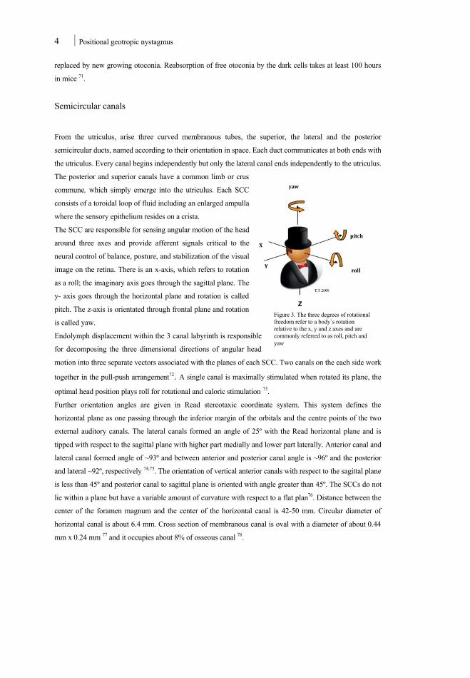
Semicircular canals
From the utriculus, arise three curved membranous tubes, the superior, the lateral and the posterior
semicircular ducts, named according to their orientation in space. Each duct communicates at both ends with
the utriculus. Every canal begins independently but only the lateral canal ends independently to the utriculus.
The posterior and superior canals have a common limb or crus
commune, which simply emerge into the utriculus. Each SCC
consists of a toroidal loop of fluid including an enlarged ampulla
where the sensory epithelium resides on a crista.
The SCC are responsible for sensing angular motion of the head
around three axes and provide afferent signals critical to the
neural control of balance, posture, and stabilization of the visual
image on the retina. There is an x-axis, which refers to rotation
as a roll; the imaginary axis goes through the sagittal plane. The
y- axis goes through the horizontal plane and rotation is called
pitch. The z-axis is orientated through frontal plane and rotation
is called yaw.
Endolymph displacement within the 3 canal labyrinth is responsible
for decomposing the three dimensional directions of angular head
motion into three separate vectors associated with the planes of each SCC. Two canals on the each side work
together in the pull-push arrangement72. A single canal is maximally stimulated when rotated its plane, the
optimal head position plays roll for rotational and caloric stimulation 73.
Further orientation angles are given in Read stereotaxic coordinate system. This system defines the
horizontal plane as one passing through the inferior margin of the orbitals and the centre points of the two
external auditory canals. The lateral canals formed an angle of 25º with the Read horizontal plane and is
tipped with respect to the sagittal plane with higher part medially and lower part laterally. Anterior canal and
lateral canal formed angle of ~93º and between anterior and posterior canal angle is ~96º and the posterior
and lateral ~92º, respectively 74,75. The orientation of vertical anterior canals with respect to the sagittal plane
is less than 45º and posterior canal to sagittal plane is oriented with angle greater than 45º. The SCCs do not
lie within a plane but have a variable amount of curvature with respect to a flat plan76. Distance between the
center of the foramen magnum and the center of the horizontal canal is 42-50 mm. Circular diameter of
horizontal canal is about 6.4 mm. Cross section of membranous canal is oval with a diameter of about 0.44
mm x 0.24 mm 77 and it occupies about 8% of osseous canal 78.
Figure 3. The three degrees of rotational freedom refer to a body`s rotation relative to the x, y and z axes and are commonly referred to as roll, pitch and yaw

Tatjana Tomanovic
5
Cupula
Each of the three ampullae of the SCC has a transverse ridge,
the crista ampullaris (Figure 4). Slopes of the crista are covered
by sensory epithelium that consists of the sensory and
supporting cells. Sensory epithelium of the lateral crista is
shaped like a rectangle with rounded corners and the surface
area is about 1 mm². The length extends from 1.7-2mm and the
width varies between 0.6-0.8mm 79. The height of the LSCC
crista is 0.40 mm and height of the covering cupula is 1.11mm 77. The cupula bridges the width of the ampulla, forming a
fluid barrier.
Morphologically, the cupula consists of the proteoglycans secreted from the supporting cells and net-shaped
tubular structures. The cupula extends from the epithelial cell surface to the roof of the ampulla and covers
the neuroepithelium.
The neuroepithelium (Figure 5) contains the hair cells, an average of 7600 79 divided into the two different
types 80. The type I hair cells are concentrated mostly in the central part of the crista and the type II cells
occupied predominantly occupy the peripheral zone. A bunch of the stereocilia and one kinocilium extend
from the body of the sensory cells 81. Stereocilia are always shorter than the kinocilium, the shortest
stereocilia are located more towards the peripheral and the tallest
stereocilia are closer to the kinocilium.
Head rotation in the plane of the semicircular canal provokes
endolymphatic flow. The inertia of the endolymph produces the forces
across the cupula, distending it and causing the displacement of the hair
bundles and hyperpolarisation or depolarization in the hair cells within in
the crista. The kinocilium is oriented towards the utriculus in the LSCC
and away from the utriculus in the vertical canal. A deviation of the hair
bundle towards the kinocilium is accompanied by depolarization of the
sensory cell; a deviation of the hair bundle in the opposite direction is
accompanied by hyperpolarization of the cell 80.
There is a lack of information about the exact topographical anatomy of the long axis of the cupula in the
plane of the LSCC. Reports of the exact shape of the cupula and its orientation are scarce because of its
vulnerability to chemical and fixation procedures. Observations in vivo have carried out on the lower
vertebrates 82. Judging from those studies the cupula has a cylindrical shape. The cupula sits on the crista
and extends towards to the ampullary roof where it is attached. In a histological study on the crista
ampullaris from 1972, Rosenhall79 noticed that there is some kind of irregularity in shape at the inferior
end of the lateral crista and at the same time it is closer to the utriculus than the superior end of the lateral
crista. Takagi et al. in 1989 83studied the spacial relations between semicircular canals and their cristae in
humans by using computer aided 3-D reconstruction. They found that the LSCC crista was not
perpendicular to the horizontal plane. They also found the 65° angle between the plane of the LSCC and
the long axis of the crista. Furthermore, the inferior end of the crista shifts posteriolaterally and
Figure 4. The ampulla of the LSCC showing the crista, hair bundles and cupula
Figure 5. Cross section of vestibular neuroepithelium showing two types of hair cells

Positional geotropic nystagmus
6
anteriomedially at its superior end. This finding indicates that crista is not perpendicular to the plane of
the LSCC and to the Reads horizontal plane, respectively. Baloh et al. 1995 3 suggested that the long axis
of the cupula is nearly parallel to the ipsilateral PSCC. However, in a recent study, Curthoys et al. 84
demonstrated that the long axis of the crista and cupula of the LSCC is almost parallel to the median
plane of the head. Bradshaw et al. in 2009 made labyrinth reconstructions from high resolution CT scans
of 34 ears and found that the degree of non-planarity for the three canals varies significantly between the
subjects 85. Therefore, knowledge of the topographical properties of the cupula and semicircular canal
would be indispensible in the understanding of the effects of the “heavy “and “light” cupula. Inclination
of the cupula is likely to be a determinant factor in nystagmus direction in pitch plane.
Vestibulo-ocular reflex During angular head movements to one side in the
horizontal plane, the LSCC on this side is excited
(Figure 6) and the canal on the other side is
inhibited. Excitation in the LSCC is a result of
cupula displacement towards the utriculus or
ampullopetal cupular deviation. Inhibition in the
coplanar canal is a result of cupula deviation away
from the utriculus or ampullofugal cupular
deviation. While the head is at rest, the primary
vestibular afferents have a tonic discharge, which is
exactly balanced between the corresponding canals.
The SCC detects angular acceleration of the head
and is responsible for the generation of the slow
phase of the rotational vestibulo-ocular reflex
(VOR). The VOR is optimally suited to reduce
retinal image slip during high frequency head
movement when visual mechanisms are insufficient
for gaze stabilization. This is a three-neuron arc
where the vestibular ganglion, vestibular nuclei and
ocular motor nuclei are relaying the head
movements into the eye movement response. For
example, activity in the left LSCC excites neurons in the left vestibular nucleus relaying activity to the
contralateral nucleus abducens and ipsilateral nucleus oculomotorius. The result is a contraction of the left
medial and right lateral musculus recti oculi followed by a reflexive eye movement to the right. At the same
time by inhibition in the ipsilateral abducens nucleus and contralateral oculomotorius nucleus the left lateral
and right medial musculus recti contribute to conjugated eye movements to the right 72.
Figure 6.

Tatjana Tomanovic
7
Positional Alcohol Nystagmus (PAN)
Substances such as alcohol 10,11,86, glycerol 19,67and heavy water change specific gravity of the cupula in
relation to the endolymph. Thereby this transforms the SCCs into gravity-sensitive receptors (the buoyancy
hypothesis) 19.
The vestibular system plays an important role in the origin of PAN 87. It cannot be elicited in bilateral
labyrinthectomized rabbits and disappears in weightlessness 13,14,88. In the first phase of PAN the cupula
becomes a gravity-sensitive receptor and causes persistent g-PDCN. The intermediate period (IP) occurs
after PAN 1 under the “falling phase” of the alcohol level in the body. Under this period of time endolymph
acquires the same concentration of ethanol as the cupula and nystagmus disappears. Finally, alcohol diffuses
out of the cupula before it leaves the endolymph. This causes the cupula to be heavy; initiating PAN 2,
which begins between 5-10 hours after cessation of drinking and is recognized by persistent a-PDCN.
When a subject after ingesting alcohol lies in supine position with his head turned sideways, so that the
LSCC is aligned with the true vertical, a persistent, mainly horizontally (g-PDCN) nystagmus will be
elicited 18. The nystagmus directed to the undermost ear is elicited by ampullopetal deviation of the cupula
in the undermost ear and an ampullofugal deviation in the uppermost ear. However, when the cupula
becomes heavy during PAN 2 the persistent nystagmus will be of opposite direction or a-PDCN 16. When
both cupulae in the LSCCs are deviated equally towards or away from the ampullae such as with head
straight forward in the prone and supine positions in the pitch plane no nystagmus will be elicited. If only
one cupula in the LSCC is too light or too heavy a horizontal nystagmus will be provoked in the supine and
prone head positions. We have used the alcohol effect on one of LSCCs to simulate the condition of the light
and heavy cupula and to analyse nystagmus directions in the pitch plane21,22.

Positional geotropic nystagmus
8
Concept of the heavy cupula
The condition of the heavy cupula is supposed to be caused by a dysfunction in one of the LSCC where the
cupula has gained a larger specific weight than the surrounding endolymph (“cupulolithiasis”). The resulting
heavy cupula makes the SCC sensitive to the linear accelerations and gravity. When a patient (Figure 7)
with a heavy cupula in one of the LSCCs is lying in the supine position on the affected ear the cupula will,
because of gravity, deviate ampullofugally and causes a persistent nystagmus towards the unaffected ear.
Ampullopetal deviation of the affected cupula in the supine position with the affected ear uppermost causes
the apogeotropic nystagmus of higher a velocity than apogeotropic nystagmus when the subject has the
affected ear undermost 59,89. This concept of different nystagmus velocity in supine side positions is based on
Ewald’s second law: “In the lateral canal ampullopetal endolymph current provokes a stronger reaction than
the ampullofugal current.”23. Nystagmus characteristics in heavy cupula: positional, persistent, no latency,
no fatigability5,90. A null position for the nystagmus in the supine position could be determined when the
patient`s head was slightly turned to one side4.
In the concept of cupulolithiasis it is considered sufficient to evaluate nystagmus velocity of the
apogeotropic nystagmus to determine the affected side, 91-93 but not consistently94.
A
B.
Figure 7. Cupula deviation in a patient with a heavy cupula in the right ear (A). Nystagmus recording in a patient with heavy cupula on the left side (B). The upper trace represents the SPV of horizontal nystagmus. Downwards shift of trace indicates slow phase to the left, upwards shift indicates slow phase to the right. The first section is when the patient’s head was in the supine left position and nystagmus was directed to the right. In the supine position with nose up (second section) nystagmus is directed to the left. When the patient’s head is turned to the right (third section) nystagmus with increased SPV is beating to the left. Nystagmus in three different head positions was recorded over circa 300 sec. The lower trace represents the SPV of vertical nystagmus.

Co
Bo
pa
ve
stu
be
lig
de
de
wo
cu
als
po
A.
B.
Figrepin zonnyrig
oncept of th
ohmer in 199
atients with au
ertigo and a p
udies after ing
ecome relative
ght cupula in
eviates ampull
eviates ampull
ould be of the
upula is quanti
so found that
osition. Nystag
.
.
gure 8 (A, B). gpresents the SPVthe upper tracene is from 45°
ystagmus, whichght (section7). N
he light cupu
90 95 and Hiru
udio-vestibula
persistent g-PD
gestion of alco
ely lighter than
n the undermo
lopetally. Wh
lofugally that
e highest inten
ification of the
t nystagmus i
gmus characte
g-PDCN in a paV of horizontal e is when patienSL to 0° (sectioh is of peak velNystagmus in di
ula
uma and Num
ar disturbance
DCN. The pat
ohol 11,18. Thi
n the surroundi
ost ear (Figur
hen patients h
leads to nysta
nsity with the
e nystagmus in
in the head fo
ristics in light
atient with a lignystagmus; the
nt’s head was inon 2 and 3). Theocity when the fferent head pos
mata in 2004
es. In our Pap
ttern of a pers
is is explained
ing endolymp
re 8) display
have the affec
agmus, directe
affected ear u
n different hea
forwards posit
t cupula: positi
ght cupula in thee lower trace repn supine left poe patient’s headpatient’s head
sitions was reco
8 reported a
per I from 200
sistent geotrop
d by the theor
h making it se
y nystagmus d
cted ear upper
ed to the healt
undermost. Ou
ad positions b
tion was oppo
ional, persisten
e left ear (A). Npresents the SPosition and nyst
d in the supine pgradually (secti
orded over circa
continuous g
06, we have p
pic nystagmus
ry that due to
ensitive to the
directed to th
rmost the cup
thy side. Theo
ur contribution
oth in yaw an
osite to that i
nt, no fatigabil
Nystagmus recoV of vertical nytagmus was dir
position (sectionion 5 and 6) wa600 sec.
Tatjana Toma
geotropic nys
presented pat
s is known fro
o alcohol the c
e gravity19. Pat
he affected cu
pula in the af
oretically the n
n to the conce
nd pitch plane.
in the head b
lity.
ording (B). The ystagmus. The frected to the lefn 4) provoked rias turned maxim
anovic 9
tagmus in
tients with
om human
cupula has
tients with
upula that
ffected ear
nystagmus
ept of light
. We have
backwards
upper trace first section ft. The zero ight beating mally to the

Positional geotropic nystagmus
10
C.
D. Figure 8. Nystagmus recording (C). The upper trace represents the SPV of horizontal nystagmus; the lower trace represents the SPV of vertical nystagmus. The first section (M1) of the upper trace is when patient’s head is in upright position, in section M2 the head is bend forward and maximally forward in M3. In both sections nystagmus is directed to the left. The fourth section (M4) is when patient’s head is bend forward and turned to the left and nystagmus is beating to the right. The fifth section is when patients head is bend forward and turned to the right and nystagmus is beating to the left. Illustration for head positions in D.

Tatjana Tomanovic
11
AIMS OF THE THESIS
This study was performed:
I. To record and describe nystagmus characteristics in different head positions in the
patients with a persistent g-PDCN
II. To analyse nystagmus directions in different head positions in experimental
conditions on hemi-labyrinthectomized subjects.
III. To correlate nystagmus findings in experimental studies with nystagmus findings
in patients with positional nystagmus of both geotropic and apogeotropic types.
IV. To analyse nystagmus in different head positions and correlate it to the results of
the vestibular tests in an extended patient study of those that have g-PDCN.
V. To find a clinical pattern in nystagmus direction to determine the affected side.
VI. To discuss the possible pathophysiological mechanism behind this condition.

Positional geotropic nystagmus
12
MATERIALS AND METHODS
Study populations Paper I In this case report we assessed six patients with g-PDCN selected from our tertiary clinic. Patients were
selected because they displayed g-PDCN during the acute onset of vertigo. None of the patients were on
drugs known to cause vestibular symptoms or nystagmus and none of the patients consumed alcohol at
least 24 h before examination.
Papers II and III This experimental interventional study was conducted on eight subjects divided into two groups.
The control (bilateral) group comprised three healthy subjects.
The experimental (unilateral) group consisted of five subjects. Subjects 1 and 2 had been
labyrinthectomized on the left side 6, and 8 years earlier. Subjects 3 and 4 had been labyrinthectomized
on the right side 9 and 6 years earlier. Subject 5 had unilateral loss of right inner ear function after
gentamicin treatment for Meniere's disease 15 years earlier. All subjects were free from attacks of vertigo.
Paper IV In this study we assessed 20 patients, 7 men and 13 women with mean age of 53 years (21-83), with g-
PDCN. They were recruited at our tertiary clinic during acute onset of vertigo and position induced
horizontal nystagmus within the last 48 hours. They were followed until 2011 when the study was
completed.
Criteria for inclusion for patients entering the study:
• Nystagmus was recorded and quantified by the slow phase velocity (SPV).
• Velocity of g-PDCN should be at least 1°/s with a duration exceeding 60 s.
• Vestibular laboratory tests should have been performed within 2 days from registration of g-
PDCN.
• No alcohol should have been consumed during the last 24 hours and the patients should be free
from drugs that possibly could elicit vestibular disability and nystagmus.
• There was no effect on nystagmus characteristics after liberatory maneuvers.
• All patients should be free from oculomotor disturbances, clinical and Magnet Resonance
Imaging signs of Central Nervous System engagement.
At the end of the study in 2011, at follow up (FU), patients included in this study underwent a final
examination to assess the following parameters:
• Frequency and duration of vertigo (the sensation of spinning or moving around) during the past
12 months.
• Recording and quantification of nystagmus in different head positions.
• Examinations using the caloric test, subjective visual horizontal (SVH), cervical vestibular
evoked myogenic potential (c-VEMP), and ocular vestibular evoked myogenic potential (o-
VEMP).

Tatjana Tomanovic
13
Ethical permission
All the subjects gave informal consent to participate. The local ethics committee approved all four
studies.
Methods
Qualitative and quantitative analysis of nystagmus
In all four Papers, video-nystagmoscopy was used for visualization of eye movements and graphical
recording of nystagmus. The peak velocity of the positional, spontaneous and post-caloric nystagmus is
considered representative for each patient and it was quantified by the slow phase velocity (SPV) in °/s.
After a slow head turn, nystagmus was recorded in each position for 60 s. Recording started 5 s after head
movement stopped.
In the different positions, pathological nystagmus was considered to be present when five or more
consecutive nystagmus beats were identified 96.
Spontaneous nystagmus was defined as nystagmus occurring when the head was kept upright and
straightforward in a position normal for the test subject. In all four studies both subjects and patients are
submitted to analysis of nystagmus characteristics in different head positions and zero zone.
Head positions
1. supine (head elevated 30°, LSCC vertical), S;
2. supine left (head turned to the left 90°), SL;
3. supine right (head turned to the right 90°), SR;
4. upright (sitting, head in normal position and
straight forward), U;
5. prone (torso and head bent forward, LSCC
vertical), P;
6. prone left (head turned to the left 90°), PL;
7. prone right (head turned to the right 90°), PR.
Figure 9. Illustration of different head positions
Positional geotropic nystagmus
14
In Papers I and IV patients were submitted to the first five of the head positions as described above (without
pronation left (PL) and pronation right (PR)). In Paper IV nystagmus was only recorded for direction in the
prone position. In Papers II and III all eight subjects were submitted three times to all seven head
positions: before alcohol ingestion, during PAN 1 and PAN 2.
Zero zone
The zero zone was determined by slowly rotating the subject's head in the supine position from left ear down
to right ear down, a zone was sought where there was no nystagmus and beyond which the nystagmus
changed direction.
Vestibular tests
The subjects in studies II and III were examined with vestibular tests one day before experiments to
evaluate the vestibular function on both sides in the bilateral group and on the healthy side in the
unilateral group. One of the labyrinthectomized subjects did not allow caloric testing. However, the
response to the head impulse test directed toward the labyrinthectomized ear was absent and without
notice to the healthy ear. The subject who had undergone unilateral gentamicin treatment was tested with
bilateral caloric irrigation. No nystagmus was elicited on the gentamicin-treated ear, neither with warm
water in the supine position or with ice water in the prone position. An absence of response was
demonstrated with c-VEMP. The c-VEMP test was normal on the healthy side in all of the subjects.
Patients in study IV during onset and follow up were submitted to the caloric test, subjective visual
horizontal (SVH) and cervical vestibular evoked myogenic potential (c-VEMP) at onset and at FU. In the
same study patients we submitted to o-VEMP at FU. All vestibular tests were performed according to
standardized procedures at our Department 97.
Caloric Test Bilateral bithermal caloric stimulation was used.
The nystagmus was quantified by the SPV. The peak velocity of the post-caloric nystagmus after four water
irrigations is considered as caloric response. The caloric side difference was calculated according to the
Jongkees and Philipszoon formula98.
A Caloric Ratio (CR) ≥ 0, 2 (20% difference) was considered pathological99.
SVH The subjective visual horizontal (SVH) is a measure of spatial orientation in the roll plane. A component in
the spatial orientation is the ability to perceive the direction of gravity and therefore dependent on the input
from the vestibular receptors. This ability can be tested quantitatively by asking the subject to align a dimly
illuminated light bar with the gravitational horizontal in a completely darkened room. Both the SVH tilt
(deviation from the true horizontal) in the upright position and the asymmetry in tilt perception, have been
suggested to reflect the asymmetries in the morphology and function of the utriculus. Test procedure and the
limits of pathology have been described earlier 100. In our present Paper we choose to show SVH test results
taking into consideration if the results were pathological or not.
Tatjana Tomanovic
15
c-VEMP Cervical VEMP are loud clicks that evoke ipsilateral relaxation of a tonically contracted sternocleidomastoid
muscle. The response is considered to be of saccular origin. Asymmetry in the click induced VEMP was
considered pathological at a 40% difference in corrected amplitudes 101.
o-VEMP At the FU the patients were also submitted to recordings of ocular evoked myogenic potentials, o-VEMP
was standardized at our department in 2010102. The stimulus for ocular VEMP was air-conducted 500 Hz
tone burst. O-VEMP consists of a negative/ positive deflection corresponding to a short latency activation of
the inferior oblique muscle of the contralateral eye. The response is considered to be mainly utriclar.
Asymmetry of 40% in amplitude is considered pathological.
Alcohol dosage and procedures for measuring blood alcohol levels
To elicit PAN in studies II and III, all participants drank 0.68 g ethanol (95%) per kg body weight, diluted
about 1:3 in a soft drink, consumed within 30 min as suggested by Jones et al.103. The presence of ethanol
on the breath was measured by an alcohol analyzer (Safeway, Paramint AB, Uppsala, Sweden) with a
detection range from 0.1‰ to 1.0‰, before ingestion and every 20 min, then again 20 min after the
breath level had reached 0‰. During PAN 1, blood alcohol concentration was also determined in mM
with a sample taken from the cubital vein (on average, 68 min after the last alcohol intake). The blood
alcohol level expressed in mM was recalculated in pro mille as suggested by the Lund Forensic Institute
(personal correspondence) (1 mM ethanol in blood = 0.038 ‰ breath ethanol). A zero alcohol breath level
was present 4–5 h after alcohol intake (14–17 measurements). PAN 2 started about 6–7 h after alcohol
intake ceased.
Questions about the frequency and duration of vertigo
In study IV at FU patients reported about the frequency and duration of vertigo (the sensation that the
things are spinning or moving around) in the past 12 months. This question is extracted from a questionnaire
(Vertigo Symptom Scale) VSS which has been validated and designed by Yardley 104. Scores are ranged
from 0 to 4. The frequency of the symptom is rated on a 5-points scale: 0 points: “never”, 1 point: “a few
times (1–3 times a year)”, 2 points: “several times (4–12 times a year)”, 3 points: “quite often (more than
once a month)” and 4 points: “very often (more than once a week)”. Duration is rated: >2 min, >1 hour,
<1hour, whole day. Each patient had the possibility to choose more than one combination, with the aim
being to achieve better descriptions of the specific vertigo type.
Statistical method
Papers II and III
Nystagmus SPV for different head positions was presented as mean. Analysis of variance was used for
statistical treatment of data. Statistically significant difference was considered at p < 0.05.

Positional geotropic nystagmus
16
Paper IV
All data were presented using descriptive statistics, mean, standard deviation, median, minimum, maximum
for continuous variables and frequency for categorical variables. In the analysis of the continuous variables
we used a non-parametric approach. To compare independent groups, the Mann-Whitney U test was used.
To assess the significance of the difference between two correlated proportions McNemar's test was used.
Correlation was estimated using the Spearman rank order correlation coefficient. We use the Fisher exact
probability test to calculate differences between independent groups.
All tests were two-sided and p< 0.05 was regarded as statistically significant.
RESULTS
Paper I
In this case report none of the six patients revealed affection of the CNS during neurological examination.
Two patients with vestibular dysfunction had associate auditory symptoms on the same side. In both
patients nystagmus in the prone position was directed to the non-affected side and in the supine position
nystagmus was directed to the affected side.
The other four patients with g-PDCN were without lateralizing symptoms. We applied the results of
nystagmus direction in different head positions from the previous two patients to estimate the affected
side. The combined nystagmus pattern with respect to the direction of nystagmus in the pitch plane and
the zero zone indicated the affected side as shown in Table 1. However the prerequisite was that the long
axis of the cupula is aligned with the ipsilateral anterior SCC.
Position Patient 1 2 3 4 5 6 Nose down (prone) ← - ← 8°/s → 3°/s ← 2°/s → 5°/s ← 6°/s Supine → - → 7°/s ← 3°/s → 2°/s ← 1°/s → 4°/s Supine left → 4°/s → 21°/s → 12°/s → 9°/s → 6°/s → 25°/s Supine right ← 9°/s ← 7°/s ← 12°/s ← 6°/s ← 4°/s ← 24°/s Zero zone - - 20°L 10°R 20°L 30°R Affected side Left Left Right? Left? Right? Left?
Table 1. Nystagmus direction and SPV in 5 different head positions and zero zone in six patients with g-PDCN.
Paper II
Alcohol level
The blood alcohol level ranged between 18 mM and 29 mM for both groups and the concentrations are
considered sufficient to elicit PAN115.

Tatjana Tomanovic
17
Nystagmus findings
Bilateral (control) group
All three subjects had no nystagmus during the series of head positions before alcohol intake. During
PAN1 all subjects displayed g-PDCN in lateral head positions both in the yaw (SR and SL) and pitch (PL
and PR) planes. We found no difference in SPV for nystagmus directed toward the right or the left for
positions SL and PR taken together, as opposed to positions SR and PL taken together. No nystagmus
was found in the pitch plane represented by P, U and S positions except in the prone position in one
subject.
Unilateral (hemi-labyrinthectomized) group
In the unilateral group all subjects demonstrated nystagmus (1-4°/s) in two or more of the various head
positions before alcohol intake. For position U that also represents spontaneous nystagmus 4/5 subjects
demonstrated nystagmus directed toward the healthy ear.
After alcohol intake (Table 2) all subjects were recorded with g-PDCN in both the yaw and pitch planes.
To evaluate the real effect of alcohol we have corrected results of nystagmus after alcohol intake for
direction and SPV with the corresponding values before alcohol intake. These corrected results are then
arranged taking direction and velocity into consideration; if it is beating to the labyrinthectomized ear or
away from it.
Table 2. Nystagmus direction and SPV during PAN 2 corrected for before alcohol intake. The five unilateral subjects were examined with the head turned to the left and right in the supine (SL, SR) and prone (PL, PR) positions. For statistical comparison, two first subjects, left-sided labyrinthectomies were converted to right-sided. L in subjects 1L–5L denotes tested left ear. ←Indicates nystagmus direction of fast phase to the right.
The mean value for nystagmus directed toward the non-labyrinthectomized ear (affected with alcohol)
was 8.5°/s, for positions SL+PR, which was significantly higher than the corresponding value, 3.9°/s, for
positions SR+PL for nystagmus toward the labyrinthectomized ear (p = 0.048).
We also found that the mean value for nystagmus SPV for SR, SL, PL and PR was lower in the unilateral
group than the corresponding value for the bilateral group. The pitch plane nystagmus, represented with
direction and SPV in the supine and prone positions was corrected for nystagmus direction and SPV
before alcohol intake (Table 3). In S and U, all but one subject (4R) displayed nystagmus directed to the
labyrinthectomized ear. In the prone position nystagmus was directed in all subjects toward the non-
labyrinthectomized ear. However, there was no significant difference in SPV between nystagmus in
positions S, U and P.
P o s i t i o n 1L 2L 3L 4L 5L SL → 9 → 8 →10 → 7 → 8 SR ← 4 ← 4 ← 5 ← 6 ← 4 PL ← 5 ← 3 ← 4 ← 1 ← 3
PR → 4 → 6 → 2 →11 →20

Positional geotropic nystagmus
18
P o s i t i o n 1L 2L 3L 4L 5L
S ← 2 ← 2 ← 1 → 1 ← 6
U ← 2 ← 2 ← 1 0 ← 2
P → 2 → 2 → 3 → 7 → 7
Table 3. The five unilateral subjects have different nystagmus directions in pitch plane. For statistical comparison, left-sided labyrinthectomies were converted to right-sided. L in subjects 1L–5L denotes tested left ear. ←Indicates nystagmus direction of fast phase to the right.
Zero zone
After the direction and SPV of nystagmus were corrected for before-alcohol intake, we found that in 4/5
subjects the nystagmus reversal occurred when the head was turned to the side affected with alcohol.
Paper III
Alcohol level
PAN 2 started about 6–7 h after alcohol intake. Zero alcohol level was measured by a breathalyser 4- 5
hours after alcohol consumption terminated.
Nystagmus findings
Bilateral (control) group
Before alcohol intake, none of the bilateral subjects had any spontaneous or position- induced nystagmus.
During PAN2 all subjects displayed an apogeotropic nystagmus in positions: SL, SR, PL and PR, mean
SPV was 4.58°/s. No nystagmus preponderance was found between right beating nystagmus in position
SL+PR, as opposed to a left-beating nystagmus in positions SR+PL. No nystagmus was found in the
supine position. In the prone position two of the subjects showed left beating (1°/s) nystagmus.
Unilateral (hemi-labyrinthectomized) group
In the unilateral group all subjects demonstrated nystagmus (1-4°/s) in two or more of the various head
positions before alcohol intake as reported in Paper II. All subjects displayed nystagmus during PAN 2 of
apogeotropic direction in both the supine and prone lateral head positions: SL, SR, PL and PR, except for
subject 4L when in position SL (Table 4).

Tatjana Tomanovic
19
Position 1L 2L 3L 4L 5L
SL ← 4 ← 4 ← 2 nc ← 5
SR → 7 → 2 → 2 → 2 → 1
PL → 5 → 1 → 1 → 3 nc
PR ← 7 ← 4 ← 4 ← 2 ← 2
Table 4. Nystagmus direction and SPV during PAN 2 corrected for before alcohol intake. The five unilateral subjects were examined with head turned to the left and right in the supine (SL, SR) and prone (PL, PR) positions. For statistical comparison, left-sided labyrinthectomies were converted to right sided. L in subjects 1L- 5L denotes tested left ear. ← indicates nystagmus direction of fast phase to the right. “nc”; no change in nystagmus direction or velocity compared to before alcohol intake. The results for nystagmus direction and SPV were corrected for the baseline value recorded before
alcohol intake. But to have a better overview of the effects of alcohol on nystagmus direction we have
organized and unified the data considering which side has been labyrinthectomized or not.
The two left sided labyrinthectomized subjects with nystagmus SPV and direction were transformed into
right-sided subjects and they were denoted as being affected on the left (L) side with alcohol. The mean
value for nystagmus velocity towards the labyrinthectomized ear was significantly no different than the
corresponding value for nystagmus towards the non-labyrinthectomized ear. Mean velocity for
ampullofugal deviation of the cupula was 3.4°/s and for ampullopetal deviation of the cupula 2.4°/s.
Difference in SPV of the apogeotropic nystagmus in the four positions (SL, SR, PL and PR) between the
bilateral and unilateral groups was not significant. In the supine position the nystagmus was directed
towards the ear affected with alcohol in three of the subjects. In the prone position, all subjects
demonstrated nystagmus directed to the labyrinthectomized ear (Table 5).
Position 1L 2L 3L 4L 5L
S → 1 nc → 5 → 1 ← 1
U ← 3 nc ← 6 → 2 ← 1
P ← 4 ← 1 ← 2 ← 1 ← 1
Table 5. The five unilateral subjects have different nystagmus direction in pitch plane. For statistical comparison, left-sided labyrinthectomies were converted to right-sided. L in subjects 1L–5L denotes tested left ear. ←Indicates nystagmus direction of fast phase to the right. nc, no change in nystagmus direction or velocity compared to before alcohol intake. (0), no nystagmus found.
Zero zone
In subjects 1, 2, 3, and 5, the zero zones were recorded when the head was turned to the side of the
affected ear. In subject 4L, no nystagmus was present in the left lateral position, but a left beating
nystagmus commenced when the inclination of the head was 10° to the left of the midline, and persisted
throughout the whole tested sequence.

Positional geotropic nystagmus
20
Paper IV
Twenty patients with g-PDCN were included in the study. Among them, the incidence of lateralizing
auditory symptoms as an indication of the affected side, occurring together with the onset of vestibular
symptoms were rare. Seven patients had previous history of benign paroxysmal positional vertigo of the
posterior canal and eight had a medical history of migraine classified according to the second edition of
the International Headache Classification (ICHD-2) criteria. In three of these patients, the incidence of g-
PDCN was associated with a migraine attack.
Onset
The median SPV for left beating nystagmus was 5,5°/s and 3,5°/s for right beating nystagmus (p=0,707).
For the 18 patients who demonstrated nystagmus in S position, the zero zone was localised on the
opposite side. In 72% of the patients, horizontal nystagmus was of opposite in directions while in the P
compared to S position. Spontaneous nystagmus was recorded in 45% of the patients with nystagmus
SPV ≤ 5°/s.
To examine whether a deviation from the resting position of the cupula (i.e., the discrepancy between the
densities of the endolymph and cupula) expressed as nystagmus in the S position was related to the
difference in g-PDCN (GSD), we analysed the correlation between velocity of the nystagmus during S
position and GSD and found a significant correlation (Figure 10; p < 0.05, rs = 0,634) . Hence, the higher
nystagmus velocity in the S position, the higher the difference in velocity of the geotropic nystagmus
(Figure 10).
Figure 10. There is a linear correlation between nystagmus velocity in supine position and a difference in velocity of the geotropic nystagmus (GSD).
In 65% of patients CR was pathological. In seven subjects, the pathologically reduced caloric reaction
was on the right side and in six subjects on the left side. To examine whether the side difference in SPV
between geotropic nystagmus directed to left or right (GSD) could serve as an indicator of the affected side,
we analysed the caloric test results as a reference of the pathologic side in relation to GSD. For the group as
a whole, there was no correlation (rs = –0.0697, p = 0.768). However, for three patients with a GSD >10°/s,
the CR showed pathology on the side with a greater SPV .

In
sid
po
CR
the
Of
fo
on
res
wi
va
su
wi
Fo
At
fro
rep
tes
Th
ne
an
At
wa
to
Th
ny
FU
SV
n theory, in ca
de. If, as here
osition should
R ≥ 0.20, ther
e direction of
f 17 patients
r c-VEMP, on
n the same sid
sults. Eight (4
ith ICHD-2),
ariable betwee
um of the four
ith a history o
ollow-up
t the end of th
om its onset w
plied positive
sting and two
herefore, a tot
euro-otologica
nd o-VEMP) in
t FU, four pat
as recorded in
P position.
he median val
ystagmus was
U (p > 0.50)
VH test was a
ses of light cu
e, the caloric
d be directed o
re was no sig
f nystagmus in
subjected to S
nly two show
de occurred in
40%) of the 2
of which 65%
en patients wi
r caloric nysta
of migraine, th
he study in 20
were invited b
ely. However,
o recovered s
tal of 10 patie
al examination
n 2011. Time
tients demons
n the S and P p
lue for nystag
disappeared.
ssessed in all
upula, the nys
side differen
opposite to th
gnificant corre
n the S position
SVH analysis
ed pathologic
only one patie
20 patients wi
% were femal
ith migraine a
agmus reaction
he sum of calo
11, all 20 pati
by mail or ph
six patients f
ufficiently an
ents (7 wome
ns using the v
for FU ranged
strated g-PDCN
positions, but
gmus in S for
For 10 patien
10 patients, o
stagmus in the
nce is regarde
he side with c
elation betwee
n (rs = -0.102
59% showed
cal results. A c
ent with sudd
ith g-PDCN h
le. Statistical
and patients w
ns (SPV) betw
oric reactions w
ients who met
hone to under
found the earl
nd had no des
en and 3 men;
vestibular test
d between 1 a
CN and one pr
t only one exh
r the same pat
nts, there was
of whom 5 (50
Figdiffleftto tCRpatsubThneg
e S position sh
d as the path
aloric impairm
en the side wi
2, p = 0.740) (F
d pathological
combination o
en deafness on
had a medica
analysis show
without migra
ween the two g
was 50°/s or m
t the inclusion
rgo a follow-u
lier vestibular
sire to partici
; mean age, 6
ts (caloric test
and 7 years.
resented with
hibited a chang
tients signific
no significant
0%) demonstra
gure 11. The indfference values (t beating nystagthe caloric ratio
R ≥ 0.2 is considthological side wbjects with GSDe positive valuegative value dep
hould be direc
hologic side, t
ment. Among
ith an impaire
Figure11).
l result. Of th
of pathologic
n the same sid
l history of m
wed no signif
aine. There w
groups; howev
more.
n criteria and p
up examinatio
r tests unpleas
ipate in exten
0 years) final
t, SVH vestib
a-PDCN. In f
ge in nystagm
cantly decreas
t difference be
ated pathologi
Tatjana Toma
dividual geotrop(right beating nygmus = GSD) in (CR) for the 20
dered pathologicwas the same on
D >10°/s and CRe depicts the leftpicts the right si
cted to the no
the nystagmu
g our 13 patie
ed caloric resp
he 13 patients
CR, SVH, an
de as the patho
migraine (in a
ficant differen
was no differen
ever, among th
participated in
on, of whom
sant and refus
nsive vestibul
lly consented
bular function
four patients n
mus direction f
sed in FU. Sp
etween CR at
ical results. A
anovic 21
pic side ystagmus –
n correlation 0 patients. cal. The nly in three R ≥ 0.2 . t side and a de.
n-affected
s in the S
nts with a
ponse and
we tested
nd cVEMP
ologic test
accordance
nce in any
nce in the
he patients
n the study
18 (80%)
sed further
ar testing.
to further
, c-VEMP
nystagmus
from the S
ontaneous
onset and
At FU, 2 of

Positional geotropic nystagmus
22
the 10 patients showed pathological results in c-VEMP; however, none of these patients showed
pathological c-VEMP results at onset.
Vertigo during the last 12 months was self-reported by 14 (78%) out of 18 patients. About 60% of the
patients have shorter attacks. Of them about 20 % have attacks more that one time per week and
additionally 20 % have attacks 1-3 times per year. Remaining 20 % could have short vertigo disability
from 4 times per year to more than 1 time in month. Characteristics for vertigo attacks shorter than 1 hour
is that 40 % of the patients had reported different frequency from more than once in month to three times
per year. Patients that reported vertigo symptom during whole day (40%) in the majority about 30%, had
problem from 1-3 times per year. Figure 12 below presents more information about different types of
duration and frequency of vertigo.
Figure 12. Vertigo attacks with respect to duration and frequency expressed in percentage of the 18 patients.
0%10%20%30%40%50%60%70%80%90%100%
<2min <1h >1h whole day
Vertigo for the last 12 months
never
>1/v
>1/m
4‐12/y
1‐3/y

Tatjana Tomanovic
23
DISSCUSSION
Methodological consideration Paper II and Paper III are based on the experimental study with alcohol. Statistical analysis for the unilateral
group have shown power ranged from 5-20. We chose to show power figures in Paper III to acknowledge
the limited sample size
• We had difficulties to recruit unilaterally labyrinthectomized subjects
• Study was time consuming both for subjects and for staff members at the Department. Only
recording of nystagmus in seven different head positions have been done at least three times
• All vestibular tests should be done the day before experiments
• Subjects would drink alcohol to the level of intoxication
We had three subjects in the control group; with the third subject we achieved good nystagmus
reproducibility. We considered it sufficient.
Paper IV
Considering the large number of patients investigated at out tertiary clinic during the period of clinical study
with patients with g-PDCN (2004-2011) the population of 20 patients is surprisingly small. This is explained
mainly by the fact that this type of dizziness is not recognized with its own characteristic and is classified by
other specialists at a primary level, probably into other diagnoses i.e., mostly BPPV. Quantification of
nystagmus and vestibular tests were performed by regular staff members and coordinated by one researcher
(T.T.)
The vestibular test was not available for all consecutive patients with g-PDCN because of organizational
difficulties. Vestibular laboratory at the Department of Hearing and Neurotological Disorders had
appointments for elected clinical activities and to arrange the acute appointment for vestibular tests battery
required extraordinary effort from staff members.
There are other factors that made it difficult to recruit patients.
• Recurrence of positional vertigo and anxiety connected to the positional and vestibular tests that
could provoke vertigo attack or make it more severe.
• Patients felt a certain level of frustration suffering from unknown kind of vestibular disturbance,
which may also be resistant to the repositioning procedure, and the choice of medication is limited.
• Examination is time consuming
About 50% of the patients from onset consented to further neurotological examinations using the vestibular
tests. Dropouts found the earlier vestibular tests unpleasant and refused further testing or some of them
recovered sufficiently and had no desire to participate in extensive vestibular testing.

Positional geotropic nystagmus
24
Nystagmus in lateral head positions – summary
In Paper I, out of the 6 patients, the median SPV for left beating nystagmus was 10.5°/s and for right
beating nystagmus was 8°/s (p= 0.1).
In Paper IV, out of the 20 patients, the median SPV for left beating geotropic nystagmus was 5.5°/s and
3.5°/s for right beating nystagmus (p = 0.775).
In Paper II, geotropic positional nystagmus during PAN 1 was found in both the bilateral and unilateral
subjects. In positions SL and SR, all subjects demonstrated geotropic nystagmus.
The bilateral subjects’ median SPV was 14°/s for left beating nystagmus and 12°/s for right beating
nystagmus. In the unilateral group we found that the mean value for nystagmus directed toward the non-
labyrinthectomized ear was 8.5°/s, which was significantly higher than the corresponding value, 3.9°/s,
for nystagmus toward the labyrinthectomized ear (p = 0.048).
In Paper III, we used the same method to analyse nystagmus SPV and direction during PAN 2 and found
significant lower nystagmus velocity then during PAN 1. The bilateral subjects’ median SPV was 3°/s for
right beating nystagmus and 5°/s for left beating nystagmus.
Furthermore, the mean value for nystagmus caused by ampullofugal deviation of the cupula in the alcohol
affected ear in the unilateral group was 3.4°/s. Nystagmus caused by ampullopetal deviation of the
affected cupula was 2.4°/s. We found nystagmus of equal velocity when the head was positioned to right
and left in supine position. This finding is in line with a clinical study on 18 patients by Ichijo 105.
In Paper IV was found a close correlation between the intensity of nystagmus in supine position and
GSD. The intensity of nystagmus in S may reflect a deviation from the resting position of the cupula; i.e.,
the magnitude of difference between the density of the endolymph and that of the cupula. This difference
has an impact on the magnitude of deviation of the cupula. When the cupula is greater influenced by
gravity, such as what occurs when the head is in a lateral position, the GSD increased. Consequently, the
lighter the cupula becomes, the more it deviates in lateral positions and the larger the difference becomes
between ampullofugal and ampullopetal deviation, as quantified by SPV.
It can be argued that in some of patients described in Paper IV, the SPV of the geotropic nystagmus was
low and could be within normal physiological ranges. However, based on our normal material a position
induced horizontal nystagmus was not found in any of the healthy subjects 96.

Tatjana Tomanovic
25
Lateralization of affected side by Ewald’s second law
Ewald’s second law states that ampullopetal stimulation is stronger than ampullofugal stimulation in
LSCC. The affected ear can be determined during supine lateral position by the side showing more
intense nystagmus in the canalolithiasis paroxysmal type and the side showing less intense nystagmus in
the cupulolithiasis type 9,93,105-108. Implementation of Ewald’s second law in determination of the affected
side has been used even in studies on patients with persistent g-PDCN 105. However, experimental data
from animal studies raised question about the validity of Ewald’s second law on human labyrinth 109.
In clinical studies, Baloh et al. demonstrated that patients with total unilateral canal paresis show
ampullopetal-ampullofugal asymmetry only at horizontal head accelerations causing nystagmus of at least
60°/s in SPV. These experimental findings provided a possible explanation of discrepancies associated
with Ewald’s second law. Nystagmus response induced by stimuli of a small magnitude would be roughly
symmetrical as opposed to stimuli of a larger magnitude that would bring about asymmetric vestibular
nerve responses 110.
Due to the small SPV found in our subjects, it is reasonable to assume that the degree of cupula deviation
is too small to permit an assessment of affected side by applying Ewald’s second law.
It can be argued that Ewald’s second law is only valid for impulse stimuli of the LSCC leading to a
transitory deviation of the cupula and not a persistent deviation of the cupula (mimicking a constant
acceleration), as would be the case in patients with persistent positional nystagmus.
Maybe it is possible to induce Ewald’s second law for nystagmus with lower velocity but this can be an
aim for future studies. Two of our findings are worth consideration. Speaking for that the law is valid for
small deviations of the cupula is the observation of the significant preponderance for ampullopetal versus
ampullofugal nystagmus of low SPV as shown in Paper II. However secondly, contradicting Ewald's second
law, is our observation in Paper IV where pathologic CR, defined as the affected side, does not correlate
with GSD but only in the three cases with GSD > 10°/s.

Positional geotropic nystagmus
26
Topographical orientation of the cupula- clinical implications
In Paper I, results from nystagmus direction in
different head positions indicated a pattern for
assessment of the affected side based on the
buoyancy hypothesis. This is when the affected
cupula in the LSCC becomes sensitive to gravity.
The occurrence of a zero zone in the supine
position suggests unilateral cupula dysfunction.
When the head of the patient, as illustrated in the
Figured 13, is positioned so that the plane of the
LSCC (dashed line) is earth-vertical and the head
is turned so that the affected cupula is aligned
with the gravitational vertical, the cupula is in a
neutral position and no nystagmus is elicited. The
nystagmus findings in both the prone and supine head positions in the pitch plane were found in all four
patients further strengthening the argument that the cupula is influenced by gravity. In Paper I we argued
that the long axis of the cupula is approximately parallel to the anterior SCC of the ipsilateral ear (Figure
13). Anatomical studies of the SCC have shown the presence of natural variability of nonplanarity and
plane orientation of the canals within the population. Theoretically, the exact anatomical arrangement
may be revealed when the function in one SCC is impaired 85.
The implication of the cupula orientation is further developed in Paper II analysing the results of the
study on unilaterally labyrinthectomized subjects during PAN1.
In the unilateral subjects in Paper II, the zero zone
was not as well demarcated, but the change in
nystagmus direction occurred when the subject's
head was turned to the side of the affected cupula.
Hence, the nystagmus pattern with the head
straightforward in the prone and supine positions,
as well as the side of the zero zone, is the opposite
to that we have reported in patients with a light
cupula in Paper I. However, if the long axis of the
cupula is parallel to the ipsilateral posterior SCC
(Figure 14), deviation of the cupula in the prone
and supine positions of the head would be quite
the opposite and in-line with the results of the
study on unilateral subjects with light cupula in
the functional ear. Furthermore we observed that in the upright position, four of the unilateral subjects
displayed nystagmus directed toward the labyrinthectomized ear, possibly a sign of a slight ampullofugal
Figure 13. Light cupula in the left LSCC with its topographical
orientation parallel to the ipsilateral anterior SCC. In this case
nystagmus in the supine position will be directed to affected
and in the prone position to the unaffected ear.
Figure 14. Light cupula in the left LSCC is in its topographical
orientation parallel to the posterior SCC. In this case nystagmus in the
supine position will be directed to unaffected ear and in the prone
position to the affected ear

Tatjana Tomanovic
27
deviation of the cupula as in this position the plane of the LSCC is not aligned with the earth's horizontal
plane.
Orientating the cupula axis parallel to the ipsilateral posterior SCC would also fit the localization of a
zero zone, which is reached when the head is slightly rotated to the side with the light cupula. In this
position the longitudinal axis of the cupula is aligned with the gravitational vertical, and no nystagmus is
elicited. In this position the cupula is in its resting position, whether it is of higher or lower density than
the surrounding endolymph. This is exactly what we have found in the study on the same unilateral
labyrinthectomized subjects during the second phase of PAN when nystagmus is of apogeotropic
direction. In all subjects, the zero zone was recorded when the head of the subject was turned towards the
side with the affected cupula. From the results in Papers II and III it could be deduced that the long
cupula axis is anteriomedially– posteriolaterally orientated in the Reid’s horizontal plane. In Paper IV we examined the nystagmus pattern in different head positions of 20 patients to compare the
experimental results from the study (Paper II) with alcohol on unilateral subjects. In the search for
objective signs that might indicate the affected side we referred to the caloric test results. If, as was the
case here, the side with caloric impairment is regarded as the pathologic side, the nystagmus in the S
position should be directed to the opposite side. However in the present study we found no correlation
between S and CR, which is illustrated in Figure 15.
The lack of conformity in study IV between the sides of vestibular impairment quantified with CR and
the direction of nystagmus in supination and the side with the most intense geotropic nystagmus
respectively could, also here, hypothetically be explained by the topographical orientation of the long axis
of the cupula.
It is possible that deviation in either direction from the median line, i.e., slightly parallel to the ipsilateral
anterior SCC or to the ipsilateral posterior SCC, could be within normal anatomic variations. Pathological processes, such as increased fluid volume within the endolymphatic system 111, which may
have been a factor in our patient cohort, could alter the orientation of the cupula axis and shift the
orientation of the axis from the median line to slightly parallel to the ipsilateral anterior SCC or to
ipsilateral posterior SCC. Based on this rationale, the nystagmus direction during the S or P position is
not a reliable indication as to the side with the light cupula.
Figure 15. Relation between nystagmus in supine position and CR. Theoretically, nystagmus in S position should be directed contra-laterally to the side with caloric impairment this was met in 7/20 patients. Sex patients have nystagmus directed towards the side with impaired caloric response. (Positive values indicate impaired caloric response on the left side and nystagmus (SPV) directed to the left.

Positional geotropic nystagmus
28
Further comments on cupula dysfunctions as sign of vestibular lesion
The normal cupula adheres to the ampullary wall and separates the ampullary space. The cupula deviates
according to the endolymphatic flow caused by head rotation shown in Figure 6. Maximally displacement
of the cupula occurs in the central region. Calculation indicates that maximal head self-induced motion
should produce the cupula motion of the midpoint of 3µm (dynamic range for cupula motion is 10 nm to
3 µm)82. Cupula play a decisive roll as the connecting link between endolymphatic flow and hair cell
stimulation. Altered cupula mechanics may play important roll in clarifying the idiopathic vestibular
disorders. An experimental study on pigeon 112 has shown that mechanical detachment of the cupula from
the wall leads to vestibular disability.
Suzuki et al performed the experimental lesional studies on cupula of bullfrog 113. He had noticed various
changes of the cupula morphology, including shrinkage, deformity, and volume enlargement. The
different level of morphological changes in the cupula was compared with the ampullary nerve action
potential. Even when the cupula is severely damaged the physiological activity of the sensory epithelia is
maintained 114,115. They assume that when the cupula shrinks, the activity to stimuli is reduced and
thereby caloric response.
The results in Paper IV showed a reduced caloric response in 65 % of the patients but the absence of
caloric response on one side was not found in any of the patients. Only two patients have been recorded
with high levels of decreased function at FU.
It can be suggested that the difference in degree of affection between the cupula and the sensory epithelia
of the crista modifies the clinical features of vestibular disorders and the results of vestibular tests. Some
of the patients had a previous history of canalolithiasis, which indicated peripheral disturbance. Some of
them had reported recurrent vertigo of various duration and frequencies. Few of the patients who had
recurrent position induced vertigo were without impairment in the vestibular tests, hypothetically
indicating a minor alteration of cupula. We must also take in account one shortcoming of the caloric test
i.e., it does not establish the sensitivity of the vestibular organs in absolute terms but only as difference in
sensitivity between the two sides .
It was established that in the series of patients in study IV the function of the inner ear was altered, not
only at a cupula level but also in the sensory structure of the otolith organ as demonstrated in the otolithic
tests. Otogelin is one of the main proteins that form the cupula and that anchor the cupula base to the
crista and also otolith membrane to the macula utriculi and sacculi 116-118. In the absence of the otogelin
gene in animal studies, the cupula is known to be detached from the crista. It can be speculated that
detachment of whole and/or parts of otolith membrane, (not only the otoliths per se) is a possible cause of
acute and residual peripheral disability found in our patients. In cats are a high proportion of vestibular
neurons that receive convergent inputs from canal/otolith nerves 119. It can be assumed that different
morphological changes on cupula and/or the otolith membrane may distort the combined otolith-
canal neural activity. However to our knowledge, it has not been recognized that horizontal nystagmus
can be elicited from the otolithic organ per se.

Tatjana Tomanovic
29
“Cupula dysfunctions ” as a part of a vestibular disorder
Careful neuro-otological examination is critical for differential diagnosis regarding positional vertigo.
Besides the present studies we have followed patients with recurrent attacks of vertigo and both the
nystagmus characteristics and the results of vestibular tests change from time to time.
One of the patients is representative of the group.
The patient, a male who was otherwise healthy, was admitted to the hospital suffering from severe vertigo
and unsteadiness along with vomiting induced by head movements. Neurological examinations including
MRT showed no abnormalities. On examination he had spontaneous nystagmus beating to the right. The
nystagmus was of the same direction both in the supine and prone positions. The supine roll test revealed
a persistent g-PDCN and the zero zone was 20° to the left. The next day he was submitted to vestibular
tests, audiometry and ABR. Audiometry and ABR showed normal results. Caloric test showed
symmetrical reactions but on a reduced level. In SVH a pathological result was recorded on the left side.
Spontaneous and position induced nystagmus was the same as the previous day but in pronation
nystagmus changed direction from right beating to left beating. He was examined with vestibular tests
after 8 months at a time when he was without any subjective vestibular symptoms. A caloric test showed
decreased reaction on the left side (CR=0,32). SVH and c-VEMP both had normal results. After two
years he was admitted to the hospital again because of the same symptoms as his first hospital visit. He
presented with a right beating spontaneous nystagmus in the supine head position, which changed to left
beating in the prone position. No nystagmus was recorded in lateral head positions. Four months later he
had an attack of vertigo with less severe symptoms than previous ones. In positional tests we found an
apogeotropic nystagmus and nystagmus to the left in supine, upright and prone positions. Five years after
the first admittance the patient was again submitted to vestibular tests. We recorded normal results in the
caloric test, positional tests and SVH. However C-VEMP showed pathological results on the left side.
In addition to this patient we examined five patients, participating in the study that over the course from
onset to follow-up demonstrated apogeotropic nystagmus in the positional tests. Another patient
displayed g-PDCN both at a time with and without subjective symptoms but the SPV of g-PDCN was
higher in the periods with subjective symptoms.
An interesting discovery was made following careful observation of nystagmus in patients with Meniere’s
disease. We noticed that during an attack of vertigo spontaneous nystagmus was prominent but as the
nystagmus subsided it was followed by g-PDCN. Hypothetically this may indicate that the relative density
between the endolymph and cupula has been affected and that the cupulae density becomes lower then the
surrounding endolymph (J.Bergenius, personal communication).
Neurological examination of position-induced nystagmus is of high diagnostic value in the management of a
patient with vertigo. The characterization of the positional nystagmus; fatigue, latency, direction and
velocity with the help of video-nystagmoscopy gives important information about the different options,
diagnosis and treatment of the positional vertigo. For example in the presence of a paroxysmal position
induced nystagmus, repositioning manoeuvres are the treatment of choice.

Positional geotropic nystagmus
30
However, in patients with persistent positional geotropic nystagmus treatment is lacking. The attacks of
vertigo tend to be recurrent and vestibular tests may show pathological results. It is probable that the “light
cupula” dizziness can be a symptom of peripheral vestibular deterioration.
Maybe is the time to take in consideration other pathophysiological mechanisms for positional nystagmus
beside the “lithiasis” theory of freely moving debris.
Tatjana Tomanovic
31
CONCLUSIONS
Paper I A persistent geotropic positional nystagmus indicates a dysfunction LSCC with a cupula of less specific
weight than the surrounding endolymph. A zero zone was found with no nystagmus, beyond which the
nystagmus changed direction when the head of the patient in the supine position was gradually rotated
from side to side. The zero zone was present when the head was turned slightly towards one side and is
thought to represent a position where the affected cupula is aligned with the gravitational vertical.
Paper II
A nystagmus pattern compatible with what is seen in patients with a light cupula in the LSCC could be
partially demonstrated during the stage of PAN 1 in hemi-labyrinthectomized subjects. All subjects
displayed a geotropic nystagmus that could be reversed when the subjects changed to the P position.
However, the nystagmus pattern with the head straightforward in the P and S positions and the
localization of the zero zone deviated from our observations in patients with a light cupula.
Paper III
After alcohol ingestion during PAN 2, unilateral deafferented subjects demonstrated a nystagmus that was
apogeotropic and changed horizontal direction when the subject’s head was changed from the supine to
prone position or vice versa. The nystagmus pattern in a sideways position, and for the majority of
observations with the head straight forward in both supination and pronation, concurs with what is
described in patients with a heavy cupula (cupulolithiasis) in the LSCC, but is opposite to what is found
in the hemi-labyrinthectomized subjects during PAN 1. In the S position, the nystagmus is directed
towards the affected ear, while in the P position, it is directed towards the unaffected ear. The zero zone
was the same for patients with heavy and light cupula and was recorded when the head of the subjects
was turned towards the side with the affected cupula. It is hypothesized that the nystagmus pattern with
the head straightforward in both S and P positions is dependent on the orientation of the long axis of the
cupula in the LSCC. The SPV of the apogeotropic nystagmus was low.
Paper IV
The present study of patients with persistent geotropic nystagmus demonstrated frequent symptoms of
subjective vestibular disability. Vestibular dysfunction in both the caloric and utricular tests was common
and was persistent throughout the observation period. The nystagmus direction during the S or P position is
not a reliable indicator of the affected side with the light cupula because different pathological processes can
alter cupula orientation. Therefore, we propose that geotropic direction changing positional nystagmus is a
sign of an inner ear disorder where the prominent clinical symptom is geotropic positional nystagmus
elicited by a light cupula in the lateral SCC.

Positional geotropic nystagmus
32
ACKNOWLEDGEMENTS
This thesis is result of collaboration and support from many people. In particular I would like to thank: All volunteers and patients who participated in my studies. My head supervisor, Doc Johan Bergenius who has taught me everything I know about dizziness. Since you became my head supervisor and co-author I am so grateful for your enthusiastic guidance through vestibular science. Without your drive and energy there would be no thesis. Elisabet Haraldsson, you part in this work has not escaped my notice. My co-supervisor Professor Sten Hellström, former head of the Department, who has encourage me throughout the whole research project and generous personal support. Robert Huhn the head of department for trying to understand my concerns and allowing me to finalize this thesis. Tessa Lauronen, for organizing me. Thank you for your friendship! A special thanks to Ulla-Britt Nyberg for examination of the patients in my clinical studies and Mariana Hansson, Kerstin Granath, Afsaneh Esmaili Mandjili and Ulla Lindblad for giving me support, taking care of my research patients and performing the tests professionally. Magnus Westin, for your helpfulness and invaluable technical support. My colleagues at the department, to helping me to get in touch with the patients with geotropic nystagmus and for doing my work when I was sitting by the computer. Esma Idrizbegovic for giving me support and always finding kind words to make me feel good. Christina Hederstierna, for giving me advice throughout whole research process and always creating a good and positive atmosphere. Luca Verrecchia for sharing with me a passion for “vertigo”. BirgittaTengroth, Karin Forsgren, Professor Ulf Rosenhall, Christian Geisler, Thomas Mellgren, Maoli Duan, my friends and collegues for making it a joyful experience to come to work every day. Belita Nilsson, Kerstin Nohlgård, my “Yrselteam”, Birgitta Westling and all my co-workers at the Department. My family-I love you!
Goran, my man, for constantly reminding me what is truly important in life.
Ognjen, my son, who gives meaning to my life.
Radmila and Kirilo Nikolic, my parents, for giving me their precious support throughout my whole life.
Hvala vam voljeni roditelji sto ste mi omogucili da danas budem ovde i budem ponosna sto ste vi moji
roditelji.
Vladan for being the best brother.
My dearest friends thank you for making my life richer, for being there for me.
For laughs and listening to me when I need to talk.
This work was supported by grants from The Acta Otolaryngologica Foundation, The Foundation for Audiological Research (Stingerfonden), The Tysta Skolan foundation, the Karolinska University Hospital and Karolinska Institutet.

Tatjana Tomanovic
33
REFERENCES 1. Lin, J., Elidan, J., Baloh, R.W. &
Honrubia, V. Direction‐changing positional nystagmus: incidence and meaning. Am J Otolaryngol 7, 306‐310 (1986).
2. Baloh, R.W., Jacobson, K. & Honrubia, V. Horizontal semicircular canal variant of benign positional vertigo. Neurology 43, 2542‐2549 (1993).
3. Baloh, R.W., Yue, Q., Jacobson, K.M. & Honrubia, V. Persistent direction‐changing positional nystagmus: another variant of benign positional nystagmus? Neurology 45, 1297‐1301 (1995).
4. Bisdorff, A.R. & Debatisse, D. A new differential diagnosis for spontaneous nystagmus: lateral canal cupulolithiasis. Ann N Y Acad Sci 956, 579‐580 (2002).
5. Bisdorff, A.R. & Debatisse, D. Localizing signs in positional vertigo due to lateral canal cupulolithiasis. Neurology 57, 1085‐1088 (2001).
6. Bergenius, J. & Tomanovic, T. Persistent geotropic nystagmus‐‐a different kind of cupular pathology and its localizing signs. Acta Otolaryngol 126, 698‐704 (2006).
7. Uemura, T., Yamaguchi, N. & Iwashima, E. Transition of nystagmus types in unilateral labyrinthine diseases. Acta Otolaryngol Suppl 330, 114‐119 (1975).
8. Hiruma, K. & Numata, T. Positional nystagmus showing neutral points. ORL J Otorhinolaryngol Relat Spec 66, 46‐50 (2004).
9. Hiruma, K., et al. Two types of direction‐changing positional nystagmus with neutral points. Auris Nasus Larynx 38, 46‐51 (2011).
10. Barany, R. Experimentelle alkoholintoxikation. Monatsschr Ohrenheilkd 45, 959‐962. (1911).
11. Aschan, G., Bergstedt, M., Goldberg, L. & Laurell, L. Positional nystagmus in man during and after alcohol intoxication. Q J Stud Alcohol 17, 381‐405 (1956).
12. Aschan, G., Bergstedt, M. & Goldberg, L. Positional Alcohol Nystagmus in Patients with Unilateral and Bilateral Labyrinthine Destructions. Confin Neurol 24, 80‐102 (1964).
13. Oosterveld, W.J. Effect of gravity on positional alcohol nystagmus (PAN). Aerosp Med 41, 557‐560 (1970).
14. Oosterveld, W.J. On the origin of positional alcohol nystagmus. Acta Otolaryngol 75, 252‐258 (1973).
15. Aschan, G. & Bergstedt, M. Positional alcoholic nystagmus (PAN) in man following repeated alcohol doses. Acta Otolaryngol Suppl 330, 15‐29 (1975).
16. Odkvist, L.M. The effect of gravity of positional alcohol nystagmus phase II in man. Acta Otolaryngol 80, 214‐219 (1975).
17. Berthoz, A., Young, L. & Oliveras, F. Action of alcohol on vestibular compensation and habituation in the cat. Acta Otolaryngol 84, 317‐327 (1977).
18. Fetter, M., Haslwanter, T., Bork, M. & Dichgans, J. New insights into positional alcohol nystagmus using three‐dimensional eye‐movement analysis. Ann Neurol 45, 216‐223 (1999).
19. Rietz, R., Troia, B.W., Yonkers, A.J. & Norris, T.W. Glycerol‐induced positional nystagmus in human beings. Otolaryngol Head Neck Surg 97, 282‐287 (1987).
20. Shigeno, K., Egami, T., Kumagami, H. & Watanabe, I. Positional nystagmus due to alteration of the specific gravity in the labyrinth. Acta Otolaryngol Suppl 481, 403‐406 (1991).
21. Tomanovic, T. & Bergenius, J. Can the nystagmus pattern in patients with a 'light cupula' be reproduced in hemi‐labyrinthectomized subjects during positional alcohol nystagmus 1? Acta Otolaryngol 131, 929‐936 (2011).
22. Tomanovic, T. & Bergenius, J. Is the nystagmus pattern in hemi‐labyrinthectomized subjects during positional alcohol nystagmus 2 similar to that found in patients with cupulolithiasis in the lateral semicircular canal? Acta Otolaryngol 133, 796‐803 (2013).
23. Ewald, E.R. Physiologische Untersuchungen uber das Endorgan des Nervus Octavus. Wiesbaden: Bergmann (1892).
24. Barany, R. Diagnose von Krankheitserscheinungen im Bereiche des Otolithenapparates. Acta

Otolaryngol (Stockh) 2, 434 – 437 (1920).
25. Nylen, C.O. Positional nystagmus; a review and future prospects. J Laryngol Otol 64, 295‐318 (1950).
26. Aschan, G., Bergstedt, M., Drettner, B., Nylen, C.O. & Stahle, J. The effect of head‐movement on positional nystagmus; electro‐nystagmography with an electric driven posture‐table. Laryngoscope 67, 884‐893 (1957).
27. Aschan, G., Bergstedt, M. & Goldberg, L. The effect of some antihistaminic drugs on positional alcohol nystagmus. Acta Otolaryngol Suppl 140, 79‐90 (1958).
28. Stenger, H.H. [Positioning nystagmus with special consideration of countercurrent transitory nystagmus induced by change of position in the sagittal plane]. Archiv fur Ohren, Nasen und Kehlkopfheilkunde, vereinigt mit Zeitschrift fur Hals, Nasen und Ohrenheilkunde 168, 220‐268 (1955).
29. Dix, M.R. & Hallpike, C.S. The pathology, symptomatology and diagnosis of certain common disorders of the vestibular system. Ann Otol Rhinol Laryngol 61, 987‐1016 (1952).
30. Cawthorne, T., Dix, M.R., Hallpike, C.S. & Hood, J.D. The investigation of vestibular function. British medical bulletin 12, 131‐142 (1956).
31. Schuknecht, H.F. Cupulolithiasis. Arch Otolaryngol 90, 765‐778 (1969).
32. Schuknecht, H.F. & Ruby, R.R. Cupulolithiasis. Adv Otorhinolaryngol 20, 434‐443 (1973).
33. Brandt, T. & Steddin, S. Current view of the mechanism of benign paroxysmal positioning vertigo: cupulolithiasis or canalolithiasis? J Vestib Res 3, 373‐382 (1993).
34. Hall, S.F., Ruby, R.R. & McClure, J.A. The mechanics of benign paroxysmal vertigo. J Otolaryngol 8, 151‐158 (1979).
35. Parnes, L.S. & McClure, J.A. Free‐floating endolymph particles: a new operative finding during posterior semicircular canal occlusion. Laryngoscope 102, 988‐992 (1992).
36. Welling, D.B., et al. Particulate matter in the posterior semicircular canal. Laryngoscope 107, 90‐94 (1997).
37. Semont, A., Freyss, G. & Vitte, E. Curing the BPPV with a liberatory maneuver. Adv Otorhinolaryngol 42, 290‐293 (1988).
38. Epley, J.M. Positional vertigo related to semicircular canalithiasis. Otolaryngol Head Neck Surg 112, 154‐161 (1995).
39. Parnes, L.S. & Price‐Jones, R.G. Particle repositioning maneuver for benign paroxysmal positional vertigo. Ann Otol Rhinol Laryngol 102, 325‐331 (1993).
40. Gacek, R.R. Cupulolithiasis. Adv Otorhinolaryngol 28, 80‐85 (1982).
41. Gacek, R.R. Cupulolithiasis and posterior ampullary nerve transection. Ann Otol Rhinol Laryngol Suppl 112, 25‐30 (1984).
42. Gacek, R.R. Pathophysiology and management of cupulolithiasis. Am J Otolaryngol 6, 66‐74 (1985).
43. McClure, J.A. Horizontal canal BPV. J Otolaryngol 14, 30‐35 (1985).
44. Pagnini, P., Nuti, D. & Vannucchi, P. Benign paroxysmal vertigo of the horizontal canal. ORL J Otorhinolaryngol Relat Spec 51, 161‐170 (1989).
45. Baloh, R.W., Furman, J.M., Halmagyi, G.M. & Allum, J.H. Recent advances in clinical neurotology. J Vestib Res 5, 231‐252 (1995).
46. Brandt, T. & Daroff, R.B. Physical therapy for benign paroxysmal positional vertigo. Arch Otolaryngol 106, 484‐485 (1980).
47. Lempert, T. & Tiel‐Wilck, K. A positional maneuver for treatment of horizontal‐canal benign positional vertigo. Laryngoscope 106, 476‐478 (1996).
48. Crevits, L. Treatment of anterior canal benign paroxysmal positional vertigo by a prolonged forced position procedure. J Neurol Neurosurg Psychiatry 75, 779‐781 (2004).
49. Hornibrook, J. A newly recognised cause of vertigo: horizontal canal variant of benign positional vertigo. N Z Med J 118, U1659 (2005).
50. Hornibrook, J. Horizontal canal benign positional vertigo. Ann Otol Rhinol Laryngol 113, 721‐725 (2004).
51. Appiani, G.C., Catania, G., Gagliardi, M. & Cuiuli, G. Repositioning maneuver for the treatment of the apogeotropic variant of horizontal canal benign
paroxysmal positional vertigo. Otol Neurotol 26, 257‐260 (2005).
52. Prokopakis, E., et al. Canalith repositioning procedures among 965 patients with benign paroxysmal positional vertigo. Audiol Neurootol 18, 83‐88 (2013).
53. von Brevern, M., et al. Epidemiology of benign paroxysmal positional vertigo: a population based study. J Neurol Neurosurg Psychiatry 78, 710‐715 (2007).
54. Babic, B.B., Jesic, S.D., Milovanovic, J.D. & Arsovic, N.A. Unintentional conversion of benign paroxysmal positional vertigo caused by repositioning procedures for canalithiasis: transitional BPPV. Eur Arch Otorhinolaryngol (2013).
55. Honrubia, V., Baloh, R.W., Harris, M.R. & Jacobson, K.M. Paroxysmal positional vertigo syndrome. Am J Otol 20, 465‐470 (1999).
56. Steddin, S. & Brandt, T. [Benign paroxysmal positional vertigo. Differential diagnosis of posterior, horizontal and anterior canalolithiasis]. Nervenarzt 65, 505‐510 (1994).
57. Korres, S., et al. Occurrence of semicircular canal involvement in benign paroxysmal positional vertigo. Otol Neurotol 23, 926‐932 (2002).
58. Brantberg, K. & Bergenius, J. Treatment of anterior benign paroxysmal positional vertigo by canal plugging: a case report. Acta Otolaryngol 122, 28‐30 (2002).
59. Asprella Libonati, G. Diagnostic and treatment strategy of lateral semicircular canal canalolithiasis. Acta Otorhinolaryngol Ital 25, 277‐283 (2005).
60. Vannucchi, P. & Pecci, R. Pathophysiology of lateral semicircular canal paroxysmal positional vertigo. J Vestib Res 20, 433‐438 (2010).
61. Califano, L., Vassallo, A., Melillo, M.G., Mazzone, S. & Salafia, F. Direction‐fixed paroxysmal nystagmus lateral canal benign paroxysmal positioning vertigo (BPPV): another form of lateral canalolithiasis. Acta Otorhinolaryngol Ital 33, 254‐260 (2013).
62. Imai, T., et al. 3D analysis of benign positional nystagmus due to cupulolithiasis in posterior
semicircular canal. Acta Otolaryngol, 1‐6 (2008).
63. Vannucchi, P., Pecci, R. & Giannoni, B. Posterior semicircular canal benign paroxysmal positional vertigo presenting with torsional downbeating nystagmus: an apogeotropic variant. Int J Otolaryngol 2012, 413603 (2012).
64. Horii, A., et al. Horizontal canal type BPPV: bilaterally affected case treated with canal plugging and Lempert's maneuver. ORL J Otorhinolaryngol Relat Spec 65, 366‐369 (2003).
65. Balatsouras, D.G. Benign paroxysmal positional vertigo with multiple canal involvement. Am J Otolaryngol 33, 250‐258 (2012).
66. Rask‐Andersen, H., Bagger‐Sjoback, D. & Lundquist, P.G. The fenestrated blood vessels of the endolymphatic sac. A freeze‐fracture and transmission electron microscopic study. Am J Otol 4, 214‐221 (1983).
67. Takumida, M., Bagger‐Sjoback, D. & Rask‐Andersen, H. The effects of glycerol on vestibular function and the endolymphatic sac after pre‐treatment with colchicine. Acta Otolaryngol Suppl 468, 59‐63 (1989).
68. Inagaki, T., et al. Model experiments of BPPV using isolated utricle and posterior semicircular canal. Auris Nasus Larynx 33, 129‐134 (2006).
69. Anniko, M. Development of otoconia. Am J Otolaryngol 1, 400‐410 (1980).
70. Andrade, L.R., Lins, U., Farina, M., Kachar, B. & Thalmann, R. Immunogold TEM of otoconin 90 and otolin ‐ relevance to mineralization of otoconia, and pathogenesis of benign positional vertigo. Hear Res 292, 14‐25 (2012).
71. Valli, P., Botta, L., Zucca, G., Valli, S. & Buizza, A. Simulation of cupulolithiasis and canalolithiasis by an animal model. J Vestib Res 18, 89‐96 (2008).
72. Purves D, A.G., Fitzpatrick D, et al., editors. in Neuroscience. 2nd edition. (Sinauer Associates, Sunderland (MA), 2001).
73. Curthoys, I.S. & Oman, C.M. Dimensions of the horizontal semicircular duct, ampulla and utricle in the human. Acta Otolaryngol 103, 254‐261 (1987).
74. Curthoys, I.S., Blanks, R.H. & Markham, C.H. Semicircular canal functional

anatomy in cat, guinea pig and man. Acta Otolaryngol 83, 258‐265 (1977).
75. Della Santina, C.C., Potyagaylo, V., Migliaccio, A.A., Minor, L.B. & Carey, J.P. Orientation of human semicircular canals measured by three‐dimensional multiplanar CT reconstruction. J Assoc Res Otolaryngol 6, 191‐206 (2005).
76. Bradshaw, A.P., et al. A mathematical model of human semicircular canal geometry: a new basis for interpreting vestibular physiology. J Assoc Res Otolaryngol 11, 145‐159.
77. Igarashi, M. Dimensional study of the vestibular apparatus. Laryngoscope 77, 1806‐1817 (1967).
78. Curthoys, I.S., Markham, C.H. & Curthoys, E.J. Semicircular duct and ampulla dimensions in cat, guinea pig and man. J Morphol 151, 17‐34 (1977).
79. Rosenhall, U. Mapping of the cristae ampullares in man. Ann Otol Rhinol Laryngol 81, 882‐889 (1972).
80. Wersall, J. Studies on the structure and innervation of the sensory epithelium of the cristae ampulares in the guinea pig; a light and electron microscopic investigation. Acta Otolaryngol Suppl 126, 1‐85 (1956).
81. Engström H, Rosenhall U. Vestibular sensory epithelia. Arch Otolaryngol. 1974 Dec;100(6):4118.
82. Oman, C.M., Frishkopf, L.S. & Goldstein, M.H., Jr. Cupula motion in the semicircular canal of the skate, Raja erinacea. An experimental investigation. Acta Otolaryngol 87, 528‐538 (1979).
83. Takagi, A., Sando, I. & Takahashi, H. Computer‐aided three‐dimensional reconstruction and measurement of semicircular canals and their cristae in man. Acta Otolaryngol 107, 362‐365 (1989).
84. Curthoys, I.S., Uzun‐Coruhlu, H., Wong, C.C., Jones, A.S. & Bradshaw, A.P. The configuration and attachment of the utricular and saccular maculae to the temporal bone. New evidence from microtomography‐CT studies of the membranous labyrinth. Ann N Y Acad Sci 1164, 13‐18 (2009).
85. Bradshaw, A.P., et al. A Mathematical Model of Human Semicircular Canal Geometry: A New Basis for Interpreting Vestibular Physiology. J Assoc Res Otolaryngol (2009).
86. Odkvist, L.M. & Oosterveld, W.J. Oxygenation and ambient air pressure influences on alcohol‐induced nystagmus in rabbits. Aviat Space Environ Med 51, 129‐131 (1980).
87. Money, K.E., Johnson, W.H. & Corlett, B.M. Role of Semicircular Canals in Positional Alcohol Nystagmus. Am J Physiol 208, 1065‐1070 (1965).
88. Oosterveld, W.J., Meineri, G. & Paolucci, G. Quantitative effect of linear acceleration on positional alcohol nystagmus. Aerosp Med 45, 695‐700 (1974).
89. Asprella‐Libonati, G. Pseudo‐spontaneous nystagmus: a new sign to diagnose the affected side in lateral semicircular canal benign paroxysmal positional vertigo. Acta Otorhinolaryngol Ital 28, 73‐78 (2008).
90. Steddin, S., Ing, D. & Brandt, T. Horizontal canal benign paroxysmal positioning vertigo (h‐BPPV): transition of canalolithiasis to cupulolithiasis. Ann Neurol 40, 918‐922 (1996).
91. Ichijo, H. Cupulolithiasis of the horizontal semicircular canal. Eur Arch Otorhinolaryngol 269, 53‐56 (2012).
92. Kim, S.H., Jo, S.W., Chung, W.K., Byeon, H.K. & Lee, W.S. A cupulolith repositioning maneuver in the treatment of horizontal canal cupulolithiasis. Auris Nasus Larynx 39, 163‐168 (2012).
93. Lee, J.B., et al. Efficacy of the "bow and lean test" for the management of horizontal canal benign paroxysmal positional vertigo. Laryngoscope 120, 2339‐2346 (2010).
94. Takaya, S. & Yamamoto, T. [Horizontal canal benign paroxysmal positional vertigo (HC‐BPPV) with direction‐changing apogeotropic nystagmus: a case with the more‐triggering side altering over a short‐term]. No To Shinkei 54, 321‐325 (2002).
95. Bohmer, A. [Sudden unilateral deafness and geotropic positional nystagmus‐‐a special form of acute cochleo‐vestibular disorders?]. HNO 38, 59‐62 (1990).
96. Geisler, C., Bergenius, J. & Brantberg, K. Nystagmus findings in healthy subjects examined with infrared videonystagmoscopy. ORL J

Otorhinolaryngol Relat Spec 62, 266‐269 (2000).
97. Tribukait, A., Brantberg, K. & Bergenius, J. Function of semicircular canals, utricles and saccules in deaf children. Acta Otolaryngol 124, 41‐48 (2004).
98. Jongkees, L.B.P., A.J. The caloric test in Menieres disease. Acta Otolaringol (Stockh) Suppl. 192, 168 (1964).
99. Bergenius, J. Caloric and oculomotor tests in neuroaudiological diagnosis. Scand Audiol Suppl 20, 1‐35 (1984).
100. Tribukait, A., Bergenius, J. & Brantberg, K. Subjective visual horizontal during follow‐up after unilateral vestibular deafferentation with gentamicin. Acta Otolaryngol 118, 479‐487 (1998).
101. Brantberg, K., Bergenius, J. & Tribukait, A. Vestibular‐evoked myogenic potentials in patients with dehiscence of the superior semicircular canal. Acta Otolaryngol 119, 633‐640 (1999).
102. Verrecchia, L., Gennser, M., Tribukait, A. & Brantberg, K. Superior vestibular dysfunction in severe decompression sickness suggests an embolic mechanism. Aviat Space Environ Med 83, 1097‐1100 (2012).
103. Jones, A.W. & Neri, A. Age‐related differences in the effects of ethanol on performance and behaviour in healthy men. Alcohol Alcohol 29, 171‐179 (1994).
104. Yardley, L., Masson, E., Verschuur, C., Haacke, N. & Luxon, L. Symptoms, anxiety and handicap in dizzy patients: development of the vertigo symptom scale. Journal of psychosomatic research 36, 731‐741 (1992).
105. Ichijo, H. Persistent direction‐changing geotropic positional nystagmus. Eur Arch Otorhinolaryngol 269, 747‐751 (2012).
106. Choung, Y.H., Shin, Y.R., Kahng, H., Park, K. & Choi, S.J. 'Bow and lean test' to determine the affected ear of horizontal canal benign paroxysmal positional vertigo. Laryngoscope 116, 1776‐1781 (2006).
107. Ichijo, H. Cupulolithiasis of the horizontal semicircular canal. Eur Arch Otorhinolaryngol (2011).
108. Lee, S.H., et al. Nystagmus during neck flexion in the pitch plane in benign
paroxysmal positional vertigo involving the horizontal canal. J Neurol Sci 256, 75‐80 (2007).
109. Trincker, D. [Structural potentials in the semicircular canal system of guinea pigs & their changes in experimental deviations of the cupula]. Pflugers Archiv : European journal of physiology 264, 351‐382 (1957).
110. Baloh, R.W., Honrubia, V. & Konrad, H.R. Ewald's second law re‐evaluated. Acta Otolaryngol 83, 475‐479 (1977).
111. Rask‐Andersen, H., DeMott, J.E., Bagger‐Sjoback, D. & Salt, A.N. Morphological changes of the endolymphatic sac induced by microinjection of artificial endolymph into the cochlea. Hear Res 138, 81‐90 (1999).
112. Helling, K., Clarke, A.H., Watanabe, N. & Scherer, H. [Morphological studies of the form of the cupula in the semicircular canal ampulla]. HNO 48, 822‐827 (2000).
113. Konomi, U., et al. Morphological change of the cupula due to an ototoxic agent: a comparison with semicircular canal pathology. Acta Otolaryngol 130, 652‐658 (2010).
114. Iimura, Y., et al. Effect of cupula shrinkage on the semicircular canal activity. Acta Otolaryngol 130, 1092‐1096 (2010).
115. Kondo, T., et al. Changes in the cupula after disruption of the membranous labyrinth. Acta Otolaryngol 132, 228‐233 (2012).
116. Bonnet, C., et al. Biallelic nonsense mutations in the otogelin‐like gene (OTOGL) in a child affected by mild to moderate hearing impairment. Gene 527, 537‐540 (2013).
117. Yariz, K.O., et al. Mutations in OTOGL, encoding the inner ear protein otogelin‐like, cause moderate sensorineural hearing loss. Am J Hum Genet 91, 872‐882 (2012).
118. Cohen‐Salmon, M., El‐Amraoui, A., Leibovici, M. & Petit, C. Otogelin: a glycoprotein specific to the acellular membranes of the inner ear. Proc Natl Acad Sci U S A 94, 14450‐14455 (1997).
119. Uchino, Y. Otolith and semicircular canal inputs to single vestibular neurons in cats. Uchu Seibutsu Kagaku 15, 375‐381 (2001).