Embed Size (px)
Citation preview

PHYSICAL METHODS OF STERILIZATION OF MICROORGANISMS
OTTO RAHNLaboratory of Bacteriology, Cornell University, Ithaca, N. Y.
CONTENTS
I. Mechanical Causes of Death................................................... 21. Grinding and shaking. 2. Pressure. 3. Sonic waves.
II. Death by Irradiation.......................................................... 81. Cause of death. 2. Effect of temperature. 3. Effect of wave length. 4.
Sensitivity of different species.III. Death by Desiccation.......................................................... 14
1. Death during desiccation. 2. Death of dry bacteria. 3. Death by dry heat.IV. Death by Low Temperatures................................................... 21
1. Subminimal temperatures. 2. Cold shock. 3. Freezing.V. Death by Moist Heat.......................................................... 24
1. Thermal death point, thermal death time. 2. Order of death. 3. Deathrates. 4. Temperature coefficients. 5. Factors controlling death by heat. 6.The unusual resistance of spores.
VI. Death by Surface Tension Depression.......................................... 361. Effects of surface tension depression. 2. Wetting agents. 3. Combinations
with disinfectants. 4. Bile solubility.
The means by which man fights micro6rganiss are ordinarily classifiedeither as chemical means (commonly called disinfectants or antiseptics), or asphysical means which include a number of very different agencies, such as heat,drying, grinding, pressure and others. While the contrast between the twotypes of agencies as such is sharp and fundamental, certain physical causes,e.g., radiations, may bring about chemical changes, and the ultimate cause ofdeath may be a chemical reaction although brought about by a physical agent.Plainly physical even in its ultimate analysis is death by mechanical destruc-tion, as by grinding. Plainly chemical is the slow death of dry bacteria whichis due to oxidation of some essential cell constituents and follows the laws ofchemical disinfection. Between the two extremes stand heat and radiationwhich destroy life by denaturation of some important cell proteins. Thisdenaturation may be considered a chemical or a physical process.
Facts can be used to greatest advantage when the reasons for the facts arecompletely understood. The object of this review is not an enumeration of facts,but an attempt to correlate the knowledge acquired about physical disinfection,and to understand how physical agents can kill bacteria, or, more generally,how they can bring about the death of any cell.The applications in the home, in industry and medicine, although of widest
use and inestimable value, cannot be considered here. Drying and freezinghave been used since prehistoric times to preserve food, but these processesdo not sterilize. They may be compared with antiseptics rather than with dis-infectants, because they prevent bacterial action and may kill a large propor-tion of micro6rganisms, but cannot be relied upon to kill all of them. The use
1
on April 29, 2020 by guest
http://mm
br.asm.org/
Dow
nloaded from

OTTO RAHN
of artificial ultraviolet light, to destroy bacteria is of rather recent date. Butthe application of heat antedates Pasteur's discovery that food spoilage is causedby micro6rganisms. Appert based his process of preserving foods by long-con-tinued heating upon the theory that the air over the food in the container was"rendered to no effect by the action of heat." A considerable canning industryhad developed long before Pasteur published his first papers.
I. MECHANICAL CAUSES OF DEATH
1. Death by grinding and shaking. It is obvious that cells are dead when theyare broken into many small pieces. Experiments with protozoa have shownthat recovery is possible only when the nucleus has remained uninjured. Withbacteria and yeasts, recovery after mechanical injury may also be expectedif the damage is slight.To what extent the smaller microorganisms might be broken up mechanically,
was not known until Buchner's famous demonstration of cell-free fermentation(1897). Yeast ground with quartz sand was still capable of changing sugar toalcohol and carbon dioxide, but had lost the power to produce colonies on nu-trient agar. This experiment calls attention to the bacteriologist's definitionof death which differs from the definitions by all other biologists. A bacterium(or yeast) is considered dead when it has lost the power to reproduce.
Bacteria can also be killed by shaking, but vigorous agitation is necessary tobring about a noticeable decrease in the number of viable cells (Campbell-Renton,1942b).An interesting combination of grinding and shaking has been studied by
Curran and Evans (1942) who shook bacterial cultures or spore suspensions withdifferent kinds of abrasives, such as glass beads, sand, or carborundum. Withuniform shaking on a mechanical shaker, the number of fatal hits per minutemust be directly proportional to the number of cells present. With n bacteriaat the start, and the fraction p killed per hour (or lOOp % per hour), the numberof dead bacteria after the first hour of shaking is np, and the number of survivorsis n(l-p). Of these survivors, the fraction p dies again during the next hour,and the fraction (1-p) survives, which makes the survivors after 2 hours of shak-ing n(l-p) (l-p). After the third hour, the survivors number n(l-p)3 and aftert hours, n(l-p)'. If we call the number of survivors b, we have
b = n(1-p)'b = (i-p)en
log b - log n=t log(l-p)-t log (1-p) = log n-log b (a)
In this equation, p is a constant, and therefore also log (1-p); and we may sub-stitute -log (l-p) = K. This simplifies equation (a) to
Kt = log n-log b = log b
2
on April 29, 2020 by guest
http://mm
br.asm.org/
Dow
nloaded from

STERILIZATION OF MICRO6RGANISMS
where n is the original number of bacteria, b the number of survivors after thours of shaking, and K a constant measuring the rate of death.' Accordingto this equation, the logarithms of survivors plotted against time must fall on astraight line. As figure 1 shows, this is actually the case.
a.:0)
z
Uf)
Ii)URSFIG. 1. SURVIVOR CURVE OF THE SPORES OF BACILLUS SUBTIIJS SHAKEN WITH GLAss
BEADS (FROM CURRAN AND EvANS, 1942)
The equation is identical with that claimed for the logarithmic order of deathby disinfectants or heat. Such identity does not indicate any relation betweenthe two causes of death. They are so entirely different that no conclusion canbe drawn from one to the other. In the shaking experiments, the order of death
1 The percentage of cells killed per hour (100 p) can be computed as follows:log (1-p) = -K
1-p = 10-K1
p = 1-
on April 29, 2020 by guest
http://mm
br.asm.org/
Dow
nloaded from

OTrO RAHN
can be predicted, it must be logarithmic, and experiment has proved the reason-ing to be correct. With chemical disinfection, this type of order was entirelyunexpected, and its explanation and even its existence has been debated formore than 30 years. The equation has been considered in some detail herebecause radiation, according to the corpuscular theory of light, may be con-sidered as a bombardment with electrons or photons or quanta, and this mustresult in a logarithmic order of death which will be discussed in a later chapter.Table 1 shows some of the data obtained by Curran and Evans. The chemical
nature of the abrasive, its degree of hardness and its specific gravity had noinfluence on the death rate. Spherical particles appeared more efficient thanangular ones, perhaps because they produced larger surfaces of contact by slid-
TABLE 1Destruction of spores suspended in buffer solution (pH 7.0) when shaken with abrasives for
5 hours at 430 rpm
SPORES OF BACILLUS COHAERENS SPORES OF BACILLUS MEGATRE-
ABRASIVE (20 g ix 25 ml OP BUFFER)
mSurvivor lrat K killed per Surivs rate K killedAtper hour) hour per ml (per hour) per hour
At start...............1,350,000 - - 950,000 - -
Particles passed Sand ............. 70,000 0.293No.20 psseedPyrex chips....... 39,800 0.342No. 20 sieve Carborundum..... 95,500 0.231but not No. Boron carbide.... 44,000 0.2974Glass beads.. .... 42,800 0.301
49.1 7,300 0.423 62.254.5 750 0.620 76.041.2 8,050 0.414 61.449.5 5,400 0.449 64.450.0 2,700 0.509 69.0
Parte p d Alundum ......... 172,000 0.179 33.8 30,200 0.300 49.8Pril80psied Emery............ 173,000 0.178 33.6 20,800 0.332 53.4No. 80 sieve Carborundum 7,900 0.446 64.2 300 0.700 80.1but not No. Boron carbide 120,000 0.210 38.3 17,400 0.347 55.1100 IGlass beads
.......30 0.931 88.3 3 1.100 92.1
ing or rolling past each other. The particle size was of great influence as maybe seen from the table. Sand was found most efficient when the grain size wasbetween sieve numbers 40 and 60, while glass beads destroyed bacteria mostrapidly when they passed sieve No. 60, but not 80. An increase in size re-sulted in a lower death rate, and a decrease had the same effect.
Vegetative cells were more sensitive than spores, but the difference was notgreat. With the spores of Bacillus cohaeren8, when shaken with glas beads indistilled water, the death rate constant was K = 0.8. Escherichia coli underidentical conditions had the constant 1.8. The same E. coli aken with thesame glass beads in broth showed a death rate constant of only 0.8, the protec-tion being probably due to the foaming. The spores of different species differconsiderably in their sensitivity to mechanical destruction, as table I shows, andthis difference is not correlated with the difference in heat resistance.
4
on April 29, 2020 by guest
http://mm
br.asm.org/
Dow
nloaded from

STERILIZATION OF MICROORGANlSMS
According to Campbell-Renton (1942b), bacteriophage is sensitive to shaking,but a great variation of sensitivity was observed with different phages. Theshaking was carried out without addition of solid particles. With Salmonellaschottmuelleri, the phage was inactivated to a much greater extent than the bac-teria. It is possible to obtain apparently phage-free cultures of bacteria by shak-ing, provided that the culture is not too heavily infected with phage.
2. Death by pressure. It is not very probable, reasoning a priori, that pressurecan affect bacteria suspended in a liquid. As there are no gas-filled spaces inthe cell, the change in pressure can result only in slight differences in volumeand the cells are not likely to be torn, unless the change is very sudden as withsupersonic waves. Pressure cannot change the cell constituents greatly, norcan it alter their relative position. The theory of Johnson, Eyring and Williams(1942) that pressure changes the equilibrium between native and denatured pro-tein does not apply in this case because in death, we are dealing with an irrevers-ible process.
Chlopin and Tammann (1903) placed 24-hour broth cultures of many dif-ferent micro6rganisms in sterile castor oil which was then subjected to pressuresup to 2900 atmospheres, the pressure being increased in steps of 500 atmospheresevery 15 minutes. A pressure of 2000 atmospheres for 4 hours at 36° killedthe entire cultures of Eberthella typhosa, Salmonella typhimurium, Vibrio chol-erae and Micrococcus agilis, i.e., they had lost the power to reproduce, but thecells were still motile. Other species were weakened, but some cells survived.Virulence was considerably decreased, and remained decreased. An exposureto 2000 atmospheres for 4 days at 14-16' killed most of the species tested, butnot Bacillus anthracis, Oidium lactis, Corynebacterium pseudodiphthericum, andbeer yeast. All of these. species were greatly weakened. Again, many of the"dead" bacteria remained motile. The increase to 2900 atmospheres did notchange the result greatly. Rapid increase and decrease of pressure had littleinfluence.
In 1914, Hite and associates attempted to preserve milk, vegetables and fruitsby pressure in place of the customary application of heat in canning. Theyfound that 100,000 pounds per square inch, at room temperature for sevendays, did not destroy some of the milk enzymes, but no culture of bacteria couldbe obtained from such samples. "In old milk, an original count of 30 or 40million bacteria per cc. may be cut down to a few hundred, or a few dozen, byan application of 100,000 pounds for 10 minutes."Grape juice, cider, peaches and pears could be sterilized by 60,000 pounds
pressure in 30 minutes, while blackberries and raspberries usually fermentedafter such treatment. Vegetables spoiled almost universally, and tomatoesbegan to sour after having been exposed to such pressure.
Experiments with pure cultures of different species showed certain differences.The logarithms of the death times plotted against the logarithms of the pressuresfell on a straight line in most cases (fig. 2). Pressures less than 30,000 pounds or2,000 atmospheres did not kill pure cultures in 3 hours. This agrees fairly wellwith Tammann's findings.
5
on April 29, 2020 by guest
http://mm
br.asm.org/
Dow
nloaded from

OTTO RAHN
A different picture is obtained when the cultures are exposed to gas pressures,because this causes a chemical change of the environment. Compressing theair above the culture to 100 atmospheres would mean approximately 100 timesas much diasolved oxygen in the medium, and this might kill even many aerobicspecies. The effect of such compressed gases is really chemical disinfection. Itbecomes physical only when the sudden release of the gas pressure tears the cell.This latter possibility will be treated at the end of this chapter.
Oxygen. That high concentrations of oxygen are toxic to bacteria, has beenknown for a very long time. In his review on inhibition of bacteria by oxygen,Rodenkirchen (1937) mentions experiments as early as 1873. Precise experi-ments with measured oxygen pressures and pure cultures were made by Porodko
65.000
w60.00055.000 -
350.000 -. __.
zo40.000 --a.
30.000 - - -.-
L75000 20 MINTE 1003;l
FIG. 2. THE DOUBLE-LOGARITHMIC RELATION BETWEEN PRESSURE AND DEATH TIME.ABOVE: SERRATIA MARCESCENS. BELOW: SACCHAROMYCES CEREVISIAE. (DATA
OF HITE, GIDDINGS AND WEAKLY, 1914)
(1905) and Berghaus (1907). Inhibition of growth for 4 days was obtained by2 atmospheres of pure oxygen with a pink yeast, Bacterium cyanogenum, Bac-terium bruneum, Alcaligenes faecalis, Pseudomonas aeruginosa, Vibrio cholerae,and BaciUus anthracis; but they were not killed completely, and they multipliedwhen the oxygen pressure was released. Other species like Pseudomonas fluo-rescens, Bacillus mycoides, Proteus vulgaris and Eberthella typhosa were inhibitedby 2.5 atmospheres of oxygen, Escherichia coli and SalmoneUa enteritidis required3 atmospheres, Sarcina lutea, Vibrio albensis, Bacillus subtilis, and Staphylococcusaureus 4 atmospheres, and another strain of E. coli as well as Serratia marcescens6.3 atmospheres. One species of micrococcus required about 10 atmospheres.The oxygen concentrations for complete sterilization are much higher. Berg-
haus found B. anthracis, Alcaligenes faecalis and Vibrio cholerae dead after 24hours' exposure to 2-4 atmospheres of oxygen pressure, but 10 representatives of
6
on April 29, 2020 by guest
http://mm
br.asm.org/
Dow
nloaded from
STERILIZATION OF MICROORGANISMS
the colon-typhoid group, as well as Pseudomonas aeruginosa and Staphylococcusaureus required 60 to 75 atmospheres.
This inhibition of bacterial development by oxygen has been used in theHofius process of milk preservation which consists in keeping the milk under 8atmospheres of oxygen pressure (120 pounds) at low temperature. Such milkis claimed to keep 2 to 3 weeks (Miller, 1936, Rodenkirchen, 1937).
Hydrogen. Very little effect of hydrogen pressure was observed by Larson,Hartzell and Diehl (1918). A pressure of 120 atmospheres never producedsterile cultures. Of E. coli, only 10 to 40% of the cells had been killed in 24hours, and the microscope showed many cells to be broken up. Unexpectedwas the result that "gram-positive bacteria would often become gram-negative,and even the acid-fast character of the tubercle bacteria was impaired."
Nitrogen at 120 atmospheres pressure did not kill the bacteria, nor did itchange their morphological characters.
Carbon dioxide has been tested by many authors. Larson et al. (1918) couldkill non-sporulating bacteria by 50 atmospheres in about 12 hours, but 40 at-mospheres had no effect whatever. Death was not due to the low pH of 3.15because the bacteria could tolerate this acidity for 48 hours when it was producedby other acids, without the pressure of CO2. Yeast cells survived the samepressure treatment for 48 hours. These authors consider the death to be dueto the "sudden change of osmotic tension."
"Gram-negative bacilli could be brought to a marked degree of disintegration,although disintegration of all the bacteria in suspension was never attained.The gram-positive cocci suffered little morphologic change aside from slightirregularity in size, and often a tendency to lose their gram-positive character."Very little disintegration was noticed when the bacteria were suspended inbroth or saline instead of distilled water.
Swearingen and Lewis (1933) assumed that the death of bacteria under highCO2 pressure was due to the formation of gas bubbles within the cell which wouldmake the cell explode when pressure was released. According to their calcula-tions, about 40 pounds (2.6 atmospheres) of surface tension pressure must beovercome to form a gas bubble of the size of a bacterium. Therefore, death byexplosion could not occur with low CO2 pressures. However, with pressures lessthan 40 pounds, they observed a slow rate of death which they ascribed to theprecipitation of certain colloid systems.The pressures used in carbonated drinks are not sufficient to produce sterility,
even after several months. Milk under 60 pounds of CO2 will sour slowly. Theresults of attempts to use C02 in food preservation are summarized in McCul-loch's book (1936).
S. Death by sonic and supersonic waves. Death by such waves is fundamentallynot different from death by mechanical agitation. It is generally assumed thatthe waves produce a very rapid succession of compressions and releases of theliquid, which tear the suspended cells to pieces. It has been suggested that therarefaction during release might go so far as to produce microscopically smallareas of vacuum. While bacteria can withstand slowly rising or decreasing
7
on April 29, 2020 by guest
http://mm
br.asm.org/
Dow
nloaded from

OTrO RAHN
pressures, the very rapid alternation injures them. The death rate rises withthe frequency of the waves, which indicates that the suddenness of change be-tween compression and release is an important factor.Harvey and Loomis (1929) proved that luminescent bacteria could be killed
by supersonic waves of approxmately 375,000 cycles per second. By exposurefor one hour or longer, complete sterility could sometimes be obtained. Wil-liams and Gaines (1930) treated cells of Escherichia coli with waves of oDly 8,800cycles, and observed a very slow decrease of about 60 to 70% of all cells perhour. The order of death was logarithmic. Chambers and Gaines (1932)found a logarithmic order for E. coli only with young cultures. The 5-dayculture contained many cells of greater resistance, and their percentage as wellas their resistance increased up to 14 days. Streptococcus lactis showed a strictly-logarithmic order even with an 8-day culture.More extensive were the experiments by Beckwith and Weaver (1936) who
used equipment similar to that of Harvey and Loomis, i.e., one that yieldedfrequencies of about 400,000 c.p.s. With such high frequencies, heat is gen-erated by the waves, and the cultures under test must be cooled. Aqueous sus-pensions of cells from a 24-hour culture of E. coli were always sterilized by a 5-minute exposure to these ultrasonic waves while a 6-hour culture ofEberthellatyphosa in broth had 1 to 10% survivors after 10 minutes' treatment. Spores ofBacillus subtilis suspended in water, after 15 minutes, had decreased only about50%. With the spores of thermophilic bacteria, 98 to 991%Owere killed whenexposed in 5% sucrose solution, and only 75% died in 20% sucrose solution.Yeast in grapejuice varied enormously; sometimes 3 utes sufficed to sterilizethe culture; at other times, a few cells survived even after 15 minutes.
Equally fluctuating results were obtained with the mixed flora of milk. Theeglciency of the method varied from 20% to 99% in terms of fatality. System-atic experiments showed that the great protection exerted by milk was due notto the fat or the lactose, but to the casein. The great retardation of death inthe presence of proteins has spoiled all hopes that this would be an efficientmethod of sterilizing milk and other foods without heat.
This method of disintegrating cells can be used to obtain certain cell consti-tuents from cell suspensions because heat-labile proteins are not denatured bysound waves. Chambers and Flosdorf (1936) produced cell-free antigens of E.typhosa and Streptococcus hemolyticus in this way.
II. DEATH BY IRRADIATION
Of the wide range of radiations existing on earth, only two regions have apronounced lethal effect on bacteria, namely, the ultraviolet range and thecathode to X-ray range. Visible rays do not affect bacteria appreciably. Thereis'a possibility of a very slight effect according to Duggar (1936, p. 1127).
Radiations can produce chemical or physical changes only when they areabsorbed. That is the reason why X-rays which have the power to penetrateorganic tissues without being absorbed are not used in practical disinfectionwhile ultraviolet rays, which are noticeably absorbed even by such transparent-substances as glass and water, are applied in many ways for sterilization.
8
on April 29, 2020 by guest
http://mm
br.asm.org/
Dow
nloaded from

STERILIZATION OF MICROORGANISMS
Color is produced by the absorption of selected wave lengths of visible light.The color of ultraviolet light is invisible to us, but its absorption by chemicalcompounds can be measured quantitatively by the absorption spectrum. Gates(1934) has shown that "the destruction spectrum of pepsin by ultraviolet agreesessentially with its absorption spectrum." This verifies the above statementthat only the absorbed rays can produce chemical changes.
Details of the effects induced by different radiations can be more readily inter-preted after a discussion of the fundamental cause of death by such rays.
1. The cause of death by irradiation is the inactivation of some essential cellconstituents by the energy of the absorbed rays. X-rays have been used formany years to produce mutations in plants and animals (see review by Duggar,1936). It is assumed that a quantum absorbed by a chromosome either destroysone or several genes, or disturbs their arrangement. If the destroyed gene isessential for multiplication, the cell may remain alive, but cannot reproduce; itbecomes sterile. Micro6rganisms are no exception. Mutations by means ofX-rays have been produced in bacteria, (Haberman and Ellsworth, 1939;Lincoln and Gowen, 1942) in yeasts, (Oster, 1934; Lacassagne et al., 1939)and in molds (Beadle and Tatum, 1941-2). Mutations of molds (Stevens,1930) and of bacteria (Sharp, 1940) have also been produced by ultravioletlight; and death by ultraviolet may well be considered to be a lethal mutation.Lea and Haines (1940) used this very term, apparently without knowledge ofRahn's (1929, 1934) explanations of death and Jordan's (1940) identical defini-tion.The corpuscular theory of light assumes that rays are minute energy pro-
jectiles moving with an enormous speed. They differ greatly in their energycontent which is released upon absorption and causes changes which may leadto death. The physicist frequently speaks of a "quantum hit" when he meansabsorption of a quantum. Therefore, death by radiation is death by bombard-ment and comparable to death by shaking with glass beads, and we must expecta strictly logarithmic order of death. Most data with cathode rays (Wyckoffand Rivers, 1930), X-rays (Wyckoff, 1930) and ultraviolet (Wyckoff, 1932;Sharp, 1939) show this. Of the survivor curves for 10 bacteria given by Sharp(1939) seven are straight, while three are concave downwards resembling thesurvivor curves of multicellular organisms. The exceptions are represented bytwo staphylococci (which form clumps) and by the thread-forming anthraxbacillus. These bacteria would be expected to produce exceptional curves(Rahn, 1930). The survivor curves of mold spores are concave downwards(Whelden et al., 1940; Zahl et al., 1939). This is typical for mold spores by allcauses of death (Rahn, 1943). The data of Gates (1929) for staphylococciare also concave downwards. The one unexplained exception is a curve of thesame shape for E. coli suspended in air, observed by Sharp (1940).The simplest case is the death by cathode rays which can be considered as a
bombardment with electrons. According to Wyckoff and Rivers (1930),"for the two motile bacilli, B. coli and B. aertrycke, the absorption of a single155 K.V. electron is sufficient to cause death. The same is undoubtedly true ofStaphylococcus aureus. Furthermore, all, or nearly all, the electrons absorbed
9
on April 29, 2020 by guest
http://mm
br.asm.org/
Dow
nloaded from

OTrO RAHN
are lethal.... The differences in sensitivity to cathode rays shown by the bac-teria studied can be explained by the purely physical factor of size."The explanation is to be sought in the great energy contained in the electron.
The same authors state that "a 150 K.V. electron will liberate about 104 ionswithin less than 0.001 mm3. Together with this ionic shower, X-rays are emittedas a consequence of electron absorption." Since 0.001 mm3 is the same as 1cubic micron, and approximately the volume of an average bacterium, one ab-sorbed electron can ionize the entire cell to complete destruction. The energyper quantum decreases as the wave length increases. With X-rays, Holweck(1929) and Lacassagne (1928) observed that a cell of Pseudomonas aeruginosacan be killed by absorption of a single quantum, but only if it is absorbed by adefinite part of the cell which they call the "sensitive zone." Wyckoff (1930)measured the average number of quantum hits required to kill a cell with X-rays
*A0
3)-
0.80 _____,
I~~~~~~~~~~~~~~
.C \
0.40
T1nutes 20 40 60 80FIG. 3. THE SURVIVAL RATIOS OF THE SPORES OF RHIZOPUS NIGRICANS UNDER THE
ACTION OF Ka X-RAYS (FROM LUYET, 1932)
of different wave lengths. From these, the size of the sensitive zone could beestimated. The results were:
Wave length,A..................... 0.56 0.71 1.5 2.3 4.0Quanta required to kill ............. 4.54 6.46 14.2 29.5 69.7Sensitive volume (cell = unit) ...... 0.22 0.16 0.07 0.034 0.014
Yeast cells are larger, their volume is of the order of magnitude of 100 41,and it cannot be expected that every electron hit will strike a vital cell constit-uent. Consequently several quanta are needed to kill a yeast cell (Wyckoffand Luyet, 1931). Mold spores are larger than yeast cells, and the logarithmicsurvivor curve of the spores of Rhizopus nigricans, figure 3, shows plainly thatmany quantum hits are necessary to kill the spore.
In the case of ultraviolet radiation, several million quanta must be absorbedbefore a cell is killed, and Wyckoff's (1932) calculation of the "sensitive zone"proved it to be only about the size of a protein molecule. Wyckoff believed thatto be impossible, and considered death by ultraviolet to be quite different fromthat by other rays. But Gowen (cf. Duggar, 1936, p. 1323) estimated that the
I
10
on April 29, 2020 by guest
http://mm
br.asm.org/
Dow
nloaded from

STERlLIZATION OF MICROORGANISMS
sensitive zone in Drosophila, which must be hit in order to produce a mutation,is about 10-18 cm3, or a cube with sides of 0.01 Iu. This is the volume of a fairlysmall protein molecule. Fricke and Demerec (1937) estimated the averagediameter of a gene to be about 25 A = 0.0025 ,. Haskins and Enzmann (1936)obtained the same value. Since death of bacteria can be considered as a lethalmutation, the measurement by Wyckoff supports this viewpoint very well.The energy in a single quantum of ultraviolet radiation seems just sufficient toinactivate the protein molecule which absorbs it, but not sufficient to causefurther effects. Thus, death occurs only when an indispensable and irreplace-able protein molecule is hit by the quantum. Quanta of visible light have lessenergy, and cannot inactivate the protein molecule even with a direct hit, andtherefore cause no death.
This simple theory of death has been questioned by Rentschler et al. (1941)who believe that "the relation between the amount of ultraviolet radiation andthe per cent of bacteria killed is determined by the distribution of bacteria ofdifferent resistivity to the radiation and is not due to the probability of hittinga vital spot in a given organism by a photon." They prove quite conclusivelythat bacteria at the stage of rapid cell division are much more sensitive thanresting bacteria, at least 5 times as sensitive according to the method of calcu-lation used. However, that does not disprove other experiments which werealmost always made with resting cells. A graded resistance cannot explain thelogarithmic order of death as Rahn (1943) has shown.These authors claim further that the single photon-hit theory can hardly
explain the fact that a sublethal dose retards the rate at which colonies developafter irradiation. However, this really should be expected. A photon hit,i.e. the absorption of a quantum of ultraviolet radiation, ionizes the immediateenvironment of the place of absorption. If no life-important gene is destroyed,there is likely to be other injury which, although reparable, may cause consid-erable delay of development.
Disturbances of the mechanism of cell division and growth coordination byrays have been frequently recorded. Luyet (1932) estimated the amount ofinjury by various rays upon the spores of Rhizopus nigricans by measuring theaverage length of mycelium per spore produced within 24 hours after exposure.He also observed spores which swelled to nearly 5 times their diameter, but neverproduced a mycelium. Oster (1934) reported giant cells of yeasts and two-cellgroups from 3 to 8 times the size of normal two-cell groups, after exposure toultraviolet. Gates (1933) described a loss of cell division, but continuance ofgrowth by E. coli after ultraviolet irradiation. Some cells continued to in-crease in size, especially in length, but did not divide, and produced filaments,sometimes 50 to 150 ,u in length, with a diameter occasionally three times nor-mal. These cells finally degenerated, or began suddenly to divide.The mechanism of cell division and co6rdination seems to be more sensitive
than that of growth as such, i.e., of organic synthesis, and this again is moresensitive than that of catabolism, of enzyme action and energy provision. Yeastcells exposed to a mercury vapor lamp lost the ability to produce colonies on agar
11
on April 29, 2020 by guest
http://mm
br.asm.org/
Dow
nloaded from

OTTO RAHN
more rapidly than the ability to ferment sugar to alcohol and carbon dioxide(Rahn and Barnes, 1933). The cells retained: after 20 minutes' exposure,1.8% of viability, 60.0% fermenting capacity; after 40 minutes' exposure, 0.7%of viability, 39.0% fermenting capacity.
2. Effect of temperature. A single quantum of ultraviolet rays or of rays ofshorter wave length, if absorbe'd at a specified location in the cell, destroys thatcell's capacity to reproduce. An increase in temperature does not increase theenergy liberated by absorption. It may, however, increase slightly the radius ofthe ionization zone around the absorbed quantum. Thereby, a slight increasein deaths may be observed at higher temperatures for such cases where thequantum hit was not close enough to the sensitive zone to cause inactivationat low temperature, but is just sufficient at the higher temperature. Thisleads to the assumption of a temperature coefficient analogous to that of photo-chemical reactions, which amounts to an almost negligible increment.
All measurements have confirmed this assumption. Hercik (1936) reporteda Qlo of 1 for the a-particles of Polonium. For ultraviolet light, Bayne-Jonesand Lingen (1923) found the value 1.15, Gates (1929) found an average of 1.1,E. Smith (1935) observed with Fusarium spores between 0 and 40 C a tempera-ture coefficient of 1.13. This is definite proof that death is not caused indirectly,e.g., through formation of toxic peroxides. The lethal effect of peroxides wouldhave a much higher temperature coefficient.
3. Effect of wave length. The death rate depends upon the number of quantaabsorbed as well as upon the energy per quantum. In the range of cathode raysand X-rays, no preferential absorption of certain wave lengths has ever beenobserved, and death depends only upon the amount of incident energy.
In the ultraviolet range, different organic compounds are characterized bytheir preferential absorption of certain wave lengths, and we must expect thestrongly absorbed wave lengths to cause more damage per erg per mm2 of inci-dent energy than those wave lengths which are but slightly absorbed. Gates(1930) determined the absorption curve for ultraviolet with Staphylococcus aureusand E. coli and found important points of similarity and of difference with thebactericidal curves. Ehrismann (1930) obtained essentially the same results.The difference begins with wave lengths longer than 2800 A. There, the greatabsorption is not accompanied by a corresponding death rate, probably becauseof the low energy per photon.
In practically all species investigated by Ehrismann, Coblentz and Fulton,Duggar and Hollaender, Gates, and Wyckoff, the greatest absorption takes placearound 2650 A, and at this wave length, the largest number of cells per erg ofincident energy is killed. With longer and with shorter wave lengths, the per-centage of killed individuals decreases. At 3300 A, the deaths per erg are lessthan 1% of that obtained near 2650 A, and at 2400 A, about 50% of this maxi-mum (see fig. 4).
In his study of the effect of the entire range of electromagnetic waves on threefungi, Johnson (1932) found that the only effect of the visible light rays was anincrease in pigment in Fusarium batatatis after exposure for a week or more. No
12
on April 29, 2020 by guest
http://mm
br.asm.org/
Dow
nloaded from
STERILIZATION OF MICROORGANISMS
effect was observed after exposure to infra-red rays above 7,000 A, or to Hert-zian waves of 50 and 100 m.
4. Sensitivity of different species. The differences between different speciesin their resistance to X-rays or cathode rays have been explained by Wyckoff(1930) to be due simply to differences in size. Almost any absorbed quantum ofthese rays is lethal, and there seems to be no significant species difference ofabsorption.
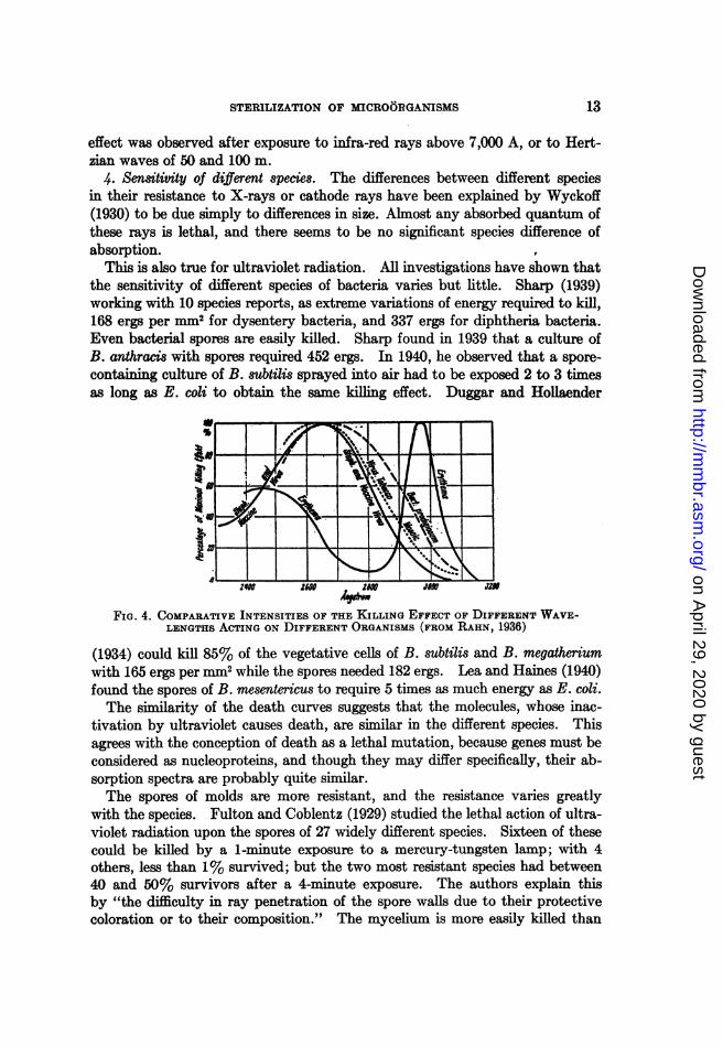
This is also true for ultraviolet radiation. All investigations have shown thatthe sensitivity of different species of bacteria varies but little. Sharp (1939)working with 10 species reports, as extreme variations of energy required to kill,168 ergs per mm2 for dysentery bacteria, and 337 ergs for diphtheria bacteria.Even bacterial spores are easily killed. Sharp found in 1939 that a culture ofB. anthracis with spores required 452 ergs. In 1940, he observed that a spore-containing culture of B. subtilis sprayed into air had to be exposed 2 to 3 timesas long as E. coli to obtain the same killing effect. Duggar and Hollaender
4~~~~~~~~~~~~~~~~~~~40
FIG. 4. COMPARATIVE INTENSITIES OF THE KILLING EFFECT OF DIFFERENT WAVE-LENGTHS ACTING ON DIFFERENT ORGANISMS (FROM RAHN, 1936)
(1934) could kill 85% of the vegetative cells of B. subtilis and B. megatheriumwith 165 ergs per mm2 while the spores needed 182 ergs. Lea and Haines (1940)found the spores of B. mesentericus to require 5 times as much energy as E. coli.The similarity of the death curves suggests that the molecules, whose inac-
tivation by ultraviolet causes death, are similar in the different species. Thisagrees with the conception of death as a lethal mutation, because genes must beconsidered as nucleoproteins, and though they may differ specifically, their ab-sorption spectra are probably quite similar.The spores of molds are more resistant, and the resistance varies greatly
with the species. Fulton and Coblentz (1929) studied the lethal action of ultra-violet radiation upon the spores of 27 widely different species. Sixteen of thesecould be killed by a 1-minute exposure to a mercury-tungsten lamp; with 4others, less than 1% survived; but the two most resistant species had between40 and 50% survivors after a 4-minute exposure. The authors explain thisby "the difficulty in ray penetration of the spore walls due to their protectivecoloration or to their composition." The mycelium is more easily killed than
13
on April 29, 2020 by guest
http://mm
br.asm.org/
Dow
nloaded from

OTTO RAHN
the spores. The spores of PeniciUium digitatum required 9 times as long anexposure as E. coli. This penicillium belonged to the 16 easily killed species;and the observation by Koller (1939) that spores of Aspergillus niger require 50to 100 times as much energy as E. coli is not contradictory.The virus of tobacco mosaic was far more resistant than the spores of B.
subtilis or of B. megatherium, but responded essentiaUy to the same wave lengths(Duggar and Hollaender, 1934).
Ultraviolet rays are widely used for the sterilization or air, especially in hos-pitals and operating rooms (see review by Hart and Sharp in Glasser's MedicalPhysics, 1944). They are also employed to decrease the contamination fromthe air of breweries, bakeries, meat and vegetable storage rooms (see review byPorter, 1940).
Ultraviolet radiation has been used for the sterilization of the water suppliesof a few cities in France. The process is efficient, but expensive. Many labora-tory attempts have been made to sterilize milk, but this method has not beenused as yet by the dairy industry (Supplee et al., 1941). Sterilization of solidobjects must necessarily be limited to the very surface, and while fair success isclaimed for meat in storage (Porter 1940), Fulton and Coblentz obtained dis-couraging results in trying to sterilize oranges. Hall and Keane (1939) couldkill all the spores of thermophilic bacteria in sugar in laboratory experiments,but in large-scale manufacturing, ultraviolet radiation destroyed only half of thespores, on account of the absorption of the rays by the sugar crystals.
III. DEATH DURING AND AFTER DESICCATION
Two different effects must be considered separately when the desiccation ofbacteria is concerned, namely, the number of fatalities due to the removal ofmoisture, and the gradual death of those bacteria which survive the change fromthe moist to the dry state. The two causes of death are quite different, and areindependent of each other.
1. Death during desiccation. The earlier theories referred only to the deathof bacteria spread in thin layers on some surface, and it was believed that bac-teria could not survive complete drying, but were protected against this occur-rence more or less completely by the capsule of the dry medium around them,and only those cells could survive which kept their natural moisture content.Modern drying methods, especially the spray-drying, leave only very thin pro-tective layers around the bacteria, and yet many survive. The percentage ofsurvivors may vary from 0 to nearly 100, depending not only upon the speciesand age of the culture, but upon the kind of medium in which the cells are sus-pended, the surface on which they are dried, and the rate and temperature ofdrying.Most important is the medium surrounding the bacteria during the act of
desiccation. Bacteria dried with their culture medium such as broth or milksurvive fairly well while suspensions of bacteria from agar surfaces or of washedbacteria have only a very small percentage of survivors.
Paul and Prall (1907) dried the staphylococci from agar surface growths, after
14
on April 29, 2020 by guest
http://mm
br.asm.org/
Dow
nloaded from

STERILlZATION OF MICROORGANISMS
suspension in water, on small stones (garnets) of uniform size in order to havebacteria free from the organic matter of the medium. A decrease of about 60%was observed in the first 24 hours, but after that, the number of survivors re-mained constant if they were kept in a vacuum at very low temperatures. Theobject of Paul and Prall's procedure was to obtain uniform bacterial suspensionsfor testing disinfectants in the absence of organic matter. However, the deadbacteria were actually merely organic matter. This becomes very evident fromthe study by Otten (1930) who made thick suspensions of bacteria from agarsurface growths in saline solutions, and dried small quantities, a few drops or 2to 1 ml, in tiny vials at room temperature in a vacuum over concentrated H2S04.He obtained a survival rate of 2 to 5% with typhoid bacteria, 0.05 to 0.005%with dysentery bacteria, and only one survivor out of 10,000, sometimes lessthan one out of a million, with the cholera vibrio. Otten varied the conditionsof drying, and observed that slow drying kilLs more bacteria than rapid drying.Quite important was the depth of the layer of dried cells. While 1 ml of a sus-pension of typhoid bacteria resulted in 0.04% survivors after drying, 0.1 mlof the same suspension dried on the same surface gave only 0.008% survivors.The same amount of suspension, dried in different containers of which one hadan exposed surface ten times as large as the other, gave survival ratios 5 to 23times as large with the smaller exposed surface. Otten then mixed the bacteriato be dried with a suspension of dead bacteria, and obtained far better survival;for instance:
1 ml of the concentrated suspension yielded....................... 2.6% survivors
fo.08% c
1 ml of a dilution (1:10) with saline yielded....... 003% i
f.3% c
1 ml of a dilution (1:10) with a suspension of dead bacteria yielded.... {2 cc
Otten emphasizes that the bacterial proteins protect the cells not by forming acover which prevents complete desiccation, but by acting as protective colloidwhich makes the drying process more gentle and less abrupt. In 1933, Ottenapplied this discovery to the drying of very sensitive species, such as the bac-teria of meningitis or whooping cough, by the addition of dead staphylococci.He also showed that dried smallpox vaccine, was protected by the lymph pro-teins and could be kept active at tropical temperature for as long as 5 years bypreservation in vacuo (fig. 5b).The majority of experiments on desiccation refer to bacteria dried with the
culture medium which is practically always of colloidal nature. Consequently,the survivor ratio is high. Rogers (1914), in his first experiments, dried freshlycurdled milk cultures of lactic streptococci by adding an equal amount of lac-tose, and blowing warm air of 43 C over the culture. About 80% of the cellsdied during this treatment (table 2), and the death rate was greatest when themoisture dropped from 10% to 5%.
Higher survival rates were obtained by spraying the cultures into a current of
15
on April 29, 2020 by guest
http://mm
br.asm.org/
Dow
nloaded from

OTrO RAHN
dry warm air. Drying was almost instantaneous, and took place at fairly lowtemperatures because of the rapid evaporation. Initial bacterial counts are notgiven, but the powdered cultures contained from 657 million to 8,590 millionviable cells. The best method, however, was the desiccation of frozen cultures.For laboratory experiments, the cultures were frozen in Petri dishes in C02-snow,placed in a cold desiccator with concentrated H2S04, or P205, and evacuated toa very high vacuum which is absolutely essential for rapid drying. 10 ml of amilk culture can thus be dried in 3 to 4 hours. The frozen and dried culturescontained between 380 and 12,670 million bacteria per gram. Neutralizationof the milk cultures resulted in higher bacterial counts before drying, but inlower counts after drying."The Bureau of Dairy Industry prepares, for distribution in the field, dried
cultures of Propionibacterium shermanii, the organisms largely responsible forthe characteristic flavor and eye formation in Swiss cheese.... The final product
TABLE 2Survival of lactic streptococci during drying of a milk culture with added lactose
SURVIVORS PER GRAMTIME OF DRYING mOPISIRECONUL NT
Moist powder Water-free basis
hours %o 59.05 785,000,000 1,917,000,0000.5 48.05 750,000,000 1,443,000,0001.0 34.71 963,000,000 1,475,000,0001.5 24.05 942,000,000 1,240,000,0002.0 10.56 916,000,000 1,024,000,0002.5 4.74 351,000,000 368,000,000
3.25 385,000,000 393,000,000
Data of Rogers, 1914.
may contain as many as 700,000,000 viable bacteria per gram" (Fundamentalsof Dairy Science, 1935, p. 432). Commercial yeast cultures are usually driedon some cereal constituents; lactic cultures for dairy starters are sometimes driedon lactose; the rapid absorption of 5% water of crystallization by anhydrouslactose may be of help in rapid drying. Bacteria in soil survive drying quite well.Rahn (1907) found that 36% of the original flora of a good farm soil was stillalive after 56 days of slow drying.The American Type Culture Collection uses drying quite extensively to
preserve cultures, because it not only avoids the necessity of continual transfers,but also prevents the formation of variants, dissociants, mutants, etc. Driedcells cannot possibly change their morphological or physiological characters.
Different species exhibit quite different resistances to desiccation. Stark andHerrington (1931) found that streptococci could tolerate the sudden changefrom moist to dry state (when the bacteria were dried in their culture medium)much better than E. coli or Lactobacillus acidophilus, while yeast and staphylo-cocci showed an intermediate tolerance.
16
on April 29, 2020 by guest
http://mm
br.asm.org/
Dow
nloaded from

STERILIZATION OF MICROORGANISMS
The survival of bacteria during the process of desiccation and afterwards is ofimportance in public health as well as in food preservation. In the manufactureof milk powder, for instance, not all bacteria are killed. Even the severe treat-ment of the drum-drying method, where the milk flows onto rotating, steam-heated drums and is scraped off as a paper-thin, dry sheet, leaves some vegeta-tive forms alive. According to Supplee and Ashbaugh (1922), usually only oneout of 10,000 bacteria survives. The very rapid spray-drying process yieldsa much higher survival ratio. The book of Hunziker (1935) has compiled alarge number of data, and the plate counts of drum-dried powder range from45 to 600,000 per gram while the spray-dried powders have the much higherlimits of 4,400 and 5,500,000. A more recent survey of 671 English milk powdersamples by Crossley and Johnson (1942) shows the wide extremes of 200 and19,500,000 bacteria per gram.
Bacteria dried on metal surfaces die when in direct contact. The fact thatmany bacteria can be obtained from coins means only that there is a layer ofprotective dirt preventing immediate contact between bacteria and metal.Ordinary glass contains free alkali, and bacteria dried on coverglasses usuallydo not survive long.
Campbell-Renton (1941) tested the resistance of bacteriophage to desicca-tion and found that the decrease is approximately logarithmic. Differentphages vary greatly in tolerance; of 15 phages for the dysentery bacteria, 8were reduced to les, than 1% of their original activity after 24 hours of drying,while 6 had more than 50% of their activity left. Once in a dry state, the vi-ability decreases very slowly if the vacuum is maintained. Even after 31 yearsof storage over P206, some phages had lost little of the activity which was leftafter the initial decrease by the drying process as such. Most resistant werethe phage "Pasteur" for Staphylococcus aureus and the phage "D M Large"for Salmonella schottmuelleri.
2. Death of dry bacteria. The death rate of dry bacteria was first studied byTh. Paul (1909) and by Paul, Birstein and Reuss (1910). Staphylococcus aureuswas dried on garnets, and the cells died slowly, and in logarithmic order, whenkept at room or incubator temperatures. The actual cause of death was foundto be oxidation. Table 3 illustrates the effect of the oxygen concentration onthe death rates which were computed from frequent plate counts. The bac-teria of Series A were kept in air and in commercial oxygen while in Series B,an intermediate oxygen concentration was used. The concentration exponents2average 0.44 which means that the death rate is approximately proportionalto the square root of the oxygen concentration. Rogers (1914) also found ahigher death rate in air or in oxygen than in vacuo, in hydrogen or carbondioxide. However, bacteria die also in the complete absence of oxygen, thoughquite slowly. The cause of this death has never been studied.
2 The concentration exponent n is calculated from the ratio of two different concentra-tions, and from the ratio of the corresponding death rate constants, by the equationC,ln K1C2J K2
17
on April 29, 2020 by guest
http://mm
br.asm.org/
Dow
nloaded from

18 OTTO RAHN
The order of death is esentially logarithmic as may be seen from fig. 5awhich shows the average decrease of viable bacteria in 9 samples of milk powderkept at 5 different moistures. The break at about 500 bacteria per gram may
TABLE 3Death rate constants of dried staphylococci at different temperatures and
oxygen concentrations
1 INITALL NUMBERSERIESl OXYGEN CONCEN DEATH RATE CONSTANTS - LOG SSERIES ~TRATION __ _ _ _ _ _ _ _ _ _ _ _ _ -
at 18.2 C at 24.9 C at 37.4 C
A 20.8 0.0017 0.0157 3.396.2 0.0034 0.0256 2.7
Concentration exponent n.... 0.46 0.32
B 20.8 0.0107 0.0264 2.154.6 0.0152 0.0369 2.096.2 - 0.0200 0.0444 1.5
Concentration exponent n ................. 0.36 0.560.50 0.46
Data of Paul, Birstein and Reuss, 1910.
FIG. 5aDecrease of bacteria in milk powder.(Data of Supplee and Ashbaugh, 1922)
-5% I 1 1
-30%-
! 2 ) 4 YEARSFIG. 5b
Decrease in activity of dry smallpoxvaccine in vacuo. (Data of Otten, 1933)
be due to spores or very resistant species. Fig. 5b shows the decrease of vi-ability of dried smallpox vaccine kept in high vacuum at room temperaturein the tropics, according to Otten (1933).The temperature coefficients of the death of dry bacteria are those to be ex-
pected for an oxidation process. The data of table 3 give an average Qlo of
on April 29, 2020 by guest
http://mm
br.asm.org/
Dow
nloaded from

STERILlZATION OF MICROORGANISMS 19
about 3 for series A, and of 2 for series B. Rogers' (1914) experiments withdried lactic cultures show a Qlo of 3.6 between 0 and 30 C.The survival of pathogenic bacteria, e.g. from dried feces, or spray-dried by
coughing and sneezing, must be known in order to prevent epidemics. Thebacteria remaining viable on dried vegetables and meats, on milk powder andegg powder decide how readily the food will spoil when moistened. Sugarcontains usually the very resistant spores of thermophilic bacteria which spoilcanned vegetables because the spores survive the canning process. While insome cases, we employ drying in order to kill as many bacteria as possible, weutilize, on the other hand, the longevity of dried bacteria as a great help in
TABLE 4Survivors of dried cholera vibrios, per million cells
SURVIVORS AFTRSTRAIN 01 VBIRIO
24 hours 5 months 4 years
Shillong 653 .................... 1,950 284 26Shillong 1077................... 12,700 4,200 5.2Shillong 610 R.................. 4,630 2,600 0Shillong 610 R.................. 1,960 155 0Rangoon R..................... 14,200 450 12Inaba S....................... 1,950 _ 97InabaRIV.................... 16,000 - 7,000Rangoon S..................... 62 3.2E1 Tor D 12 .................... 11,000 - 50E1TorD 12.................... 21 15E1TorD31.................... 4,100 30E1TorD35.................... 51,000 550E1TorD 35.................... 100 12El TorD 6.................... 2,150 - 0E1TorD 33.................... 4,700 - 0
Data of Campbell-Renton, 1942.
providing cultures for commercial purposes: magic yeast, starters for dairypurposes, and nodule bacteria dried on soil.
Bacteria are also dried to prevent them from changing their properties bymutation, dissociation or adaptation. Most of these cultures, if they are toremain alive for a very long time, are kept in a vacuum and at very low tem-peratures. How they gradually decrease, may be seen from Campbell-Renton's(1942a) experiences with the very sensitive cholera vibrios kept at room tem-perature in a high vacuum over P205 (table 4).
S. Death by dry heat. Dry cells display no life functions; the enzymes are notactive in the absence of moisture; even endogenous catabolism has ceased. Thecells die from oxidation, and when the temperature is raised above the maximaltemperature of the species under test, death is still due only to oxidation. Thereis no coagulation of proteins because dry proteins do not coagulate when heatedto 100 C, and dry enzymes retain their activity. All experiments show that at
on April 29, 2020 by guest
http://mm
br.asm.org/
Dow
nloaded from

OTTO RAHN
higher temperatures, bacteria die more rapidly, but the gradual increase in thedeath rate is due merely to an increase in the rate of oxidation; there is noabrupt change in the death rate at the maximum temperature of growth, norat any other point.
Otten (1930) found dried typhoid, dysentery and cholera bacteria able tosurvive 37 C for many months, 42 C for several weeks, 58 to 60 C for 7 to 10days, and even 100 C for 1 to 2 hours. Boysen (see Rahn, 1932 p. 309) meas-ured the death rate of yeast dried on infusorial earth and on sand. The tem-perature coefficient between 60 and 98 C fluctuated between 4.2 and 6.8 whilebetween 30 and 50 C, it varied from 2.1 to 4.1.Though dry heat is used in all laboratories to sterlize Petri dishes, pipettes
and other equipment, there seems to be a surpnsing lack of a systematic study
TABLE 5Death times (in minutes) of the spores of Clostridium botulinum exposed to dry heat
TEMP. I II III IV V
1100 >120 >120 >120 >140 115-1201150 >120 >120 >120 >110 80- 851200 110-115 95-100 110-115 > 120 100-1051250 >60 >60 40-45 45- 501300 55- 60 30- 35 40-45 55- 601350 35- 40 35- 40 >65 >651400 30- 35 40- 45 60- 65 15-20 15- 201450 25- 30 15- 20 25- 30 10- 151500 20- 25 15- 20 25- 30 10- 151550 10- 15 10- 15 25- 30 5-10 10- 151600 20- 25 15- 20 20- 25 10-15 10- 151650 15- 201700 10- 151750 5- 10
1800 5- 10
Data of Tanner and Dack, 1922.
of the death rates at high temperature. In 1921, Ayers and Mudge measuredthe death times of E. coli and of a heat-resistant lactic type by drying aqueoussuspensions from agar slants on tin strips, and placing these in wide test tubesin an oil bath. They found that in order to be killed in 30 minutes, E. colineeded 60 C in milk, but 71-82 C in hot air; the lactic type needed 76 C in milk,but 110 C in hot air; a sporeformer needed more than 132 C in hot air.
All other experiments by these authors were made upon milk cans that werestill wet when placed in hot air, so that no precise line between moist and dryheat could be drawn. In the experiments by Dahlberg and Marquardt (1932)the dairy utensils to be sterilized in dry heat were also placed into the heaterwhile still wet.The most detailed data that could be found are those of Tanner and Dack
(1922) who swabbed sterile test tubes with cultures of Clostridium botulinum,
20
on April 29, 2020 by guest
http://mm
br.asm.org/
Dow
nloaded from

STERILIZATION OF MICROORGANISMS
dried them, and determined the death times at temperatures ranging from 110to 180 C. The results are given in table 5.The temperature coefficients of death by dry heat are very low. From the
data of table 5, the temperature coefficients for the entire range are 1.75, 1.85,1.65, 1.59 and 1.83. This may seem contradictory to the Qlo of about 3 shownin table 3. However, those coefficients refer to temperatures between 18 and37 C. As will be explained in the section on Death by Moist Heat, temperaturecoefficients decrease slightly with increasing temperature. A reaction with aQio of 3 at 20 to 30 C will display at 160 to 170 C a Qlo of only 1.7 (see table 10).Therefore, the results of Tanner and Dack (table 5) and those by Paul et al.(table 3) are not in disagreement.The obvious consequence of these low temperature coefficients is that an in-
crease of 10 degrees does not reduce the heating time greatly. With a Qloof 1.7, the temperature must be raised 13 degrees in order to halve the steriliz-ing time. This low coefficient is the reason why the various laboratory manualsdisagree widely on the times and temperatures necessary to sterilize dry glass-ware. There is no disagreement about the sterilization of media in the auto-clave because the temperature coefficient in this case is so high that an increaseof 2 to 3 C cuts the sterilizing time in half.
IV. DEATH BY LOW TEMPERATURES
1. Subminimal temperatures. Most bacteria cease to grow at temperatures5 to 10 C above the freezing point, and when they cannot grow, they die withoutbeing frozen, though very slowly. Hilliard and Davis (1918) suspended cells ofEscherichia coli in glucose solutions and subjected them to temperatures as lowas -6 C which did not freeze the solution. About 50% of the cells died in3 hours while in parallel suspensions in water which crystallized, 93 to 99% ofthe cells were killed. Streptococcus lactis which cannot multiply at temperaturesbelow 5 C was held by Rahn and Bigwood (1939) at 0 to 2 C. The originalnumber of 227 million cells per ml of milk decreased in 114 days to
16,000 per ml when the culture was not treated;34,000 per ml when the culture was neutralized at the start;
450,000 per ml when the air was replaced by nitrogen;63,000,000 per ml when the culture was neutralized and kept under nitrogen.
Apparently, death is primarily due to a change of some essential cell constituentby oxidation which is prevented or repaired above the minimum temperature,i.e. as long as the temperature permits the synthetic mechanisms of the cell tofunction. At 0 C, the oxygen concentration is twice as high as at 30 C.
2. Cold shock. Bacteria may also die from cold shock. Sherman and Cam-eron (1934) could kill about 95% of very young cells of E. coli by cooling themvery suddenly from 45 C to 10 C while gradual cooling during 30 minutes causedno injury. Some other species were less sensitive. In older cultures, only asnall percentage of the cells died. The cause of death is not known. B6lehra-dek (1935 p. 147) states: "Under the action of cold, the cellular content is some-
21
on April 29, 2020 by guest
http://mm
br.asm.org/
Dow
nloaded from

OTTO RAHN
times displaced in an atypical way." Several examples are given. It seemsprobable that the suddenness of chilling is likely to enhance such displacement.Two experiments on the effect of cold shock upon higher organisms are avail-
able for comparison. Kylin (1917) observed complete cessation of plasmastreaming in the alga Nitella clavata after sudden cooling from 20 to 3 C. Hereas with bacteria, the emphasis is on suddenness. It is imaginable that a veryrapid temperature change disrupts the cell mechanisms either by upsettingchemical equilibria, or by spatially disconnecting some cell functions whichdepend upon each other. The observation by Plough (1942) that temperatureshocks increase the mutation rate, seems less likely to explain the death of sucha high percentage of bacteria although we have leamed to look upon death ofbacteria as a lethal mutation (see p. 9).
TABLE 6Death by continuous freezing and by alternate freezing and thawing
(Numbers indicate plate counts per ml.)
CONTINUOUS FREEZING ALTiRNATE FIEEZING
Eberthella typhosa
Inoculum..................... 40,896 Inoculum..................... 40,89624 hrs....................... 29,780 Frozen 3 times.903 days ....................... 1,800 Frozen S times .04 days....................... 950 Frozen 6 times .05 days ....................... 2,490
Serratia marcescens
Inoculum...................... 339,516 Inoculum ...................... 339,51624 hrs...................... 36,410 Refrozen once.2,57030 hrs....................... 41,580 Refrozen 2 times.27548 hrs....................... 14,440 Refrozen 3 times.1596 hrs....................... 4,850 Refrozen 4 times .0
Data of Hilliard and Davis, 1918.
S. Freezing. When the water surrounding the bacteria changes to ice, thewater inside of the cell usually solidifies too, as its freezing point does not differgreatly from that of the medium. Solidification of the water prevents anykind of metabolic action, and there is some analogy between frozen bacteriaand dry bacteria. In both cases, the act of transferring bacteria from thenormal into the anhydrous state is a severe ordeal and kilLs many cells, butthose which survive die at a very slow rate if kept anhydrous. Hilliard andDavis (see table 6) compared alternate freezing and thawing with the holdingof frozen bacteria at -1 C. After freezing and thawing 5 successive times,all cells in cultures of E. typhosa and Serratia marceseens were dead while afterremaining undisturbed in the frozen state for 4 days, several thousand cellsremained alive, and most of those that were dead had been killed during theinitial freezing.
22
on April 29, 2020 by guest
http://mm
br.asm.org/
Dow
nloaded from

STERiLIZATION OF MICROORGANISMS
The greatest injury by the act of freezing must be due to the change of waterto ice which is accompanied by expansion, by crystal formation, and by col-loidal changes. Expansion is not so likely to injure the rather elastic cell, butsharp-edged ice crystals may puncture the plasma membrane. Some colloidalsolutions, after freezing and thawing, remain unchanged while others show aseparation of the concentrated colloid from the liquid phase, the melted crystals.Such separation is commonly observed in frozen cells of plants and animals,and results in death of the cell.
Crystallization requires the presence of seed crystals or the formation ofcrystallization nuclei by a special collision of water molecules. The probabilityof such collisions is greatly reduced by colloids. According to Callow (1925),the addition of 3% gelatin to water reduces its velocity of crystallization to1/350 of the normal rate. Ice formation in cells will therefore be slow. Thenumber of nuclei per cell will also depend upon the volume involved, and icefornation in bacteria will be slower than in the much larger cells of plant leaves.This may account for the fact that as a rule, not all cells in a suspension of bac-teria are killed by a single freezing.
Freezing involves several causes of death, and the most common cause, injuryby ice crystals, is quantitatively unpredictable. Thus no order of death canbe expected, and no order has been observed. The survivors of the freezingprocess die at a very slow rate when kept in the frozen state. Bacteria, yeastcells and mold spores have been known to survive for several years in the frozencondition. It is impossible to sterilize foods or even water by freezing.A different picture is obtained when freezing is accomplished very rapidly to
very low temperatures, e.g. by immersion in liquid air. Then, the water in thecells is not changed to ice crystals, but to a glass-like, amorphous mass. Luyetand Gehenio (1940) speak of this change as vitrification, and of the physicalstate as vitreous. Water in the vitreous state may change to the crystallineice stage, and this happens more readily at higher temperatures, when the vitreousmass is warmed slowly to a temperature near the freezing point. If cells aresuccessfully vitrified without formation of ice, they can be held at low tempera-tures for a very long time without danger of ice formation. This danger exists,however, during the thawing process. Bacteria which have survived vitrifica-tion, may thus be killed during the thawing. Rapid thawing will prevent this.
It is not surprising, therefore, that Kyes and Potter (1939) found tuberclebacteria to survive rapid freezing and thawing 20, 40, 80 and even 200 timeswhen frozen in steel test tubes in liquid air, and thawed in hot water. Onlyone experiment was made with C02-ice in glass tubes, and no growth in vitrocould be obtained after 25 alternations of slow freezing and thawing. Storagein the refrigerator for 6 years at -3 C killed all bacteria because the refrigeratorwas defrosted twice each year which meant twelve very slow freezings andthawings. What seemed offhand to be a most severe treatment, namely, therapid change over 200 C in liquid air, proved to be rather harmless, thanks tovitrification. The literature on this point, and on freezing generally, has beenreviewed critically by Luyet and Gehenio (1940).
23
on April 29, 2020 by guest
http://mm
br.asm.org/
Dow
nloaded from

OTTO RAHN
V. DEATH BY MOIST HEAT
Heat is applied in two different ways for the destruction of bacteria. Glass-ware and certain instruments and materials are sterilized with dry heat. Theeffect of high temperatures on dry bacteria has been discussed in the precedingpages. Other materials are heated when wet, e.g., foods in the canning process,milk, beer and wine during pasteurization, culture media for bacteria, andsurgical dressings. For this process, the term "moist heat" is most commonlyused although Chick, in the first detailed quantitative study, spoke of it as"death by hot water." The cause of death in moist heating is quite differentfrom that in dry heating, and the rules applying to the one method do not fitthe other. Death by dry heat is primarily an oxidation process; death by moistheat is due to coagulation of some protein in the cell.
1. Thermal death poinl and thermal death time. The standard of comparisonof heat tolerance of different species was originally the Thermal Death Point,i.e., the lowest temperature at which a suspension of bacteria is killed in 10minutes (see Descriptive Chart, Society of American Bacteriologists). Thismethod cannot give comparable results unless the conditions are standardizedas to age of culture, approximate number of cells, pH of suspension, dimensionsof test tubes and thickness of glass in the test tubes.
Research workers in the canning industry found it more suitable for theirpurposes to keep the temperature constant and to vary the time. ThermalDeath Time is the shortest time necessary to kill all bacteria in a given suspen-sion at a given temperature. Bigelow and Esty (1920) suspended the bacteriaor spores to be tested in clear juices from canned foods, distributed the suspen-sion uniformly among a number of small narrow glass tubes, sealed these com-pletely by fusing the glass, and dropped them into an oil bath of constant tem-perature. Tubes were removed at different times and incubated; survival ofany bacteria became evident by clouding of the medium. A review of variousslight alterations of technique is given by Beamer and Tanner (1939a). It isnecessary to determine the initial number of cells or spores, because the thermaldeath time is longer with larger inocula.
2. The order of death. The order of death by heat is logarithmic. From theearliest quantitative measurements by Chick (1910) to the extensive investiga-tions by Watkins and Winslow (1932), death of vegetative cells as well asdeath of spores has been found to be logarithmic. The investigations byBigelow and others of the National Canners Association and the many studies ofheat sterilization of spores of Clostridium botulinum, e.g., by Weiss (1921) andEsty and Meyer (1922) have confirmed this. A number of experiments byBeamer and Tanner (1939a, b) with vegetative cells of bacteria and with yeastsgave the same order.The customary explanation that death is brought about by heat inactivation
of the enzymes (see e.g., Isaacs, 1935) cannot be correct because, for mathe-matical reasons, a logarithmic order is possible only when death is due to thedestruction of a single molecule in the cell (Rahn 1929, 1943). To be sure,Edwards and Rettger (1937) found that washed cells of bacteria, held for 24
OA
on April 29, 2020 by guest
http://mm
br.asm.org/
Dow
nloaded from

STERILIZATION OF MICROORGANISMS
hours between 40 and 60 C, lost all their respiratory enzymes at temperaturesnear the maximum for growth. However, enzyme deterioration of washedcells without food held for such a long time under very abnormal conditionspermits no conclusions as to the behavior of the same cells in a suitable medium.Rahn and Schroeder (1941) repeated the experiment using the method of Ed-wards and Rettger for enzyme analysis, but they measured viable cells and en-zyme content in the same sample of cell suspensions of Bacillus cereus suspendedin phosphate buffer at 46 C and 50 C. The first line of data in table 7 shows that99% of the cells were dead when only 14% of the peroxidase and 20% of thecatalase had been inactivated. Enzyme coagulation could not possibly havebeen the cause of death. Similar results had been obtained with yeast byRahn and Barnes (1933).
TABLE 7Death of cells and loss of enzyme activity of Bacillus cereus under the action of heat
TEMPERATURE TIME OF EXPOSURE VIABLE CELLS (PLATE CATALASE DEHYDROGENASECOUNT)
Percentage of cells or enzymes rem ining
C minutes
46 10 1.1 80 8620 0.04 65 8640 0.002 48 6780 0.00001 38 46
50 5 2.4 8910 0.006 56 5720 <0.00002 56 940 <0.000002 48 -
Data of Rahn and Schroeder, 1941.
Microbial enzymes continue to function for a considerable time at tempera-tures above the maximum for growth. Table 8 shows that centrifuged cells ofStreptococcus lactis suspended in buffer solution with glucose ferment rapidlyat 42 C and even at higher temperatures. This strain multiplies most rapidlyat 33 C and cannot multiply above 41 C, yet at 42 C, the energy availablefrom fermentation is far higher than at 33 C. Multiplication at 42 C cannot behandicapped by the heat inactivation of the enzymes, but by the inactivation ofthe synthetic catalysts or the cell division mechanism. Similar data havebeen obtained with yeast which grows most rapidly at 29 C, ceases to grow at35 C, but ferments at 44 C more rapidly than at 29 C (Rahn, 1932, p 132).The mathematical necessity that death must be brought about by destruction
of a single molecule brings us back to the definition given in the chapter onradiation that death is a lethal mutation. We may assume that heat coagulationof a single gene prevents reproduction. Such a cell is sterile, and according tobacteriological standards, it is dead, though its enzyme content may not be ex-hausted. However, the inactivation of the growth mechanism is likely to make
25
on April 29, 2020 by guest
http://mm
br.asm.org/
Dow
nloaded from

O2rrO RAHN
repair and replacement of inactivated molecules impossible, and the enzymecontent of such sterile cells must gradually decrease and this is evident in table 8.The decreasing enzyme content is the consequence of inhibited growth, and notits cause.
S. Death rates. Regardless of whether we accept the one or the other explana-tion of the cause of the logarithmic order of death, its existence permits us tocompute death rates and to draw conclusions from them which are independentof any explanation. Death rates make it possible to compare the heat resistanceof different species at the same temperature, or the heat resistance of one speciesat different temperatures. It also enables us to describe in quantitative temsthe effect of environmental factors, such as concentration of the medium or itspH, upon heat sterilization.
TABLE 8Per cent lactic acid produced in buffer plus glucose by centrifuged cells of Streptococcus lactis
(1.315,000,000 cells per ml.)
TEM- MINUTESPERA-TURE 10 20 30 40 50 60 90 120 150 180 240
30 0.036 0.072 0.108 0.144 0.189 0.234 0.279 0.36033 0.059 0.107 0.144 0.198 0.252 0.306 0.360 0.43737 0.072 0.135 0.189 0.261 0.315 0.369 0.405 0.48140 0.108 0.180 0.243 0.315 0.385 0.441 0.486 0.54942 0.099 0.171 0.243 0.315 0.385 0.437 0.473 0.49545 0.054 0.099 0.144 0.207 0.221 0.297 0.351 0.36947 0.063 0.099 0.130 0.198 0.216 0.270 0.306 0.30650 0.036 0.063 0.090 0.108 0.117 0.144 0.171 0.171
Data of Dorn and Rahn, 1939.
For the application of heat in the preservation of foods, the death rate con-stant is too c3umbersome, and simpler constants have been introduced. Bakerand McClung (1939) measured the time required, at a certain temperature,to reduce the bacteria to 0.01% of their original number. More elastic in itsapplications is the Decimal Reduction Time (D.R.T.) by Katzin, Sandholzerand Strong (1942) which is the time required to reduce the bacterial populationto 10% of the original number. Accordingly, a doubling of D.R.T. must re-duce the populAtion to 1% and heating for 4X (D.R.T.) reduces it to 0.01%,so that the constant of Baker and McClung is identical with 4 times the DecimalReduction Time.Ayers and Johnson (1914), in their study of pasteurization of milk, observed
that in cultures of E. coli and of lactic streptococci, a ver few cells were some-times found which were much more resistant than the great average. Theyintroduced the term "majority thermal deathpoint" for the lowest temperaturewhich kills the large majority of cells in 10 minutes. This phenomenon hadbeen described in detail by Gage and Stoughton (1906) who worked with E. coli.
26
on April 29, 2020 by guest
http://mm
br.asm.org/
Dow
nloaded from

STERILIZATION OF MICROORGANISMS
Beamer and Tanner (1939a, b) gave a clearer picture by drawing the logarithmicsurvivor curves which showed a sharp break, indicating that the remaining celLshad a much greater, but uniform resistance. These experiments included,besides the colon-typhoid group, also several yeasts. Gage and Stoughtoncould find no evidence of spore formation, and they demonstrated that thegreater heat tolerance was not inheritable. In all reported cases, the percentageof resistant cells has been less than 0.1. Whatever the explanation, the greatpractical importance of these very few highly resistant cells in commercial pas-teurization of all kinds of foods is obvious. Chambers and Gaines (p. 8) had asimilar experience when killing bacteria by sonic waves.Very complicated is the mathematical treatment used by Ball (1923, 1928)
in the study of the temperatures required in the canning of vegetables. Thisproblem involves not only the heat sterilization of spores, but also the heatconductivity of the cans and their contents. The z and F factors which play animportant role in these equations will be discussed later.
4. The temperature coefficient of death by moist heat. The death rate constantsare either obtained from plate counts at certain exposure times, from the equa-
initial numbertion Kt = log surivor or they may be computed from the thermal deathsurvvors
times. In this latter method, the initial inoculum for all tubes is the same forany series of experiments and the final number of survivors is also the same,namely less than 1, so that the quotient of initial number over survivors isconstant."
If the death time is determined at the temperatures T1 and T2 (T2 beinghigher), we may call the corresponding death rate constants K1 and K2, and thedeath times t1 and t2. Since the order of death is logarithmic, we have the for-mula
K1 t1 = log initial number Ksurvivors
or
K2 tiK1 t2
This quotient indicates how much more rapidly death proceeds at the highertemperature T2. This is the temperature coefficient for the temperature in-crease T2- T1. For comparative purposes, it is customary to give the coeffi-
3 The thermal death times are not precise values. Between the last sample that showedviable bacteria and the first that showed none, some time has passed. During this inter-val, the number of survivors was reduced to less than 1 per sample. In all experiments,the number of survivors was identically the same at some moment between these twocritical times, but the exact moment is not known. All death time data have a certainrange of possible error, the magnitude of which depends upon the spacing of the timeintervals. The number of survivors is never zero, but becomes very small, e.g., 1 in 100liters, 1 in 1,000 liters, etc.
27
on April 29, 2020 by guest
http://mm
br.asm.org/
Dow
nloaded from

OTTO RAHN
cient for an increse of 10 C which is designated as Qlo. The formula for con-version from Qn to Qio is indicated in the following relation:
10 10
Qio = Qn = n
A number of such temperature coefficients of heat disinfection have beencompiled by Rahn (1932, pp. 320-323). For spores, they are fairly uniform,between 8 and 10 at temperatures from 100 to 135 C. With vegetative cells
TABLE 9Temperature coefficients of disinfection by moist heat
Qio IN NUTRIENT BROTH Qlo IN TOMATO JUICE
pH 7.05 pH 4.2
Temperature interval
55-60 C 6045 C 5540 C 60-65 C
Eberthella typhosa.28.9 26.1 20.2 2.7Salmonella paratyphosa. 37.7 10.3 .Salmonella scottmuelleri. 42.0 33.7 3.3Salmonella aertrycke.23.7 9.0 5.2 27.7Salmonella enteritidis 36.0 15.3 9.8 -
Staphylococcus aureus.28.9 59.2 10.1 3.9
Qlo IN BROTH Qlo IN BROTH Qio IN GRAPE JUICE
pH 6.8 pH 3.8 pH 2.6
Temperature interval
55-60 C 60-65 C 55-60 C 60-65 C 55-60 C 60-65 C
Debar,yomyces globosus. 4.0 12.2 6.5Monilia candida. 4.0 71.0 - 63.0Saccharomyces ellipsoideus 11.6 12.2 15.4 9.0 21.3 19.6Torula monosa.20.6 7.0 7.5 8.0 6.4 29.2
Data of Beamer and Tanner, 1939a, b.
at 50 to 80 C, they are usually higher. Some more recent data for vegetativecells are given in table 9.The coefficients computed from the data by Beamer and Tanner fluctuate
greatly, each value being the result of only one experiment. This fluctuation isdue to the fact that the determination of death rates generally is subject toconsiderable experimental error; the possibility of error is greatly enlargedwhen the quotient of two such death rates is computed. Most of the coefficientsare above 10, and where they are lower, it is due to the survival of a few indi-viduals with much higher resistance, e.g., in the case of DebaryomycesandMonilia(Rahn, 1943). The high temperature coefficient makes it practically certainthat death by heat is a coagulation (or denaturation) process, as such hightemperature coefficients are very rare except with protein coagulation.
28
on April 29, 2020 by guest
http://mm
br.asm.org/
Dow
nloaded from
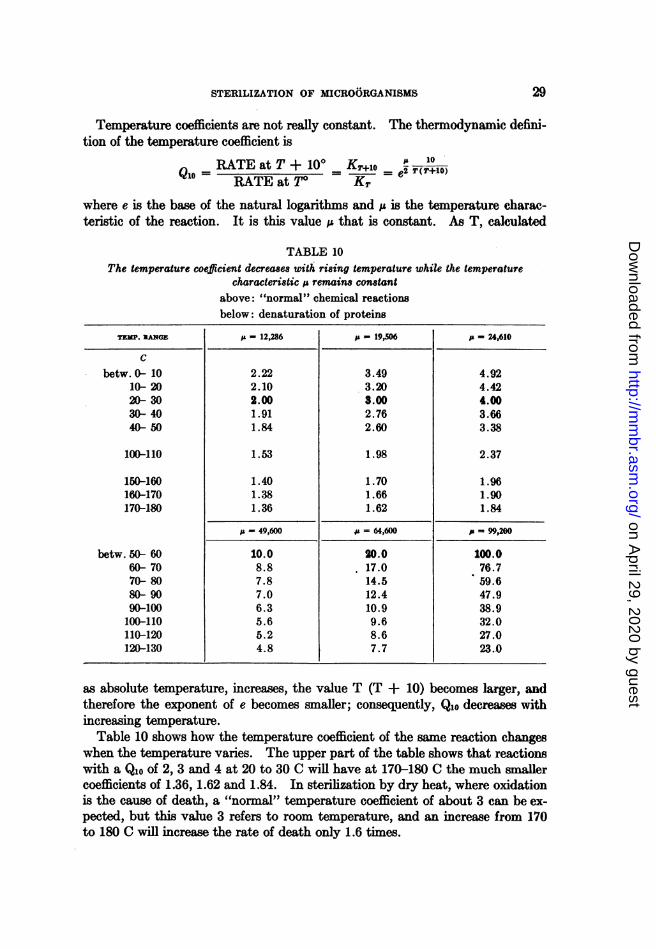
STERlLIZATION OF MICROORGANISMS
Temperature coefficients are not really constant. The thermodynamic defini-tion of the temperature coefficient is
Q - RATE at T + 100 KT+ralo = 10QIO= =~~___ e2 T(T+10)10 RATE at To Kr
where e is the base of the natural logarithms and ,u is the temperature charac-teristic of the reaction. It is this value ,u that is constant. As T, calculated
TABLE 10The temperature coefficient decreases with rising temperature while the temperature
characteristic a remains constantabove: "normal" chemical reactionsbelow: denaturation of proteins
ju = 12X286 isz 19,506 j p 24,610
3.493.203.002.762.60
1.98
1.701.661.62
4.924.424.003.663.38
2.37
1.961.901.84
i = 49,600 u =64,60 p =9920
20.017.014.512.410.99.68.67.7
100.076.759.647.938.932.027.023-0
as absolute temperature, increases, the value T (T + 10) becomes larger, andtherefore the exponent of e becomes smaller; consequently, Qlo decreases withincreasing temperature.
Table 10 shows how the temperature coefficient of the same reaction changeswhen the temperature varies. The upper part of the table shows that reactionswith a Qlo of 2, 3 and 4 at 20 to 30 C will have at 170-180 C the much smallercoefficients of 1.36, 1.62 and 1.84. In sterilization by dry heat, where oxidationis the cause of death, a "normal" temperature coefficient of about 3 can be ex-pected, but this value 3 refers to room temperature, and an increase from 170to 180 C will increase the rate of death only 1.6 times.
2.222.102.001.911.84
1.53
1.401.381.36
TEMP. lNGE
Cbetw.0- 10
10- 2020- 3030- 4040- 50
100-110
150-160160-170170-180
betw. 50- 6060- 7070- 8080- 9090-100100-110110-120120-130
10.08.87.87.06.35.65.24.8
29
on April 29, 2020 by guest
http://mm
br.asm.org/
Dow
nloaded from
o0rro RAHN
The lower part of table 10 deals with protein reactions such as coagulation,denaturation, or inactivation, which have very high temperature coefficients.A coefficient of 100 at 50 C corresponds to a Qlo of 23 at 120 C. The fact thatthe Qlo of spore sterilization in canning averages about 9, while that of milkpasteurization is about 20, does not prove that we are dealing with differentreactions. The same reaction with a Qlo of 20 near 60 C has at 120 C aQioof 8.So long as temperature effects are studied within the narrow limits of life,
i.e., between 10 and 50 C, the assumption of a constant Qlo does not introduce agreat ehtor, but when the range becomes as wide as in the study of death byheat, the assumption of a constant temperature coefficient may be very mis-leading.Two different methods of pasteurization of milk are permitted by the New
York State Department of Health, namely the holding process which consistsof holding the milk for 30 minutes at 143 F, and the short-time, high tempera-ture process, usually called the flash heat process, which requires holding for15 seconds at 160 F. The temperatures are 9.44 C apart (71.11-61.67) and theheating time of the one process is 120 times that of the other. This means,that the authorities assumed a Q9.44 (which is practically Qlo) of 120, a veryhigh temperature coefficient for this range.
It is obvious that the temperature coefficients of death cannot be extrapolatedinto the region of growth. If a bacterium can multiply, though very poorly,at 40 C, but dies at 50 C, the temperature coefficient will equal the death rateat 50 C divided by zero which means Qio = oo. A sharp drop from infinity tothe actual temperature coefficient of inactivation of the life processes must beexpected, and very high coefficients must be expected at the lowest lethal tem-peratures. This accounts for the high coefficients found for Escherichia colinear 50 C since its maximum temperature of growth is 47 C. Watkins (1933)computed the increase of death rate between 50 and 55 C to correspond to aQlo of 560. Baker and McClung (1939) found for the same bacterium in threeexperiments:
between 51.7 and 54.5 ....................... Qio = 296.0 315.0 16.6between 54.5 and 57.3 C.......................... 22.7 21.6 408.0
Average between 51.7 and 57.3 C................. 86.7 70.8 89.2
Henderson Smith (1923) computed from his experiments with the spores ofBotrytis cinerea the following temperature coefficients:
between 31 and 37 C Qio = 690between 37 and 44.3 C 132between 44.3 and 47.0 C 92.8between 47.0 and 50.3 C 29.5
The data on the death rates of spores show a far greater uniformity and con-stancy than those for vegetative cells, because all data on spores refer to tem-peratures which were at least 30 C higher than the maximal growth temperature,
30
on April 29, 2020 by guest
http://mm
br.asm.org/
Dow
nloaded from

STERILIZATION, OF MICROORGANISMS
while many of the experiments with vegetative cells were made within 10 or15 C of the maximum for growth.
5. Factors controlling death by heat. Sensitivity to heat varies with the spe-cies. Pasteurization of milk by holding at 63 C for 30 minutes destroys allpathogenic bacteria except the spores, and kills many species of saprophytes,but not all. With some species such as lactobacilli, the death rate is low, andsometimes, thermophilic bacteria multiply during pasteurization. The sporesof many species can survive a short period of boiling, but the spores of Methano-bacterium omelianskii are killed at 80 C in 10 minutes (Barker 1940). Thecause of the great resistance of bacterial spores will be discussed at the end ofthis chapter.The medium in which bacteria are suspended can inuence the death rate.
The best-known example is the effect of the acidity of the medium. Bacteriadie more rapidly in acid or alkaline media than in neutral suspensions. This sug-gests, of course, the assumption of chemical disinfection rather than physicalcoagulation, but two facts speak against this. In chemical disinfection, thetemperature coefficients should be relatively low while table 9 shows that theyare, on the average, higher in acid than in neutral media. Further, yeastwhich grows better in acid media is killed by heat more easily in acid than inneutral media (Beamer and Tanner, 1939b).
Extensive experiments on the effect of pH on the heat resistance of sporeshave been carried out by Weiss (1921), Esty and Meyer (1922) and others.The study by Townsend, Esty and Baselt (1938) may serve as a more recentexample. These authors did not use death rates or temperature coefficients,but the factors F and z which Ball had introduced in 1923 for the computationof processing times in the canning industry. As these two values are frequentlyused in the canning industry which furnishes the largest amount of data fordeath by heat, the meaning of these factors will be discussed here.
The value F is the thermal death time of the species at 121 C or 250 F. Te number zis the temperature increase, in degrees Fahrenheit, necessary to reduce the death time toone-tenth. We introduce the temperature coefficient for 1 C, Qi, which can be transformedinto Qlo by the simple relation Qlo = Q' °. A few pages earlier, it has been shown thatthB ratio of the death rates at temperatures T2 and T1 can be measured by the correspondingdeath times, viz.,
K2 tbK1 t2
This ratio is the temperature coefficient for n° = T2- T1 degrees, hence
t2
The definition of z specifies that with an increase of z°F, or 0.555zC, the death time
t2 is one-tenth of th. Therefore, n = O.M555z and L = 10.t2
Q1=Q1~~~~~1Q0 l Q° =log 10t2
0.555x log Q, = log 10 = 1
31
on April 29, 2020 by guest
http://mm
br.asm.org/
Dow
nloaded from

OTTO RAHN
We have seen that Qlo = Ql, therefore
log Qio = 10 log Qi = 10 = 180.555z z
18Z log Qlo
The value of z or Qlo indicates the slope of the straight line obtained by plotting the loga-rithms of death times against temperature. The value F gives one point on this curve, andthus, F and z (or its corresponding Qlo) are sufficient to characterize the thermal resistanceof the spores of a species at any temperature.
The authors studied in detail the heat resistance of the spores of two strainsof Clostridium botulinum, and of a bacillus isolated from spoiled canned corn.The summarized results are given in table 11 where the z values have beenconverted into Qlo. With the bacillus spores, the value F, the death time at
TABLE 11The heat resistance of the spores of three bacteria, expressed by the values F and Q0o
BACILLuS 3679 C. BOTULINum 62A c. BOTuLINum 213BMEDIUM OF SPORE pH
SUSPENSIONF* QioFi Qio F Qio
Phosphate buffer. 7.0 4.00 12.1 1.70 12.5 2.00 10.0Asparagus.5.4 3.30 7.1 0.39 15.9 0.39 15.9Peas.5.4 3.00 11.6 0.30 22.0 1.40 14.2Spinach.5.4 2.60 9.8 0.65 14.5 0.68 14.5Mik.6.3 2.60 7.8 0.45 16.8 0.50 18.2
Average 9.7 16.3 14.6
* Death time in minutes at 121 C.Experiments of Townsend, Esty and Baselt, 1938.
121 C (250 F), was not greatly affected by the medium, but the spores of the twoclostridia survived in the neutral phosphate buffer 2 to 4 times as long as inmilk and in the vegetable juices. The differences cannot be explained by pHalone. It has been observed in commercial canning that the heat tolerance ofspores is not the same in different vegetables, but the cause is not known. Thetemperature coefficient with the spores of the two clostridia was higher in thevegetable juices than in the buffer, whereas the spores of the bacillus showed theopposite. In the buffer, the three behaved nearly alike. Tanner (1944, p. 962)speaks of a. "food factor" which is the ratio of death time in food juice to deathtime in neutral phosphate buffer.
Different from this chemical effect is the protection afforded by concentratedsolutions. The well-known increase of the thermal resistance of bacteria throughaddition of sugar is usually explained by partial dehydration of the protoplasm.In concentrated solutions of sugar, proteins are not coagulated by heat (Beilins-son, 1929). According to Fay (1934), egg albumin which coagulated in 3 to4 minutes at 60 C, required 23 and 31 minutes respectively in molar solutions
32
on April 29, 2020 by guest
http://mm
br.asm.org/
Dow
nloaded from

STERILIZAT1ON OF MICROORGANISMS
of glucose and sucrose, and 73 minutes and more than 24 hours, respectively,in twice molar solutions. Rennet enzyme dissolved in water was completelyinactivated at 70 C in 15 minutes, but molar CaCl2 protected it for 45 minutes;and in 4 molar sucrose it lost only half of its strength in 5 hours. K. von Angererand Kuster (1939) confirmed the assumption that bacteria become partly de-hydrated by comparing the turbidities of bacterial suspensions in water and inconcentrated solutions.The time of contact is of importance. Fay obtained only 2.5% survivors of
E8cherichia coli upon heating immediately after suspending the cells in 50% su-crose, whereas the same heating left 79% of the cells unharmed if they had beenin the sugar solution for 2 hours before heating.
TABLE 12The harmful effect of high temperature on liquids containing reducing sugars (in
half-molar concentration)Survivors of E. coli after being heated for 8 minutes at 54 C in variously treated sugar
solutions at pH 7.0.
PERCENTAGE SURVIVORS IN MEDIUM PREVIOUSLY STERILIZED DV
CONCENTRATION SUGAR Autoclaving Filtration
In buffer In broth In buffer In broth
0 (control) 1.3 7.2 1.9 3.9
9 glucose 0.08 2.7 5.3 7.99 galactose 0.04 1.8 4.3 8.017.3 lactose 0.09 1.6 18.0 1l.817.3 maltose 0 3.1 5.0 7.3
17.3 sucrose 9.2 15.0 12.0 10.09.1 mannitol 4.5 10.8 5.5 8.94.1 glycerol 3.2 9.4 3.7 6.4'
Data of Baumgartner, 1938.
Some inconsistencies in the results by various investigators are explainedbyBaumgartner (1938) as being caused by harmful decomposition products'origi-nating from reducing sugars during autoclaving. This is prevented by asepticfiltration. Table 12 shows that the percentages of survivors in the last threesolutions, representing non-reducing compounds, are nearly the same in auto-claved and filtered media while the four media containing reducing sugarshave become quite toxic by autoclaving.The theory of dehydration of the protoplasm is too simple to fit the facts.
Protection is not proportional to molarity (table 12), disaccharides protect morethan monosaccharides but maltose seems to be an exception. Von Angerer andKuister (1939) measured the death rates of Escherichia coli and Staphylococcus sp.at 56 C in concentrated solutions of various carbohydrates and other easilysoluble substances. It was found that starch, gum arabic, urea, pyruvate,
33'
on April 29, 2020 by guest
http://mm
br.asm.org/
Dow
nloaded from

OTTO RAHN
glycocoll, calcium chloride, or phosphate buffer offered no protection whilegrycol, glycerol, sorbitol and various mono- and di-saccharides retarded deathvery noticeably.
The Unusuc1 Residance of Spores is not limited to heat. It is very conspicuouswith disinfectants, but does not extend to irradiation (see p. 13). It has beenthe cause of many investigations because bacterial spores are the most resistantorganisms existing. The spores of molds, although more hardy than the my-celium, are killed readily by boiling, and the spores of yeasts are only slightlymore resistant than the vegetative cells.The oldest explanation, the assumption of a practically impermeable spore
wall, can hardly be applied in the case of heat. A wall of only 0.1 ,u thick-ness could not possibly insulate the cell contents against high temperature forseveral hours. Nor could such extreme lack of heat conduction protect sporese.g., for 440 minutes at 100 C, but for only 5 minutes at 120 C. Absence ofmoisture has been another explanation. According to early investigations, thespecific gravity of spores seemed to be about 1.35 to 1.4 which indicated alowmoisture content. Since dry proteins are not coagulated by heat, this explana-tion seemed plausible. However, newer measurements indicate that sporescontain nearly as much moisture as vegetative cells (Virtanen and Pulkki,1933; Henry and Friedman, 1937). K. von Angerer and Kiister (1939) could ex-tract water: from spores by suspending them in concentrated sugar solutions.Virtanen anid Pulkki assumed that the enzymes of the spores were in an inactiveform, and therefore resistant. Friedman and Henry (1938) explained this re-sistance as due to the large amount of bound water in the spores. From measure-ments of the freezing points of spore suspensions in water and in sucrose solution,the bound water for three different species of bacilli could be calculated to 69,63 and 59%0 of the weight of moist spores whereas the corresponding figuresfor vegetative cells were 0, 18 and 28%. The calculations are based on tempera-ture differences of a few hundredths of one degree, and probably have a largeexperimental error.
0. B. Williams (1929) approached the problem from a different angle. Heshowed that the ash content of spores is low, and as proteins do not coagulatereadily in the absence of electrolytes, the low electrolyte content of the sporesmay be responsible for their heat resistance.A different viewpoint was investigated by Rosel von Angerer (1939) who con-
sidered the fairly high fat content of spores as a possible means of protection.Long extraction with fat solvents decreased heat resistance while impregnationwith paraffin (dissolved in carbon tetrachloride) doubled it. Presence or ab-sence of oxygen did not affect the death time. She could not verify the theoryof Sobermheim and Mundel (1938) that spores adsorbed on porous materialssuch as soil are much more resistant. Of all the materials tested, only granu-lated coal increased the death time significantly.However, the most probable explanation is the assumption that the protein
in spores is different from that of vegetative cells (Heim, 1938). The tempera-ture of denaturation of different proteins is by no means the same. Among
34
on April 29, 2020 by guest
http://mm
br.asm.org/
Dow
nloaded from
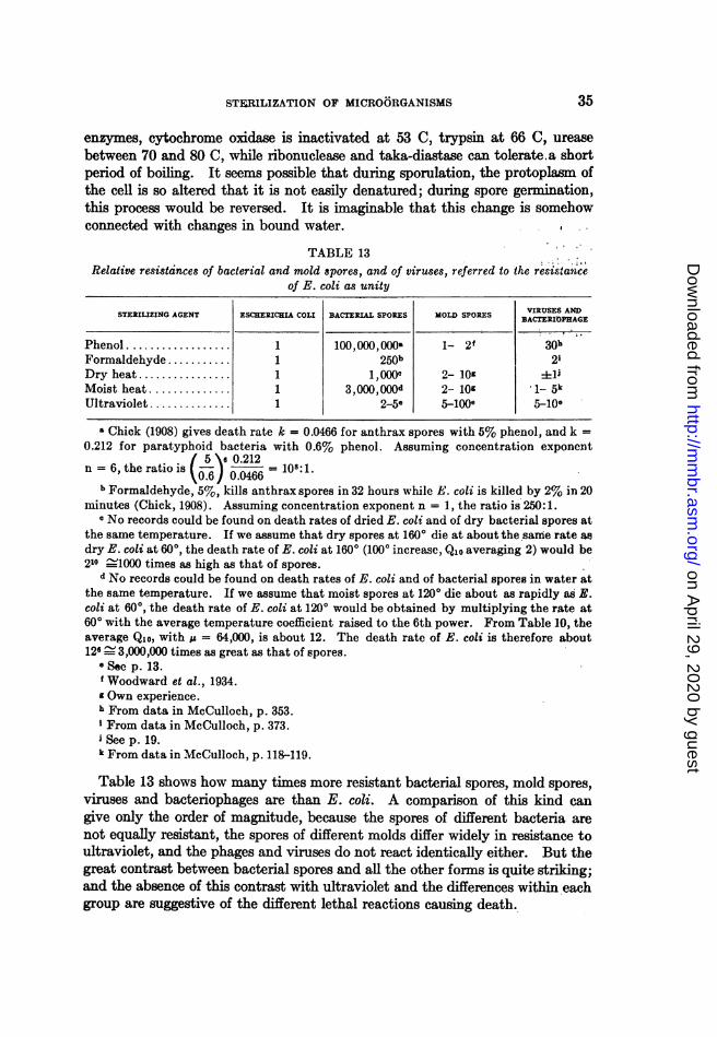
STERILIZATION OF MICROORGANISMS
enzymes, cytochrome oxidase is inactivated at 53 C, trypsin at 66 C, ureasebetween 70 and 80 C, while ribonuclease and taka-diastase can tolerate.a shortperiod of boiling. It seems possible that during sporulation, the protoplasm ofthe cell is so altered that it is not easily denatured; during spore germination,this process would be reversed. It is imaginable that this change is somehowconnected with changes in bound water.
TABLE 13Relative resistdnces of bacterial and mold spores, and of viruses, referred to the rest.stance
of E. coli as unity
STERIILIZING AGENT ESCHERICHIA COLI BACTERIAL SPORES MOLD SPORES BACTRUIOAGE
Phenol .................. 1 100,000,000a 1- 2f 30hFormaldehyde .......... 1 250b 2'Dry heat ................ 1 1,0 2- lMoist heat .............. 1 3,000,00od 2- 10 1_ 5kUltraviolet .............. 1 2-5e 5-lOOe 5l10e
Chick (1908) gives death rate k = 0.0466 for anthrax spores with 5% phenol, and k =0.212 for paratyphoid bacteria with 0.6% phenol. Assuming concentration exponent
/5 6e 0.212n= 6, the ratio is 0.1
= 108:1.' t~~0.6) 0.046610.b Formaldehyde, 5%, kills anthrax spores in 32 hours while E. coli is killed by 2% in 20
minutes (Chick, 1908). Assuming concentration exponent n = 1, the ratio is 250:1.e No records could be found on death rates of dried E. coli and of dry bacterial spores at
the same temperature. If we assume that dry spores at 1600 die at about the same rate asdry E. coli at 600, the death rate of E. coli at 160° (100° increase, Qlo averaging 2) would be210 .'1000 times as high as that of spores.
d No records could be found on death rates of E. coli and of bacterial spores in water atthe same temperature. If we assume that moist spores at 1200 die about as rapidly as E.coli at 600, the death rate of E. coli at 1200 would be obtained by multiplying the rate at600 with the average temperature coefficient raised to the 6th power. From Table 10, theaverage Qlo, with p = 64,000, is about 12. The death rate of E. coli is therefore about126 - 3,000,000 times as great as that of spores.
e See p. 13.f Woodward et al., 1934.g Own experience.h From data in McCulloch, p. 353.iFrom data in McCulloch, p. 373.See p. 19.
k From data in McCulloch, p. 118-119.
Table 13 shows how many times more resistant bacterial spores, mold spores,viruses and bacteriophages are than E. coli. A comparison of this kind cangive only the order of magnitude, because the spores of different bacteria arenot equally resistant, the spores of different molds differ widely in resistance toultraviolet, and the phages and viruses do not react identically either. But thegreat contrast between bacterial spores and all the other forms is quite striking;and the absence of this contrast with ultraviolet and the differences within eachgroup are suggestive of the different lethal reactions causing death.
35
on April 29, 2020 by guest
http://mm
br.asm.org/
Dow
nloaded from

OTrO RAHN
Experimentation with the resistance of spores is complicated by a great varia-bility of this property due to environmental conditions. A summary of theliterature is given by Theophilus and Hammer (1938). Bacterial spores willoccasionally remain dormant for a long time before germinating. This accountsfor the difficulty encountered in the sterilization of food by intermittent heatingon successive days. An extensive study has been made by Morrison and Rett-ger (1930). According to Evans and Curran (1943) incubation of spores atsublethal temperatures, e.g., 65 C, accelerates the rate of their germination.
VI. SURFACE TENSION DEPRESSION
1. Effects of surface tension depression. Many compounds which lower thesurface tension of water are good disinfectants. Best known among these are
TABLE 14Surface tension of solutions killing Staphylococcus aureus in 6 minutes
SURFACE TENSION IN DYNESCONC.
In water During test
Phenol .................................1.25 41.5 42.0&--Cresol ............................... 1.00 37.5 37.5Hexyl resorcinol ......................... 0.025 37.5 37.5Thymol ............................... 00.0625 38.5 38.5Chlorothymol ........................... 0.25 49.0 49.0Mercuric chloride.............;0.025 70.0 68.5M-erthiolate...0.025 51.0 51.5Mertiolae .............................. 0251.01.5Mercurochrome ......................... 1 71.5 65.0Iodine ............................... 0.02 59.5 53.5
Data of Gershenfeld and Witlin, 1941.
some of the fatty acids, the alcohols and phenols, the soaps, the dyes, the bilesalts, and many of the large number of modem wetting agents and detergents.
If the decrease in surface tension were the fundamental cause of death, thebacteria should die when a certain tension is reached, regardless of the chemicalstructure of the compound used. This is not the case. Ayers, Rupp and John-son (1923) found streptococci inhibited at different surface tensions when differ-ent depressants were used. According to Frobisher (1926), Staphylococcusaureus is completely inhibited at a surface tension of 44 dynes per cm whenproduced by sodium glycocholate, but multiplies readily at as low a tension as34 dynes. when produced by sodium oleate. Gershenfeld and Witlin (1941)determined the concentrations required to kill Staphylococcus aureus between5 and 10 minutes, and measured the surface tensions of the killing solutions.Table 14 shows that there is no correlation.
It may be argued that no direct parallelism between lethal concentration andsurface tension can be expected because the lethal effect takes place at theinterface between medium and cel membrane, and this interfacial tension need
36
on April 29, 2020 by guest
http://mm
br.asm.org/
Dow
nloaded from

STERILIZATION OF MICROORGANISMS
not be parallel to the tension between medium and air. The interfacial tensionbetween cell and medium cannot be measured. Davis (1927) measured the dropsize of oleic acid in chloride solutions, and compared this with the concentrationof different chlorides necessary to inhibit bacterial growth. He believed thatthe toxic effects of the cations can be explained by interfacial tension "if bac-teria are looked upon as analogous to oil drops." This analogy has not beengenerally accepted, and the data presented to support this view show consider-able exceptions to the rule.
It must be concluded from these and many other experimental results thatdeath is not caused by change in surface tension, but by some chemical reactionof the surface tension depressant upon some vital cell constituent. However,the subject cannot be dismissed entirely as chemical disinfection because of dif-ferences in the mode of action, and of interaction. The difference arises pri-marily from the concentration of surface-active substances at the surface andinterfaces. They act upon the cell not according to the concentration in theliquid, but according to the much higher concentration at the cell surface.This explains perhaps the fact that so many surface-active substances are gooddisinfectants and antiseptics.
2. Wetting agents. The soaps which have been almost the only efficientdetergents for more than a thousand years are gradually being replaced bysynthetic detergents which decrease the surface tension greatly, but are effi-cient in acid as well as in alkaline media, and are not precipitated by hard water.These modern "wetting agents" can be divided into anionic, cationic, and un-dissociated detergents. The composition and origin of a number of these sub-stances is given by Baker et al. (1941a, b). In the cationic detergents, theorganophilic (or hydrophobic) group is positively charged, as in Emulsol 660 B =(lauryl pyridinium)+ (chloride)-; and in the anionic detergents, this group isnegative as in (Na)+ (lauryl sulfate)-. The only neutral, non-ionized detergentmentioned is Demal, representing an organic ester.Domagk (1935) called attention to the antiseptic properties of this new group
of chemical products. According to Baker et al., (1941b) the cationic deter-gents, as a group, are quite efficient disinfectants, capable of killing gram-posi-tive and gram-negative bacteria in 10 minutes at a concentration of 0.015%(1: 6000), or even 0.003%. A few prepatations were less efficient. The anionicdetergents did not kill these bacteria even after 90 minutes in concentration of0.1%. No data on surface tension are given.
Cationic and anionic detergents neutralize and precipitate each other. Theaction of Zephiran is inhibited by decyl sulfate or sodium taurocholate (Bakeret al., 1941c) or by Aerosol 0 T (Gershenfeld and Perlstein, 1943). Differentfrom this ionic reaction is the protection of bacteria against detergents byphospholipids, lecithin and related compounds, if they are added either beforeor simultaneously with the detergent (Baker et al., 1941c). Addition after thedetergent had no effect. Compounds which did not reduce surface tensiondid not counteract the bactericidal effect. On the other hand, phospholipidsdid not decrease the germicidal effect of several mercury compounds. Thus,
37
on April 29, 2020 by guest
http://mm
br.asm.org/
Dow
nloaded from

OTrO RAHN
the germicidal power of detergents and its decrease by other substances isinterlinked with a surface tension effect. The neutral compound Demal coun-teracted cationic as well as anionic detergents; it also counteracted gramicidin.Valko and DuBois (1942) found that if a strongly germicidal detergent is
mixed with a weak one of the same charge, the total efficiency is smaller thanthat of the stronger germicide, because the weaker one has occupied manyplaces on the bacterial surface, and the surface concentration of the strongdisinfectant is decreased. This is an indirect proof for the claim that surfacetension as such is not the deciding lethal factor. The same authors (1944) alsoshow that bacteria "killed" by a cationic detergent may be revived by additionof a high molecular anion if this is applied within a certain time of perhaps 10 to30 minutes. They consider the reversible death as due to ion exchange in thecell, similar to the death by mercury salts, i.e., a chemical phenomenon.The germicidal efficiency of these detergents depends largely upon the pH
of the medium in which they act. The anionic detergents resemble benzoic orsalicylic acid in their rapid increase of efficiency with acidification of the me-dium (Rahn and Conn, 1944). The analogy permits the conclusion that onlythe undissociated acid molecules of these compounds have bactericidal properties.Gershenfeld and Milanick (1941) found the following concentrations, in ppm,necessary to kill Staphylococcus aureus in 5 minutes:
At pH
4 5 6 7 8 9
Aerosol OT.29 33 250 >10,000 >10,000 >10,000Tergitol 4& 4 T........ 140 250 500 >10,000 >10,000 >10,000TritonK 2.>10,000 10,000 3,300 1,250 110 55
The last compound is a cationic detergent, and in this case, the base in itsundissociated state is probably the effective agent.
S. Combination of disinfectants with surface tension depressants. When surface-active substances are added to a disinfectant solution, they may decrease itsgermicidal efficiency. Hampil (1928) obtained such marked inhibition of thebactericidal action of various phenols by the addition of soap as to indicate theimpossibility of producing a germicidal soap by incorporating s;mall quantitiesof a phenolic compound. As an example may be given the concentration ofbutyl resorcinol required to kill Staphylococcus aureus between 1 and 2 minutesat different soap concentrations:
% Na oleate................ 0 0.2 0.5 1.0 3.0 5.0
% disinfectant required............ <0.05 0.067 0.1 0.2 0.5 0.5
The same has been found to be true with mixtures of mercury compounds andsoap, unless the soap is very much diluted (Frobisher, 1927; Rettger et al., 1929;Schaffer and Tilley, 1930; Tilley, 1939). The general explanation is that somuch soap is concentrated at the surface that the other, stronger disinfectantcannot reach the cell as readily as without soap.
630
on April 29, 2020 by guest
http://mm
br.asm.org/
Dow
nloaded from

STERILIZATION OF MICRO6RGANISMS
It is frequently stated in literature that a decreased surface tension ofthemedium must increase the efficiency of any antiseptic because the rate of diffu-sion through the cell membrane is increased. For example, Frobisher (1944)points out that aqueous iodine solutions do not spread easily on the skin whilean alcoholic solutiot has a much greater wetting power. This leads him to theconclusion in the next paragraph that solutions of disinfectants in dilute alco-hol or in similar solvents which have low surface tensions, are much more likelyto be effective than when dissolved in water. However, Gershenfeld and Miller'sdata (1932) prove this to be a poor example because with iodine, the difference
TABLE 15Concentrations, in per cent, required to kill Staphylococcus aureus in 10, but not
in 5 minutes
pH
7 6 1 5 4
Aerosol concentration, when added
0.02 0.008 0.0017 0.0011
Phenol.1.25 1.17 1.11 1-1.05same + aerosol . .56 0.42 -0.50 0.33 0.26
Mercuric chloride. .0067 0.004-0.005 0.004-0.005 0.0025-0.00275same + aerosol. 0.005-0.0067 0.0033-0.004 0.00275 0.0020-0.0022
Merthiolate. 0.014 0.014-0.02 0.0056-0.0067 0.0033same + aerosol.0.0028-0.0033 0.0033 0.0028 0.0020-0.0022
Hexyl resorcinol. 0.033 0.025 -0.033 0.013-0.02 0.0067-0.0084same + aerosol. .004-0.005 0.004 0.0033-0.004 0.0025-0.00275
Zonite. .05 0.125 -0.167 0.167 0.25 -0.33same + aerosol.0.033 0.125 0.125 -0.167 0.20
Zephiran.....0...........0.00125-0.00142 0.005 >0.2 >0.2same + aerosol.......... >0.2 >0.2 >0.2 >0.2
Data of Gershenfeld and Perlstein, 1941.
between aqueous and alcoholic solutions is slight, and in favor of the aqueoussolution. Frobisher's generalization is erroneous. The surface of wood orskin which contains airspaces, is not comparable with the spaces betweenmolecules of a cell membrane which are already completely surrounded by watermolecules.However, while not a general phenomenon, the efficiency of some disinfectants
at specified concentrations may be increased by surface tension depressants.The increase may be quite noticeable. Gershenfeld and Witlin (1941) couldfind no change in the efficiency of disinfectants by the addition of detergents,but in the same volume, a little later, Gershenfeld and Perlstein did report suchan effect (table 15). It is worth noting that at pH 7, Aerosol in concentration
39
on April 29, 2020 by guest
http://mm
br.asm.org/
Dow
nloaded from
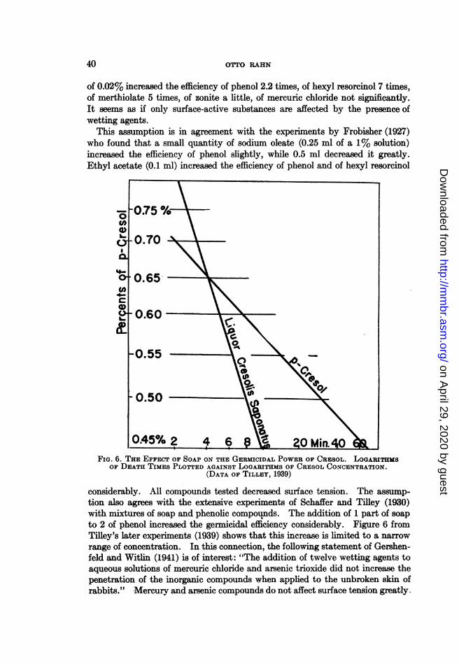
rOTTO RAHN
of 0.02% increased the efficiency of phenol 2.2 times, of hexyl resorcinol 7 times,of merthiolate 5 times, of zonite a little, of mercuric chloride not significantly.It seems as if only surface-active substances are affected by the presence ofwetting agents.
This assumption is in agreement with the experiments by Frobisher (1927)who found that a small quantity of sodium oleate (0.25 ml of a 1% solution)increased the efficiency of phenol slightly, while 0.5 ml decreased it greatly.Ethyl acetate (0.1 ml) increased the efficiency of phenol and of hexyl resorcinol
FIG. 6. THE EFFECT OF SOAP ON THE GERMICIDAL POWER OF CRESOL. LOGARITHMSOF DEATH TIMES PLOTTED AGAINST LOGARITHMS OF CRESOL CONCENTRATION.
(DATA OF TILLEY, 1939)
considerably. All compounds tested decreased surface tension. The assump-tion also agrees with the extensive experiments of Schaffer and Tilley (1930)with mixtures of soap and phenolic compounds. The addition of 1 part of soapto 2 of phenol increased the germicidal efficiency considerably. Figure 6 fromTilley's later experiments (1939) shows that this increase is limited to a narrowrange of concentration. In this connection, the following statement of Gershen-feld and Witlin (1941) is of interest: "The addition of twelve wetting agents toaqueous solutions of mercuric chloride and arsenic trioxide did not increase thepenetration of the inorganic compounds when applied to the unbroken skin ofrabbits." Mercury and arsenic compounds do not affect surface tension greatly.
40
on April 29, 2020 by guest
http://mm
br.asm.org/
Dow
nloaded from

STERILIZATION OF MICROORGANISMS
4. Bile solubility. Another phenomenon usually ascribed to surface tensiondepression is the bile solubility of certain bacteria, notably pneumococci. Hoberand Hober (1942) treat this solubility in a general way, although they are dis-cussing primarily blood cells. They describe the action of bile salts and deter-gents as follows:
"Their molecules are composed of two portions, one polar hydrophilic,which has a tendency to anchor the molecule to water, and one nonpolarhydrophobic which is attracted towards the nonaqueous phase. The resultis a molecular orientation at the interface.... An increase of hydrophilicaffinity will lead to a stronger pull on the nonaqueous phase toward the waterand may have a disintegrating or a dispersing effect. Stronger organophilicproperties on the other hand will favor a wetting action, viz., the fixation of afilm of water on the nonaqueous phase or its separate components. Regard-ing especially the interface between a cell and its surroundings, hydration,swelling and dissolution can be due to the wetting action on the colloidalstructures or the micellae. Protein molecules are unfolded or disrupted bythe pull, and thus denatured. In conjugated proteins, the bonds betweenthe protein part and the prosthetic group are severed, viruses and enzymesare inactivated, and the final result of such effects on cells is cytolysis."This phenomenon of complete dissolution by such compounds as bile and bile
salts is not caused by the decreased surface tension as such, for other depressntslike the alcohols, phenols and saponin do not dissolve bacteria. Of specialinterest in this differentiation between dissolving and non-dissolving agents isthe report by Falk and Yang (1926) that sodium oleate dissolves washed pneu-mococci when re-suspended in distilled water, but not when re-suspended in0.85% NaCl solution. Leonard and Feirer (1927) reported that Endamoebacoli, lodamoeba williamsi, Leptospira icterohaemorrhagiae, Trypanosoma lewisi,and Trichomonas hominis are instantly destroyed and disappear completely oncontact with hexyl resorcinol solutions while bacteria do not. The reviewer hasobserved that cationic detergents did not dissolve pneumococci at pH 9.8 wherethey were most efficient as germicides. While germicidal power depends pri-marily on the concentration of the undissociated base, dissolving power dependsupon other properties.Our knowledge of the interaction between surface tension depressants and
bacteria is not at all perfect or complete. The sterilizing properties of thecompounds of this group are certainly not due to the decreased interfacial ten-sion as such. However, the intensity of the chemical reaction of these com-pounds is influenced by their effect upon interfacial tension, and they may, bythis effect, increase or decrease the germicidal power of other disinfectants.
REFERENCESAYERS, S. H. AND JOHNSON, W. T. 1914 Ability of streptococci to survive pasteuriza-
tion. J. Agr. Research, 2, 321-334.AYERS, S. H. AND MUDGE, C. S. 1921 Hot air sterilization of dairy utensils. J. Dairy
Sci., 4, 79-90.AYERS, S. H., RupP, P. AND JOHNSON, W. T. 1923 The influence of surface tension de-
pressants on the growth of streptococci. J. Infectious Diseases, 33, 202-216.
41
on April 29, 2020 by guest
http://mm
br.asm.org/
Dow
nloaded from

OTTO RAHN
BAKER, E. E. AND MCCLUNG, L. S. 1939 Determination of the heat resistance of non-spore-forming bacteria. Food Research, 4, 21-29.
BAKER, Z., HARRISON, R. W. AND MILLER, B. F. 1941a Action of synthetic detergentson the metabolism of bacteria. J. Exptl. Med., 73, 249-271.
BAKER, Z., HARRISON, R. W. AND MILLER, B. F. 1941b The bactericidal action of syn-thetic detergents. J. Exptl. Med., 74, 611-620.
BAKER, Z., HARRISON, R. W. AND MILLER, B. F. 1941c Inhibition by phospholipids ofthe action of synthetic detergents on bacteria. J. Exptl. Med., 74, 621-637.
BALL, C. 0. 1923 Thermal process time for canned food. Nat. Research Council Bull. 37.BALL, C. 0. 1928 Mathematical solution of problems on thermal processing of canned
food. Univ. Calif. Pub. Public Health, 1, 15-245.BARKER, H. A. 1940 The isolation and culture of Methanobacterium omelianskii. Antonie
van Leeuwenhoek, 6, 201-220.BAUMGARTNER, J. G. 1938 Heat sterilized reducing sugars and their effect on the thermal
resistance of bacteria. J. Bact., 36, 369-382.BAYNE-JONES, S. AND VAN DER LINGEN, J. S. 1923 The bactericidal action of ultra-violet
light. Bull. Johns Hopkins Hosp., 34, 11-16.BEADLE, G. W. AND TATUm, E. L. 1941 Genetic control of biochemical reactions in Neuro-
spora. Proc. Natl. Acad. Sci., U. S., 27, 499-506.BEAmER, P. R. AND TANNER, F. W. 1939a Resistance of non-spore-forming bacteria to
heat. Zentr. Bakt. Parasitenk. Infek. II, 100, 81-98.BEAMER, P. R. AND TANNER, F. W. 1939b Heat resistance studies on selected yeasts.
Ibid, 100, 202-211.BzCKwIrH, T. D. AND WEAVER, C. E. 1936 Sonic energy as a lethal agent for yeast and
bacteria. J. Bact., 32, 361-373.BEILINSSON, A. 1929 Thermostabilisation der Eiweisslosungen mit Rohrzucker und
Glyzerin. Biochem. Z., 213, 399-405.B#.LEHRLDEK, J. 1935 Temperature and Living Matter. Borntraeger, Berlin.BERaGE.us, W. H. 1907 tlber die Wirkung der Kohlensiure, des Sauerstoffs und des Was-
serstoffs auf Bakterien bei verschiedenen Druckh6hen. Arch. Hyg., 62, 172-200.BIGELOW, W. D. AND ESTY, J. R. 1920 The thermal death point in relation to time of
typical thermophilic organisms. J. Infectious Diseases, 27, 602-617.B1JCHNER, E. 1897 Alkoholische Garung ohne Hefezellen. Ber. deut. chem. Ges., 30,
117-124 and 1110-1113.CALLOW, E. H. 1925 The velocity of ice crystallization through supercooled gelatin gels.
Proc. Royal Soc., lOBA, 305-323.CAMPBELL-RENTON, M. L. 1941 Experiments on drying and on freezing bacteriophage.
J. Path. Bact., 53, 371-384.CAMPBELL-RENTON, M. L. 1942a The recovery of cholera vibrios after drying. J. Path.
Bact., 54, 121-125.CAMPBELL-RENTON, M. L. 1942b Experiments on shaking bacteriophage. J. Path.
Bact., 54, 235-245.CHAMBERS, L. A. AND FLOSDORF, E. W. 1936 Sonic extraction of labile bacterial constit-
uents. Proc. Soc. Exptl. Biol. Med., 34, 631-636.C1HAMBERS, L. A. AND GAINES, N. 1932 Some effects of intense audible sound on living
organims and cells. J. Cellular Comp. Physiol., 1, 451-473.CHICK, H. 1908 An investigation of the laws of disinfection. J. Hyg., 8, 92-158.CHICK, H. 1910 The process of disinfection by chemical agencies and hot water. J.
Hyg., 10, 237-286.CHLOPIN, G. W. uwD TAMMANN, G. 1903 Ueber den Einfluss hoher Drucke auf Mikroor-
ganismen. Z. Hyg., 45, 171-204.COBLENTZ, W. W. AND FurLToN, H. R. 1924 A radiometric investigabion of the germicidal
action of ultra-violet radiation. Bur. Standards, Sci. Papers, 19, 641-680.
42
on April 29, 2020 by guest
http://mm
br.asm.org/
Dow
nloaded from

STERILIZATION OF MICROORGANISMS 4
CROSSLEY, E. L. AND JOHNSON, W. A. 1942 Bacteriological aspects of the manufacture ofspray-dried milk and whey powders, including some observations concerning moisturecontent and solubility. J. Dairy Research, 13, 5-44.
CURRAN, H. R. AND EVANS, F. R. 1942 The killing of bacterial spores in fluids by agita-tion with small inert particles. J. Bact., 43, 125-140.
DAHLBERG, A. C. AND MARQUARDT, J. C. 1932 Sterilization of dairy utensils with dryheat. N. Y. State Exp. Station, Geneva. Bull. 612.
DAVIS, N. 1927 Interfacial tension and bacterial growth. J. Bact., 13, 381-386.DOMAGK, G. 1935 Eine neue Klasse von Desinfektionsmitteln. Deut. med. Wochschr.,
61, 829-832.DORN, F. L. AND RAHN, 0. 1939 Definition versus measurement of optimal temperature.
Arch. Mikrobiol., 10, 6-12.DUGGAR, B. M. 1936 Biological Effects of Radiation. McGraw-Hill, New York.DUGGAR, B. M. AND HOLLAENDER, A. 1934 Irradiation of plant viruses and of micro-
organisms with monochromatic light. J. Bact., 27, 219-256.EDWAfDS, 0. F., AND RETTGER, L. F. 1937 The relation of certain respiratory enzymes
to the maximum growth temperatures of bacteria. J. Bact., 34, 489-515.EHRISMANN, 0. 1930 tlber den Mechanismus der bactericiden Wirkung ultravioletter
Strahlen. Z. Hyg, 111, 618-643.ESTY, J. R. AND MEYER, K. F. 1922 The heat resistance of the spores of B. botulinus
and allied anaerobes. J. Infectious Diseases, 31, 650-663.EVANS, F. R. AND CuRRAN, H. R. 1943 The accelerating effect of sublethal heat on spore
germination in mesophilic aerobic bacteria. J. Bact., 46, 513-523.FALK, I. S. AND YANG, S. Y. 1926 The lysis of pneumococci by sodium oleate. J. In-
fectious Diseases, 38, 8-13.FAY, A. C. 1934 The effect of hypertonic sugar solutions on the thermal resistance of
bacteria. J. Agr. Research, 48, 453-468.FRICKE, H. AND DEMEREC, M. 1937 The influence of wave-length on genetic effects of
X-rays. Proc. Nat. Acad. Sci., U. S., 23, 320-327.FRIEDMAN, C. A. AND HENRY, B. S. 1938 Bound water content of vegetative and spore
forms of bacteria. J. Bact., 36, 99-105.FROBISHER, M. 1926 Relations of surface tension to bacterial phenomena. J. Infec-
tious Diseases, 38, 66-91.FROBISHER, M. 1927 Studies upon the relationship between surface tension and the ac-
tion of disinfectants, with special reference to hexylresorcinol. J. Bact., 13, 163-182.FROBISHER, M. 1944 Fundamentals of Bacteriology. 3d ed. p. 113. W. B. Saunders,
Philadelphia.FULTON, H. R. AND COBLENTZ, W. W. 1929 The fungicidal action of ultraviolet radiation.
J. Agr. Research, 38, 159-168.Fundamentals of Dairy Science. 1935 2d ed. Reinhold Publ. Co., New York.GAGE, S. AND STOUGHTON, G. 1906 A study of the laws governing the resistance of B.
coli to heat. Tech. Quart., 19, 41-49.GATES, F. L. 1929 A study of the bactericidal action of ultraviolet light. I and II.
J. Gen. Physiol., 13, 231-248 and 249-260.GATES, F. L. 1930 A study of the bactericidal action of ultraviolet light. III. J. Gen.
Physiol., 14, 31-42.GATES, F. L. 1933 The reaction of individual bacteria to irradiation with ultraviolet
light. Science, 77, 350.GATES, F. L. 1934 The absorption of ultra-violet radiation by crystalline pepsin. J.
Gen. Physiol., 18, 265-278.GERSHENFELD, L. AND MILANICK, V. E. 1941 Bactericidal and bacteriostatic properties
of surface tension depressants. Am. J. Pharm., 113, 306-326.GERSHENFELD, L. AND MILLER, R. E. 1932 The bactericidal efficiency of iodine solutions.
J. Am. Pharm. Assoc., 21, 894-903.
43
on April 29, 2020 by guest
http://mm
br.asm.org/
Dow
nloaded from

OTTO RAHN
GERSHENFELD, L. AND PERLSTEIN, D. 1941 The effect of Aerosol 0 T and hydrogen ionconcentration on the bactericidal efficiency of antiseptics. Am. J. of Pharm., 113,237-255.
GERSHENFELD, L. AND WITLIN, B. 1941 Surface tension reducents in bacterial solutions.Am. J. of Pharm., 113, 215-236.
GLASSER, 0. 1944 Medical Physics. Year Book Publishers, Chicago.HABERMAN, S. AND ELLSWORTH, L. D. 1939 The effect of X-rays on the dissociative rates
of certain bacteria. J. Bact., 38, 237-238.HALL, H. H. AND KEANE, J. C. 1939 Effect of radiant energy on thermophilic organisms
in sugar. Ind. Eng. Chem., 31, 1168-1170.HAmPIL, B. 1928 The effect of pure soaps on the bactericidal properties of phenolic
germicides. J. Bact., 16, 287-300.HARVEY, E. N. AND LooMIs, A. L. 1929 The destruction of luminous bacteria by high
frequency sound waves. J. Bact., 17, 373-376.HASKINS, C. P. AND ENZMANN, E. V. 1936 A determination of the magnitude of the cell
"sensitive volume" associated with the white-eye mutation in X-rayed DrosophilaII. Proc. Natl. Acad. Sci., U. S., 22, 397-400.
HEIM, L. 1938 Lehrbuch der Bakteriologie. 6th ed. p. 204.HENRY, B. S. AND FRIEDMAN, C. A. 1937 The water content of bacterial spores. J. Bact.,
33, 323-329.HER&IK, F. 1936 Action of ultraviolet light on spores and vegetative forms of B. mega-
therium sp. J. Gen. Physiol., 20, 589-594.HILLIARD, C. M. AND DAVIS, M. A. 1918 The germicidal action of freezing temperatures
by bacteria. J. Bact., 3, 423-431.HITE, B. H., GIDDINGS, N. J. AND WEAKLY, C. E. 1914 The effect of pressure on certain
microorganisms encountered in the preservation of fruits and vegetables. West Va.Agr. Exp. Sta. Bull. 146.
HMBER, R. AND H6BER, J. 1942 The influence of detergents on some physiological phe-nomena, especially on the properties of the stellate cells of the frog liver. J. Gen.Physiol., 25, 705-715.
HOLWECK, F. 1929 Production de rayons X monochromatiques de grande longueurd'onde. Action quantique sur les microbes. Compt. rend. Par-is, 188, 197-199.
HUNZIKER, 0. F. 1935 Condensed Milk and Milk Powder. La Grange, Illinois.IsAAcs, M. L. 1935 In F. P. Gay, Agents of Disease and Host Resistance. p. 228. C. C.
Thomas, Baltimore, Md.JOHNSON, F. H. 1932 Effects of electromagnetic waves on fungi. Phytopathology, 22,
277-300.JOHNSON, F. H., EYRING, H. AND WILLIAMS, R. W. 1942 The nature of enzyme inhibitions
in bacterial luminescence: sulfanilamide, urethane, temperature and pressure. J.Cellular Comp. Physiol., 20, 247-268.
JORDAN, P. 1940 Theoretische Untersuchungen uber die T6tung von Mikroben durchStrahlungen. Third Internat. Congr. Microbiol., New York, 263.
KATZIN, L. I., SANDHOLZER, L. A. AND STRONG, M. E. 1942 Application of the decimalreduction time principle to a study of the resistance of coliform bacteria to pasteuriza-tion. J. Bact., 45, 265-272.
KOLLER, L. R. 1939 Bactericidal effect of ultraviolet radiation produced by low pressuremercury vapor lamp. J. Applied Phys. 10, 624-630.
KYES, P. AND POTTER, T. S. 1939 The resistance of avian tubercle bacilli to low tempera-tures with especial reference to multiple changes in temperature. J. Infectious Dis-eases, 64, 123-134.
KYLIN, H. 1917 Ueber die Kalteresistenz der Meeresalgen. Ber. deut. bot. Ges., 35,370-384.
LACASSAGNE, A. 1929 Action des rayons x de grande longueur d'onde sur les microbes.Compt. rend. Paris, 188, 200-202.
Al
on April 29, 2020 by guest
http://mm
br.asm.org/
Dow
nloaded from

STERILIZATION OF MICROORGANISMS
LACASSAGNE, A., SCHOEN, M. AND BERAUD, P. 1939 Contribution A l'etude des radio-races de ldvures. II. Ann. fermentations. (Paris), 5, 130-152.
LARsON, W. P., HARTZELL, T. B. AND DIEHL, H. S. 1918 The effect of high pressures onbacteria. J. Infectious Diseases, 22, 271-279.
LEA, D. E. AND HAINES, R. B. 1940 The bactericidal action of ultra-violet light. J.Hyg., 40, 162-171.
LEONARD, V., AND FEIRER, W. A. 1927 The bactericidal activity of hexylresorcinol inglycerine. Bull. Johns Hopkins Hosp., 41, 21-35.
LINCOLN, R. E. AND GOWEN, J. W. 1942 Mutation of Phytomonas stewartii by X-rayirradiation. Genetics, 27, 441-462.
LUYET, B. J. 1932 The effects of ultra-violet, X-, and cathode rays o'n the spores ofMucoraceae. Radiology, 18, 1019-1022.
LUYET, B. J. AND GEHENIO, P. M. 1940 Life and Death at Low Temperatures. Mono-graph No. 1, Biodynamica, Normandy, Mo.
MCCULLOUGH, E. C. 1936 Disinfection and Sterilization. Philadelphia, Lea & Febiger.MORRISON, E. W. AND RETTGER, L. F. 1930 Bacterial Spores. I. A study in heat re-
sistance and dormancy. J. Bact., 20, 299-311.MORRISON, E. W. AND RETTGER, L. F. 1930 Bacterial Spores. II. A study of bacterial
spore germination in relation to environment. J. Bact., 20, 313-342.MtOLLER, J. 1936 Bakteriologische Untersuchungen nach dem Hofius-Verfahren be-
handelter Milch. Z. Fleisch-Milchhyg., 46, 276-277.OSTER, R. H. 1934 Results of irradiating Saccharomyces with monochromatic ultra-
violet light. J. Gen. Physiol., 18, 251-254.OTTEN, L. 1930 Die Trockenkonservierung von pathogenen Bakterien. Zentr. Bakt.
Parasitenk. Infek. I. Orig., 116, 199-210.OTTEN, L. 1933 Trockenlymphe. II. Z. Hyg., 114, 705-719.PAUL, TH. 1909 Der chemische Reaktionsverlauf beim Absterben trockener Bakterien
bei niederen Temperaturen. Biochem Z., 18, 1-13.PAUL, TH., BIRSTEIN, G. UND REUSS, A. 1910 Beitrag zur Kinetik des Absterbens der
Bakterien in Sauerstoff verschiedener Konzentration und bei verschiedenen Tempera-turen. Biochem. Z., 25, 367-400.
PAUL, TH. UND PRALL, F. 1907 Die Wertbestimmung von Desinfektionsmitteln mitStaphylokokken, die bei der Temperatur der flussigen Luft aufbewahrt wurden, Arb.kaiserl. Gesundh., 26, 73-129.
PLOUGH, H. H. 1942 Temperature and spontaneous mutation. Biol. Symposia, 6,9-20.
PORODKO, TH. 1904 Studien fiber den Einfluss der Sauerstoffspannung auf pflanzlicheMikroorganismen. Jahrb. wiss. Botan., 41, 142.
PORTER, T. R. 1940 Ultraviolet finds new applications. Food Industries, 12,51-53.RAHN, 0. 1908 Bakteriologische Untersuchungen uber das Trocknen des Bodens.
Zentr. Bakt. Parasitenk. Infek., II, 20, 38-61.RAHN, 0. 1929 The size of bacteria as the cause of the logarithmic order of death. J.
Gen. Physiol., 13, 179-205.RAHN, 0. 1930 The non-logarithmic order of death of some bacteria. J. Gen. Physiol.,
13, 395-407.RAHN, 0. 1932 Physiology of Bacteria. Blakiston, Philadelphia.RAHN, 0. 1934 Chemistry of Death. Cold Spring Harbor Symposia of Quant. Biol.,
2, 70-77.RAHN, 0. 1936 Invisible Radiations of Organisms. Borntraeger, Berlin.RAHN, 0. 1943 The problem of the logarithmic order of death in bacteria. Biodynamica,
4, 81-130.RAHN, 0. AND BARNES, M. N. 1933 An experimental comparison of different criteria of
death in yeast. J. Gen. Physiol., 16, 579-592.RAHN, 0. AND BIGWOOD, F. M. 1939 The effect of low temperature on Streptococcus lactis.
Arch. Mikrobiol., 10, 1-5.
45
on April 29, 2020 by guest
http://mm
br.asm.org/
Dow
nloaded from
4vrro RAHN
RAHN, 0. AND CONN, J. E. 1944 Effect of increase in acidity on antiseptic efficiency.Ind. Eng. Chem., 36, 185-187.
RAHN, O., AND SCHROEDER, W. R. 1941 Inactivation of enzymes as the cause of death ofbacteria. Biodynamica, 3, 199-208.
RENTSCHLER, H. C., NAGY, R., AND MOUROMSEFF, G. 1941 Bactericidal effect of ultra-violet radiation. J. Bact., 41, 745-774.
RETTGER, L. F., VALLEY, G. AND PLASTRIDGE, W. N. 1929 Studies on the disinfectantproperties of certain alkyl phenols. I. Normal butyl resorcinol. Zentr. Bakt. Para-sitenk. Infek. I Orig., 110, 80-92.
RODENKIRCHEN, J. 1937 Untersuchungen uber das Hofius-Verfahren. Milchw. Forsch.18, 255-264.
ROGERS, L. A. 1914 The preparation of dried cultures. J. Infectious Diseases, 14,100-123.
SCHAFFER, J. M. AND TILLEY, F. W. 1930 Germicidal efficiency of soaps and of mixturesof soaps with sodium hydroxide or with phenols. J. Agr. Research, 41, 737-747.
SHARP, D. G. 1939 The lethal action of short ultraviolet rays on several common patho-genic bacteria. J. Bact., 37, 447-460.
SHARP, D. G. 1940 The effects of ultraviolet light on bacteria suspended in air. J.Bact., 39, 535-547.
SHERMAN, J. M. AND CAMERON, G. M. 1934 Lethal environmental factors within the nat-ural range of growth. J. Bact., 27, 341-348.
SMITH, E. C. 1935 Effects of ultraviolet radiation and temperature on Fusarium. TorreyBotan. Club Bull., 62, 45-57.
SMITH, J. HENDERSON. 1923 The killing of Botrytis cinerea by heat. Ann. AppliedBiol., 10, 335-348.
SOBERNHEIM, G. UND MtNDEL, 0. 1936 Grundsatzliches zur Technik der Sterilisations-prufung. Z. Hyg., 118, 328-345.
STARK, C. N. AND HERRINGTON, B. L. 1931 The drying of bacteria and the viability ofdry bacterial cells. J. Bact., 21, 13-14.
STEVENS, F. L. 1930 The response to ultraviolet radiation shown by various races ofGlomerelUa cingulata. Am. J. Botany, 17, 870-881.
SUPPLEE, G. C. AND ASHBAUGH, V. J. 1922 Bacterial content of milk powder. J. DairySci., 5, 216-228.
SUPPLEE, G. C., FLANIGAN, G. E. AND JENSEN, 0. G. 1941 The lethal effectiveness ofultraviolet rays applied to milk. J. Dairy Sci., 24, 1055-1070.
SWEARINGEN, J. S. AND LEWIS, I. M. 1933 The nature of the effect of CO2 under pressureupon bacteria. J. Bact., 26, 201-210.
TANNER, F. W. 1944 Microbiology of Foods, 2d ed. Garrard Press, Champaign, Ill.TANNER, F. W. AND DACK, G. M. 1922 Clostridium botulinum. J. Infectious Diseases,
31, 92-100.TATUM, E. L. AND BEADLE, G. W. 1942 Genetic control of biochemical reactions in Neu-
rospora: an "aminobenzoicless" mutant. Proc. Natl. Acad. Sci., U. S., 28, 234-243.THEOPHILUS, D. R. AND HAMMER, B. W. 1938 Influence of growth temperature on the
thermal resistance of some bacteria from evaporated milk. Iowa State Agric. Expt.Sta. Research Bull. 244.
TILLEY, F. W. 1939 An experimental study of the relation between concentration of dis-infectants and time required for disinfection. J. Bact., 38, 499-510.
TOWNSEND, C. T., ESTY, J. R. AND BASELT, F. C. 1938 Heat-resistance studies on sporesof putrefactive anaerobes in relation to determination of safe processes for cannedfoods. Food Research, 3, 323-346.
VALKO, E. I. AND DuBois, A. S. 1942 Observations on the anti-bacterial action of sur-face active cations. J. Bact., 44, 394-395.
VALxO, E. I. AND DuBois, A.S. 1944 The anti-bacterial action of surface active cations.J. Bact., 47, 15-25.
46
on April 29, 2020 by guest
http://mm
br.asm.org/
Dow
nloaded from

STERILIZATION OF MICROORGANISMS
VIRTANEN, A. I. UND PULKKI, L. 1933 Biochemische Untersuchungen uber Bakterien-sporen. Arch. Mikrobiol., 4, 99-122.
v. ANGERER, K. uiD KtSTER, E. 1939 tyber die Verz6gerung des Hitzetodes von Bak-terien und Sporen durch hochkonzentrierte Medien als Funktion des Tyndall-Effektes.Arch. Hyg., 132, 57-97.
v. ANGERER, R. 1939 Untersuchungen uber die Ursachen der Resistenz von Bazillen-sporen. Arch. Hyg., 121, 12-55.
WATKINS, J. H. 1932 The rate of mortality of bacteria exposed to alkalinity and heat.Thesis. Yale University.
WATKINS, J. H. AND WINSLOW, C.-E. A. 1932 Factors determining the rate of mortalityof bacteria exposed to alkalinity and heat. J. Bact., 24, 243-265.
WEISS, H. 1921 The heat resistance of spores with especial reference to the spores ofB. botulinu8. J. Infectious Diseases, 28, 70-92.
WHELDEN, R. M., BuaCHWALD, C. E., COOPER, F. S., AND HAsKINs, C. P. 1940 Electronbombardment of biological naterials. II. J. Gen. Physiol., 23, 391400.
WILLIAMS, 0. B. 1929 The heat resistance of bacterial spores. J. Infectious Diseases,44, 421-465.
WILLIMS, 0. B. AND GAINES, N. 1930 The bactericidal effects of high frequency soundwaves. J. Infectious Diseases, 47, 485-489.
WOODWARD, G. J., KINGERY, L. B., AND WILLIAms, R. J. 1934 The fungicidal power ofphenol derivatives. J. Lab. Clin. Med., 19, 1216-1223.
WYCKOFF, R. W. G. 1930 The killing of certain bacteria by X-rays. J. Exptl. Med.,52, 435-446.
WYCKOFF, R. W. G. 1930 The killing of colon bacilli by X-rays of different wave lengths.Ibid., 769-780.
WYCKOFF, R. W. G. 1932 The killing of colon bacilli by ultraviolet light. J. Gen.Physiol., 15, 351-361.
WYCKOFF, R. W. G. AND LuYET, B. J. 1931 The effects of X-rays, cathode, and ultra-violet rays on yeast. Radiology, 17, 1171-1175.
WYCKOFF, R. W. G. AND RIVERS, T. M. 1930 The effect of cathode rays upon certain bac-teria. J. Exptl. Med., 51, 921-932; Proc. Soc. Exptl. Biol. Med., 27, 312-314.
ZAHL, P. A., KOLLR, L. R. AND HASKNS, C. P. 1939 The effects of ultra-violet radiationson spores of the fungus Aspergillus niger. J. Gen. Physiol., 22, 689-698.
47
on April 29, 2020 by guest
http://mm
br.asm.org/
Dow
nloaded from