Embed Size (px)
Citation preview

Ontogeny of ramified CD45+ cells in avian embryo and
in the ganglia of the enteric nervous system
Doctoral theses
Dr. Dóra Dávid László
Semmelweis University
Molecular Medicine School of Ph.D. Studies
Tutor: Dr. Nagy Nándor PhD, associate professor
Opponents: Dr. Engelmann Péter PhD, associate professor
Dr. Jakus Zolán PhD, associate professor
Head of final exam comitee: Dr. Kiss András DSc, professor
Members of final exam comitee: Dr. Gócza Elen DSc, scientific advisor
Dr. Krenács Tibor DSc, senior research
fellow
Budapest
2018

2
1. Introduction
During vertebrate development, hematopoiesis, which is the process of proliferation,
asymmetric self renewal and differentiation of hematopoietic stem cells (HSC), is responsible
for blood cell formation. This process occurs at anatomically distinct sites, starting in extra-
embryonic mesoderm (primitive hematopoiesis) and continues in the dorsal aorta of the
embryo, ultimately seeding the para-aortic mesenchyme and fetal liver, after which HSC
colonize the primary lymphoid organs and the bone marrow (definitive hematopoiesis). The
avian embryo has been an exceptional model system for investigating hematopoiesis for over
a century. Since the original demonstration, in chick-quail chimeric embryos, which the
definitive HSC originate from an intra-embryonic source, namely from the ventral wall of the
dorsal aorta and replace the extra-embryonically located yolk-sac-derived HSC, numerous
research laboratories have used this model organism to reveal the origin and fate of
embryonic HSC.
In Amniotes, the highly glycosylated cell surface protein CD34 and the common leukocyte
antigen CD45 (a transmembrane glycoprotein with phosphotyrosine function) are considered
to be pan-HSC markers. Notably, CD45 is not expressed on erythrocytes and endothelial
cells, whereas CD34 expression is heterogeneous and is expressed by endothelial cells.
Similar to mammals, the CD34 gene in chick is expressed by differentiating chicken HSC
cells but no avian CD34-specific antibody has been reported. Therefore, in avian embryo, as
in mouse and human, CD45 is considered the most specific cell surface marker of the
hematopoietic lineage.
In avian embryos, as in mammalian embryos, HSC appear first in the extra-embryonic yolk-
sac blood islands and predominantly generate a transient wave of erythroid and thrombocytic
cells. The use of chick-quail chimeras and QH1 monoclonal antibody, which specifically
labels quail endothelial and hematopoietic cells has demonstrated that, in addition to the
erythroid-thrombocytic lineage, the yolk sac can also generate circulating endothelial cells
and primitive macrophages. However, experimental evidence strongly supports that yolk-sac-
derived myeloid cells invade the neural tube to differentiate into microglia and migrate to the
ectoderm to give rise to epidermal dendritic cells but the contribution of yolk-sac-derived
cells to the developing lymphoid organs is still a matter of debate.
The endothelial-associated intra-aortic clusters develop between E3–4 in chicken embryo,
protrude into the lumen of the aorta and give rise to definitive HSC. The newly generated

3
aorta-associated HSCs are either released into the circulation or ingress into the mesenchyme
of the dorsal mesentery forming the next place for aorta-associated hematopoiesis, the para-
aortic foci. The CD45+ pool of chick HSCs expands in the para-aortic region between E6 and
E8 and disappears a few days before the onset of primary lymphoid organ development.
Although chicken HSCs can be easily identified by CD45 expression, a detailed distribution
pattern and immunophenotypic derivatives of this cell type have not been described.
The CNS and the ENS are both of neuroectodermal origin, although the ENS is formed by
enteric neural crest cells (ENCCs) which migrate away from the neuroectoderm to give rise to
the intrinsic neurons and glia of the intestinal tract. The CNS includes a third population of
cells, referred to as microglia, which are highly ramified cells first described by del Rio-
Hortega. Although microglia are considered glial cells, they derive from hematopoietic, not
neural crest precursors and are capable of antigen presentation. Multiple roles for microglia
have been demonstrated in the developing and mature CNS, including contributing to
learning-dependent synapse formation, phagocytosis and neuroprotection during
inflammation and ischemia, synaptic pruning, and participation in crosstalk with neurons
through fractalkine (FKN) and its receptor, CX3CR1. In the ENS, however, no cells
corresponding to microglia have been identified. Recently, a
CSF1R+/CX3CR1+/CD11b+/MHCII+ macrophage population in the muscularis externa layer
(muscularis macrophages, MMs) was found closely opposed to enteric ganglia in rodents.
These cells appear to play a role in neuro-immune crosstalk between the mucosa-associated
lymphatic tissue of the gut and the ENS. Further, the fractalkine receptor, CX3CR1, is
uniquely expressed on intestinal macrophages and microglia, and not on other tissue
macrophages. While the presence of MMs has been described, the existence of an
intraganglionic population of macrophages and its embryologic origin has not been previously
reported.
In my doctoral theses we demonstrate the first time a thorough analysis of the spatiotemporal
appearance, colonisation and differentiation of embryonic CD45+ cells, demonstrating
cellular morphology and immunophenotypes as well. During the characterisation of
embryonic CD45+ cells we acknowledged the presence of a ramified macrophage population
in the wall of the developing gut tube, close to the myenteric plexus. This observation raises
the possibility that in the ganglia of the ENS in addition to neurons and glial cells of neural
crest origin, a third population of cells is also present.

4
2. OBJECTIVES
1. To identify the first CD45+ cells of intra- and extra-embryonic tissues before and after they
were connected by the vasculature.
2. To characterize the immunophenotype and to map the tissue distribution of CD45+ HSC
during early chicken development.
3. To determine the contribution of yolk-sac-derived HSC to the developing bursa of
Fabricius (BF).
4. To characterize the ganglion associated CD45+ cell population in the avian and mammalian
enteric nervous system and to reveal their ontogeny by the use of embryomanipulation
techniques.
Using double-immunofluorescence staining, we precisely evaluated the expression of specific
hematopoietic and lymphomyeloid markers. We obtained evidence that CD45 antigen was
expressed first in the yolk-sac blood island during primitive hematopoiesis. When green
fluorescent protein (GFP)-expressing chick embryos were grafted into the yolk sac of normal
chick embryos, extra-embryonic derived GFP+ cells colonized all organ primordia. Moreover,
when fragments of GFP+ yolk sac were recombined with normal embryonic BF and co-
cultured on the choriallantoic membrane, ramified GFP+CD45+ cells migrated to the
developing lymphoid follicles to differentiate into bursal secretory dendritic cells (BSDC).
Using intestinal chorioallantoic chimeras we showed that enteric ganglia-associated CD45+
cells are of hematopoietic and not of neural crest origin. During the immunophenotypical
characterisation of intraganglionic CD45+ cells we revealed their macrophage signature, and
that they express similar cell surface molecules as microglia.

5
3. METHODS
3.1 Animals
Fertilized White Leghorn chicken (Gallus gallus domesticus) eggs were obtained from
commercial breeders and incubated at 38 °C. Transgenic GFP-expressing chicken eggs were
provided by the courtesy of Prof. Helen Sang, The Roslin Institute, University of Edinburgh.
Embryos were staged according to the developmental tables of Hamburger and Hamilton
(HH) or the number of embryonic days (E). Colony stimulating factor 1 receptor-GFP
(CSF1RGFP) chicken were obtained from The Roslin Institute. CX3CR1GFP transgenic mice
were kindly provided by Dr. Hans-Christian Reinecker, Massachusetts General Hospital,
Boston. The design conditions of the animal experiments were approved by the Animal
Ethical Committee of Semmelweis University, Budapest, Hungary.
3.2 Immunocytochemistry
Samples were fixed in 4% formaldehyde in phosphate buffered saline (PBS) for 1 hour, rinsed
with PBS, and infiltrated with 15% sucrose/PBS overnight at 4°C. The medium was changed
to 7.5% gelatin containing 15% sucrose at 37°C for 1–2 hr, and the tissues rapidly frozen at -
60°C in isopentane. Frozen sections were cut at 12μm for epifluorescent imaging or 20 μm for
confocal microscopy, collected on poly-L-lysine–coated slides, and stained by
immunocytochemistry. Frozen sections were incubated with primary antibodies for 45
minutes, followed by biotinylated goat anti-mouse IgG and avidin-biotinylated peroxidase
complex. Endogenous peroxidase activity was quenched with 3% hydrogen peroxide for 10
minutes. The binding sites of the primary antibodies were visualized by 4-chloro-1-naphthol.
For double immunofluorescence staining the sections were incubated with the first primary
antibody at room temperature for 45 min. followed by second primary antibody. Secondary
antibodies (Alexa Fluor 594 and 488 conjugated anti–mouse IgG, Alexa Fluor 594 conjugated
anti–mouse IgM, Alexa Fluor 594 and 488 conjugated anti-mouse IgG2a, and IgG1, Alexa
Fluor 647 and 488 conjugated anti–goat, Alexa Fluor 488 conjugated anti–rabbit from
Invitrogen) were used for 1 hour. Mitotic cells were detected by using rabbit polyclonal
antiphospho-histone H3 antibody. Anti-activated caspase-3 antibody was used to detect
apoptosis. Cell nuclei were counterstained with DAPI (4′,6–diamidine–2–phenylidole–
dihydrochloride). Sections were covered with aqueous Poly/Mount and examined by using a
Nikon Eclipse 80i microscope or with a Nikon A1R laser-scanning confocal microscope.
Images were compiled using ImageJ and Adobe Photoshop.

6
3.4 India ink injections and in ovo yolk-sac culture
To visualize the establishment of the circulation in chicken embryo, a Narishige microinjector
device with a 50-μl Hamilton syringe was used to inoculate embryos intracardially between
stages HH10–13 with 2 μl Pelikan India ink diluted 1:10 in PBS (n = 21). For in ovo yolk-sac
cultures, 36-h-old (HH10) embryos (at this stage, no anastomosis is present between the yolk-
sac blood islands and the vasculature of the embryo) were dissected out from the blastodisc by
using Moria Pascheff-Wolff Spring scissors. To ensure the survival of the embryo-ablated
yolk sac, the artificial inner margins of the yolk sacs were pinched with fine forceps. As a last
step, the pinched margins were sealed with a single parallel cut with Pascheff scissors to
prevent leakage of the yolk and the eggs were further incubated for 2 to 5 days at 38 °C. At
the end of incubation, the yolk sac was dissected out from the egg, cut into 1 × 1 cm size,
fixed in 4 % PFA for 2 h, embedded in gelatin and processed for immunostaining (n = 8).
3.5 Chick-quail parabiosis
During the experiment we orientated the 24 hour chick parabiotic embryo to provide the
largest possible attachment surface for the quail parabiotic embryo placed next to it, in a
common egg-shell. We incubated the eggs at 38ºC degree in our laboratory. From the 12
incubated parabiotic embryonic pairs, after 3 days 7 were alive, after 5 days 3, after 7 days 2
parabiotic pair were viable, that means 17% survival rate.
3.6 Yolk-sac chimera
During the experiment chick (White leghorn) and GFP-chick eggs were incubated
horizontally for 35–40 h at 38 °C until they developed to stage HH10. After the proper
staging of the embryo, the vitelline membrane was removed from the surface by using a
tungsten needle. The central area of the exposed blastoderm of a GFP-chick embryo was
dissected out and replaced with a non-GFP chick embryo. To ensure the adherence between
the associated donor and host blastodisc, free margins were stitched with horizontal cuts by
using Pascheff scissors. After the grafting step, eggs were returned to the incubator (n = 32)
and allowed to develop for up to an additional 72 h, once circulatory connections had formed
through the suture, and the GFP+ yolk-sac-derived cells had colonized the embryo. Yolk-sac
chimeras were fixed at E5 (n = 6) and immunostained. To follow the contribution of yolk-sac-
derived cells to later stages and in particular to the B-cell-specific primary lymphoid organ,
namely the BF, the E9 chicken bursa (stage before B cell colonization) was recombined with

7
fragments of GFP-chick yolk sac cultured 48 h after embryo ablation at stage HH10. The
recombinants were grafted onto the E9 choriallantoic membrane (CAM) for 7 days (n = 24).
3.9 Chick-quail coelomic explants
To generate quail-chick chimeras, the hindgut was removed from E8 quail embryos and were
transplanted into the coelomic cavity of E3 chick embryos. During the experiment the
vitelline membrane and amnion were carefully opened under the right wing bud. A
longitudinal incision was made between the wing bud and heart by using a sterile tungsten
needle. The isolated hindgut, labeled with sterile charcoal, was introduced into the coelomic
cavity of the host avian embryo by using a blunt-end glass needle. The eggs were closed with
adhesive tape and allowed to develop at 38°C for 14 days. The total number of chimeras
produced was 8. The survival rate for the chimeras was 75%.

8
Figure 1. Schematic presentation of embryomanipulational techniques. A) chick-quail parabiosis B) GFP
yolk-sac chimera C) in vitro bursa Fabricii – GFP yolk sac recombination and CAM culture D) india ink
injections and in ovo yolk-sac culture E) GFP-CAM culture of embryonic intestinal explant E) chick-quail
coelomic explant
3.11 Chorioallantoic membrane (CAM) transplants
During the experiment, gut segments from E8 chick embryos were dissected and transplanted
onto the chorioallantoic membrane (CAM) of E8 GFP expressing chick embryos. During
grafting, a small portion of the CAM was gently traumatized by laying a strip of sterile lens
paper onto the surface of the epithelium and then removing it immediately. The dissected
intestine was placed over the junction of blood vessels on the traumatized area of the CAM
and incubated for 9 days. The graft, together with the surrounding CAM, was excised, fixed in
4% buffered formaldehyde and embedded in gelatin. These studies included a total of 21
chimeric experiments coming from three separate series.
4. RESULTS
4.1 Emergence of CD45-immunoreactive cells in the early chick embryo
Although CD45 is the only known cell surface marker expressed on the early chick HSC, a
detailed analysis of its developmental expression pattern has not been described. In this study,
we examined by immunohistochemistry the embryonic appearance and tissue distribution of
CD45+ cells from E2 (onset of chick hematopoiesis) to E10 (hematopoietic colonization of
the primary lymphoid organs) stage in the chick embryo. Expression of CD45 was first
detected at HH13, when two types of cells were identified: round CD45+ cells observed in the
lumen of the blood islands and elongated CD45+ cells situated in the mesenchyme between
the blood islands. No CD45-immunoreactive cells were found in the embryo proper. By
HH15-16 (50–56 hours of development), following the onset of circulation between extra-
embryonic and intra-embryonic compartments, the first round CD45+ cells were detected
inside the embryonic blood vessels. Starting at HH15, a second CD45+ cell type was
identified within the blood islands, where a subset of round CD45+ cells co-expressed
CD51/61 chick thrombocytic marker. In addition to circulating cells, CD45 antigen was also
expressed by a large number of ramified cells with a slender cell body and long thin
processes. These ramified cells were concentrated in the cranial mesenchyme around the
neuroepithelium. CD45+ cells with elongated and ramified morphology were also present

9
outside the vasculature in the trunk mesenchyme as early as HH17. Round CD45 cells were
attached to the ventral side of the aortic endothelium, forming intra-aortic clusters previously
reported to represent sites of definitive HSC emergence in the chick embryo. Approximately
12 h later, at HH21, the majority of CD45 cells had become scattered throughout the
embryonic mesenchyme and showed a ramified morphology.
4.2 Differentiation of CD45+ cells in early hematopoietic and primary lymphomyeloid
organs
By E5 (HH26), the rapidly increasing CD45+ ramified cells were dispersed around the
notochord and neural tube, occasionally appearing in the neuroepithelium and surface
ectoderm. Numerous highly ramified CD45-immunoreactive cells could be detected in the
developing liver, pancreas and heart, in the mesenchyme surrounding the gut epithelium and
in the limb bud mesenchyme. Immunohistochemistry of MHC II showed the presence of
single cells lying in the mesenchyme around the notochord and having a similar morphology
to ramified CD45+ cells. Staining with Grl2 mAb (chick granulocyte marker) revealed a
group of amoeboid-shaped cells in the limb bud and in the mesenchyme surrounding the
aorta. Cells with a similar morphology were immunoreactive for the macrophage-specific
Lep100 (LAMP1 lisosome associated membrane protein 1) but negative for other macrophage
markers, such as 74.2 and KUL01 (MRP1 mannose receptor protein 1). Acid-phosphatase
histochemistry confirmed the macrophage identity of these CD45+Grl2+Lep100+ cells. At
E5, the round CD45+ cells were mostly found as circulating cells inside blood vessels and
represented either CD45+CD51/61+ thrombocytes or undifferentiated CD45+CD51/61-
HSCs.
In the developing chick embryo, CD45+ round cells that were initially present as intra-aortic
clusters at E3, ingressed ventrally to establish a new site of intraembryonic hematopoiesis, the
para-aortic foci. CD45 immunoreactivity in the para-aortic foci was first detected in cross-
sections at E6. Large populations of the round CD45+ cells were diffusely spread in the dorsal
mesentery between the aorta and esophagus down to the mesonephros. CD51/61+ cells were
also detected in the para-aortic region. Double-immunofluorescence demonstrated that 49.9 ±
19.6% of CD45 cells in the para-aortic foci stained with the thrombocytic lineage marker
CD51/61. No CD3 (pan-T cell marker) or chB6 (pan-B cell marker) expression was observed
in this region.

10
While investigating the immunophenotype of CD45+ para-aortic foci, we also observed
CD45+ cell aggregates present caudal to the mesonephros, where CD45+ cells formed a
complete ring around the coeliac artery. At E8, a well-developed CD45+ periarterial
hematopoietic sheath was found around the coeliac artery, possibly representing a new
hematopoietic focus. At E8, ramified CD45+ cells populated virtually all tissues including the
neuroepithelium. Highly ramified CD45+ cells occasionally occurred in the epidermis
possibly representing precursors for Langerhans cells. Many CD45+ ramified cells around the
neural tube expressed MHC II (23.5 ± 12.6 %) but were negative for classic macrophage
markers, including Grl2+ and Lep100+, which were frequently observed in the mesenchyme
around the aorta, gut and interdigital area of the limb buds. These cells produced acid
phosphatase and had an amoeboid morphology. In the limb bud, uniformly distributed
ramified CD45+ cells were clearly distinct from the amoeboid-shaped CD45+ cell grouped in
the interdigital mesenchyme. Immunohistochemistry revealed a close association between
Lep100+ macrophages and areas rich in caspase-3 expression, marking the interdigital regions
of the chick limb bud. From E8 to E10, the number of CD45+ ramified cells steadily
increased throughout the embryo in a pattern similar to that observed at earlier stages.
At this stage, the emerging lymphomyeloid organs (spleen, thymus, BF) were also colonized
by CD45+ cells. Spleen primordium started to develop as an appendage dorsal to the
duodenum and dorsal pancreatic bud at day E4 and contained rare round-shaped CD45+ cells,
even at this early stage. By E10, dispersed CD45+ cells were identified in large numbers
throughout the splenic mesenchyme. The staining of consecutive sections showed that many
round CD45+ cells co-expressed B-cell-specific chB6 antigen, including in the mesenchyme
around the coeliac artery. At E10, CD51/61+thrombocytes were also present in the
developing spleen, as were MHC II+ cells with a ramified morphology. The thymic epithelial
rudiment arose from pharyngeal endoderm at E5 and was colonized by circulating CD45+
HSC starting at HH29. At E10, the thymic primordium formed near the jugular vein and
vagus nerve and contained mostly round CD45+ cells. These round cells expressed CD3 (pan-
T cell marker), whereas ramified cells expressed MHC II. The BF is the primary lymphoid
organ for B cell development. The BF primordium develops at E5 as an epithelial
diverticulum from the ectodermal part of the cloacal plate and is surrounded by the tailbud
mesenchyme. CD45+ cells were first observed at E8 when a few round and ramified cells
appeared in the loose mesenchyme of the BF. At E10, three distinct cell types were observed
in the BF by CD45 immunofluorescence: (1) a few round CD45+chB6+ B cells were evenly

11
scattered in the mesenchyme; (2) small aggregates of round CD45+ Lep100 macrophages
were concentrated in the proximal part of the BF mesenchyme; (3) CD45+MHC II+ cells with
a stellate morphology were seen grouped under the BF epithelium.
4.3 Yolk-sac-derived CD45+ cells colonize chick embryo
Since yolk-sac hematopoietic progenitor cells differentiate into various cell types, we
hypothesized that the slightly ramified and elongated CD45+ cells observed in the HH13 yolk
sac gave rise to intra-embryonic CD45+MHC II+ ramified cells. To test this, we cultured de-
embryonated HH10 stage yolk sac in ovo. At this stage, the blood circulation between the
embryo and extra-embryonic yolk sac has not yet been established. We confirmed this by
India ink injection into the heart tube. Yolk sac cultures were generated by surgical ablation
of the India-ink-injected HH10 chick embryo from the egg. By day 3, well-developed blood
islands highly packed with aggregated peroxidase expressing erythrocytes formed throughout
the extra-embryonic mesoderm. The yolk sac was then dissected from the egg and
subsequently prepared sections were stained with phosphohistone-H3, showing that cell
proliferation occurred continuously during the in ovo culture period, whereas we found no
evidence of caspase-3 immunoreactivity. CD45 immunostaining of the cultured yolk sac
revealed that morphologically two types of cells developed within the isolated extra-
embryonic yolk-sac mesenchyme: round CD45 cells found primarily in the enlarged blood
islands and ramified CD45 cells distributed uniformly in the extra-embryonic mesenchyme.
Some of the round CD45+ cells expressed CD51/61, whereas some ramified cells expressed
MHC II.
To test whether the yolk sac was the source of the intra-embryonic ramified CD45+ cells, we
generated yolk-sac chimeras by using two different methodologies. In the first experiment, we
surgically ablated the embryo at the HH10 stage from GFP-expressing chicken embryo and
replaced it with an age-matched non-GFP embryo. GFP-labeled yolk-sac-derived cells fully
colonized the grafted embryo and showed strong CD45 expression. GFP+ cells were
uniformly scattered in the intraembryonic mesenchyme, including the neuroepithelium. All
GFP+ cells were labeled with CD45 and their distribution pattern was similar to that of
CD45+ ramified cells in the normal embryo. Additional staining to characterize the
immunophenotype of the GFP+ cells within the embryo revealed that both MHC II and
CD51/61 were strongly expressed. Thrombocyte-specific CD51/61 stained only round GFP+
cells and did not label the ramified cells, whereas MHC II marked only the ramified cells.

12
GFP+ cells with amoeboid morphology were also seen in the chimeras but were restricted to
specific areas, such as the subaortic mesenchyme and expressed Lep100. As expected, CD45+
HSC in the intra-aortic ridge did not express GFP, since they were generated intra-
embryonically.
4.4 Yolk-sac origin of bursal secretory dendritic cells (BSDC)
To determine the colonization and differentiation pattern of yolk-sac-derived cells at later
stages and, in particular, during development of the primary lymphoid organs, fragments of
yolk sac from HH10 GFP+ embryo were recombined with normal E9 chick embryo BF and
co-cultured on E9 chick CAM. After 7 days, well-developed CD45+ lymphoid follicles
formed in cultured chick BF developed similarly to those in vivo. Donor-derived GFP+ cells
were observed throughout the BF, especially in the forming follicles. All the follicular GFP+
cells had typical ramified morphology and expressed CD45. To characterize the GFP+ cells
further, staining with various antibodies was performed. chB6 staining showed that GFP+
cells were not B lymphocytes. GFP+ cells were also MHC II+, but MHC II was present in all
the cells in the lymphoid follicles. Therefore, we used mAb 74.3, a specific and late marker of
avian dendritic cells and found intense immunoreactivity in most GFP+ cells, confirming that
the BSDC were derived from transplanted yolk-sac cells. The yolk-sac/BF recombinants
contained chimeric lymphoid follicles in which some 74.3+ cells were GFP+, and others were
GFP-. This occurred because both the GFP-derived yolk sac and the host bursa provided
74.3+ BSDC to the graft.
4.5 CD45+ cells of hematopoietic origin in the ganglia of chick and murine ENS
While characterizing the distribution and ontogeny of CD45+ hematopoietic cells in the chick
embryo, we noted the presence of a ramified CD45+ cell type characteristic of macrophages
within the gut mesenchyme. The cells appeared to be closely associated with the ENS and we
therefore sought to further characterize this relationship. At E18, immunofluorescent staining
of chick midgut shows the presence of ramified CD45+ cells within the nerve of Remak
(NoR) and within the submucosal and myenteric ganglia. This observation suggested the
presence of a population of hematopoietic-derived cells within the enteric ganglia, which are
thought to contain only neural crest-derived neurons and glia. To confirm the hematopoietic
origin of this new cell population, E8 hindgut was grafted onto the CAM of a host E9 GFP-
expressing chick embryo. After 9 days, the grafted hindgut was examined and GFP+ cells can

13
be seen throughout the wall of the intestine, including interspersed within the NoR and
ganglia. The GFP+ cells present within the myenteric ganglia have a highly ramified
appearance and express CD45, consistent with their hematopoietic origin.
We next characterized the immunophenotype of the hematopoietic cells in the enteric ganglia
of postnatal animals. Staining of ileum from a 6-week-old chicken reveals the continued
presence of CD45+ ramified cells in the muscular layer, including within myenteric ganglia.
Ramified CD45+ cells also express MHC type II, CSF1R and chB6. chB6 recognizes B cells
within the avian lymphomyeloid organs and also marks CD45+ mucroglia. ChB6 is a 70-kD
homodimer transmembrane protein with a highly glycosylated extracellular domain. Its
function is not entirely known, though it has been reported to control cell survival, apoptosis
and adhesion during B cell development. We find chB6 expressed by B cells scattered
throughout the gut mucosa, microglia in the cerebellum, and a population of ramified cells
within the enteric ganglia. The absence of chick B cell-specific EIVE12 or CD1 antigens on
the intestinal macrophages suggests that these hematopoietic-derived cells within the enteric
ganglia do not represent B cells. CSF1R is a marker of monocytes and macrophages in the
chick, as well as microglia. We find CSF1R expressed not only by the MMs, but also by the
CD45+/chB6+ ramified cells in the enteric ganglia. 74.2 antibody labels an unknown
cytoplasmic molecule that is highly restricted to phagocytic tissue macrophages. Interestingly,
mAb 74.2 and other chick-specific tissue macrophage markers, including 68.2 and KUL01
mark cells outside, but not within, the ganglia. Together, these results show that avian enteric
ganglia include ramified cells with a macrophage- and microglia-specific immunophenotype
distinct from the tissue macrophage-like cells, suggesting that these “intraganglionic
macrophages” (IMs) represent a novel myeloid cell population. To determine whether a
similar population of IMs is present in mammals, we used CX3CR1GFP
transgenic mice, in
which GFP labels macrophages and microglia, which are known to express CX3CR1.
CX3CR1-GFP-expressing cells are present within the myenteric ganglia, interdigitating
among the Hu+ enteric neurons.
Based on our findings, we conclude that enteric ganglia in both avians and rodents contain, in
addition to neural crest-derived neurons and glia, a third population of cells that has not been
previously described. These cells are hematopoietic-derived, highly-ramified, and express
markers consistent with a macrophage/microglia signature: CD45+/MHCII+/CSF1R+/chB6+
in chick and CX3CR1+, F4/80+, CD11b+, and MHC II in mice. The immunophenotype of the
IMs, however, supports the idea that these cells may represent microglia. Based on their
14
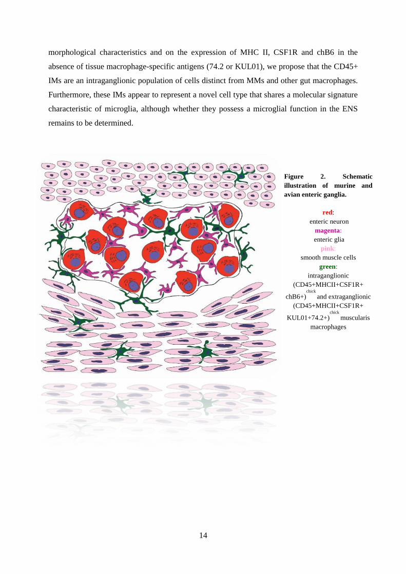
morphological characteristics and on the expression of MHC II, CSF1R and chB6 in the
absence of tissue macrophage-specific antigens (74.2 or KUL01), we propose that the CD45+
IMs are an intraganglionic population of cells distinct from MMs and other gut macrophages.
Furthermore, these IMs appear to represent a novel cell type that shares a molecular signature
characteristic of microglia, although whether they possess a microglial function in the ENS
remains to be determined.
Figure 2. Schematic
illustration of murine and
avian enteric ganglia.
red:
enteric neuron
magenta:
enteric glia
pink:
smooth muscle cells
green:
intraganglionic
(CD45+MHCII+CSF1R+
chB6+)chick
and extraganglionic
(CD45+MHCII+CSF1R+
KUL01+74.2+)chick
muscularis
macrophages

15
5. Conclusions
1. We demonstrated that extraembryonically two types of CD45+ cells differentiates: CD45+
cells with round morphology in the lumen of blood islands, and ramified cells in the
extraembryonic mesenchyme. In later stages of development round cells are localised
associated to the aorta and its impaired visceral branches, ramified cells are found scattered
throughout the embryonic mesenchyme.
2. In addition to the already described intraaortic folds and paraaortic region, we identified a
novel hematopoietic locus around the visceral branches of the abdominal aorta, and named it
„periarterial hematopoietic sheath”. According to immunostainings of B-cell specific
antibodies we hypothesize, that B-cell precursors arise in this anatomical location before the
colonisation of lymphoid organs.
3. We established a novel method of embryomanipulation for the culture of the yolk-sac. With
combination of parabiosis, in ovo yolk-sac culture, yolk-sac chimera techniques and
immunocytochemical analysis, we confirmed, that progenitor cells of intraembryonic CD45+
ramified cells are derived from the yolk-sac, and after migration to the embryo, they colonise
the organ primordia, where they differentiate to MHCII expressing dendritic, or „stellate”
cells.
4. We revealed the yolk-sac origin of bursal secretory dendritic cells (BSDC) with in vitro
embryonic bursa Fabricii – yolk sac recombination and in ovo chorioallantoic membrane
cultures.
5. With intestinal chorioallantoic and coelomic explants we demonstrated, that blood-borne
CD45+ cells colonise the developing enteric ganglia in the chick embryo. These
hematopoietic CD45+ cells later differentiate to ramified cells and express cell surface
molecules similar to microglia.
6. We showed that the immunophenotype of intraganglionic and extraganglionic ramified
cells are different in the adult chick gut. While CD45, MHC-II, and CSF1R cell surface
molecules are expressed in both cell types, the expression of avian B-cell and microglia
marker chB6 is specific to intraganglionic cells. In contrast, monocyte-derived tissue
macrophage markers 74.2 and KUL-01 are solely expressed in extraganglionc cells.

16
6. Publications of the author
6.1 Publications related to the doctoral theses :
Dora, D., Fejszák, N., Goldstein, A.M., Minkó, K., Nagy, N., 2017. Ontogeny of ramified
CD45 cells in chicken embryo and their contribution to bursal secretory dendritic cells. Cell
and Tissue Research 368, 353–370. doi:10.1007/s00441-017-2595-y
IF: 3,043
Dora, D., Arciero, E., Hotta, R., Barad, C., Bhave, S., Kovacs, T., Balic, A., Goldstein, A.M.,
Nagy, N., 2018. Intraganglionic macrophages: A new population of cells in the enteric
ganglia. Journal of Anatomy. doi:10.1111/joa.12863
IF: 2,479
6.2 Other publications:
Nagy, N., Barad, C., Hotta, R., Bhave, S., Arciero, E., Dora, D., Goldstein, A.M., 2018.
Collagen 18 and agrin are secreted by enteric neural crest cells to remodel their
microenvironment and regulate their migration during ENS development., n.d. .
Development. doi:10.1242/dev.160317
IF: 5,413