Embed Size (px)
Citation preview

Aquaculture 318 (2011) 162–168
Contents lists available at ScienceDirect
Aquaculture
j ourna l homepage: www.e lsev ie r.com/ locate /aqua-on l ine
Ontogeny of the digestive tract and enzymatic activity in white seabass,Atractoscion nobilis, larvae
Mario A. Galaviz a, Alejandra García-Gasca b, Mark Drawbridge c,Carlos Alfonso Álvarez-González d, Lus M. López a,e,⁎a Programa de Maestría y Doctorado en Oceanografía Costera, Facultad de Ciencias Marinas, Universidad Autónoma de Baja California (UABC), PO Box 76, Ensenada B.C., 22860, Mexicob Centro de Investigación en Alimentación y Desarrollo, Unidad Mazatlán (CIAD), PO Box 711, Mazatlán, Sinaloa, C.P. 82010, Mexicoc Hubbs-SeaWorld Research Institute, 2595 Ingraham St., San Diego, CA 92109, USAd Laboratorio de Acuicultura Tropical DACBIOL-UJAT, Carr Vhsa-Cárdenas Km 0.5, Bosques de Saloya, Villahermosa, Tabasco, Méxicoe Facultad de Ciencias Marinas, Universidad Autónoma de Baja California (UABC), PO Box 76, Ensenada B.C. 22860, Mexico
⁎ Corresponding author at: UABC, PO Box 76, Ensenad646 1744570x147; fax: +52 646 1744103.
E-mail address: [email protected] (L.M. López).
0044-8486/$ – see front matter © 2011 Elsevier B.V. Adoi:10.1016/j.aquaculture.2011.05.014
a b s t r a c t
a r t i c l e i n f oArticle history:Received 19 May 2010Received in revised form 18 April 2011Accepted 5 May 2011Available online 14 May 2011
Keywords:LarvaeOntogenyHistologyDigestive systemDigestive enzymesWhite seabass
The development of the digestive system and digestive enzyme activity in white seabass larvae, Atractoscionnobilis, were analyzed from hatching until 40 days post hatch (dph) using histological and biochemicalapproaches. The development of the digestive system in A. nobilis larvae was similar to that reported for othermarine fish species. Larvae at 3 dph (0.55±0.001 mg wet weight and 3.6±0.02 mm total length), cultured at18 °C in seawater, presented all the structures (i.e. differentiation of the alimentary canal into the buccopharynx,esophagus, anterior andposterior intestines, pancreaswith zymogengranules, liver, gall bladder andopenmouth)necessary for the digestion and absorption of nutrients such as proteins and lipids (primarily). At this time, thelarvae had fully-developed digestive systems that allowed them to digest inert feed and to absorb nutrientsthroughout the intestinewalls. On the other hand,most digestive enzyme activitieswere detected at themomentof hatching. Trypsin activity was 0.80±0.16 mU/mg protein at 1 dph (0.51±0.001 mg wet weight larvae), andincreased gradually during the following days, but most notably after the initial exogenous feeding at 4 dph. Thespecific activity of chymotrypsin was 7.21±1.29 mU×10−4/mg protein at 1 dph and reached peak level (15.9±1.02 mU×10−4/mg protein) at 18 dph (6.6±0.003 mg wet weight larvae). The specific activity of leucineaminopeptidase increased continuously from 1.31±0.05 mU×10−3/mg protein at 1 dph to 15.91±0.40 mU×10−3/mg protein at 18 dph. The activity of α-amylase at 1 dph was 1.35±0.09 U/mg protein,increasing to 8.07±0.98 U/mg protein at 16 dph. The activity of pepsin was detected at a very low level (0.71±0.53 U/mg protein) at 10 dph, and a stepwise increase in activity was observed between 16 and 20 dph, reachingmaximum level (13.92±0.09 U/mg protein) at 40 dph. These results indicate that the digestive tract developsrapidly in this species and that the stomach becomes functional between 16 and 18 dph. It should, therefore, bepossible to start weaning the fish at this young age.
a BC 22860, Mexico. Tel.: +52
ll rights reserved.
© 2011 Elsevier B.V. All rights reserved.
1. Introduction
In southern California and Baja California, the white seabassAtractoscion nobilis supports an important commercial and sportfishery (Vojkovich and Reed, 1983). Its wide acceptance and highmarket value led to over-harvesting the wild stocks in many areas(Drawbridge and Kent, 1997), which in turn led to the development ofa comprehensive stock replenishment program that was initiated insouthern California in 1983 (Kent et al., 1995). As the productioncapacity of A. nobilis has increased, so too has the interest in applyingthe technology to commercial farming (Drawbridge and Kent, 2001).Research related to the nutritional requirements of this species is
limited; our research group has reported on macronutrient selectionand the effects of digestible protein, lipid, and carbohydrate require-ments on juveniles and young adults (Durazo et al., 2010; Jirsa et al.,2010; López et al., 2006; 2009).
In typical aquaculture operations, weaning is extended 1 or2 weeks after the digestive tract is fully developed in order to reducebody malformation andmortality, or performed as early as possible toreduce the cost of live food production. For this reason, knowledge ofearly ontogeny of the digestive tract and digestive enzyme activity infish larvae is of value for establishing appropriate feeding andweaning routines for target aquaculture species (Baglole et al.,1997; Zambonino-Infante and Cahu, 2001), as in the case of giltheadsea bream Sparus aurata (Sarasquete et al., 1995), red drum Sciaenopsocellatus (Lazo et al., 2000), California halibut Paralichthys californicus(Alvarez-González et al., 2006; Gisbert et al., 2004), yellow croakerPseudosciaena crocea (Ma et al., 2005; Mai et al., 2005), bullseye puffer

163M.A. Galaviz et al. / Aquaculture 318 (2011) 162–168
Sphoeroides annulatus (García-Gasca et al., 2006), and spotted sandbass Paralabrax maculatofasciatus (Alvarez-González et al., 2008; Peñaet al., 2003), among others.
The objective of this study was to describe the development of thedigestive system and the activity of the main digestive enzymes in A.nobilis larvae fed live food and a compound microdiet from hatchinguntil 40 days post hatch (dph). This information will be useful forimproving current larval rearing practices and feeding protocols, andreducing weaning costs of this fish species.
2. Materials and methods
2.1. Eggs and larval fish rearing
Fertilized eggs of A. nobilis were obtained from the Hubbs-SeaWorld Research Institute's marine finfish hatchery in Carlsbad,CA, USA. The eggs were produced by wild-caught brood fishmaintained in four separate groups of 50 fish in equal sex ratios.Adult A. nobiliswere induced to spawn using photothermal control tosimulate natural seasonal cycles that were phase-shifted amonggroups to provide eggs year-round. Adult A. nobilis were estimated toweight 15–25 kg and were fed fresh fish and squid 5 days per weekwith supplementation of vitamins (Jirsa et al., 2010) every other day.The eggs were transported to the Fish Culture and Biotechnology Unitof the School ofMarine Science, University of Baja California (UABC) atEnsenada, Mexico. Eggs were treated with 100 ppm formalin for 1 hand stocked at a density of 100 eggs/L in a 1600-L cone bottom tankwith 18 °C seawater recirculated at a rate of 1.5–2 L/min through afluidized bed, UV sterilizer, and foam fractionator.
Eggs hatched approximately 48 h after fertilization. Yolk-saclarvae were stocked at a density of 30 individuals/L in nine 100-Lexperimental tanks. Beginning at 4 dph, larvae were fed three timesper day (08:00, 12:00, and 17:00 h) exclusively with Artemiametanauplii (Salt Creek Inc., Salt Lake City, UT, USA) enriched withlipid emulsion (Bio-Marine Algamac 3050™) at a concentration of0.6 g/L. The Artemia were supplied at a concentration of 5 nauplii/mLonly up to 15 dph. At 16 dph, the amount of live food was decreasedand a combination of enriched Artemia metanauplii and formulateddiet (Otohime Japanese Marine Weaning Diet, Red Mariculture;protein 52.11%, lipid 16.3%, ash 11.2%, particle size 200–1410 μm)was supplied. The weaning period was complete at 24 dph, when livefood was no longer supplied. Larvae were fed themicrodiet from 24 to40 dph (end of the trial).
2.2. Sampling
Depending on size, between 20 and 150 larvae were collectedusing a 200 μm dip net, 1 h after the first daily feeding. Samples werecollected daily starting at 0 dph (free embryos) through to 6 dph,every 2 days from 8 dph until 20 dph, and every 4 days thereafteruntil 40 dph. After sampling, larvae were anesthetized with tricainemethanesulfonate (MS 222), rinsed with distilled water, freeze-dried,and stored at −80 °C until analysis.
Supplementary samples were collected from the rearing tankseach day after hatching until 6 dph and on 8, 12, 15, 18, 24, 36, and40 dph. These samples were used to measure total length and wetweight of larvae. Additionally, an average total length (measured tothe nearest 0.1 mm) was calculated for each sampling day bymeasuring the larvae under a dissecting microscope using a digitizingcamera and PAXcam2 (PAX-it version 6, MIS Inc., USA) software. Wetweight (measured to the nearest 0.1 mg) was calculated by weighingthe subsample of larvae using an analytical balance (SartoriusGottingen, Germany; precision of 0.1 mg), then counting the larvaecontained in the subsample. Individual larval weight was determinedby dividing the subsample weight by the number of larvae in thesample.
2.3. Enzymatic activity
For each sampling time, three samples of 150 larvae until 10 dph,50 larvae until 20 dph, and 20 larvae until 40 dph were used forenzymatic analyses. Because of the difficulties associated withdissecting and removing the digestive tract of small larvae, wholebody homogenates were used for enzymatic analyses in larvaeyounger than 16 dph. After this age, the larvae digestive system wasdissected on a glass slide supported on a frozen mini-table.
Each sample was homogenized with a tissue grinder in 1 mL ofdistilled water chilled to 4 °C. A subsample of the homogenate wasstored at −80 °C until later analysis of leucine aminopeptidaseactivity. The remaining sample was centrifuged at 14,000g for 30 minat 4 °C, and the supernatants were stored at −80 °C until analysis.
2.3.1. Proteases (endopeptidases)The level of soluble protein in pooled sampleswas determined using
themethod described by Bradford (1976). Trypsin activity was assayedaccording to Erlanger et al. (1961), using BAPNA (N-α-Benzoyl-DL-arginine p-nitroanilide) as substrate. The mixtures were incubated at37 °C and the absorbance of the reaction products was measured at410 nm. Chymotrypsin activity was measured by the method ofHummel (1959), as modified by Applebaum et al. (2001), using BTEE(N-Benzoyl-L-tyrosine ethyl ester) as substrate. The mixtures wereincubated at 37 °C and the absorbance of the reaction products wasmeasured at 256 nm. The reaction of trypsinwas stoppedby adding30%acetic acid. One unit of enzyme activity was defined as 1 μg nitroanilidereleased per minute, using a molar extinction coefficient of 8.8 fortrypsin and of 964 for chymotrypsin.
Leucine aminopeptidase was measured at 37 °C as suggested byAppel (1974), using leucine P-nitroanilide as substrate. The reactionof leucine aminopeptidase was stopped by adding 30% acetic acid. Themixtures were incubated at 37 °C and the absorbance of the reactionproducts was measured at 410 nm. One unit of enzyme activity wasdefined as 1 μg nitroanilide released per minute, using a molarextinction coefficient of 8.2.
Acid proteinase (pepsin) activity was evaluated as described bySarath et al. (1989), using 2% hemoglobin as substrate. The enzymecrude extracts and the substrate were incubated at 37 °C and theabsorbance of the reaction products measured at 280 nm. One unit ofenzyme activity was defined as 1 μg tyrosine released per minute,using the molar extinction coefficient of 0.005.
2.3.2. Alpha-amylaseThe α-amylase assay was performed according to Vega-Villasante
et al. (1993), using soluble starch (1%) as substrate. One unitcorresponded to the amount of enzyme required to increase by 0.01units the absorbance at 540 nm per minute.
Specific and total enzyme activities in digestive extracts weredetermined using the following equations:
(1) Units/mL=(Δabs reaction final volume (mL)) /(MEC·time(min) extract volume (mL));
(2) Units/mg protein=Units per mL/mg of soluble protein
Δabs represents the increased absorbance at a determinedwavelength and MEC represents the molar extinction coefficient forthe product of the reaction (mL/μg/cm). The results are presentedusing Eqs.(1) and (2), and all assays were carried out in triplicate.
2.4. Histology
Conventional histology and hematoxylin–eosin (H&E) stainingwere performed on larvae fixed in 2% paraformaldehyde for 24 h at4 °C, before being washed, dehydrated, cleared, and embedded inparaffin. Sagittal sections (5 μm) were obtained with a conventionalmicrotome, placed on gelatin-coated slides, re-hydrated, and H&E-

164 M.A. Galaviz et al. / Aquaculture 318 (2011) 162–168
stained. Histological sections were viewed under a light microscopeand photographed with an Infinity digital camera and the PAXcam2software (PAX-it version 6, MIS Inc., USA).
2.5. Statistics
Specific and total digestive enzyme activities in larval extractswere expressed as the mean±SD. Homogeneity of variances andnormality tests were performed. The specific activity was calculatedusing the one-way analysis of variance (ANOVA) procedure. Multiplecomparisons of enzymatic activity over time were obtained by aTukey comparison test. All statistics were conducted using Sigma-Plot11.0 forWindows (Systat Software Inc.). A significance level of Pb0.05was used.
3. Results
A. nobilis showed exponential growth for wet weight and totallength from hatching until the end of the study (40 dph) (Fig. 1).
3.1. Enzymatic activity
Trypsin specific activity was detected as early as 1 dph (0.80±0.16 mU/mg protein), when larvae measured 2.5±0.02 mm total lengthand 0.51±0.001 mg wet weight, and increased gradually during thefollowing days, but most notably after the initial exogenous feeding,between 4 and 5 dph, until 14 dph (5.84±0.20 mU/mg protein). Afterthis time, trypsin activity decreased until 18 dph, before increasing at24 dph and again at 32 dph (9.75±0.36 mU/mg protein), and thendecreased until the experiment was concluded at 40 dph (Fig. 2A).
The specific activity of chymotrypsin was detected at 1 dph (7.21±1.29 mU×10−4/mg protein). Maximum activity (15.92±1.02 mU×10−4/mg protein) was measured 2 days after weaning from Artemianauplii to a microdiet, at 18 dph. Chymotrypsin activity then decreaseduntil 20 dph and remained constant thereafter (Fig. 2B).
The specific activity of leucine aminopeptidase was detected at1 dph (1.31±0.05 mU×10−3/mg protein), and it increased contin-uously until 18 dph (15.91±0.40 mU×10−4/mg protein). Thisincrease was followed by a steady decline until 20 dph, when theactivity increased again until 28 dph (Fig. 2C).
The specific activity of acid protease (pepsin) was detected at10 dph (0.71±0.53 U/mg protein), and a stepwise increase in activitywas observed until the end of the experiment. The first increase was
Fig. 2. Mean digestive enzyme activity during Atractoscion nobilis larviculture (±SD,three pooled samples of 100 larvae until 10 dph, 50 larvae until 20 dph, and 20 larvaeuntil 40 dph). (A) Specific activity of trypsin, (B) specific activity of chymotrypsin, and(C) specific activity of leucine aminopeptidase.
Fig. 1. Mean wet weight (●, mg) and total length (■, cm) (±SD, three pooled samplesof 150 larvae until 10 dph, 50 larvae until 20 dph, and 20 larvae until 40 dph) ofAtractoscion nobilis larvae.
observed at 20 dph (3.61±1.3 U/mg protein), 4 days after theappearance of the first gastric glands. The activity decreased at24 dph and increased again, reaching maximum level at 40 dph(13.92±0.1 U/mg protein) (Fig. 3A).
The activity of α-amylase peaked at 16 dph (8.07±0.98 U/mgprotein), coinciding with the start of weaning period. The activitydecreased at 18 dph, but increased again at 20 dph (4.44±0.38 U/mgprotein) and fluctuated until the end of the experiment (Fig. 3B).
3.2. Histology
At hatching (0.51±0.003 mg and 1.3±0.001 mm larval size), thedigestive system of A. nobilis larvae appeared as a straight andundifferentiated tube, not opened to the exterior (buccopharynx and

Fig. 3. Mean digestive enzyme activity during Atractoscion nobilis larviculture (±SD,three pooled samples of 100 larvae until 10 dph, 50 larvae until 20 dph, and 20 larvaeuntil 40 dph). (A) Specific activity of pepsin and (B) specific activity of amylase.
Fig. 4. Sagittal sections of the Atractoscion nobilis larvae at days 0 (A), 3 (B), 8 (C) and 12 (Dcentral nervous system; ck, cephalic kidney; e, esophagus; ey, eye; ga, gill arch; h, heart; l,intestine; pin, primordial intestine; sb, swim bladder and ud, urinary duct. H&E staining.
165M.A. Galaviz et al. / Aquaculture 318 (2011) 162–168
anus not differentiated), lying dorsally to a large yolk-sac (Fig. 4A).During yolk-sac absorption (1–2 dph), the rudimentary digestivesystem became differentiated into buccopharynx, esophagus, andintestine. By 3 dph the mouth was open, the swim bladder wasdifferentiated, the liver continued to develop, and the posteriorintestine and some connective tissue became visible (Fig. 4B).
Histological analyses revealed that the urinary duct, swim bladder,intestine, liver, and pancreas were all developed by 3 dph, and by8 dph basophilic cells of the diffuse exocrine pancreas were visibleand the intestine was lined by ciliated columnar epithelial cells withbasal nuclei (Fig. 4C).
At 12 dph, the esophagus was well developed and the stomachwas visible as an extension of the esophagus. The liver continued toincrease in size and revealed the bile duct between the liver and theexocrine cells of the pancreas. The urinary duct connected to the anuswas also well developed, and the embryonic kidney (pronephros) andthe heart were visible in the histological section (Fig. 4D).
Gastric glands were identified by the presence of small groups ofcubic cellswith aprominent central nucleus and the formationof a smallglandular acinus in the mucosa, and were fully differentiated at 16 dph(Fig. 5A). The size and number of gastric glands increased with age.Goblet cells appeared and increased in number and the liver increasedsignificantly in size at 16 dph. All thedigestive organswere visible in thehistological section, and became fully developed between 18 and32 dph. During this stage, the most obvious changes in the morphologyof the digestive tract were the following: 1) the formation of thestomach, 2) the appearance of small vacuolar inclusions in theenterocytes of the medium intestine (Fig. 5B), 3) liver enlargement,and 4) embedding of exocrine pancreas into the medium intestine.
There was also a rapid increase in the number of gastric glands andin the size of the stomach (Fig. 5C). At 24 dph the intestine loop wasformed to accommodate the increasing length of the digestive tractinside the abdominal cavity (Fig. 5D). Themorphology of the digestivesystem resembled that of the adult by day 32 post hatching, with anenlarged, fully developed stomach containing a large number ofgastric glands (Fig. 6A). The stomach was connected to the anteriorintestine through the pyloric sphincter, which was covered by a thickmuscular wall (Fig. 6B).
) post hatch. Abbreviations: ai, anterior intestine; b, buccopharynx; bd, bile duct; cns,liver; m, mouth; mf, muscular fibers; n, notochord; ep, exocrine pancreas; pi, posterior
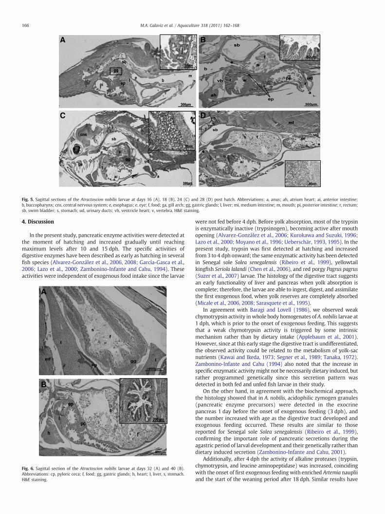
Fig. 5. Sagittal sections of the Atractoscion nobilis larvae at days 16 (A), 18 (B), 24 (C) and 28 (D) post hatch. Abbreviations: a, anus; ah, atrium heart; ai, anterior intestine;b, buccopharynx; cns, central nervous system; e, esophagus; e, eye; f, food; ga, gill arch; gg, gastric glands; l, liver; mi, medium intestine; m, mouth; pi, posterior intestine; r, rectum;sb, swim bladder; s, stomach; ud, urinary ducts; vh, ventricle heart; v, vertebra. H&E staining.
166 M.A. Galaviz et al. / Aquaculture 318 (2011) 162–168
4. Discussion
In the present study, pancreatic enzyme activities were detected atthe moment of hatching and increased gradually until reachingmaximum levels after 10 and 15 dph. The specific activities ofdigestive enzymes have been described as early as hatching in severalfish species (Alvarez-González et al., 2006, 2008; García-Gasca et al.,2006; Lazo et al., 2000; Zambonino-Infante and Cahu, 1994). Theseactivities were independent of exogenous food intake since the larvae
Fig. 6. Sagittal section of the Atractoscion nobilis larvae at days 32 (A) and 40 (B).Abbreviations: cp, pyloric ceca; f, food; gg, gastric glands; h, heart; l, liver, s, stomach.H&E staining.
were not fed before 4 dph. Before yolk absorption, most of the trypsinis enzymatically inactive (trypsinogen), becoming active after mouthopening (Alvarez-González et al., 2006; Kurokawa and Suzuki, 1996;Lazo et al., 2000; Moyano et al., 1996; Ueberschär, 1993, 1995). In thepresent study, trypsin was first detected at hatching and increasedfrom 3 to 4 dph onward; the same enzymatic activity has been detectedin Senegal sole Solea senegalensis (Ribeiro et al., 1999), yellowtailkingfish Seriola lalandi (Chen et al., 2006), and red porgy Pagrus pagrus(Suzer et al., 2007) larvae. The histology of the digestive tract suggestsan early functionality of liver and pancreas when yolk absorption iscomplete; therefore, the larvae are able to ingest, digest, and assimilatethe first exogenous food, when yolk reserves are completely absorbed(Micale et al., 2006, 2008; Sarasquete et al., 1995).
In agreement with Baragi and Lovell (1986), we observed weakchymotrypsin activity inwhole body homogenates of A. nobilis larvae at1 dph, which is prior to the onset of exogenous feeding. This suggeststhat a weak chymotrypsin activity is triggered by some intrinsicmechanism rather than by dietary intake (Applebaum et al., 2001).However, since at this early stage the digestive tract is undifferentiated,the observed activity could be related to the metabolism of yolk-sacnutrients (Kawai and Ikeda, 1973; Segner et al., 1989; Tanaka, 1972).Zambonino-Infante and Cahu (1994) also noted that the increase inspecific enzymatic activitymight not be necessarily dietary induced, butrather programmed genetically since this secretion pattern wasdetected in both fed and unfed fish larvae in their study.
On the other hand, in agreement with the biochemical approach,the histology showed that in A. nobilis, acidophilic zymogen granules(pancreatic enzyme precursors) were detected in the exocrinepancreas 1 day before the onset of exogenous feeding (3 dph), andthe number increased with age as the digestive tract developed andexogenous feeding occurred. These results are similar to thosereported for Senegal sole Solea senegalensis (Ribeiro et al., 1999),confirming the important role of pancreatic secretions during theagastric period of larval development and their genetically rather thandietary induced secretion (Zambonino-Infante and Cahu, 2001).
Additionally, after 4 dph the activity of alkaline proteases (trypsin,chymotrypsin, and leucine aminopeptidase) was increased, coincidingwith the onset of first exogenous feeding with enriched Artemia naupliiand the start of the weaning period after 18 dph. Similar results have

167M.A. Galaviz et al. / Aquaculture 318 (2011) 162–168
been reported for turbot Scophthalmus maximus (Cousin et al., 1987),European seabass Dicentrarchux labrax (Cahu and Zambonino-Infante,1997), P. californicus and P. maculatofasciatus (Alvarez-González et al.,2006, 2008). The pattern of activity coincided with that of other brushborder enzymes, which should indicate a maturation of enterocytes(Hakim et al., 2007; Zambonino-Infante and Cahu, 2001); this wascorroborated by the increment of villi and microvilli in the intestinalbrush border of larvae between 18 and 32 dph.
The development of the digestive system in A. nobilis larvae wassimilar to that reported for other marine fish species, particularlyP. crocea (Mai et al., 2005), bluefin tuna Thunnus thynnus, andyellowfin tuna Thunnus albacares (Kaji et al., 1996; 1999). All thesespecies possessed an undifferentiated and rudimentary digestivesystem, which became differentiated into buccopharynx, esophagus,intestine, and rectum at 3 dph. Accordingly, similar results wereobserved in our study at 3 and 4 dph, when the major changes in thedevelopment of the digestive system were observed, such as thedifferentiation of enterocytes, buccopharynx, and esophagus; thefolding of the intestinal mucosa; and the development of the liver.
According to Segner et al. (1994), the cytological differentiation ofenterocytes indicates that the intestine is suitable for the absorption ofnutrients and the beginning of exogenous feeding. In A. nobilis, thisdifferentiation coincided with the folding of the intestinal mucosa andsubsequent division into anterior and posterior intestine. The folding ofthe digestive tract is common in Paracanthopterygii and Acanthopterygiilarvae; however, when these changes occur may vary between speciesdue to differences between larval stages (Govoni, 1980;Hachero-Cruzadoet al., 2009).
In our study, the gastric glands began to differentiate at 16 dph andthe acid protease (pepsin) activity was detected as early as 10 dph,which in some cases suggests the presence of a functional stomach atthis premature age. Differences betweenmorphological development ofgastric glands and pepsin secretion have also been observed in D. labrax(Vu, 1983), S. senegalensis (Ribeiro et al., 1999), and P. californicus(Alvarez-González et al., 2006), andmaybedue to individual differencesor, more likely, to differences resulting from the use of histological andbiochemical analytical techniques. Moyano et al. (1996) discussed thatthepossible causeof theabsence of pepsin in themucosaof thedigestivetract walls of S. aurata larvae could be compensated by micro-pinocytosis and intracellular digestion of protein, probably by pepti-dases and cathepsin. Cahu et al. (2003) and Zambonino-Infante andCahu (2007) reported that D. labrax larvae can be supplied a mix of livefood and artificial diet for the first exogenous feeding as soon as thelarvae have opened the mouth, even if the gastric glands have not yetdeveloped; however, this depends on the type of live food that isprovided and on the weaning process from live to formulated feed.
On the other hand, although α-amylase activity was low in A. nobilislarvae, it was detected at the time of hatching, concurring with theresults reported for P. californicus (Alvarez-González et al., 2006) andP.maculatofasciatus (Alvarez-González et al., 2008). This activity appearsto be an integral component of the enzymatic systemoffish larvaeduringdevelopment, since it has been detected at the start of development andits presence does not seem to be induced by the diet. However, the levelof α-amylase activity may change depending on the composition of thediet (Moyano et al., 1996; Péres-Borla et al., 1998; Zambonino-Infanteand Cahu, 2001); in our experiment peak activity occurred at 16 dph,which coincided with the introduction of the extruded microdiet.Additionally, the high levels of α-amylase during early larval develop-ment in A. nobilis were not induced by enriched live food and may bebetter explained as a result of programmedgene expression, as suggestedby Zambonino-Infante and Cahu (2001). As live food did not offer asuitable substrate for this enzyme, substrate-mediated inductionwas notproduced and the activity decreased significantly. In summary, twopeakswereobserved forα-amylase activity, thefirst at theonsetof theweaningperiod (16 dph) and the secondwhen larvaewere fed only themicrodietat 32 dph. These increases in α-amylase activity may have been due to
the presence of a certain amount of carbohydrate in the formulated feedsgiven the weaned larvae (Cara et al., 2003).
5. Conclusions
Digestive enzymatic activity was observed in A. nobilis larvae at thetime of hatching and during the absorption of the yolk-sac. Thestomach became functional between 16 and 18 dph. At this time, thelarvae had a fully-developed digestive system that allowed to digestinert feed and to absorb nutrients. Our findings suggest that this age isappropriate to initiate the weaning process in this species.
Acknowledgements
This work was supported by the Mexican Council for Science andTechnology (CONACyT, SNI-1 funding and doctoral scholarship No.164557) and by internal research grants from the AutonomousUniversity of Baja California (UABC). The authors would like toacknowledge the valuable comments and suggestions made by Prof.Domenico Voltolina, and thank Deyanira Rodarte and Yesica Solorzanofor their technical assistance, and Prof. Enric Gisbert for his valuableadvice on the description of the development of the digestive system.Fertilized eggs of A. nobilis were donated by Hubbs-SeaWorld ResearchInstitute as part of California's Ocean Resources Enhancement andHatcheryProgramoperatedunder authorityof theCaliforniaDepartmentof Fish and Game, USA.
References
Alvarez-González, C.A., Cervantes-Trujano, M., Tovar-Ramírez, D., Conklin, D.E.,Nolasco, H., Gisbert, E., Piedrahita, R., 2006. Development of digestive enzymes inCalifornia halibut Paralichthys californicus larvae. Fish Physiology and Biochemistry31, 83–93.
Alvarez-González, C.A., Moyano-López, F.J., Civera-Cerecedo, R., Carrasco-Chávez, V.,Ortiz-Galindo, J.L., Dumas, S., 2008. Development of digestive enzyme activity inlarvae of spotted sand bass Paralabrax maculatofasciatus. I: Biochemical analysis.Fish Physiology and Biochemistry 34, 373–384.
Appel, W., 1974. Leucine aminopeptidase determination with L-leucinamide assubstrate. In: Bergmeyer, H.U. (Ed.), Methods of Enzymatic Analysis. AcademicPress, New York, New York, USA, pp. 954–958.
Applebaum, S.L., Perez, R., Lazo, J.P., Holt, G.J., 2001. Characterization of chymotrypsinactivity during early ontogeny of larval red drum (Sciaenops ocellatus). FishPhysiology and Biochemistry 25, 291–300.
Baglole, C.J., Murray, H.M., Goff, G.P., Wright, G.M., 1997. Ontogeny of the digestive tractduring larval development of yellowtail flounder: a light microscopic and mucoushistochemical study. Journal of Fish Biology 51, 120–134.
Baragi, V., Lovell, R.T., 1986. Digestive enzyme activities in striped bass from firstfeeding through larval development. Transactions of the American Fisheries Society115, 478–484.
Bradford, M.M., 1976. A rapid and sensitive method for the quantization of microgramquantities of protein utilizing the principle of protein dye binding. AnalyticalBiochemistry 72, 248–254.
Cahu, C., Zambonino-Infante, J.L., 1997. Is the digestive capacity of marine fish larvaesufficient for compound diet feeding? Aquaculture International 5, 151–160.
Cahu, C., Zambonino Infante, J., Takeuchi, T., 2003. Nutritional components affectingskeletal development in fish larvae. Aquaculture 227, 245–258.
Cara, J.B., Moyano, F.J., Cardenas, S., Fernandez-Diaz, C., Yufera, M., 2003. Assessment ofdigestive enzyme activities during larval development of white bream. Journal ofFish Biology 63, 48–58.
Chen, B.N., Qin, J.G., Kumar, S.M., Hutchinson, W.G., Clarke, S.M., 2006. Ontogeneticdevelopment of digestive enzymes in yellowtail kingfish Seriola lalandi larvae.Aquaculture 260, 264–271.
Cousin, J.B.C., Baudin-Laurencin, F., Gabaudan, J., 1987. Ontogeny of enzymatic activities infed and fasting turbot, Scophthalmus maximus L. Journal of Fish Biology 30, 15–33.
Drawbridge, M.A., Kent, D.B., 1997. The white seabass (Atractoscion nobilis) as acandidate species for open ocean culture: a review based on four years of culture innear-shore cages. Proceedings of the Second International Conference on OpenOcean Aquaculture. Maui, Hawaii. April 23–25, pp. 203–211.
Drawbridge, M.A., Kent, D.B., 2001. Culture of marine fish. California's living resource: astatus report. California Department of Fish and Game 2001, 510–512 December.
Durazo, E., Cruz, A.C., López, L.M., Lazo, J.P., Drawbridge, M., Viana, M.T., 2010. Effects ofdigestible protein levels in isonitrogenous diets on growth performance and tissuecomposition of juvenile Atractoscion nobilis. Aquaculture Nutrition 16, 54–60.
Erlanger, B., Kokowsky, N., Cohen, W., 1961. The preparation and properties of two newchromogenic substrates of trypsin. Archives of Biochemistry andBiophysics 95, 271–278.

168 M.A. Galaviz et al. / Aquaculture 318 (2011) 162–168
García-Gasca, A., Galaviz, M., Gutiérrez, J.N., García-Ortega, A., 2006. Development ofthe digestive tract, trypsin activity and gene expression in eggs and larvae of thebullseye puffer fish Sphoeroides annulatus. Aquaculture 256, 366–376.
Gisbert, E., Piedrahita, R.H., Conklin, D.E., 2004. Ontogenetic development of thedigestive system in California halibut (Paralichthys californicus) with notes onfeeding practices. Aquaculture 232, 455–470.
Govoni, J.J., 1980. Morphological, histological, and functional aspects of alimentarycanal and associated organ development in larval Leiostomus xanthurus. Review ofCanadian Biology 39, 69–80.
Hachero-Cruzado, I., Ortiz-Delgado, J.B., Borrega, B., Herrera, M., Navas, J.I., Sarasquete,C., 2009. Larval organogenesis of flatfish brill Scophalmus rhombus L: histologicaland histochemical aspects. Aquaculture 286, 138–149.
Hakim, Y., Rowland, S.J., Guy, J.A., Mifsud, C., Uni, Z., Harpaz, S., 2007. Effects of genetic strainand holding facility on the characteristics of alkaline phosphatase and brush borderenzymes in silver perch (Bidyanus bidyanus). Aquaculture Research 38, 361–372.
Hummel, B.C.W., 1959. A modified spectrophotometric determination of chymotrypsin,trypsin and thrombin. Canadian Journal of Biochemistry and Physiology 37 (12),1393–1399.
Jirsa, D., Davis, D.A., Drawbridge, M., 2010. Development of a practical soy-based diet forwhite seabass. North American Journal of Aquaculture. doi:10.1577/A09-078.1.
Kaji, T., Tanaka, M., Takahashi, Y., Oka, M., Ishibashi, N., 1996. Preliminary observationson development of Pacific bluefin tuna Tunnus thynnus (Scombridae) larvae rearedin the laboratory, with special reference to the digestive system. Marine andFreshwater Research 47, 261–269.
Kaji, T., Tanaka, M., Oka, M., Takeuchi, H., Ohsumi, S., Teruya, K., Hirokawa, J., 1999.Growth and morphological development of laboratory-reared yellowfin tunaThunnus albacares larvae and early juveniles, with special emphasis on thedigestive system. Fisheries Science 65, 700–707.
Kawai, S., Ikeda, S., 1973. Studies on digestive enzymes of fishes. Part 4. Development ofthe digestive enzymes of carp and black sea bream after hatching. Bulletin of theJapanese Society for the Science of Fish 39, 877–881.
Kent, D.B., Ford, R.F., Drawbridge, M.A., 1995. Accomplishments and roadblocks of amarine stock enhancement program for white seabass in California. AmericanFisheries Society Symposium 15, 492–498.
Kurokawa, T., Suzuki, T., 1996. Formation of the diffuse pancreas and the developmentof digestive enzyme synthesis in larvae of Japanese flounder Paralichthys olivaceus.Aquaculture 141, 267–276.
Lazo, J.P., Holt, G.J., Arnold, C.R., 2000. Ontogeny of pancreatic enzymes in larval reddrum Sciaenops ocellatus. Aquaculture Nutrition 6, 183–192.
López, L.M., Torres, A.L., Durazo, E., Drawbridge, M., Bureau, D., 2006. Effects of lipid ongrowth and feed utilization of white seabass (Atractoscion nobilis) fingerlings.Aquaculture 253, 557–563.
López, L.M., Durazo, E., Viana, M.T., Drawbridge, M., Bureau, D., 2009. Dietary lipid levelseffect on performance, body composition and fatty acid profile of juvenile whiteseabass, Atractoscion nobilis. Aquaculture 289, 101–105.
Ma, H., Cahu, C., Zambonino, J., Yu, H., Duan, Q., Le Gall, M.M., Mai, K., 2005. Activities ofselected digestive enzymes during larval development of large yellow croakerPseudosciaena crocea. Aquaculture 245, 239–248.
Mai, K., Yu, H., Ma, H., Duan, Q., Gisbert, E., Zambonino Infante, J., Cahu, C., 2005. Ahistological study on the development of the digestive system of Pseudosciaenacrocea larvae and juveniles. Journal of Fish Biology 67, 1094–1106.
Micale, V.M., Garrafo, L., Genovese, M., Spedicato, M.T., Muglia, U., 2006. The ontogenyof the alimentary tract during larval development in common pandora Pagelluseryhtrinus L. Aquaculture 251, 354–365.
Micale, V., Di Giancamillo, A., Domeneghini, C., Mylonas, C.C., Nomikos, N., Papadakis,I.E., Muglia, U., 2008. Ontogeny of the digestive tract in sharpsnout sea breamDiplodus puntazzo (Cetti, 1777). Histology and Histopathology 23, 1077–1091.
Moyano, F.J., Díaz, M., Alarcón, F.J., Sarasquete, M.C., 1996. Characterization of digestiveenzyme activity during development of gilthead sea bream (Sparus aurata). FishPhysiology and Biochemistry 15, 121–130.
Peña, R., Dumas, S., Villalejo-Fuerte, M., Ortiz-Galindo, J., 2003. Ontogenetic develop-ment of the digestive tract in reared spotted sand bass Paralabrax maculatofasciatuslarvae. Aquaculture 219, 633–644.
Péres-Borla, O., Martone, C.B., Sanchez, J.J., 1998. Protease I inhibitor system infishmuscle.A comparative study. Comparative Biochemical and Physiology 119B, 101–105.
Ribeiro, L., Zambonino-Infante, J.L., Cahu, C.L., Dinis, M.T., 1999. Development ofdigestive enzymes in larvae of Solea senegalensis, Kaup 1858. Aquaculture 170,465–473.
Sarasquete, M.C., Polo, A., Yufera, M., 1995. Histology and histochemistry of thedevelopment of the digestive system of larval gilthead sea bream Sparus aurata L.Aquaculture 130, 79–82.
Sarath, G., De la Monte, R.S., Warner, F.W., 1989. Protease assay methods. In: Beyon, R.J.,Bond, J.S. (Eds.), Proteolytic Enzymes: A Practical Approach. Oxford UniversityPress, New York, New York, USA, pp. 25–56.
Segner, H., Rosch, V., Schmidt, H., von Poeppinghausen, K.J., 1989. Digestive enzymes inlarval Coregonus lavaratus. Journal of Fish Biology 35, 249–263.
Segner, H., Storch, V., Reinecke, M., Kloas, W., Hanke, W., 1994. The development offunctional digestive and metabolic organs in turbot Scophthalmus maximus. MarineBiology 119, 471–486.
Suzer, C., Kamacı, H.O., Çoban, D., Saka, Ş., Fırat, K., Ozkara, B., Ozkara, A., 2007. Digestiveenzyme activity of the red porgy (Pagrus pagrus, L.) during larval developmentunder culture conditions. Aquaculture Research 38, 1178–1785.
Tanaka, M., 1972. Studies on the structure and function of the digestive system inteleost larvae. Part 5: epithelial changes in the posterior gut and protein digestion.Japanese Journal of Ichthyology 19, 172–180.
Ueberschär, B., 1993. Measurement of proteolytic enzyme activity: significance andapplication in larval fish research. Part III. In: Walther, B.T., Fhyn, H.J. (Eds.),Physiological and Biochemical Aspects of Fish Development. University of Bergen,Norway, pp. 233–239.
Ueberschär, B., 1995. The use of tryptic enzyme activity measurement as a nutritionalcondition index: laboratory calibration data and field application. ICES MarineScience Symposium 201, 119–129.
Vega-Villasante, F., Nolasco-Soria, H., Civera-Cerecedo, R., 1993. The digestive enzymesof the Pacific brown shrimp (Penaeus californiensis) I. Properties of amylase activityon digestive tract. Comparative Biochemistry and Physiology 112, 123–129.
Vojkovich, M., Reed, J.R., 1983. White seabass, Atractoscion nobilis, in California-Mexican waters: status of fishery. CalCOFI Report 24, 79–83.
Vu, T.T., 1983. Etude histoenzymologique des activities proteasiques dans le tubedigestif des larves et des adultes de bar, Dicentrarchus labrax (L). Aquaculture 32,57–69.
Zambonino-Infante, J.L., Cahu, C., 1994. Development and response to a diet change ofsome digestive enzymes in seabass (Dicentrarchus labrax) larvae. Fish Physiologyand Biochemistry 12, 399–408.
Zambonino-Infante, J.L., Cahu, C.L., 2001. Ontogeny of the gastrointestinal tract ofmarine fish larvae. Comparative Biochemistry and Physiology 130C, 477–487.
Zambonino-Infante, J.L., Cahu, C.L., 2007. Dietary modulation of some digestiveenzymes and metabolic processes in developing marine fish: applications to dietformulation. Aquaculture 268, 98–105.