Embed Size (px)
Citation preview

RESEARCH PAPER
Omeprazole and PGC-Formulated Heparin Binding EpidermalGrowth Factor Normal i zes Fast ing B lood Glucoseand Suppresses Insulitis in Multiple Low Dose StreptozotocinDiabetes Model
Gerardo M. Castillo & Akiko Nishimoto-Ashfield & Aryamitra A. Banerjee & Jennifer A. Landolfi & Alexander V. Lyubimov &
Elijah M. Bolotin
Received: 27 March 2013 /Accepted: 4 June 2013 /Published online: 21 June 2013# Springer Science+Business Media New York 2013
ABSTRACTPurpose Our objective was to develop novel nanocarriers(protected graft copolymer, PGC) that improve the stability ofheparin binding EGF (HBEGF) and gastrin and then to use PGC-formulated HBEGF (PGC-HBEGF) and Omeprazole (+/−PGC-gastrin) for normalizing fasting blood glucose (FBG) andimproving islet function in diabetic mice.Methods HBEGF, PGC-HBEGF, Omeprazole, Omeprazole +PGC-HBEGF, Omeprazole + PGC-gastrin + PGC-HBEGFand epidermal growth factor (EGF) + gastrin were tested inmultiple low dose streptozotocin diabetic mice.Results Omeprazole + PGC-HBEGF normalized FBG and isbetter than EGF + gastrin at improving islet function and decreas-ing insulitis. Groups treated with Omeprazole, Omeprazole +PGC-HBEGF, or EGF + gastrin have significantly improved isletfunction versus saline control. All animals that received PGC-HBEGF had significantly reduced islet insulitis versus saline control.Non-FBG was lower for Omeprazole + PGC-gastrin + PGC-HBEGF but Omeprazole + PGC-HBEGF alone showed betterFBG and glucose tolerance.Conclusions Omeprazole + PGC-HBEGF provides a sustainedexposure to both EGFRA and gastrin, improves islet function, anddecreases insulitis in multiple low dose streptozotocin diabetic mice.Although HBEGFor EGF elevates non-FBG, it facilitates a reductionof insulitis and, in the presence of Omeprazole, provides
normalization of FBG at the end of treatment. The study demon-strates Omeprazole and PGC-HBEGF is a viable treatment fordiabetes.
KEY WORDS growth factors . insulitis . islet function .nanocarrier . type 1 diabetes
ABBREVIATIONSEGFRA Epidermal growth factor receptor agonistFBG Fasting blood glucoseHBEGF Heparin-binding EGFKd Dissociation constantnon-FBG Non-fasting blood glucosePGC Protected graft copolymerT1D Type 1 diabetesT2D Type 2 diabetes
INTRODUCTION
The hallmark of diabetes is hyperglycemia which can resultfrom the depletion or lack of insulin producing β-cells in thepancreas, or a decrease of sensitivity of the body’s cells toinsulin, or both. For type 1 diabetes (T1D), without β-cells orwith very few β-cells, as reflected by the absence or very lowlevel of blood insulin, the treatment has been frequent insulininjection for over half a century now. Transplantation ofinsulin-producing pancreatic islet cells has been shown to beeffective in partially reversing T1D in patients but a chronicimmunosuppressive regimen is needed to prevent rejection.Toxicity associated with immunosuppressive therapy contrib-utes to a decrease in transplanted islet viability through inter-ference with islet cell division. In addition, this approach is
G. M. Castillo (*) : A. Nishimoto-Ashfield : E. M. BolotinPharmaIN Corporation( 19805North Creek Parkway, Suite 200, Bothell, Washington, 98011, USAe-mail: [email protected]
A. A. Banerjee : J. A. Landolfi : A. V. LyubimovToxicology Research Laboratory, Department of PharmacologyThe University of Illinois at Chicago, College of MedicineChicago, Illinois, USA
Pharm Res (2013) 30:2843–2854DOI 10.1007/s11095-013-1112-6

limited to a subgroup of diabetics and by the availability ofdonor islets. Regenerating missing or insufficient β-cells and/orshutting down the underlying autoimmune disease that attacksβ-cells are recent major research goals. These goals are moti-vated by advances in the understanding of growth factors andhormones involved in β-cell regeneration and the developmentof various agents that are able to modulate the autoimmunedisease. The measure of remaining β-cell mass correlates posi-tively with and is directly related to insulin staining of thepancreas (1) and the latter correlates negatively with fastingblood glucose level (2). In this study, we used insulin stainingand fasting blood glucose as reporters for an increase in β-cellmass or regeneration as a result of treatment, irrespective ofwhether regeneration was a result of 1) β-cell proliferation, 2)trans-differentiation of cells of the pancreatic duct into β-cells, 3)trans-differentiation of glucagon-producing α-cells into β-cells,or any combination thereof (see Fig. 1). Evidence exists indicat-ing that epidermal growth factor receptor agonist (EGFRA) is atrans-differentiation factor that primes specific cells in the pan-creas to differentiate into β-cell precursors, while gastrin facili-tates β-cell maturation and proliferation (1–3). In fact, combi-nation therapy of EGFRA and gastrin increases β-cell mass andreverses hyperglycemia in diabetic mice (1–3). Because bloodhalf-lives of gastrin and EGFRA are only a few minutes (4–6),infusion ormultiple daily administrations have been required todemonstrate 1) increase in β-cell mass, 2) reversal of diabetes, 3)islet regeneration, and 4) potential induction of immune-tolerance (1–3). The fact that EGFRA and gastrin regenerateβ-cells and induce immune tolerance (2,6) raises the excitingpossibility of a cure for diabetes without transplantation andimmune suppression. The practical human application of thistreatment is limited by the lack of blood stability of gastrin andEGF as well as the gastric hyperacidity induced by gastrin.Although this combination treatment has been approved bythe FDA for a Phase 1 clinical trial, the involvement of EGF in
various cancers is a concern. This may limit the duration oftreatment and possible re-treatment of patients using this com-bination therapy. To alleviate future risks or safety concerns, weselected soluble heparin-binding EGF (HBEGF) for this study;HBEGF has no cytoplasmic domain which has been implicatedin the self-sustaining cycle of tumor growth. We chose to useOmeprazole, an over-the-counter proton pump inhibitor thatprevents stomach hyperacidity, and showed that it can increaseblood gastrin in a sustained manner. The safety of Omeprazolehas been established by over three decades of use. However,before human use, new safety studies must be done for Omep-razole combined with HBEGF to establish the boundary ofsafety compared to duration and frequency of treatment. In thisstudy, we evaluated whether endogenous gastrin inductionusing Omeprazole can increase β-cell mass; use of Omeprazolehas the advantage of eliminating stomach hyperacidity, a sideeffect of exogenous gastrin administration. In fact, a recentretrospective human study (7) showed that type 2 diabetes(T2D) patients using Omeprazole have significantly lowerHbA1c (a surrogate for a two-week blood glucose average) thanthose patients who do not; in the present study we found similarresults in diabetic mice. The present study also showed: 1)development of nanocarriers made of protected graft co-polymer (PGC) that bind HBEGF and gastrin; 2) formulatedPGC-HBEGF and PGC-gastrin that have extended bloodstability compared to unformulated HBEGF and gastrin; 3)Omeprazole induces sufficient endogenous gastrin elevation(15 to 120-fold) to replace gastrin or PGC-gastrin administra-tion; and 4) once a day treatment with PGC-HBEGF andOmeprazole normalizes FBG, improves islet function, and de-creases insulitis in multiple low dose streptozotocin diabeticmice. In addition, we show that combined PGC-HBEGF andOmeprazole, given once a day, is equivalent to, or better than,a combination of EGF and gastrin, given three times a day, atproviding sustained exposure to EGFRA and gastrin,
Fig. 1 Shown are variouspathways proposed for β-cellregeneration, depending on theexperimental model used.Regeneration of β-cells can be aresult of 1) β-cell proliferation, 2)trans-differentiation of glucagon-producing α-cells into β-cells, 3)trans-differentiation of cells of thepancreatic duct into β-cells, or anycombination. The agents thathave been shown experimentallyto cause these changes areindicated and those with questionmarks have been shown to occurbut the mechanism remainscontroversial because of thepossible contribution of othermechanisms.
2844 Castillo et al.
improving glucose homeostasis, improving islet function, anddecreasing insulitis in multiple low dose streptozotocin diabeticmice.
MATERIALS AND METHODS
Synthesis of PGC
The PGC used for HBEGF was made by linking a chondroi-tin sulfate (mw=31–54 kDa Pfaltz&Bauer, Waterbury, CT)backbone with multiple amino-terminated polyethylene gly-col (amino PEG, mw=5 kDa; Laysan Bio Inc. Arab, AL)chains at a weight ratio of 1:2 (chondroitin sulfate:PEG)using a N-hydroxysuccinimide and 1-Ethyl-3-[3-dimethylaminopropyl]carbodiimide hydrochloride facilitat-ed reaction as previously described (8), see Fig. 2. PGC forgastrin was synthesized in a similar manner but with PEI(branched polyethyleneimine; mw=10 kDa, Polysciences,Inc. Warrington, PA) as the backbone instead of chondroitinsulfate. The backbone amino groups of PEI were linked to,in order: 1) multiple carboxyl terminated PEG (Laysan Bio,Inc., Arab, AL) up to 44% amino saturation, 2) stearic acidup to 86% saturation, and 3) Iodomethane up to 100%saturation, using the same facilitated reaction as above forthe first and second reactions, as previously described (8).
Binding of HBEGF and Gastrin to PGC
For preliminary binding studies, PGC (0.1–2 mg PGC forHBEGF; 1 and 4 mg of PGC for gastrin) in 250 ul bufferedsaline (100 mM HEPES/100 mM NaCl) was loaded andincubated for 2 h at room temperature with 10 ug of soluble
HBEGF (Recombinant human HBEGF, 86 amino acids,9.7 kDa, DLQEADLDLL RVTLSSKPQA LATPNKEEHGKRKKKGKGLG KKRDPCLRKY KDFCIHGECKYVKELRAPSC ICHPGYHGER CHGLSL, PeproTech,RockyHill, NJ) or 20 and 8 ug of stable human gastrin (gastrin,17 amino acids, 2.08 kDa, Pyr-GPWLEEEEEAYGWLDF-NH2, Anaspec, Freemont, CA). After 2 h, the incubationmixture was processed as previously described (8) except thatthe analysis of bound and free was done using a HBEGFELISA (R&D Systems, Minneapolis, MN) or gastrin EIA(Assay Designs-Enzo Life Sciences, Plymouth Meeting, PA).For the determination of dissociation constant (Kd), 2 mg ofPGC (or 0.25mg for gastrin) in 250 ul buffered saline (100mMHEPES/100mMNaCl) was mixed with 5–120 ug of HBEGF(or 12.5–75.0 ug of gastrin) and incubated for 2 h at RT (n=3)and processed as above. Using free and bound information,Scatchard plots were constructed from which the Kd andcapacity of the carriers were calculated (slope is −1/Kd andx-intercept is capacity).
Pharmacokinetics of PGC-HBEGF, PGC-Gastrin,and Omeprazole Induction of Endogenous Gastrin
Animal use followed the “Principles of Laboratory AnimalCare” in NIH publication #85-23, revised in 1985 and wasapproved by the Pacific Northwest Diabetes Research Insti-tute animal use committee. Aqueous mixtures of PGC andHBEGF (0.5% loading or 30 mg PGC with 0.15 mgHBEGF) or PGC and gastrin (2% and 0.5% loading) wereincubated for 2 h and lyophilized. The lyophilized formula-tions and the HBEGF or gastrin control (without PGC) weredissolved in saline and injected subcutaneously (s.c.) intoBALB/c mice (n=3) at 0.3 mg HBEGF/Kg or 3 mggastrin/Kg. Blood samples were collected from the retro-orbital sinus at various times and the serum level of HBEGFor gastrin was measured as described above. Omeprazole inDMSO (50 and 100 mg/kg) was injected intraperitoneally(i.p.) daily (9) for 1 week in BALB/c mice (n=5) and bloodsamples were collected retro-orbitally prior to the first injec-tion (baseline control), and prior to the 2nd-5th Omeprazoleinjections. The blood level of endogenous gastrin was mea-sured as described above.
Testing of the Combined PGC-HBEGFand Omeprazole Treatment in the Multiple LowDose Streptozotocin Diabetic Mouse Model
Male CD-1 mice, 4–5 weeks old, were given freshStreptozotocin (40 mg/Kg, i.p. injection) in 50 mM Na-Citrate buffer, pH 4.5, for 5 consecutive days to inducechemical injury to the islet cells. The immune response thatfollowed has been well documented in the literature, consis-tent with T1D. Non-FBG was monitored (Ascensia Contour
Fig. 2 The structure of the Protected Graft Copolymer (PGC) used forHBEGF consists of a backbone polymer (central line), protective PEG chains(parallel lines), and a binding region shown by the circled negative charges alongthe chondroitin chain with 16 available sites; the peptide or free drug (darkcircles) is in equilibrium, favoring the loaded PGC. The PGC used for gastrin 17has a hydrophobic core similar to that previously published (8) but with differentbackbone. The average hydrodynamic diameter of this PGC is 20 nm.
Omeprazole & Growth Factor with Nanocarrier for Type 1 Diabetes 2845

Glucometer and test strips, Bayer, Mishawaka, IN) daily for7 days and twice a week thereafter using tail-tip snip bloodcollection (5 ul). The 28-day treatments started on day 8 aftergrouping the diabetic animals to have similar starting averageglucose, see Table I. HbA1c was measured on days 11 and 31,prior to drug administration for that day, using a Bayer A1cNow + (Sunnyvale, CA). After day 35, mice (n=5) were fastedfor 16 h and an intraperitoneal glucose tolerance test (IPGTT)was performed by injecting glucose (1.0 mg/g body weight)and monitoring glucose levels at various times over 2 h toevaluate islet β-cell function. In addition, 5′-BrDU (Bromo-2′-deoxyuridine, 50 mg/kg) was injected along with glucose as acell division marker (3). Animals were euthanized by CO2
inhalation and various organs were harvested for histopathol-ogy and immunohistochemical analysis.
Histology
Formalin fixed, paraffin-embedded sections (3×5 um sections,200 um apart, per animal) of pancreata were stained withhematoxylin-eosin (2) for light microscopic evaluation.“Insulitis” or lymphocyte infiltration of the islets was evaluated.The average total number of islets with insulitis per total isletsobserved per section (n=15 sections) was determined blindly ineach group. Insulin content was evaluated in adjacent serialsections (15 sections/group) via immunohistochemistry (2).The insulin content of the islets was scored blindly using thefollowing scoring system: 0=no staining, 1=<50% β-cellsstaining in an individual islet, 2=50–75% β-cells staining,3=75–90% β-cells staining, 4=>90% β-cells staining. BrDUimmunostaining was performed as previously described (3).Stomachs were processed and surface areas of various regionswere measured. Sections (3×5 um sections, 200 um apart, peranimal) from the glandular area of the fundus and antrumwere stained with hematoxylin-eosin. In addition to evaluationfor tumors, sections were evaluated for lymphocytic inflamma-tion, mucosal epithelial cell vacuolar degeneration and necro-sis and glandular ectasia. These abnormalities are scored
following: Minimal=1, Mild=2, Moderate=3, andSevere=4; for glandular ectasia: Rare=1 and Several=2.
RESULTS
PGCs bind to various EGFRAs and gastrin. Initially wesynthesized and tested at least 20 different PGCs with abackbone of polylysine (8,10,11) for binding to three epider-mal growth factors receptor agonists (EGFRAs) that bind tothe same receptor (EGF, transforming growth factor alphaor TGF-a, and Heparin binding EGF or HBEGF). Over95% of the carriers we tested did not bind any of theEGFRAs. The PGCs that did bind EGF, HBEGF, andTGF-alpha efficiently were DTPA-copper containing PGCswhere each DTPA picks up two copper ions. When thiscopper chelate containing PGC was tested for EGF binding,it showed 0% free EGF at 0.5% loading (0.5mgEGF/100mgof PGC) with a Kd of 900 nM and a capacity of ~15 EGFmolecules per carrier molecule by Scatchard plot analysis.We also found that chondroitin sulfate-based PGC bindsHBEGF with a Kd of 800 nM and a capacity of 16 HBEGFmolecules per PGC. The chondroitin sulfate-based PGC hasadditional low affinity sites for HBEGF with a Kd of 7 uMand a capacity of 50 HBEGF per PGC but these sites will beof low significance in the high salt and protein environmentin the blood. For this study, we chose to use the solubleHBEGF because it lacks a tumorigenic cytoplasmic domain(12–14) for better safety than the previously published EGF(2), a factor that must be considered if repeated treatmentsbecome necessary. We chose the chondroitin sulfate-basedPGC for the animal studies because chondroitin sulfate has ahistory of human use for alleviating symptoms of arthritis.However, with copper being antibacterial by itself, the cop-per containing HBEGF formulation is potentially useful intreating diabetic foot ulcers since topical EGF, such asRegen-D®, has been shown to facilitate the healing of dia-betic foot ulcers.
Table I Treatment Groups. Group (Gr) 1 was Normal Mice and Gr 2–8 were Multiple Low Dose Streptozotocin Diabetic Mice
Group Description Dose Frequency
1 Saline/DMSO non-diabetic control Match maximum volume 1×/day
2 Saline/DMSO diabetic control Match maximum volume 1×/day
3 Unformulated HBEGF 0.3 mg HBEGF/Kg 1×/day
4 PGC-HBEGF 0.3 mg HBEGF/Kg 1×/day
5 Omeprazole 50 mg Omeprazole/Kg 1×/day
6 Omeprazole + PGC-HBEGF 50 mg Omeprazole/Kg 0.3 mg HBEGF/Kg 1×/day
7 Omeprazole + PGC-HBEGF + PGC-gastrin 50 mg Omeprazole/Kg 0.3 mg HBEGF/Kg 1×/day3 mg gastrin/Kg
8 EGF + gastrin (literature control) 0.1 mg EGF/Kg 3×/day3 mg gastrin/Kg
2846 Castillo et al.

We also successfully formulated human gastrin usingPGC with hydrophobic core made with 10 kDa PEI as thebackbone instead of previously described polylysine (8) and44% of the amino groups linked to 5 kDa PEG, 42% linkedto stearic acid, and the remaining 14% were methylated.This PGC binds gastrin with a Kd of 1 uM and a capacity of15 gastrin molecules per PGC. However, because Omepra-zole can increase gastrin in the blood, the use of PGC-gastrinmay not be needed. The binding of HBEGF and gastrin toPGC is reversible, with a defined Kd. Because they are insolution, they were expected to provide a degree of automat-ic control of the amount of free drugs in the blood, limitedonly by blood components, such that when the concentrationof free drug decreases the PGC will release more drug tosatisfy the equilibrium based dissociation constant, Kd, be-tween PGC and drug. Essentially this is a drug reservoir inthe blood and the mechanism is as previously described (8).
PGC Formulations of HBEGF and Gastrin HaveSignificantly Larger Blood Area Under the Curve,and Longer Half-Life
As expected, when the PGC-formulated HBEGF and gastrinwere subcutaneously administered into mice, a larger bloodarea under the curve and a longer half-life were observed(Fig. 3), consistent with results from other PGC-formulatedpeptides (8,15,16). PGC increased HBEGF blood area underthe curve and half-life by 10-fold compared to unformulatedHBEGF, and allowed for much greater/prolonged exposure(24 h) of the animals to HBEGF from a single subcutaneousinjection.
Omeprazole Provides a More Sustained Elevationof Endogenous Gastrin That is Superiorto the Administration of a High Dose of ExogenousGastrin
When mice were given 50 and 100 mg Omeprazole/kg dailyfor 1 week, the first 24 h endogenous gastrin level in theblood increased by 4- and 37-fold (CV or 100*SD/mean of9% and 67%, respectively) from the baseline (0.035 ng/ml),respectively. Greater variability in the level of gastrin wasobserved during the first 24 h perhaps because it takes sometime for cells to reach the maximum synthetic capacity. Onthe fourth day blood gastrin levels of 100- and 126-fold (CVof 1% and 4.5%, respectively) over baseline were observedfor animals that received 50 and 100 mg Omeprazole/kgdaily, respectively. The fourth day results also show thatthere is no tachyphylaxis from repeated administration ofOmeprazole. The dose used in mice was lower than theprevious report (136 mg/Kg) (9) but achieved a much higherlevel of endogenous gastrin, likely due to the use of a freshlyprepared solution of Omeprazole in DMSO each time.
Omeprazole has a rat LD50 of greater than 2 g/Kg andthe standard human dose of Omeprazole to treat heartburnis 10–40 mg/person making the use of Omeprazole in dia-betes significantly safer and without the side effect of stomachhyperacidity from exogenous gastrin. Omeprazole providesa more sustained elevation of endogenous gastrin that issuperior to the administration of high doses of exogenousgastrin, either PGC-formulated or unformulated.
PGC-HBEGF + Omeprazole Given Once a Dayis Better Than EGF + Gastrin Given 3-Times a Dayin Improving Glucose Homeostasis, Islet Function,and Reducing Insulitis in Multiple Low DoseStreptozotocin Diabetic Mice
Because EGF and HBEGF can transiently increase non-FBG (17) during treatment, the ability to improve islet health(1–3) was evaluated post treatment by looking at FBG,IPGTT glucose area under the curve (gAUC), and islethistology. The group treated once a day with combined
Fig. 3 Serum HBEGF or gastrin (G17) in mice (n=3) after single s.c. dosewith and without PGC (0.5% loading; Mean, ng/mL, error bars are SD). Thelimit of detection of the assay (LOD) is 0.02 ng/ml; the endogenousbackground is below the limit of detection of the assay. The AUC of HBEGFincreases from 3.90 to 40.98 ng/ml*hr in the presence of PGC and theapparent half-life increases from 0.7 to 7 h. The AUC of gastrin increasesfrom 70 to 166 ng/ml*hr in the presence of PGC and the apparent half-lifeincreases from 0.7 to 1.8 h.
Omeprazole & Growth Factor with Nanocarrier for Type 1 Diabetes 2847

PGC-HBEGF and Omeprazole (Gr 6) had normal averageFBG at the end of the treatment (Figs. 4 and 5) where 60% ofanimals were euglycemic (below 120 mg/dl). In contrast,only 20% of animals in Gr 8 (unformulated EGF and gastringiven 3×/day) were euglycemic. Compared to vehicle con-trol (Gr 2), positive control (Gr 8) treatment improved β-cellfunction consistent with the literature (1–3) but treatmentonce a day with combined PGC-HBEGF and Omeprazole(Gr 6) was even more effective (Figs. 4 and 5 and Table III).Later in the treatment, Gr 6 had significantly lower non-FBG and gAUC during the treatment period compared toGr 2 (Table II). In contrast the positive control, HBEGF, andPGC-HBEGF groups all had similar non-FBG profiles dur-ing the treatment period, consistent with hyperglycemic ef-fect of both HBEGF and EGF, confounded by the absence ofgastrin to counteract this effect (Fig. 6 and Table II). At theend of the treatment, Gr 6 and Gr 5 had significantlyimproved islet function by IPGTT gAUC compared to Gr2 (Table III, p=0.0052 and p=0.0135, respectively). Al-though higher, the average weights of all peptide-treatedgroups at the end of the treatment are not statistically differ-ent from the vehicle group. Power analysis indicated that alarge standard deviation in weight (1.76 g) would requiren=15 animals for weight to be a useful predictor of meta-bolic health, unless weight changes are very dramatic whichwould likely reach significance even with n=8. BrDU stain-ing in the exocrine pancreas, particularly near the duct, washigher in the combined Omeprazole and PGC-HBEGFtreated group than normal and diabetic controls (not shown).Treatment with Omeprazole + PGC-HBEGF is also associ-ated with a higher incidence of insulin staining in the exocrinepancreas than normal and diabetic controls (not shown). As-sociation between these two observations (BrDU and insulin)
in the exocrine pancreas will require a separate future studywith sufficient power to reach a statistically valid conclusion.However, it is already established that HBEGF gene trans-duction promotes the proliferation of pre-existing β-cells anddifferentiation of duct cells into β-cells, resulting in an increasein β-cell mass and improved glucose tolerance (18).
During the Treatment, a Lower Non-FBG is Associatedwith the Presence of Omeprazole or Gastrin WhichAppears to be Counteracted by HBEGF or EGF
All groups that received Omeprazole have significantly low-er average non-FBG near the end of the treatment (Table II,Gr 5 and Gr 6, p<0.04) compared to the saline treated group(Gr 2) and the group that received additional exogenousgastrin had the lowest non-FBG (Gr 7). The non-FBG ofthose that received HBEGF alone (Gr 3) or PGC-HBEGFalone (Gr 4) are close to saline with Gr 3 above saline and Gr4 below saline, perhaps because in Gr 4 PGC limits theamount of hyperglycemic free HBEGF. When PGC-HBEGF is combined with Omeprazole (Gr 6), non-FBGdrops significantly indicating that Omeprazole and/or gas-trin is a good blood glucose lowering agent. The observationthat HBEGF is a hyperglycemic agent is further supportedby the fact that when Omeprazole alone (Gr 5) wassupplemented with PGC-HBEGF (Gr 6) an increase in
Fig. 4 Diagram of the in vivo Blood Glucose level during and after theindicated treatment of multiple low dose streptozotocin diabetic mice. Thediagram was derived from the best fit curve of the data and is for illustrativepurposes only. CD1 mice (n=8/group; n=5/group for IPGTT) weretreated with multiple low dose Streptozotocin (40 mg/Kg daily for 5 days)to induce chemical injury followed by auto immune injury. PGC-HBEGFand HBEGF alone are similar to saline (not shown). Asterisks indicatesusceptibility to hyperglycemic effect of HBEGF (17) during treatment whileenhancing islet recovery. Proper evaluation of the effect of HBEGF contain-ing treatment must be by FBG, IPGTT, and Histology after the treatmentends. It appears that too much gastrin improves blood glucose duringtreatment but can limit islet recovery based on FBG.
Fig. 5 Average fasting blood glucose (FBG; mean+/−SEM) value at Day 36;n=5 in this case because the other animals were used for other purpose.Groups are as in Table I. The group treated with Omeprazole + PGC-HBEGF (Gr 6) has significantly lower FBG than vehicle control Gr 2(p<0.033). None of the other groups are significantly different from group2 based on n=5. However, based on the FBG standard deviation and poweranalysis a much larger n is needed to have a power of 80% to not erroneouslyconclude a lack of significant difference between Gr 2 and Gr 5 or Gr 8 (p=0.061 and p= 0.165, respectively). Considering the significant difference inIPGTTresults between Gr 2 and Gr 5 or Gr 8 (Table III, p= 0.014 and p=0.002, respectively), the differences in FBG between Gr 2 and Gr 5 or Gr8 are likely to be significant at n= 6 or 10, respectively. The average FBG isnormal (114+/− 9.2; mean+/−SEM) for the Omeprazole + PGC-HBEGFgroup (Gr 6) and better than the EGF + gastrin group 3×/day (Gr 8).Comparisons of Gr 3 vs. Gr 4, Gr 3 vs. Gr 7, Gr 4 vs. Gr 7, and Gr 5 vs.Gr 6, indicated no significant difference (p>0.7, for all 4 comparisons).
2848 Castillo et al.

non-FBG was seen. When the treatment with Omeprazoleand PGC-HBEGF (Gr 6) was supplemented with exogenousgastrin (Gr 7), a further drop in non-FBG was observed,consistent with the role of gastrin as observed in the Omep-razole only group, and, because of this consistency, it isunlikely to be due to the PGC that was used for gastrin.However, this supplementation with exogenous gastrinresulted in an increase in FBG, perhaps as a result of over-stimulation of β-cells that could compromise their overallhealth. This indicates that exogenous gastrin supplementa-tion may not be advisable and the development of PGC forgastrin may not be a productive endeavor in the treatment ofdiabetes. The hyperglycemic effect of HBEGF is supportedby previous observations (17,19) but nevertheless Omepra-zole with PGC-HBEGF (Gr 6) showed much improved β-cellfunction at the end of treatment with better FBG, IPGTTgAUC, and a reduction of insulitis, which is a better treat-ment outcome. This also confirms the beneficial role ofEGFRA in islet health despite its hyperglycemic effect.
PGC Alone had no Impact on the Alleviating DiabeticCondition
It is clear that the improvement of the diabetic condition isnot caused by PGC because the comparison of HBEGFalone versus PGC-HBEGF (Gr 3 and Gr 4, respectively)shows little difference in: non-FBG at day 31 or non-FBGgAUC during treatment (Table II); or FBG at Day 36 (Fig. 5)and insulitis in Gr 4 is not lower than in Gr 3 (Table IV andFig. 7). HBEGF and PGC-HBEGF treatment reduced
insulitis compared to the Gr 2 control (Fig. 7 and Table IV),without a significant improvement in diabetic condition(Fig. 5 and Table III), thus PGC alone does not improvediabetic condition.
The Statistical Power of the Experimental Designis Limited to a Dramatic Effect That Wouldbe of Practical Significance
Insulin staining shows that the islet insulin content is signif-icantly higher in both the Omeprazole alone (Gr 5) andcombined Omeprazole and PGC-HBEGF (Gr 6) treatedgroups compared to saline (Table V). It should be noted thatthe design of the experiment is powered to detect only adramatic effect that is substantial enough to make a practicaldifference in the treatment rather than any subtle effect.Such possible subtle effect which will not be detectable atn=5 include differences between Gr 3 vs. Gr 4, Gr 3 vs. Gr 7,Gr 4 vs. Gr 7, and Gr 5 vs. Gr 6. These comparisons will needlarger numbers of samples to have appropriate statisticalpower and will be the subject of future study: the dissectionof the contribution of each component in the formulation.
No Tumor was Detected in the StomachUpon Histological Examination at the Endof the Treatment Period
An independent pathologist evaluated H&E stained tissuesections blindly after the short but effective 28 days treatment(2.3 year human equivalent); no abnormal growths or
Table II Glucose Homeostasis Parameters Showing Glycemia During the Treatment, Group (Gr) Descriptions are in Table I, n=8
Group Gr 1 Gr 2 Gr 3 Gr 4 Gr 5 Gr 6 Gr 7 Gr 8
Non-FBG near the endof treatment, day 31
mg/dl, mean 136 608 619 599 497 536 414 640
SEM 3 18 16 21 37 25 53 10
p value versus Gr 2 <0.0001 – 0.6549 0.7497 0.0173a 0.0348a 0.0038a 0.1425
Non-FBG gAUC duringtreatment, day 7-day 31
mg/dl, mean 3176 11614 12006 10863 9292 10526 8594 11266
SEM 44 758 318 516 918 439 1059 743
p value versus Gr 2 <0.0001 – 0.6408 0.4265 0.0714 0.2346 0.0360a 0.7479
HbA1c during treatment,week4-week1
Δ HbA1c %, mean −0.4 2.0 2.4 1.8 1.1 1.6 0.9 2.2
SEM 0.1 0.2 0.3 0.3 0.2 0.3 0.4 0.3
p value versus Gr 2 <0.0001 – 0.2860 0.5879 0.0067b 0.2860 0.0275b 0.5879
aOmeprazole (Gr 5), Omeprazole + PGC-HBEGF (Gr 6), and Omeprazole + PGC-HBEGF + PGC-gastrin (Gr 7) treated animals have significantly lowernon-FBG at day 31 than vehicle control (Gr 2). However, only Gr 7 (those that received the induction of endogenous gastrin, exogenous gastrin, plus PGC-HBEGF) have significantly lower glucose area under the curve during the treatment period, indicating that additional gastrin lowers blood sugar during thetreatment but may have contributed to higher FBG after the treatment. In addition, early time points prior to the full effect of the treatment diluted the non-FBG glucose area under the curve (gAUC) results as seen by comparison with the non-FBG at day 31b Gr 5 and Gr 7 have significantly less increase in HbA1c compared to Gr 2 control during the treatment period. Gr 6 and literature positive control Gr 8 didnot show any decrease in HbA1c formation compared to vehicle control Gr 2 during the treatment, which may be due to a hyperglycemic effect of theformulations, despite improvement of islet function after the treatment as seen from IPGTT
Note that Omeprazole alone (Gr 5) or Omeprazole + PGC-HBEGF + PGC-gastrin (Gr 7) showed reduced non-fasting blood glucose during treatment,indicating that gastrin can partially overcome the hyperglycemic effect of HBEGF
Omeprazole & Growth Factor with Nanocarrier for Type 1 Diabetes 2849
neoplasms were detected in tissue sections from the stomachor duodenum, the organs with the most cancer risk in T1D(20) and the target of gastrin-induced ECL cell hypertrophy/hyperplasia. No tumors were found in the other organs (notshown). Compared to controls, no significant increase in thetotal duodenum/stomach surface area was observed in any ofthe animals that received the formulations for 28 days. Thisresult is consistent with a 30-week long study of HBEGFdelivered using adenoviral vector (a more sustained deliverymethod) where no neoplasm was observed in liver, kidney,intestine, or muscle (18). This is also consistent with long-term
use of Omeprazole in humans which does not cause neoplasm(21). Over-expression of EGF and gastrin for at least 14 weeksin mice is safe. One may argue that cancer nodes in ourspecific treatment (Omeprazole + PGC-HBEGF) did notgrow long enough to be detectable; this will be the subject offuture studies. Elevation of gastrin, EGF, and/or HBEGF inany of the animals, if present, was not associated with mor-phologic changes in the gastric EC cells. We observed a 6%incidence of vacuolar degeneration of mucosal epithelial cellsin all groups which was not associated with any particulartreatment. The significance of this is unknown.
Fig. 6 Average non-fasting bloodglucose (non-FBG) values duringthe treatment period (n=8).Groups (Gr) are described inTable I.
Table III Islet Function: IPGTT gAUC (g/dl × min) and T-Test p Value Compared to Gr 2, n=5, Group (Gr) Descriptions are in Table I
Group Gr 1 Gr 2 Gr 3 Gr 4 Gr 5 Gr 6 Gr 7 Gr 8
Mean 19.8 74.9 73.2 64.1 53.4 47 65 61.3
SEM 0.59 1.2 8.5 7.9 6.1 6.4 6.3 2.3
p value versus Gr 2 <0.0001 – 0.8496 0.2252 0.0135a 0.0052a 0.1736 0.0019a
a IPGTT glucose area under the curve (gAUC) results showed that Gr 5, Gr 6, and Gr 8 have significantly lower gAUC than vehicle control Gr 2. Gr 7 which issimilar to Gr 5 but received extra exogenous gastrin did not get the benefit of improved islet function (IPGTT) seen in Gr 5 but effectively counteracted thehyperglycemic effect of HBEGF during the treatment
2850 Castillo et al.
DISCUSSION
Brand et al. (1) showed that 1 week of subcutaneousEGF/gastrin infusion (145 ug EGF:260 ug gastrin/Kg/day)in diabetic STZ rats (at 10 days after STZ treatment)prevented a further increase of FBG, from 360 mg/dl to363+/−46 mg/dl, n=7, whereas the FBG of the untreatedgroup increased from 360 mg/dl to 499+/−26 mg/dl, n=6.FBG of the treated group did not normalize (1) but deterio-ration was prevented along with improved glucose toleranceand increased β-cell mass with an increase in BrDU labeling(1). Similar and lasting improvement was found in Alloxandiabetic mice (3). Interestingly, for newly diagnosed NODmice, an extremely low concentration of EGF/gastrin (1 ugEGF/3 ug gastrin/Kg/day given i.p. twice a day for 14 days)has also been reported to be effective in reversing diabetes (2,22), indicating that perhaps NODmice are easier to treat andrequire a lower dose of EGF/gastrin than either STZ- orAlloxan- diabetics. Similar results were obtained usingBetacellulin, another EGFRA (23). Reversal to euglycemia is
more difficult when done 8 days after STZ induction (1,3).Our results were better evenwhen animals were treated 8 daysafter the initial STZ treatment with a less frequently admin-istered long acting PGC-HBEGF along with Omeprazole.This treatment normalizes FBG, improves islet function, anddecreases insulitis, and is better than an EGF/gastrin combi-nation in both efficacy and convenience, despite the hypergly-cemic effect of HBEGF (17,19). The present study shows thata combination of PGC-HBEGF and Omeprazole given oncea day is in line with, if not better than, the EGF/gastrininfusion results of Brand et al. (1) and Rooman et al. (3). It isinteresting that bariatric surgeries that eliminate the acidproducing fundus from the food path improve the diabeticcondition independent of weight loss (24,25).
Both gastrin and GLP-1 are capable of inducing β-cellmaturation, but they are not sufficient for neogenesis(1,2,26–28) without at least one trans-differentiation factorfrom any of the ErbB ligands (such as EGF, HBEGF, TGF-a, amphiregulin, betacellulin, epiregulin, epigen, andneuroregulin (1,26,29)). These factors are also released and
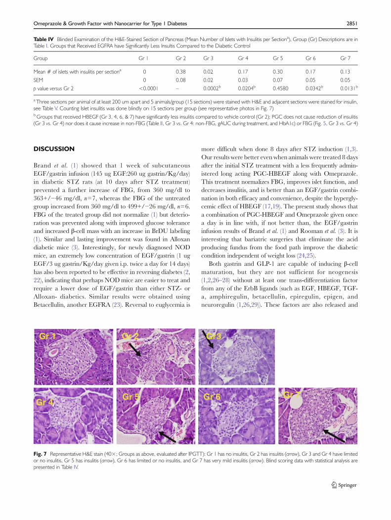
Table IV Blinded Examination of the H&E-Stained Section of Pancreas (Mean Number of Islets with Insulitis per Sectiona), Group (Gr) Descriptions are inTable I. Groups that Received EGFRA have Significantly Less Insultis Compared to the Diabetic Control
Group Gr 1 Gr 2 Gr 3 Gr 4 Gr 5 Gr 6 Gr 7
Mean # of islets with insulitis per sectiona 0 0.38 0.02 0.17 0.30 0.17 0.13
SEM 0 0.08 0.02 0.03 0.07 0.05 0.05
p value versus Gr 2 <0.0001 – 0.0002b 0.0204b 0.4580 0.0342b 0.0131b
a Three sections per animal of at least 200 um apart and 5 animals/group (15 sections) were stained with H&E and adjacent sections were stained for insulin,see Table V. Counting Islet insulitis was done blindly on 15 sections per group (see representative photos in Fig. 7)b Groups that received HBEGF (Gr 3, 4, 6, & 7) have significantly less insulitis compared to vehicle control (Gr 2); PGC does not cause reduction of insulitis(Gr 3 vs. Gr 4) nor does it cause increase in non-FBG (Table II, Gr 3 vs. Gr 4: non-FBG, gAUC during treatment, and HbA1c) or FBG (Fig. 5, Gr 3 vs. Gr 4)
Gr 1 Gr 2
Gr 4Gr 5 Gr 7Gr 6
Gr 3
Fig. 7 Representative H&E stain (40×; Groups as above, evaluated after IPGTT): Gr 1 has no insulitis, Gr 2 has insulitis (arrow), Gr 3 and Gr 4 have limitedor no insulitis, Gr 5 has insulitis (arrow), Gr 6 has limited or no insulitis, and Gr 7 has very mild insulitis (arrow). Blind scoring data with statistical analysis arepresented in Table IV.
Omeprazole & Growth Factor with Nanocarrier for Type 1 Diabetes 2851
facilitate repairs during duct ligation (27), partial pancreatec-tomy, cellophane wrapping of the gland, alloxan, STZ, andgamma interferon (2,30,31). Co-administration of ErbB li-gand, along with GLP-1 or gastrin, causes neogenesis andproliferation of islet β-cells (1,2,26–28). However, it is criticalto pick an ErbB ligand that is less likely to be associated withtumor formation. Unlike cell membrane bound HBEGF, thesoluble HBEGF utilized in this study lacks an intracellulardomain (12) that can initiate a self-sustaining signal of tumorformation. Use of soluble HBEGF will keep the signal poten-tially self-limiting once treatment is over. Our evaluation ofvarious tissues (stomach, pancreas, kidney, liver, and spleen)indicated that the 28 day treatment did not cause neoplasm.In particular, neither neoplasm nor hyperplasia were observedin the stomach which is also the target of gastrin action(causing ECL cells hyperplasia). While T2D has increased riskof cancer in several organs (32) due to insulin and obesity,T1D cancer risk is mainly in the stomach due to sustained H.pylori induced inflammation (20). For T1D, treatment thatreducesH. pylori induced inflammation should also reduce thisrisk; our data indicates that PGC-HBEGF reduces insulitis.The absence of neoplasm is consistent with a 30 weeks longstudy of HBEGF delivered using adenoviral vector (a moresustained delivery method) that looked at the effect on liver,kidney, intestine, and muscle (18). In addition, tissue injuriesrelease ErB ligand and yet tissue injuries rarely lead to cancer(2,27,30,31), perhaps because it is not sustained. Similarly, useof Omeprazole for over 30 years in humans does not causeneoplasm (21,33,34). Anticipated human treatment will occurover a few weeks and the risk of this treatment may not begreater than immune suppression treatments which lowerresistance to infection and cancer.
This study did not address the reasonwhyHBEGF causes areduction or elimination of insulitis or whether the hypergly-cemic effect of HBEGF can be dissociated from the elimina-tion of insulitis and its ability to induce trans-differentiation. Aprevious adoptive transfer model study indicated that EGFand gastrin induce immunoregulatory cells that prevent
autoimmunity (2). The mechanism responsible for this obser-vation will be the subject of future studies. Whatever themechanism, this approach offers exciting prospects for thetreatment and/or prevention of T1D without the use ofimmunosuppressant drugs that can increase the risk of infec-tion, such as anti-CD3, tacrolimus or rapamycin, with thelatter two also inhibiting β-cell division (35–37). Althoughthe comparison of insulitis between HBEGF and PGC-HBEGF treated groups showed that PGC does not causethe reduction of insulitis, the PGC seems to increase insulitisto some extent. Despite wide use of chondroitin sulfate toalleviate arthritis symptoms, it is possible that thechondroitin-containing PGC causes inflammation althoughit is likely that PGC reduces the anti-inflammatory effect ofHBEGF because of lowering the amount of free HBEGFneeded to suppress inflammation while prolonging the expo-sure. Paradoxically, hyperglycemia and inflammation facili-tate islet cell regeneration (38) and a proper balance will berequired to achieve full regeneration in addition to trans-differentiation of other cells into β-cells. In this study, bothhyperglycemia and inflammation are affected by HBEGF butwhether these effects are independent and distinct from theinduction of trans-differentiation, and whether one can besuppressed while maintaining the other effect, is not clear.Previous studies indicated that prolongation of exposure byfrequent administration or infusion is necessary for β-cellrecovery. However, we do not know the level and durationof the exposure sufficient for each of the following: 1) trans-differentiating cells into to β-cells, 2) reducing autoimmunityto a less harmful and more beneficial level, and 3) the degreeof beneficial hyperglycemia needed to assist gastrin-induced β-cell proliferation. In fact, the beneficial effect of an EGFRAcan reverse at a higher bolus dose (23), perhaps becauseextreme hyperglycemia causes glucotoxicity in β-cells whichoutweighs suppression of inflammation that destroys β-cells. Inthe present study, the hyperglycemic effect of HBEGF ismoderated by PGC and is clearly reduced by theOmeprazole-induced gastrin elevation. Perhaps those groups
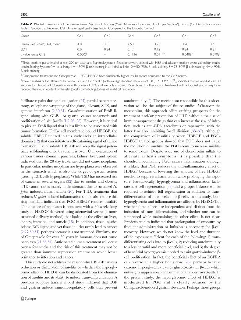
Table V Blinded Examination of the Insulin-Stained Section of Pancreas (Mean Number of Islets with Insulin per Sectiona), Group (Gr) Descriptions are inTable I. Groups that Received EGFRA have Significantly Less Insulin Compared to the Diabetic Control
Group Gr 1 Gr 2 Gr 4 Gr 5 Gr 6 Gr 7
Insulin Islet Scorea; 0–4, mean 4.0 3.0 2.50 3.73 3.70 3.6
SEM 0.0 0.24 0.19 0.12 0.24 0.21
p value versus Gr 2 0.0003 – 0.1136 0.0111b 0.0486b 0.0703c
a Three sections per animal of at least 200 um apart and 5 animals/group (15 sections) were stained with H&E and adjacent sections were stained for insulin.Insulin Scoring System: 0=no staining, 1=<50% β-cells staining in an individual islet, 2=50–75% β-cells staining, 3=75–90% β-cells staining, 4=>90%β-cells stainingbOmeprazole treatment and Omeprazole + PGC-HBEGF have significantly higher insulin scores compared to the Gr 2 controlc Power analysis of the difference between Gr 2 and Gr 7 of 0.6 (with average standard deviation of 0.8 (0.21SEM*151/2 )) indicates that we need at least 30sections to rule out lack of significance with power of 80% and we only analyzed 15 sections. In other words, treatment with additional gastrin may havereduced the insulin content of the islet β-cells contributing to loss of analytical resolution
2852 Castillo et al.

that experienced the strong hyperglycemic effect of HBEGFduring treatment will have glucotoxicity while those that havebeen tempered by Omeprazole or PGC (by limiting theamount of freeHBEGF) will not; the latter will have improvedregeneration assisted by suppression of inflammation andtrans-differentiation of other cells into β-cells. The literaturecontrol group (Gr 8), which also partially recovers, was treatedwith EGF and gastrin 3×/day. Gr 8 also showed hyperglyce-mia during treatment, which may also have been partiallysuppressed by gastrin 3×/day to minimize glucotoxicity andfacilitate recovery.
PGC uses reversible binding of peptide/protein drugs toprovide an automatic control of the amount of free drug inthe blood such that when the concentration of free drugdecreases, the PGC releases more drug to satisfy the Kd,thus acting as a drug reservoir/buffer in the blood. Thecomplex is encased in a protective PEG polymer, shieldingboth the PGC and drug from elimination by the reticuloen-dothelial system and enzyme degradation (8,39). This com-plex is large enough (15–30 nm) to escape from glomerularclearance (4 nm) that normally affects most drugs. Because ofthe size of PGC, there is preferential accumulation at sites ofhigh vascular permeability such as inflamed pancreas (40). Infact, PGC has been used to deliver GLP-1 for once a weekadministration in diabetic-ZDF rats and has efficacy that isequivalent to exendin-4 administered twice a day (8); twice aweek administration of PGC-GLP-1 is more effective thantwice a day administration of exendin-4 (8).
The novel findings in this study include the use of Omep-razole as a substitute for gastrin, the use of PGC to improveblood stability of growth factors relevant to diabetes (EGF,HBEGF, gastrin), and the improved approach to treat diabe-tes using a combination of Omeprazole and PGC-HBEGF.This approach has the distinct advantage of replacing inject-able labile gastrin with an FDA-approved orally availablemedication. Despite the hyperglycemic effect of HBEGF dur-ing treatment, we found that it is responsible for the reductionof insulitis while Omeprazole (or gastrin) is responsible forreducing blood glucose and increasing islet insulin content. Acombination of PGC-HBEGF and Omeprazole normalizesFBG, improves islet function, and reduces insulitis in multiplelow dose streptozotocin diabetic mice.
ACKNOWLEDGMENTS AND DISCLOSURES
This work was supported by the SBIR Grant # DK084724from National Institute of Diabetes and Digestive and Kid-ney diseases of the National Institute of Health. There hasbeen no prior publication of the present study.
G.M.C. conceived the approach, designed the experi-ments, researched data, and wrote the manuscript. A.N.A.contributed to the design of the experiments, researcheddata, and reviewed/edited the manuscript. A.A.B.. and
J.L.L. researched data and reviewed/edited the manuscript.A.V.L. reviewed/edited the manuscript. E.M.B. contributedto the design of the experiments and reviewed/edited themanuscript. The authors thank Ms. Cynthia Jones for helpwith preparing the manuscript, and Mr. ManShun Lai andDr. Sandra Reichstetter for technical assistance. G.M.C.,A.N.A., C.C.J., M.S.L., S.R., and E.M.B. are employees ofPharmaIN Corp.; A.A.B.., J.A.L., and A.V.L. are employeesof the University of Illinois.
REFERENCES
1. Brand SJ, Tagerud S, Lambert P, Magil SG, Tatarkiewicz K,Doiron K, et al. Pharmacological treatment of chronic diabetes bystimulating pancreatic beta-cell regeneration with systemic co-ad-ministration of EGF and gastrin. Pharmacol Toxicol.2002;91(6):414–20.
2. Suarez-Pinzon WL, Yan Y, Power R, Brand SJ, Rabinovitch A.Combination therapy with epidermal growth factor and gastrinincreases beta-cell mass and reverses hyperglycemia in diabeticNOD mice. Diabetes. 2005;54(9):2596–601.
3. Rooman I, Bouwens L. Combined gastrin and epidermal growthfactor treatment induces islet regeneration and restoresnormoglycaemia in C57Bl6/J mice treated with alloxan.Diabetologia. 2004;47(2):259–65. Epub 2003/12/11.
4. Lev-Ran A, Hwang DL, Ben-Ezra J, Williams LE. Origin of uri-nary epidermal growth factor in humans: excretion of endogenousEGF and infused [131I]-human EGF and kidney histochemistry.Clin Exp Pharmacol Physiol. 1992;19(10):667–73. Epub 1992/10/01.
5. Feng J, Mehta VB, El-Assal ON, Wu D, Besner GE. Tissue distri-bution and plasma clearance of heparin-binding EGF-like growthfactor (HB-EGF) in adult and newborn rats. Peptides.2006;27(6):1589–96. Epub 2005/12/21.
6. Suarez-Pinzon WL, Power RF, Yan Y, Wasserfall C, Atkinson M,Rabinovitch A. Combination therapy with glucagon-like peptide-1and gastrin restores normoglycemia in diabetic NOD mice.Diabetes. 2008;57(12):3281–8.
7. Mefford IN, Wade EU. Proton pump inhibitors as a treatmentmethod for type II diabetes. Med Hypotheses. 2009;73(1):29–32.Epub 2009/03/24.
8. Castillo GM, Reichstetter S, Bolotin EM. Extending residence timeand stability of peptides by Protected Graft Copolymer (PGC)excipient: GLP-1 example. Pharm Res. 2012;29(1):306–18. Epub2011/08/11.
9. Zavros Y, Rieder G, Ferguson A, Samuelson LC, Merchant JL.Hypergastrinemia in response to gastric inflammation suppressessomatostatin. Am J Physiol Gastrointest Liver Physiol. 2002;282(1):G175–83. Epub 2001/12/26.
10. Castillo GM, Bolotin EM, Nishimoto-Ashfield A. Anionic-corecomposition for delivery of therapeutic agents, and methods ofmaking and using the same. USPTO. 2012;App No 20120190097.
11. Bolotin E. Compositions for delivery of therapeutics and othermaterials, and methods of making and using the same (now pat-ent#7,138,105). 2003;App No 20030224974.
12. Wang X, Mizushima H, Adachi S, Ohishi M, Iwamoto R, MekadaE. Cytoplasmic domain phosphorylation of heparin-binding EGF-like growth factor. Cell Struct Funct. 2006;31(1):15–27. Epub2006/03/25.
Omeprazole & Growth Factor with Nanocarrier for Type 1 Diabetes 2853

13. Hieda M, Isokane M, Koizumi M, Higashi C, Tachibana T,Shudou M, et al. Membrane-anchored growth factor, HB-EGF,on the cell surface targeted to the inner nuclear membrane. J CellBiol. 2008;180(4):763–9. Epub 2008/02/27.
14. Adam RM, Danciu T, McLellan DL, Borer JG, Lin J, ZurakowskiD, et al. A nuclear form of the heparin-binding epidermal growthfactor-like growth factor precursor is a feature of aggressive transi-tional cell carcinoma. Cancer Res. 2003;63(2):484–90. Epub 2003/01/25.
15. Reichstetter S, Castillo GM, Lai M, Nishimoto-Ashfield A,Banerjee A, Bogdanov A, et al. Protected Graft Copolymer (PGC)basal formulation of insulin as potentially safer alternative toLantus(R) (Insulin-Glargine): a streptozotocin-induced, diabeticsprague dawley rats study. Pharm Res. 2012;29(4):1033–9. Epub2011/12/29.
16. Reichstetter S, Castillo GM, Rubinsteinb I, Nishimoto-Ashfield A,Lai M, Jones CC, et al. Protected graft copolymer excipient leads toa higher acute maximum tolerated dose and extends residence timeof vasoactive intestinal peptide significantly better than stericallystabilized micelles. Pharm Res. 2013;30:670–82.
17. Grau M, Tebar F, Ramirez I, Soley M. Epidermal growth factoradministration decreases liver glycogen and causes mildhyperglycaemia in mice. Biochem J. 1996;315(Pt 1):289–93.Epub 1996/04/01.
18. Kozawa J, Tokui Y, Moriwaki M, Li M, Ohmoto H, Yuan M, et al.Regenerative and therapeutic effects of heparin-binding epidermalgrowth factor-like growth factor on diabetes by gene transductionthrough retrograde pancreatic duct injection of adenovirus vector.Pancreas. 2005;31(1):32–42. Epub 2005/06/22.
19. Cameron CM, Kostyo JL, Papkoff H. Nonmammalian growthhormones have diabetogenic and insulin-like activities.Endocrinology. 1985;116(4):1501–5. Epub 1985/04/01.
20. Zendehdel K, Nyren O, Ostenson CG, Adami HO, Ekbom A, YeW. Cancer incidence in patients with type 1 diabetes mellitus: apopulation-based cohort study in Sweden. J Natl Cancer Inst.2003;95(23):1797–800. Epub 2003/12/05.
21. Bateman DN, Colin-Jones D, Hartz S, Langman M, Logan RF,Mant J, et al. Mortality study of 18 000 patients treated withomeprazole. Gut. 2003;52(7):942–6.
22. Reddy S, Cheung CC, Chai RC, Rodrigues JA. Persistence ofresidual beta cells and islet autoimmunity during increasing dura-tion of diabetes in NODmice and experimental approaches towardreversing new-onset disease with bioactive peptides. Ann N Y AcadSci. 2008;1150:171–6.
23. Li L, Seno M, Yamada H, Kojima I. Betacellulin improves glucosemetabolism by promoting conversion of intraislet precursor cells tobeta-cells in streptozotocin-treated mice. Am J Physiol EndocrinolMetab. 2003;285(3):E577–83. Epub 2003/08/06.
24. Rizzello M, Abbatini F, Casella G, Alessandri G, Fantini A, LeonettiF, et al. Early postoperative insulin-resistance changes after sleevegastrectomy. Obes Surg. 2010;20(1):50–5. Epub 2009/11/17.
25. Spector D, Shikora S. Neuro-modulation and bariatric surgery fortype 2 diabetes mellitus. Int J Clin Pract Suppl. 2010;166:53–8.Epub 2010/08/18.
26. Brand SJ. Prolonged efficacy of islet neogenesis therapy methodswith a gastrin/CCK receptor ligand and an EGF receptor ligand
composition in subjects with preexisting diabetes. USPTO. 2006;Pat No 6992060B2: .
27. Rooman I, Lardon J, Bouwens L. Gastrin stimulates beta-cellneogenesis and increases islet mass from transdifferentiated but notfrom normal exocrine pancreas tissue. Diabetes. 2002;51(3):686–90.
28. Song SY, Gannon M, Washington MK, Scoggins CR, MeszoelyIM, Goldenring JR, et al. Expansion of Pdx1-expressing pancreaticepithelium and islet neogenesis in transgenic mice overexpressingtransforming growth factor alpha. Gastroenterology. 1999;117(6):1416–26.
29. Wilson KJ, Gilmore JL, Foley J, Lemmon MA, Riese 2nd DJ.Functional selectivity of EGF family peptide growth factors: impli-cations for cancer. Pharmacol Ther. 2009;122(1):1–8. Epub 2009/01/13.
30. Bonner-Weir S, Baxter LA, Schuppin GT, Smith FE. A secondpathway for regeneration of adult exocrine and endocrine pancre-as. A possible recapitulation of embryonic development. Diabetes.1993;42(12):1715–20.
31. Gu D, Sarvetnick N. Epithelial cell proliferation and islet neogenesisin IFN-g transgenic mice. Development. 1993;118(1):33–46.
32. Vigneri P, Frasca F, Sciacca L, Pandini G, Vigneri R. Diabetes andcancer. Endocrine-Relat Cancer. 2009;16(4):1103–23. Epub2009/07/22.
33. Singh P, Indaram A, Greenberg R, Visvalingam V, Bank S. Longterm omeprazole therapy for reflux esophagitis:follow-up in serumgastrin levels, EC cell hyperplasia and neoplasia. World JGastroenterol. 2000;6(6):789–92. Epub 2002/01/31.
34. Ligumsky M, Lysy J, Siguencia G, Friedlander Y. Effect of long-term, continuous versus alternate-day omeprazole therapy on se-rum gastrin in patients treated for reflux esophagitis. J ClinGastroenterol. 2001;33(1):32–5.
35. Hyder A, Laue C, Schrezenmeir J. Effect of the immunosuppressiveregime of Edmonton protocol on the long-term in vitro insulinsecretion from islets of two different species and age categories.Toxicol In Vitro. 2005;19(4):541–6. Epub 2005/04/14.
36. Gangemi A, Salehi P, Hatipoglu B, Martellotto J, Barbaro B,Kuechle JB, et al. Islet transplantation for brittle type 1 diabetes:the UIC protocol. Am J Transplant. 2008;8(6):1250–61.
37. Lopez-Talavera JC, Garcia-Ocana A, Sipula I, Takane KK,Cozar-Castellano I, Stewart AF. Hepatocyte growth factor genetherapy for pancreatic islets in diabetes: reducing the minimal islettransplant mass required in a glucocorticoid-free rat model ofallogeneic portal vein islet transplantation. Endocrinology.2004;145(2):467–74. Epub 2003/10/11.
38. Akirav EM, Baquero MT, Opare-Addo LW, Akirav M, Galvan E,Kushner JA, et al. Glucose and inflammation control islet vasculardensity and {beta}-cell function in NOD mice: control of isletvasculature and vascular endothelial growth factor by glucose.Diabetes. 2011. Epub 2011/02/11.
39. Bogdanov Jr AA, Mazzanti M, Castillo G, Bolotin E. ProtectedGraft Copolymer (PGC) in imaging and therapy: a platform for thedelivery of covalently and non-covalently bound drugs.Theranostics. 2012;2(6):553–76. Epub 2012/06/28.
40. Medarova Z, Castillo G, Dai G, Bolotin E, Bogdanov A, Moore A.Noninvasive magnetic resonance imaging of microvascular changesin type 1 diabetes. Diabetes. 2007;56(11):2677–82.
2854 Castillo et al.