Embed Size (px)
Citation preview

Journal of Clinical Pathology, 1979, 32, 171-178
Gastrin cells and fasting serum gastrin levels induodenal ulcer patientsA quantitative study based on multiple biopsy specimens
JUAN PIRIS AND R. WHITEHEADIFrom the Department ofPathology, Harkness Laboratories, Radcliffe Infirmary, Oxford OX2 6HE, UK
SUMMARY The number of gastrin-producing cells in biopsy specimens from the gastric and duodenalmucosae in 19 duodenal ulcer patients was quantitated using a morphometric method. The levelof immunoreactive gastrin in a sample of fasting serum obtained at the time of the biopsy was
measured by radioimmunoassay. The results show no significant difference when compared withthose from a group of normal control volunteers. Moreover, there was no correlation between thenumbers of gastrin-producing cells and the fasting serum gastrin level in either controls or duodenalulcer patients.It is widely believed that hypersecretion of gastrichydrochloric acid is a factor involved in the aetiologyof some duodenal ulcers. Although some controversyremains, most authors believe that the level offasting serum gastrin in patients with duodenalulcers is not significantly higher than that of thenormal population (Trudeau and McGuigan, 1970,1971; Korman et al., 1971; Ganguli and Hunter,1972; McGuigan and Trudeau, 1973; Stadil andRehfeld, 1973), the main source of gastrin being thegastrin-producing cells in the stomach. A number ofpublished reports suggest that in some duodenalulcer patients there is hyperplasia of gastrin-produc-ing cells in the pyloric antrum (Solcia et al., 1970;Polak et al., 1972; Cowley et al., 1973; Ganguliet al., 1974). Only a few of these reports are basedon reliable, objective, quantitative methods, andnone has used multiple biopsy specimens obtainedwith the fibreoptic endoscope. Our paper presentsthe results of a quantitative study of gastrin-produc-ing cells in the stomach of 19 patients suffering fromduodenal ulcer who had multiple gastric biopsies.
Material and methods
Nineteen patients diagnosed as having duodenalulceration were studied. All gave a typical clinicalhistory of abdominal pain and dyspepsia. Each had abarium meal examination and gastroduodenoscopyon at least one occasion. An ulcer crater was
'Present address: Department of Pathology, School ofMedicine, The Flinders University of South Australia,Bedford Park, South Australia 5042.
Received for publication 12 July 1978
demonstrated by the radiologist or seen at endoscopyin all patients. There were 15 males and four femaleswith a mean age of 38 4 ± 13 years, ranging from16 to 62 years.A group of 15 young adults with no recognisable
disease of the gastrointestinal tract acted as controls.Seven of them were healthy volunteers; the remain-ing eight were examined by endoscopy and biopsyfor a variety of clinical reasons, and all were foundto be normal. The results of the investigationsperformed in this group of controls can be foundelsewhere (Piris, 1975; Piris and Whitehead, 1975).There were seven males and eight females, withages ranging from 17 to 53 (mean 30 4 ± 9 5)years.Endoscopy was performed in bothgroups after
an overnight fast. At the same time a sample ofblood was taken for measurement of serum gastrinlevels by radioimmunoassay. After careful examina-tion of the complete gastric and duodenal mucosae,biopsies were obtained from the duodenum (duo-denal cap and proximal descending duodenum) andfrom five standard sites in the stomach: (1) thepyloric antrum on the lesser curvature between thepyloric ring and the incisura angularis; (2) at aboutthe middle of the lesser curve; (3) opposite to thison the greater curve; (4) high on the lesser curve atabout 3 cm from the cardia; and (5) the fundus.In patients with duodenal ulcers, biopsies were alsotaken from the ulcer edge.
DEMONSTRATION OF GASTRIN CELLS
Gastrin-producing cells (G-cells) were demonstratedin biopsy specimens obtained using an immuno-
171
on 7 March 2019 by guest. P
rotected by copyright.http://jcp.bm
j.com/
J Clin P
athol: first published as 10.1136/jcp.32.2.171 on 1 February 1979. D
ownloaded from

Juan Piris and R. Whitehead
peroxidase method with antisera raised in rabbitsagainst synthetic human gastrin I (ICI), as describedearlier (Piris and Whitehead, 1974). Briefly, speci-mens were formalin-fixed and embedded in paraffin;6p. sections were then exposed to anti-gastrin serum(diluted 1:10 with phosphate buffer saline (PBS),pH 7-2 for 60 minutes, and after washing in PBSthe sections were exposed to swine anti-rabbit IgGlabelled with peroxidase. The bound peroxidase wasdeveloped by treating with diaminobenzidine tetra-hydrochloride containing hydrogen peroxide(Nakane and Pierce, 1967). The specificity of thereactions shown in Figs 2, 3, and 4 was establishedby preabsorbing the antisera with synthetic humangastrin I; normal rabbit serum did not produce anystaining of gastrin cells. In the final preparation, thegastrin-producing cells stain dark brown, and back-ground tissues, whose structural features are wellshown, appear a light green.
QUANTITATION OF G-CELLSThe morphometric method for the quantitation ofgastrin-producing cells related to the volume ofepithelium, which was described in a previous paper(Piris and Whitehead, 1975), was slightly modifiedby the addition of lines to the grid. This was donefor the purpose of measuring the length of themuscularis mucosae in accordance with the principledeveloped by Short (1950). It assumes that if aseries of lines of a constant length are randomly caston a cut surface of a composite object, the number oftimes that the lines 'cut' the linear component to bemeasured is proportional to its total length in thesection. A modification of this principle to estimatevolume-surface ratios was used by Dunnill andWhitehead (1972), who obtained an index relatingthe surface to volume ratio of the mucosa of smallintestinal biopsy specimens. Weibel (1963) describeda template for the dual purpose of point counting
Fig. 1 Morphometric grid with diagrammatic representation of antral mucosasuperimposed (mm = muscularis mucosae).
172
on 7 March 2019 by guest. P
rotected by copyright.http://jcp.bm
j.com/
J Clin P
athol: first published as 10.1136/jcp.32.2.171 on 1 February 1979. D
ownloaded from
Gastrin cells andfasting serum gastrin levels in duodenal ulcer patients
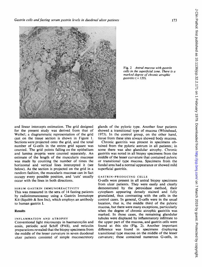
Fig. 2 Antral mucosa with gastrincells in the superficial zone. There is amarked degree of chronic atrophicgastritis ( x 120).
and linear intercepts estimation. The grid designedfor the present study was derived from that ofWeibel; a diagrammatic representation of the gridcast on the tissue section is shown in Figure 1.Sections were projected onto the grid, and the totalnumber of G-cells in the entire grid square wascounted. The grid points falling on the epitheliumand lamina propria were counted separately. Anestimate of the length of the muscularis mucosaewas made by counting the number of times thehorizontal and vertical lines intercepted it (seebelow). As the section is projected on the grid in arandom fashion, the muscularis mucosae can in factoccupy every possible position, and 'cuts' usuallyoccur with the lines in both directions.
SERUM GASTRIN IMMUNOREACTIVITYThis was measured in the sera of 14 fasting patientsby radioimmunoassay using a Gastrin ImmutopeKit (Squibb & Son Inc), which employs an antibodyto human gastrin I.
Results
INFLAMMATION AND ATROPHYConventional light microscopy in haematoxylin andeosin, periodic acid Schiff (PAS), and reticulinpreparations revealed that the biopsy specimens fromthe middle of the lesser curvature in seven duodenalulcer patients consisted of simple mucosecretory
glands of the pyloric type. Another four patientsshowed a transitional type of mucosa (Whitehead,1973). In the control group, on the other hand,tissue from these sites always showed body mucosa.
Chronic gastritis was present in specimens ob-tained from the pyloric antrum in all patients; insome there was also glandular atrophy. Chronicgastritis was noted in all biopsy specimens from themiddle of the lesser curvature that contained pyloricor transitional type mucosa. Specimens from thefundal area had a normal appearance or showed mildsuperficial gastritis.
GASTRIN-PRODUCING CELLSG-cells were present in all antral biopsy specimensfrom ulcer patients. They were easily and clearlydemonstrated by the peroxidase method, theircytoplasm appearing densely stained and fullygranulated, thus contrasting with the cells in thecontrol cases. In general, G-cells were in the usuallocation, that is, the middle third of the pyloricmucosa, but there were many exceptions, particularlywhen the degree of chronic atrophic gastritis wasmarked. In those cases, the remaining glandulartubules were displaced by inflammatory infiltrate tothe upper part of the mucosa, and gastrin cells werefound at this site (Fig. 2). Another importantdifference was found in specimens displayingtransitional type mucosa on the middle of the lessercurvature; these contained numerous G-cells, in
173
on 7 March 2019 by guest. P
rotected by copyright.http://jcp.bm
j.com/
J Clin P
athol: first published as 10.1136/jcp.32.2.171 on 1 February 1979. D
ownloaded from

Juan Piris and R. Whitehead
I
4
44
'Imy
4..
**.'..
S
Fig. 3 Transitional type mucosa from the middle of the lesser curvature. Numerous gastrin cells are seen ( x 460).
Fig. 4 Gastrin cells in duodenal Brunner's glands ( x 560).
174
MM:
on 7 March 2019 by guest. P
rotected by copyright.http://jcp.bm
j.com/
J Clin P
athol: first published as 10.1136/jcp.32.2.171 on 1 February 1979. D
ownloaded from

Gastrin cells andfasting serum gastrin levels in duodenal ulcer patients 175
contrast with those of control cases in which they QUANTITATION OF GASTRIN CELLSwere rarely present (Fig. 3). The results of the quantitative study are given in
Biopsy specimens obtained from remaining sites Tables 1 and 2. Owing to atrophy and inflammationwithin the stomach showed a body type of mucosa in the antrum of patients with duodenal ulceration,with no gastrin cells. there was considerable variation in the relative
In duodenal biopsy specimens there was no differ- volume proportions occupied by the epithelialence in distribution of G-cells between controls and component and lamina propria in these biopsypatients. At this site G-cells are found in small specimens. This could be objectively quantified bynumbers in gland crypts and occasionally in means of the counts of points falling on epitheliumBrunner's glands (Fig. 4). (E) and on lamina propria (LP), which are respect-
Table 1 Control group
No. Sex Age E LP I G GII index SG
1 M 25 658 417 262 186 0-71 422 F 27 1178 978 270 417 1-54 573 F 34 547 436 118 195 1-65 334 M 34 699 388 229 202 0-88 555 F 20 650 382 280 192 0-69 436 F 22 649 375 221 120 0 54 687 M 30 933 656 255 184 0-72 698 F 17 737 536 227 278 1-23 959 M 43 A 946 630 290 267 0-92 -
B <610 M 28 A 1026 612 324 238 0 74 63
B <2011 M 32 A 835 766 265 228 0-86 -
B <412 F 53 968 572 260 307 1-18 5413 F 30 1062 670 309 438 1-42 -
14 M 21 1712 886 522 470 0 90 5515 F 38 714 475 228 86 0-38 -
E=Points falling on epithelium; LP= Points falling on lamina propria; I = Intercepts with muscularis mucosae; G = Number of gastrin cells;SG = Serum gastrin (pg/ml); A = Biopsy antrum; B = Biopsy middle of lesser curve.
Table 2 Duodenal ulcer group
No. Sex Age E LP I G GII GII index SG
1 M 38 812 537 229 113 0 49 0-49 602 M 30 576 468 112 120 1-07 1-07 243 F 34 A 584 632 206 279 1-36 1*36 76
B <104 M 34 A 531 611 192 43 0-22
B 529 619 176 100 0-57 0-79 132S M 26 A 874 866 191 485 2-54
B 295 234 74 82 1 11 3-65 -
6 M 24 324 816 142 140 0-99 0-99 _7 F 38 598 717 174 281 1-62 1-62 1058 M 40 A 567 459 138 131 0-95
B 514 445 113 101 0-89 1-84 309 M 53 A 423 397 113 160 1-42
B 378 367 86 166 1-93 3 35 9010 M 44 685 711 147 66 0 45 0 4511 M 28 A 634 788 224 148 0-66
B 300 449 95 83 0-87 1-5312 M 24 A 662 465 120 112 0-93 0-93 _
B 10-1513 M 44 514 479 127 101 0-79 0-79 8514 M 37 A 425 275 84 257 3-06
B 330 158 116 95 0-82 3-88 7515 F 34 401 534 139 175 1-26 1-26 9516 M 30 426 565 165 191 1-16 1-16 60 517 M 16 392 406 135 126 0-93 0-93 9318 F 46 A 663 602 226 197 0-87 0-87 75-5
B <1019 M 62 976 1051 346 643 1-86 1-86 58-S
See footnote to Table I
on 7 March 2019 by guest. P
rotected by copyright.http://jcp.bm
j.com/
J Clin P
athol: first published as 10.1136/jcp.32.2.171 on 1 February 1979. D
ownloaded from

Juan Piris and R. Whitehead
ively proportional to the areas occupied by thesetwo components (Weibel, 1963; Piris and Whitehead,1975). In addition, it was found that the thicknessof the complete mucosa from muscularis mucosaeto surface epithelium was not comparable for allpatients. This total mucosal volume is proportionalto the figure obtained by adding the points fallingon epithelium to those falling on the lamina propria(that is, E + LP). For the above reasons it is clearthat a comparison of the number of gastrin cells perunit volume of epithelium between control subjectsand patients with duodenal ulceration is not validsince numbers of G-cells and/or the volume ofepithelium could change and thus alter the ratio.Therefore it was necessary to introduce the measure-ment of a fixed parameter, which is not altered by thedisease. The length of the muscularis mucosaefulfils this criterion. For this reason the number ofgastrin cells per unit length of muscularis mucosae(G/l) was chosen as the best means of comparingthe results in both groups.An additional difficulty encountered was the
presence of significant numbers of gastrin cells inspecimens from the middle of the lesser curve in someduodenal ulcer patients. This was not encountered inthe control group.For the purpose of comparing the G-cell density
in these patients and the controls, the ratio G/t(gastrin cells per unit length of muscularis mucosae)obtained in the antral specimen (A) and in the tissuefrom the middle of the lesser curve (B) were addedtogether. The result, which is no longer mathematic-ally a ratio, has been called the G/I index. In thosecases in which G-cells were found only in the antralbiopsy specimen (A) and in the control cases, theG/I index is identical with the ratio of gastrin cellsto intercepts with the muscularis mucosae obtainedin the antral specimen (Fig. 5).
STATISTICAL ANALYSIS OF DATAThis was done using non-parametric statisticaltests since it could not be assumed that the numbersof gastrin cells in both groups-controls and duo-denal ulcer subjects were distributed in accordancewith the normal distribution. In fact the variance-ratio, or F-test, demonstrated the inequality of thecorresponding variances for the two populations.The Mann-Whitney U test- a ranking test-wasused to compare the G-cell index of the 19 patientshaving duodenal ulceration with those of the 15controls. The value of U obtained was 88, which isnot significant at the 50 level.
SERUM GASTRINSerum gastrin levels were not available in five cases.The mean serum gastrin level in 14 patients with
4 0
30.
20.
10.
-(A)-
(B)
1-- Antrum Lesser-C
*I
S
Fig. 5 Ratios of G-cells to intercepts with mnuscularismucosae (G/I) for the specimens obtained fiom theantrum (A) and lesser carve (B) and the two sitescombined (A + B) in duodenal ulcer patients and controls.Using the Mann- Whitney U test, the correspondingvalues of U are: (A) U = 113; (B) U = 97,(A + B) U = 88, which are not significant at the 50level.
duodenal ulcer was 75-61 pg/ml with a standarddeviation of 28-50. There was no significant differ-ence between these values and those of the controlgroup (11 patients, mean 57 64 pg/ml, SD 16 60).
SERUM GASTRIN AND G-CELL NUMBERSThere was no correlation (r = 003 1) between fastingserum gastrin levels and the G-cell index for the 14patients for whom these two sets of data wereavailable. The presence of gastrin cells in themiddle of the lesser curve was not associatedwith an increased fasting serum gastrin level.
Structural changes of antral mucosaIt has been said that the antral biopsy specimens fromthe duodenal ulcer patients showed a degree ofchronic gastritis and atrophy. Since the inflammatoryinfiltrate is concentrated in the lamina propria ofthe mucosa, the proportion of volume occupied byit should be larger than that of the non-inflamedcases such as the controls. An accurate measure-ment of the volume of lamina propria can beobtained by relating the number of points falling onthe lamina (LP) to the intercepts (I) with the muscu-laris mucosae. The mean ratio LP/I for the controlgroup (Table 1) was 2 25 ± 0-69. The mean ratiofor the 19 patients with duodenal ulcer was 3 65 +
0
176
00
0
0 A
on 7 March 2019 by guest. P
rotected by copyright.http://jcp.bm
j.com/
J Clin P
athol: first published as 10.1136/jcp.32.2.171 on 1 February 1979. D
ownloaded from

Gastrin cells and fasting serum gastrin levels in duodenal ulcer patients 177
0.80. The difference in the ratios of the two groupsas measured by the Mann-Whitney U test gives avalue of U of 26, which is significant at the 0-2%level. In order to test whether or not the degree ofinflammation in the antrum did have an effect onthe number of gastrin cells, a correlation test betweenthe ratio LP/I and the G/1 index was carried out.The correlation coefficient for the 19 duodenal ulcerpatients was 0.07, which is not significant.
Discussion
It appears from the data presented above that thedensity of gastrin cells in patients with duodenalulcer is not significantly higher than that of normalcontrols. This is so when the two groups are com-pared as a whole but in the case of duodenal ulcerpatients the density of gastrin cells in the antrumshows a wider range, from normal to a moderate(up to twofold) increase.
It is also worth noting that in some of the duodenalulcer patients-six out of 19-considerable numbersof gastrin cells were found in the metaplasticmucosa of the lesser curve; in spite of this, when thenumbers of G-cells of the antrum and of the lessercurve are independently compared in the two groups,no significant difference emerges.
It has been shown by Stave and Brandtzaeg(1976) that the density of gastrin cells is greatestaround the pylorus and decreases proximally in thestomach. An exception to this pattern was, however,found in some of our patients in whom there was asevere degree of antntis, and in them a higherdensity of gastrin cells is found (cases 4, 9, and 11)in the specimen obtained from the middle of thelesser curve.
It is also of interest that the gastrin cells in theduodenal ulcer patients were more intensely stainedthan those of the controls; this indicates that, in thefasting state, the cells contain more gastrin in theircytoplasm and may provide an explanation for theincreased response to gastrin-releasing stimulifound in duodenal ulcer disease.We did not find in our duodenal ulcer patients
two populations, one with normal or lower numbersof gastrin cells, the other with much larger numbersof gastrin cells, as described by Ganguli et al.(1974), even when the numbers of gastrin cells wererelated to the intercepts with the muscularis mucosae,which gives a ratio independent of the presence anddegree ofinflammation and/or atrophy in the antrum.More recently, a report of the number of gastrin
cells in the antrum of patients with duodenal ulcer(Creutzfeldt et al., 1976) has appeared, based on theresults of counting gastrin cells in single biopsyspecimens from the pyloric antrum or duodenal
mucosa. In addition, counting was limited to themid-zone of the antral mucosa, which is the areawhere most gastrin cells are to be found in normalhuman stomach, but, as illustrated in Fig. 2, not inall cases where there is inflammation and atrophy.In their investigation the size of the area used forcounting antral G-cells (0.35 x 0-23 mm) and thenumber of fields counted (at least 10) were seeminglychosen in an arbitrary manner. It must be pointedout that, in the presence of a marked inflammatoryinfiltrate, the volume occupied by the laminapropria will be expanded, and that therefore withinone of their 'counting areas' fewer glands areincluded and consequently possibly fewer gastrincells. If, for instance, an increase in the number ofgastrin cells within each gland has taken place, thiscould be missed by counting a smaller number ofglands. From the quantitative data presented here itis clear that marked differences in relative volumeof epithelium and of lamina propria occur in thepresence of inflammation. Using their method,Creutzfeldt et al. (1976) could not detect anysignificant differences in the number of G-cells ofpatients with duodenal ulcer and of controls. Ourstudy paradoxically agrees with their conclusionsbut not with the methods whereby such conclusionswere reached. It is also interesting to find that thenumber of gastrin cells appears to be independent ofthe degree of inflammation of the antrum, as indi-cated by poor correlation between ratios of volumelamina propria to length of muscularis mucosae andG-cells per length of muscularis mucosae.
We thank Dr A. T. H. Robb-Smith for makingavailable the facilities of his department; Mr R. S.Davison, Mr N. D. Thomas, and Mrs Sue Canningfor excellent technical help; and Mrs Hazel Christianfor handling the manuscript.One of us (JP) was supported by a scholarship from
the Wellcome Trust.We are indebted to Dr S. C. Truelove for allowing
us to study his patients and to Dr M. Roca, whoobtained the biopsy specimens.
References
Cowley, D. J., Dymock, I. W., Boyes, B. E., Wilson,R. Y., Stagg, B. H., Lewin, M. R., Polak, J. M.,and Pearse, A. G. E. (1973). Zollinger-Ellison syndrometype I: clinical and pathological correlations in a case.Gut, 14, 25-29.
Creutzfeldt, W., Arnold, R., Creutzfeldt, C., and Track,N. S. (1976). Mucosal gastrin concentration, molecularforms of gastrin, number and ultrastructure of G-cellsin patients with duodenal ulcer. Gut, 17, 745-754.
on 7 March 2019 by guest. P
rotected by copyright.http://jcp.bm
j.com/
J Clin P
athol: first published as 10.1136/jcp.32.2.171 on 1 February 1979. D
ownloaded from

178 Juan Piris and R. Whitehead
Dunnill, M. S., and Whitehead, R. (1972). A method forthe quantitation of small intestinal biopsy specimens.Journal of Clinical Pathology, 25, 243-246.
Ganguli, P. C., and Hunter, W. M. (1972). Radio-immunoassay of gastrin in human plasma. Journal ofPhysiology, 220, 499-510.
Ganguli, P. C., Polak, J. M., Pearse, A. G. E., Elder,J. B., and Hegarty, M. (1974). Antral-gastrin-cellhyperplasia in peptic-ulcer disease. Lancet, 1, 583-586.
Korman, M. G., Soveny, C., and Hansky, J. (1971).Serum gastrin in duodenal ulcer. Part 1. Basal levelsand effect of food and atropine. Gut, 12, 899-902.
McGuigan, J. E., and Trudeau, W. L. (1973). Differencesin rates of gastrin release in normal persons andpatients with duodenal ulcer disease. New EnglandJournal of Medicine, 288, 64-66.
Nakane, P. K., and Pierce, G. B., Jr. (1967). Enzyme-labeled antibodies: preparation and application for thelozalization of antigens (Letter). Journal of Histo-chemistry and Cytochemistry, 14, 12, 929-931.
Piris, J. (1975). Serum gastrin and gastrin cells in thehuman stomach. D.Phil. thesis, University ofOxford.
Piris, J., and Whitehead, R. (1974). An immunoperoxidasetechnique for the identification of gastrin-producingcells. Journal of Clinical Pathology, 27, 798-799.
Piris, J., and Whitehead, R. (1975). Quantitation of G-cells in fiberoptic biopsy specimens and serum gastrinlevels in healthy normal subjects. Journal of ClinicalPathology, 28, 636-638.
Polak, J. M., Stagg, B., and Pearse, A. G. E. (1972).Two types of Zollinger-Ellison syndrome: immuno-fluorescent, cytochemical and ultrastructural studies of
the antral and pancreatic gastrin cells in differentclinical states. Gut, 13, 501-512.
Short, R. H. D. (1950). Alveolar epithelium in relationto growth of the lung. Philosophical Transactions, B,235, 35-36.
Solcia, E., Capella, C., and Vasallo, G. (1970). Endocrinecells of the stomach and pancreas in states of gastrichypersecretion. Reudiconti Romani di Gastro-enterol-ogia, 2, 147-158.
Stadil, F., and Rehfeld, J. F. (1973). Determination ofgastrin in serum. An evaluation of the reliability of aradioimmunoassay. Scandinavian Journal of Gastro-enterology, 8, 101-112.
Stave, R., and Brandtzaeg, P. (1976). Immunohisto-chemical investigation of gastrin-producing cells(G cells): the distribution of G cells in resected humanstomachs. Scandinavian Journal of Gastroenterology,11, 705-712.
Trudeau, W. L., and McGuigan, J. E. (1970). Serumgastrin levels in patients with peptic ulcer disease.Gastroenterology, 59, 6-12.
Trudeau, W. L., and McGuigan, J. E. (1971). Relationsbetween serum gastrin levels and rates of gastrichydrochloric acid secretion. New England Journal ofMedicine, 284, 408-412.
Weibel, E. R. (1963). Principles and methods for themorphometric study of the lung and other organs.Labor-atory Investigation, 12, 131-155.
Whitchead, R. (1973). Mucosal Biopsy of the Gastro-intestinal Tract. Saunders, London.
Requests for reprints to: Dr J. Piris, University Depart-ment of Pathology, Harkness Laboratories, RadcliffeInfirmary, Oxford OX2 6HE.
on 7 March 2019 by guest. P
rotected by copyright.http://jcp.bm
j.com/
J Clin P
athol: first published as 10.1136/jcp.32.2.171 on 1 February 1979. D
ownloaded from

![BMC Cancer | Home page - Gastrin activates autophagy and ......gastric epithelial cells [14]. Gastrin has been found to stimulate proliferation of cancer cell lines isolated from the](https://img.dokumen.tips/doc/110x75/611338783576842c4f73986e/bmc-cancer-home-page-gastrin-activates-autophagy-and-gastric-epithelial.jpg)