Embed Size (px)
Citation preview
DRAFT – AS of September 19, 2017
DO NOT REPRODUCE
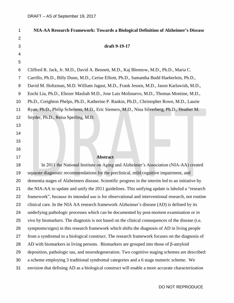
NIA-AA Research Framework: Towards a Biological Definition of Alzheimer’s Disease 1
2
draft 9-19-17 3
4
5
Clifford R. Jack, Jr. M.D., David A. Bennett, M.D., Kaj Blennow, M.D., Ph.D., Maria C. 6
Carrillo, Ph.D., Billy Dunn, M.D., Cerise Elliott, Ph.D., Samantha Budd Haeberlein, Ph.D., 7
David M. Holtzman, M.D. William Jagust, M.D., Frank Jessen, M.D., Jason Karlawish, M.D., 8
Enchi Liu, Ph.D., Eliezer Masliah M.D., Jose Luis Molinuevo, M.D., Thomas Montine, M.D., 9
Ph.D., Creighton Phelps, Ph.D., Katherine P. Rankin, Ph.D., Christopher Rowe, M.D., Laurie 10
Ryan, Ph.D., Philip Scheltens, M.D., Eric Siemers, M.D., Nina Silverberg, Ph.D., Heather M. 11
Snyder, Ph.D., Reisa Sperling, M.D. 12
13
14
15
16
Abstract 17
In 2011 the National Institute on Aging and Alzheimer’s Association (NIA-AA) created 18
separate diagnostic recommendations for the preclinical, mild cognitive impairment, and 19
dementia stages of Alzheimers disease. Scientific progress in the interim led to an initiative by 20
the NIA-AA to update and unify the 2011 guidelines. This unifying update is labeled a “research 21
framework”, because its intended use is for observational and interventional research, not routine 22
clinical care. In the NIA AA research framework Alzheimer’s disease (AD) is defined by its 23
underlying pathologic processes which can be documented by post-mortem examination or in 24
vivo by biomarkers. The diagnosis is not based on the clinical consequences of the disease (i.e. 25
symptoms/signs) in this research framework which shifts the diagnosis of AD in living people 26
from a syndromal to a biological construct. The research framework focuses on the diagnosis of 27
AD with biomarkers in living persons. Biomarkers are grouped into those of β-amyloid 28
deposition, pathologic tau, and neurodegeneration. Two cognitive staging schemes are described: 29
a scheme employing 3 traditional syndromal categories and a 6 stage numeric scheme. We 30
envision that defining AD as a biological construct will enable a more accurate characterization 31

DRAFT – AS of September 19, 2017
DO NOT REPRODUCE
and understanding of the sequence of events that lead to cognitive impairment as well as the 32
multi factorial etiology of dementia. This approach also will enable a more precise approach to 33
interventional trials where specific pathways can be targeted in the disease process and in the 34
appropriate people. Importantly, the validity of this construct should be determined in more 35
diverse populations. 36
37
DRAFT – AS of September 19, 2017
DO NOT REPRODUCE
1. Preamble 38
Alzheimer’s disease (AD) was initially defined as a clinico-pathologic entity which was 39
diagnosed definitely at autopsy and in life as possible or probable AD. Over time, however, the 40
distinction between neuropathologic change and clinical symptoms has become blurred. 41
Consequently the term AD is often used to describe two very different entities: a prototypical 42
clinical syndrome without neuropathologic verification, or AD neuropathologic changes. 43
However, a syndrome is not an etiology but rather a clinical consequence of one or more 44
diseases. A biological rather than a syndromal definition of AD is a logical step toward greater 45
understanding of the mechanisms underlying its clinical expression. Disease modifying 46
interventions must engage biologically defined targets and the dementia syndrome does not 47
denote a specific biological target(s). In addition, the most rational framework with which to 48
discover interventions that prevent or delay the initial onset of symptoms is a biologically based 49
definition of the disease that encompasses both the clinical and the preclinical phases. This will 50
advance the public health. Thus a framework suitable for interventional trials should be founded 51
on a biologically based definition of AD and the framework should be harmonized between 52
interventional and observational research. 53
Neuropathologic examination is the standard for defining AD and there are validated 54
biomarkers that are proxies for AD neuropathologic change. We propose a research framework 55
grounded on a biomarker based definition of AD in living people. In many situations, however, 56
biomarker characterization of research participants is not possible. Research without biomarkers 57
has and will continue to constitute a vital part of our efforts to understand the dementia and MCI 58
syndromes. The presence of a biologically based research framework does not devalue research 59
without biomarkers; the two approaches are complimentary. Also, this framework does not limit 60
but rather enhances research into broadly defined dementia by providing a biologically based 61
definition of one cause of dementia - AD. 62
The AD field is fortunate that biomarkers of important categories of neuropathologic 63
change, i.e. -amyloid deposition, pathologic tau, and neurodegeneration, have been and are 64
being developed. This framework is focused on characterizing research participants with these 65
biomarkers. AD biomarker characterization will identify some research participants who have no 66
AD biomarker abnormalities as well as some who likely have diseases other than AD. This 67
research framework does not ignore these individuals but rather provides a system for 68

DRAFT – AS of September 19, 2017
DO NOT REPRODUCE
characterizing them alongside individuals who are in the Alzheimer’s continuum. The 69
framework is also expandable to incorporate new biomarkers. 70
71
72
2. Background: Rationale for updating 2011 NIA-AA guidelines for Alzheimer’s disease 73
74
In 2011 the National Institute on Aging and Alzheimer’s Association (NIA-AA) created 75
separate sets of diagnostic guidelines for the symptomatic or “clinical” stages of Alzheimer’s 76
disease (AD) which were mild cognitive impairment (MCI) and dementia 1,2
. Recommendations 77
were also created for a stage of AD in individuals without overt symptoms, called “preclinical 78
AD” 3. The criteria for the symptomatic stages were intended, in part, to aid clinicians in 79
diagnostic decision making, and in part to provide researchers a common framework to define 80
these clinical stages 1,2,4
. The recommendations for preclinical AD were not designed for routine 81
clinical care but rather to provide researchers a common language to identify and stage research 82
participants who were not cognitively impaired but had abnormal AD biomarkers 3,4
. The 83
framework described in this document has that same intention – to give researchers a common 84
language. 85
Since the publication of the 2011 guidelines, data has continued to accumulate indicating 86
that the cognitive decline in AD occurs continuously over a long period 5-7
, and that progression 87
of biomarker measures is also a continuous process that begins prior to symptoms 8-13
. Thus the 88
disease is regarded to be a continuum rather than 3 distinct clinically defined entities 14
. This 89
concept was already recognized but was not formalized in the 2011 NIA AA guidelines 3,4
. 90
A common theme in the 2011 recommendations was the use of imaging and 91
cerebrospinal fluid (CSF) biomarkers. In symptomatic individuals, biomarkers were used to 92
refine confidence that AD pathologic changes contributed to a person’s cognitive impairments 93
1,2,4. In the case of pre-clinical AD, biomarkers were used to define the construct
3. In the 2011 94
recommendations, biomarker evidence of cerebral -amyloidosis in the absence of cognitive 95
symptoms was proposed as sufficient to diagnose preclinical AD. While amyloid biomarkers 96
were placed at the apex of the biomarker hierarchy preclinically 3, all AD biomarkers, including 97
those reflecting neurodegeneration, were placed on equal footing in the MCI and dementia 98
guidelines 1,2
. While this discrepancy was noted at the time 4, there is now a consensus that 99

DRAFT – AS of September 19, 2017
DO NOT REPRODUCE
application of biomarkers should be harmonized conceptually across the disease continuum and 100
that biomarkers of neurodegeneration are not equivalent to those reflecting amyloid and 101
pathologic tau accumulation 15
. 102
A major motivation for updating the 2011 guidelines has been the evolution in thinking 103
about biomarkers. Studies published since 2011 have reinforced the idea that certain imaging and 104
CSF biomarkers are valid proxies for neuropathologic changes of AD. Imaging-to-autopsy 105
comparison studies have established that amyloid PET imaging is a valid in vivo surrogate for -106
amyloid deposits (in brain parenchyma or vessel walls) 16-23
. It is also now widely accepted that 107
CSF A42 (or the A42/40 ratio) is a valid indicator of the abnormal pathologic state associated 108
with cerebral -amyloid deposition 24
. An additional development has been the introduction of 109
PET ligands for pathologic tau 25-27
. By contrast, additional research has highlighted the fact that 110
measures of neurodegeneration or neuronal injury that are commonly used in AD research - 111
MRI, FDG PET, and CSF total tau (T-tau) - are not specific for AD but rather are nonspecific 112
indicators of damage that may derive from a variety of etiologies 28
. 113
Based on this background, NIA-AA leadership formed a work group whose charge was 114
to examine the 2011 guidelines in the context of current scientific knowledge and if appropriate 115
update them. Members of the workgroup were selected by NIA-AA leadership with the goals of 116
providing a range of scientific expertise, broad representation of different institutions and 117
professional organizations involved with AD research, and gender and geographic diversity 118
(including both within the US and international scientists). 119
120
3. Guiding principles for updating NIA-AA guidelines for AD 121
122
The charge to the 2018 NIA-AA work group was to unify and update the 2011 123
recommendations in a manner that is consistent with current understanding of the AD 124
continuum. The work group approached this mandate with several guiding principles. 125
First, the overall objective was to create a scheme for defining and staging the disease 126
across its entire spectrum. Experience with the 2011 NIA AA recommendations has shown that 127
a common framework for defining and staging the disease facilitates standardized reporting of 128
research findings across the field 29-44
. 129

DRAFT – AS of September 19, 2017
DO NOT REPRODUCE
Second, we determined that that these recommendations should be cast as a “research 130
framework”; not as diagnostic criteria or guidelines. Unlike the 2011 NIA-AA criteria for MCI 131
or AD dementia based on clinical criteria (i.e. without biomarkers) 1,2
, the 2018 research 132
framework is not intended for general clinical practice. It is called a “research framework” 133
because it needs to be validated and modified if needed before being adopted into general 134
clinical practice. There are two categories of studies that will achieve this: longitudinal cohort 135
studies and randomized placebo controlled trials. Cohort studies, particularly community and 136
population based cohorts, will examine the extent to which temporal relationships and patterns of 137
signs, symptoms and biomarkers expected by this framework align with what is observed. These 138
results will support convergent and divergent validity. Trials showing that an intervention 139
modifies both biomarkers and signs and symptoms will establish criterion validity (i.e. a disease 140
modifying effect). Other areas of medicine have used this approach to define pathologic 141
processes using biomarkers, for example, bone mineral density, hypertension, hyperlipidemia 142
and diabetes are defined by biomarkers. Interventions on these biomarkers have been shown to 143
reduce the likelihood of developing fractures, myocardial and cerebral infarctions 45,46
. 144
Third, the committee recognized the research framework must function in two major 145
applications – observational cohort studies and interventional trials. 146
The committee took a step wise approach to creating the 2018 research framework by 147
posing a series of questions where each incremental step built on earlier conclusions. 148
149
4. The term “Alzheimer's disease” refers to an aggregate of neuropathologic changes and 150
thus is defined in vivo by biomarkers and by post mortem examination, not by clinical 151
symptoms 152
153
We approached the definition of Alzheimer’s disease with awareness of the distinction 154
between a syndrome and a disease. Some will argue that a specific syndrome, i.e. a multi domain 155
amnestic dementia (after other potential etiologies have been excluded), should define AD in 156
living people. Our position, however, is that dementia is not a “disease” but rather is a syndrome 157
composed of signs and symptoms that can be caused by multiple diseases, one of which is AD. 158
As we elaborate in the following paragraph, there are two major problems with using a syndrome 159
to define AD; one, it is neither sensitive nor specific for the neuropathologic changes that define 160
DRAFT – AS of September 19, 2017
DO NOT REPRODUCE
the disease, and two, it cannot identify individuals who have the disease but do not (yet) manifest 161
signs or symptoms 47,48
. These problems support a definition of disease that advances the public 162
health goals of a diagnosis that leads to biologically targeted treatment and the ability to 163
prescribe treatment to prevent or delay disability. 164
It is now well established that the prototypical multi domain amnestic dementia 165
phenotype historically used to define AD dementia 49
does not rule in AD pathologic change at 166
autopsy 50-52
. From 10% to 30% of individuals clinically diagnosed as AD dementia by experts 167
do not display AD neuropathologic changes at autopsy 50
and a similar proportion have normal 168
amyloid PET or CSF A42 studies 53-62
. Thus the multi domain amnestic dementia phenotype is 169
not specific; it can be the product of other diseases as well as AD 51
. Non amnestic clinical 170
presentations, i.e. language, visuospatial, and executive disorders, may also be due to AD 63-66
. 171
Thus the prototypical clinical phenotype is not necessarily sensitive for AD neuropathologic 172
changes. In addition, AD neuropathologic changes are often present without signs or symptoms, 173
especially in older persons. Thirty to forty percent of cognitively unimpaired elderly persons 174
have AD neuropathologic changes at autopsy 67,68,69
and a similar proportion have abnormal 175
amyloid biomarkers 32,53-55,60,70-73
. The fact that an amnestic multi domain dementia is neither 176
sensitive nor specific for AD neuropathologic change suggests that cognitive symptoms are not 177
an ideal way to define AD. 178
The traditional approach to incorporating biomarkers into models of AD began with 179
patients’ clinical symptoms, which appear late in the disease, and worked backwards to relate 180
symptoms to biomarker findings. The committee recommends a different approach where the 181
neuropathologic changes detected by biomarkers define the disease. Defining AD by 182
neuropathologic change independent from clinical symptoms is a profound shift in thinking. For 183
many years AD was conceived as a clinical-pathological construct 49
; it was assumed that if an 184
individual had typical amnestic multi domain symptoms they would have AD neuropathologic 185
changes at autopsy and if symptoms were absent they would not have AD at autopsy. 186
Symptoms/signs defined the presence of the disease in living persons and therefore the concepts 187
of symptoms and disease became interchangeable. AD later became a clinical-biomarker 188
construct with International Work Group (IWG) 64,74,75
and 2011 NIA-AA guidelines where 189
biomarkers were used to support a diagnosis of AD in symptomatic individuals, but the 190
definition of AD was not divorced from clinical symptoms (with the exceptions of the 2011 NIA 191
DRAFT – AS of September 19, 2017
DO NOT REPRODUCE
AA recommendations on preclinical AD and IWG criteria in autosomal dominate mutation 192
carriers, and NIA AA neuropathologic guidelines). 193
194
195
5. AD biomarkers 196
Various imaging and CSF biomarkers are widely used in AD and brain aging research. 197
In order to meet the committee’s mandate of arriving at a generalizable research framework, it is 198
helpful to reduce the complexity that results from the variety of available biomarkers. The 199
committee addressed this by following the recommendations from a recent position paper that 200
outlined an unbiased descriptive classification scheme for biomarkers used in AD and brain 201
aging research 15
. The scheme (which is labeled ATN) recognizes three general groups of 202
biomarkers based on the nature of the pathologic process that each measures (Table 1) 15
. 203
Biomarkers of -amyloid plaques (labeled “A)” are cortical amyloid PET ligand binding 76,77
or 204
low CSF Aβ42 78-80
. Biomarkers of fibrillar tau (labeled “T”) are elevated CSF phosphorylated 205
tau (P-tau) and cortical tau PET ligand binding 79,81
. Biomarkers of neurodegeneration or 206
neuronal injury (labeled “N”) are CSF total tau (T-tau) 82
, FDG PET hypometabolism and 207
atrophy on MRI 83-89
. 208
A limitation of the 2011 NIA-AA recommendations was grouping biomarkers into just 2 209
categories – amyloid and tau-related neurodegeneration. Tauopathy and neurodegeneration were 210
placed into the same biomarker category. In persons with only AD it is reasonable to assume 211
that neurodegeneration is closely associated with pathologic tau. However, it is increasingly 212
recognized that neurodegeneration/injury, even in classic AD brain regions, also occurs in non-213
AD conditions. This is particularly so in elderly individuals where co morbidities are common 90
. 214
ATN classification provides a solution to this problem which is to separate biomarkers that are 215
specific for pathologic tau deposits from those that are nonspecific measures of 216
neurodegeneration/neuronal injury. 217
The ATN system was designed with both a CSF and an imaging biomarker in each of the 218
3 biomarker groups (Table 1) 15
. Thus complete ATN biomarker characterization of research 219
participants is possible using either imaging or CSF biomarkers alone. However, some research 220
groups may prefer a mixture of imaging and CSF biomarkers for ATN characterization. For 221

DRAFT – AS of September 19, 2017
DO NOT REPRODUCE
example when lumbar puncture and MRI are accessible but PET is not, investigators may choose 222
to use CSF Aβ42 and P-tau as the A and T biomarkers and MRI as the N biomarker. 223
224
6. Definition of AD 225
226
Once the committee agreed that AD should be defined as a biologic construct that is 227
identified by biomarkers in living people, the next logical question was: what biomarker 228
signature or profile(s) defines AD? The committee agreed that only biomarkers that are specific 229
for hallmark AD proteinopathies (i.e. Aβ and pathologic tau) should be considered as potential 230
biomarker definitions of the disease. Different possible biomarker profiles were considered. 231
Numerous studies have shown that cognitively unimpaired individuals with abnormal 232
amyloid biomarkers have more rapid progression of atrophy, hypometabolism and 233
clinical/cognitive decline than individuals without biomarker evidence of β-amyloid deposition 234
12,32,80,91-97 The proportion of amyloid PET positive clinically normal individuals by age nearly 235
perfectly parallels the (increasing) age specific prevalence of individuals clinically diagnosed as 236
AD dementia 15-20 years later 53
. The first biomarkers to become abnormal in carriers of 237
deterministic AD mutations are those of β-amyloid 8-10,13
. These data suggest a causal up-stream 238
role for β-amyloid in the pathogenesis of AD; and while β-amyloidosis alone is insufficient to 239
cause cognitive deterioration directly, it may be sufficient to cause downstream pathologic 240
changes (i.e. tauopathy and neurodegeneration) that ultimately lead to cognitive deterioration. 241
These findings are supported by clinic-pathologic studies as well 98,99
. Consequently a widely 242
held view is that amyloid biomarkers represent the earliest evidence of AD neuropathologic 243
change currently detectable in living persons. This suggests that abnormal β-amyloidosis 244
biomarkers alone could serve as the defining signature of AD. However, both β-amyloid and 245
paired helical filament (PHF) tau deposits are required to fulfill neuropathologic criteria for AD 246
100,101 which suggests that evidence of abnormalities in both β-amyloid and pathologic tau 247
biomarkers should be present in order to apply the label “Alzheimer’s disease” in a living person 248
(Fig 1). With these considerations in mind, the committee agreed on the following definitions. 249
An individual with biomarker evidence of Aβ deposition alone (abnormal amyloid PET 250
scan or low CSF Aβ 42 or 42/40 ratio) with a normal pathologic tau biomarker would be 251
assigned the label “Alzheimer’s pathologic change” (Table 2) (Fig 2). The term “Alzheimer’s 252
DRAFT – AS of September 19, 2017
DO NOT REPRODUCE
disease” would be applied if biomarker evidence of both Aβ and pathologic tau was present (Fig 253
1). Alzheimer’s pathologic change and Alzheimer’s disease are not regarded as separate entities 254
but earlier and later phases of the “Alzheimer’s continuum” (an umbrella term that includes both 255
Alzheimer’s pathologic change and Alzheimer’s disease). These definitions are applied 256
independently from clinical symptoms. These definitions meet our specifications to function 257
equally well across the disease spectrum: from early through late life onset, from pre 258
symptomatic through symptomatic phases, and for typical and atypical clinical presentations. 259
260
261
7. Staging 262
263
We next developed a system for staging severity. Our guiding principles were the 264
following. Two types of information about the patient are staged independently from each other: 265
1) grading disease severity using biomarkers, and 2) grading the severity of cognitive 266
impairment. Measures used to define AD must be specific for the disease while measures used to 267
stage severity need not be. Thus different measures have different roles. Aβ biomarkers 268
determine whether or not an individual is in the Alzheimer’s continuum. Pathologic tau 269
biomarkers determine if someone who is in the Alzheimer’s continuum has AD, since both Aβ 270
and tau are required for a neuropathologic diagnosis of the disease. Neurodegenerative/ neuronal 271
injury biomarkers and cognitive symptoms, neither of which is specific for AD, are used only to 272
stage severity not to define the presence of the Alzheimers continuum. 273
274
8. Biomarker profiles and categories 275
In many research studies it will be most appropriate to treat biomarkers of amyloid, 276
pathologic tau and neurodegeneration/neuronal injury as continuous measures without 277
employing normal/abnormal cut points. However biomarkers used in medicine often use a cut 278
point denoting normal vs abnormal values to support management decisions for an individual 279
patient. The need for discrete categorization of biomarker continua is also obvious for AD 280
clinical trials where hard cutpoints serve as inclusion/exclusion criteria. We recognize from the 281
experience of more mature biomarker defined disease such as cardiovascular disease and 282
osteoporosis that as knowledge of biomarkers and other factors increase, the biomarker 283

DRAFT – AS of September 19, 2017
DO NOT REPRODUCE
categorization may change from using cut-points of “normal” or abnormal,” to multi-factorial 284
and multidimensional scoring systems (see for example FRAX criteria for osteoporosis). 285
The addition of a normal/abnormal cut point for each ATN biomarker group results in 8 286
different ATN “biomarker profiles” (Table 2); A+T-N-, A+T+N+, etc. Based on the definitions 287
of Alzheimer’s pathologic change and AD outlined earlier, the ATN biomarker system with cut 288
points assigns every individual one of three “biomarker categories” (Table 2): 1) individuals 289
with normal AD biomarkers; 2) those in the Alzheimer’s continuum (subdivided into 290
Alzheimer’s pathologic change and AD); and, 3) those with a normal amyloid biomarker but 291
with abnormal T or N, or both. This latter biomarker profile implies evidence of one or more 292
neuropathologic processes other than AD 102
and has been labeled “suspected non Alzheimer’s 293
pathophysiology” (SNAP) 37
. 294
It is worthwhile re-emphasizing that, like the 2012 NIA-AA classification system for AD 295
neuropathic change 100,101
, ATN scoring of biomarkers is independent from clinical symptoms. 296
The rate of cognitive decline is significantly greater for cognitively impaired and 297
unimpaired individuals who have abnormalities in both an amyloid biomarker and a second 298
biomarker type which could be CSF tau (T- tau or P- tau), atrophy or hypo metabolism in 299
comparison to individuals who have neither or only one of these biomarker abnormalities 29-
300
34,38,39,41-44. These data firmly establish that more advanced disease defined by biomarkers 301
predicts more rapid cognitive decline. Thus a solid evidence base exists proving that 302
combinations of biomarker abnormalities are useful for staging the Alzheimer’s continuum. 303
While the term stage is more familiar, we use the term “biomarker profile” (Table 2) 304
because the term stage implies a sequence – i.e. stage 1 always precedes stage 2, etc. Many in the 305
field are convinced that amyloidosis induces or facilitates the spread of pathologic tau, and that 306
tauopathy in turn is a proximate cause of neurodegeneration. If so then the logical biomarker 307
sequence of AD would be: A+T-N- then A+T+N- then A+T+N+ 103
. It is not certain though 308
where the A+T-N+ profile would fit in a sequential staging scheme. A likely possibility is that 309
A+T-N+ represents evidence of comorbidity – i.e. A+T- represents Alzheimer’s pathologic 310
change while N+ represents evidence of non-AD neurodegeneration/neuronal injury 104
(see Fig 311
3). Biomarker-autopsy studies are needed to clarify this. We can, however, be confident that 312
A+T-N- represents an early neuropathologic stage while A+T+N+ represents the most advanced. 313
Staging disease severity is thus accomplished by combining binary information from each of the 314
DRAFT – AS of September 19, 2017
DO NOT REPRODUCE
3 biomarker groups; the more biomarker groups that are abnormal, the more advanced the 315
pathologic stage 103
. 316
8.1 Alternatives to binary biomarker groups: Given that Alzheimer’s pathologic change and AD 317
are defined by biomarkers, a single cut point is needed in many situations. However, as pointed 318
out in the ATN position paper 15
, other options are possible. In many research situations 319
biomarkers are best treated as continuous variables. For example, the risk of short term cognitive 320
decline increases continuously with worsening N biomarkers and this may be true of T 321
biomarkers as well 105,106
. 322
Situations can be also envisioned where a three range (2 cut points) approach might be 323
useful 15,107
. If these 3 ranges were labeled, clearly normal (0), intermediate range (1), clearly 324
abnormal (2), then a 2 cut point biomarker profile might look like A2T
1N
0, etc. Designating an 325
intermediate range using 2 cut points has evolved in other diseases for clinical care, for example, 326
pre hypertension and pre-diabetes have proved to be useful constructs in medicine. 327
328
8.2 Personalized medicine: The ATN system moves AD research in the direction of 329
personalized medicine by coding pathologic change in three categories for each research 330
participant and allows for future flexibility by adding other biomarkers as they are discovered 331
and validated. This level of granularity in biomarker classification, perhaps combined with 332
genetic and clinical information, will presumably be useful in tailoring treatment to the 333
individual when various treatments become available. 334
335
9. Characteristics and limitations of biomarkers 336
337
9.1 CSF vs imaging biomarkers: While we place imaging and CSF biomarkers into common 338
groups a fundamental difference between the two should be recognized. CSF biomarkers are 339
measures of the concentrations of proteins in CSF from the lumbar sac that reflect the rates of 340
both production (protein expression or release/secretion from neurons or other brain cells) and 341
clearance (degradation or removal) at a given point in time 108,109
. Imaging measures, on the 342
other hand, represent the magnitude of the neuropathologic load or damage accumulated over 343
time. Low CSF Aβ42 is therefore best considered a biomarker of a pathologic state that is 344
associated with amyloid plaque formation and not a measure of amyloid plaque load as amyloid 345

DRAFT – AS of September 19, 2017
DO NOT REPRODUCE
PET is. Similarly, CSF P-tau is best considered a biomarker of a pathologic state that is 346
associated with PHF tau formation and not a measure of pathologic tau deposits as tau PET is. 347
Discordances between imaging and CSF biomarkers may occur 35,40,110-113
. In some 348
situations discordance in normal/abnormal labels between an imaging and CSF biomarker within 349
a study is simply a product of how cut points were established that can be rectified by adjusting 350
cut points. The continuous relationship between CSF Aβ42 and amyloid PET, however, is “L-351
shaped” rather than linear 110,111,114
. This may be due to a temporal off set between these 2 352
measures 115-117
. In the limited data currently available, tau PET ligand binding is linearly 353
correlated with elevated CSF P tau 109,118,119
, however, the correlation is imperfect. Given these 354
observations one might ask how could a CSF and an imaging measure be used as biomarkers of a 355
common pathologic process – e.g. amyloidosis, pathologic tau or neurodegeneration/neuronal 356
injury? The answer lies in the chronic nature of AD which spans years- to-decades. Thus an 357
ongoing active pathologic state, denoted by CSF, and the accumulation of neuropathologic load, 358
denoted by imaging, will agree over the long term. 359
360
9.2 Tau PET: Tau PET is a new modality and the ligands that have been evaluated to date are 361
considered first generation compounds. These compounds suffer from some limitation, the most 362
common being off target binding. However, at least one first generation ligand has emerged as a 363
legitimate biomarker of 3R/4R PHF tau deposits 27
. Autoradiographic studies have shown that 364
the most widely studied ligand, Flortaucipir (formerly T807 and AV1451), does not bind to 365
amyloid plaques, TDP43, argyrophillic grains or alpha synuclein. AV1451 binds weakly or not at 366
all to sole 4R or sole 3R tau deposits in primary tauopathies 120-122
. In vivo imaging to autopsy 367
comparisons also indicate specific binding of AV1451 to PHF tangles 22
. Elevated tau PET 368
binding in both medial temporal structures and neocortex is strongly associated with positive 369
amyloid PET scans and with clinical impairment across the normal aging to dementia clinical 370
spectrum 119,123-129
. High ligand binding predicts future clinical worsening 130,131
. Longitudinal 371
accumulation correlates with concurrent clinical decline 131
. New tau PET ligands are in the 372
early stages of development and there is optimism that some of the limitations of the first 373
generation compounds will be addressed in the next generation of tau PET ligands. 374

DRAFT – AS of September 19, 2017
DO NOT REPRODUCE
9.3 CSF T tau and P tau: The most thoroughly examined P-tau epitope as a CSF biomarker for 375
AD is Threonine 181 (P-tau181) 132
, but other assays for the concentration of P-tau231 and P-376
tau199 correlate tightly with P-tau181 and show very similar diagnostic accuracy 133
. CSF levels 377
of T-tau and P-tau are tightly correlated within cohorts of AD patients and controls 134
, and the 378
correlation between CSF T tau and P tau is typically much higher than between CSF T tau and 379
MRI or FDG PET 35,109
. Therefore it is reasonable to ask why not place both CSF T tau and P tau 380
in the pathologic tau biomarker group? The answer lies in the divergent behavior of these two 381
measures in other diseases. There is a marked temporary increase in T-tau, with no change in P 382
tau, in traumatic brain injury and stroke that correlates with the severity of neuronal damage 383
135,136. It is difficult to rationalize how changes in T tau in such patients can be attributed to brain 384
PHF tau deposition. Further, in Creutzfeldt-Jakob disease, a disorder characterized by very rapid 385
neurodegeneration but not PHF tau accumulation, there is a very marked increase in CSF T-tau 386
(10-20 times more than in AD), while P-tau shows no or minor change 137,138
. The only disorder 387
that consistently shows an increase in CSF P-tau is AD 132
, while this biomarker is normal in 388
other neurodegenerative disorders. The level of CSF Ptau also does correlate with severity of 389
PHF tau accumulation post-mortem 81,139
. Taken together these data indicate that CSF T-tau 390
reflects the intensity of neuronal damage at a specific point 108
while elevated CSF P-tau reflects 391
an abnormal pathologic state associated with PHF tau formation. 392
393
9.4 Biomarkers of neurodegeneration or neuronal injury: Biomarkers in the N category (Table 394
1) are indicators of neurodegeneration or neuronal injury from many causes; they are not specific 395
for neuronal damage due to AD. In any individual the proportion of observed 396
neurodegeneration/injury that can be attributed to AD vs other possible co morbid conditions 397
(most of which have no extant biomarker) is unknown. This is a recognized limitation of this 398
category of biomarkers. However, the combination of an abnormal MRI, CSF T tau, or FDG 399
PET study with an abnormal amyloid biomarker provides much more powerful prediction of 400
future cognitive decline 29-34,38,39,41-44
than an abnormal amyloid study alone. This is logical given 401
that neurodegeneration particularly synapse loss is the aspect of AD neuropathologic change that 402
correlates most closely with symptoms 140
. Thus the neurodegeneration / neuronal injury 403
biomarker group provides important pathologic staging information and for this reason it seems 404
inadvisable to eliminate this class of biomarkers from the AD research framework. 405
DRAFT – AS of September 19, 2017
DO NOT REPRODUCE
It is important to note some differences among biomarkers in the N group. 108
Atrophy on 406
MR likely reflects cumulative loss and shrinkage of the neuropil 141-143
. CSF T tau likely 407
indicates the intensity of neuronal injury at a given point in time 105,108,144,145
. FDG PET likely 408
indicates both cumulative loss of the neuropil and functional impairment of neurons. These 409
differences may result in discordances 35,42,109,113,146
. 410
411
9.5 Limitations: None of the biomarkers are as sensitive as direct examination of tissue at 412
autopsy. Absolute sensitivity of amyloid PET relative to an autopsy gold standard has been 413
assessed 147
. Typical cut points used for 18
F amyloid PET ligands roughly label individuals with 414
none to sparse neuritic plaques normal and individuals with moderate to high neuritic plaque 415
load and Thal phase 4-5 abnormal 17,21
. A typical cut point used for 11
C PIB approximately labels 416
individuals with Thal phase 0-1 normal and individuals with Thal phase 2 -5 abnormal 20
. Thus, a 417
negative amyloid PET should not be equated with the complete absence of β-amyloid in the 418
brain or even with absent sparse neuritic plaques. Clinico-pathologic studies suggest that low 419
levels of pathologic changes are associated with subtle cognitive deficits among cognitively 420
unimpaired persons 7,148
. The amount of pathologic tau that can be present in the brain below the 421
in vivo tau PET detectable threshold is unknown at this time. This limitation is important to bear 422
in mind when considering the distinction between Alzheimer’s pathologic change and AD which 423
hinges on in vivo detection of pathologic tau deposits; however, neither CSF P tau nor tau PET 424
are expected to identify minimal neurofibrillary changes that are detectable by neuropathologic 425
examination. Similarly, the number of neurons or neuronal processes that must be lost in order to 426
detect atrophy on MRI or hypometabolism on FDG PET is not known. For every biomarker there 427
must be an in vivo limit of detection. For this reason we use the terms normal/abnormal for 428
biomarkers rather than positive/negative. Normal/abnormal implies that the test detects what it is 429
capable of within acknowledged limits, and is not an absolute measure of neuropathologic 430
changes in the brain. 431
The 2018 research framework is designed around biomarker technology that is presently 432
available rather than what would be ideal. ATN biomarkers are available in many research 433
settings at the present time. Other proteintopathies, e.g. -synuclein and TDP43, are associated 434
with AD pathogenesis or frequently co-occur with AD pathologic changes 149,150
; however, 435

DRAFT – AS of September 19, 2017
DO NOT REPRODUCE
validated biomarkers are not presently available for these. Likewise, micro infarcts, hippocampal 436
sclerosis and agyrophillic grains are commonly observed in the brains of the elderly but no 437
reliable markers exist for these either. The ATN biomarker scheme is expandable to incorporate 438
new biomarkers. For example, a vascular biomarker group could be added, i.e. ATNV, when a 439
notion of what constitutes V+ is developed. And, when biomarkers for TDP and --synuclein are 440
developed, ATN can be expanded to incorporate these as well. An important pathologic process 441
in AD is activation of the innate immune system with both astrocytosis and microgliosis 151
. 442
This process is involved in the risk and progression of AD. There are not yet reliable markers of 443
these changes though some are emerging 152,153
. CSF neurogranin is presumed to measure 444
synaptic degeneration and loss 154,155
and neurofilament light chain 156
to measure axonal injury. 445
When they have been more thoroughly studied, these measures should serve as biomarkers of 446
damage to the neuropil in the “N” group of biomarkers. 447
448
9.6 Biomarkers other than ATN: While we focus on biomarkers of AD we emphasize that other 449
biomarkers have a valuable role to play. MRI provides useful information about cerebro vascular 450
disease. Although a biomarker for alpha-synuclein does not yet exist, decreased striatal 451
dopamine transporter uptake of 123
I-2β-carbomethoxy-3β-(4-iodophenyl)-N-(3-fluoropropyl) 452
nortropane (123
I-FP-CIT) single photon emission computed tomography (DAT scan) is thought to 453
reflect nigrostriatal degeneration in Lewy body disease 157
. Likewise, the FDG PET cingulate 454
island sign is often present in Lewy body disease 158
. These tests may provide useful information 455
about non AD pathologic processes and may be used alone or concordantly with ATN 456
biomarkers to provide a more complete picture of the heterogeneous etiologic nature of 457
dementia. For example, in an individual with an A+T-N+ biomarker profile and a hemispheric 458
infarction(s), atrophy is attributable at least in part to vascular brain injury. 459
The fact that most dementia is multi factorial presents a challenge both for diagnosis and 460
treatment. It is highly likely that in individuals with multiple brain neuropathologic processes 461
each makes some contribution to the individual’s cognitive impairment. However, the fact that 462
biomarkers of all causes of dementia do not exist at present should not prevent investigators from 463
studying the disease for which a useful suite of biomarkers does exist – AD. In an individual 464
with multiple neuropathologic processes, treating one of them (i.e. AD) should have a beneficial 465

DRAFT – AS of September 19, 2017
DO NOT REPRODUCE
effect. Therefore using biomarkers to aid in discovery of treatments for AD should not be 466
delayed until biomarkers of all possible etiologies for dementia have been developed. 467
468
469
10. Cognitive staging 470
Like biomarkers, cognitive performance exists on a continuum. An obvious approach to 471
cognitive staging therefore is to use continuous instruments. Continuous cognitive measures may 472
be the preferred outcome measure in many modern clinical trials 159
. The committee felt it was 473
also appropriate to outline categorical cognitive staging schemes. In the 2011 NIA-AA 474
guidelines cognitive staging was implicit rather than explicit. Three different documents were 475
published describing preclinical AD, MCI, and dementia; however, these categories have at 476
times been interpreted to indicate three distinct entities. In 2018 we avoid the notion of separate 477
entities, and instead use the terminology staging the cognitive continuum. 478
One of the specifications of the NIA AA research framework was that it be applicable in 479
two distinct research contexts – interventional trials and observational research. In many if not 480
most modern AD interventional trials, individuals are selected for inclusion with the aid of 481
biomarkers. The studies are concerned only with a defined portion of the population – those in 482
the Alzheimer’s continuum. For observational research on the other hand the research questions 483
often require that all members of a recruited sample are included (those with non-AD pathologic 484
changes, normal AD biomarkers, and those in the Alzheimer’s continuum). In these studies 485
research questions often hinge on the presence of heterogeneity within the cohort –which is 486
screened out of AD trial cohorts. We therefore outline 2 types of categorical clinical staging 487
schemes. The first is syndromal categorical cognitive staging which employs traditional 488
syndromal categories and is applicable to all members of a recruited cohort (i.e. includes all 489
biomarker profiles). The second is a numeric clinical staging scheme that is applicable only to 490
those in the Alzheimer’s continuum. 491
The committee also recognized that cognitive staging had to function both when prior 492
longitudinal clinical or cognitive testing evaluations were available for participants, or when 493
prior information is unavailable and the participant is being evaluated for the first time. 494
495
496
DRAFT – AS of September 19, 2017
DO NOT REPRODUCE
10.1 Syndromal categorical cognitive staging: The syndromal cognitive staging scheme divides 497
the cognitive continuum into 3 traditional categories – Cognitively Unimpaired (CU), MCI, and 498
dementia with dementia further subdivided into mild, moderate and severe (table 3). This 3-499
category division serves as the basis for cognitive categorization in many large ongoing studies 500
53,160-162. Many in the research community feel that it has been and continues to be effective for 501
clinical research and that abandoning it would unnecessarily disrupt ongoing studies. Dividing 502
the cognitive continuum into these 3 syndromal categories also has been adopted by many 503
medical practitioners 163
. It has also been codified for clinical practice in the DSM 5 criteria 164
504
by the mild cognitive disorder (essentially MCI) and major cognitive disorder (essentially 505
dementia) labels. 506
While the definitions of CU, MCI and dementia (Table 3) are largely the same as in the 2011 507
NIA AA guidelines there are differences. For example the 2011 guidelines included only those 508
cognitively unimpaired individuals who had an abnormal amyloid biomarker study (i.e. 509
preclinical AD). In contrast in the NIA AA research framework the definition of CU is 510
independent from biomarker findings. In the 2011 guidelines for MCI, the diagnosis was based 511
on clinical judgment when all available information about the patient was considered. In the NIA 512
AA research framework the diagnosis can be based on clinical judgment and/ or on cognitive test 513
performance. In the 2011 guidelines an amnestic multi domain dementia was labeled “probable 514
or possible AD by clinical criteria” without requiring biomarker documentation of AD. In the 515
NIA AA research framework the labels CU, MCI and dementia denote only severity of cognitive 516
impairment and are not used to infer its etiology. 517
518
Nomenclature: Every individual will have both a biomarker profile and a cognitive stage. 519
Many researchers indicated a preference to retain traditional descriptive terms from 2011 that 520
combined these two sources of information. In Table 4 we illustrate descriptive terminology 521
combining biomarker profile and a cognitive stage which retains nomenclature from 2011 but 522
does depart from 2011 naming in some ways. For example the label “Alzheimer’s disease with 523
MCI (2018)” is used rather than “MCI due to Alzheimer’s disease (2011)”. By this we indicate 524
that although the person has an AD biomarker profile, we cannot know if their cognitive deficit 525
is attributable to AD alone or in addition to other potential comorbidities. In Table 4 we further 526
recognize contributions of co morbidities for individuals with an A+T-N+ biomarker profile with 527

DRAFT – AS of September 19, 2017
DO NOT REPRODUCE
the descriptive phrase “Alzheimer’s and concomitant suspected non Alzheimer’s pathologic 528
change”. By this we imply that in an A+T-N+ MCI individual both Alzheimer’s and non-529
Alzheimer’s pathologic change may be contributing to the individual’s impairment. The NIA 530
AA framework naming convention places the biomarker category in the lead position. In 531
addition to carrying forward NIA AA 2011 terminology we also incorporate the term “prodromal 532
AD” from the IWG which many investigators find useful (Table 4). 533
An alternative approach to descriptive names is to simply combine ATN biomarker profile 534
with cognitive stage without using descriptive phrases; that is, combine the row and column 535
names from table 4 without the descriptive phrases in the body of the table; for example, 536
“A+T+N+ dementia” instead of “Alzheimer’s disease with dementia”. Some groups may prefer 537
this “row and column” naming approach. 538
539
Table 4 illustrates the principle that biomarker profile and cognitive staging represent 540
independent sources of information. For a given cognitive stage (i.e. a given column in Table 4) 541
every biomarker profile will be present in the population. Likewise different cognitive stages 542
may be present in the population among people with the same biomarker profile (i.e. a given row 543
in Table 4). Many effects can blur the relationship between neuropathologic severity and 544
cognitive symptoms at the individual level. These include protective factors, such as cognitive 545
reserve 165-167
, as well as risk factors, such as co morbid pathologic processes 168,169,170
. 546
Table 5 illustrates the principle that biomarker profiles within the Alzheimer’s continuum 547
raise or lower the risk of short term cognitive decline; and that cognitive stage provides 548
additional independent information about the risk of future cognitive decline. 549
550
10.2 Numeric clinical staging: The committee also created a “numeric clinical staging scheme” 551
(Table 6) that avoided traditional syndromal labels and is specific for only those in the 552
Alzheimer’s continuum. This staging scheme reflects the sequential evolution of AD from an 553
initial stage characterized by the appearance of abnormal AD biomarkers in asymptomatic 554
individuals. As biomarker abnormalities progress the earliest subtle symptoms become 555
detectable. Further progression of biomarker abnormalities is accompanied by progressive 556
worsening of cognitive symptoms culminating in dementia. A common application for this 557
DRAFT – AS of September 19, 2017
DO NOT REPRODUCE
numeric cognitive staging scheme would be interventional trials since it is applicable only to 558
individuals who are in the Alzheimer’s continuum. 559
It is apparent that numeric stages 1-6 (Table 6) bear a close resemblance to the global 560
deterioration scale 171
with the important distinction that the global deterioration scale was 561
created in the pre biomarker era. Stage 1 (Table 6) is defined by biomarker evidence of the 562
Alzheimer’s continuum in asymptomatic individuals. Stage 2 describes the earliest detectable 563
clinical consequence of the Alzheimer’s continuum and is similar to “stage 3 preclinical AD” in 564
the 2011 NIA AA guidelines 3. Stage 3 describes cognitive impairment that is not severe enough 565
to result in significant functional loss. Stages 4-6 describe progressively worse functional loss. 566
The nature of decline or impairment in stages 2 - 6 may involve any cognitive domain(s) – not 567
only memory. We suspect that finding individuals in stages 3-6 with an A+T-N- profile will be 568
uncommon, as clinical symptoms are typically associated with evidence of neuronal injury. We 569
also suspect that A+T-N+ biomarker profiles in symptomatic individuals may be due to the 570
combination of Alzheimer’s and non Alzheimer’s pathologic change. However, both of these 571
biomarker profiles are included in all 6 numeric stages for research purposes. 572
The syndromal categories in Table 3 and numeric stages in table 6 obviously point to similar 573
constructs. A cognitively unimpaired individual who also has no subjective or objective evidence 574
of subtle decline (Table 3) and Stage 1 (Table 6) both describe an asymptomatic state. A 575
cognitively unimpaired individual who has subjective or objective evidence of subtle decline 576
(Table 3) is similar to Stage 2 (Table 6). MCI (Table 3) and Stage 3 (Table 6) both describe 577
cognitive impairment short of dementia. Mild, moderate and severe dementia (Table 3) is 578
identical to stages 4-6 (Table 6). 579
However, since the two staging systems address different needs there are important 580
differences between them. First, numeric staging is only applicable to those in the Alzheimer’s 581
continuum while syndromal categorical staging includes all biomarker profiles. Second, stage 2 582
is called out as a distinct transitional stage between asymptomatic (stage 1) and mildly impaired 583
(stage 3) in the numeric scheme (table 6) but there is no separate category between clinically 584
unimpaired and MCI in the syndromal categorical scheme. Our reasoning was that if an 585
individual is in the Alzheimer’s continuum, then it is reasonable to label subjective complaints or 586
evidence of subtle cognitive decline as a transitional stage attributable to the pathologic process. 587
However, in the syndromal categorical scheme (table 3) where abnormal biomarkers are not 588

DRAFT – AS of September 19, 2017
DO NOT REPRODUCE
required, it is not reasonable to assume that subjective complaints (which are very common in 589
aging) represent a symptom of any specific disease(s). Third, neurobehavioral symptoms are 590
treated differently between the two staging systems. While cognitive symptoms represent the 591
core clinical feature of AD, in some individuals the initial presentation may be neurobehavioral 592
(e.g. depression, anxiety, apathy) rather than cognitive 172
. Therefore in the numeric scheme an 593
individual may be placed into stage 2 on the basis of neurobehavioral symptoms alone – i.e. 594
without evident cognitive decline. To reflect this we use the term “clinical staging” rather than 595
cognitive staging to recognize that early clinical manifestations of AD may be either cognitive or 596
neurobehavioral. Individuals must have cognitive impairment to be placed into numeric stages 3 597
- 6 173
. We recognize though that neurobehavioral symptoms often do not have a 598
neurodegenerative etiology. Thus, our position is that without biomarker abnormalities indicating 599
the presence of a neurodegenerative disease, it is not reasonable to classify patients with isolated 600
neurobehavioral symptoms as having MCI or dementia. Consequently, cognitive symptoms are 601
required for inclusion in these categories in the syndromal staging scheme which is not limited to 602
individuals in the Alzheimer’s continuum. 603
Because only 4 biomarker profiles are eligible for numeric staging, the committee saw an 604
opportunity to streamline nomenclature. In this shorthand naming scheme the four Alzheimer’s 605
continuum biomarker profiles are labeled a-d: 606
a) A+T-N- 607
b) A+T-N+ 608
c) A+T+N- 609
d) A+T+N+ 610
Thus, individuals can be fully described by a single number/letter combination denoting numeric 611
clinical stage and biomarker profile- i.e. stage 1a, stage 2c, etc. 612
613
11. Implementation 614
The committee avoided making specific recommendations for many implementation 615
details. Our objective was to outline a general research framework that could be adapted by 616
individual research groups to their own research goals and environment. For example, different 617
research groups will employ the cognitive testing battery and cut points that best fit their own 618
research samples. 619

DRAFT – AS of September 19, 2017
DO NOT REPRODUCE
Evaluation of images may be by visual interpretation or by quantitative methods. 620
Methods of image quantification vary among research groups and are constantly being refined. 621
For tau PET, FDG and MRI the locations of the abnormalities are closely related to symptoms 622
and thus quantification methods should be sensitive to location 174
. This is not the case for 623
amyloid PET, however, where ligand uptake appears diffusely throughout the cortex and its 624
topography is not directly related to symptoms 63,175
. Cut points must be determined and age 625
norming biomarker cut points is controversial. Arguments have been made that 626
neurodegenerative biomarkers should be age normed because loss of neuropil is closely tied with 627
ageing. By contrast a strong argument can be made that any amyloid or pathologic tau detected 628
by a biomarker is abnormal regardless of age and thus age norming biomarker cutpoints is 629
inappropriate. The distinction between normal aging and age related disease has been debated 630
for decades and we do not presume to settle this here. This is ultimately a matter of selecting the 631
definitions that best serve the goal of those definitions.. 632
Initiatives to standardize imaging and CSF biomarker measures exist , e.g., the Centiloid 633
Project 176
, EADC-ADNI Harmonized Protocol for hippocampal segmentation 177
, Alzheimer’s 634
Association Global Biomarkers Standardization Consortium 178
and International Federation of 635
Clinical Chemistry Working Group for CSF proteins 179
. These efforts are the subject of ongoing 636
research but universal standards have not yet been established 180
. For amyloid imaging, where 637
over a decade of data are available, different ligands, methods of image acquisition, and image 638
processing can result in different thresholds when compared to neuropathologic standards 639
20,21,181. These issues are currently less understood for pathologic tau imaging, but the questions 640
are equally tractable. The committee avoided taking a proscriptive approach to these 641
methodologic issues with the assumption that this was best left to expert work groups and 642
individual research centers. 643
644
12. Genetics 645
Genetics is not formally included in the research framework because our concept of 646
disease rests on neuropathologic change (that can be detected by biomarkers). In contrast genic 647
variants do not measure pathologic change but rather indicate an individual’s risk for developing 648
pathologic change. For example, inheritance of an APOE 4 allele neither defines the presence of 649
Alzheimer’s pathologic change or AD, nor does it indicate any particular stage of the disease. 650

DRAFT – AS of September 19, 2017
DO NOT REPRODUCE
The penetrance of the classic autosomal dominate mutations in APP, PSEN1, or PSEN2, 651
is essentially 100% and for this reason it could be argued that these mutations confer a 652
pathologic state that exists from conception. However, our definitions of AD pathologic change 653
and AD are based on biomarker evidence of disease, and our current biomarkers do not detect 654
pathologic processes in mutation carriers at very young age. 655
656
13. Clinical research without biomarkers or with incomplete biomarker information 657
Although incorporation of biomarkers into clinical research is already widespread and 658
growing, we recognize that in some settings it may not be feasible to obtain biomarkers, such as 659
areas without access to the necessary laboratories and imaging facilities, persons who are 660
reluctant to participate in research studies, or low and middle income countries without adequate 661
financial resources to support biomarker research. In other cases, a study may simply not be able 662
to justify the cost and participant burden, such as large, longitudinal, community-based cohort 663
studies that can tolerate the loss of diagnostic precision more than it can tolerate the bias that will 664
be introduced by modest participation rates in biomarker data collections. Finally, there may be 665
research studies that do not require biomarker evidence of AD to achieve the specific goals of the 666
research program such as studies of non-specific cognitive decline or dementia. Clinical research 667
without biomarkers therefore remains a valuable component of the research landscape that will 668
continue to provide important contributions. 669
Investigators involved in studies without biomarkers may wish to employ the traditional 670
terms possible or probable AD dementia for research participants who display a prototypical 671
syndrome (although these terms are not employed in the NIA AA research framework). Such 672
studies provide valuable information on the burden of disability. In both the 1984 49
and in the 673
2011 NIA AA 1 criteria for AD dementia a probabilistic assumption about AD pathologic 674
changes was inferred from the clinical presentation alone. AD neuropathologic change is 675
documented in 80%, or more of cases with a traditional clinical diagnosis of “AD dementia” 50-
676
52,149,169,182-184. However, 40% or more of cognitively unimpaired individuals over age 80 have 677
AD neuropathologic changes at autopsy or by biomarkers 60,185,186
. Thus multi domain amnestic 678
dementia is reasonably good at identifying the presence of AD neuropathologic changes but is 679
incapable of identifying the absence of AD neuropathologic changes. This situation is analogous 680
to inferring cerebral infarction from a clinical diagnosis of stroke which can be made, albeit with 681
DRAFT – AS of September 19, 2017
DO NOT REPRODUCE
less diagnostic fidelity, in the absence of MRI based solely on a history and neurologic 682
examination. What cannot be done without MRI is make a diagnosis of subclinical or silent 683
stroke which is present in about 25% -30% of older persons 187-189
. Similarly, without biomarkers 684
one has no information on preclinical AD. 685
A related issue is that many studies will not have biomarker data for complete ATN 686
characterization of study participants. Because tau PET is relatively new, incomplete biomarker 687
information will occur in studies that use imaging for amyloid and neurodegenerative biomarker 688
characterization but lack tau PET. Participants in these studies may be categorized on the basis 689
of information that is available i.e. A+ places the participant in the “Alzheimer’s continuum”, A-690
N- is normal biomarkers and A-N+ is suspected non-AD pathologic change (Table 2). A second 691
common situation where biomarker data will be incomplete is studies with MRI or FDG PET, 692
but without either PET or CSF molecular biomarkers for amyloid and tau. In this situation, while 693
MRI or FDG PET cannot be used to indicate the Alzheimer’s continuum, they can be highly 694
useful as measures of neurodegeneration which in turn is a powerful predictor of future clinical 695
course. 696
697
14. Comparison to IWG 698
In addition to the NIA AA, the other group that has established diagnostic guidelines for 699
AD that incorporate biomarkers is the international work group (IWG) 64,74,75
. In the most recent 700
formal IWG document, published in 2014 75
, the diagnosis of AD required the presence of 701
cognitive symptoms plus an AD biomarker signature. This could be either an abnormal amyloid 702
PET study or both abnormal CSF Aβ and tau. The NIA-AA research framework aligns with 703
these criteria in recognizing that neither hypometabolism nor atrophy are specific for AD and 704
thus cannot be used to support a diagnosis of AD. One difference though is that we regard CSF T 705
tau as a nonspecific marker of neuronal injury while the IWG 2014 treats the combination of 706
elevated T tau and low Aβ 42 as a biomarker signature that is specific for AD. In addition, tau 707
PET was not available in 2014 and thus was not included in the 2014 IWG criteria. In addition to 708
an AD biomarker signature, cognitive symptoms (specifically either a typical or a known 709
atypical AD phenotype) were also required to diagnose AD in IWG 2014. Individuals with 710
symptoms that fell short of dementia were labeled prodromal AD. Asymptomatic individuals 711
with deterministic autosomal dominant mutations and those with Down’s syndrome were an 712
DRAFT – AS of September 19, 2017
DO NOT REPRODUCE
exception and were labeled presymptomatic AD. Cognitively unimpaired individuals with an 713
abnormal amyloid PET study or a CSF study demonstrating both abnormal Ab and tau were 714
labeled “asymptomatic at risk for AD”. The most significant difference between 2014 IWG and 715
the NIA AA reproach framework is that, with the exception of genetically determined AD, the 716
2014 IWG diagnosis of AD in living persons required both biomarker and clinical findings and 717
therefore was not purely a biological construct. 718
In a paper on preclinical AD (published in 2016 14
that may be considered part of the 719
IWG series), the diagnosis of AD was extended to include asymptomatic individuals with 720
biomarker evidence of both A and tau. In contrast to IWG 2014, symptoms were no longer 721
required to reach a diagnosis of AD. Some differences with the NIA AA research framework 722
remain however. Preclinical AD 2016 defines a cognitively unimpaired individual with an 723
abnormal A biomarker and normal tau (A+T-) as “at risk for AD, asymptomatic A+” and one 724
with A-T+ as “at risk for AD, asymptomatic T+”. We label the former Alzheimer’s pathologic 725
change and the latter suspected non Alzheimer’s pathologic change (in keeping with the NIA AA 726
pathologic definition of primary age related tauopathy as not Alzheimer’s disease 100,101
). 727
Importantly, the NIA AA research framework uses “at risk” in a much different connotation, 728
referring to asymptomatic individuals with biomarker evidence of AD as having AD but being 729
“at risk” of subsequent cognitive decline (as opposed to “at risk” for AD). While differences 730
remain, IWG 2016 and the NIA research framework are aligned on the key issue that the 731
combination of an abnormal Ab and tau biomarker constitutes AD regardless of cognitive 732
symptoms and thus AD is a biologically defined entity throughout its continuum. This is an 733
important step toward harmonization. 734
735
15. Future directions 736
The design of this frame work poses many readily testable questions, questions that are 737
essential for validating the framework. The degree to which this framework adds value to the AD 738
research field will be determined by this research. Most of the biomarker data to date has been 739
largely been generated from highly educated people of European ancestry and it will be 740
necessary to evaluate this framework in diverse cohorts across a range of ethnic and socio-741
economic groups 190
. Similarly, much of the biomarker data to date has been generated from 742
DRAFT – AS of September 19, 2017
DO NOT REPRODUCE
highly selected clinic samples and evaluation of the framework in population based samples is 743
needed. 744
PET biomarkers of amyloid 16-21
or pathologic tau 120,121
deposition or MRI measures of 745
neurodegeneration/neuronal injury 141,142
have been convincingly validated using tissue to tissue 746
or image to tissue comparisons. However, CSF biomarkers reflect a complex interaction among 747
many different physiologic rates and validation is more difficult than with imaging. 748
Development of physiologically based methods to validate CSF biomarkers would be extremely 749
helpful. 750
We recognize that current biomarkers used in AD research are either expensive or 751
invasive. The current generation of biomarkers is invaluable for discovery; however, widespread, 752
routine clinical use will be facilitated by the development of less expensive and invasive 753
biomarkers. For example, new ultrasensitive immunoassay techniques may enable measurement 754
of minute amounts of brain specific proteins in blood samples 191
. Some candidate blood 755
biomarkers such as neurofilament light protein show promise as non-disease specific tools to 756
identify neurodegeneration 192
. Plasma β-amyloid measures now show promise as a screening 757
test 193
. In the future, less invasive/expensive blood-based biomarker tests along with genetics, 758
clinical and demographic information will likely play an important screening role in selecting 759
individuals for more expensive/invasive biomarker testing. This has been the history in other 760
biologically defined diseases such as cardiovascular disease (see for example the 2013 761
ACC/AHA Guideline on the Treatment of Blood Cholesterol to Reduce Atherosclerotic 762
Cardiovascular Risk in Adults) 194
. 763
The NIA-AA research framework defines the presence and severity of AD by biomarkers 764
and treats cognitive impairment as a symptom/sign of the disease rather than the definition of the 765
disease. This approach should enhance efforts to understand both the biology of AD and the 766
multi factorial etiology of dementia which has been obscured to some extent in the past by 767
equating amnestic multi domain dementia with the presence of AD neuropathologic changes; 768
and, by equating the absence of the prototypical dementia syndrome with the absence of AD 769
neuropathologic changes. This approach can be adopted for other neurodegenerative disorders 770
when specific biomarkers of other proteinopathies (-synuclein, TDP43 and 3R or 4R 771
tauopathies) become available. 772
773

DRAFT – AS of September 19, 2017
DO NOT REPRODUCE
774
775
776
777
778
779
780
781
782
Text Box #1 - Glossary 783
Alzheimer disease (AD) – refers to β-amyloid plaques and pathologic tau deposits, defined
in vivo by abnormal biomarkers of β-amyloid and pathologic tau (both are required)
Alzheimer’s pathologic change – early stage of Alzheimer’s continuum, defined in vivo by
an abnormal β-amyloid biomarker with normal pathologic tau biomarker
Alzheimer’s continuum – refers to individuals with biomarker designation of either AD or
Alzheimer’s pathologic change
Biomarker group – refers to three different pathologic processes a biomarker can measure:
β-amyloid (A), pathologic tau (T) and neurodegeneration/neuronal injury (N)
Biomarker profile – binarizing each of the 3 biomarker groups into normal/abnormal (+/-)
results in 8 possible biomarker profiles – e.g. A+T-N-, A+T+N-, etc.
Biomarker category – biomarker profiles are grouped into three possible biomarker
categories: normal AD biomarkers, A-T-N-; Alzheimer’s continuum, any A+ combination;
non Alzheimer’s pathologic change (i.e. SNAP), A-T+N-, A-T-N+, or A-T+N+.
Cognitively Unimpaired (CU) – cognitive performance in the non-impaired range for that
individual – defined as not MCI or demented
Neurobehavioral symptoms – symptoms attributable to mood or behavioral disorders – e.g.
anxiety, depression, apathy
Transitional cognitive decline –cognitive performance in the non-impaired range but with a
subjective complaint of cognitive decline, a subtle decline measured on longitudinal cognitive
testing, or both.
DRAFT – AS of September 19, 2017
DO NOT REPRODUCE
784
785
786
787
788
789
790
791
792
Text Box #2 – changes from NIA AA 2011 793
794
795
References 796
The NIA AA research framework builds on but implements a number of changes from the
2011 NIA AA guidelines. In the research framework the term AD refers to pathologic
processes and therefore in living persons is defined by biomarkers. Thus, the terms probable
and possible AD based on clinical presentation alone are not used. AD is defined as a
continuous process in both cognitive and biomarker domains (research framework) rather than
as three separate clinical entities (2011). Characterization of pathologic processes by
biomarkers is harmonized across the disease continuum in the research framework.
Biomarkers are grouped into those of β-amyloid, pathologic tau, and neurodegeneration or
neuronal injury; unlike 2011 where tau and neurodegeneration/neuronal injury biomarkers were
placed into the same category. Unlike 2011, biomarker staging includes all members of the
population - i.e. individuals in the Alzheimer’s continuum, with non-AD pathologic changes
and with normal biomarker profiles. While AD is defined by biomarkers, severity is staged by
both biomarkers and cognitive symptoms. The research framework outlines 2 different
systems for staging the severity of cognitive symptoms. A syndromal categorical scheme
which largely preserves the three clinical categories from 2011 – cognitively unimpaired, MCI
and dementia. This is applicable to all members of the population regardless of biomarker
profile. A numeric clinical staging scheme that is applicable only to individuals in the
Alzheimer’s continuum.
DRAFT – AS of September 19, 2017
DO NOT REPRODUCE
797
1. McKhann GM, Knopman DS, Chertkow H, et al. The diagnosis of dementia due to 798 Alzheimer's disease: Recommendations from the National Institute on Aging and the 799 Alzheimer's Assocation Workgroup. Alzheimers Dement. 2011;7(3):263-269. 800
2. Albert MS, DeKosky ST, Dickson D, et al. The diagnosis of mild cognitive impairment 801 due to Alzheimer's disease: Recommendations from the National Institute on Aging and 802 Alzheimer's Association Workgroup. Alzheimers Dement. 2011;7(3):270-279. 803
3. Sperling RA, Aisen PS, Beckett LA, et al. Toward defining the preclinical stages of 804 Alzheimer's disease: recommendations from the National Institute on Aging-Alzheimer's 805 Assocation workgroups on diagnostic guidelines for Alzheimer's disease. Alzheimers 806 Dement. 2011;7(3):280-292. 807
4. Jack CR, Jr., Albert MS, Knopman DS, et al. Introduction to the recommendations from 808 the National Institute on Aging-Alzheimer's Association workgroups on diagnostic 809 guidelines for Alzheimer's disease. Alzheimers Dement. 2011;7(3):257-262. 810
5. Resnick SM, Sojkova J, Zhou Y, et al. Longitudinal cognitive decline is associated with 811 fibrillar amyloid-beta measured by [11C]PiB. Neurology. 2010;74(10):807-815. 812
6. Wilson RS, Leurgans SE, Boyle PA, Schneider JA, Bennett DA. Neurodegenerative 813 basis of age-related cognitive decline. Neurology. 2010;75(12):1070-1078. 814
7. Monsell SE, Mock C, Hassenstab J, et al. Neuropsychological changes in asymptomatic 815 persons with Alzheimer disease neuropathology. Neurology. 2014;83(5):434-440. 816
8. Bateman RJ, Xiong C, Benzinger TL, et al. Clinical and Biomarker Changes in 817 Dominantly Inherited Alzheimer's Disease. The New England journal of medicine. 818 2012;367(9):795-804. 819
9. Benzinger TL, Blazey T, Jack CR, Jr., et al. Regional variability of imaging biomarkers in 820 autosomal dominant Alzheimer's disease. Proc Natl Acad Sci U S A. 821 2013;110(47):E4502-4509. 822
10. Fleisher AS, Chen K, Quiroz YT, et al. Associations Between Biomarkers and Age in the 823 Presenilin 1 E280A Autosomal Dominant Alzheimer Disease Kindred: A Cross-sectional 824 Study. JAMA Neurol. 2015;72(3):316-324. 825
11. Villemagne VL, Burnham S, Bourgeat P, et al. Amyloid beta deposition, 826 neurodegeneration, and cognitive decline in sporadic Alzheimer's disease: a prospective 827 cohort study. Lancet Neurol. 2013;12(4):357-367. 828
12. Villemagne VL, Pike KE, Chetelat G, et al. Longitudinal assessment of Abeta and 829 cognition in aging and Alzheimer disease. Ann Neurol. 2011;69(1):181-192. 830
13. Fagan AM, Xiong C, Jasielec MS, et al. Longitudinal Change in CSF Biomarkers in 831 Autosomal-Dominant Alzheimer's Disease. Sci Transl Med. 2014;6(226):226ra230. 832
14. Dubois B, Hampel H, Feldman HH, et al. Preclinical Alzheimer's disease: Definition, 833 natural history, and diagnostic criteria. Alzheimers Dement. 2016;12(3):292-323. 834
15. Jack CR, Jr., Bennett DA, Blennow K, et al. A/T/N: An unbiased descriptive classification 835 scheme for Alzheimer disease biomarkers. Neurology. 2016;87(5):539-547. 836
16. Ikonomovic MD, Klunk WE, Abrahamson EE, et al. Post-mortem correlates of in vivo 837 PiB-PET amyloid imaging in a typical case of Alzheimer's disease. Brain. 2008;131(Pt 838 6):1630-1645. 839
17. Fleisher AS, Chen K, Liu X, et al. Using positron emission tomography and florbetapir 840 F18 to image cortical amyloid in patients with mild cognitive impairment or dementia due 841 to Alzheimer disease. Arch Neurol. 2011;68(11):1404-1411. 842
18. Clark CM, Pontecorvo MJ, Beach TG, et al. Cerebral PET with florbetapir compared with 843 neuropathology at autopsy for detection of neuritic amyloid-beta plaques: a prospective 844 cohort study. Lancet Neurol. 2012;11(8):669-678. 845

DRAFT – AS of September 19, 2017
DO NOT REPRODUCE
19. Clark CM, Schneider JA, Bedell BJ, et al. Use of Florbetapir-PET for Imaging B-Amyloid 846 Pathology. JAMA: The Journal of the American Medical Association. 2011;305(3):275-847 283. 848
20. Murray ME, Lowe VJ, Graff-Radford NR, et al. Clinicopathologic and 11C-Pittsburgh 849 compound B implications of Thal amyloid phase across the Alzheimer's disease 850 spectrum. Brain. 2015;138(Pt 5):1370-1381. 851
21. Thal DR, Beach TG, Zanette M, et al. [(18)F]flutemetamol amyloid positron emission 852 tomography in preclinical and symptomatic Alzheimer's disease: Specific detection of 853 advanced phases of amyloid-beta pathology. Alzheimers Dement. 2015;11(8):975-985. 854
22. Ikonomovic M, Abrahamson E, Kofler J, et al. Neuropathology and biochemical 855 correlations of [F-18]AV-1451 and [C-11]PiB PET imaging in a subject with Alzheimer's 856 disease. Paper presented at: 11th Human Amyloid Imaging; Jan 11-13, 2017, 2017; 857 Miami, Florida. 858
23. Seo SW, Ayakta N, Grinberg LT, et al. Regional correlations between [11C]PIB PET and 859 post-mortem burden of amyloid-beta pathology in a diverse neuropathological cohort. 860 Neuroimage Clin. 2017;13:130-137. 861
24. Blennow K, Mattsson N, Scholl M, Hansson O, Zetterberg H. Amyloid biomarkers in 862 Alzheimer's disease. Trends Pharmacol Sci. 2015;36(5):297-309. 863
25. Villemagne VL, Fodero-Tavoletti MT, Masters CL, Rowe CC. Tau imaging: early 864 progress and future directions. The Lancet Neurology. 2015;14(1):114-124. 865
26. Villemagne VL, Furumoto S, Fodero-Tavoletti MT, et al. In vivo evaluation of a novel tau 866 imaging tracer for Alzheimer's disease. Eur J Nucl Med Mol Imaging. 2014;41(5):816-867 826. 868
27. Chien DT, Bahri S, Szardenings AK, et al. Early Clinical PET Imaging Results with the 869 Novel PHF-Tau Radioligand [F-18]-T807. J Alzheimers Dis. 2013;34(2):457-468. 870
28. Wirth M, Madison CM, Rabinovici GD, Oh H, Landau SM, Jagust WJ. Alzheimer's 871 disease neurodegenerative biomarkers are associated with decreased cognitive function 872 but not beta-amyloid in cognitively normal older individuals. J Neurosci. 873 2013;33(13):5553-5563. 874
29. Knopman DS, Jack CR, Jr., Wiste HJ, et al. Brain injury biomarkers are not dependent 875 on beta-amyloid in normal elderly. Ann Neurol. 2013;73(4):472-480. 876
30. Mormino EC, Betensky RA, Hedden T, et al. Synergistic Effect of beta-Amyloid and 877 Neurodegeneration on Cognitive Decline in Clinically Normal Individuals. JAMA Neurol. 878 2014;71(11):1379-1385. 879
31. Vos SJ, Xiong C, Visser PJ, et al. Preclinical Alzheimer's disease and its outcome: a 880 longitudinal cohort study. Lancet Neurol. 2013;12(10):957-965. 881
32. van Harten AC, Smits LL, Teunissen CE, et al. Preclinical AD predicts decline in memory 882 and executive functions in subjective complaints. Neurology. 2013;81(16):1409-1416. 883
33. Caroli A, Prestia A, Galluzzi S, et al. Mild cognitive impairment with suspected 884 nonamyloid pathology (SNAP): Prediction of progression. Neurology. 2015;84(5):508-885 515. 886
34. Burnham SC, Bourgeat P, Dore V, et al. Clinical and cognitive trajectories in cognitively 887 healthy elderly individuals with suspected non-Alzheimer's disease pathophysiology 888 (SNAP) or Alzheimer's disease pathology: a longitudinal study. The Lancet Neurology. 889 2016;15(10):1044-1053. 890
35. Vos SJ, Gordon BA, Su Y, et al. NIA-AA staging of preclinical Alzheimer disease: 891 discordance and concordance of CSF and imaging biomarkers. Neurobiol Aging. 892 2016;44:1-8. 893
36. Mormino EC, Papp KV, Rentz DM, et al. Heterogeneity in Suspected Non-Alzheimer 894 Disease Pathophysiology Among Clinically Normal Older Individuals. JAMA Neurol. 895 2016;Epub ahead of print. 896
DRAFT – AS of September 19, 2017
DO NOT REPRODUCE
37. Jack CR, Jr., Knopman DS, Weigand SD, et al. An operational approach to National 897 Institute on Aging-Alzheimer's Association criteria for preclinical Alzheimer disease. Ann 898 Neurol. 2012;71(6):765-775. 899
38. Petersen RC, Aisen P, Boeve BF, et al. Mild cognitive impairment due to Alzheimer 900 disease in the community. Ann Neurol. 2013;74(2):199-208. 901
39. Wisse LE, Butala N, Das SR, et al. Suspected non-AD pathology in mild cognitive 902 impairment. Neurobiol Aging. 2015;36(12):3152-3162. 903
40. Gordon BA, Blazey T, Su Y, et al. Longitudinal beta-Amyloid Deposition and 904 Hippocampal Volume in Preclinical Alzheimer Disease and Suspected Non-Alzheimer 905 Disease Pathophysiology. JAMA Neurol. 2016;Epub ahead of print. 906
41. Wirth M, Villeneuve S, Haase CM, et al. Associations Between Alzheimer Disease 907 Biomarkers, Neurodegeneration, and Cognition in Cognitively Normal Older People. 908 JAMA Neurol. 2013;70(12):1512-1519. 909
42. Toledo JB, Weiner MW, Wolk DA, et al. Neuronal injury biomarkers and prognosis in 910 ADNI subjects with normal cognition. Acta Neuropathol Commun. 2014;2(1):26. 911
43. Prestia A, Caroli A, van der Flier WM, et al. Prediction of dementia in MCI patients based 912 on core diagnostic markers for Alzheimer disease. Neurology. 2013;80(11):1048-1056. 913
44. Vos SJ, Verhey F, Frolich L, et al. Prevalence and prognosis of Alzheimer's disease at 914 the mild cognitive impairment stage. Brain. 2015;138(Pt 5):1327-1338. 915
45. Greene JA. The abnormal and the pathological: Cholesterol, statins, and the threshold of 916 disease. In: Tone A, Siegel Watkins E, eds. Medicating Modern America: Prescription 917 Drugs in History. New York New York University Press; 2007:193-228. 918
46. Karlawish J. Desktop medicine. JAMA. 2010;304(18):2061-2062. 919 47. Sperling RA, Jack CR, Jr., Aisen PS. Testing the right target and right drug at the right 920
stage. Sci Transl Med. 2011;3(111):111cm133. 921 48. Sperling RA, Karlawish J, Johnson KA. Preclinical Alzheimer disease-the challenges 922
ahead. Nat Rev Neurol. 2013;9(1):54-58. 923 49. McKhann G, Drachman D, Folstein M, Katzman R, Price D, Stadlan EM. Clinical 924
diagnosis of Alzheimer's disease: report of the NINCDS-ADRDA Work Group under the 925 auspices of Department of Health and Human Services Task Force on Alzheimer's 926 Disease. Neurology. 1984;34(7):939-944. 927
50. Nelson PT, Head E, Schmitt FA, et al. Alzheimer's disease is not "brain aging": 928 neuropathological, genetic, and epidemiological human studies. Acta Neuropathol. 929 2011;121(5):571-587. 930
51. Serrano-Pozo A, Qian J, Monsell SE, et al. Mild to moderate Alzheimer dementia with 931 insufficient neuropathological changes. Ann Neurol. 2014;75(4):597-601. 932
52. Barnes LL, Leurgans S, Aggarwal NT, et al. Mixed pathology is more likely in black than 933 white decedents with Alzheimer dementia. Neurology. 2015;85(6):528-534. 934
53. Rowe CC, Ellis KA, Rimajova M, et al. Amyloid imaging results from the Australian 935 Imaging, Biomarkers and Lifestyle (AIBL) study of aging. Neurobiol Aging. 936 2010;31(8):1275-1283. 937
54. Rowe CC, Ng S, Ackermann U, et al. Imaging beta-amyloid burden in aging and 938 dementia. Neurology. 2007;68(20):1718-1725. 939
55. Jack CR, Jr., Lowe VJ, Senjem ML, et al. 11C PiB and structural MRI provide 940 complementary information in imaging of Alzheimer's disease and amnestic mild 941 cognitive impairment. Brain. 2008;131(Pt 3):665-680. 942
56. Salloway S, Sperling R, Fox NC, et al. Two phase 3 trials of bapineuzumab in mild-to-943 moderate Alzheimer's disease. N Engl J Med. 2014;370(4):322-333. 944
57. Doody RS, Thomas RG, Farlow M, et al. Phase 3 trials of solanezumab for mild-to-945 moderate Alzheimer's disease. N Engl J Med. 2014;370(4):311-321. 946
DRAFT – AS of September 19, 2017
DO NOT REPRODUCE
58. Zwan MD, Bouwman FH, Konijnenberg E, et al. Diagnostic impact of [18F]flutemetamol 947 PET in early-onset dementia. Alzheimers Res Ther. 2017;9(1):2. 948
59. Ossenkoppele R, Prins ND, Pijnenburg YA, et al. Impact of molecular imaging on the 949 diagnostic process in a memory clinic. Alzheimers Dement. 2013;9(4):414-421. 950
60. Jansen WJ, Ossenkoppele R, Knol DL, et al. Prevalence of cerebral amyloid pathology 951 in persons without dementia: A meta-analysis. JAMA. 2015;313(19):1924-1938. 952
61. Johnson KA, Sperling RA, Gidicsin CM, et al. Florbetapir (F18-AV-45) PET to assess 953 amyloid burden in Alzheimer's disease dementia, mild cognitive impairment, and normal 954 aging. Alzheimers Dement. 2013;9(5 Suppl):S72-83. 955
62. Rodrigue KM, Kennedy KM, Devous MD, Sr., et al. beta-Amyloid burden in healthy 956 aging: regional distribution and cognitive consequences. Neurology. 2012;78(6):387-957 395. 958
63. Rabinovici GD, Jagust WJ, Furst AJ, et al. Abeta amyloid and glucose metabolism in 959 three variants of primary progressive aphasia. Ann Neurol. 2008;64(4):388-401. 960
64. Dubois B, Feldman HH, Jacova C, et al. Revising the definition of Alzheimer's disease: a 961 new lexicon. Lancet Neurol. 2010;9(11):1118-1127. 962
65. Murray ME, Graff-Radford NR, Ross OA, Petersen RC, Duara R, Dickson DW. 963 Neuropathologically defined subtypes of Alzheimer's disease with distinct clinical 964 characteristics: a retrospective study. Lancet Neurol. 2011;10(9):785-796. 965
66. Ossenkoppele R, Jansen WJ, Rabinovici GD, et al. Prevalence of amyloid pet positivity 966 in dementia syndromes: A meta-analysis. JAMA. 2015;313(19):1939-1949. 967
67. Bennett DA, Schneider JA, Arvanitakis Z, et al. Neuropathology of older persons without 968 cognitive impairment from two community-based studies. Neurology. 2006;66(12):1837-969 1844. 970
68. Price JL, Davis PB, Morris JC, White DL. The distribution of tangles, plaques and related 971 immunohistochemical markers in healthy aging and Alzheimer's disease. Neurobiol 972 Aging. 1991;12(4):295-312. 973
69. Knopman DS, Parisi JE, Salviati A, et al. Neuropathology of cognitively normal elderly. J 974 Neuropathol Exp Neurol. 2003;62(11):1087-1095. 975
70. Mintun MA, Larossa GN, Sheline YI, et al. [11C]PIB in a nondemented population: 976 potential antecedent marker of Alzheimer disease. Neurology. 2006;67(3):446-452. 977
71. Aizenstein HJ, Nebes RD, Saxton JA, et al. Frequent amyloid deposition without 978 significant cognitive impairment among the elderly. Arch Neurol. 2008;65(11):1509-1517. 979
72. Donohue MC, Jacqmin-Gadda H, Le Goff M, et al. Estimating long-term multivariate 980 progression from short-term data. Alzheimers Dement. 2014;10(5 Suppl):S400-410. 981
73. van Harten AC, Visser PJ, Pijnenburg YA, et al. Cerebrospinal fluid Abeta42 is the best 982 predictor of clinical progression in patients with subjective complaints. Alzheimers 983 Dement. 2013;9(5):481-487. 984
74. Dubois B, Feldman HH, Jacova C, et al. Research criteria for the diagnosis of 985 Alzheimer's disease: revising the NINCDS-ADRDA criteria. Lancet Neurol. 986 2007;6(8):734-746. 987
75. Dubois B, Feldman HH, Jacova C, et al. Advancing research diagnostic criteria for 988 Alzheimer's disease: the IWG-2 criteria. Lancet Neurol. 2014;13(6):614-629. 989
76. Klunk WE, Engler H, Nordberg A, et al. Imaging brain amyloid in Alzheimer's disease 990 with Pittsburgh Compound-B. Ann Neurol. 2004;55(3):306-319. 991
77. Villain N, Chetelat G, Grassiot B, et al. Regional dynamics of amyloid-beta deposition in 992 healthy elderly, mild cognitive impairment and Alzheimer's disease: a voxelwise PiB-PET 993 longitudinal study. Brain. 2012;135(Pt 7):2126-2139. 994
78. Fagan AM, Roe CM, Xiong C, Mintun MA, Morris JC, Holtzman DM. Cerebrospinal fluid 995 tau/beta-amyloid(42) ratio as a prediction of cognitive decline in nondemented older 996 adults. Arch Neurol. 2007;64(3):343-349. 997
DRAFT – AS of September 19, 2017
DO NOT REPRODUCE
79. Mattsson N, Zetterberg H, Hansson O, et al. CSF biomarkers and incipient Alzheimer 998 disease in patients with mild cognitive impairment. JAMA. 2009;302(4):385-393. 999
80. Visser PJ, Verhey F, Knol DL, et al. Prevalence and prognostic value of CSF markers of 1000 Alzheimer's disease pathology in patients with subjective cognitive impairment or mild 1001 cognitive impairment in the DESCRIPA study: a prospective cohort study. Lancet Neurol. 1002 2009;8(7):619-627. 1003
81. Buerger K, Ewers M, Pirttila T, et al. CSF phosphorylated tau protein correlates with 1004 neocortical neurofibrillary pathology in Alzheimer's disease. Brain. 2006;129(Pt 1005 11):3035-3041. 1006
82. Blennow K, Hampel H, Weiner M, Zetterberg H. Cerebrospinal fluid and plasma 1007 biomarkers in Alzheimer disease. Nat Rev Neurol. 2010;6(3):131-144. 1008
83. Seab JP, Jagust WJ, Wong ST, Roos MS, Reed BR, Budinger TF. Quantitative NMR 1009 measurements of hippocampal atrophy in Alzheimer's disease. Magn Reson Med. 1010 1988;8(2):200-208. 1011
84. Fox NC, Crum WR, Scahill RI, Stevens JM, Janssen JC, Rossor MN. Imaging of onset 1012 and progression of Alzheimer's disease with voxel-compression mapping of serial 1013 magnetic resonance images. Lancet. 2001;358(9277):201-205. 1014
85. Minoshima S, Giordani B, Berent S, Frey KA, Foster NL, Kuhl DE. Metabolic reduction in 1015 the posterior cingulate cortex in very early Alzheimer's disease. Ann Neurol. 1016 1997;42(1):85-94. 1017
86. Besson FL, La Joie R, Doeuvre L, et al. Cognitive and Brain Profiles Associated with 1018 Current Neuroimaging Biomarkers of Preclinical Alzheimer's Disease. J Neurosci. 1019 2015;35(29):10402-10411. 1020
87. Dickerson BC, Bakkour A, Salat DH, et al. The cortical signature of Alzheimer's disease: 1021 regionally specific cortical thinning relates to symptom severity in very mild to mild AD 1022 dementia and is detectable in asymptomatic amyloid-positive individuals. Cereb Cortex. 1023 2009;19(3):497-510. 1024
88. Knopman DS, Jack CR, Jr., Wiste HJ, et al. Selective worsening of brain injury 1025 biomarker abnormalities in cognitively normal elderly persons with beta-amyloidosis. 1026 JAMA Neurol. 2013;70(8):1030-1038. 1027
89. Landau SM, Harvey D, Madison CM, et al. Associations between cognitive, functional, 1028 and FDG-PET measures of decline in AD and MCI. Neurobiol Aging. 2011;32(7):1207-1029 1218. 1030
90. Kovacs GG, Milenkovic I, Wohrer A, et al. Non-Alzheimer neurodegenerative 1031 pathologies and their combinations are more frequent than commonly believed in the 1032 elderly brain: a community-based autopsy series. Acta Neuropathol. 2013;126(3):365-1033 384. 1034
91. Rowe CC, Bourgeat P, Ellis KA, et al. Predicting Alzheimer disease with beta-amyloid 1035 imaging: results from the Australian imaging, biomarkers, and lifestyle study of ageing. 1036 Ann Neurol. 2013;74(6):905-913. 1037
92. Nordberg A, Carter SF, Rinne J, et al. A European multicentre PET study of fibrillar 1038 amyloid in Alzheimer's disease. Eur J Nucl Med Mol Imaging. 2013;40(1):104-114. 1039
93. Skoog I, Davidsson P, Aevarsson O, Vanderstichele H, Vanmechelen E, Blennow K. 1040 Cerebrospinal fluid beta-amyloid 42 is reduced before the onset of sporadic dementia: a 1041 population-based study in 85-year-olds. Dement Geriatr Cogn Disord. 2003;15(3):169-1042 176. 1043
94. Gustafson DR, Skoog I, Rosengren L, Zetterberg H, Blennow K. Cerebrospinal fluid 1044 beta-amyloid 1-42 concentration may predict cognitive decline in older women. J Neurol 1045 Neurosurg Psychiatry. 2007;78(5):461-464. 1046

DRAFT – AS of September 19, 2017
DO NOT REPRODUCE
95. Donohue MC, Sperling RA, Petersen R, Sun CK, Weiner MW, Aisen PS. Association 1047 Between Elevated Brain Amyloid and Subsequent Cognitive Decline Among Cognitively 1048 Normal Persons. JAMA. 2017;317(22):2305-2316. 1049
96. Petersen RC, Wiste HJ, Weigand SD, et al. Association of Elevated Amyloid Levels With 1050 Cognition and Biomarkers in Cognitively Normal People From the Community. JAMA 1051 Neurol. 2016;73(1):85-92. 1052
97. Papp KV, Rentz DM, Mormino EC, et al. Cued memory decline in biomarker-defined 1053 preclinical Alzheimer disease. Neurology. 2017;88(15):1431-1438. 1054
98. Bennett DA, Schneider JA, Wilson RS, Bienias JL, Arnold SE. Neurofibrillary tangles 1055 mediate the association of amyloid load with clinical Alzheimer disease and level of 1056 cognitive function. Arch Neurol. 2004;61(3):378-384. 1057
99. Mortimer JA, Snowdon DA, Markesbery WR. The effect of APOE-epsilon4 on dementia 1058 is mediated by Alzheimer neuropathology. Alzheimer Dis Assoc Disord. 2009;23(2):152-1059 157. 1060
100. Montine TJ, Phelps CH, Beach TG, et al. National Institute on Aging-Alzheimer's 1061 Association guidelines for the neuropathologic assessment of Alzheimer's disease: a 1062 practical approach. Acta Neuropathol. 2012;123(1):1-11. 1063
101. Hyman BT, Phelps CH, Beach TG, et al. National Institute on Aging-Alzheimer's 1064 Association guidelines for the neuropathologic assessment of Alzheimer's disease. 1065 Alzheimers Dement. 2012;8(1):1-13. 1066
102. Landau SM, Horng A, Fero A, Jagust WJ. Amyloid negativity in patients with clinically 1067 diagnosed Alzheimer disease and MCI. Neurology. 2016;86(15):1377-1385. 1068
103. Jack CR, Jr., Knopman DS, Jagust WJ, et al. Tracking pathophysiological processes in 1069 Alzheimer's disease: an updated hypothetical model of dynamic biomarkers. Lancet 1070 Neurol. 2013;12(2):207-216. 1071
104. Jack CR, Jr., Wiste HJ, Weigand SD, et al. Age-specific and sex-specific prevalence of 1072 cerebral beta-amyloidosis, tauopathy, and neurodegeneration in cognitively unimpaired 1073 individuals aged 50-95 years: a cross-sectional study. The Lancet Neurology. 2017. 1074
105. Buchhave P, Minthon L, Zetterberg H, Wallin AK, Blennow K, Hansson O. Cerebrospinal 1075 Fluid Levels of beta-Amyloid 1-42, but Not of Tau, Are Fully Changed Already 5 to 10 1076 Years Before the Onset of Alzheimer Dementia. Arch Gen Psychiatry. 2012;69(1):98-1077 106. 1078
106. Jack CR, Jr., Wiste HJ, Vemuri P, et al. Brain beta-amyloid measure and magnetic 1079 resonance imaging atophy both predict time-to-progression from mild cognitive 1080 impairment to Alzheimer's disease. Brain. 2010;133(11):3336-3348. 1081
107. Klunk WE, Cohen A, Bi W, et al. Why we need two cutoffs for amyloid-imaging: Early 1082 versus Alzheimer's-like amyloid-positivity. Paper presented at: Alzheimer's Association 1083 International Conference2012; Vancouver, British Columbia, Canada. 1084
108. Blennow K, Hampel H. CSF markers for incipient Alzheimer's disease. The Lancet 1085 Neurology. 2003;2(10):605-613. 1086
109. Gordon BA, Friedrichsen K, Brier M, et al. The relationship between cerebrospinal fluid 1087 markers of Alzheimer pathology and positron emission tomography tau imaging. Brain. 1088 2016;139(Pt 8):2249-2260. 1089
110. Landau SM, Lu M, Joshi AD, et al. Comparing positron emission tomography imaging 1090 and cerebrospinal fluid measurements of beta-amyloid. Ann Neurol. 2013;74(6):826-836. 1091
111. Palmqvist S, Zetterberg H, Mattsson N, et al. Detailed comparison of amyloid PET and 1092 CSF biomarkers for identifying early Alzheimer disease. Neurology. 2015;85(14):1240-1093 1249. 1094
112. Jagust WJ, Landau SM, Shaw LM, et al. Relationships between biomarkers in aging and 1095 dementia. Neurology. 2009;73(15):1193-1199. 1096
DRAFT – AS of September 19, 2017
DO NOT REPRODUCE
113. Alexopoulos P, Kriett L, Haller B, et al. Limited agreement between biomarkers of 1097 neuronal injury at different stages of Alzheimer's disease. Alzheimers Dement. 1098 2014;10(6):684-689. 1099
114. Fagan AM, Mintun MA, Mach RH, et al. Inverse relation between in vivo amyloid imaging 1100 load and cerebrospinal fluid Abeta42 in humans. Ann Neurol. 2006;59(3):512-519. 1101
115. Mattsson N, Insel PS, Donohue M, et al. Independent information from cerebrospinal 1102 fluid amyloid-beta and florbetapir imaging in Alzheimer's disease. Brain. 2015;138(Pt 1103 3):772-783. 1104
116. Palmqvist S, Mattsson N, Hansson O. Cerebrospinal fluid analysis detects cerebral 1105 amyloid-beta accumulation earlier than positron emission tomography. Brain. 1106 2016;139(Pt 4):1226-1236. 1107
117. Vlassenko AG, McCue L, Jasielec MS, et al. Imaging and cerebrospinal fluid biomarkers 1108 in early preclinical Alzheimer disease. Ann Neurol. 2016;80(3):379-387. 1109
118. Chhatwal JP, Schultz AP, Marshall GA, et al. Temporal T807 binding correlates with 1110 CSF tau and phospho-tau in normal elderly. Neurology. 2016;87(9):920-926. 1111
119. Brier MR, Gordon B, Friedrichsen K, et al. Tau and Aβ imaging, CSF measures, and 1112 cognition in Alzheimer’s disease. Sci Transl Med. 2016;8(338):338ra366-338ra366. 1113
120. Marquie M, Normandin MD, Vanderburg CR, et al. Validating novel tau positron 1114 emission tomography tracer [F-18]-AV-1451 (T807) on postmortem brain tissue. Ann 1115 Neurol. 2015;78(5):787-800. 1116
121. Lowe VJ, Curran G, Fang P, et al. An autoradiographic evaluation of AV-1451 Tau PET 1117 in dementia. Acta Neuropathol Commun. 2016;4(1):58. 1118
122. Marquie M, Normandin MD, Meltzer AC, et al. Pathological correlations of [F-18]-AV-1119 1451 imaging in non-alzheimer tauopathies. Ann Neurol. 2017;81(1):117-128. 1120
123. Cho H, Choi JY, Hwang MS, et al. In vivo cortical spreading pattern of tau and amyloid in 1121 the Alzheimer disease spectrum. Ann Neurol. 2016;80(2):247-258. 1122
124. Johnson KA, Shultz A, Betensky RA, et al. Tau positron emission tomographic imaging 1123 in aging and early Alzheimer's disease. Ann Neurol. 2016;79(1):110-119. 1124
125. Scholl M, Lockhart SN, Schonhaut DR, et al. PET Imaging of tau deposition in the aging 1125 human brain. Neuron. 2016;89(5):971-982. 1126
126. Lowe V, Wiste HJ, Pandey M, et al. Tau-PET imaging with AV-1451 in Alzheimer's 1127 disease. Paper presented at: Human Amyloid Imaging; Jan 14, 2016; Miami Beach, FL. 1128
127. Schwarz AJ, Yu P, Miller BB, et al. Regional profiles of the candidate tau PET ligand 1129 18F-AV-1451 recapitulate key features of Braak histopathological stages. Brain. 1130 2016;139(Pt 5):1539-1550. 1131
128. Sperling RA, Schultz AP, Rentz DM, et al. The A4 study: Preliminary analyses of 1132 baseline tau PET imaging. Paper presented at: 11th Human Amyloid Imaging; Jan 11-1133 13, 2017, 2017; Miami, Florida. 1134
129. Cho H, Choi JY, Lee SH, et al. Excessive tau accumulation in the parieto-occipital cortex 1135 characterizes early-onset Alzheimer's disease. Neurobiol Aging. 2017;53:103-111. 1136
130. Devous Sr MD, Navitsky M, Siderowf A, et al. Baseline 18F Flortaucipir SUVR, but not 1137 amyloid or cognition, predicts cognitive decline over 18 months in Phase 2 trial subjects. 1138 Paper presented at: 11th Human Amyloid Imaging; Jan 11-13, 2017, 2017; Miami, 1139 Florida. 1140
131. Hanseeuw B, Becker A, Schultz AP, et al. Longitudinal tau accumulation is associated 1141 with cognitive decline in normal elderly. Paper presented at: 11th Human Amyloid 1142 Imaging; Jan 11-13, 2017, 2017; Miami, Florida. 1143
132. Olsson B, Lautner R, Andreasson U, et al. CSF and blood biomarkers for the diagnosis 1144 of Alzheimer's disease: a systematic review and meta-analysis. The Lancet Neurology. 1145 2016;15(7):673-684. 1146

DRAFT – AS of September 19, 2017
DO NOT REPRODUCE
133. Hampel H, Buerger K, Zinkowski R, et al. Measurement of phosphorylated tau epitopes 1147 in the differential diagnosis of Alzheimer disease: a comparative cerebrospinal fluid 1148 study. Arch Gen Psychiatry. 2004;61(1):95-102. 1149
134. Blennow K, Wallin A, Agren H, Spenger C, Siegfried J, Vanmechelen E. Tau protein in 1150 cerebrospinal fluid: a biochemical marker for axonal degeneration in Alzheimer disease? 1151 Mol Chem Neuropathol. 1995;26(3):231-245. 1152
135. Hesse C, Rosengren L, Andreasen N, et al. Transient increase in total tau but not 1153 phospho-tau in human cerebrospinal fluid after acute stroke. Neurosci Lett. 1154 2001;297(3):187-190. 1155
136. Ost M, Nylen K, Csajbok L, et al. Initial CSF total tau correlates with 1-year outcome in 1156 patients with traumatic brain injury. Neurology. 2006;67(9):1600-1604. 1157
137. Skillback T, Rosen C, Asztely F, Mattsson N, Blennow K, Zetterberg H. Diagnostic 1158 performance of cerebrospinal fluid total tau and phosphorylated tau in Creutzfeldt-Jakob 1159 disease: results from the Swedish Mortality Registry. JAMA Neurol. 2014;71(4):476-483. 1160
138. Buerger K, Otto M, Teipel SJ, et al. Dissociation between CSF total tau and tau protein 1161 phosphorylated at threonine 231 in Creutzfeldt-Jakob disease. Neurobiol Aging. 1162 2006;27(1):10-15. 1163
139. Tapiola T, Alafuzoff I, Herukka SK, et al. Cerebrospinal fluid {beta}-amyloid 42 and tau 1164 proteins as biomarkers of Alzheimer-type pathologic changes in the brain. Arch Neurol. 1165 2009;66(3):382-389. 1166
140. Terry RD, Masliah E, Salmon DP, et al. Physical basis of cognitive alterations in 1167 Alzheimer's disease: synapse loss is the major correlate of cognitive impairment. Ann 1168 Neurol. 1991;30(4):572-580. 1169
141. Bobinski M, de Leon MJ, Wegiel J, et al. The histological validation of post mortem 1170 magnetic resonance imaging-determined hippocampal volume in Alzheimer's disease. 1171 Neuroscience. 2000;95(3):721-725. 1172
142. Zarow C, Vinters HV, Ellis WG, et al. Correlates of hippocampal neuron number in 1173 Alzheimer's disease and ischemic vascular dementia. Ann Neurol. 2005;57(6):896-903. 1174
143. Barkhof F, Polvikoski TM, van Straaten EC, et al. The significance of medial temporal 1175 lobe atrophy: a postmortem MRI study in the very old. Neurology. 2007;69(15):1521-1176 1527. 1177
144. van Rossum IA, Vos SJ, Burns L, et al. Injury markers predict time to dementia in 1178 subjects with MCI and amyloid pathology. Neurology. 2012;79(17):1809-1816. 1179
145. Roe CM, Fagan AM, Grant EA, et al. Amyloid imaging and CSF biomarkers in predicting 1180 cognitive impairment up to 7.5 years later. Neurology. 2013;80(19):1784-1791. 1181
146. Chetelat G, Ossenkoppele R, Villemagne VL, et al. Atrophy, hypometabolism and clinical 1182 trajectories in patients with amyloid-negative Alzheimer's disease. Brain. 2016;139(Pt 1183 9):2528-2539. 1184
147. Roberts BR, Lind M, Wagen A, et al. Biochemically-defined pools of Aβ-amyloid in 1185 Alzheimer's disease: correlation with Aβ-PET imaging and a first approximation of 1186 accumulation rates of Aβ. Brain. 2017;In press. 1187
148. Bennett DA, Wilson RS, Boyle PA, Buchman AS, Schneider JA. Relation of 1188 neuropathology to cognition in persons without cognitive impairment. Ann Neurol. 1189 2012;72(4):599-609. 1190
149. Sonnen JA, Larson EB, Crane PK, et al. Pathological correlates of dementia in a 1191 longitudinal, population-based sample of aging. Ann Neurol. 2007;62(4):406-413. 1192
150. James BD, Wilson RS, Boyle PA, Trojanowski JQ, Bennett DA, Schneider JA. TDP-43 1193 stage, mixed pathologies, and clinical Alzheimer's-type dementia. Brain. 2016. 1194
151. Thambisetty M, An Y, Nalls M, et al. Effect of complement CR1 on brain amyloid burden 1195 during aging and its modification by APOE genotype. Biol Psychiatry. 2013;73(5):422-1196 428. 1197

DRAFT – AS of September 19, 2017
DO NOT REPRODUCE
152. Craig-Schapiro R, Perrin RJ, Roe CM, et al. YKL-40: a novel prognostic fluid biomarker 1198 for preclinical Alzheimer's disease. Biol Psychiatry. 2010;68(10):903-912. 1199
153. Piccio L, Deming Y, Del-Aguila JL, et al. Cerebrospinal fluid soluble TREM2 is higher in 1200 Alzheimer disease and associated with mutation status. Acta Neuropathol. 1201 2016;131(6):925-933. 1202
154. Thorsell A, Bjerke M, Gobom J, et al. Neurogranin in cerebrospinal fluid as a marker of 1203 synaptic degeneration in Alzheimer's disease. Brain Res. 2010;1362:13-22. 1204
155. Kester MI, Teunissen CE, Crimmins DL, et al. Neurogranin as a Cerebrospinal Fluid 1205 Biomarker for Synaptic Loss in Symptomatic Alzheimer Disease. JAMA Neurol. 2015:1-1206 7. 1207
156. Zetterberg H, Skillback T, Mattsson N, et al. Association of Cerebrospinal Fluid 1208 Neurofilament Light Concentration With Alzheimer Disease Progression. JAMA Neurol. 1209 2016;73(1):60-67. 1210
157. McKeith I, O'Brien J, Walker Z, et al. Sensitivity and specificity of dopamine transporter 1211 imaging with 123I-FP-CIT SPECT in dementia with Lewy bodies: a phase III, multicentre 1212 study. The Lancet Neurology. 2007;6(4):305-313. 1213
158. Lim SM, Katsifis A, Villemagne VL, et al. The 18F-FDG PET cingulate island sign and 1214 comparison to 123I-beta-CIT SPECT for diagnosis of dementia with Lewy bodies. J Nucl 1215 Med. 2009;50(10):1638-1645. 1216
159. Mormino EC, Papp KV, Rentz DM, et al. Early and late change on the preclinical 1217 Alzheimer's cognitive composite in clinically normal older individuals with elevated 1218 amyloid-beta. Alzheimers Dement. 2017. 1219
160. Petersen RC, Aisen PS, Beckett LA, et al. Alzheimer's Disease Neuroimaging Initiative 1220 (ADNI): clinical characterization. Neurology. 2010;74(3):201-209. 1221
161. Knopman DS, Penman AD, Catellier DJ, et al. Vascular risk factors and longitudinal 1222 changes on brain MRI: the ARIC study. Neurology. 2011;76(22):1879-1885. 1223
162. Lopez OL, Klunk WE, Mathis C, et al. Amyloid, neurodegeneration, and small vessel 1224 disease as predictors of dementia in the oldest-old. Neurology. 2014;83(20):1804-1811. 1225
163. Petersen RC, Lopez O, Armstrong MJ, et al. Practice guideline update: Mild cognitive 1226 impairment 1227
Report of the Guideline Development, Dissemination, and Implementation Subcommittee of the 1228 American Academy of Neurology. Neurology. In Press 2017. 1229 164. Association AP, Force APAD-T. Diagnostic and statistical manual of mental disorders : 1230
DSM-5. 2013:xliv, 947 p. 1231 165. Reed BR, Mungas D, Farias ST, et al. Measuring cognitive reserve based on the 1232
decomposition of episodic memory variance. Brain. 2010;133(Pt 8):2196-2209. 1233 166. Rentz DM, Locascio JJ, Becker JA, et al. Cognition, reserve, and amyloid deposition in 1234
normal aging. Ann Neurol. 2010;67(3):353-364. 1235 167. Vemuri P, Lesnick TG, Przybelski SA, et al. Effect of lifestyle activities on Alzheimer 1236
disease biomarkers and cognition. Ann Neurol. 2012;72(5):730-738. 1237 168. Schneider JA, Arvanitakis Z, Bang W, Bennett DA. Mixed brain pathologies account for 1238
most dementia cases in community-dwelling older persons. Neurology. 1239 2007;69(24):2197-2204. 1240
169. Schneider JA, Arvanitakis Z, Leurgans SE, Bennett DA. The neuropathology of probable 1241 Alzheimer disease and mild cognitive impairment. Ann Neurol. 2009;66(2):200-208. 1242
170. Sonnen JA, Santa Cruz K, Hemmy LS, et al. Ecology of the aging human brain. Arch 1243 Neurol. 2011;68(8):1049-1056. 1244
171. Reisberg B, Ferris SH, de Leon MJ, Crook T. The Global Deterioration Scale for 1245 assessment of primary degenerative dementia. The American journal of psychiatry. 1246 1982;139(9):1136-1139. 1247
DRAFT – AS of September 19, 2017
DO NOT REPRODUCE
172. Ismail Z, Smith EE, Geda Y, et al. Neuropsychiatric symptoms as early manifestations of 1248 emergent dementia: Provisional diagnostic criteria for mild behavioral impairment. 1249 Alzheimers Dement. 2016;12(2):195-202. 1250
173. Fischer CE, Qian W, Schweizer TA, et al. Determining the impact of psychosis on rates 1251 of false-positive and false-negative diagnosis in Alzheimer's disease. Alzheimer's & 1252 Dementia: Translational Research & Clinical Interventions. 2017;3(3):385-392. 1253
174. Maass A, Landau S, Baker S, et al. Comparison of multiple tau-PET measures as 1254 biomarkers in aging and Alzheimer's Disease. Neuroimage. In Press 2017. 1255
175. Lockhart SN, Scholl M, Baker SL, et al. Amyloid and tau PET demonstrate region-1256 specific associations in normal older people. Neuroimage. 2017;150:191-199. 1257
176. Klunk WE, Koeppe RA, Price JC, et al. The Centiloid Project: Standardizing quantitative 1258 amyloid plaque estimation by PET. Alzheimer's & dementia. 2015;11(1):1-15. 1259
177. Frisoni GB, Jack CR, Jr., Bocchetta M, et al. The EADC-ADNI Harmonized Protocol for 1260 manual hippocampal segmentation on magnetic resonance: evidence of validity. 1261 Alzheimers Dement. 2015;11(2):111-125. 1262
178. Carrillo MC, Blennow K, Soares H, et al. Global standardization measurement of 1263 cerebral spinal fluid for Alzheimer's disease: an update from the Alzheimer's Association 1264 Global Biomarkers Consortium. Alzheimers Dement. 2013;9(2):137-140. 1265
179. Kuhlmann J, Andreasson U, Pannee J, et al. CSF Abeta1-42 - an excellent but 1266 complicated Alzheimer's biomarker - a route to standardisation. Clin Chim Acta. 1267 2017;467:27-33. 1268
180. Mormino EC, Brandel MG, Madison CM, et al. Not quite PIB-positive, not quite PIB-1269 negative: Slight PIB elevations in elderly normal control subjects are biologically 1270 relevant. Neuroimage. 2012;59(2):1152-1160. 1271
181. Villeneuve S, Rabinovici GD, Cohn-Sheehy BI, et al. Existing Pittsburgh Compound-B 1272 positron emission tomography thresholds are too high: statistical and pathological 1273 evaluation. Brain. 2015;138(Pt 7):2020-2033. 1274
182. Nelson PT, Jicha GA, Schmitt FA, et al. Clinicopathologic correlations in a large 1275 Alzheimer disease center autopsy cohort: neuritic plaques and neurofibrillary tangles "do 1276 count" when staging disease severity. J Neuropathol Exp Neurol. 2007;66(12):1136-1277 1146. 1278
183. Troncoso JC, Zonderman AB, Resnick SM, Crain B, Pletnikova O, O'Brien RJ. Effect of 1279 infarcts on dementia in the Baltimore longitudinal study of aging. Ann Neurol. 1280 2008;64(2):168-176. 1281
184. Au R, Seshadri S, Knox K, et al. The Framingham Brain Donation Program: 1282 neuropathology along the cognitive continuum. Curr Alzheimer Res. 2012;9(6):673-686. 1283
185. Braak H, Thal DR, Ghebremedhin E, Del Tredici K. Stages of the pathologic process in 1284 Alzheimer disease: age categories from 1 to 100 years. J Neuropathol Exp Neurol. 1285 2011;70(11):960-969. 1286
186. Jack CR, Jr., Wiste HJ, Weigand SD, et al. Age-specific population frequencies of 1287 cerebral beta-amyloidosis and neurodegeneration among people with normal cognitive 1288 function aged 50-89 years: a cross-sectional study. The Lancet Neurology. 1289 2014;13(10):997-1005. 1290
187. Smith EE, Saposnik G, Biessels GJ, et al. Prevention of Stroke in Patients With Silent 1291 Cerebrovascular Disease: A Scientific Statement for Healthcare Professionals From the 1292 American Heart Association/American Stroke Association. Stroke; a journal of cerebral 1293 circulation. 2017;48(2):e44-e71. 1294
188. Gupta A, Giambrone AE, Gialdini G, et al. Silent Brain Infarction and Risk of Future 1295 Stroke: A Systematic Review and Meta-Analysis. Stroke; a journal of cerebral circulation. 1296 2016;47(3):719-725. 1297
DRAFT – AS of September 19, 2017
DO NOT REPRODUCE
189. Vermeer SE, Longstreth WT, Jr., Koudstaal PJ. Silent brain infarcts: a systematic 1298 review. The Lancet Neurology. 2007;6(7):611-619. 1299
190. Ighodaro ET, Nelson PT, Kukull WA, et al. Challenges and Considerations Related to 1300 Studying Dementia in Blacks/African Americans. J Alzheimers Dis. 2017;60(1):1-10. 1301
191. Andreasson U, Blennow K, Zetterberg H. Update on ultrasensitive technologies to 1302 facilitate research on blood biomarkers for central nervous system disorders. Alzheimer's 1303 & dementia. 2016;3:98-102. 1304
192. Mattsson N, Andreasson U, Zetterberg H, Blennow K. Association of Plasma 1305 Neurofilament Light With Neurodegeneration in Patients With Alzheimer Disease. JAMA 1306 Neurol. 2017;74(5):557-566. 1307
193. Ovod V, Ramsey KN, Mawuenyega KG, et al. Amyloid beta concentrations and stable 1308 isotope labeling kinetics of human plasma specific to central nervous system 1309 amyloidosis. Alzheimers Dement. 2017;Epub ahead of print. 1310
194. Stone NJ, Robinson JG, Lichtenstein AH, et al. 2013 ACC/AHA guideline on the 1311 treatment of blood cholesterol to reduce atherosclerotic cardiovascular risk in adults: a 1312 report of the American College of Cardiology/American Heart Association Task Force on 1313 Practice Guidelines. Circulation. 2014;129(25 Suppl 2):S1-45. 1314
1315

DRAFT – AS of September 19, 2017
DO NOT REPRODUCE
Table 1 - ATN biomarker grouping
(A) Aggregated -amyloid or associated pathologic state
CSF Ab 42, or 42/40 ratio
Amyloid PET
(T) Aggregated tau (neurofibrillary tangles) or associated pathologic state
CSF phosphorylated tau
Tau PET
(N) Neurodegeneration or neuronal injury
Anatomic MRI
FDG PET
CSF total tau
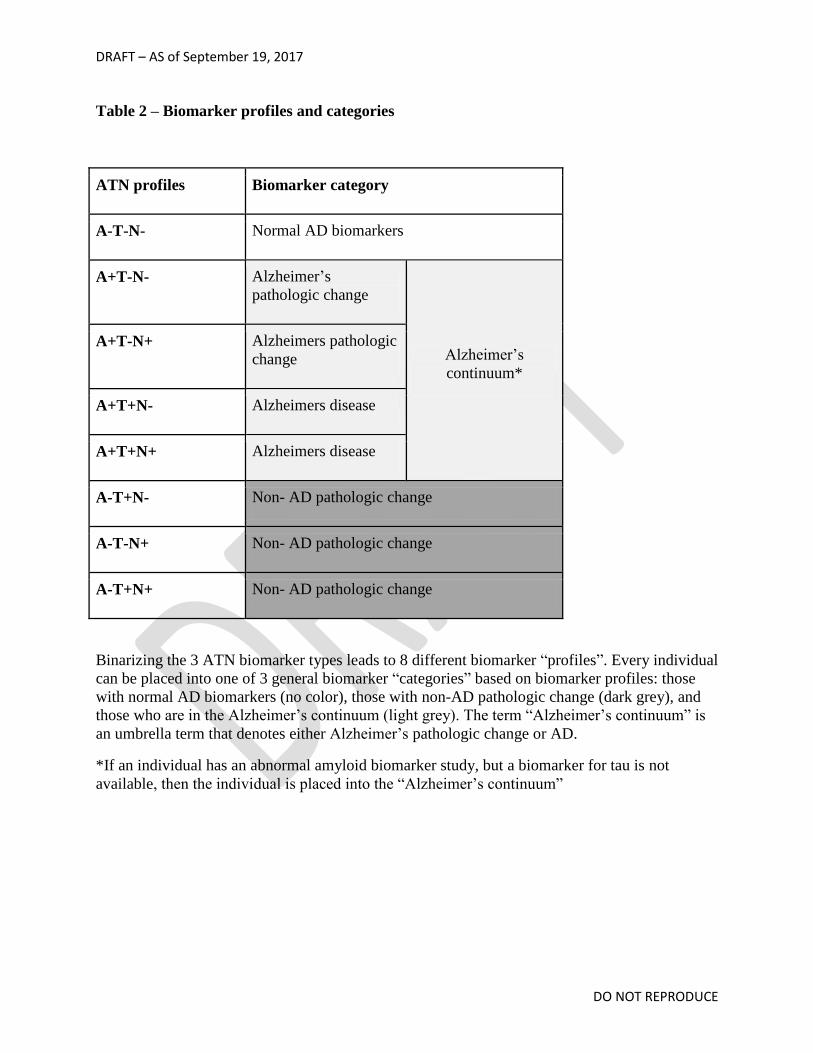
DRAFT – AS of September 19, 2017
DO NOT REPRODUCE
Table 2 – Biomarker profiles and categories
ATN profiles Biomarker category
A-T-N- Normal AD biomarkers
A+T-N- Alzheimer’s
pathologic change
Alzheimer’s
continuum*
A+T-N+ Alzheimers pathologic
change
A+T+N- Alzheimers disease
A+T+N+ Alzheimers disease
A-T+N- Non- AD pathologic change
A-T-N+ Non- AD pathologic change
A-T+N+ Non- AD pathologic change
Binarizing the 3 ATN biomarker types leads to 8 different biomarker “profiles”. Every individual
can be placed into one of 3 general biomarker “categories” based on biomarker profiles: those
with normal AD biomarkers (no color), those with non-AD pathologic change (dark grey), and
those who are in the Alzheimer’s continuum (light grey). The term “Alzheimer’s continuum” is
an umbrella term that denotes either Alzheimer’s pathologic change or AD.
*If an individual has an abnormal amyloid biomarker study, but a biomarker for tau is not
available, then the individual is placed into the “Alzheimer’s continuum”
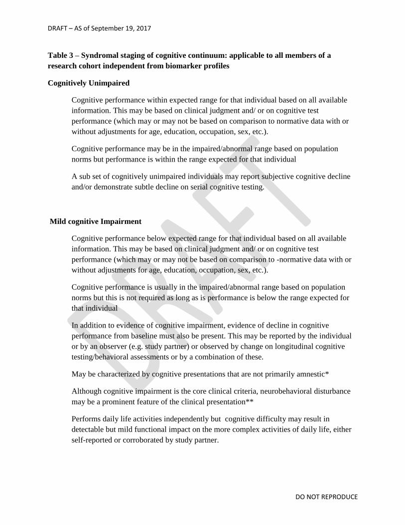
DRAFT – AS of September 19, 2017
DO NOT REPRODUCE
Table 3 – Syndromal staging of cognitive continuum: applicable to all members of a
research cohort independent from biomarker profiles
Cognitively Unimpaired
Cognitive performance within expected range for that individual based on all available
information. This may be based on clinical judgment and/ or on cognitive test
performance (which may or may not be based on comparison to normative data with or
without adjustments for age, education, occupation, sex, etc.).
Cognitive performance may be in the impaired/abnormal range based on population
norms but performance is within the range expected for that individual
A sub set of cognitively unimpaired individuals may report subjective cognitive decline
and/or demonstrate subtle decline on serial cognitive testing.
Mild cognitive Impairment
Cognitive performance below expected range for that individual based on all available
information. This may be based on clinical judgment and/ or on cognitive test
performance (which may or may not be based on comparison to -normative data with or
without adjustments for age, education, occupation, sex, etc.).
Cognitive performance is usually in the impaired/abnormal range based on population
norms but this is not required as long as is performance is below the range expected for
that individual
In addition to evidence of cognitive impairment, evidence of decline in cognitive
performance from baseline must also be present. This may be reported by the individual
or by an observer (e.g. study partner) or observed by change on longitudinal cognitive
testing/behavioral assessments or by a combination of these.
May be characterized by cognitive presentations that are not primarily amnestic*
Although cognitive impairment is the core clinical criteria, neurobehavioral disturbance
may be a prominent feature of the clinical presentation**
Performs daily life activities independently but cognitive difficulty may result in
detectable but mild functional impact on the more complex activities of daily life, either
self-reported or corroborated by study partner.

DRAFT – AS of September 19, 2017
DO NOT REPRODUCE
Dementia
Substantial progressive cognitive impairment that affects several domains and/or
neurobehavioral symptoms. May be reported by the individual or by an observer (e.g.
study partner) or observed by change on longitudinal cognitive testing
Cognitive impairment and/or neurobehavioral symptoms result in clearly evident
functional impact on daily life. No longer fully independent/requires assistance with daily
life activities. This is the primary feature differentiating dementia from MCI.
May be subdivided into mild, moderate and severe
* For MCI and dementia: Cognitive impairment may be characterized by presentations that are
not primarily amnestic
**For MCI and dementia: Although cognition is the core feature, neurobehavioral changes - e.g.
changes in mood, anxiety, or motivation – commonly co-exist and may be a prominent part of
the presentation.
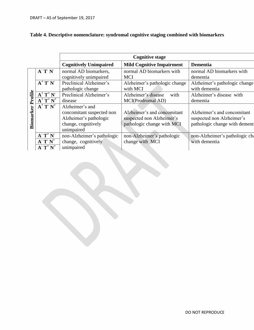
DRAFT – AS of September 19, 2017
DO NOT REPRODUCE
Table 4. Descriptive nomenclature: syndromal cognitive staging combined with biomarkers
Cognitive stage
Cognitively Unimpaired Mild Cognitive Impairment Dementia
Bio
mark
er P
rofi
le
A- T
- N
- normal AD biomarkers,
cognitively unimpaired
normal AD biomarkers with
MCI
normal AD biomarkers with
dementia
A+ T
- N
- Preclinical Alzheimer’s
pathologic change
Alzheimer’s pathologic change
with MCI
Alzheimer’s pathologic change
with dementia
A+ T
+ N
- Preclinical Alzheimer’s
disease
Alzheimer’s disease with
MCI(Prodromal AD)
Alzheimer’s disease with
dementia A+ T
+ N
+
A+ T
- N
+ Alzheimer’s and
concomitant suspected non
Alzheimer’s pathologic
change, cognitively
unimpaired
Alzheimer’s and concomitant
suspected non Alzheimer’s
pathologic change with MCI
Alzheimer’s and concomitant
suspected non Alzheimer’s
pathologic change with dementia
A- T
+ N
- non-Alzheimer’s pathologic
change, cognitively
unimpaired
non-Alzheimer’s pathologic
change with MCI
non-Alzheimer’s pathologic change
with dementia
A
- T
- N
+
A- T
+ N
+

DRAFT – AS of September 19, 2017
DO NOT REPRODUCE
Table 5. Risk of short term cognitive decline based on biomarker profile and
cognitive stage
Syndromal Cognitive Stage
Bio
mark
er P
rofi
le
Cognitively
unimpaired
MCI dementia
A- T
- N
-
normal AD biomarkers,
cognitively unimpaired
normal AD
biomarkers with
MCI
normal AD
biomarkers with
dementia
A+
T- N
-
Preclinical Alzheimer’s
pathologic change
Alzheimer’s
pathologic change
with MCI
Alzheimer’s
pathologic change
with dementia
A+
T- N
+
Alzheimer’s and
concomitant suspected
non Alzheimer’s
pathologic change,
cognitively unimpaired
Alzheimer’s and
concomitant
suspected non
Alzheimer’s
pathologic change
with MCI
Alzheimer’s and
concomitant suspected
non Alzheimer’s
pathologic change
with dementia
A+
T+
N-
Preclinical Alzheimer’s
disease
Alzheimer’s disease
with MCI
(Prodromal AD)
Alzheimer’s disease
with dementia A+
T+
N+
Non-Alzheimer’s continuum profiles are not included in table because the risk associated with
different combinations of T+N-, T+N+, T-N+ among A- individuals has not been established
rate of short term clinical progression expected to be low
rate of short term clinical progression expected to be high

DRAFT – AS of September 19, 2017
DO NOT REPRODUCE
Table 6: Numeric clinical staging - applicable only to individuals in the Alzheimer’s
pathologic continuum
Stage 1
Performance within expected range on objective cognitive tests. Cognitive test
performance may be compared to normative data of the investigators choice, with or
without adjustment (again the choice of the investigators) for age, sex, education, etc.*
Does not report recent decline in cognition or new onset of neurobehavioral symptoms of
concern
No evidence of recent cognitive decline or new neurobehavioral symptoms by report of
an observer (e.g. study partner) or by longitudinal cognitive testing if available
Stage 2
Normal performance within expected range on objective cognitive tests.
Transitional cognitive decline: decline in previous level of cognitive function which may
involve any cognitive domain(s) (i.e. not exclusively memory).
May be documented through subjective report of cognitive decline that is of
concern to the participant
Represents a change from individual baseline within past 1-3 years, and
persistent for at least 6 months
May be corroborated by informant but not required
OR may be documented by evidence of subtle decline on longitudinal cognitive
testing but not required
Or may be documented by both subjective report of decline as well as objective
evidence on longitudinal testing
Although cognition is the core feature, mild neurobehavioral changes - e.g. changes in
mood, anxiety, or motivation – may co-exist. In some individuals the primary compliant
may be neurobehavioral rather than cognitive. Neurobehavioral symptoms should have a
clearly defined recent onset which persists and cannot be explained by life events. **
No functional impact on daily life activities
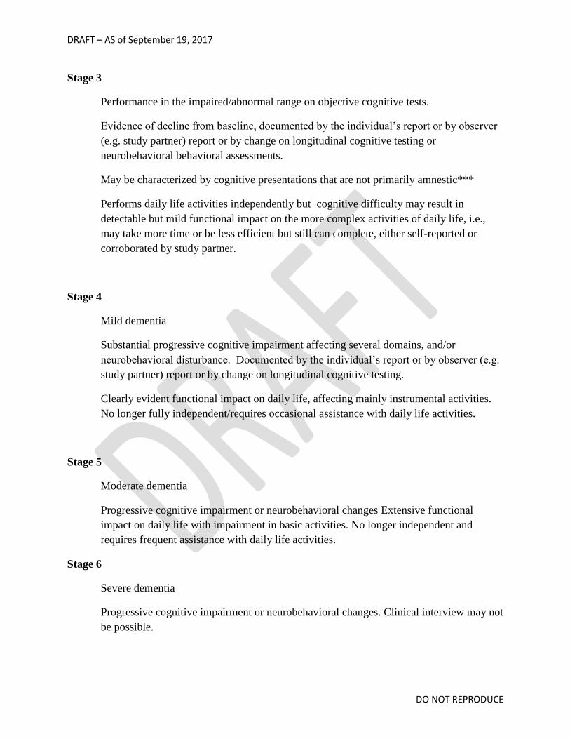
DRAFT – AS of September 19, 2017
DO NOT REPRODUCE
Stage 3
Performance in the impaired/abnormal range on objective cognitive tests.
Evidence of decline from baseline, documented by the individual’s report or by observer
(e.g. study partner) report or by change on longitudinal cognitive testing or
neurobehavioral behavioral assessments.
May be characterized by cognitive presentations that are not primarily amnestic***
Performs daily life activities independently but cognitive difficulty may result in
detectable but mild functional impact on the more complex activities of daily life, i.e.,
may take more time or be less efficient but still can complete, either self-reported or
corroborated by study partner.
Stage 4
Mild dementia
Substantial progressive cognitive impairment affecting several domains, and/or
neurobehavioral disturbance. Documented by the individual’s report or by observer (e.g.
study partner) report or by change on longitudinal cognitive testing.
Clearly evident functional impact on daily life, affecting mainly instrumental activities.
No longer fully independent/requires occasional assistance with daily life activities.
Stage 5
Moderate dementia
Progressive cognitive impairment or neurobehavioral changes Extensive functional
impact on daily life with impairment in basic activities. No longer independent and
requires frequent assistance with daily life activities.
Stage 6
Severe dementia
Progressive cognitive impairment or neurobehavioral changes. Clinical interview may not
be possible.

DRAFT – AS of September 19, 2017
DO NOT REPRODUCE
Complete dependency due to severe functional impact on daily life with impairment in
basic activities, including basic self-care.
* For stages 1-6: Cognitive test performance may be compared to normative data of the
investigators choice, with or without adjustment (choice of the investigators) for age, sex,
education, etc.
**For stages 2-6: Although cognition is the core feature, neurobehavioral changes - e.g. changes
in mood, anxiety, or motivation – may co-exist.
***For stages 3-6: Cognitive impairment may be characterized by presentations that are not
primarily amnestic

DRAFT – AS of September 19, 2017
DO NOT REPRODUCE
Fig 1. Alzheimer’s disease with dementia. 75 yo woman with amnestic multi domain dementia,
abnormal amyloid PET (a), tau PET (b,c) and atrophy on MRI (d). Biomarker profile A+T+N+.

DRAFT – AS of September 19, 2017
DO NOT REPRODUCE
Fig 2. Preclinical Alzheimer’s pathologic change. Cognitively unimpaired 67 yo man.
Abnormal amyloid PET (top row), no uptake on tau PET (middle row), no atrophy on MR
(bottom row). Biomarker profile A+T-N- .
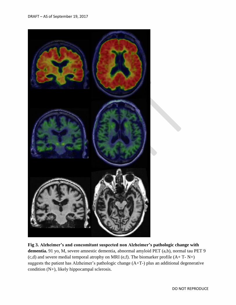
DRAFT – AS of September 19, 2017
DO NOT REPRODUCE
Fig 3. Alzheimer’s and concomitant suspected non Alzheimer’s pathologic change with
dementia. 91 yo, M, severe amnestic dementia, abnormal amyloid PET (a,b), normal tau PET 9
(c,d) and severe medial temporal atrophy on MRI (e,f). The biomarker profile (A+ T- N+)
suggests the patient has Alzheimer’s pathologic change (A+T-) plus an additional degenerative
condition (N+), likely hippocampal sclerosis.