Embed Size (px)
Citation preview

Neuron, Vol. 47, 267–280, July 21, 2005, Copyright ©2005 by Elsevier Inc. DOI 10.1016/j.neuron.2005.06.011
The Coordinated Mapping of Visual Spaceand Response Features in Visual Cortex
Hongbo Yu,1,3 Brandon J. Farley,1,3 Dezhe Z. Jin,2
and Mriganka Sur1,*1Department of Brain and Cognitive SciencesPicower Institute for Learning and MemoryMassachusetts Institute of TechnologyCambridge, Massachusetts 021392Department of PhysicsThe Pennsylvania State UniversityUniversity Park, Pennsylvania 16802
Summary
Whether general principles can explain the layouts ofcortical maps remains unresolved. In primary visualcortex of ferret, the relationships between the mapsof visual space and response features are predictedby a “dimension-reduction” model. The representa-tion of visual space is anisotropic, with the elevationand azimuth axes having different magnification. Thisanisotropy is reflected in the orientation, ocular domi-nance, and spatial frequency domains, which areelongated such that their directions of rapid change,or high-gradient axes, are orthogonal to the high-gra-dient axis of the visual map. The feature maps arealso strongly interdependent—their high-gradient re-gions avoid one another and intersect orthogonallywhere essential, so that overlap is minimized. Our re-sults demonstrate a clear influence of the visual mapon each feature map. In turn, the local representationof visual space is smooth, as predicted when manyfeatures are mapped within a cortical area.
Introduction
One of the keys to understanding cortical function isdefining the rules that govern the organization of neu-rons according to their response properties. As somecortical areas are traversed, neuronal response proper-ties change in a regular manner, forming systematicmaps with highly stereotyped structures. Additionally,more than one response property can be mappedsimultaneously within an area, as has been shown invisual, somatosensory, and auditory cortex (Hubel andWiesel, 1963; Sur et al., 1981; Linden and Schreiner,2003; Friedman et al., 2004). What determines theforms that these maps take? One theoretical proposalis that the structures reflect constraints imposed by “di-mension-reduction,” or a need to smoothly map several(more than two) response properties onto a two-dimen-sional cortical surface (Kohonen, 1982a; Durbin andMitchison, 1990; Obermayer et al., 1990; Swindale,1991). An explicit goal of dimension-reduction modelsis to minimize the connection length between neuronsthat share similar response properties, an approachalso used in “elastic net” models (Goodhill and Will-
*Correspondence: [email protected]
3 These authors contributed equally to this work.shaw, 1990) and “wire-length minimization” algorithms(Koulakov and Chklovskii, 2001) for explaining thestructure of cortical columns.
Primary visual cortex (V1) is a model area for examin-ing this issue since neurons in the region are mappedaccording to several well-understood response proper-ties (i.e., features), including receptive field position,ocular dominance, orientation, and spatial frequency.The columnar organization of the area implies that themultiple feature maps are superimposed over a singletwo-dimensional cortical sheet. To accommodate therepresentation of all the features, dimension-reductionmodels predict specific spatial relationships betweenfeature maps (Durbin and Mitchison, 1990; Obermayeret al., 1990). However, recent studies have questionedthe relevance of such principles to cortical maps, inparticular the map of visual space (Das and Gilbert,1997; Bosking et al., 2002; Buzas et al., 2003).
A major prediction of dimension-reduction models isthat, at locations where the value of a given featurechanges rapidly across cortex, that of other indepen-dent features should change slowly; that is, there is anegative correlation between the gradient magnitudesof different maps (Obermayer et al., 1992). A corollaryto this principle is that, at locations where the gradientmagnitudes of two features are necessarily high, thecontours of the two maps, and hence their gradient di-rections, are orthogonal. Experimental studies havedemonstrated relationships for some pairs of functionalmaps consistent with these predictions. For example,locations where the preferred orientation map gradientis high, i.e., orientation pinwheels, are situated towardthe center of ocular dominance and spatial frequencycolumns, where the latter maps have low gradients. Ad-ditionally, ocular dominance and orientation contoursintersect near-orthogonally at the edges of ocular dom-inance columns, where the ocular dominance gradientis high (Blasdel and Salama, 1986; Bartfeld and Grin-vald, 1992; Obermayer and Blasdel, 1993; Hubener etal., 1997).
In contrast, results from experiments that have exam-ined the retinotopic and orientation maps appear in-consistent with the model predictions: whereas theoriginal models (Durbin and Mitchison, 1990; Obermayeret al., 1990) predicted a negative correlation between thegradients of these two maps, such that receptive fieldlocations should change slowly in locations where ori-entation preferences change rapidly, experimental re-sults show either a positive correlation (Das and Gil-bert, 1997) or no correlation (Bosking et al., 2002;Buzas et al., 2003). But the original models includedonly retinotopy and orientation as mapped features;more recent studies have demonstrated that the pre-dicted relationships between the retinotopic and othermaps are dependent on the number of features that areincluded in the model simulations (Swindale, 2004). Itremains to be examined how experimentally measuredrelationships compare to model predictions, when themodel simulations include all of the features known tobe mapped within the cortical region of question.

Neuron268
We reasoned that an examination of the relationships tvbetween the retinotopic and other maps in V1 should
address three related questions: (1) does the overall (cstructure of the retinotopic map influence the mapping
of other features, in a manner consistent with dimen- 3asion-reduction principles; (2) are other feature maps re-
lated to each other by these principles, and (3) is the f(mapping of other features in turn reflected in the local
structure of the retinotopic map? Since the relation- faships between the retinotopic and other maps have
proven elusive, we examined a case where the dimen- masion-reduction model makes a clear prediction: when
the retinotopic map magnification differs for the two maaxes of visual space, the model predicts clear conse-
quences for the remaining maps. The examination of agmaps in V1 of ferrets provided a unique opportunity to
test this prediction. We found that, indeed, the retino- gtopic map powerfully influences the layout of othermaps, as the model predicts, and that the other maps e
Emaintain specific local relationships with one another.Finally, the retinotopic map is predicted to be locally i
csmooth in the case where three other variables aremapped within a cortical area. Our results demonstrate i
aa strong interdependence between each visual cortexfunctional map, including the retinotopic map. n
lhResultsstThe Dimension-Reduction ModellTo model how several features could be mapped
across the two-dimensional cortical surface, we usedvthe self-organizing feature map algorithm (Kohonen,r1982b; Obermayer et al., 1992). In this algorithm, eachupossible feature combination within a multidimensionalnresponse feature space (representing x and y axis reti-anotopic location, orientation, ocular dominance, and1spatial frequency) is represented at a location in cortex.tIn other words, the cortex achieves complete “coverage”wof the feature space. In addition, the algorithm has aomechanism to promote “continuity” in the cortical rep-
resentations; i.e., nearby locations on the cortical sur-face are forced to represent similar features so that or- Pderly feature maps are formed. The constraints of Ocoverage and continuity act in opposition and are com- amonly employed in some form in many computational Wmodels of visual cortex (Erwin et al., 1995; Swindale, o1996). We asked what form the feature mapping was mpredicted to take given these constraints and, in partic- mular, how a global anisotropy in the retinotopic map was hpredicted to influence the layouts of the other maps. oWe reasoned that a match between the simulation pre- wdictions and in vivo experimental observations would tsuggest that the mapping of visual space and other efeatures occurs in conformity with a dimension-reduc- wtion strategy. t
aFPredicted Influence of the Retinotopic Map
on the Structures of Other Maps wmTo determine the predicted influence of retinotopy on
the mapping of other features, we compared the resultsgof simulations in which visual space was represented
either anisotropically or isotropically. To model an an- w1isotropy, we used a retinotopic map whose magnifica-
ion was four times greater along the elevation axis ofisual space representation than along the azimuth axisFigure 1A). This degree of anisotropy was chosen be-ause it resembles that found in ferret V1 (see Figure). We observed a strong influence of the visual mapnisotropy on the layouts of the remaining mapsormed in the simulation: domains in the orientationFigure 1A), ocular dominance (Figure 1B), and spatialrequency (see Figure S1A in the Supplemental Datavailable online) maps were highly elongated. Further-ore, the domains from the latter maps were elongated
long a common axis; this axis was parallel to the azi-uth (low-magnification) axis of the retinotopic map
nd orthogonal to the elevation (high-magnification)xis. It follows from these observations that the high-radient axis of retinotopy is orthogonal to the high-radient axis of the other maps.To quantify this observation, we calculated the gradi-
nt vector at every pixel of each functional map (seexperimental Procedures). The direction of this vector
ndicates the axis along which the mapped variablehanges maximally. We found a bias toward orthogonal
ntersection angles between the visual map gradientnd the gradients of either the orientation, ocular domi-ance, or spatial frequency maps (Figure 1C, dotted
ines). Further, in those locations where the latter mapsad a high-gradient magnitude, there was an eventronger tendency for their gradient direction to be or-hogonal to that of the visual map (Figure 1C, solidines).
In contrast to the above results, when an isotropicisual map was used in the simulation, domains in theesulting orientation (Figure 1D), ocular dominance (Fig-re 1E), and spatial frequency (Figure S1E) maps wereot elongated. As a result, these domains did not alignlong a particular axis of the retinotopic map (FigureF). Taking these results together, our model predictshat a global anisotropy in visual space representationill strongly and specifically influence the layouts ofther maps.
redicted Gradient Relationships betweenrientation, Ocular Dominance,nd Spatial Frequency Mapse next examined the relationships between the model’s
rientation, ocular dominance, and spatial frequencyaps. A fundamental prediction of dimension-reductionodels, which has previously been described, is that the
ighest gradient regions of multiple maps should avoidne another. We asked whether this could still occurith a strong anisotropy in the retinotopic map. Orien-
ation, ocular dominance, and spatial frequency gradi-nt maps from the same region of the model cortexere obtained. We found that the anisotropy caused
he highest-gradient regions of these three maps tolign along a common axis of the cortex (Figure 2A; cf.igure 2F). Nevertheless, the highest-gradient regionsere precisely interleaved and appeared to occupyostly nonoverlapping regions of cortex (Figure 2A).To quantify this relationship, all pixels within each
radient map were assigned a value between 1 and 10,here 1 indicated that the gradient was in the lowest0th percentile of all gradient values within that map,

Mapping Multiple Features in Visual Cortex269
Figure 1. Effect of Visual Space Anisotropyon the Layouts of Other Feature Maps, asPredicted by a Dimension-Reduction Model
The retinotopic map was either anisotropic(A–C) or isotropic (D–F); it is illustrated in (A)and (D) by iso-elevation (horizontal in figure)and iso-azimuth (vertical in figure) contours;all contour intervals are identical. (A and D)The orientation map, denoted by color cod-ing each pixel according to its preferred ori-entation. (B and E) The ocular dominancemap. (C and F) Percent of pixels that havean intersection angle, within each 10° rangewhose center is indicated by the x axis tickmark, between the retinotopic gradient andthe gradient of the orientation (black lines),ocular dominance (blue lines), or spatial fre-quency (red lines) map. Dotted lines indicatethe calculation over all pixels of the cortex.Solid lines indicate the calculation over onlythose pixels whose orientation, ocular domi-nance, or spatial frequency gradients werewithin the highest 30th percentile.
and 10 the highest 10th percentile. The values from allthree maps were averaged together at each pixel in theregion, and the standard deviation of this average overthe entire cortex was measured. We found that thestandard deviation was smaller if the actual gradientmaps were used (Figure 2B, red dotted bar) in the cal-culation, compared to when the pixels in each gradientmap were randomly shuffled (Figure 2B, blue histo-gram). Thus, there is a tendency for the average gradi-ent magnitude of all three maps to remain constantacross the cortical surface. This demonstrates that thehighest-gradient regions of the three maps mutuallyavoid one another.
To analyze directly whether the high-gradient regionsof two maps avoid one another, we compared their gra-dients at corresponding pixels. Pixels were binned intoten groups according to their ocular dominance gradi-ent percentile, and the average orientation gradientover all pixels within each group was determined andplotted (Figure 2C). A negative correlation can be seen,suggesting that the highest gradient regions of thesetwo maps avoid one another. The same relationship ex-ists between the orientation and spatial frequencymaps (Figure S1B).
These gradient correlations among maps of orienta-tion, ocular dominance, and spatial frequency also heldwhen the initial retinotopic map was isotropic (Figures2F–2H and Figure S1F). Thus, a fundamental predictionof dimension-reduction models is that high-gradient re-
gions of multiple maps avoid one another, and it holdswhether the retinotopic map is anisotropic or isotropic.
Predicted Contour Relationships betweenOrientation, Ocular Dominance, and SpatialFrequency MapsAbove we described a fundamental prediction of di-mension-reduction models, that high-gradient regionsof multiple maps avoid one another. A second majorprediction of this class of models, which is common tomany cortical mapping algorithms, is that contours ofdifferent maps should cross at perpendicular angles(Erwin et al., 1995; Swindale, 1996). However, the con-straint imposed by the anisotropy in the visual mapchallenges this prediction. With an anisotropic visualmap, the orientation, ocular dominance, and spatial fre-quency domains are elongated along a parallel axis ofcortex (see Figure 2A). This would seem to preclude thecontours of these maps from crossing at perpendicu-lar angles.
To examine this issue, we plotted the distribution ofthe intersection angles, over all map pixels, betweenthe orientation and ocular dominance gradient vectors.When the visual map was isotropic, the intersection an-gles were orthogonal on average, as expected fromprevious studies (Figure 2J, red line). In contrast, whenan anisotropic visual map was used, the intersectionangles had no bias toward orthogonality (Figure 2E, red

Neuron270
Figure 2. Predicted Effect of Visual Space Map Anisotropy on the Relationships between Other Feature Maps
The retinotopic map was either anisotropic (A–E) or isotropic (F–J). (A and F) High-gradient regions (pixels with gradients within the top 30thpercentile) of orientation (blue), ocular dominance (green), spatial frequency (red), or two or more maps (white) are indicated. (B and G)Standard deviation across cortex of the average gradient magnitude of the orientation, ocular dominance, and spatial frequency maps (reddotted line). Histogram of this standard deviation calculation after pixels within each map were randomly shuffled, in 10,000 cases (bluehistogram). (C and H) Pixels are grouped into ten bins according to their ocular dominance gradient percentile, and the mean orientationgradient for each group is indicated. (D and I) Orientation (color) contours superimposed on ocular dominance (black) contours. Gray pixelsindicate locations where the gradient of both the orientation and ocular dominance maps are within the highest 30th percentile. (E and J)Percent of pixels that have an intersection angle, within each 10° range whose center is indicated, between the orientation and oculardominance gradients. Red line shows the calculation over all pixels. Blue line indicates the calculation for those pixels where the gradientsof both maps are within the highest 30th percentile.
line). Thus, an anisotropy in the visual map precludes lran overall orthogonality between the remaining maps.
We next show, however, that the remaining maps omhave specific local regions where orthogonality is main-
tained. In the anisotropic case, a clear bias toward or- mtthogonal intersection angles between the orientation
and ocular dominance gradient vectors emerged spe- tgcifically in those locations where the maps of these fea-
tures had overlapping high gradients (Figure 2D, graytregions; Figure 2E, blue line). For the isotropic case,
the bias toward intersection angles between orientation ecand ocular dominance gradients similarly became
stronger in those locations where the two maps have ethigh gradients (Figures 2I and 2J). The relationships be-
tween the contours of orientation and spatial frequency omwere very similar to those between orientation and ocu-
ar dominance (Figures S1C, S1D, S1G, and S1H). Theesults suggest that the overall orthogonality betweenrientation and ocular dominance (or spatial frequency)aps can vary, based on the nature of the retinotopicap. But the model predicts that, regardless of the na-
ure of the retinotopic map, orthogonality will consis-ently occur between the remaining maps in their high-radient overlap regions.Together, the relationships we have described reflect
he model’s strategy for smoothly accommodating sev-ral features simultaneously on the two-dimensionalortical surface. In particular, they predict what influ-nce the retinotopic map should have on other maps ifhe mapping of visual space is interdependent with thatf other features. Whether the visual space and otheraps in visual cortex demonstrate these relationships

Mapping Multiple Features in Visual Cortex271
requires experimental examination. We next comparedthese modeling predictions to the results we obtainedexperimentally in ferret V1.
Experimental Results from Ferret Visual Cortex:An Anisotropy in the V1 Retinotopic MapWe determined the layout of the retinotopic map in fer-ret V1 (Figure 3, inset) using optical imaging of intrinsicsignals (see Experimental Procedures). The presenta-
Figure 3. Optical Imaging Reveals that the Retinotopic Map in Fer-ret V1 Is Anisotropic
(Inset) Dorsal view of right hemisphere of ferret brain, indicatingthe orientation of the functional maps for all figures; outlined areaindicates the region of cortex imaged. (A–B) “Single-condition” reti-notopic maps. (A) Cortical activity pattern (activated pixels aredarker) elicited by a flashing vertical bar. The stimulus position dif-fered by 3° in horizontal space between successive panels, as indi-cated. (B) Activity elicited by a horizontal bar. The stimulus positiondiffered by 3° in vertical space between successive panels. (C) Azi-muth position preference map. Each pixel is color coded accordingto its preferred relative azimuth position in visual space. 9° of hori-zontal space was represented over 0.64 mm of cortex. (D) Elevationposition preference map. 9° of vertical space was represented over2.8 mm. Scale bar in (C) represents 1 mm and also applies to (A),(B), and (D).
tion of a vertical bar activated a narrow, mediolaterallyelongated strip of cortex in V1, which represents an iso-azimuth contour (Figure 3A). As the vertical bar shiftedfrom central to more peripheral regions of the visualfield, the region of activation in V1 shifted posteriorly. Ahorizontal bar, on the other hand, activated a strip ofcortex that was elongated along the anteroposterioraxis of the cortex (Figure 3B). This strip represents aniso-elevation contour. Downward movement of the hori-zontal bar across the visual field caused the activatedregion to shift from lateral to medial across cortex (Fig-ure 3B).
With our retinotopy stimulation paradigm, the stimu-lus passes over the receptive field of each cortical loca-tion periodically and causes a periodic activation pat-tern across time (data not shown) for each pixel. Apixel’s relative receptive field location can be deter-mined by its relative phase of activation within the stim-ulus cycle (Engel et al., 1994; Sereno et al., 1995; Kalat-sky and Stryker, 2003; Mrsic-Flogel et al., 2003). Wecalculated the phase of the Fourier transform of eachpixel’s response time course, in response to stimulationby a horizontally or vertically drifting bar, and con-structed maps for relative horizontal (Figure 3C) andvertical (Figure 3D) visual space, respectively.
While the general layout we show for the visual spacemap is in agreement with previous reports (Law et al.,1988; White et al., 1999), we additionally find a pro-nounced anisotropy in the representation of visualspace (Figure 3). A much greater amount of cortex isdevoted to the representation of a given amount of vi-sual field elevation than for the equivalent amount ofazimuth. In general, for the region of ferret V1 accessi-ble to optical imaging, the magnification factor for theelevation axis of visual space was approximately threeto five times greater than that for azimuth (Table S1).
The magnification anisotropy was confirmed by elec-trophysiological recordings. Receptive field positionsand sizes were determined using the reverse correla-tion method of receptive field mapping. A representa-tive case is shown in Figure 4. On- and off-subfieldswere summed to calculate the aggregate receptive fieldat each recording site (Figures 4A and 4B). The distribu-tion of the receptive fields recorded at multiple sites(Figure 4A) revealed a wide range of azimuth positionpreferences (within a small anteroposterior extent ofcortex) and a narrower range of elevations (Figure 4C).The composite retinotopic map confirmed that themagnification for the elevation axis of visual space wasmuch greater than that for the azimuth axis (Figures 4Dand 4E; note similarity to optical imaging-derived mapsin Figures 3C and 3D).
Influence of the Global Visual Map Anisotropyon Feature MapsGiven the pronounced anisotropy of the retinotopicmap in ferret visual cortex, we examined whether itwould impact the layout of other functional maps, asthe dimension-reduction model predicted. We first ex-amined the map of orientation in V1, which was ob-tained by comparing the cortical activation pattern inresponse to grating stimuli presented at four different

Neuron272
Figure 4. Multiunit Electrophysiological Recordings Confirm the Anisotropy of the Retinotopic Map in Ferret V1
(A) Image of cortical vasculature showing 14 recording sites. (B) Example multiunit receptive fields recorded at the two sites indicated in (A).The strength of the response to stimuli presented in each location of the stimulus grid (dotted box in [C]) is indicated (as percent maximumresponse). (C) Receptive field locations for all recording sites shown in (A). (D and E) Azimuth (D) and elevation (E) position preference mapfor the region of cortex shown in (A) (see Experimental Procedures). The dotted line in (D) indicates the approximate location of the V1/V2border. Scale bar in (A) represents 1 mm and also applies to (D)–(E); bar in (B) is 3°.
orientations. Though this map has been previously de- riscribed in ferret (Rao et al., 1997; White et al., 2001), a
salient feature that we noticed was an anisotropy of the sEorientation domains. They were often elongated along
the anteroposterior dimension of cortex (Figures 5A– fd5F). This was also true for the ocular dominance do-
mains. The ocular dominance map in the binocular por- rStion of V1, located at the most posterolateral edge of
cortex (Law et al., 1988; Redies et al., 1990; White et o(al., 1999), contains ocular dominance columns that in-
terleave similarly to those in V1 of the cat and macaque. tiIn this part of V1, ocular dominance domains were an-
isotropic: they were elongated along the anteroposte- a
n anisotropy in the domains of the orientation, ocularFigure 5. Orientation, Ocular Dominance,and Spatial Frequency Domains Are Aniso-tropic
(A–D) Cortical activity patterns elicited by thepresentation of grating stimuli of the orienta-tion indicated. (E) Orientation preference mapwith retinotopic contours superimposed. Verti-cal lines represent iso-azimuth contours, andhorizontal lines are iso-elevation contours. (F)Detailed view of the region from (E) that isoutlined. All contour intervals are 1.5°. (G)Ocular dominance preference map. (H) Spa-tial frequency preference map. Scale barsare 1 mm. Scale bar in (A) applies to (A)–(E),(G), and (H). Orientation, ocular dominance,and spatial frequency maps in this figurewere obtained from different animals.
ior dimension of cortex (Figure 5G), similarly to theso-orientation domains. Finally, we examined whetherpatial frequency is mapped in ferret visual cortex (seexperimental Procedures), and in what manner. We
ound that alternating high and low spatial frequencyomains exist in V1 that are elongated along the ante-
oposterior dimension of cortex (Figure 5H and Figure2A). The mapping of spatial frequency was verified inne animal by multiunit electrophysiological recordings
Figures S2B–S2D). Taken together, these results matchhose predicted by our model, suggesting that an an-sotropy of the visual map is indeed accompanied by
Mapping Multiple Features in Visual Cortex273
dominance, and spatial frequency maps (cf. Figures 1Aand 1B and Figure S1A).
We next directly compared the layout of the retino-topic map with that of other maps. By examining retino-topic contours superimposed on the orientation map(Figures 5E, 5F, and 6A), it can be seen that the high-gradient axes of these two maps are orthogonal. Toquantify this relationship, we calculated the pixel-by-pixel intersection angles between the retinotopic gradi-ent and the gradients of the other maps (see Experi-mental Procedures). Across the cortex, the percentageof pixels having a 60°–90° intersection angle would be33.3% for a random distribution into the three bins of0°–30°, 30°–60°, and 60°–90°. For the retinotopic andorientation maps, the number was 44.1% ± 4.5%, sig-nificantly higher than the chance level (p < 0.005, t test;n = 8 animals). For retinotopy and ocular dominance,the number was 48.0% ± 2.8% of pixels (p < 0.005,t test, n = 4 animals), and for retinotopy and spatialfrequency, the number was 51.2% ± 4.9% of pixels(p < 0.005, t test, n = 4 animals). Thus there was a pre-ponderance of near-orthogonal intersection angles be-tween the retinotopic gradient and the gradients of theother maps, which reflects the global relationship be-tween the anisotropy of the retinotopic map and theelongation of feature map domains.
Quantifying this in another way, we grouped pixels
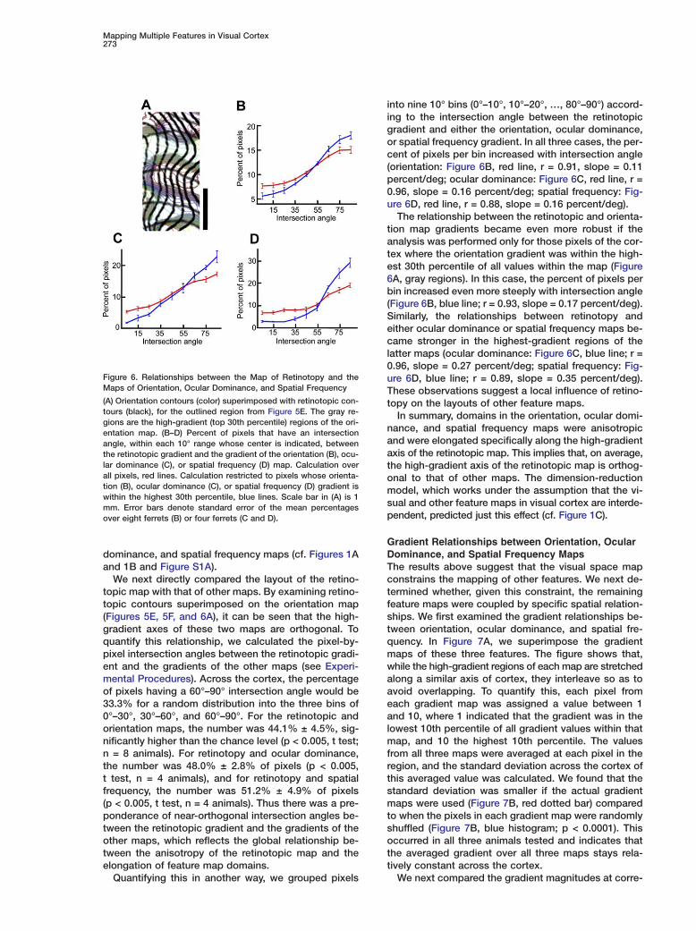
Figure 6. Relationships between the Map of Retinotopy and theMaps of Orientation, Ocular Dominance, and Spatial Frequency
(A) Orientation contours (color) superimposed with retinotopic con-tours (black), for the outlined region from Figure 5E. The gray re-gions are the high-gradient (top 30th percentile) regions of the ori-entation map. (B–D) Percent of pixels that have an intersectionangle, within each 10° range whose center is indicated, betweenthe retinotopic gradient and the gradient of the orientation (B), ocu-lar dominance (C), or spatial frequency (D) map. Calculation overall pixels, red lines. Calculation restricted to pixels whose orienta-tion (B), ocular dominance (C), or spatial frequency (D) gradient iswithin the highest 30th percentile, blue lines. Scale bar in (A) is 1mm. Error bars denote standard error of the mean percentagesover eight ferrets (B) or four ferrets (C and D).
into nine 10° bins (0°–10°, 10°–20°, …, 80°–90°) accord-ing to the intersection angle between the retinotopicgradient and either the orientation, ocular dominance,or spatial frequency gradient. In all three cases, the per-cent of pixels per bin increased with intersection angle(orientation: Figure 6B, red line, r = 0.91, slope = 0.11percent/deg; ocular dominance: Figure 6C, red line, r =0.96, slope = 0.16 percent/deg; spatial frequency: Fig-ure 6D, red line, r = 0.88, slope = 0.16 percent/deg).
The relationship between the retinotopic and orienta-tion map gradients became even more robust if theanalysis was performed only for those pixels of the cor-tex where the orientation gradient was within the high-est 30th percentile of all values within the map (Figure6A, gray regions). In this case, the percent of pixels perbin increased even more steeply with intersection angle(Figure 6B, blue line; r = 0.93, slope = 0.17 percent/deg).Similarly, the relationships between retinotopy andeither ocular dominance or spatial frequency maps be-came stronger in the highest-gradient regions of thelatter maps (ocular dominance: Figure 6C, blue line; r =0.96, slope = 0.27 percent/deg; spatial frequency: Fig-ure 6D, blue line; r = 0.89, slope = 0.35 percent/deg).These observations suggest a local influence of retino-topy on the layouts of other feature maps.
In summary, domains in the orientation, ocular domi-nance, and spatial frequency maps were anisotropicand were elongated specifically along the high-gradientaxis of the retinotopic map. This implies that, on average,the high-gradient axis of the retinotopic map is orthog-onal to that of other maps. The dimension-reductionmodel, which works under the assumption that the vi-sual and other feature maps in visual cortex are interde-pendent, predicted just this effect (cf. Figure 1C).
Gradient Relationships between Orientation, OcularDominance, and Spatial Frequency MapsThe results above suggest that the visual space mapconstrains the mapping of other features. We next de-termined whether, given this constraint, the remainingfeature maps were coupled by specific spatial relation-ships. We first examined the gradient relationships be-tween orientation, ocular dominance, and spatial fre-quency. In Figure 7A, we superimpose the gradientmaps of these three features. The figure shows that,while the high-gradient regions of each map are stretchedalong a similar axis of cortex, they interleave so as toavoid overlapping. To quantify this, each pixel fromeach gradient map was assigned a value between 1and 10, where 1 indicated that the gradient was in thelowest 10th percentile of all gradient values within thatmap, and 10 the highest 10th percentile. The valuesfrom all three maps were averaged at each pixel in theregion, and the standard deviation across the cortex ofthis averaged value was calculated. We found that thestandard deviation was smaller if the actual gradientmaps were used (Figure 7B, red dotted bar) comparedto when the pixels in each gradient map were randomlyshuffled (Figure 7B, blue histogram; p < 0.0001). Thisoccurred in all three animals tested and indicates thatthe averaged gradient over all three maps stays rela-tively constant across the cortex.
We next compared the gradient magnitudes at corre-

Neuron274
Figure 7. Relationships between the Orientation, Ocular Dominance, and Spatial Frequency Maps
(A) High-gradient regions (top 30th percentile) of orientation (blue), ocular dominance (green), spatial frequency (red), or two or more maps(white). (B) Standard deviation across cortex of the average gradient magnitude of the orientation, ocular dominance, and spatial frequencymaps (red dotted line). Histogram of standard deviations after pixels within each map were randomly shuffled, in 150,000 cases (blue histo-gram). (C and F) Pixels are grouped into ten bins according to their ocular dominance (C) or spatial frequency (F) gradient percentile, and themean orientation gradient for each group is indicated. (D and G) Colored lines representing orientation contours are superimposed on blacklines representing ocular dominance (D) or spatial frequency (G) contours. Gray regions indicate locations where high-gradient regions (top30th percentile) of the orientation and ocular dominance (D) or spatial frequency (G) maps coincided. (E and H) Percent of pixels that havean intersection angle, within each 10° range whose center is indicated, between the orientation gradient and the ocular dominance (E) orspatial frequency (H) gradients. Average over all pixels, red line. Calculation for high orientation and ocular dominance (E) or spatial frequency(H) gradient overlap regions, blue line. Scale bar in (A) is 1 mm, bars in (D) and (G) are 0.5 mm. Error bars (C, E, F, and H) denote standarderror of the mean values over five ferrets.
sponding pixels in orientation and ocular dominance pmmaps. We found that the average orientation gradient
calculated over all pixels whose ocular dominance gra- twdient was within the highest 20th percentile was signifi-
cantly lower than that calculated for pixels whose ocu- talar dominance gradient was within the lowest 20th
ercentile (average difference of 1.9 ± 0.6 deg/pixel;eans of 6.6 and 8.5 deg/pixel, respectively; paired t
est, p < 0.001, n = 5 animals). In addition, all the pixelsithin a region were binned into ten groups according
o their ocular dominance gradient percentile, and theverage orientation gradient within each group was de-

Mapping Multiple Features in Visual Cortex275
termined. We found a strong negative correlation be-tween the mean orientation gradient and the oculardominance gradient percentile (r = −0.77, slope = −0.03deg/pixel/percentile; Figure 7C).
The gradient relationships also held between orienta-tion and spatial frequency maps. The average orienta-tion gradient calculated for pixels whose spatial fre-quency gradient was within the highest 20th percentilewas significantly lower than that calculated for pixelswhose spatial frequency gradient was within the lowest20th percentile (average difference of 2.1 ± 0.8 deg/pixel; means of 6.3 and 8.4 deg/pixel, respectively;paired t test, p < 0.001, n = 5 animals). There was astrong negative correlation between the mean orienta-tion gradient and the spatial frequency gradient per-centile, which held throughout the cortex (r = −0.71,slope = −0.02 deg/pixel/percentile; Figure 7F).
The results demonstrate that, in ferret V1, where theretinotopic map imposes a strong constraint on the re-maining maps, the latter maintain specific relationshipswith one another. Their highest-gradient regions avoidone another, so that their combined gradient remainsconstant across the cortical surface. These relationshipsclosely match the predictions of our dimension-reductionmodel (cf. Figures 2B and 2C and Figure S1B).
Contour Relationships between Orientation, OcularDominance, and Spatial Frequency MapsWe next examined whether the contours of orientation,ocular dominance, and spatial frequency maps tend tointersect at near-perpendicular angles. Previous studieshave shown that this occurs in cat and monkey V1. Butit may not occur in ferret V1 since (as noted above) thedomains of orientation, ocular dominance, and spatialfrequency maps are elongated along a parallel (ratherthan perpendicular) axis of cortex, reflecting the visualmap anisotropy (see Figure 7A).
The contour lines of a superimposed orientation andocular dominance map are illustrated in Figure 7D.Across the cortex, we found no significant tendency forthe gradient vectors of the two maps to intersect atnear-perpendicular angles. Quantification revealed that36.2% ± 1.8% of pixels had gradients that intersectedat an angle between 60° and 90°, which is not signifi-cantly different from the chance percentage of 33.3%(p > 0.1, t test, n = 5 animals). By grouping the pixelsinto nine 10° bins according to their intersection angles,the percent of pixels per bin did not increase greatlywith intersection angle (Figure 7E, red line; r = 0.76,slope = 0.037 percent/deg). However, for the pixels ofthe cortex where both the orientation and ocular domi-nance gradients were within the highest 30th percen-tile, a clear tendency for orthogonal intersection anglesbetween gradient vectors of the two maps emerged(Figure 7D, gray region; Figure 7E, blue line; r = 0.89,slope = 0.20 percent/deg). Considering this region,49.3% ± 5.0% of pixels had gradient vector intersectionangles within 60°–90° (p < 0.01, t test, n = 5 animals).
The relationships between the orientation and spatialfrequency contours (Figure 7G) were similar. Across thecortex, there was only a weak tendency for orthogonalintersection angles between the gradients of the twomaps, with 34.3% ± 2.8% of pixels having gradient in-
tersection angles within 60°–90° (p > 0.1, t test, n =5 animals). By grouping the pixels into nine 10° binsaccording to their intersection angles, the percent ofpixels per bin did not change greatly with intersectionangle (Figure 7H, red line; r = 0.28, slope = 0.011 per-cent/deg). In contrast, for the pixels of the cortex whereboth the orientation and spatial frequency gradientswere within the highest 30th percentile, there was aclear tendency for orthogonal intersection angles be-tween the gradient vectors of the two maps (Figure 7G,gray regions; Figure 7H, blue line, r = 0.77, slope = 0.16percent/deg). In that case, 47.1% ± 1.4% of gradientvectors had intersection angles within 60°–90° (p <0.005, t test, n = 5 animals).
The results demonstrate, as our model predicted,that the visual map anisotropy decreases the overalltendency for perpendicular intersections between gra-dients of orientation and ocular dominance (and spatialfrequency) maps. But the maps maintain strong orthog-onal relationships in those locations where they haveoverlapping high gradients (cf. Figure 2 and Figure S1).
Influence of Feature Maps on Local Retinotopy:Model and ExperimentWe showed above that the structure of the retinotopicmap is reflected in the layouts of other feature maps.We lastly wanted to determine whether, in turn, the lo-cal structure of the retinotopic map is influenced by themapping of these other features. Previous simulationstudies predicted that the orientation map would havea strong influence on the retinotopic map (Durbin andMitchison, 1990; Obermayer et al., 1990), but thosesimulations included only orientation and retinotopy asmapped features. We compared the predictions of sim-ulations using only orientation and retinotopy to thosethat included additional features known to be mappedin ferret V1 and examined how the predictions in eachcase related to experimental relationships.
In the retinotopy-orientation simulation (the retino-topic map was anisotropic), we found that the retino-topic contours were distorted (Figure 8A). Iso-elevationlines were, in general, more widely spaced than averageat pinwheel centers (dots in Figure 8A) and extremes ofthe orientation gradient map (although some counter-examples exist). We plotted the retinotopic gradientpercentile as a function of the orientation gradient per-centile (Figure 8C, red line) and found a clear negativecorrelation. Thus, in the retinotopy-orientation simula-tion, the detailed structure of the retinotopic map is vis-ibly influenced by the orientation map, as has beenshown previously with isotropic retinotopy (Durbin andMitchison, 1990; Obermayer et al., 1990).
The results differed when the simulations included,besides orientation and retinotopy, the additional fea-tures of ocular dominance and spatial frequency (fourcomponents total, as in Figures 1 and 2). In this case,we did not find distortions in the retinotopic map thatcorrelated with orientation pinwheels (Figure 8B), andthere was no strong relationship between the gradientmagnitudes of orientation and retinotopy (Figure 8C,blue line). Further, the retinotopy contours were smoothercompared to the retinotopy-orientation simulation.
We compared these predictions to experimentally

Neuron276
Figure 8. Local Gradient Relationships be-tween the Retinotopic and Other FeatureMaps
(A) Retinotopic contours are shown at highresolution from a two-component (retinotopyand orientation) simulation (retinotopic azi-muth contour intervals represent four timesthe extent of visual space than elevationcontour intervals). The background repre-sents the normalized orientation gradient (10indicates that a pixel is within the highest10th percentile gradient, and 1 the lowest10th percentile). Black dots represent orien-tation pinwheels. (B) Retinotopic contoursare shown at high resolution from a four-
component (retinotopy, orientation, ocular dominance, spatial frequency) simulation, as used throughout the manuscript (except in [A]).Retinotopic contour intervals same as in (A). Background represents the average normalized gradient over the orientation, ocular dominance,and spatial frequency maps. Black dots represent orientation pinwheels. (C) Pixels are grouped into ten bins according to their orientationgradient percentile, and the mean retinotopic gradient percentile for each group is indicated. Red line, from two-component simulation; blueline, from four-component simulation; black line, from optical-imaging experimental data.
measured relationships in ferret V1, where optical im- eaaging was used to obtain retinotopic and orientation
maps (see Figure 5). We found no strong correlation tnbetween the gradient magnitudes of retinotopy and ori-
entation (Figure 8C, black line), or between the gradient mamagnitudes of retinotopy and either ocular dominance
or spatial frequency (data not shown). We additionally wameasured the local relationships between the retino-
topic and orientation maps using electrophysiologicalotechniques (Figure S3) and found no strong correlation
between the orientation gradient and either the recep- agtive field gradient or the degree of receptive field over-
lap. These experimentally measured relationships be- iatween retinotopy and orientation were thus similar to
the predictions of the four-component simulation and trsuggest that the retinotopic map may not be visibly dis-
torted locally by the orientation map in the case where ttseveral features are mapped within a cortical area.raDiscussionfiOur study presents a comprehensive description of the
mapping of visual space and of multiple other features rTwithin a cortical area. By directly comparing modeling
and experimental results, we show that the spatial rela- dstionships between cortical maps, including those
between visual space and other features, occur in con- atformity with a dimension-reduction strategy. This sug-
gests that the constraints which drive map formation Hein the model, continuity (representing each feature
smoothly across cortex) and coverage uniformity (rep- tdresenting each feature combination), may play a central
role in determining the functional organization of vi- msual cortex.
TWThe Retinotopic Map and Other Feature
Maps Are Interdependent (oRecent experimental studies have come to conflicting
conclusions regarding whether the structure of the reti- ttnotopic map is interdependent with that of other maps.
In ferret V1, we find evidence for an interdependence: sethe retinotopic map in this species is strongly aniso-
tropic, and the anisotropy is reflected in the layouts of 1cother maps. Specifically, the gradient vectors of the ori-
ntation, ocular dominance, and spatial frequency mapslign along a specific axis of the retinotopic map, sohat the highest-gradient axis of retinotopy is orthogo-al to the highest-gradient axes of the remaining featureaps. Our model suggests that these relationships playrole in coordinating the mapping of visual space alongith multiple additional features within a single corticalrea.While we suggest that retinotopy influences the lay-
uts of other feature maps, some other factors havelso been implicated. A role for the V1/V2 border is sug-ested by observations that ocular dominance columns
n primates run perpendicular to this border (LeVay etl., 1985; Florence and Kaas, 1992). Since the retino-opic map and the V1/V2 border often have a specificelationship with one another, it can be difficult to dis-inguish between the influences of these two factors onhe layouts of feature maps. However, in some corticalegions, retinotopy and the V1/V2 border are notligned. In macaque V1, for example, they can deviaterom each other, and here it was found that ocular dom-nance patterns follow local changes in the axis of theetinotopic anisotropy (Blasdel and Campbell, 2001).his favors the hypothesis that retinotopy itself has airect relationship with other feature maps. Simulationtudies have demonstrated that the shape of a corticalrea is an additional factor that may influence the struc-ures of feature maps (Bauer, 1995; Wolf et al., 1996).owever, the shape of a cortical area also likely influ-nces the degree of anisotropy of the retinotopic map;hus, cortical area shape may influence feature mapsirectly, or indirectly via its influence on the retinotopicap, which in turn influences the other feature maps.
he Local Pattern of Retinotopyhether local distortions exist in the retinotopic map
at a subcolumnar scale) that relate to the mapping ofther features, such as pinwheel centers of the orien-ation map, has remained a question of particular in-erest. Early dimension-reduction models predictedpecific local relationships between retinotopy and ori-ntation (Durbin and Mitchison, 1990; Obermayer et al.,990), whereas experimental measurements failed toonfirm the predictions (Das and Gilbert, 1997; Bosking

Mapping Multiple Features in Visual Cortex277
et al., 2002; Buzas et al., 2003). However, a recent mod-eling study suggested that predictions of dimension-reduction models, regarding the relationships betweenretinotopy and other feature maps, are dependent onhow many features are mapped within a cortical area(Swindale, 2004). In agreement with this, our studiessuggest that the local relationships in ferret V1 betweenretinotopy and orientation maps are in close agreementwith model predictions when a realistic number of fea-tures are simulated in the model.
For example, we show that low-gradient retinotopyregions are predicted to coincide with high-gradientorientation regions when these are the only two fea-tures mapped within an area. But if ocular dominanceand spatial frequency maps also exist, then low retino-topic gradient regions should coincide with high-gradi-ent regions of these additional maps as well. Given ourobservation that the high-gradient regions of orienta-tion, ocular dominance, and spatial frequency maps oc-cur in nonoverlapping locations of cortex, each of thesefeature maps should have opposing effects on the reti-notopic map and smooth out the distorting effects ofone another. In this case, the local retinotopic map ispredicted to be smooth and to have no visible distor-tions that correlate with any other single map. Theselocal relationship predictions are in agreement with ourexperimental results as well as those recently mea-sured in other species (Bosking et al., 2002; Buzas etal., 2003). It remains possible that local distortions existin the retinotopic map that cannot be detected by in-trinsic-signal optical imaging or electrophysiologicalmethods.
Contour Relationships between Multiple MapsMapping variables along orthogonal axes of cortex haslong been suggested as a means to accommodate asmooth representation of multiple variables within asingle cortical area (Hubel and Wiesel, 1977). However,since it is not possible for the domains of more thantwo maps to be mutually orthogonal, this cannot repre-sent a general solution to the problem. In a modelingstudy (Obermayer et al., 1992), a relationship was sug-gested that could hold between more than two maps:that orthogonality occurs between any two maps whenthe high-gradient regions of those maps coincide. Ourdata directly confirm this prediction with experimentalevidence. We find that although ocular dominance andorientation are not mapped along orthogonal axes (theirdomains are often elongated along a parallel axis), thecontours of these maps intersect at perpendicular an-gles when high-gradient regions of the two maps coin-cide (and similarly for spatial frequency and orien-tation).
The general prediction that two features are mappedorthogonally in their high-gradient overlap regions mayrelate to previous observations in cat V1 that oculardominance and orientation contours intersect at or-thogonal angles in ocular dominance border regions(Bartfeld and Grinvald, 1992; Hubener et al., 1997). Itmay also relate to the finding in macaque V1 that or-thogonality between orientation and ocular dominancecontours increases in “linear” regions of the orientationmap (Obermayer and Blasdel, 1993).
In some cases, two features can be mapped orthogo-nally throughout the cortex, not only locally. For exam-ple, orientation domains run along a specific axis of theretinotopic map in cat V2 (Cynader et al., 1987), and ourresults show that orientation, ocular dominance, andspatial frequency domains do the same in ferret V1. Wesuggest that this occurs due to the strong anisotropyin retinotopy within these two cortical areas; in thesecases, the strong retinotopy gradient, which pointsalong a constant axis of cortex, will cause the high-gradient regions and in turn the domains in other fea-ture maps to be elongated along a constant axis.
While one recent study suggested that no orthogonalrelationships exist between orientation and oculardominance maps in ferret visual cortex (White et al.,2001), we found strong relationships. We suggest thatthe difference lies in the part of visual cortex examined:White and colleagues analyzed the V1/V2 border re-gion, which has very large monocular domains. We an-alyzed the region containing interleaving ocular domi-nance columns, and we detect relationships consistentwith those found in functionally similar regions of catand primate.
Separability of Response Maps in V1An assumption implicit in our study, that separablemaps of multiple response properties exist in V1, issupported by numerous studies. It has previously beendemonstrated that the orientation map (in binocular re-gions of cortex) is not dependent on the eye to whichstimuli are presented; conversely, the map of ocular dom-inance is invariant to stimulus orientation (Blasdel,1992). Further, the overall structure of the retinotopicmap is not dependent on stimulus orientation (Blasdeland Campbell, 2001). While the receptive field of a neu-ron can differ for the two eyes, this relationship doesnot lead to systematic changes in retinotopic magnifi-cation, which implies that within binocular regions ofcortex the overall pattern of retinotopy is not depen-dent on the eye to which stimuli are presented. Thus,we suggest that the maps of orientation, ocular domi-nance, and retinotopy are separable.
Whether orientation and spatial frequency maps canbe approximated as separable from one another hasalso been examined. It has been shown, using sine-wave gratings, that the structure of the orientation mapis not altered by the stimulus spatial frequency (Issaet al., 2000). On the other hand, the preferred spatialfrequency of some neurons can change with orientation(Webster and De Valois, 1985). While one study sug-gests that the spatial frequency map has some depen-dence on stimulus orientation (Issa et al., 2000), our re-sults suggest that the structure of the differential spatialfrequency map in the region of ferret V1 we examinedremains largely invariant to orientation.
Responses to texture stimuli are also consistent withseparable maps of orientation and spatial frequency.An optical imaging study (Basole et al., 2003) showed,using texture stimuli composed of short line segments,that changing the bar length or axis of motion causeschanges in the cortical population response. In particu-lar, an activation pattern produced by long bars (grat-ings) moving orthogonally was reproduced by short

Neuron278
Abars moving obliquely. Such equivalence has been pre-Tviously described using interdigitating gratings com-pposed of short line segments, which can elicit patternsb
of cortical activity that resemble activity patterns due sto orthogonal gratings (Sheth et al., 1996). But com- (
rpared to gratings, short line segments contain atbroader range of spatial frequencies and orientations inatheir Fourier spectrum, and the population response tosshort segments in these studies is fully compatible withE
both the known spatiotemporal responses of V1 neu- mrons (Mante and Carandini, 2003) and the existence of f
tseparable maps of orientation and spatial frequencyw(Baker and Issa, 2005; Mante and Carandini, 2005).
OExperimental Procedures F
pComputational Model wTo simulate the mapping of response features across the cortex we sused the Kohonen self-organizing map algorithm (Kohonen, 41982b), as modified by Obermayer (Obermayer et al., 1992). A mul- 0tidimensional feature space is defined, where each stimulus is rep- Mresented as a multicomponent vector Vs = (xs, ys, qs cos(2fs), qs fsin(2fs), zs, fs) within this space. Here x and y correspond to azi- nmuth and elevation retinotopic position, respectively, q is orienta- ntion selectivity, f is orientation preference, z is ocular dominance, mand f is spatial frequency preference. The feature x ranges from (0,X), y from (0, Y), q from (0, Q), f from (0, π), z from (0, Z), and f from t(0, F). The stimuli are mapped onto a cortical surface, which is crepresented as a two-dimensional grid of points with size N × N. 2Each cortical point r = (i, j) has a receptive field defined as Wr = (xr, cyr, qr cos(2fr), qr sin(2fr), zr, fr). At the beginning of the simulation, athe maps are initialized as xr = i/N, yr = j/N, qr = Q/2, fr = π/2, zr = lZ/2, and fr = F/2. The maps are formed through iterations (1.5 mil- ilion) of three steps. (1) A stimulus Vs is chosen one at a time ran- cdomly from the complete feature space, assuming uniform distribu- wtions of each feature. (2) The cortical point rc = (ic, jc), whose spreferred features are closest to those of the stimulus, is identified Ias the “winner.” The closeness of the feature is measured with the sEuclidian distance between the vectors rVs − Wrr
2. (3) The preferred wfeatures of the cortical points are updated according to the equa- ttion �Wr = αh(r)(V − Wr). Here, α is the learning rate, r is the cortical tdistance between a given cortical point (i, j) and the winner rc, and th(r) = exp(−r2/σ2) is the neighborhood function. The neighborhood pfunction restricts the changes in receptive fields to those cortical spoints nearby the winner (in cortical distance). e
We used the following parameters for the simulations: N = 513, tσ = 5, α = 0.02, X = ρN, Y = N, Q = 40, Z = 60, F = 60. Here, ρ is the televation:azimuth magnification ratio of the retinotopic map and lwas chosen to match the magnification anisotropy that exists in tthe ferret or to simulate an isotropic map. Thus, the anisotropy was aachieved by mapping different extents of azimuth and elevation svisual space onto a square cortex. Alternately, a similar anisotropy ocould be obtained by simulating the mapping of an isotropic visual ospace onto an oval-shaped model cortex; similar map relationships mresulted in both cases (data not shown). The maps displayed and ianalyzed were derived from a portion of the model cortex that didnot include the boundary regions. We found that the gradient vec- Ator relationships between pairs of maps persisted for a range of Wsimulation parameters (Q from 30 to 50, Z or F from 50 to 80, and σ efrom 5.0 to 5.5), while the degree or strength of these relationships psystematically varied within these ranges. We were able to produce srealistic orientation maps only when the orientation selectivity (q) pwas allowed to vary across the cortex; a number of parameter and yannealing regimes were attempted to achieve a fixed orientation tselectivity. The parameters used in the manuscript were chosenso that the relative wavelengths of multiple maps, as well as theorientation pinwheel density, matched between the simulation andexperimental data. We found that while the map structures a
−changed significantly during the initial simulation iterations, theydid not change greatly between 1.5 million (the number used in this a
fstudy) and 6 million (the maximum number tested) iterations.
nimalsen adult ferrets were used in these experiments. Animals wererepared for acute experiments according to protocols approvedy MIT’s Animal Care and Use Committee. Details have been de-cribed (Rao et al., 1997). Anesthesia was induced with ketamine25 mg/kg) and xylazine (1.5 mg/kg) and maintained with isofluo-ane (1.0% to 1.5% in 70:30 mixture of N2O/O2) delivered through aracheal cannula using artificial respiration. Fluid maintenance waschieved with a 50:50 mixture of 5% dextrose and lactated Ringer’solution, supplemented with Norcuron (0.25 mg/kg/hr) for paralysis.xpired CO2 was maintained at 4%, and the anesthesia level wasonitored continuously. A craniotomy and durotomy were per-
ormed to expose V1. A chamber was mounted on the skull aroundhe exposed region and filled with agarose (1.5% in saline). Thisas covered by a cover glass and then silicone oil.
ptical Imagingor a description of the optical imaging procedures, see the Sup-lemental Data. For orientation and spatial frequency maps, stimuliere presented binocularly and consisted of drifting, full-fieldquare-wave gratings having one of four orientations (separated by5°), one of four fundamental spatial frequencies (0.08, 0.125,.225, or 0.375 cycles/deg), and a temporal frequency of 1 Hz.onocularly presented drifting gratings (four orientations, spatial
requency of 0.125 cycles/deg) were used to obtain ocular domi-ance maps. For further details on how orientation, ocular domi-ance, and spatial frequency maps were obtained, see the Supple-ental Data.To generate retinotopic maps, we used a periodic visual stimula-
ion paradigm (presented to the contralateral eye) combined withontinuous data acquisition optical imaging (Kalatsky and Stryker,003). For azimuth maps, the stimulus consisted of elongated verti-al bars (1° × 30°) separated by 20°, which each flashed at 3 Hznd shifted their azimuthal location by 0.66° every second. A given
ocation of space was thus stimulated every 30 s. Light-reflectancemages were captured at 1 Hz. For elevation maps, the stimulusonsisted of elongated horizontal bars (1° × 40°) separated by 15°,hich drifted continuously at a rate of 1°/s in the elevation dimen-ion. Each location of space was thus stimulated every 15 s.mages were captured at 3 Hz. Each retinotopic stimulus trial con-isted of 8 to 24 cycles of stimulation, after which a blank screenas shown for 25 s, and each experiment consisted of 5 to 15 such
rials. The light-reflectance data were averaged in-phase over allrials and cycles. Each frame of this averaged response (45 framesotal for elevation, 30 for azimuth) thus consisted of the activationattern resulting from stimulation during a restricted phase of thetimulus cycle. To obtain the retinotopic single-condition maps,ach frame was subtracted from the mean of all the frames andhen filtered (gaussian filter, standard deviation of 0.06 mm). To de-ermine each pixel’s preferred receptive field position, we calcu-ated the phase of the Fast Fourier Transform (FFT) at the stimula-ion frequency, on the time course response of each pixel (Kalatskynd Stryker, 2003). Since the optical imaging signals follow thetimulation with an unknown lag time, this method provides mapsf relative rather than absolute retinotopy. All of our analyses relynly on relative retinotopy values; for display purposes, we esti-ated the absolute values based on published maps of retinotopy
n ferret (Law et al., 1988).
nalysis of Map Structures and Relationshipse used identical procedures for analyzing our computational and
xperimental data. Gradient maps were computed from the low-ass filtered maps of retinotopy, orientation, ocular dominance, orpatial frequency as the two-dimensional spatial derivative at eachixel. Let A(x, y) be the value at a pixel (x, y), dx = (A(x + 1, y) − A(x,)) and dy = (A(x, y + 1) − A(x, y)). Then the gradient vector magni-ude at (x, y) is
√dx2 + dy2,
nd the gradient vector angle is (180/π)atan(dy/dx) and ranges from90 to 90. For orientation, dx and dy were corrected to take intoccount circularity. The gradient magnitude describes how much aeature is changing around a given pixel in the map, and the gradi-

Mapping Multiple Features in Visual Cortex279
ent angle indicates the axis of cortex along which the feature ischanging maximally (and is orthogonal to the map contour at thatpixel).
The retinotopy gradient angle at a pixel was defined as being thedirection orthogonal to the elevation gradient angle and approxi-mates the cortical axis along which retinotopy changes maximally(see Supplemental Data). To obtain the retinotopy gradient magni-tude at each pixel, we calculated the gradient magnitude sepa-rately for both the azimuth and elevation maps and summed them.The orientation gradient map was derived from the orientation an-gle map, i.e., the map of f (see Supplemental Data), and the oculardominance and spatial frequency gradient maps were derived fromthe ocular dominance and spatial frequency preference maps, re-spectively.
The gradient percentile indicates the percentage of pixels withina region whose gradient is at or below the gradient of the pixel inquestion. The gradient magnitude comparison of two maps, andthe associated plots, were obtained by first grouping the pixelswithin a region into ten equal-sized bins according to the gradientmagnitude percentile of one feature map (the x axis tick marks indi-cate the mean percentile for each bin). Then, for all pixels of eachbin, the average gradient value (or percentile) of the second featuremap was calculated. The gradient intersection angle comparisonsof two maps, and the associated plots, were obtained by calculat-ing the pixel-by-pixel difference in gradient directions for two fea-ture maps (which range from 0° to 90°). The percent of total pixelsfrom the region falling in each of nine 10° bins (0°–10°, 10°–20°, …,80°–90°) is plotted.
ElectrophysiologyFor a description of the electrophysiology, see the SupplementalData.
Supplemental DataThe Supplemental Data include four figures, one table, and Supple-mental Experimental Procedures. They can be found with this arti-cle online at http://www.neuron.org/cgi/content/full/47/2/267/DC1/.
Acknowledgments
The authors wish to thank James Schummers, Jitendra Sharma,and Christine Waite for technical assistance; Nathan Wilson andDavid Lyon for comments on the manuscript; and Peter Wiesingand Beau Cronin for helpful discussions. B.J.F. was supported bya predoctoral fellowship from NSF, and D.J. was supported byHHMI. This work was supported by NIH grant EY07023 to M.S.
Received: November 19, 2004Revised: April 15, 2005Accepted: June 2, 2005Published: July 20, 2005
References
Baker, T.I., and Issa, N.P. (2005). Cortical maps of separable tuningproperties predict population responses to complex visual stimuli.J Neurophysiol., in press. Published online March 9, 2005.
Bartfeld, E., and Grinvald, A. (1992). Relationships between orienta-tion-preference pinwheels, cytochrome oxidase blobs, and ocular-dominance columns in primate striate cortex. Proc. Natl. Acad. Sci.USA 89, 11905–11909.
Basole, A., White, L.E., and Fitzpatrick, D. (2003). Mapping multiplefeatures in the population response of visual cortex. Nature 424,986–990.
Bauer, H.U. (1995). Development of oriented ocular dominancebands as a consequence of areal geometry. Neural Comput. 7,36–50.
Blasdel, G.G. (1992). Differential imaging of ocular dominance andorientation selectivity in monkey striate cortex. J. Neurosci. 12,3115–3138.
Blasdel, G., and Campbell, D. (2001). Functional retinotopy of mon-key visual cortex. J. Neurosci. 21, 8286–8301.
Blasdel, G.G., and Salama, G. (1986). Voltage-sensitive dyes reveala modular organization in monkey striate cortex. Nature 321, 579–585.
Bosking, W.H., Crowley, J.C., and Fitzpatrick, D. (2002). Spatialcoding of position and orientation in primary visual cortex. Nat.Neurosci. 5, 874–882.
Buzas, P., Volgushev, M., Eysel, U.T., and Kisvarday, Z.F. (2003).Independence of visuotopic representation and orientation map inthe visual cortex of the cat. Eur. J. Neurosci. 18, 957–968.
Cynader, M.S., Swindale, N.V., and Matsubara, J.A. (1987). Func-tional topography in cat area 18. J. Neurosci. 7, 1401–1413.
Das, A., and Gilbert, C.D. (1997). Distortions of visuotopic mapmatch orientation singularities in primary visual cortex. Nature 387,594–598.
Durbin, R., and Mitchison, G. (1990). A dimension reduction frame-work for understanding cortical maps. Nature 343, 644–647.
Engel, S.A., Rumelhart, D.E., Wandell, B.A., Lee, A.T., Glover, G.H.,Chichilnisky, E.J., and Shadlen, M.N. (1994). fMRI of human visualcortex. Nature 369, 525.
Erwin, E., Obermayer, K., and Schulten, K. (1995). Models of orien-tation and ocular dominance columns in the visual cortex: a criticalcomparison. Neural Comput. 7, 425–468.
Florence, S.L., and Kaas, J.H. (1992). Ocular dominance columnsin area 17 of Old World macaque and talapoin monkeys: completereconstructions and quantitative analyses. Vis. Neurosci. 8, 449–462.
Friedman, R.M., Chen, L.M., and Roe, A.W. (2004). Modality mapswithin primate somatosensory cortex. Proc. Natl. Acad. Sci. USA101, 12724–12729.
Goodhill, G.J., and Willshaw, D.J. (1990). Application of the elasticnet algorithm to the formation of ocular dominance stripes. Net-work-Comp. Neural. 1, 41–59.
Hubel, D.H., and Wiesel, T.N. (1963). Shape and arrangement ofcolumns in cat’s striate cortex. J. Physiol. 165, 559–568.
Hubel, D.H., and Wiesel, T.N. (1977). Ferrier lecture. Functional ar-chitecture of macaque monkey visual cortex. Proc. R. Soc. Lond.B. Biol. Sci. 198, 1–59.
Hubener, M., Shoham, D., Grinvald, A., and Bonhoeffer, T. (1997).Spatial relationships among three columnar systems in cat area 17.J. Neurosci. 17, 9270–9284.
Issa, N.P., Trepel, C., and Stryker, M.P. (2000). Spatial frequencymaps in cat visual cortex. J. Neurosci. 20, 8504–8514.
Kalatsky, V.A., and Stryker, M.P. (2003). New paradigm for opticalimaging: temporally encoded maps of intrinsic signal. Neuron 38,529–545.
Kohonen, T. (1982a). Analysis of a simple self-organizing process.Biol. Cybern. 44, 135–140.
Kohonen, T. (1982b). Self-organized formation of topologically cor-rect feature maps. Biol. Cybern. 43, 59–69.
Koulakov, A.A., and Chklovskii, D.B. (2001). Orientation preferencepatterns in mammalian visual cortex: a wire length minimizationapproach. Neuron 29, 519–527.
Law, M.I., Zahs, K.R., and Stryker, M.P. (1988). Organization of pri-mary visual cortex (area 17) in the ferret. J. Comp. Neurol. 278,157–180.
LeVay, S., Connolly, M., Houde, J., and Van Essen, D.C. (1985). Thecomplete pattern of ocular dominance stripes in the striate cortexand visual field of the macaque monkey. J. Neurosci. 5, 486–501.
Linden, J.F., and Schreiner, C.E. (2003). Columnar transformationsin auditory cortex? A comparison to visual and somatosensory cor-tices. Cereb. Cortex 13, 83–89.
Mante, V., and Carandini, M. (2003). Visual cortex: seeing motion.Curr. Biol. 13, R906–R908.
Mante, V., and Carandini, M. (2005). Mapping of stimulus energy inprimary visual cortex. J. Neurophysiol., in press. Published onlineMarch 9, 2005.

Neuron280
Mrsic-Flogel, T., Hubener, M., and Bonhoeffer, T. (2003). Brain map-ping: new wave optical imaging. Curr. Biol. 13, R778–R780.
Obermayer, K., and Blasdel, G.G. (1993). Geometry of orientationand ocular dominance columns in monkey striate cortex. J. Neu-rosci. 13, 4114–4129.
Obermayer, K., Ritter, H., and Schulten, K. (1990). A principle forthe formation of the spatial structure of cortical feature maps. Proc.Natl. Acad. Sci. USA 87, 8345–8349.
Obermayer, K., Blasdel, G.G., and Schulten, K. (1992). Statistical-mechanical analysis of self-organization and pattern formation dur-ing the development of visual maps. Phys. Rev. A. 45, 7568–7589.
Rao, S.C., Toth, L.J., and Sur, M. (1997). Optically imaged maps oforientation preference in primary visual cortex of cats and ferrets.J. Comp. Neurol. 387, 358–370.
Redies, C., Diksic, M., and Riml, H. (1990). Functional organizationin the ferret visual cortex: a double-label 2-deoxyglucose study. J.Neurosci. 10, 2791–2803.
Sereno, M.I., Dale, A.M., Reppas, J.B., Kwong, K.K., Belliveau, J.W.,Brady, T.J., Rosen, B.R., and Tootell, R.B. (1995). Borders of multi-ple visual areas in humans revealed by functional magnetic reso-nance imaging. Science 268, 889–893.
Sheth, B.R., Sharma, J., Rao, S.C., and Sur, M. (1996). Orientationmaps of subjective contours in visual cortex. Science 274, 2110–2115.
Sur, M., Wall, J.T., and Kaas, J.H. (1981). Modular segregation offunctional cell classes within the postcentral somatosensory cortexof monkeys. Science 212, 1059–1061.
Swindale, N.V. (1991). Coverage and the design of striate cortex.Biol. Cybern. 65, 415–424.
Swindale, N.V. (1996). The development of topography in the visualcortex: a review of models. Network-Comp. Neural. 7, 161–247.
Swindale, N.V. (2004). How different feature spaces may be repre-sented in cortical maps. Network 15, 217–242.
Webster, M.A., and De Valois, R.L. (1985). Relationship betweenspatial-frequency and orientation tuning of striate-cortex cells. J.Opt. Soc. Am. A 2, 1124–1132.
White, L.E., Bosking, W.H., Williams, S.M., and Fitzpatrick, D.(1999). Maps of central visual space in ferret V1 and V2 lack match-ing inputs from the two eyes. J. Neurosci. 19, 7089–7099.
White, L.E., Bosking, W.H., and Fitzpatrick, D. (2001). Consistentmapping of orientation preference across irregular functional do-mains in ferret visual cortex. Vis. Neurosci. 18, 65–76.
Wolf, F., Bauer, H.U., Pawelzik, K., and Geisel, T. (1996). Organiza-tion of the visual cortex. Nature 382, 306–307.