Embed Size (px)
Citation preview

Summary. Nestin is an intermediate filament proteinexpressed in dividing cells during the early stages ofdevelopment in the CNS, PNS and in myogenic andother tissues. Upon differentiation, nestin becomesdownregulated and is replaced by tissue-specificintermediate filament proteins. Interestingly, nestinexpression is reinduced in the adult during pathologicalsituations, such as the formation of the glial scar afterCNS injury and during regeneration of injured muscletissue. Although it is utilised as a marker of proliferatingand migrating cells very little is known about itsfunctions or regulation. In depth studies on thedistribution and expression of nestin in mitotically activecells indicate a complex role in regulation of theassembly and disassembly of intermediate filamentswhich together with other structural proteins, participatein remodeling of the cell. The role of nestin in dynamiccells, particularly structural organisation of the cell,appears strictly regulated by phosphorylation, especiallyits integration into heterogeneous intermediate filamentstogether with vimentin or α-internexin.
Key words: Nestin, Intermediate filaments,Cytoskeleton
Introduction
Nestin is an intermediate filament protein expressedpredominantly in rapidly dividing progenitor cells ofdeveloping and regenerating tissues. Cell divisionrequires that cytoplasmic and nuclear compartments bedisassembled, reorganized and partitioned into daughtercells. These processes of extensive remodeling areorchestrated by components of the cytoskeleton, acomposite of microtubules (20 nm in diameter),intermediate filaments (8-12 nm in diameter) and actinmicrofilaments (6 nm in diameter) (Geisler et al., 1989;Klymkowsky, 1996; Ku et al., 1996; Fuchs and
Cleveland, 1998; Goldman et al., 1999). Intermediate filaments, of which nestin is a member,
comprise more than forty individual proteins that can bedivided into six main classes (I-VI) based on theirmolecular structure (Lendahl et al., 1990; Steinert andLiem, 1990). Class I and class II are basic and acidickeratins of epithelial cells; class III proteins includedesmin, GFAP, peripherin and vimentin; class IVconsists of neurofilaments and α–internexin and class Vare nuclear lamins. Nestin comprises a novel class VIintermediate filament protein (Lendahl et al., 1990).Intermediate filament proteins are differentiallyexpressed in tissues and depending on the cell type maycomprise from 1 to 85% of total protein, where they arearranged as homogenous or heterogeneous polymers(Zehner, 1991; Goldman, 2001).
Changes within the spatial and temporal expressionof intermediate filament proteins regulate remodeling ofthe cell cytoskeleton during development. This isparticularly striking in the CNS where intermediatefilaments exhibit sequential expression; preimplantationembryos express cytokeratins (Classes I and II);following neurulation, multipotent CNS cells expressnestin (class VI) and vimentin (class III). Finallyterminal differentiation involves down-regulation ofnestin and induction of neurofilaments (class IV) inneurons or GFAP (class III) in astrocytes (Steinert andLiem, 1990).
Identified in 1985 (Hockfield and McKay, 1985),nestin is expressed in the majority of mitotically activeCNS and PNS progenitors that give rise to both neuronsand glia (Cattaneo and McKay, 1990; Lendahl et al.,1990; Lendahl, 1997; Mujtaba et al., 1998). Nestin isalso found in myogenic precursors of skeletal muscleand heart (Lendahl et al., 1990; Sejersen and Lendhal,1993; Kachinsky et al., 1994, 1995), as well as in thedeveloping tooth bud (Terling et al., 1995), testis(Fröjdman et al., 1997) and hair follicle sheathprogenitor cells of the skin (Li et al., 2003).
Nestin is downregulated in all cells upondifferentiation (Zimmerman et al., 1994; Lothian andLendahl, 1997), but reappears transiently after injury tomuscle or the CNS where it has been found in reactive
Review
Nestin structure and predicted function in cellular cytoskeletal organisationK. Michalczyk and M. ZimanSchool of Biomedical and Sports Science, Edith Cowan University, Joondalup, Western Australia, Australia
Histol Histopathol (2005) 20: 665-671
Offprint requests to: Dr. Mel Ziman, School of Biomedical and SportsScience, Edith Cowan University, 100 Joondalup Drive, Joondalup,Western Australia, Australia 6027. e-mail: [email protected]
http://www.hh.um.es
Histology andHistopathology
Cellular and Molecular Biology
astroglia of the brain and in ependymal cells of the ratspinal cord after injury (Lendahl, 1997; Krum andRosenstein, 1999; Namiki and Tator, 1999; Pekny et al.,1999; Vaittinen et al., 2001). Moreover, adult tissuessuch as CNS and skin contain small populations ofnestin positive stem/progenitor cells (Johansson et al.,2002; Li et al., 2003). In fact, nestin is now wildly usedas a marker for stem cells that characteristically displayfeatures such as multipotency, self renewal andregeneration, yet little is known about nestin function. Inthis review the basic biological properties of nestin aredescribed and possible functional roles in cellremodeling during mitosis are explored.
The nestin gene and its evolution
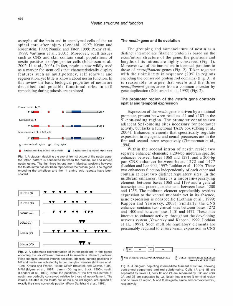
The grouping and nomenclature of nestin as adistinct intermediate filament protein is based on theexon/intron structure of the gene; the positions andlengths of its introns are highly conserved (Fig. 1).Moreover two of the introns are in identical positions tothose of neurofilament genes (Fig. 2). Taken togetherwith their similarity in sequence (20% in regionsencoding the conserved protein rod domains) (Fig. 3), itis reasonable to argue that nestin and the threeneurofilament genes arose from a common ancestor bygene duplication (Dahlstrand et al., 1992) (Fig. 2).
The promoter region of the nestin gene controlsspatial and temporal expression
Expression of the nestin gene is driven by a minimalpromoter, present between residues -11 and +183 in the5’-non-coding region. The promoter contains twoadjacent Sp1-binding sites necessary for promoteractivity, but lacks a functional TATA box (Cheng et al.,2004). Enhancer elements that specifically regulateexpression in myogenic and neural precursors are in thefirst and second intron respectively (Zimmerman et al.,1994).
Within the second intron of nestin reside twoseparate enhancer elements; a 204-bp midbrain specificenhancer between bases 1068 and 1271, and a 206-bppan-CNS enhancer between bases 1272 and 1477(Lothian and Lendahl, 1997; Lothian et al., 1999). Thesetwo enhancers function independently of each other andcontain at least two distinct regulatory sites. In themidbrain enhancer, there is a midbrain-specifyingelement, between bases 1068 and 1199 and a generaltranscriptional potentiator element, between bases 1200and 1255. The midbrain element reproducibly restrictsexpression to the ventral midbrain yet in its absence,gene expression is nonspecific (Lothian et al., 1999;Kappen and Yaworsky, 2003). Similarly, the CNSenhancer contains two critical sites between bases 1272and 1400 and between bases 1401 and 1477. These sitesinteract to enhance activity throughout the developingnervous system (Yaworsky and Kappen, 1999; Lothianet al., 1999). Such multiple regulatory elements arepresumably required to ensure nestin expression in CNS
666
Nestin structure and function
Fig. 1. A diagram depicting the exon/intron structure of the nestin gene;the intron pattern is conserved between the human, rat and mousenestin genes. The first three introns are in identical positions howeverthe fourth intron has not been reported for the human gene. The regionsencoding the α-helices and the 11 amino acid repeats have beenshaded.
Fig. 2. A schematic representation of intron positions in the genesencoding the six different classes of intermediate filament proteins.Filled triangles indicate intronic positions. Identical intronic positions inNF and nestin are indicated by larger triangles. Keratins (Ichinose et al.,1988; Krauss and Franke, 1990); GFAP (Balcarek and Cowan, 1985);NFM (Myers et al., 1987); Lamin (Döring and Stick, 1990); nestin(Lendahl et al., 1990). Note: the positions of the first two introns ofnestin are perfectly conserved relative to those of neurofilament. Theintrons, situated in the fourth coil of the a-helical region, are spliced atexactly the same nucleotide position (From Dahlstrand et al., 1992).
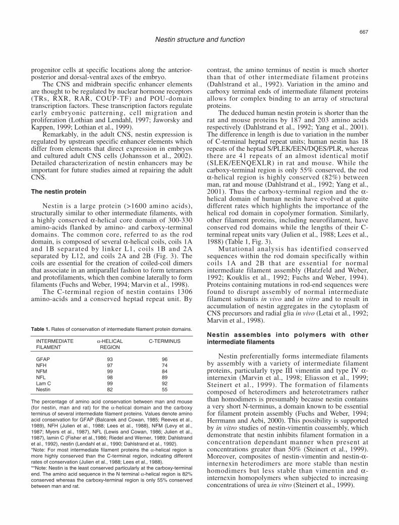
Fig. 3. A diagram depicting intermediate filament structure, showingconserved sequences and rod subdomains. Coils 1A and 1B areseparated by linker L1, coils 1B and 2A are separated by L12, and coils2A and 2B are separated by L2. Nestin has a short N terminal regionand no linker L2 region. N and C designate amino and carboxyl termini,respectively.
progenitor cells at specific locations along the anterior-posterior and dorsal-ventral axes of the embryo.
The CNS and midbrain specific enhancer elementsare thought to be regulated by nuclear hormone receptors(TRs, RXR, RAR, COUP-TF) and POU-domaintranscription factors. These transcription factors regulateearly embryonic patterning, cell migration andproliferation (Lothian and Lendahl, 1997; Jaworsky andKappen, 1999; Lothian et al., 1999).
Remarkably, in the adult CNS, nestin expression isregulated by upstream specific enhancer elements whichdiffer from elements that direct expression in embryosand cultured adult CNS cells (Johansson et al., 2002).Detailed characterization of nestin enhancers may beimportant for future studies aimed at repairing the adultCNS.
The nestin protein
Nestin is a large protein (>1600 amino acids),structurally similar to other intermediate filaments, witha highly conserved α-helical core domain of 300-330amino-acids flanked by amino- and carboxy-terminaldomains. The common core, referred to as the roddomain, is composed of several α-helical coils, coils 1Aand 1B separated by linker L1, coils 1B and 2Aseparated by L12, and coils 2A and 2B (Fig. 3). Thecoils are essential for the creation of coiled-coil dimersthat associate in an antiparallel fashion to form tetramersand protofilaments, which then combine laterally to formfilaments (Fuchs and Weber, 1994; Marvin et al., 1998).
The C-terminal region of nestin contains 1306amino-acids and a conserved heptad repeat unit. By
contrast, the amino terminus of nestin is much shorterthan that of other intermediate filament proteins(Dahlstrand et al., 1992). Variation in the amino andcarboxy terminal ends of intermediate filament proteinsallows for complex binding to an array of structuralproteins.
The deduced human nestin protein is shorter than therat and mouse proteins by 187 and 203 amino acidsrespectively (Dahlstrand et al., 1992; Yang et al., 2001).The difference in length is due to variation in the numberof C-terminal heptad repeat units; human nestin has 18repeats of the heptad S/PLEK/EEN/DQES/PLR, whereasthere are 41 repeats of an almost identical motif(SLEK/EENQEXLR) in rat and mouse. While thecarboxy-terminal region is only 55% conserved, the rodα-helical region is highly conserved (82%) betweenman, rat and mouse (Dahlstrand et al., 1992; Yang et al.,2001). Thus the carboxy-terminal region and the α-helical domain of human nestin have evolved at quitedifferent rates which highlights the importance of thehelical rod domain in copolymer formation. Similarly,other filament proteins, including neurofilament, haveconserved rod domains while the lengths of their C-terminal repeat units vary (Julien et al., 1988; Lees et al.,1988) (Table 1, Fig. 3).
Mutational analysis has identified conservedsequences within the rod domain specifically withincoils 1A and 2B that are essential for normalintermediate filament assembly (Hatzfeld and Weber,1992; Kouklis et al., 1992; Fuchs and Weber, 1994).Proteins containing mutations in rod-end sequences werefound to disrupt assembly of normal intermediatefilament subunits in vivo and in vitro and to result inaccumulation of nestin aggregates in the cytoplasm ofCNS precursors and radial glia in vivo (Letai et al., 1992;Marvin et al., 1998).
Nestin assembles into polymers with otherintermediate filaments
Nestin preferentially forms intermediate filamentsby assembly with a variety of intermediate filamentproteins, particularly type III vimentin and type IV α-internexin (Marvin et al., 1998; Eliasson et al., 1999;Steinert et al., 1999). The formation of filamentscomposed of heterodimers and heterotetramers ratherthan homodimers is presumably because nestin containsa very short N-terminus, a domain known to be essentialfor filament protein assembly (Fuchs and Weber, 1994;Herrmann and Aebi, 2000). This possibility is supportedby in vitro studies of nestin-vimentin coassembly, whichdemonstrate that nestin inhibits filament formation in aconcentration dependant manner when present atconcentrations greater than 50% (Steinert et al., 1999).Moreover, composites of nestin-vimentin and nestin-α-internexin heterodimers are more stable than nestinhomodimers but less stable than vimentin and α-internexin homopolymers when subjected to increasingconcentrations of urea in vitro (Steinert et al., 1999).
667
Nestin structure and function
Table 1. Rates of conservation of intermediate filament protein domains.
INTERMEDIATE α-HELICAL C-TERMINUSFILAMENT REGION
GFAP 93 96NFH 97 74NFM 99 84NFL 98 89Lam C 99 92Nestin 82 55
The percentage of amino acid conservation between man and mouse(for nestin, man and rat) for the α-helical domain and the carboxyterminus of several intermediate filament proteins. Values denote aminoacid conservation for GFAP (Balcarek and Cowan, 1985; Reeves et al.,1989), NFH (Julien et al., 1988; Lees et al., 1988), NFM (Levy et al.,1987; Myers et al., 1987), NFL (Lewis and Cowan, 1986; Julien et al.,1987), lamin C (Fisher et al.,1986; Riedel and Werner, 1989; Dahlstrandet al., 1992), nestin (Lendahl et al., 1990; Dahlstrand et al., 1992).*Note: For most intermediate filament proteins the α-helical region ismore highly conserved than the C-terminal region, indicating differentrates of conservation (Julien et al., 1988; Lees et al., 1988). **Note: Nestin is the least conserved particularly at the carboxy-terminalend. The amino acid sequence in the N terminal α-helical region is 82%conserved whereas the carboxy-terminal region is only 55% conservedbetween man and rat.
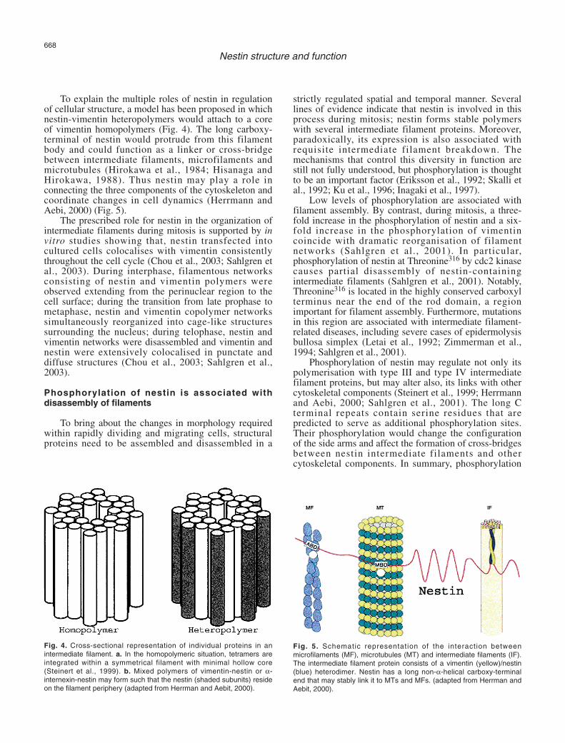
To explain the multiple roles of nestin in regulationof cellular structure, a model has been proposed in whichnestin-vimentin heteropolymers would attach to a coreof vimentin homopolymers (Fig. 4). The long carboxy-terminal of nestin would protrude from this filamentbody and could function as a linker or cross-bridgebetween intermediate filaments, microfilaments andmicrotubules (Hirokawa et al., 1984; Hisanaga andHirokawa, 1988). Thus nestin may play a role inconnecting the three components of the cytoskeleton andcoordinate changes in cell dynamics (Herrmann andAebi, 2000) (Fig. 5).
The prescribed role for nestin in the organization ofintermediate filaments during mitosis is supported by invitro studies showing that, nestin transfected intocultured cells colocalises with vimentin consistentlythroughout the cell cycle (Chou et al., 2003; Sahlgren etal., 2003). During interphase, filamentous networksconsisting of nestin and vimentin polymers wereobserved extending from the perinuclear region to thecell surface; during the transition from late prophase tometaphase, nestin and vimentin copolymer networkssimultaneously reorganized into cage-like structuressurrounding the nucleus; during telophase, nestin andvimentin networks were disassembled and vimentin andnestin were extensively colocalised in punctate anddiffuse structures (Chou et al., 2003; Sahlgren et al.,2003).
Phosphorylation of nestin is associated withdisassembly of filaments
To bring about the changes in morphology requiredwithin rapidly dividing and migrating cells, structuralproteins need to be assembled and disassembled in a
strictly regulated spatial and temporal manner. Severallines of evidence indicate that nestin is involved in thisprocess during mitosis; nestin forms stable polymerswith several intermediate filament proteins. Moreover,paradoxically, its expression is also associated withrequisite intermediate filament breakdown. Themechanisms that control this diversity in function arestill not fully understood, but phosphorylation is thoughtto be an important factor (Eriksson et al., 1992; Skalli etal., 1992; Ku et al., 1996; Inagaki et al., 1997).
Low levels of phosphorylation are associated withfilament assembly. By contrast, during mitosis, a three-fold increase in the phosphorylation of nestin and a six-fold increase in the phosphorylation of vimentincoincide with dramatic reorganisation of filamentnetworks (Sahlgren et al., 2001). In particular,phosphorylation of nestin at Threonine316 by cdc2 kinasecauses partial disassembly of nestin-containingintermediate filaments (Sahlgren et al., 2001). Notably,Threonine316 is located in the highly conserved carboxylterminus near the end of the rod domain, a regionimportant for filament assembly. Furthermore, mutationsin this region are associated with intermediate filament-related diseases, including severe cases of epidermolysisbullosa simplex (Letai et al., 1992; Zimmerman et al.,1994; Sahlgren et al., 2001).
Phosphorylation of nestin may regulate not only itspolymerisation with type III and type IV intermediatefilament proteins, but may alter also, its links with othercytoskeletal components (Steinert et al., 1999; Herrmannand Aebi, 2000; Sahlgren et al., 2001). The long Cterminal repeats contain serine residues that arepredicted to serve as additional phosphorylation sites.Their phosphorylation would change the configurationof the side arms and affect the formation of cross-bridgesbetween nestin intermediate filaments and othercytoskeletal components. In summary, phosphorylation
668
Nestin structure and function
Fig. 4. Cross-sectional representation of individual proteins in anintermediate filament. a. In the homopolymeric situation, tetramers areintegrated within a symmetrical filament with minimal hollow core(Steinert et al., 1999). b. Mixed polymers of vimentin-nestin or α-internexin-nestin may form such that the nestin (shaded subunits) resideon the filament periphery (adapted from Herrman and Aebit, 2000).
Fig. 5. Schematic representation of the interaction betweenmicrofilaments (MF), microtubules (MT) and intermediate filaments (IF).The intermediate filament protein consists of a vimentin (yellow)/nestin(blue) heterodimer. Nestin has a long non-α-helical carboxy-terminalend that may stably link it to MTs and MFs. (adapted from Herrman andAebit, 2000).
and dephosphorylation of nestin may modulaterespectively, disassembly and assembly of intermediatefilaments within supramolecular structures and thuscontrol dynamic changes in cell ultastructure (Hirokawaet al., 1984; Steinert et al., 1999).
Similarly, phosphorylation has been shown toregulate the spatial organization of intermediate filamentproteins such as vimentin (Chou et al., 1989, 1990). Infact, during mitosis, the disassembly of vimentinintermediate filaments requires the presence andphosphorylation of both nestin and vimentin (Chou etal., 2003). To induce the disassembly of vimentinpolymers during mitosis, nestin works in concert withthe ubiquitous mitotic kinase, maturation/M-phase-promoting factor, MPF, which phosphorylates vimentinat Ser-55 in the amino-terminal head domain, the regionrequired for dimerisation (Aubin et al., 1980; Jones etal., 1985; Chou et al., 1996, 2003; Sahlgren et al., 2003).The role of nestin in vimentin disassembly was recentlyconfirmed by downregulation of nestin with specificsiRNAs in vitro, which blocked the disassembly ofvimentin intermediate filaments in mitotic cells (Chou etal., 2003).
The disassembly of vimentin intermediate filamentsis not a feature of all cells – in fact, many cell types donot express nestin. In dividing cells in which nestin isabsent and there is no apparent disassembly of vimentinfilaments, there is alternately, a restricted localdisassembly of intermediate filaments at the cleavagefurrow in late cytokinesis (Yasui et al., 2001).
Nestin association with cytoplasmic trafficking inrapidly dividing progenitor cells
The advantage of mitotic disassembly of vimentinfilaments for cells expressing nestin remains unknown.Mitotic and spreading interphase cells, containingnonfilamentous keratin or vimentin, are able to moveprotein particles at high speeds along microtubules withmolecular motors kinesin and dynein (Prahlad et al.,1998; Windoffer and Leube, 1999; Helfand et al., 2002).It is possible that this ensures the rapid transport ofprecursor molecules between various cytoplasmiccompartments. Therefore, nestin expression may beassociated with increased cytoplasmic trafficking inprogenitor cells undergoing rapid rounds of division,interspersed with active interphase migration. Suchcharacteristics are common features of cells in earlydeveloping nerve and muscle tissues (Lendahl et al.,1990; Sejersen and Lendahl, 1993; Kachinsky et al.,1995; Vaittinen et al., 1999) and in regenerating adulttissues (Frisen et al., 1995; Vaittinen et al., 1999).
Nestin may also play a role in the asymmetricallocation of cytoskeletal and other cellular factors todaughter cells. For example, within the developingneural tube, ventricular cells continue to proliferatewhereas differentiated cells migrate towards the pialsurface. Polarized distribution of material betweendividing and differentiating daughter cells within the
neuroepithelium may be caused by nestin-mediateddisassembly and uneven partitioning of motile vimentinparticles during mitosis (Frederiksen and McKay, 1988;Rakic, 1988; Chou et al., 2003).
Conclusion
Intermediate filaments represent the least understoodpart of the cytoskeleton. Although many parameters areknown, the reasons for the existing diversity ofintermediate proteins as well as their individualfunctions remain unknown. Because nestin is expressedin the majority of mitotically active CNS and PNSprogenitors, it is currently widely used as a marker forneural stem cells yet its apparent diversity of roles inheterogeneous cells is still not completely understood.Several researchers have performed complexexperiments and detailed analyses to ascertain itsfunctions. While these have aided in the understandingof the complexity of cell dynamics, the intricate role ofnestin in cellular proliferation during development andregeneration remains to be conclusively defined.
References
Aubin J., Osborn M., Franke W. and Weber K. (1980). Intermediatefilaments of the vimentin-type and the cytokeratin-type aredistributed differently during mitosis. Exp. Cell Res. 129, 149-165.
Balcarek J. and Cowan N. (1985). Structure of the mouse glial fibrillaryacidic protein gene: implications for the evolution of the intermediatefilament multigene family. Nucl. Acids Res. 13, 5527-5543.
Cattaneo E. and McKay R. (1990). Proliferation and differentiation ofneuronal stem cells regulated by nerve growth factor. Nature 347,762 -765.
Cheng L., Jin Z., Liu L., Yan Y., Li T., Zhu X. and Jing N. (2004).Characterization and promoter analysis of the mouse nestin gene.FEBS Lett. 565, 195-202.
Chou Y-H., Rosevear E. and Goldman R. (1989). Phosphorylation anddisassembly of intermediate filaments in mitotic cells. Proc. Natl.Acad. Sci. USA 86, 1885-1889.
Chou Y-H., Bischoff J., Beach D. and Goldman R. (1990). Intermediatefilament reorganization during mitosis is mediated by p34cdc2phosphorylation of vimentin. Cell 62, 1063-1071.
Chou Y-H., Opal P., Quinlan R. and Goldman R. (1996). The relativeroles of specific N- and C-terminal phosphorylation sites in thedisassembly of intermediate filament in mitotic BHK-21 cells. J. CellSci. 109, 817-826.
Chou Y-H., Khuon S., Herrmann H. and Goldman R. (2003). Nestinpromotes the phosphorylation-dependent disassembly of vimentinintermediate filaments during mitosis. Mol. Biol. Cell 14, 1468-1478.
Dahlstrand J., Zimmerman L., McKay R. and Lendahl U. (1992).Characterization of the human nestin gene reveals a closeevolutionary relationship to neurofilaments. J. Cell Sci. 103, 589-597.
Döring V. and Stick R. (1990). Gene structure of nuclear lamin LIII ofXenopus laevis; a model for the evolution of IF proteins from alamin-like ancestor. EMBO J. 9, 4073-4081.
Eliasson C., Sahlgren C., Berthold C., Stakeberg J., Celis J., BetsholtzC., Eriksson J. and Pekny M. (1999). Intermediate filament protein
669
Nestin structure and function
partnership in astrocytes. J. Biol. Chem. 274, 23996-24006.Eriksson J., Brautigan D., Vallee R., Olmstedt S., Fujiki H. and Goldman
R. (1992). Cytoskeletal integrity in interphase cells requires proteinphosphatase activity. Proc. Natl. Acad. Sci. USA 89, 11093-11097.
Fisher D., Chaudhary N. and Blobel G. (1986). cDNA sequencing ofnuclear lamins A and C reveals primary and secondary structurehomology to intermediate filaments. Proc. Nat. Acad. Sci. USA 83,6450-6454.
Frederiksen K. and McKay R. (1988). Proliferation and differentiation ofrat neuroepithelial precursor cells in vivo. J. Neurosci. 8, 1144-1151.
Frisen J., Johansson C., Torok C., Risling M. and Lendahl U. (1995).Rapid, widespread, and longlasting induction of nestin contributes tothe generation of glial scar tissue after CNS injury. J. Cell Biol. 131,453-464.
Fröjdman K., Pelliniemi L., Lendahl U., Virtanen I. and Eriksson J.(1997). The intermediate filament protein nestin occurs transiently indifferentiating testis of rat and mouse. Differentiation 61, 243-249.
Fuchs E. and Weber K. (1994). Intermediate filaments: structure,dynamics, function, and disease. Annu. Rev. Biochem. 63, 345-382.
Fuchs E. and Cleveland D. (1998). A structural scaffolding ofintermediate filaments in health and disease. Science 279, 514-519.
Geisler N., Hatzfeld M. and Weber K. (1989). Phosphorylation in vitro ofvimentin by protein kinases A and C is restricted to the headdomain. Identification of the phosphoserine sites and their influenceon filament formation. Eur. J. Biochem. 1, 441-447.
Goldman R. (2001). Worms reveal essential functions for intermediatefilaments. Proc. Natl. Acad. Sci. USA 98, 7659-7661.
Goldman R., Chou Y-H., Prahlad V. and Yoon M. (1999). Intermediatefilaments: dynamic processes regulating their assembly, motility, andinteractions with other cytoskeletal systems. FASEB. J. 13, 261-265.
Hatzfeld M. and Weber K. (1992). A synthetic peptide representing theconsensus sequence motif at the carboxy-terminal end of the roddomain inhibits intermediate filament assembly and disassemblespreformed filaments. J. Cell Biol. 116, 157-166.
Helfand B., Mikami A., Vallee R. and Goldman R. (2002). A requirementfor cytoplasmic dynein and dynactin in intermediate filament networkassembly and organization. J. Cell Biol. 157, 795-806.
Herrmann H. and Aebi U. (2000). Intermediate filaments and theirassociates: multi-talented structural elements specifyingcytoarchitecture and cytodynamics. Curr. Opin. Cell Biol. 12, 79-90.
Hirokawa N., Glicksman N. and Willard M. (1984). Organization ofmammalian neurofilament polypeptides within the neuronalcytoskeleton. J. Cell Biol. 98, 1523-1536.
Hisanaga S. and Hirokawa N. (1988). Structure of the peripheraldomains of neurofilaments revealed by low angle rotary shadowing.J. Mol. Biol. 202, 297-305.
Hockfield S. and McKay R. (1985). Identification of major cell classes inthe developing mammalian nervous system. J. Neurosci. 5, 3310-3328.
Ichinose Y., Morita T., Zhang F., Srimahasongeran S., Tondella M.,Matsumoto M., Nozaki M. and Matsushiro A. (1988). Nucleotidesequence and structure of the mouse cytokeratin endo B gene.Gene 70, 85-95.
Inagaki M., Inagaki N., Takahashi T. and Takai Y. (1997).Phosphorylation-dependent control of structures of intermediatefilaments: a novel approach using site- and phosphorylation state-specific antibodies. J. Biochem. (Tokyo) 121, 407-414.
Johansson C., Lothian C., Molin M., Okano H. and Lendahl U. (2002).Nestin enhancer requirements for expression in normal and injured
adult CNS. J. Neurosci. Res. 69, 784-794. Jones J., Goldman A., Yang H. and Goldman R. (1985). The
organizational fate of intermediate filament networks in two epithelialcell types during mitosis. J. Cell Biol. 100, 93-102.
Julien J-P., Grosveld F., Yazdanbaksh K., Flavell D., Meijer D. andMushynski W. (1987). The structure of a human neurofilament gene(NF-L): a unique exon-intron organization in the intermediatefilament family. Biochim. Biophys. Acta 909, 10-20.
Julien J., Côte F., Beaudet L., Sidky M., Flavell D., Grosveld F. andMushynski W. (1988). Sequence and structure of the mouse genecoding for the largest neurofilament subunit. Gene 68, 307-314.
Kachinsky A., Dominov J. and Miller J. (1994). Myogenesis and theintermediate filament protein nestin. Dev. Biol. 165, 216-228.
Kachinsky A., Dominov J. and Miller J. (1995). Intermediate filaments incardiac myogenesis: Nestin in the developing mouse heart. J.Histochem. Cytochem. 43, 843-847.
Kappen C. and Yaworsky P. (2003). Mutation of a putative nuclearreceptor binding site abolishes activity of the nestin midbrainenhancer. Biochim. Biophys. Acta 1625, 109-115.
Klymkowsky M. (1996). Intermediate filaments as dynamic structures.Cancer Metastasis Rev. 15, 417-428.
Kouklis P., Traub P. and Georgatos S. (1992). Involvement of theconsensus sequence motif at coil 2b in the assembly and stability ofvimentin filaments. J. Cell Sci. 102, 31-41.
Krauss S. and Franke W. (1990). Organization and sequence of thehuman gene encoding cytokeratin 8. Gene 86, 241-249.
Krum J. and Rosenstein J. (1999). Transient coexpression of nestin,GFAP, and vascular endothelial growth factor in mature reactiveastroglia following neural grafting or brain wounds. Exp. Neurol. 160,348-360.
Ku N-O., Liao J., Chou C-F. and Omary B. (1996). Implications ofintermediate filament protein phosphorylation. Cancer Metastasis15, 11093-11097.
Lees J., Shneidman P., Skuntz S., Carden M. and Lazzarini R. (1988).The structure and organization of the human heavy neurofilamentsubunit (NF-H) and the gene encoding it. EMBO J. 7, 1947- 1955.
Lendahl U. (1997). Transgenic analysis of central nervous systemdevelopment and regeneration. Acta Anaesthesiol. Scand. Suppl.110, 116 -118.
Lendahl U., Zimmerman L. and McKay R. (1990). CNS stem cellsexpress a new class of intermediate filament protein. Cell 60, 585-595.
Letai A., Coulombe P. and Fuchs E. (1992). Do the ends justify themean? Proline mutations at the ends of the keratin coiled-coil rodsegment are more disruptive than internal mutations. J. Cell Biol.116, 1181-1195.
Levy E., Liem R., D’Eustachio P. and Cowan N. (1987). Structure andevolutionary origin of the gene encoding mouse NF-M, the middle-molecular-mass neurofilament protein. Eur. J. Biochem. 166, 71-77.
Lewis S. and Cowan N. (1986). Anomalous placement of introns in amember of the intermediate f i lament multigene family: anevolutionary conundrum. Mol. Cell. Biol. 6, 1529-1534.
Li L., Mignone J., Yang M., Matic M., Penman S., Enikolopov G. andHoffman, R. (2003). Nestin expression in hair follicle. Proc. Natl.Acad. Sci. USA 100, 9958-9961.
Lothian C. and Lendahl U. (1997). An evolutionarily conserved region inthe second intron of the human nestin gene directs gene expressionto CNS progenitor cells and to early neural crest cells. Eur. J.Neurosci. 9, 452-462.
670
Nestin structure and function
Lothian C., Prakash N., Lendahl U. and Wahlstrom G. (1999).Identification of both general and region-specific embryonic CNSenhancer elements in the nestin promoter. Exp. Cell Res. 248, 509-519.
Marvin M., Dahlstrand J., Lendahl U. and McKay R. (1998). A rod enddeletion in the intermediate filament protein nestin alters itssubcellular localization in neuroepithelial cells of transgenic mice. J.Cell Sci. 111, 1951-1961.
Mujtaba T., Mayer-Proschel M. and Rao M. (1998) A common neuralprogenitor for the CNS and PNS. Dev. Biol. 200, 1-15.
Myers M., Lazzarini R., Lee V., Schlaepfer W. and Nelson D. (1987).The human mid-size neurofilament subunit: a repeated proteinsequence and the relationship of its gene to the intermediatefilament gene family. EMBO J. 6, 1617-1626.
Namiki J. and Tator C. (1999). Cell proliferation and nestin expression inthe ependyma of the adult rat spinal cord after injury. J.Neuropathol. Exp. Neurol. 58, 489-498.
Pekny M., Johansson C., Eliasson C., Stakeberg J., Wallen A.,Perlmann T., Lendahl U., Betsholtz C., Berthold C. and Frisen J.(1999). Abnormal reaction to central nervous system injury in micelacking glial fibrillary acidic protein and vimentin. J. Cell Biol. 145,503-514.
Prahlad V., Yoon M., Moir R., Vale R. and Goldman R. (1998). Rapidmovements of vimentin on microtubule tracks: kinesin-dependentassembly of intermediate filament networks. J. Cell Biol. 143, 159-117.
Rakic P. (1988). Specification of cerebral cortical areas. Science 241,170-176.
Reeves S., Helman L., Allison A. and Israel M. (1989). Molecular cloningand primary structure of human glial fibrillary acidic protein. Proc.Nat. Acad. Sci. USA 86, 5178-5182.
Riedel W. and Werner D. (1989). Nucleotide sequence of the full-lengthmouse lamin C cDNA and its deduced amino acid sequence.Biochim. Biophys. Acta 1008, 119-122.
Sahlgren C., Mikhailov A., Hellman J., Chou Y-H., Lendahl U., GoldmanR. and Eriksson J. (2001). Mitotic reorganization of the intermediatefilament protein nestin involves phosphorylation by cdc2 kinase. J.Biol. Chem. 276, 16456-16463.
Sahlgren C., Mikhailov A., Vaittinen S., Pallari A., Kalimo H., Pant H.and Eriksson J. (2003). Cdk5 regulates the organization of nestinand its association with p35. Mol. Cell. Biol. 23, 5090-5106.
Sejersen T. and Lendahl U. (1993). Transient expression of theintermediate filament nestin during skeletal muscle development. J.
Cell Sci. 106, 1291-1300.Skalli O., Chou Y-H. and Goldman R. (1992). Intermediate filaments: not
so tough after all. Trends Cell Biol. 2, 308-312.Steinert P. and Liem R. (1990). Intermediate filament dynamics. Cell 60,
521-523.Steinert P., Chou Y-H., Prahlad V., Parry A., Marekov L., Wu K., Jang
S-I. and Goldman R. (1999). A high molecular weight intermediatefilament-associated protein in BHK-21 cells is nestin, a Type VIintermediate f i lament protein. J. Biol. Chem. 274, 9881-9890.
Terling C., Rass A., Midsiadis T., Fried K., Lendahl U. and WroblewskiJ. (1995). Expression of the intermediate filament nestin duringrodent tooth development. Int. J. Dev. Biol. 39, 947-956.
Vaittinen S., Lukka R., Sahlgren C., Rantanen J., Hurme T., Lendahl U.,Eriksson J. and Kalimo H. (1999). Specific and innervation-regulatedexpression of the intermediate f i lament protein nestin atneuromuscular and myotendinous junctions in skeletal muscle. Am.J. Pathol. 154, 591-600.
Vaittinen S., Lukka R., Sahlgren C., Hurme T., Rantanen J., Lendahl U.,Eriksson J. and Kalimo H. (2001). The expression of intermediatefilament nestin as related to vimentin and desmin in regeneratingskeletal muscle. J. Neuropathol. Exp. Neurol. 60, 588-597.
Windoffer R. and Leube R. (1999). Detection of cytokeratin dynamics bytime-lapse fluorescence microscopy in living cells. J. Cell Sci. 112,4521-4534.
Yang J., Cheng L., Yan Y., Bian W., Tomooka Y., Shiurba R. and JingN. (2001). Mouse nestin cDNA cloning and protein expression in thecytoskeleton of transfected cells. Biochim. Biophys. Acta 1520, 251-254.
Yasui Y., Goto H., Matsui S., Manser E., Lim L., Nagata K. and InagakiM. (2001). Protein kinases required for segregation of vimentinfilaments in mitotic process. Oncogene 20, 2868-2876.
Yaworsky P. and Kappen C. (1999). Heterogenity of neural progenitorcells revealed by enhancers on the nestin gene. Dev. Biol. 205, 309-321.
Zehner Z. (1991). Regulation of intermediate filament gene expression.Curr. Opin. Cell Biol. 3, 67-74.
Zimmerman L., Parr B., Lendahl U., Cunningham M., McKay R., GavinB., Mann J., Vassileva G. and McMahon A. (1994). Independentregulatory elements in the nestin gene direct trans gene expressionto neural stem cells or muscle precursors. Neuron 12, 11-24.
Accepted January 5, 2004
671
Nestin structure and function