Embed Size (px)
Citation preview
Simulated Cytoskeletal Collapse via Tau DegradationAustin Sendek1,2,3, Henry R. Fuller2,3, N. Robert Hayre2,3, Rajiv R. P. Singh2,3, Daniel L. Cox2,3*
1 Department of Applied Physics, Stanford University, Palo Alto, California, United States of America, 2 Department of Physics, University of California Davis, Davis,
California, United States of America, 3 Institute for Complex Adaptive Matter, University of California Davis, Davis, California, United States of America
Abstract
We present a coarse-grained two dimensional mechanical model for the microtubule-tau bundles in neuronal axons inwhich we remove taus, as can happen in various neurodegenerative conditions such as Alzheimers disease, tauopathies,and chronic traumatic encephalopathy. Our simplified model includes (i) taus modeled as entropic springs betweenmicrotubules, (ii) removal of taus from the bundles due to phosphorylation, and (iii) a possible depletion force betweenmicrotubules due to these dissociated phosphorylated taus. We equilibrate upon tau removal using steepest descentrelaxation. In the absence of the depletion force, the transverse rigidity to radial compression of the bundles falls to zero atabout 60% tau occupancy, in agreement with standard percolation theory results. However, with the attractive depletionforce, spring removal leads to a first order collapse of the bundles over a wide range of tau occupancies for physiologicallyrealizable conditions. While our simplest calculations assume a constant concentration of microtubule intercalants tomediate the depletion force, including a dependence that is linear in the detached taus yields the same collapse. Applyingpercolation theory to removal of taus at microtubule tips, which are likely to be the protective sites against dynamicinstability, we argue that the microtubule instability can only obtain at low tau occupancy, from 0.06–0.30 depending uponthe tau coordination at the microtubule tips. Hence, the collapse we discover is likely to be more robust over a wide rangeof tau occupancies than the dynamic instability. We suggest in vitro tests of our predicted collapse.
Citation: Sendek A, Fuller HR, Hayre NR, Singh RRP, Cox DL (2014) Simulated Cytoskeletal Collapse via Tau Degradation. PLoS ONE 9(8): e104965. doi:10.1371/journal.pone.0104965
Editor: Chad A. Dickey, University of South Florida Alzheimer’s Institute, United States of America
Received March 21, 2014; Accepted July 16, 2014; Published August 27, 2014
Copyright: � 2014 Sendek et al. This is an open-access article distributed under the terms of the Creative Commons Attribution License, which permitsunrestricted use, distribution, and reproduction in any medium, provided the original author and source are credited.
Data Availability: The authors confirm that all data underlying the findings are fully available without restriction. We have posted all the data for themicrotubule simulations at http://dx.doi.org/10.6084/m9.figshare.1044310 http://dx.doi.org/10.6084/m9.figshare.1023176 http://dx.doi.org/10.6084/m9.figshare.1023087
Funding: This work was supported by United States National Science Foundation Grant DMR-1207624 (A.S., H.F., N.R.H., R.R.P.S., D.L.C). It was also supported byInternational Institute for Complex Adaptive Matter, United States National Science Foundation Grant DMR-0844115 (computing cluster) (N.R.H, D.L.C.). Thefunders had no role in study design, data collection and analysis, decision to publish, or preparation of the manuscript.
Competing Interests: The authors have declared that no competing interests exist.
* Email: [email protected]
Introduction
Neurodegenerative conditions such as Alzheimer’s disease (AD),
Parkinson’s, and those resulting from chronic traumatic enceph-
alopathy (CTE) damage arising in contact sports, represent a
massive public health threat that annually impacts tens of millions
worldwide. Finding routes to prevention or treatment prior to
irreversible changes in the affected cells remain important goals.
Part of the difficulty with this task is the complexity of the
conditions themselves. For example, in Alzheimer’s disease, while
it is generally agreed, per the ‘‘amyloid cascade hypothesis’’ (see
[1] and [2]) that the initial trigger of AD is the production and
subsequent aggregation of A-beta protein (ABP) into oligomers
(ABO), the way in which the ABOs trigger cell damage and
eventual death remains unclear.
Neuro-fibrillary tangles (NFTs), consisting largely of hyperpho-
sphorylated tau protein aggregates, have been observed in post-
mortem tissues of AD victims. These NFTs are correlated with
regions of A-beta aggregates and neuronal damage or death [3]
confirms that ABOs trigger processes which lead to modifications
of the tau protein. NFTs and tau associated damage also arise in
CTE [4]. Tau proteins play a critical role in the microtubule
bundles (MTBs) of the axon cytoskeleton, by promoting the
assembly of axonal microtubules and inhibiting dynamic instability
(DI) [5], and by crosslinking the microtubules in the bundles. The
integrity of the MTBs is critical for neuronal function: they provide
mechanical stability to axons for decades of human life, and they
serve as the highways for fast and slow axonal transport of
neurotransmitters and organelles to and from the synaptic regions
of the neuron. Clearly, at the cellular level, a significant marker of
irreversible damage in conditions affecting tau proteins would be
loss of integrity of the MTBs, and thus a point of no return for
intervention.
Studies of cultured neurons exposed to high concentrations of
ABOs have confirmed that the ABOs can be internalized, and
once internalized these trigger production of proteinases which
can cut taus, and kinases which can phosphorylate taus [6]. Both
are important, since full length taus are needed to assure the
proper spacing of microtubules, and hyperphosphorylated taus
(HPTs) may tend to dissociate from the negatively charged
microtubule surfaces.
If tau detachment from microtubule binding sites exceeds 50%,
this should lead to collapse of MTBs due to the dynamic instability
of individual microtubules. In experiments on cultured hippo-
campal neurons exposed to massive concentrations of ABOs
(1000–10000 times physiological ABP concentrations in vivo) MT
loss is indeed observed [7], with the lateral density decreasing by a
factor of 25 compared to unexposed cells. However, these high
PLOS ONE | www.plosone.org 1 August 2014 | Volume 9 | Issue 8 | e104965
ABO concentrations may obscure more subtle changes in the
MTBs. Specifically, since the taus provide transverse mechanical
stability, MTB transverse rigidity may be lost when a majority of
the taus are still intact: random removal of connecting springs in a
lattice of otherwise noninteracting cylinders leads to a loss of
transverse mechanical stiffness at the rigidity percolation threshold
near 60% of spring occupancy as we discuss below. Moreover, as
the HPTs no longer favorably associate with the surface of
microtubules, they can lower the free energy by gaining
translational entropy through reduced excluded volume between
microtubules, yielding a fluctuation mediated attraction between
microtubules. This will, in effect, add an inward radial pressure to
the MTBs as taus are removed.
In this paper, we develop a two dimensional mechanical model
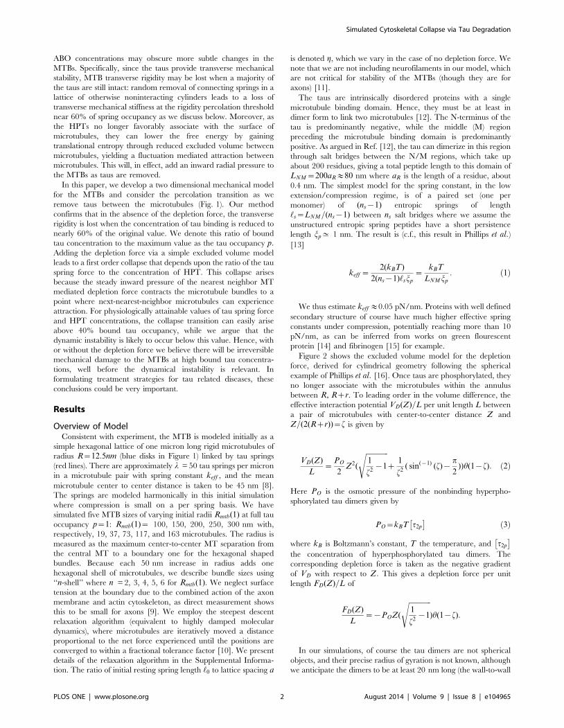
for the MTBs and consider the percolation transition as we
remove taus between the microtubules (Fig. 1). Our method
confirms that in the absence of the depletion force, the transverse
rigidity is lost when the concentration of tau binding is reduced to
nearly 60% of the original value. We denote this ratio of bound
tau concentration to the maximum value as the tau occupancy p:Adding the depletion force via a simple excluded volume model
leads to a first order collapse that depends upon the ratio of the tau
spring force to the concentration of HPT. This collapse arises
because the steady inward pressure of the nearest neighbor MT
mediated depletion force contracts the microtubule bundles to a
point where next-nearest-neighbor microtubules can experience
attraction. For physiologically attainable values of tau spring force
and HPT concentrations, the collapse transition can easily arise
above 40% bound tau occupancy, while we argue that the
dynamic instability is likely to occur below this value. Hence, with
or without the depletion force we believe there will be irreversible
mechanical damage to the MTBs at high bound tau concentra-
tions, well before the dynamical instability is relevant. In
formulating treatment strategies for tau related diseases, these
conclusions could be very important.
Results
Overview of ModelConsistent with experiment, the MTB is modeled initially as a
simple hexagonal lattice of one micron long rigid microtubules of
radius R~12:5nm (blue disks in Figure 1) linked by tau springs
(red lines). There are approximately l = 50 tau springs per micron
in a microtubule pair with spring constant keff , and the mean
microtubule center to center distance is taken to be 45 nm [8].
The springs are modeled harmonically in this initial simulation
where compression is small on a per spring basis. We have
simulated five MTB sizes of varying initial radii Rmtb(1) at full tau
occupancy p~1: Rmtb(1)~ 100, 150, 200, 250, 300 nm with,
respectively, 19, 37, 73, 117, and 163 microtubules. The radius is
measured as the maximum center-to-center MT separation from
the central MT to a boundary one for the hexagonal shaped
bundles. Because each 50 nm increase in radius adds one
hexagonal shell of microtubules, we describe bundle sizes using
‘‘n-shell’’ where n = 2, 3, 4, 5, 6 for Rmtb(1). We neglect surface
tension at the boundary due to the combined action of the axon
membrane and actin cytoskeleton, as direct measurement shows
this to be small for axons [9]. We employ the steepest descent
relaxation algorithm (equivalent to highly damped molecular
dynamics), where microtubules are iteratively moved a distance
proportional to the net force experienced until the positions are
converged to within a fractional tolerance factor [10]. We present
details of the relaxation algorithm in the Supplemental Informa-
tion. The ratio of initial resting spring length ‘0 to lattice spacing a
is denoted g, which we vary in the case of no depletion force. We
note that we are not including neurofilaments in our model, which
are not critical for stability of the MTBs (though they are for
axons) [11].
The taus are intrinsically disordered proteins with a single
microtubule binding domain. Hence, they must be at least in
dimer form to link two microtubules [12]. The N-terminus of the
tau is predominantly negative, while the middle (M) region
preceding the microtubule binding domain is predominantly
positive. As argued in Ref. [12], the tau can dimerize in this region
through salt bridges between the N/M regions, which take up
about 200 residues, giving a total peptide length to this domain of
LNM~200aR&80 nm where aR is the length of a residue, about
0.4 nm. The simplest model for the spring constant, in the low
extension/compression regime, is of a paired set (one per
monomer) of (ns{1) entropic springs of length
‘s~LNM=(ns{1) between ns salt bridges where we assume the
unstructured entropic spring peptides have a short persistence
length jp^ 1 nm. The result is (c.f., this result in Phillips et al.)[13]
keff ~2(kBT)
2(ns{1)‘sjp
~kBT
LNM jp
: ð1Þ
We thus estimate keff &0.05 pN/nm. Proteins with well defined
secondary structure of course have much higher effective spring
constants under compression, potentially reaching more than 10
pN/nm, as can be inferred from works on green flourescent
protein [14] and fibrinogen [15] for example.
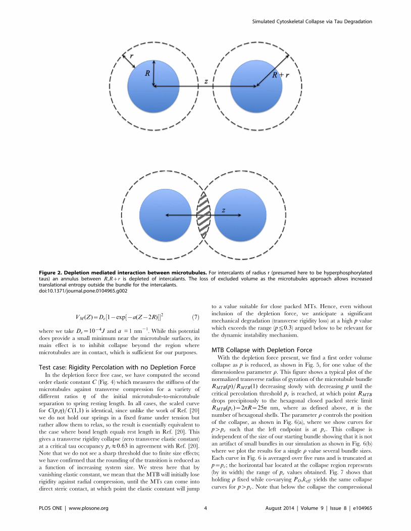
Figure 2 shows the excluded volume model for the depletion
force, derived for cylindrical geometry following the spherical
example of Phillips et al. [16]. Once taus are phosphorylated, they
no longer associate with the microtubules within the annulus
between R, Rzr. To leading order in the volume difference, the
effective interaction potential VD(Z)=L per unit length L between
a pair of microtubules with center-to-center distance Z and
Z=(2(Rzr))~f is given by
VD(Z)
L~
PO
2Z2(
ffiffiffiffiffiffiffiffiffiffiffiffiffi1
f2{1
sz
1
f2( sin({1) (f){
p
2))h(1{f): ð2Þ
Here PO is the osmotic pressure of the nonbinding hyperpho-
sphorylated tau dimers given by
PO~kBT t2p
� �ð3Þ
where kB is Boltzmann’s constant, T the temperature, and t2p
� �the concentration of hyperphosphorylated tau dimers. The
corresponding depletion force is taken as the negative gradient
of VD with respect to Z: This gives a depletion force per unit
length FD(Z)=L of
FD(Z)
L~{POZ(
ffiffiffiffiffiffiffiffiffiffiffiffiffi1
f2{1
s)h(1{f):
In our simulations, of course the tau dimers are not spherical
objects, and their precise radius of gyration is not known, although
we anticipate the dimers to be at least 20 nm long (the wall-to-wall
Simulated Cytoskeletal Collapse via Tau Degradation
PLOS ONE | www.plosone.org 2 August 2014 | Volume 9 | Issue 8 | e104965
distance between microtubules) and 5–10 nm thick. For simplicity
we have taken r~R = 12.5 nm. We find, in the region of
collapse(Z&35nm), that the magnitude of the depletion force per
unit length divided by the osmotic pressure wD(Z) is weakly
dependent upon r and given approximately by
wD(Z)~FD(Z)
LPO
&{2:0{5:5r nm: ð4Þ
wD varies only from 256 nm to 285 nm as r varies from 10–
15 nm. Hence, the main determinant of the depletion force is the
magnitude of the osmotic pressure.
We note that taking the radius of gyration of the tau dimers to
be 10–15 nm makes them larger than the observed (calculated)
value of 6.5(6.9) nm for tau monomers [17]. However, these
monomers are in a compressed conformation in which the positive
and negative charges comingle. The relatively extended confor-
mation necessary for the dimer must be larger.
The upper limit on the osmotic pressure associated with
hyperphosphorylated tau dimers is clearly the bound concentra-
tion of dimers, t2b½ �, in the nondegraded axon. Using the volume
between microtubules, we estimate t2b½ � = 320 micromolar, which
gives an osmotic pressure of 800 Pa. We note that the density of
bound taus in the MT bundles is significantly larger than the ^2-4
micromolar estimated concentrations of free tau monomers, as has
been observed before [18].
It is convenient to characterize our model bundles with
depletion force in terms of dimensionless ratio r of the spring
force constant per unit length compared to the osmotic pressure,
which is given by
r~lkeff
PO
: ð5Þ
For l = 50/micron, keff = 0.05 pN/nm, and PO = 800 Pa,
r~3:125.
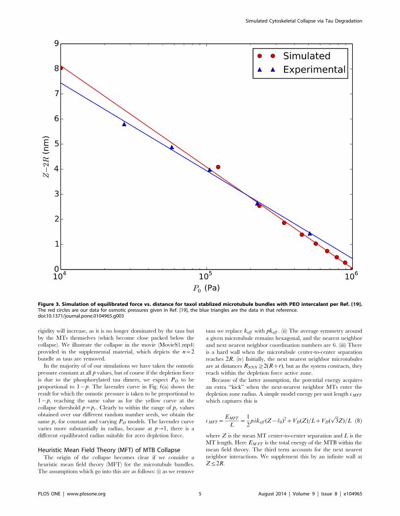
0.1 Test Case: simulation of taxol stabilized microtubuleswith PEO intercalants
We note that the depletion force has been observed in in vitroexperiments with taxol stabilized microtubule bundles intercalated
with PEO polymers (and no taus) [19]. In this case the only
additional interaction between the micotubules is via screened
Coulomb coupling, which is estimated by the force per unit
microtubule surface area PES
PES(Z)~0:078exp({(Z{2R)=LD)nN
nm2ð6Þ
with the Debye length LD = 1.47 nm. We add this force to our
depletion force to test our steepest descents equilibration as shown
in Fig. 3. Over a factor of 100 variation in PO, the agreement
between theory and the data in Fig. 11 of Needleman et al. [19] is
good. Note that given the large rest length of the taus, the
interaction PES is irrelevant prior to any collapse and so we
neglect it in our model for neuronal microtubules.
0.2 Model MT repulsion for axonal MTBsIn our simulations with taus for axonal MTBs, we mimic the
combined screened electrostatic repulsion/steric repulsion be-
tween microtubules by a Morse potential of the form
Figure 1. Two Dimensional Microtubule Bundle (MTB) Model. Microtubules are treated as rigid cylinders of diameter 25 nm (blue disks) withcenter-to-center distance of 45 nm (a). Taus are treated as springs, with 50 per micron length of a microtubule pair. To model tau depletion, taus areremoved at random and the system is re-equilibrated with the steepest descents method described in the text (b).doi:10.1371/journal.pone.0104965.g001
Simulated Cytoskeletal Collapse via Tau Degradation
PLOS ONE | www.plosone.org 3 August 2014 | Volume 9 | Issue 8 | e104965
VM (Z)~De 1{exp {a(Z{2R)½ �½ �2 ð7Þ
where we take De~10{4J and a = 1 nm21. While this potential
does provide a small minimum near the microtubule surfaces, its
main effect is to inhibit collapse beyond the region where
microtubules are in contact, which is sufficient for our purposes.
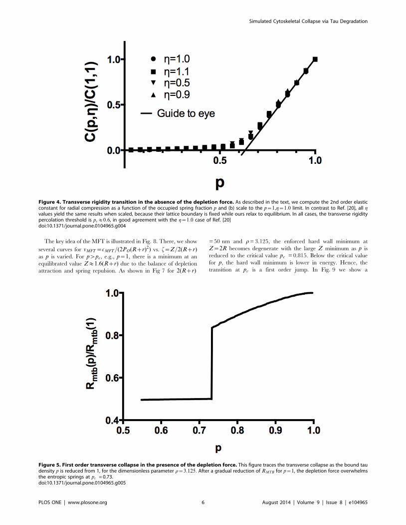
Test case: Rigidity Percolation with no Depletion ForceIn the depletion force free case, we have computed the second
order elastic constant C (Fig. 4) which measures the stiffness of the
microtubules against transverse compression for a variety of
different ratios g of the initial microtubule-to-microtubule
separation to spring resting length. In all cases, the scaled curve
for C(p,g)=C(1,1) is identical, since unlike the work of Ref. [20]
we do not hold our springs in a fixed frame under tension but
rather allow them to relax, so the result is essentially equivalent to
the case where bond length equals rest length in Ref. [20]. This
gives a transverse rigidity collapse (zero transverse elastic constant)
at a critical tau occupancy pc&0:63 in agreement with Ref. [20].
Note that we do not see a sharp threshold due to finite size effects;
we have confirmed that the rounding of the transition is reduced as
a function of increasing system size. We stress here that by
vanishing elastic constant, we mean that the MTB will initially lose
rigidity against radial compression, until the MTs can come into
direct steric contact, at which point the elastic constant will jump
to a value suitable for close packed MTs. Hence, even without
inclusion of the depletion force, we anticipate a significant
mechanical degradation (transverse rigidity loss) at a high p value
which exceeds the range (pƒ0:3) argued below to be relevant for
the dynamic instability mechanism.
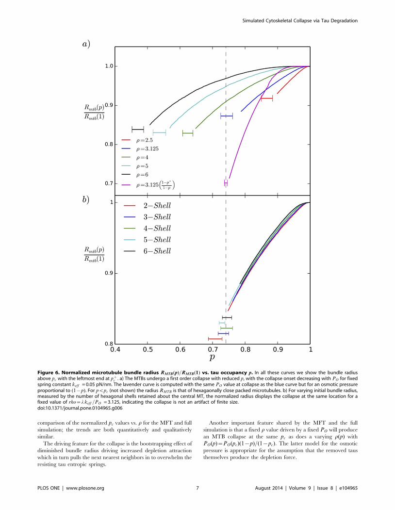
MTB Collapse with Depletion ForceWith the depletion force present, we find a first order volume
collapse as p is reduced, as shown in Fig. 5, for one value of the
dimensionless parameter r. This figure shows a typical plot of the
normalized transverse radius of gyration of the microtubule bundle
RMTB(p)=RMTB(1) decreasing slowly with decreasing p until the
critical percolation threshold pc is reached, at which point RMTB
drops precipitously to the hexagonal closed packed steric limit
RMTB(pc)~2nR~25n nm, where as defined above, n is the
number of hexagonal shells. The parameter r controls the position
of the collapse, as shown in Fig. 6(a), where we show curves for
pwpc such that the left endpoint is at pc: This collapse is
independent of the size of our starting bundle showing that it is not
an artifact of small bundles in our simulation as shown in Fig. 6(b)
where we plot the results for a single r value several bundle sizes.
Each curve in Fig. 6 is averaged over five runs and is truncated at
p~pc; the horizontal bar located at the collapse region represents
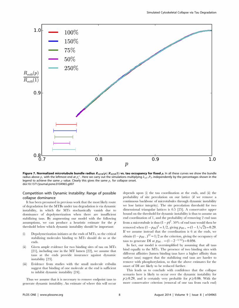
(by its width) the range of pc values obtained. Fig. 7 shows that
holding r fixed while co-varying PO,keff yields the same collapse
curves for pwpc. Note that below the collapse the compressional
Figure 2. Depletion mediated interaction between microtubules. For intercalants of radius r (presumed here to be hyperphosphorylatedtaus) an annulus between R,Rzr is depleted of intercalants. The loss of excluded volume as the microtubules approach allows increasedtranslational entropy outside the bundle for the intercalants.doi:10.1371/journal.pone.0104965.g002
Simulated Cytoskeletal Collapse via Tau Degradation
PLOS ONE | www.plosone.org 4 August 2014 | Volume 9 | Issue 8 | e104965
rigidity will increase, as it is no longer dominated by the taus but
by the MTs themselves (which become close packed below the
collapse). We illustrate the collapse in the movie (MovieS1.mp4)
provided in the supplemental material, which depicts the n~2bundle as taus are removed.
In the majority of of our simulations we have taken the osmotic
pressure constant at all p values, but of course if the depletion force
is due to the phosphorylated tau dimers, we expect PO to be
proportional to 1{p: The lavender curve in Fig. 6(a) shows the
result for which the osmotic pressure is taken to be proportional to
1{p, reaching the same value as for the yellow curve at the
collapse threshold p~pc: Clearly to within the range of pc values
obtained over our different random number seeds, we obtain the
same pc for constant and varying PO models. The lavender curve
varies more substantially in radius, because at p?1, there is a
different equilibrated radius suitable for zero depletion force.
Heuristic Mean Field Theory (MFT) of MTB CollapseThe origin of the collapse becomes clear if we consider a
heuristic mean field theory (MFT) for the microtubule bundles.
The assumptions which go into this are as follows: (i) as we remove
taus we replace keff with pkeff . (ii) The average symmetry around
a given microtubule remains hexagonal, and the nearest neighbor
and next nearest neighbor coordination numbers are 6. (iii) There
is a hard wall when the microtubule center-to-center separation
reaches 2R: (iv) Initially, the next nearest neighbor microtubules
are at distances RNNN§2(Rzr), but as the system contracts, they
reach within the depletion force active zone.
Because of the latter assumption, the potential energy acquires
an extra ‘‘kick’’ when the next-nearest neighbor MTs enter the
depletion zone radius. A simple model energy per unit length EMFT
which captures this is
EMFT~EMFT
L~
1
2plkeff (Z{‘0)2zVD(Z)=LzVD(
ffiffiffi3p
Z)=L ð8Þ
where Z is the mean MT center-to-center separation and L is the
MT length. Here EM FT is the total energy of the MTB within the
mean field theory. The third term accounts for the next nearest
neighbor interactions. We supplement this by an infinite wall at
Zƒ2R.
Figure 3. Simulation of equilibrated force vs. distance for taxol stablized microtubule bundles with PEO intercalant per Ref. [19].The red circles are our data for osmotic pressures given in Ref. [19], the blue triangles are the data in that reference.doi:10.1371/journal.pone.0104965.g003
Simulated Cytoskeletal Collapse via Tau Degradation
PLOS ONE | www.plosone.org 5 August 2014 | Volume 9 | Issue 8 | e104965
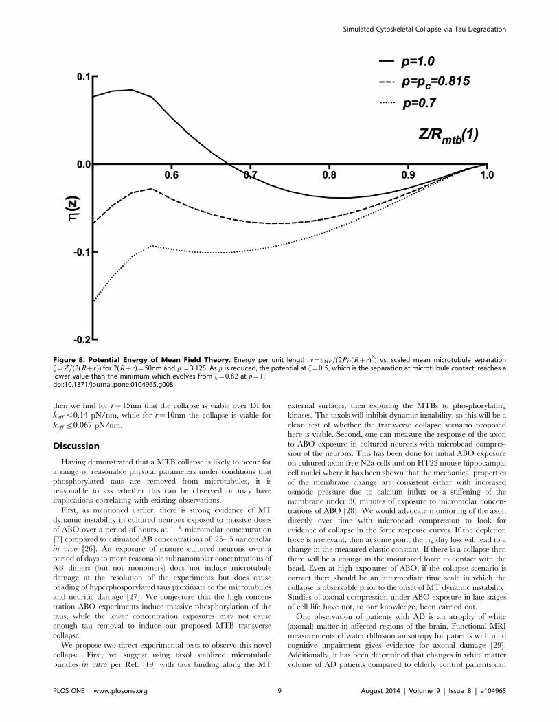
The key idea of the MFT is illustrated in Fig. 8. There, we show
several curves for nMFT~EMFT=(2PO(Rzr)2) vs. f~Z=2(Rzr)as p is varied. For pwpc, e.g., p~1, there is a minimum at an
equilibrated value Z&1:6(Rzr) due to the balance of depletion
attraction and spring repulsion. As shown in Fig 7 for 2(Rzr)
= 50 nm and r = 3.125, the enforced hard wall minimum at
Z~2R becomes degenerate with the large Z minimum as p is
reduced to the critical value pc = 0.815. Below the critical value
for p, the hard wall minimum is lower in energy. Hence, the
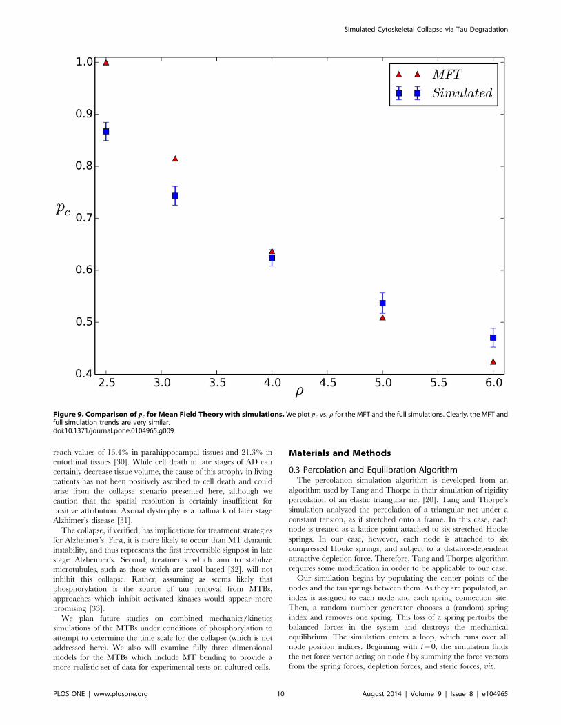
transition at pc is a first order jump. In Fig. 9 we show a
Figure 4. Transverse rigidity transition in the absence of the depletion force. As described in the text, we compute the 2nd order elasticconstant for radial compression as a function of the occupied spring fraction p and (b) scale to the p~1,g~1:0 limit. In contrast to Ref. [20], all gvalues yield the same results when scaled, because their lattice boundary is fixed while ours relax to equilibrium. In all cases, the transverse rigiditypercolation threshold is pc&0:6, in good agreement with the g~1:0 case of Ref. [20]doi:10.1371/journal.pone.0104965.g004
Figure 5. First order transverse collapse in the presence of the depletion force. This figure traces the transverse collapse as the bound taudensity p is reduced from 1, for the dimensionless parameter r~3:125: After a gradual reduction of RMTB for p~1, the depletion force overwhelmsthe entropic springs at pc = 0.73.doi:10.1371/journal.pone.0104965.g005
Simulated Cytoskeletal Collapse via Tau Degradation
PLOS ONE | www.plosone.org 6 August 2014 | Volume 9 | Issue 8 | e104965
comparison of the normalized pc values vs. r for the MFT and full
simulation; the trends are both quantitatively and qualitatively
similar.
The driving feature for the collapse is the bootstrapping effect of
diminished bundle radius driving increased depletion attraction
which in turn pulls the next nearest neighbors in to overwhelm the
resisting tau entropic springs.
Another important feature shared by the MFT and the full
simulation is that a fixed r value driven by a fixed PO will produce
an MTB collapse at the same pc as does a varying r(p) with
PO(p)~PO(pc)(1{p)=(1{pc): The latter model for the osmotic
pressure is appropriate for the assumption that the removed taus
themselves produce the depletion force.
Figure 6. Normalized microtubule bundle radius RMTB(p)=RMTB(1) vs. tau occupancy p. In all these curves we show the bundle radiusabove pc with the leftmost end at pz
c . a) The MTBs undergo a first order collapse with reduced p, with the collapse onset decreasing with PO for fixedspring constant keff = 0.05 pN/nm. The lavender curve is computed with the same PO value at collapse as the blue curve but for an osmotic pressureproportional to (1{p). For pvpc (not shown) the radius RMTB is that of hexagaonally close packed microtubules. b) For varying initial bundle radius,measured by the number of hexagonal shells retained about the central MT, the normalized radius displays the collapse at the same location for afixed value of rho~l keff =PO = 3.125, indicating the collapse is not an artifact of finite size.doi:10.1371/journal.pone.0104965.g006
Simulated Cytoskeletal Collapse via Tau Degradation
PLOS ONE | www.plosone.org 7 August 2014 | Volume 9 | Issue 8 | e104965
Competition with Dynamic Instability: Range of possiblecollapse dominance
It has been presumed in previous work that the most likely route
of degradation for the MTBs under tau degradation is via dynamic
instability, in which the MTs stochastically vanish due to
dominance of depolymerization when there are insufficient
stabilizing taus. By augmenting our model with the following
assumptions, we can produce a heuristic estimate for the pthreshold below which dynamic instability should be important:
(i) Depolymerization initiates at the ends of MTs, so the critical
stabilizing molecules binding to MTs should do so at the
ends.
(ii) Given ample evidence for two binding sites of tau on MTs
[21], including one in the MT lumen [22], we assume that
taus at the ends provide insurance against dynamic
instability [23].
(iii) Evidence from studies with the small molecule eribulin
suggest that binding of one molecule at the end is sufficient
to inhibit dynamic instability [24].
Thus we assume that it is necessary to remove endpoint taus to
generate dynamic instability. An estimate of where this will occur
depends upon (i) the tau coordination at the ends, and (ii) the
probability of site percolation on our lattice (if we remove a
continuous backbone of microtubules through dynamic instability
we lose lattice integrity). The site percolation threshold for two
dimensional triangular lattices is 0.5 [25]. A conservative upper
bound on the threshold for dynamic instability is thus to assume an
end coordination of 1, and the probability of removing 2 end taus
from a microtubule is thus (1{p)2: 50% of end taus would then be
removed when (1{pDI )2~1=2, giving pDIz~(1{1=ffiffiffi2p
)~0:29.
If we assume instead that the coordination is 6 at the ends, we
obtain (1{pDI{)12~1=2 as the criterion, giving the occupancy of
taus to generate DI at pDI{~(1{2{1=12)~0:056.
In fact, our model is oversimplified by assuming that all taus
bind equally to the MTs. The presence of two binding sites with
different affinities (lumen binding taus have a higher affinity than
surface taus) suggest that the stabilizing end taus are harder to
remove with phosphorylation, so that the above estimates for the
onset of DI are likely to be reduced further.
This leads us to conclude with confidence that the collapse
scenario here is likely to occur over the dynamic instability for
p§0:29, and is certainly very probable for p§0:06. With the
more conservative criterion (removal of one tau from each end)
Figure 7. Normalized microtubule bundle radius RMTB(p)=RMTB(1) vs. tau occupancy for fixed r. In all these curves we show the bundleradius above pc with the leftmost end at pz
c . Here we carry out the simulations multiplying keff ,PO independently by the percentages shown in thelegend to achieve the same r value. Clearly this gives the same pc for collapse onset.doi:10.1371/journal.pone.0104965.g007
Simulated Cytoskeletal Collapse via Tau Degradation
PLOS ONE | www.plosone.org 8 August 2014 | Volume 9 | Issue 8 | e104965
then we find for r~15nm that the collapse is viable over DI for
keff ƒ0:14 pN/nm, while for r~10nm the collapse is viable for
keff ƒ0:067 pN/nm.
Discussion
Having demonstrated that a MTB collapse is likely to occur for
a range of reasonable physical parameters under conditions that
phosphorylated taus are removed from microtubules, it is
reasonable to ask whether this can be observed or may have
implications correlating with existing observations.
First, as mentioned earlier, there is strong evidence of MT
dynamic instability in cultured neurons exposed to massive doses
of ABO over a period of hours, at 1–5 micromolar concentration
[7] compared to estimated AB concentrations of .25–.5 nanomolar
in vivo [26]. An exposure of mature cultured neurons over a
period of days to more reasonable subnanomolar concentrations of
AB dimers (but not monomers) does not induce microtubule
damage at the resolution of the experiments but does cause
beading of hyperphosporylated taus proximate to the microtubules
and neuritic damage [27]. We conjecture that the high concen-
tration ABO experiments induce massive phosphorylation of the
taus, while the lower concentration exposures may not cause
enough tau removal to induce our proposed MTB transverse
collapse.
We propose two direct experimental tests to observe this novel
collapse. First, we suggest using taxol stablized microtubule
bundles in vitro per Ref. [19] with taus binding along the MT
external surfaces, then exposing the MTBs to phosphorylating
kinases. The taxols will inhibit dynamic instability, so this will be a
clean test of whether the transverse collapse scenario proposed
here is viable. Second, one can measure the response of the axon
to ABO exposure in cultured neurons with microbead compres-
sion of the neurons. This has been done for initial ABO exposure
on cultured axon free N2a cells and on HT22 mouse hippocampal
cell nuclei where it has been shown that the mechanical properties
of the membrane change are consistent either with increased
osmotic pressure due to calcium influx or a stiffening of the
membrane under 30 minutes of exposure to micromolar concen-
trations of ABO [28]. We would advocate monitoring of the axon
directly over time with microbead compression to look for
evidence of collapse in the force response curves. If the depletion
force is irrelevant, then at some point the rigidity loss will lead to a
change in the measured elastic constant. If there is a collapse then
there will be a change in the monitored force in contact with the
bead. Even at high exposures of ABO, if the collapse scenario is
correct there should be an intermediate time scale in which the
collapse is observable prior to the onset of MT dynamic instability.
Studies of axonal compression under ABO exposure in late stages
of cell life have not, to our knowledge, been carried out.
One observation of patients with AD is an atrophy of white
(axonal) matter in affected regions of the brain. Functional MRI
measurements of water diffusion anisotropy for patients with mild
cognitive impairment gives evidence for axonal damage [29].
Additionally, it has been determined that changes in white matter
volume of AD patients compared to elderly control patients can
Figure 8. Potential Energy of Mean Field Theory. Energy per unit length n~EMF=(2PO(Rzr)2) vs. scaled mean microtubule separationf~Z=(2(Rzr)) for 2(Rzr)~50nm and r = 3.125. As p is reduced, the potential at f~0:5, which is the separation at microtubule contact, reaches alower value than the minimum which evolves from f~0:82 at p~1.doi:10.1371/journal.pone.0104965.g008
Simulated Cytoskeletal Collapse via Tau Degradation
PLOS ONE | www.plosone.org 9 August 2014 | Volume 9 | Issue 8 | e104965
reach values of 16.4% in parahippocampal tissues and 21.3% in
entorhinal tissues [30]. While cell death in late stages of AD can
certainly decrease tissue volume, the cause of this atrophy in living
patients has not been positively ascribed to cell death and could
arise from the collapse scenario presented here, although we
caution that the spatial resolution is certainly insufficient for
positive attribution. Axonal dystrophy is a hallmark of later stage
Alzhimer’s disease [31].
The collapse, if verified, has implications for treatment strategies
for Alzheimer’s. First, it is more likely to occur than MT dynamic
instability, and thus represents the first irreversible signpost in late
stage Alzheimer’s. Second, treatments which aim to stabilize
microtubules, such as those which are taxol based [32], will not
inhibit this collapse. Rather, assuming as seems likely that
phosphorylation is the source of tau removal from MTBs,
approaches which inhibit activated kinases would appear more
promising [33].
We plan future studies on combined mechanics/kinetics
simulations of the MTBs under conditions of phosphorylation to
attempt to determine the time scale for the collapse (which is not
addressed here). We also will examine fully three dimensional
models for the MTBs which include MT bending to provide a
more realistic set of data for experimental tests on cultured cells.
Materials and Methods
0.3 Percolation and Equilibration AlgorithmThe percolation simulation algorithm is developed from an
algorithm used by Tang and Thorpe in their simulation of rigidity
percolation of an elastic triangular net [20]. Tang and Thorpe’s
simulation analyzed the percolation of a triangular net under a
constant tension, as if stretched onto a frame. In this case, each
node is treated as a lattice point attached to six stretched Hooke
springs. In our case, however, each node is attached to six
compressed Hooke springs, and subject to a distance-dependent
attractive depletion force. Therefore, Tang and Thorpes algorithm
requires some modification in order to be applicable to our case.
Our simulation begins by populating the center points of the
nodes and the tau springs between them. As they are populated, an
index is assigned to each node and each spring connection site.
Then, a random number generator chooses a (random) spring
index and removes one spring. This loss of a spring perturbs the
balanced forces in the system and destroys the mechanical
equilibrium. The simulation enters a loop, which runs over all
node position indices. Beginning with i~0, the simulation finds
the net force vector acting on node i by summing the force vectors
from the spring forces, depletion forces, and steric forces, viz.
Figure 9. Comparison of pc for Mean Field Theory with simulations. We plot pc vs. r for the MFT and the full simulations. Clearly, the MFT andfull simulation trends are very similar.doi:10.1371/journal.pone.0104965.g009
Simulated Cytoskeletal Collapse via Tau Degradation
PLOS ONE | www.plosone.org 10 August 2014 | Volume 9 | Issue 8 | e104965
~FFi~~FFspring,iz~FFD,iz~FFsteric,i: ð9Þ
We then update the position~rri(t) to the new fictive time tzdtby adding a distance proportional to the net force
~rri(tzdt)~~rri(t)za~FFi ð10Þ
where a is an adjustable parameter used to control the step size.
We use a~10{6nm/pN. This fictive molecular dynamics assumes
the springs are overdamped or critically damped so that the nodes
do not oscillate as they relocate.
Once the last node is moved, we check to see if the system has
equilibrated to within a tolerance of 10{6 nm at each position
during the previous loop; if not, we rerun the force relaxation loop
until convergence at this tolerance is achieved. We average our
results over 10–15 different starting random number seeds.
To estimate the second order elastic constant C at a given pvalue, we uniformly stretch or compress the area by a small
dimensionless strain E~Da=a where a is the average bond length,
reequilibrate at the new strain value, and take C from
C~(E(zE)zE({E)){2E(E~0)½ �
E2ð11Þ
where E is the total potential energy. We take E sufficiently small
that we are in the quadratic in strain region.
Supporting Information
Movie S1
(MP4)
Acknowledgments
We acknowledge useful conversations with S. Feinstein, N. Hall, A. Karsai,
G.Y. Liu, C. DeCarli, and A. Levine.
Author Contributions
Conceived and designed the experiments: AS DLC HRF RRPS NRH.
Performed the experiments: AS HRF DLC. Analyzed the data: AS HRF
DLC RRPS. Contributed reagents/materials/analysis tools: NRH RRPS.
Contributed to the writing of the manuscript: DLC AS HRF RRPS NRH.
References
1. Hardy JA, Higgins GA (1992) The amyloid cascade hypothesis. Science 256:
184–185.
2. Hardy JA, Selkoe DJ (2002) The amyloid hypothesis of alzheimer’s disease:progress and problems on the road to therapeutics. Science 297: 353–356.
3. Fein JA, Sokolow S, Miller CA, Vinters HV, Yang F, et al. (2008) Co-localization of amyloid beta and tau pathology in alzheimer’s disease
synaptosomes. Am J Pathology 172: 1683–1692.4. McKee AC, Cantu RC, Nowinski CJ, Hedley-Whyte ET, Gavett BE, et al.
(2009) Chronic traumatic encephalopathy in athletes: Progressive tauopathy
following repetitive head injury. J Neuropathol Exp Neurol 68: 709–735.5. Wang JZ, Liu F (2008) Microtubule-associated protein tau in development,
degeneration, and protection of neurons. Progress in Neurobiology 85: 148–175.6. Reifert J, Hartung-Cranston D, Feinstein SC (2011) Amyloid b-mediated cell
death of cultured hippocampal neurons reveal extensive tau fragmentation
without increased full-length tau phos-phorylation. J Biol Chem 286: 20792–20811.
7. Zempel H, Thies E, Mandelkow E, Mandelkow EM (2010) A-beta oligomerscause localized ca2+ elevation, missorting of endogenous tau into dendrites, tau
phosphorylation, and destruction of microtubules and spines. J Neurosci 30:11938–11950.
8. Hirokawa N (1994) Microtubule organization and dynamics dependent on
microtubule-associated proteins. Curr Op Cell Biol 6: 74–81.9. Pontes B, Ayala Y, Fonseca ACC, Romao LF, Amaral RF, et al. (2013)
Membrane elastic properties and cell function. PLoS One 8: e67708.10. Press W, Teukolsky S, Vetterling W, Flannery B (2007) Numerical Recipes.
Cambridge: Cambridge Univ. Press, 3rd edition, 516–520 pp.
11. Brady ST, Colman DR, Brophy PJ (2013) Subcellular organization of thenervous system: Organelles and their functions. In: Squire L, editor,
Fundamental Neuroscience, Burlington MA: Academic Press. 3rd edition, p. 97.12. Rosenberg KJ, Ross JL, Feinstein HE, Feinstein SC, Israelachvili J (2008)
Complementary dimerization of microtubule-associated tau protein: Implica-
tions for microtubule bundling and tau-mediated pathogenesis. Proc Natl AcadSci USA 105: 7445–7450.
13. Phillips R, Kondev J, Theriot J (2008) Physical Biology of the Cell. New York:Garland Science, 1st edition, 313–315 pp.
14. Dietz H, Berkemeier F, Bertz M, Rief M (2006) Anisotropic deformationresponse of single protein molecules. Proc Nat Acad Sci USA 34: 12724–12728.
15. Chtcheglova LA, Shubeita GT, ASekatskii S, Dietler G (2004) Force
spectroscopy with a small dithering of afm tip: A method of direct andcontinuous measurement of the spring constant of single molecules and
molecular complexes. Biophys J 86: 1177–1184.16. Phillips R, Kondev J, Theriot J (2008) Physical Biology of the Cell. New York:
Garland Science, 1st edition, 526–528 pp.
17. Mylonas E, Hascher A, Bernado P, Blackledge M, Mandelkow E, et al. (2008)Domain conformation of tau protein studied by solution small-angle x-ray
scattering. Biochemistry 47: 10345–10353.
18. King ME, Ahuja V, Binder LI, Kuret J (1999) Ligand-dependent tau filament
formation: implications for alzheimer’s disease progression. Biochemistry 38:
14851–14859.
19. Needleman D, Ojeda-Lopez MA, Raviv U, Ewert K, Miller HP, et al. (2005)
Radial compression of microtubules and the mechanism of action of taxol and
associated proteins. Biophys J 89: 3410–3423.
20. Tang W, Thorpe MF (1988) Percolation of elastic networks under tension. Phys
Rev B 37: 5539–5551.
21. Chau MR, Radeke MJ, deInes C, Barasoain I, Kohlstaedt LA, et al. (1998) The
microtubule-associated protein tau crosslinks to two distinct sites on each a and btubulin monomer via separate domains. Biochemistry 37: 17692–17703.
22. Kar S, Fan J, Smith MJ, Goedert M, Amos LA (2003) Repeat motifs of tau bind
to the insides of microtubules in the absence of taxol. EMBO J 22: 70–77.
23. Hawkins TL, Sept D, Mogessie B, Straube A, Ross J (2013) Mechanical
properties of doubly stabilized microtubule filaments. Biophys J 104: 1517–
1528.
24. Smith J, Wilson L, Azarenko O, Zhu XJ, Lewis BM, et al. (2010) Eribulin binds
at microtubule ends to a single site on tubulin to suppress dynamic instability.
Biochemistry 49: 1331–1337.
25. Sykes MF, Essam JW (1964) Exact critical percolation probabilities for site and
bond problems in two dimensions. J Math Phys 5: 1117–1127.
26. Bero AW, Yan P, Roh JH, Cirrito JR, Steward FR, et al. (2011) Neuronal
activity regulates the regional response to amyloid-beta deposition. Nature
Neurosci 14: 750–756.
27. Jin M, Shepardson N, Yang T, Chen G, Walsh D, et al. (2011) Soluble amyloid
beta-protein dimers isolated from alzheimer cortex directly induce tau
hyperphosphorylation and neuritic damage. Proc Nat Acad Sci (USA) 108:
5819–5824.
28. Lulevich V, Zimmer CC, Hong HS, Jin LW, Liu GY (2010) Single-cell
mechanics provides a sensitive and quantitative means for probing amyloid-bpeptide and neuronal cell interactions. Proc Nat Acad Sci USA 107: 13872–
13877.
29. Huang J, Friedland RP, Auchus AP (2007) Diffusion tensor imaging of normal-
appearing white matter in mild cognitive impairment and early alzheimer
disease: Preliminary evidence of axonal degeneration in the temporal lobe.
Am J Neuroradiology 28: 1943–1948.
30. Salat DH, Greve DN, Pacheco JL, Quinn BT, Helmer KG, et al. (2009)
Regional white matter volume differences in nondemented aging and
alzheimer’s disease. NeuroImage 44: 1247–1258.
31. Lingor P, Koch JC, Toenges L, Baehr M (2012) Axonal degeneration as a
therapeutic target in the cns. Cell Tissue Res 349: 289–311.
32. Michaelis ML (2006) Ongoing in vivo studies with cytoskeletal drugs in
transgenic mice. Current Alzheimer Research 3: 215–219.
33. Mueller BK, Mack H, Teusch N (2005) Rho kinase, a promising drug target for
neurological disorders. Nature Rev Drug Discov 4: 387–398.
Simulated Cytoskeletal Collapse via Tau Degradation
PLOS ONE | www.plosone.org 11 August 2014 | Volume 9 | Issue 8 | e104965