Embed Size (px)
Citation preview

Histol Histopathol (1999) 14: 501-509
001: 10.14670/HH-14.501
http://www.hh.um.es
Histology and Histopathology
From Cell Biology to Tissue Engineering
Invited Review
Cytoskeletal proteins connecting intermediate filaments to cytoplasmic and nuclear periphery K.Ojabali School of Medicine, Pitie-Salpetriere, Cytoskeleton and Development, Paris, France
Summary. Interm ed iate filaments (I Fs), together with microtubules and microfil ament s build up the cytoskeleton of most eukaryotic cells. Cytoplasmic IFs form a dense fil ament network radiating from the nucleus and ex tendin g to th e pl asma membrane. The assoc iati on between the cytoplasmic and nuclear surfaces appears to provide a continuous link important for the organisat ion of th e cytopla m, for cellular co mmuni ca ti o n, and possibly for the transport into and ou t of the nucl eus. Cy topl as mi c IFs ap proach the nucl ea r surface, thin fibrils see m to connect the IFs with the nuclea r pore complexes and a direct interaction of cytoplasmic IFs with the nuclear lamin B has been observed by in vitro binding studies. However, none of the components that cross- link IFs to the nucleus has bee n unambiguously identified. Furthermore, if a direct interaction between cytoplasmic IFs and the nuclear lam in B occurs in vivo, the question of how cytoplasmic IFs get access to the nuclear interior remains to be resolved. The association of IFs with th e plasma membranes involves different components, some of which are cell type specific. Two specialised complexes in epi thelial cells: the desmosome and the hemidesmoso me, serve as attachment sites for kera tin filaments. Des moplakin is considered as the cross-linking co mpon ent of IFs to th e desmosomal plaque, whereas BPAG 1 (b ullous pemphigoid antige n) would cross-link IFs at the hemidesmosomal plaque. In other ce ll types the modality of how IFs are anchored to the plasma membrane is less well understood. It involves differe nt co mp o ne nts such as th e spectrin based membrane ske le to n, ankyrin, myos in , pl ec tin and certainly man y o ther st ill unrav e ll ed partners. Association between th e IFs and cellular membranes plays an important role in determining cell shape and tissue integrity. Thus, the identification and characterisa tion of the components involved in these interactions will be crucial for und ers tanding the function o f intermediate filaments.
Offprint requests to: Karima Oj abali. Facu lte de Medecine, Pit ie·
Salpetriere, CNRS·URA 2115, Cytosquelette et Developpement, 105.
Boulevard de I'Hopital. 75634 Paris Cedex 13. France. Fax: 00 33 t 53 60 08 02
Key words: Intermediate filament protein, Cy toske leton, Intermediate fil ament associated protein
Introduction
Recent works demonstrate that intermed iate filament ( IF) networ ks contribute to mai ntain th e s tru c tura l integrity of the ce ll and provide mechanical strength for tissue (S teinert and Roop, 1988). Mutations in different keratin proteins have been id entified as the cause of seve ral ti ss ue-specifi c human diseases (Fuchs, 1996; Fuchs and Cleveland, 1998). Such mutations resulted in a disorganised IF network that rendered cells very fragile and incapable of resisting any extracellular stress. The ove rall intrace llul ar organisation of IFs with their int e racti ng pa rtn e rs see ms to be import a nt for maintaining the shape and the pl asticity of the cell.
Int er medi a te filaments (IFs) constitute majo r cytoskeletal components of the eukaryoti c cy top lasm and of the nuclear lamina. Dcpending on cell type and developmental state, IFs can be assembled from single int e rm edi a te filament pro te in s ( IFPs) or from a combi nation thereof. IFPs constitute a multigene family whose members can be grouped into six categories (IVI) (Stewart , J990; Fuchs and Weber, 1994). In contrast to the ot her two filament systems, IFs are built from a multitude of developmentally regu lated and differentiation-specific subunits in such a way that the conserved a -helical rod domains of the subunits constitute the fil ament body, and th e non conserved , non a-helical terminal polypeptide regions are largely exposed on the filament surface (Ste inert and Roop, 1988). The surface reg io ns vary in s ize and sequ ence and correspond to different biochemical proper ti es, as well as to different interact ions with components and structures characteristic of developing and terminally differentiated cells (Ste inert et aI., 1985).
In a cellular context, cytoplasmic IFs are radiall y distributed from the nuclear membrane towards the cell surface (Goldman et aI. , ] 985). This implies site-specific recognition between IF ubunits, and binding to specific proteins of the different cellular structures. Analysis of
502
Intermediate filament and membrane interactions
IF function is difficult because of the complexity of its interaction partners. IFs have been shown to interact with the plasma membrane, nuclear envelope, mitochondria, microtubules (Georgatos and Maison, 1996), and actin filament bundles (Goldman et a!., 1986). Interactions of IFs with the plasma membrane occur at specialised attachment sites, focal adhesions, des mosomes and hemidesmosomes . A new family named plakin consisting of desmoplakin, bullous pemphigoid antigen 1, envoplakin and plectin, is thought to mediate IF association with contact structures at the plasma membrane (Schmidt et aI., 1994; Barradoti and Sonnenberg, 1996). Most of these proteins are expressed in different tissue and cell types, therefore suggesting different modes of attachment for the IFs depending on the subset of IFPs expressed in a cell type according to its developmental and differentiation stage.
IFs interact with microtubules and microfilaments
The IF-microtubule interaction was unravelled long ago. In most vimentin-containing cells, vimentin filaments form an extended network that stretches from the vicinity of the nucleus to the cellular periphery. This network of IFs appears to be established primarily through interaction with microtubules (Geiger and Singer, 1980). Depolymerization of the microtubule system leads to a collapse of the vimentin network into a dense coil near the cell centre (Blose et aI., 1984). This phenomenon suggests a dependence of IF network organisation upon the integrity of the microtubule network, implying a physico-chemical linkage between the two systems. Structural evidence for the existence of cross-bridges between the two cytoskeletal elements has been shown for the neurofilament IFs where MAP2 and kinesin have been implicated (Hirokawa, 1982; Leterrier et aI., 1982; Gyoeva and Gelfand, 1991). Interaction between IFs and micro filaments is generally apparent only upon drug disruption of the microtubule system. In the absence of microtubules, IFs undergo a microfilament-dependent centripetal collapse,which requires energy (Tint et aI., 1991). Currently, we assume that IFs are dragged outward through interactions with microtubules, and pulled inward through interactions with the microfilament system. Recent electron microscopy studies have revealed 2 nm linking elements connecting all three cytoskeletal systems. Plectin was identified as a versatile cross-linker (Svitkina et aI., 1996). Furthermore, plectin has a wide distribution among tissues and species, and is able to interact with different types of IFs such as vim entin, desmin, peripherin and neurofilament proteins, as well as with itself (Foisner and Wiche, 1991; Errante et a!., 1994). In vitro interactions showed also that plectin binds to the a-helical rod domain of vimentin (Foisner et aI., 1988). Interestingly, in cells lacking IFs, plectin is capable of cross-linking cytoskeletal structures independently of the IFs (Svitkina et aI., 1996). Plectin may enable sufficient cross-linking of the cytoplasm to itself and therefore may playa key role
in maintaining ti ssue integrity. This could explain the absence of obvious phenotypes in null vimentin mice (Colucci-Guyon et aI., 1994).
Connections of the IFs to the nuclear envelope
The nuclear envelope comprises three distinct regions: the outer nuclear membrane, the inner nuclear membrane and the nuclear pore complexes. The nuclear pore complexes are inserted into the nuclear membranes where the inner and outer membranes merge to form a pore. The outer nuclear membrane is continuous with the endoplasmic reticulum. The inner membrane faces the nucleoplasm and is linked to the nuclear lamina (Aebi et a!., 1986). The lamina is a thin fibrous structure composed of the nuclear lamins type A and B (Nigg, 1989) which appear to constitute a major structural framework for the nuclear envelope (Gerace and Blobel, 1980; Aebi et a!., 1986). The nuclear lam ins bind directly to chromosomes (Glass et a!., 1993), polynucleosomes, matrix-associated DNA and core histones (Taniura et a!., 1995). Several integral membrane proteins of the inner nuclear membrane are assumed to interact directly with the nuclear lamina (Georgatos et a!., 1994): the lamin B receptor (LBR) (Worman et a!., 1988) and the lamin-associated polypeptides (LAPs) (Foisner and Gerace, 1993). The LAPs (LAP1 A, LAP1 B, LAP1 C and LAP2) are typically integral membrane proteins with a single trans-membrane domain. LAP1 A and B represent splicing variants of LAP] C (Martin et a!., 1995). The LAPs bind directly to lamin para-crystals under in vivo conditions; LAP1 A and 1 B interact with all lamin types, while LAP2 associates exclusively with B-type lamins (Foisner and Gerace, 1993). LAP1 C binds to lamin A under in vivo conditions (Powell and Burke, 1990). The LBR is assumed to have eight membrane spanning segments, and a large positively charged amino-terminal domain facing the nucleoplasm that exhibits both lamin B and DNA binding activity (Worman et a!., 1990). LBR associates with at least two other proteins: p18 and p34 (Georgatos et a!., 1994).
There is one other group of nuclear envelope components with which the lamina might interact, namely the nuclear pore complexes. Structural studies as well as biochemical experiments clearly indicate such an association (Aebi et a!., 1986; Goldberg and Allen, 1992). This latter association must involve, at the very minimum, B-type lam ins, as these are the only members of the lamin family ubiquitously expressed in vertebrate somatic cells. However, no nuclear pore complex proteins which exhibit lamin-binding activity have been identified so far. Furthermore, several recent publications have reported the existence of lamins which are not associated with the envelope within the nuclear interior (Hozak et aI., 1995).
Several electron microscopy s tudies refer to an association of the cytoplasmic IFs with the nuclear envelope in a variety of cells (Metuzals et aI., 1988). The IFs are seen to loop and follow the nuclear periphery or

503 Intermediate filament and membrane interactions
to connect with the nuclear pore-complex (CarmoFonseca et aI., 1987). The fact that IFs are attached to the nucleus is further suggested by their persistence after nuclei isolation (Staufenbiel and Deppert, 1982). In vitro binding studies using isolated vimentin, desmin, and avian erythrocyte nuclear membrane, have revealed the existence of IF attachment sites along the nuclear envelope (Georgatos et aI., 1987). The binding was localised to the carboxy-terminal tail domain of the type III IFs and the nuclear receptor was identified as lamin B (Georgatos and Blobel, 1987; Djabali et aI., 1991). In addition, rabbits immunised with a synthetic peptide representing the proximal part of the carboxy-terminal region of peripherin , which is required for lamin B binding in vitro, produce anti-idiotypic antibodies that recognise lamin B (Djabali et aI., 1991). These results suggest that lamin B is a physiological receptor for IFs at the nuclear envelope. However, there still is a topographical problem to be resolved in order to explain how cytoplasmic IFs associate with the nuclear lam ins. Some IFs may traverse nuclear pore complexes since these are the only known channels between the nuclear interior and the cytoplasm. Ultrastructural studies at sites where IFs interact with the nuclear envelope will be required for us to understand how IFs are anchored . Immunofluorescence microscopy of cultured cells reveals a close association of plectin with the nuclear surface (Herrmann and Wiche, 1983). Solid phase binding assays show that plectin binds specifically to lamin B and not to lamin A and C. Phosphorylation of plectin and lamin B by different protein kinases significantly decreases the binding property between these two proteins (Foisner et aI., 1991). These results may indicate that during mitosis, since lam ins become hyperphosphorylated (Franke, 1987), the association between plectin and lamin B is disrupted which would suggest a highly dynamic mode of interaction between theses two partners. In addition to the binding between plectin and lamin B, binding between cytoplasmic IFs and lamin B, may also indicate an alternative link between cytoplasmic IFs and the nuclear lam in B. These different interactions are possible because plect in is a very elongated molecule self assembling into various shapes (Foisner and Wiche, 1987), and therefore may be able to pass through the nuclear pore and cross-link the IFs from the different compartments. It is noteworthy that cells lacking cytoplasmic IFs exhibit abnormal nuclear morphology (Serria et aI. , 1994). In these cells, the nucleus appears irregular with prominent folding or invaginations, while cells containing normal IF networks exhibit a uniform and smooth nucleus. These observations clearly imply an important role of the cytoplasmic IFs in positioning and maintaining the integrity of the nuclear envelope and, the shape of the nucleus.
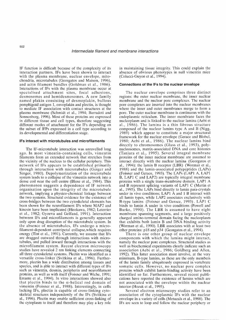
Our current knowledge is compatible with four alternative ways in which IFs could be anchored to the nuclear envelope (Fig. 1). 1) Cytoplasmic IFs may interact directly with the nuclear lamin B located on the inner surface of the nuclear envelope . However, a
topological problem will have to be resolved to understand how the cytop lasmic IFs can enter the nucleus in order to interact with the nuclear lamin B. Either the IFs somehow cross the nuclear envelope to get access to the inner surface of the nuclear envelope or they get access by entering through the nuclear pore complexes. 2) A crosslinking component, plectin, may mediate the interaction between cytoplasmic IFs and the nuclear envelope. 3) Components of the nuclear pore complex exposed to the cytoplasmic face may be responsible for the anchoring of IFs. So far, none of these elements has been reported apart from the existence of fibrils extending from the surface of the nuclear pore to the cytoplasm (Wiese and Wilson, 1993). 4) Another scenario could be the existence of IF associated-proteins loca lised on the extra-cellular nuclear membrane which would provide a direct link to the cytoplasmic IFs. Which of these four scenarios is true, remains to be established. Their respective research will certainly provide important insight into the function of IFPs.
Cytoplasm
Outer membrane
LAMIN
Nucleoplasm NPC
Fig. 1. Drawing of the nuclear envelope, sketching how IFs could connect the nucleus. IFs could interact with the nuclear envelope in four possible ways: 1) IF interacts with the lamin B by crossing the nuclear pore; 2) IF interacts with 2-nm fibrils exposed at the cytoplasmic surface of the nuclear pore; 3) IF interacts with cytoplasmic components of the nuclear pore complex ; and 4) IFs are anchored to the nuclear membrane via specific interactions with components associated to the outer nuclear membrane. IF: intermediate filament; F: 2 nm fibrils; Rlam: receptor for lamins; RIF: receptor for IF; NP: cytoplasmic components of the nuclear pore; NPC: nuclear pore complex.

504
Intermediate filament and membrane interactions
Connecti on s between IFs and the cytoplasmic membrane
The identification of the modality of IF interactions to the plasma membrane is complex because it involves several cytoskeletal components: actin filaments, microtubules and other membrane-associated proteins such as integrins, spectrin, plectin, a-actinin (Fig. 2). Specific interactions between the cytoskeleton and membraneassociated components are observed when capping of cell surface receptors occurs, or when cells become adherent to a substratum and when cell-cell contacts are formed (Lee et aI., 1988). The mechanism by which such special regions are organised indicates a highly dynamic process. A central role in assembly and maintenance of cell adhesions is attributed to the sub membrane plaques of cell-cell and cellextracellular contacts that link the adhesion receptors to the actin cytoskeleton (Bershadsky et a!., 1995). These structures consist of protein complexes that are specific either to cell-extracellular matrix junctions (such as talin, paxillin) or to cell-cell junctions (a- and /3-catenin, and plakoglobin) or that are shared by both types of adhesion (vinculin, a-actinin, zyxin and tensin) (Ben-Ze'ev, 1997) . Thus, actin filaments appear to be a key element in linking IFs to the plasma membrane.
IF interactions in non-specialised areas of the cytoplasmic membrane
Studies of the erythrocyte membrane provided the first isolation of the putative element involved in linking IFs to cortical actin. The major constituents of the submembranous erythrocyte cytoskeleton are a- and /3-spectrin forming a filamentous lattice attached to the membrane by ankyrin (Bennett, 1985). Recently, spectrin and ankyrin have been identified also in other cell types (Bennett, 1992). In vitro interaction using inverted erythrocyte membrane vesicles shows that vimentin binds to ankyrin (Georgatos and Marchesi, 1985), and that the amino-terminal domains of vimentin and desmin specifically bind to ankyrin in overlay assays (Georgatos et aI., 1987). Association of vimentin and desmin with erythrocyte spectrin is also observed (Langley and Cohen, 1987). Brain spectrin binds neurofilament proteins to a greater extent than does erythrocyte spectrin. Plectin is also reported to bind to spectrin (Herrmann and Wiche, 1987). Moreover, plectin interacts with several IF subunits (Foisner et a!., 1988) and may in fact mediate the linkage of IFs to spectrin and thus to the actin cell cortex. This possibility is supported by previous experiments showing that microinjection of anti-spectrin antibodies into fibroblasts results in a collapse of the IF network (Mangeat and Burridge, 1984). In conclusion, the emerging picture is that interactions between IFs and submembrane cortex involve three molecules: ankyrin, spectrin and plectin.
IF associations at cell-cell junctions
Epithelial cells exhibit two major types of adhering junctions: adherens junctions anchoring actin microfilaments and desmosomes anchoring intermediate filaments to the plasma membrane (Koch and Franke, 1994; Schmidt et aI., 1994). In adherens junctions, intracellular adhesion is caused by classical cadherins such as E- or N-cadherin (Geiger and Ayalon, 1992). Cadherins represent a distinct family of singletransmembrane domain glycoproteins. They form dimers in the plane of the membrane which in turn connect to those from opposing cells (Shapiro et a!., 1995). The carboxyl-terminal cytoplasmic domain of cadherins interacts directly with the central region of the two related proteins plakoglobin and /3-catenin (Ozawa et a!. , 1989; Sacco et a!., 1995). Furthermore, these proteins interact with a-catenin which then connects the membrane-associated complex to the actin cytoskeleton either directly or indirectly by association with a-actinin (another actin-binding protein) (Knudsen et a!., 1995; Huber et a!., 1997). IFs may be linked directly to the adhering junction, since actin can bind the vimentin tail domain, or indirectly by involving a cross-linking element such as plectin (Cary et a!., 1994; Svitkina et a!. , 1996). However, the in situ localisation of the association between IFs and components of the adhering junction remains to be established.
IFs are anchored at desmosomes, which are constituted by desmosome-specific cadherins: desmocollins and desmogleins (Koch and Franke, 1994; Chidgey, 1997). Desmosomal cadherins are glycosylated type I transmembrane proteins. Their homology to classical cadherins is strongest in their extracellular domains. Intracellular plaques of both types of adhering junctions contain one common component: plakoglobin. In desmosomes, plakoglobin specifically interacts with desmocollin and desmoglein but not with a-catenin (Chitaev et aI., 1996). Furthermore, desmocollin binds desmoplakins, which are cytoplasmic proteins of 220 kDa and 250 kDa derived from the same gene by alternative splicing. The carboxyl-terminal domain of desmoplakin interacts with the amino-terminal region of the basic keratins as well as with vimentin (Kouklis et a!., 1994). Therefore, desmoplakin provides a direct link between the IFs and the desmosomal plaque. However, other components are probably also involved in the interaction with IFs since experiments based on the overexpression of the carboxyl-terminal domain of desmoplakin produced only a partial collapse of the IFs from the desmosome (Stappenbeck and Green, 1992). An additional linker to IFs may be Band 6 or plakophilin, which binds to keratin and interacts in a similar fashion as plakoglobin with the same central region of desmoglein (Hatzfeld et a!., 1994; Marthur et a!. , 1994).
IF interactions to cell surface junctions
Hemidesmosomes are multi protein complexes that

505
Intermediate filament and membrane interactions
mediate the adhesion of epithelial cells to the underlying basement membrane and connect elements of the cytoskeleton to the extracellular matrix (Green and Jones, 1996). The keratin filaments are anchored to the cell surface at sites of the cytoplasmic plaques that are associated with hemidesmosomes. The plaque components involve at least two proteins: Bullous pemphigoid antigen 1 (BPAG1) (Stanley et aI., 1988), and plectin (Foisner and Wiche , 1991) . Studies demonstrate that both proteins are located at the cytoplasmic surface of the hemidesmosomes, where keratins are inserted (Jones et aI., 1994). Furthermore, BPAG1 shares sequence homology with plectin and desmoplakin (Tanaka et aI., 1991), and BPAG1 binds in vitro to keratin through their carboxyl-terminal domains (Yang et aI. , 1996). In conclusion, the interaction of keratins with hemidesmosomes is mediated by BPAG 1 and plectin.
Connections of the neuronal intermediate filament proteins
In epidermal tissues, several studies have characterised specific sites of attachment for keratin IFs. However, little is known on how neurofilaments (NFs) are anchored to the cytoplasmic surface of neurons. The neuronal cytoskeleton provides a framework that defines, supports, and maintains the shape of neurons. A highly crosslinked network of microtubules, neurofilaments, and specific associated proteins forms the major cytoplasmic structural units of the neuronal cell body, the dendrites and the axons. The NFs are composed of three proteins: NF-L, NF-M, NF-H. These proteins appear to be synthesised in the perikaryon region of the cell and are slowly transported down to the peripheral axonal processes. The NF proteins assemble as obligate heteropolymers, in which NF-M and NF-H are anchored to a core of NF-L through their central domains (Hirokawa et aI., 1984). The tail domains of the NF proteins are rich in charged amino acids, particularly glutamic acid. The charged tails of NF-M and NF-H protrude from the filament core and form at least a portion of the fibrous interconnections between parallel neurofilaments and microtubules in axons (Hirokawa et aI. , 1984). NF proteins are highly phosphorylated; the level of phosphorylation varies as they are transported from the cell body to the peripheral axon (Nixon et aI., 1994). Studies using monoclonal antibodies have distinguished phosphorylated and non-phosphorylated epitopes on the NFs. When these antibodies were applied to sections of rat brain, it was shown that certain nerve cell bodies, their dendrites and the portion of the proximal axon possessed non phosphorylated neurofilaments, and that long fibers, including terminal axons, contained phosphorylated neurofilaments (Sternberger and Sternberger, 1983) . In addition, the state of phosphorylation of NF proteins was directly correlated to the packing of the NFs and to the axonal caliber (Nixon et aI., 1994) .
Interaction between neurofilament proteins and the other cytoskeletal elements
In vitro studies have identified some putative components that could be implicated in the association between neuronal IFs and the cell periphery. Crosslinking components between NFs and microfilaments have recently been identified. In addition to plectin (Foisner and Wiche, 1991), another IF-microfilament cross-bridging protein has recently been revealed. The dystonin gene has been identified to be responsible for the mouse neurodegenerative disease dystonia musculorum (Brown et aI., 1995) and as being an isoform of the epidermal BPAG1e. Furthermore, the ablation of the BPAG1 gene in mice produced rapid sensory neuron degeneration identical to the symptoms of dystonia musculorum (Guo et aI., 1995). These observations lead to the conclusion that epidermal BPAG1e and its two neuronal variants BPAG1n1 and BPAG1n2 (also called dystonins) are alternatively spliced products of the same gene (Yang et aI., 1996). The neuronal BPAG 1 has the same sequence in the rod and carboxyl-terminal domain as the epidermal isoform, whi le the amino-terminal domain is more extended and contains an actin-binding site, absent in BPAG1e (Brown et aI., 1995). Furthermore, imm unohistochemistry shows that BPAG1n is found predominatly in axon termini, and not in the cell body of sensory neurons
Cytoplasm
Membrane
Ext racellular
ankyrin binding protein
spectrin binding protein
Fig. 2. Summary of several IF interactions in the neuronal context. IFs interact with the microtubule and microfilament systems. IFs can be anchored to the cell periphery by associations with different cytoskeletal components such as: 1) interaction with ankyrin; 2) interaction with spectrin; and 3) interaction with the cortical actin mediated by plectin or BPAG1n.

506
Intermediate filament and membrane interactions
(Yang et ai., 1996), whereas similar studies, in BPAGln knockout mice revealed that NF networks were disorganised and that axonal degeneration was accompanied by a reduction of NFs, especially in areas close to the axonal membrane (Yang et ai., 1996).
Possible candidates for linking NFs to the cell periphery
Brain spectrin has been localised and found associated to the plasmalemma in cell bodies, dendrites, axons, and nerve terminals (Kordeli et aI., 1986). Spectrin is an essential component of the membranerelated cytoskeleton and links actin filaments to the plasma membrane. The spectrin cytoskeleton is attached to the plasma membrane by membrane proteins such as ankyrin (Bennett, 1992) or by association between several integral membrane proteins such as neuronal cell adhesion molecule (N-CAM 180) (Pollerberg et aI., 1987). Furthermore, several in vitro studies using purified spectrin show the association of spectrin with Factin , the microtubule-associated protein Tau, and with neurofilament protein NF-L (Carlier et aI., 1984; Frappier et ai. , 1987). The multiplicity of membraneassociated domains in spectrin, together with the variability of spectrin ligands, may be responsible for selective targeting of spectrin to functionally distinct membrane domains and may therefore provide specific anchoring sites for the NFs to specialised domains of the peripheral membrane.
In vitro assays demonstrate that ankyrin binds specifically the amino-terminal domain of vimentin and desmin (Georgatos et ai., 1987). Similar assays have provided evidence that ankyrin could as well interact with peripherin, a neuronal IF of the same type (Djabali et ai., unpublished). However, binding of ankyrin to the neurofilament proteins has not been reported so far. Interestingly, multiple isoforms of ankyrin are expressed in the nervous system with diversity due to distinct genes, as well as to alternative splicing of mRNAs. The ankyrin B is located in neuronal processes while ankyrin R is confined to the cell bodies and dendrites; and ankyrin node is localised at axonal initial segments and nodes of Ranvier (Bennett, 1992). Furthermore, ankyrinbinding glycoproteins have been identified as members of the neuronal cell adhesion molecules (N-CAMs) (Davis et aI. , 1993). Associations of N-CAMs with ankyrin, and of ankyrin to NFs, may provide an example for a series of protein-protein interactions extending from the extracellular space to the cytoplasm and may therefore constitute a novel signalling pathway.
Figure 2 summarises different possible models of how NFs could be attached to the cell periphery. These models have been hypothesised based on the different components identified as cross-linking elements for IFs and the plasma membrane which have been essentially provided by studies of epithelial cells and erythrocyte membranes. Based on these findings, I propose three alternative ways that may be implicated in NF-cytoplasmic membrane interactions. 1) NFs could directly
interact with spectrin which has been shown to be associated with the cortical cytoplasm of cell bodies, dendrites, and axons (Levin and Willard, 1981). Furthermore, spectrin could also cross-link the NFs between themselves, as well as to the microfilament system. 2) NFs could associate with ankyrin, a structural protein located on the cytoplasmic surface of the plasma membrane (Bennett, 1992). Moreover, ankyrin exhibits recognition sites for integral membrane proteins, which may contribute to specific regions of distribution in neurons and thereby, may contribute to the formation of specific multi-protein complexes for NF attachment. 3) Another alternative could be that NFs are indirectly linked to the plasma membrane. NFs could be crosslinked through associated proteins such as plectin and lor BPAGln. These proteins have been shown to interact with NFPs (Foisner and Wiche, 1991; Yang et ai., 1996). Furthermore, plectin and BPAG1n have been implicated in crosslinking NFs to the microfilament system.
Conclusion
The exact function of intermediate filaments still remains enigmatic. However, in the case of defective genes encoding some of the IFPs, it has been demonstrated that they are responsible for degenerative diseases (Fuchs, 1996). The intra-cytoarchitectural organisation of IFs and their mode of interaction with specific components may provide further insight into their function. For example, in epithelial cells, keratin proteins are connected to hemidesmosomes that are integrin-mediated adhesive junctions. Furthermore, the keratin IFs are also attached to desmosomes, that are cadherin-mediated cell-cell junctions. Both multi-protein complexes have been implicated in cell signalling events. These findings may suggest that IFs can act as signal transducers, relaying information from the extracellular matrix, or from cell-cell adhesion to the nucleus, which would indicate a new dynamic property of these IFPs. To date, several cross-linking proteins have been characterised, and some of them have been identified as important in the intracellular organisation of the IF network. It is now clear that an intact and well organised cytoplasmic IF is required for the cell to maintain its integrity. Identification and characterisation of the components involved in IF arrangement would probably help in the near future to unravel the real physiological role of the different IF subunits.
Acknowledgements. I would like to thank Dr. M-M. Portier (CNRS-URA 2115) for her constant interest and support of the work. I am grateful to B. Rost (EMBL, Heidelberg), D. Martin (Paris) , G. Piron and B de Nechaud (CNRS-URA 2115) for useful discussion and critical reading of the manuscript. The work of the laboratory of M-M . Portier was supported by Centre National de la Recherche Scientifique (CNRS,
URA 2115) , Institut National de la Sante et de la Recherche Medicale (INSERM, grant 910810), Association Fran<;aise contre les Myopathies (AFM) and Direction des Recherches-Etudes et Techniques (DRET, grant 94-2541 A).

507
Intermediate filament and membrane interactions
References
Aebi U., Cohn J.B. and Gerace L.L. (1986) . The nuclear lamina is a meshwork of intermediate-type filaments. Nature 323, 560-564.
Barradoti L. and Sonnenberg A. (1996) . Hemidesmosomes: roles in adhesion , signaling and human diseases. Curro Opin. Cell BioI. 8, 647-656.
Ben-Ze'ev A. (1997) . Cytoskeletal adhesion proteins as tumor suppressors. Curro Opin. Cell BioI. 9, 99-108.
Bennett V. (1985) . The membrane skeleton of human erythrocyte and its implications for more complex cells. Annu. Aev. Biochem. 54, 273-304.
Bennett V. (1992) . Ankyrins : adaptors between diverse plasma membrane proteins and cytoplasm. J. BioI. Chem. 267, 8703-8706.
Bershadsky A.D ., Yehuda-Levenberg S. and Geiger B. (1995). Molecular interactions in the submembrane plaque of cell-cell and cell-matrix adhesions. Acta. Anat. 154, 46-62.
Blose S.H., Meltze 0 .1. and Feramisco J.A. (1984) . 10-nm filaments are induced to collapse in living cells microinjected with monoclonal and polyclonal antibodies against tubulin. J. Cell BioI. 98, 847-859.
Brown A., Bernier G. and Mathieu M.A. and J. Kothary A. (1995) . The mouse dystonia musculorum gene is a neuronal isoform of bullous pemphygoid antigen 1. Nature Gen. 10, 301-306.
Carlier M.F. , Simon C. , Cassoly A. and Pradel L.A. (1984) . Interaction between microtubule-associated protein tau and spectrin. Biochim. 66, 305-311.
Carmo-Fonseca M., Cidadao A.J . and David-Ferreira D.F. (1987). Filamentous cross-bridges link intermediate filaments to the nuclear pore complexes. Eur. J. Cell BioI. 45, 282-290.
Cary A.B., Klymkowsky MW., Evans A.M. , Domingo A., Dent J.A. and Backhus L.E. (1994) . Vimentin 's tail interacts with actin-containing structures in vivo. J. Cell Sci. 107, 1609-1622.
Chidgey MAJ. (1997) . Desmosomes and disease. Histol. Histopathol. 12, 1159-1168.
Chitaev N.A., Leube A.E., Troyanovsky A.B., Eshkind L.G. and Franke
WW. (1996). The binding of plakoglobin to desmosomal cadherins: patterns of binding sites and topogenic potential. J. Cell BioI. 133, 359-369.
Colucci-Guyon E., Portier M.-M. , Dunia I. , Paulin D., Pournin S. and Babinet C. (1994) . Mice lacking vimentin develop and reproduce without an obvious phenotype. Cell 79, 679-694.
Davis J.a ., McLaughlin T. and Bennett V. (1993). Ankyrin -binding proteins related to nervous system cell adhesion molecules : candidates to provide transmembrane and intercellular connections in adult brain. J. Cell BioI. 121 , 121-133.
Djabali K , Portier M.-M., Gros F., Blobel G. and Georgatos S.D. (1991 ). Network antibodies identify nuclear lamin B as a physiological attachment site for peripherin intermediate filaments. Cell 64, 109-121
Errante L.D., Wiche G. and Shaw G. (1994) . Distribution of plectin , an
intermediate filament-associated protein, in the adult rat central nervous system. J. Neurosci. Aes. 37, 515-528.
Foisner A. and Gerace L. (1993) . Integral membrane proteins of the nuclear envelope interact with lamins and chromosomes , and binding is modulated by mitotic phosphorylation . Cell 73, 1267-1279.
Foisner A. and Wiche G. (1987) . Structure and hydrodynamic properties of plectin molecules. J. Mol. BioI. 198, 515-531.
Foisner A. and Wiche G. (1991) . Intermediate filaments -associated proteins. Curro Opin. Cell BioI. 3, 75-81.
Foisner A., Leichtrfried F.E. , Herrman H., Small J.v., Lawson D. and
Wiche G. (1988) . Cytoskeletion -associated plectin: In situ localization , in vitro reconstitution , and binding to immobilized intermediate filament proteins. J. Cell BioI. 106, 723-733.
Foisner A. , Traub P. and Wiche G. (1991). Protein kinase A- and protein Kinase C-regulated interaction of plectin with lamin Band vimentin. Proc. Natl. Acad. Sci. USA 88, 3812-3816.
Franke W.W. (1987). Nuclear lamins and cytoplasmic intermediate filament proteins: a growing multigene family. Cell 48, 3-4.
Frappier T., Aegnouf F. and Pradel L.A. (1987) . Binding of brain spectrin to the neurofilament subunit protein. Eur. J. Biochem. 169, 651-657.
Fuchs E. (1996). The cytoskeleton and disease: genetic disorders of intermediate filaments. Annu . Aev. Genet. 30, 197-231 .
Fuchs E. and Cleveland D.W. (1998). A structural scaffolding of intermediate filaments in health and disease. Science 279, 514-519.
Fuchs E. and Weber K. (1994) . Intermediate filaments : structure, dynamics, function, disease. Annu. Aev. Biochem. 63, 345-382.
Geiger B. and Ayalon O. (1992) . Cadherins. Annu . Aev. Cell BioI. 8, 307-332.
Geiger B. and Singer S.J . (1980) . Association of microtubules and intermediate filaments in chicken gizzard cells as detected by double immunofluorescence. Proc. Natl. Acad. Sci. USA 77, 4769-4773.
Georgatos S.D. and Blobel G. (1987). Lamin B constitutes an intermediate filament attachment site at the nuclear envelope. J. Cell Biol.l05, 117-125.
Georgatos S.D. and Maison C. (1996). Integration of intermediate filaments into cellular organelles. Int. Aev. Cytol. 164, 91 -138.
Georgatos S.D. and Marchesi V.T. (1985). The binding of vimentin to human erythrocyte membranes: A model system for the study of intermdiate filament-membrane interactions. J. Cell BioI. 100, 1955-1961 .
Georgatos S.D., Meier J. and Simons G. (1994) . Lamins and laminassociated proteins. Curr. Opin. Cell BioI. 6, 347-353.
Georgatos S.D., Weber K., Geisler N. and Blobel G. (1987) . Binding of two desmin derivatives to the plasma membrane and the nuclear envelope of avian erythrocytes: Evidence for a conserved site
specificity in intermediate filament-membrane interactions. Proc . Nalt. Acad . Sci. USA 84, 6780-6784.
Gerace L. and Blobel G. (1980). The nuclear envelope lamina is reversibly de polymerized during mitosis. Cell 19, 277-287.
Glass CA, Glass J.A. , Taniura H., Hasel KW., Blevitt J.M. and Gerace L. (1993). The a-helical rod domain of human lamins A and C contains a chromatin binding site. EMBO J. 12, 4413-4423.
Goldberg M.W. and Allen T.D. (1992) . High resolution scanning electron microscopy of the nuclear envelope: demonstration of a new, regular, fibrous lattice attached to the baskets of the nucleoplasmic face of the nuclear pores. J. Cell BioI. 119, 1429-1440.
Goldman A.D., Goldman A., Green K. , Jones J., Lieska N. and Yang H.Y . (1985) . Intermediate filaments : possible functions as cytoskeletal connecting links between the nucleus and the cell surface. Ann . NY Acad . Sci . 455, 1-17.
Goldman A.D ., Goldman A., Green K, Jones J., Lieska N. and Yang H.Y. (1986). Intermediate filaments networks: organization and possible functions of a diverse group of cytoskeletal elements. J. Cell Sci. 5, 69-97.
Green K.J . and Jones J.C .A. (1996) . Desmosomes and hemidesmosomes: structure and function of molecular components . FASEB J. 10, 871-881.
Guo L. , Degenstein L. , Dowling J. , Yu a .c ., Wollmann A. , Perman B.

508
Intermediate filament and membrane interactions
and Fuchs E. (1995). Gene targeting of BPAG1 : abnormalities in mechanical strenght and cell migration in stratified squamous
epithelia and severe neurologic degeneration. Cell 81 , 233-243. Gyoeva F.K. and Gelfand V.1. (1991). Coalignement of vimentin
intermediate filaments with microtubules depends on kinesin. Nature 353, 445-448.
Hatzfeld M., Kristjansson G.!', Plessmann U. and Weber K. (1994). Band 6 protein, a major constituent of desmosomes from stratified epithelia, is a novel member of the armadillo mulligen family. J. Cell Sci. 107, 2259-2270.
Herrmann H. and Wiche G. (1983) . Specific in situ phosphorylation of plectin in detergent-resistant cytoskeletons from cultured chinese
hamster ovary cells. J. BioI. Chem. 258, 14610-14618. Herrmann H. and Wiche G. (1987). Plectin and IFAP- 300 K are
homologous proteins binding to microtubule-associated proteins 1
and 2 and to the 240 kilodalton subunit of spectrin. J. BioI. Chem. 262,1320-1325.
Hirokawa N. (1982). Cross-linker sytem between neurofilaments , microtubules and membranous organelles in frog axons revealed by
the quick-freeze deep-etching method. J. Cell BioI. 94, 129-142. Hirokawa N., Glicksman M.A. and Willard M.B. (1984) . Organization of
mammalian neurofilament polypeptides within the neuronal cy1oskeleton. J. Cell BioI. 98, 1523-1536.
Hozak P., Sasseville A.M.J., Raymond Y. and Cook P.R. (1995). Lamin proteins form internal nucleoskeleton as well as a peripheral lamina in human cells. J. Cell Sci. 108, 635-644.
Huber 0., Krohn M. and Kemler A. (1997) . A specific domain in 0-
catenin mediates binding to f3-catenin or plakoglobin . J. Cell Sci. 110, 1759-1765.
Jones J.C.R., Asmuth J., Baker S.E., Langhofer M., Roth S.1. and Hopkinson S.B. (1994) . Hemidesmosomes: extracellular matrix/ intermediate filament connectors. Exp. Cell Res. 213, 1-11.
Knudsen K.A., Soler A.P. , Johnson K.R. and Wheelock M.J. (1995) . Interaction of alpha-actinin with the cadherin/catenin cell-cell
adhesion complex via alpha-catenin. J. Cell BioI. 130,67-79. Koch P.J. and Franke W.W. (1994). Desmosomal cadherins: another
growing multigene family of adhesion molecules. Curro Opin. Cell BioI. 6, 682-687.
Kordeli E. , Cartaud J. , Nghiem H.O., Pradel L.A., Dubreuil C., Paulin D. and Changeux J.P. (1986). Evidence for a polarity in the distribution of proteins from the cy1oskeleton in Torpedo marmorata electrocy1e. J. Cell BioI. 102,748-760.
Kouklis D.P., Hutton E. and Fuchs E. (1994). Making a connection : direct binding between keratin intermediate filaments and desmosomal proteins. J. Cell BioI. 127, 1049-1060.
Langley A.J.J. and Cohen C.M. (1987). Cell type-specific association between two types of spectrin and two types of intermediate filaments. Cell. Moti!. Cy1osk. 8,165-173.
Lee J.K., Black J.D., Repasky E.A. and Bankert A.B. (1988). Activation induced a rapid reorganization of spectrin in lymphocy1es. Cell 55, 807-816.
Leterrier J.F., Liem A.K. and Shelanski M. (1982). Interaction between
neurofilaments and microtubule-associated proteins, a possible mechanism for interorganellar bridging. J. Cell BioI. 95, 982-986.
Levin J. and Willard M. (1981). Fodrin axonally transported polypeptides
associated with the internal periphery of many cells. J. Cell BioI. 90, 631-643.
Mangeat P.H . and Burridge K. (1984) . Immunoprecipitation of nonerythrocyte spectrin within live cells following microinjection of
specific antibodies: relation to cy10skeletal structures. J. Cell BioI.
98, 1363-1377. Marthur M. , Goodwin L. and Cowin P. (1994). Interactions of a
desmosomal cadherin, Dsgl, with plakoglobin. J. Bioi. Chem. 269,
14075-14080. Martin L. , Crimaudo C. and Gerace L. (1995) . cDNA cloning and
characterization of lamina-associated polypeptide 1 C (LAPCl C) an integral protein of the inner nuclear membrane. J. BioI. Chem. 270,
8822-8828. Metuzals J., Robitaille Y., Houghton S., Gauthier S. and Leblanc R.
(1988) . Paired helical filaments and the cytoplasmic-nuclear
interface in Alzheimer's disease. J. Neurocy1ol. 17,827-833. Nigg E.A. (1989) . The nuclear envelope. Curr. Opin. Cell BioI. 1, 435-
440. Nixon R.A., Paskevich P.A., Sihag RX and Thayer C.Y. (1994).
Phosphorylation on carboxyl terminus domains of neurofilament proteins in retinal ganglion cell neurons in vivo: influences on regional neurofilament accumulation, interneuronfilament spacing,
and axon caliber. J. Cell BioI. 126, 1031-1046. Ozawa M., Baribault H. and Kemler R. (1989). The cy10plasmic domain
of the cell adhesion molecule uvomurilin associates with three independent proteins structurally related in different species. EMBO J. 8, 1711 -1717.
Pollerberg G.E. , Burridge K. , Krebs K.E. , Goodman S.R. and Schachner M. (1987). The 180 KD component of the neuronal cell adhesion molecule N-CAM is involved in cell-cell adhesion and cy1oskeletonmembrane interactions. Cell Tissue Res. 250, 227-236.
Powell L. and Burke W.B. (1990). Internuclear exchange of an inner nuclear protein (p55) in heterokaryons: in vivo evidence for the interaction of p55 with the nuclear lamina. J. Cell BioI. 111 , 2225-
2234. Sacco P.A., McGranahan M., Wheelock M.J. and Johnson K.R. (1995).
Identification of plakoglobin domains required for association with Ncadherin and alpha-catenin. J. BioI. Chem. 270, 20201-20206.
Schmidt A. , Heid H.w., Schafer S., Nuber U.A., Zimbelmann R. and Franke W.W. (1994). Desmosomes and cy10skeletal architecture in epithelial differentiation: cell type-specific plaque components and
intermediate filament anchorage. Eur. J. Cell BioI. 65, 229-245. Serria A.J., Lieber J.G., Nordeen S.K. and Evans R.M. (1994). The
presence or absence of a vimentin-like intermediate filament network affects the shape of the nucleus in SW-13 cells. J. Cell Sci. 107, 1593-1607.
Shapiro L., Fannon A.M. , Kwong P.O. , Thompson A., Lehman M.S., Grubel G., Legrand J., Nielson A., Colman J. and Hendrickson W.A. (1995). Structural basis of cell-cell adhesion by cadherins. Nature 374 , 327-336.
Stanley J.R., Tanaka T., Mueller S., Klaus-Kovtun V. and Roop D.
(1988) . Isolation of cDNA for bullous pemphigoid antigen by use of patients' autoantibodies. J. Clin. Invest. 82, 1864-1870.
Stappenbeck T.S. and Green K.J. (1992) . The desmoplakin carboxyl terminus coaligns with and specifically disrupts intermediate filament networks when expressed in culture cells. J. Cell BioI. 116, 1197-
1209. Staufenbiel M. and Deppert W. (1982). Intermediate filament systems
are collapsed onto the nuclear surface after isolation of nuclei from tissue cullure cells. Exp. Cell Res. 138,207-214.
Steinert P.M. and Roop D.A. (1988) . Molecular and cellular biology of intermediate filaments. Annu . Rev. Biochem. 57, 593-625.
Steinert P.M., Steven A.C. and Roop D.R. (1985) . The molecular
509
Intermediate filament and membrane interactions
biology of intermediate filaments. Cell 42, 411-419. Sternberger L.A. and Sternberger N.H. (1983). Monoclonal antibodies
distinguish phosphorylated and nonphosphorylated forms of neurofilaments in situ. Proc . Natl. Acad. Sci. USA 80, 6126-
6130. Stewart M. (1990). Intermediate filaments: structure, assembly and
molecular interactions. Curr. Opin. Cell BioI. 2, 91-100. Svitkina T.M., Verkhovsky A.B. and Borisy G.G. (1996). Plectin
sidearms mediate intercation of intermediate filaments with
microtubules and other components of the cy1oskeleton. J. Cell BioI.
135, 991-1007. Tanaka T., Parry DAD., Klaus-Kov1un V., Steinert P.M. and Stanley
J.R. (1991). Comparison of molecularly cloned bullous pemphigoid
antigen to desmoplakin I confirms that they define a new family of cell adhesion junction plaque proteins. J. BioI. Chem. 266, 12555-
12559. Taniura H. , Glass C. and Gerace L. (1995). A chromatin binding site in
the tail domain of nuclear lam ins that interacts with core histones. J.
Cell BioI. 131, 33-44.
Tint I.S., Hollenbeck P.J., Verkhovsky A.B. , Surgucheva I.G. and
Bershadsky A.D. (1991). Evidence that intermediate filament reorganisation is induced by ATP-dependent contraction of acto
myosin cortex in permeabilized fibroblast. J. Cell Sci. 98, 375-384.
Wiese C. and Wilson K.L. (1993). Nuclear membrane dynamics. Curro Opin. Cell BioI. 5, 387-394.
Worman H.J., Evans C.D. and Blobel G. (1990). The lamin B receptor of the nuclear envelope inner membrane: a poly1opic protein with eight
potential transmembrane domains. J. Cell BioI. 111 , 1535-1542. Worman H.J., Yuan J., Blobel G. and Georgatos S.D. (1988). A lamin B
receptor in the nuclear envelope. Proc. Natl. Acad. Sci. USA 85,
8531-8534. Yang Y., Dowling J. , Yu a.c., Kouklis P., Cleveland D.W. and Fuchs E.
(1996). An essential cytoskeletal linker protein connecting actin
microfilaments to intermediate filaments. Cell 86, 655-665.
Accepted July 27, 1998
![RhoA Cytoskeletal Pathway to Transplantation...superfamily of small GTP-binding proteins. These proteins occur ... regulation of focal adhesions and actin stress fibers assembly [8,29],](https://img.dokumen.tips/doc/110x75/60f458f03515ca574b71d30e/rhoa-cytoskeletal-pathway-to-transplantation-superfamily-of-small-gtp-binding.jpg)