Embed Size (px)
Citation preview
© 2017. Published by The Company of Biologists Ltd.
Nestin contributes to skeletal muscle homeostasis and regeneration
Julia Lindqvist1, 2*, Elin Torvaldson1, 2*, Josef Gullmets1, 2, 3, Henok Karvonen1, 2,
Andras Nagy4, Pekka Taimen3, and John E. Eriksson1, 2#
1Cell Biology, Biosciences, Faculty of Science and Engineering, Åbo Akademi University, Turku, Finland
2Turku Centre for Biotechnology, University of Turku and Åbo Akademi University, Turku, Finland
3Department of Pathology, University of Turku and Turku University Hospital, Turku, Finland
4Lunenfeld-Tanenbaum Research Institute, Mount Sinai Hospital, Toronto, Canada
*These authors contributed equally to this work
#Corresponding author
Key words: Intermediate filaments, nestin, Cdk5, muscle, regeneration, differentiation
Jour
nal o
f Cel
l Sci
ence
• A
dvan
ce a
rtic
le
JCS Advance Online Article. Posted on 21 July 2017
Summary
Muscles from nestin knockout mice have decreased mass, are prone to spontaneous
degeneration/regeneration and have longer recovery time after injury. This phenotype is
linked to disturbed proliferation of satellite cells.
Abstract
Nestin, a member of the cytoskeletal family of intermediate filaments, regulates the onset of
myogenic differentiation through bidirectional signaling with the Cdk5-kinase. Here we show
that these effects are also reflected at the organism level, as there is a loss of skeletal muscle
mass in nestin-/- (NesKO) mice, reflected as reduced lean mass in the mice. Further
examination of muscles in male mice revealed that these effects stemmed from nestin-
deficient muscles being more prone to spontaneous regeneration. When the regeneration
capacity of the compromised NesKO muscle was tested by muscle injury experiments, a
significant healing delay was observed. NesKO satellite cells showed delayed proliferation
kinetics in conjunction with an elevation in p35 levels and Cdk5 activity. These results reveal
that nestin-deficiency generates a spontaneous regenerative phenotype in skeletal muscle that
relates to a disturbed proliferation cycle, which is associated with uncontrolled Cdk5 activity.
Jour
nal o
f Cel
l Sci
ence
• A
dvan
ce a
rtic
le
INTRODUCTION
Intermediate filament proteins (IFs) are part of the cytoskeleton that provides mechanical
stability to the cell. In addition, they are crucial regulators of cell signaling and contribute to
several important cellular functions (Gruenbaum and Aebi, 2014; Hyder et al., 2011).
Through their association with organelles, structural proteins and signaling executors, IFs are
necessary components of the signaling pathways that drive cell migration, adhesion,
proliferation, differentiation and death. IFs have the ability to spontaneously form homo- or
heteropolymers, which are fused into long filamentous structures that are constantly
remodeled according to the cells’ needs.
The use of genetically engineered mice have revealed essential structural functions for major
muscle IFs, such as for desmin, the lack of which disturbs sarcomeres as well as organelle
positioning in muscle (Li et al., 1996; Milner et al., 1996; Milner et al., 2000). In addition, the
knockout or mutation of the IFs lamin A/C (Kubben et al., 2011; Sullivan et al., 1999),
synemin α/β (García-Pelagio et al., 2015; Li et al., 2014) and keratin 19 (Stone et al., 2007)
hamper proper myoblast function. A multitude of myopathic desmin and lamin A/C mutations
have been identified in human patients, affecting both skeletal and cardiac muscle (Carboni et
al., 2013; Goldfarb and Dalakas, 2009; Worman et al., 2009). Different types of muscle
injuries are common in all age groups, including myopathies and externally induced injures.
Such muscular conditions are affecting the patient's quality of life negatively, and are costly
to society, as they require extensive rehabilitation. Severe muscle injury is characterized by a
regenerative condition that leads to the activation of muscle stem cells. Prolonged
regeneration or inflammation may eventually lead to fibrosis, which severely hampers muscle
function. During muscle regeneration, the differentiation of myoblasts, re-innervation of
muscle and vascularization, all contribute to healing of the injured site (Ciciliot and
Schiaffino, 2010). These physiological processes are not completely characterized, and basic
research is therefore of great importance.
The IF protein nestin has received much attention as a progenitor cell marker (Wiese et al.,
2004), particularly in the developing central nervous system, where it was first discovered
(Hockfield and McKay, 1985). Contrary to many other IFs, nestin has a remarkably short
amino-terminus, which hinders its self-polymerization; instead, nestin forms heteropolymers
with other IFs, such as vimentin and desmin (Sjöberg et al., 1994b; Steinert et al., 1999).
Nestin expression is induced during development of the central nervous system and skeletal
Jour
nal o
f Cel
l Sci
ence
• A
dvan
ce a
rtic
le
muscle (Sejersen and Lendahl, 1993). In mice, nestin is expressed during muscle development
in the early limb bud and its expression sustains in muscle until birth, after which nestin is
down-regulated (Carlsson et al., 1999; Sejersen and Lendahl, 1993; Wroblewski et al., 1997).
In the adult myofibers, nestin expression is limited to the neuromuscular and myotendinous
junctions (Carlsson et al., 1999; Vaittinen et al., 1999). Nestin protein is also expressed in the
proliferating progeny of satellite cells, the myoblasts, and transiently in differentiating
myotubes (Sahlgren et al., 2003).
Our previous studies show that nestin regulates the differentiation of myogenic precursor cells
(Pallari et al., 2011; Sahlgren et al., 2003). The down-regulation of nestin in myoblasts was
found to increase the differentiation rate through regulation of Cyclin-dependent kinase 5
(Cdk5)-activator turnover (Pallari et al., 2011). A bi-directional interplay between nestin and
Cdk5 has also been described in apoptotic neuronal progenitor cells (Sahlgren et al., 2006).
Studies in mice lacking nestin have in turn shown that nestin has an important role in the
development and maintenance of neuromuscular synapses through regulation of appropriate
Cdk5 activation (Mohseni et al., 2011; Yang et al., 2011). Upon muscle injury, nestin
expression is up-regulated in differentiating myoblasts and regenerating myotubes (Vaittinen
et al., 2001). Nestin is also upregulated in regenerating myofibers in Duchenne-Becker
muscular dystrophy and myositis (Sjöberg et al., 1994a).
While the accumulated data indicate that nestin plays a part in myoblast functions of both
healthy and diseased muscle, we employed nestin knockout mice (Mohseni et al, 2011) to
examine how nestin-deficiency affects muscle homeostasis under normal conditions and
during regeneration in vivo. Our results show that nestin-deficiency generates a spontaneous
regenerative phenotype in skeletal muscle that relates to a disturbed proliferation cycle.
Jo
urna
l of C
ell S
cien
ce •
Adv
ance
art
icle
RESULTS
Mice lacking nestin weigh less and have less muscle mass
In this work, we utilized nestin-/- (NesKO) mice to make a comprehensive study of the
structure and function of nestin-deficient skeletal muscle. First, the body composition of the
mice was analyzed. When NesKO male mice were weighed at different ages, we found that
these mice consistently weighed less than the wildtype counterparts at the age of 3 months
(Fig. 1A), a difference that was less obvious in younger mice (data not shown). No difference
in food consumption and spontaneous activity level of the mice was observed (Supplemental
Fig. S1), suggesting that the difference in body weight derives from factors affecting tissue
and/or body homeostasis rather than changed behavior. The manifestation of body weight
difference became increasingly marked in aged (>15 months old) male mice (Fig. 1B), where
the average weight difference increased from less than one gram in 3 months old mice to,
more than 7 grams in mice that were >15 months. To dissect which tissues were affected in
the NesKO mice, the fat and lean mass (muscle mass) content of 3 month old mice was
measured with Echo-MRI body composition analysis. Corresponding to the lighter weight of
NesKO mice, we found that the average lean mass was significantly decreased in the mutants
(Fig. 1C), while the average fat mass was similar in both genotypes (Fig. 1C), demonstrating
that the weight loss is specifically related to non-adipose tissues. Aged NesKO males had an
even greater difference in lean mass compared to wildtype (Fig. 1D). The phenotype is
gender-independent, as 3 month old female mice weighed less, and had less lean mass alike
the males (Supplemental Fig. S2A, B), but for practical reasons, male mice were used for all
the succeeding experiments. To confirm the assumption of a muscle defect, individual
muscles from the hind limbs of 3 month old male mice of both genotypes were weighed. In
agreement with the decreased lean mass content in the whole body measurements, the hind
limb muscles tibialis anterior (TA) and extensor digitorum longus (EDL) were found to be
significantly lighter in NesKO mice (Fig. 2A), whereas the soleus muscle did not reveal
genotype-dependent weight variations. When myofiber area was measured from the soleus
and EDL muscles of WT and NesKO mice, no obvious difference could be observed (Fig. 2B,
C), suggesting that myofiber caliber is not affected per se.
Jour
nal o
f Cel
l Sci
ence
• A
dvan
ce a
rtic
le
NesKO skeletal muscle is characterized by the presence of spontaneously regenerating
myofibers
The previous results prompted us to investigate the state of NesKO muscle in greater detail.
Muscle samples were collected for histology from several hind limb muscles to investigate
whether nestin has a role in maintenance of muscle tissue during baseline conditions in adult
mice. Although the overall morphology of NesKO myofibers was relatively normal, we found
that the number of myofibers with centrally located nuclei (CLN), a standard marker for
regenerating muscle (Yin et al., 2013), was higher in the NesKO muscles. This was most
obvious in the TA and soleus muscles (Fig. 2D, E). The more obvious effects may reflect the
different use of the muscles, as they are typical weight-bearing muscles, or alternatively,
differences in fiber type composition. This suggests that in the absence of nestin, muscle
fibers are intrinsically more prone to undergo spontaneous regeneration, a phenotype, which
is likely to emerge as more severe under specific stressful conditions, in specific muscles.
Together, these observations point towards an intrinsic defect in nestin null muscle. As the
NesKO muscles weigh less, but contain equally sized myofibers, the results suggest that the
mice do not have a myofiber maturation defect that would result in poor myofibers, but
simply have less skeletal muscle. This is a phenotype which is also reflected in the whole
body analysis of the animals. As indicated by the increased number of regenerating myofibers
in NesKO mice already at a fairly young age, it seems as NesKO mice have underlying
problems with muscle homeostasis, the outcome of which is reflected as reduced muscle mass.
NesKO myoblasts have increased Cdk5 kinase activity
Our previous studies have identified nestin as a regulator of the pace and onset of myoblast
differentiation through a Cdk5-dependent mechanism (de Thonel et al., 2010; Pallari et al.,
2011; Sahlgren et al., 2003). Therefore, it was of interest for us to assess if the genetic
depletion of nestin would affect myoblast differentiation. For this purpose, we employed
primary myoblast cultures, which are prepared by mincing of muscle tissue, resulting in
release of a large amount of myogenic cells suitable for producing adequate amounts of
samples for biochemical purposes. The primary myoblasts were isolated from adult male mice
and immunolabeled and blotted with nestin antibody to demonstrate that the NesKO
myoblasts are devoid of nestin (Fig. 3A, B). The primary myoblasts from both genotypes
formed myotubes in culture when differentiation was induced. In addition, the protein levels
of other muscle IFs were similar between genotypes, not revealing any obvious compensatory
Jour
nal o
f Cel
l Sci
ence
• A
dvan
ce a
rtic
le
expression in NesKO myoblasts (Fig. 3B). Next, several time points (indicated as hours after
induction of differentiation) were analyzed through Western blotting for the expression of the
differentiation markers troponin T and desmin, to study whether nestin ablation would affect
the pace of primary myoblast differentiation. While in our previous study we observed an
accelerated differentiation along with activation of the Cdk5-pathway after nestin siRNA in
C2C12 cells (Pallari et al., 2011), we could not detect a difference in differentiation kinetics
in NesKO primary myoblasts (Fig. 3C). However, we did observe that myoblasts from adult
NesKO mice showed markedly increased levels of the Cdk5 activator p35. This was
particularly obvious when myoblasts are still in a proliferative state (0 hours) and during early
stages of differentiation (24 hours), i.e. when myoblasts have entered the myogenic program
but before myotube formation (Fig. 3C). As differentiation proceeded, the overall protein
levels of p35 were downregulated progressively in both genotypes. Although p35 levels were
elevated in myoblasts from NesKO mice, we did not observe any consistent changes in p25
levels (seen as a faint band transiently at the 24 hour time point in Fig. 3C) between
genotypes.
To confirm that the p35 up-regulation directly affects Cdk5 kinase activity, Cdk5 was
immunoprecipitated from primary myoblasts and used for a kinase activity assay, where Cdk5
phosphorylated histone H1 in vitro in the presence of γ-32P-ATP. The results confirmed that
Cdk5 activity is dramatically upregulated in nestin-deficient myoblast cultures (Fig. 3D), as
already suggested by the increased p35 levels. Hence, there is the distinct possibility that in
both proliferating and differentiating myoblasts, nestin balances p35 levels and Cdk5 activity,
(Mohseni et al., 2011; Pallari et al., 2011). This activation seems to serve a different purpose
than amplifying differentiation, which is not surprising, as Cdk5 has many action modes,
depending on how it is activated and where it is located (Contreras-Vallejos et al., 2012; Shah
and Lahiri, 2014).
NesKO satellite cells show disturbed proliferation
The Nestin locus has been reported to be active in satellite cells and is considered to
characterize the quiescent state of these cells (Day et al., 2007). Furthermore, nestin
expression has been correlated with proliferation in several non-myogenic cell models (Daniel
et al., 2008; Li et al., 2015; Tschaharganeh et al., 2014; Zhao et al., 2014). Therefore, we
found it relevant to study the behavior of NesKO muscle stem cells in more detail. To achieve
a pure culture model, which would still enable us to study the satellite cell behavior in a
Jour
nal o
f Cel
l Sci
ence
• A
dvan
ce a
rtic
le
maximally natural setting, myofiber cultures were utilized as an in vivo-like system that
allows for long-term live cell imaging. First, to assess if the absence of nestin influences the
overall number of satellite cells under basal conditions, freshly isolated myofibers were fixed
immediately after isolation (0 h) and immunolabeled with the satellite cell-specific marker
Pax7. No obvious nestin-dependent differences in the amount of Pax7+ satellite cells were
observed (Fig. 4A). Next, to characterize satellite cell commitment to the myogenic lineage,
myofiber explants were incubated in floating conditions for 72 h, allowing satellite cell
activation to occur on top of the fibers, a process which is characterized by the induction of
MyoD expression when cells become committed to the myogenic lineage. After 72 hours, the
live myofibers were fixed, and immunolabeled with Pax7 (marker for quiescent and activated
satellite cells) and MyoD (expressed in committed satellite cells) antibodies, to assess the
commitment and differentiation potential of satellite cells. Quantification of the ratio of
Pax7+, Pax7+/MyoD+ double-positive and MyoD+ cells showed no consistent change in
satellite cell commitment (Fig. 4B).
To address whether proliferation of satellite cells was affected, myofibers were allowed to
attach to Matrigel-coated cell culture dishes for 48 hours, after which they were imaged 96
hours with live phase contrast microscopy. In this experimental model, satellite cells migrate
out from the fibers and proliferate in their vicinity to then fuse and form new muscle fibers or
strengthen the existing fibers (Supplemental video 1). First, we followed the proliferative
behavior of individual satellite cells through lineage tracking. When the times between two
cell divisions for individual cells were extracted from the data, NesKO satellite cells were
revealed to have a one hour longer doubling time (Fig. 4C). These results prompted us to
further study the proliferation kinetics of the knockout myoblasts. For this purpose, satellite
cell numbers were quantified with Cell-IQ Analyser from comparable image fields and
plotted against time, revealing that NesKO satellite cell counts lagged in comparison to WT
(Fig. 4D), corresponding well to the lag in doubling time (Fig. 4C).
In summary, our results suggest that knockout of nestin does not alter quiescent satellite cell
numbers, their myogenic commitment, or the behavior of myoblasts upon a differentiation
stimulus when assessed by western blotting. Instead, the myogenic NesKO cells show a delay
in their cell proliferation kinetics, which may be coupled to deregulation of Cdk5, the activity
of which has been associated with cell cycle exit during myogenic differentiation (Lazaro et
al., 1997). The effects on proliferation are, in the end, likely to be consequential during tissue
stress and at times of active cell proliferation.
Jour
nal o
f Cel
l Sci
ence
• A
dvan
ce a
rtic
le
Muscle healing after injury is delayed in mice lacking nestin
Next, we wanted to investigate whether the depletion of nestin would affect muscle healing in
vivo. To this end, a muscle regeneration study was performed, in which traumatic muscle
injury was mimicked by a transverse incision of the TA muscle of 2 months old WT and
NesKO mice. Using immunohistochemistry, we initially confirmed that nestin is indeed
expressed in regenerating WT muscle following injury (Fig. 5A). Nestin null muscle lacked
nestin immunoreactivity, as expected (Supplemental Fig. S3). During the earliest time points
(3 days post-injury), nestin was clearly detected in the proliferating WT myoblasts (Fig. 5A).
At 7 and 14 days after injury, the newly formed myotubes showed intense nestin reactivity,
especially at the tips of the tubes. The expression of nestin sustained until 28 days post- injury,
when it could be detected in the still regenerating myofibers (Fig. 5A). Therefore, 28 days
after injury was selected as an endpoint for the regeneration study. At this time the acute
phase, characterized by necrosis, inflammation, and degeneration, is over, but regeneration is
still detectable. Whole muscles were cut in successive parallel planes, and the sections closest
to the site of injury were used for quantification of the scar size. The regenerating area was set
as the area of scar tissue and regenerating myofibers (determined by the presence of CLN),
compared to the total muscle area. The regenerating area was significantly greater in the
NesKO mice, suggesting that nestin is required for normal muscle healing (Fig. 5B). This is in
line with the results demonstrating a higher proportion of spontaneous regeneration in
uninjured NesKO muscle. While nestin-deficiency clearly hampered regenerative capacity,
the overall sequence of events in muscle healing seems to be comparable between WT and
NesKO (Fig. 5C, D). Taken together, our results show that nestin is required for maintenance
of skeletal muscle physiology and proper muscle healing after injury.
DISCUSSION
We have previously shown that nestin acts as a scaffold for Cdk5 during myogenic
differentiation (Pallari et al., 2011; Sahlgren et al., 2003). Due to this link between nestin and
myogenic differentiation, the purpose of the current study was to address the role of nestin in
skeletal muscle function and regeneration at the organism level. The study revealed that
NesKO mice weigh less and have less lean mass. The detailed analysis of body composition
determined that the weight loss is a result of reduction in muscle mass. Nestin is, however,
also expressed in cardiac muscle during development (Kachinsky et al., 1995). Although we
did not study this cell type extensively, we could not observe any obvious changes in the
Jour
nal o
f Cel
l Sci
ence
• A
dvan
ce a
rtic
le
histology of the heart. Instead of failing maturation of individual muscle fibers, the muscle
defect seems be related to a regeneration problem, as our results showed increased
regeneration in NesKO myofibers, and that the situation was further exacerbated upon injury
of the muscles. This effect was illustrated by increased presence of CLN in un-injured muscle,
and by delayed regeneration of injured muscle. CLN has long been regarded as a marker for
regeneration, but there is also growing evidence that proper localization of myonuclei is
required for normal muscle function. Consequently, loss of nuclear positioning has been
linked to myopathies (Cadot et al., 2015; Folker and Baylies, 2013). Hence, the increased
ratio of CLN we detect in NesKO muscle could be due to increased regeneration or a
consequence of a phenotype related to a muscle disorder due mispositioning of nuclei. While
our interpretation is that the observed phenotype is linked to elevated regeneration, the
possibility that nestin-deficiency could be coupled to nuclear positioning in some other way
will require further examination.
Intriguingly, we also found that NesKO satellite cells had slower proliferation kinetics in
myofiber explant cultures, although the number of quiescent satellite cells was unaffected.
Two key phenomena in literature support the notion that nestin would be associated with cell
proliferation. Firstly, nestin is known to be expressed in several rapidly expanding cells, such
as in many progenitor cell types and in various cancers (Tampaki et al., 2014; Wiese et al.,
2004). Secondly, nestin downregulation has been repeatedly shown to negatively affect the
cell cycle and proliferation of many cell types (Daniel et al., 2008; Li et al., 2015;
Tschaharganeh et al., 2014; Zhao et al., 2014). In light of these observations, it is highly
plausible that the regenerative muscle phenotype and delayed muscle healing observed in the
NesKO mice would be consequences of the decreased proliferation rate of the satellite cells. It
is widely acknowledged that defects in myogenic proliferation, caused by defective satellite
cell-associated signaling proteins, contribute to flawed muscle function and regeneration (Shi
and Garry, 2006; Yin et al., 2013). While the observed delay in proliferation of the NesKO
satellite cells may appear relatively mild during the early phases of satellite cell expansion
and examined in in vitro conditions, it may have serious implications for muscle functions,
durability, and homeostasis during times of heavy cell expansion in vivo, such as during
healing after injury, or regeneration and expansion following physical activity. In this respect,
proper satellite cell activation and proliferation are indispensable for the process of muscle
maintenance and regeneration.
Jour
nal o
f Cel
l Sci
ence
• A
dvan
ce a
rtic
le
The results presented in this work support our previously established concept of nestin-
mediated regulation of Cdk5 activity, showing that nestin serves as a scaffold for Cdk5
(Sahlgren et al. 2003; 2006; Pallari et al., 2011). Related to the scaffolding role, we have
shown that nestin siRNA in C2C12 myoblasts promoted Cdk5 activity through stabilization of
the Cdk5 activator p25 (Pallari et al., 2011). Related to these observations, we observed in the
primary NesKO myoblast cultures that Cdk5 activity was dramatically increased, although
here through stabilization of p35. Thereby, depletion of the inhibitory nestin scaffold in the
NesKO mice unleashes Cdk5 kinase activity by increasing the levels of its activator p35. As
we have earlier shown that the generation of p25 is the key determinant to allow Cdk5-
dependent differentiation to occur (de Thonel et al., 2010), and we could not detect consistent
changes in the levels of p25 in NesKO myoblasts, it is perhaps not surprising that myoblast
differentiation occurred normally in this setting. Although the differentiation-related Cdk5
activity is likely to be similar in both genotypes, the total Cdk5 activity was strongly
upregulated in NesKO myoblasts. This is likely to affect several aspects of myoblast function,
as Cdk5 activity is essential for myogenesis (Lazaro et al., 1997, Philpott et al., 1997).
Correspondingly, in nestin knockdown mice, the regulation of the Cdk5 activity associated
with neuromuscular junctions was observed to be disrupted leading to disturbed acetylcholine
receptor clustering (Yang et al., 2011). Hence, our former and current results highlight that
nestin, in different systems, has differential effects on Cdk5 signaling. Especially the effects
on the p35/p25 turnover seem to be highly dependent on the model system. Nonetheless, it is
evident that nestin constitutes an important part of the Cdk5-restricting machinery in
myoblasts. While Cdk5 is known to be essential for the course of myogenesis (Lazaro et al.,
1997; Philpott et al., 1997), its direct targets (besides nestin) that drive myogenesis are not
known. In fact, it has been suggested that either too little or too much Cdk5 activity can
negatively impact myogenesis (Philpott et al., 1997), which opens the possibility that Cdk5
acts at multiple levels of the myogenic process and has several important myogenic substrates.
In future studies, it would be essential to understand the molecular roles of Cdk5 in both
proliferating and differentiating myoblasts, and the distinction between the different activators
during the course of differentiation, to understand the underlying signaling cascade that
contributes to the NesKO phenotype.
IFs are crucial for organ integrity and healing, as has been established by the role of vimentin
in wound healing (Cheng et al., 2016; Eckes et al., 2000). There are a number of mouse
strains genetically deficient in different IFs that show dramatic regenerative muscle
Jour
nal o
f Cel
l Sci
ence
• A
dvan
ce a
rtic
le
phenotypes, such as in the case of lamin A/C (Kubben et al., 2011; Sullivan et al., 1999) and
desmin (Li et al., 1996; Milner et al., 1996). These studies reinforce the view that IFs play an
essential role in the maintenance of muscle architecture, integrity and in muscle stem cell
function. Knockout of less well-understood and less abundant IFs have also displayed roles
for these proteins in muscle functioning. For example, the synemin null mice exhibit a mild
regenerative muscle phenotype at baseline conditions, which under closer investigation
revealed alterations in myoblast signaling, with consequences on muscle hypertrophy (García-
Pelagio et al., 2015; Li et al., 2014). Similarly to our observations with the NesKO mice,
synemin null mice have an increased number of CLN in skeletal muscles (García-Pelagio et
al., 2015; Li et al., 2014). These phenotypes show that both these highly specialized IFs play
important roles in skeletal muscle biology, although both mouse models develop and function
largely normally. Hence, our results are supportive of the well-recognized notion that IFs act
as stress protectors in tissues (Toivola et al., 2010). Skeletal muscle is under constant physical
and oxidative stress, which sets high requirements for a functional stress response system.
While IFs are direct targets of stress-induced phosphorylation, the removal of the IF networks,
such as that of nestin, leaves the tissue with inapt cell signaling, thereby resulting in the tissue
being more prone to injury-associated tissue dysfunction.
Nestin is an IF protein with highly specific expression patterns during both development and
differentiation. The specific expression of nestin both in developing myofibers (in the present
study) and in fully differentiated neuromuscular junctions (Yang et al. 2011) has been related
to bidirectional signaling between nestin and Cdk5. The established paradigm of bidirectional
signaling between nestin and Cdk5 paves the way towards a better understanding of the
molecular causalities behind myodegenerative diseases.
Jo
urna
l of C
ell S
cien
ce •
Adv
ance
art
icle
MATERIALS AND METHODS
Animals
NesKO and wildtype mice in C57BL/6 background (Mohseni et al., 2011) were housed at the
Central Animal Laboratory of the University of Turku on a 12:12 h light-dark cycle and given
standard rodent chow and water ad libitum.
For the in vivo regeneration study, 2 months old male wild type (WT) and NesKO mice were
used. The mice were anesthetized with isoflurane and a transverse incision was made with a
razor blade through the skin and half of the muscle thickness in the midbelly of the left tibialis
anterior (TA) muscle. The skin was then sutured and the animals were allowed to move freely
in their cages. The mice received pain relief in the form of buprenorfine (Temgesic, Reckitt
Benckiser Healthcare) 0.1 mg/kg i.p. 0 and 8 h post-operative as well as carprofen (Rimadyl,
Pfizer Animal Health) 5 mg/kg s.c. 0, 24 and 48 h post-operative. The animals were
euthanatized by carbon dioxide inhalation and cervical dislocation at indicated time points,
and the TA muscles were dissected immediately and processed for paraffin embedding. The
animal experiments were approved by the Finnish National Animal Experiment Board and
performed according to The Finnish Act on Animal Experimentation (62/2006).
Histology
Tissues were fixed in 3% paraformaldehyde (PFA) in phosphate buffered saline (PBS),
dehydrated and embedded in paraffin. 2 μm thick sections were cut and collected on
Superfrost Plus slides (Thermo Scientific). For tissue staining, the sections were de-
paraffinized, rehydrated and stained with hematoxylin and eosin (H&E), or alternatively,
tissue samples were blocked in Rodent Block M (Biocare Medical), incubated in primary
antibody and species-specific BrightVision Poly-HRP secondary antibody (ImmunoLogic)
when performing HRP-based immunohistochemistry.
Picrosirius red was used to stain collagen deposits and Fast green FCF (Sigma-Aldrich) was
used to counterstain muscle tissue in injured muscle sections. The sections were heated at
60°C for 45 minutes, hydrated and then incubated in a staining solution containing 0.1%
Picrosirius red and 0.1% FCF green in 1.2% picric acid solution for 60 minutes. Samples were
washed in water, dehydrated and mounted.
Jour
nal o
f Cel
l Sci
ence
• A
dvan
ce a
rtic
le
Body composition measurement
The Echo-MRI body composition analyzer (EchoMRI LLC) was used to determine the lean
and fat mass content of mice. This technology utilizes nuclear magnetic resonance to
determine the amount of fat and lean mass of live conscious mice. Each mouse was measured
twice.
Isolation of primary myoblasts and cell culture
Cells from adult mice were isolated as previously described (Danoviz and Yablonka-Reuveni,
2012). Briefly, TA, extensor digitorum longus (EDL) and gastrocnemius muscles were
isolated from adult mice and the muscles were digested in Dispase II (Stem Cell Technologies)
and collagenase D (Roche Diagnostics), satellite cells released by titration, collected by
centrifugation (1000 x g, 10 minutes), and finally the cells were cultured on gelatin-coated
dishes in standard growth medium for 3 days, and induced to differentiate by changing to
differentiation medium. Standard growth medium for proliferating myoblast cultures
consisted of Dulbecco´s Modified Eagle serum (DMEM) (Sigma-Aldrich), 100 U/ml
penicillin and 100 μg/ml streptomycin, 2 mM L-glutamine, 1 mM sodium pyruvate (Sigma-
Aldrich), 20% fetal calf serum (Gibco), 10% horse serum (HyClone) and 1% chick embryo
extract made in-house as previously described (Danoviz and Yablonka-Reuveni, 2012).
Myoblasts were differentiated in 1% horse serum in DMEM with penicillin and streptomycin,
2 mM L-glutamine, and 1 mM sodium pyruvate. Cells and myofibers were grown at 37°C, 5%
CO2 in humidified incubators.
Myofiber isolation and imaging
EDL muscles from both hind legs of 3 month old male mice were used for myofiber
preparation according to (Keire et al., 2013). Briefly, muscles were dissected, digested in type
I collagenase (Calbiochem) for 60 min and fibers were released by careful serial titration and
washing. For confocal imaging purposes, myofibers were either immediately fixed in 3% PFA
to assess the numbers of satellite cells, or grown individually in floating conditions in
standard growth medium on horse serum-coated tissue culture dishes for 72 hours to allow
satellite cell activation on top of the myofibers, until fixed in PFA and immunostained for
myogenic markers.
For continuous phase contrast imaging with Cell-IQ imaging platform (CM Technologies),
isolated myofibers were allowed to attach to the bottom of Matrigel (BD Biosciences)-coated
Jour
nal o
f Cel
l Sci
ence
• A
dvan
ce a
rtic
le
cell culture plates for 48 hours. Subsequently, imaging was performed with CO2 supply at
37°C in standard growth medium for an additional 96 hours to study the proliferation and
expansion of emerging satellite cells (Supplemental Fig S4). A representative video of the
myofiber culture is available as Supplemental video 1.
Immunocytochemistry
Cells were washed in PBS after fixation, permeabilized with 0.5% Triton-X 100 for 10
minutes and blocked in 1% bovine serum albumin (BSA)/PBS solution for one hour. Primary
antibody (Supplementary table 1) was added at an antibody-specific concentration and
incubated for two hours. Coverslips were washed three times in PBS before addition of
secondary antibodies (species-specific Alexa Fluor 488 or 555, Invitrogen). After one hour of
incubation in secondary antibody, samples were again washed in PBS, and mounted in
ProLong Gold reagent with 4',6-diamidino-2-phenylindole (DAPI) (Life Technologies).
Myofibers were immunolabeled similarly to cells, with some modifications: floating
individual myofibers were blocked in 5% BSA/PBS for two hours and incubated in primary
antibody solution for two more hours. Last, myofibers were mounted on coverslips in
ProLong Gold mounting agent with DAPI (Life Technologies).
Microscopy and image analysis
LSM 780 confocal microscope (Zeiss) was used for imaging of cells and myofibers, and ZEN
2012 (Zeiss), ImageJ or BioimageXD software were used to process images. Pannoramic 250
Slide Scanner (3D Histech) was used for brightfield imaging of histological samples and
CaseViewer and PannoramicViewer (3D Histech) software were used to export images, as
well as for manual quantification of injury areas and myofiber size in muscle sections. The
regenerating muscle area (injury area over total muscle area) was calculated from six separate
histological sections per mouse, of which an average value was calculated for each mouse.
The regenerating muscle area was defined by the presence of myofibers containing centrally
located nuclei residing in immediate vicinity of each other.
To quantify satellite cell numbers from live Cell-IQ microscopy, nine image fields per
genotype from three independent experiments were processed with similar settings using Cell-
IQ Analyser software (CM Technologies) to quantify the number of cells emerging from the
attached myofibers. The area occupied by myofibers in each image was comparable in all
samples (3-8% of the total pixel area). The number of cells per field was plotted against time.
Jour
nal o
f Cel
l Sci
ence
• A
dvan
ce a
rtic
le
To estimate satellite cell cycle length, manual cell lineage tracking was performed with the
Fiji plugin TrackMate from the Cell-IQ imaging data. Cells were tracked for four generations
corresponding to approximately the first 40 hours of imaging. The time between two cell
divisions was calculated from the data and plotted.
To enhance the visibility of the collagen in the Picrosirius red and Fast green FCF staining the
red color was extracted from the images using color deconvolution with the setting “Feulgen
Light Green” in Fiji/ImageJ (Schindelin et al., 2012). The resulting image was then converted
to a binary image using Fiji.
Western blotting
Cells were lysed in Laemmli lysis buffer, separated on acrylamide gels using SDS-PAGE and
transferred to polyvinylidene fluoride membranes (BioRad). After blocking in milk,
membranes were incubated in primary antibody overnight. All used antibodies are tabulated
in Supplementary table 1. After washing, species-specific horseradish peroxidase (HRP)-
conjugated secondary antibodies were applied to the membranes, which were washed after
incubation and detected using enhanced chemiluminiscence reagent (GE Healthcare) on X-ray
films (Fujifilm).
Kinase assay
Primary myoblasts from 3 months old male mice were induced to differentiate for 24 h, lysed
30 minutes in lysis buffer (50 mM Tris pH 8.0, 150 mM NaCl, 1% Nonidet P-40, 0.5%
sodium deoxycholate, 0.05% SDS, 5 mM EDTA, 5 mM EGTA, Complete Protease Inhibitor
Cocktail [Roche Diagnostics], PhosSTOP Phosphatase Inhibitor Cocktail [Roche
Diagnostics]) and centrifuged. The supernatant was pre-cleared with sepharose G beads (GE
Healthcare) and incubated with Cdk5 antibody for one hour. Protein G sepharose was added
and samples were incubated under rotation for 2.5 hours. Samples were washed once with
lysis buffer and two times with kinase reaction buffer (50 mM HEPES pH 7.2, 0.1 mM
EDTA, 0.1 mM EGTA, 5 mM MgCl2). A mixture of ATP and 3 μCi [γ-
32P] ATP was added
to the beads to a final concentration of 100 μM. Histone H1 (Sigma-Aldrich) was
phosphorylated in a kinase reaction for 30 minutes at 30°C, and lysed in Laemmli lysis buffer.
The samples were run on 12.5% SDS-PAGE and stained with coomassie brilliant blue. After
drying, the 32
P-labeled histone was visualized on X-ray film (Fujifilm).
Jour
nal o
f Cel
l Sci
ence
• A
dvan
ce a
rtic
le
Statistics
GraphPad Prism 6 (GraphPad Software) was used to analyze statistical significance using
unpaired Student´s t-test, unpaired Student´s t-test with Welch´s correction or Mann-Whitney
test when samples were not normally distributed (D'Agostino-Pearson omnibus normality test
or Shapiro-Wilk normality test) depending on the experimental setup. All results are presented
as mean±s.e.m. P<0.05 is considered significant and marked with asterisk (p<0.05= *,
p<0.005= **, p<0.001= ***, p<0.0001= ****), n.s. stands for not statistically significant
(p>0.05). N≥3 in all the experiments.
Author contributions
Conceptualization JL, ET and JEE; Investigation JL, ET, JG and HK; Formal Analysis JL;
Resources AN and PT; Writing - Original Draft JL, ET and JEE; Supervision JEE and PT;
Funding Acquisition JEE and PT.
ACKNOWLEDGEMENTS
Sinikka Collanus is acknowledged for assistance in histological applications. Turku
Bioimaging and Cell Imaging Core at Turku Centre for Biotechnology are acknowledged for
assistance with microscopy. JE was supported by the Sigrid Jusélius Foundation, the
Academy of Finland, the Finnish Cancer Foundations, the Magnus Ehrnrooth Foundation, the
Foundation “Drottning Victorias Frimurarestiftelse”, and the Endowment of the Åbo Akademi
University; JL was supported by the Turku Graduate School of Biomedical Sciences; JG was
supported by Turku Doctoral Network in Molecular Biosciences; PT was supported by the
Finnish Medical Foundation.
Jour
nal o
f Cel
l Sci
ence
• A
dvan
ce a
rtic
le
REFERENCES
Cadot, B., Gache, V. and Gomes, E. R. (2015). Moving and positioning the nucleus in
skeletal muscle – one step at a time. Nucleus 6, 373–381.
Carboni, N., Mateddu, A., Marrosu, G., Cocco, E. and Marrosu, M. G. (2013). Genetic
and clinical characteristics of skeletal and cardiac muscle in patients with lamin A/C
gene mutations. Muscle Nerve 48, 161–170.
Carlsson, L., Li, Z., Paulin, D. and Thornell, L. E. (1999). Nestin is expressed during
development and in myotendinous and neuromuscular junctions in wild type and
desmin knock-out mice. Exp. Cell Res. 251, 213–223.
Cheng, F., Shen, Y., Mohanasundaram, P., Lindström, M., Ivaska, J., Ny, T. and
Eriksson, J. E. (2016). Vimentin coordinates fibroblast proliferation and keratinocyte
differentiation in wound healing via TGF-β–Slug signaling. Proc. Natl. Acad. Sci. U. S.
A. 113, E4320–E4327.
Ciciliot, S. and Schiaffino, S. (2010). Regeneration of Mammalian Skeletal Muscle: Basic
Mechanisms and Clinical Implications. Curr. Pharm. Des. 16, 906–914.
Contreras-Vallejos, E., Utreras, E. and Gonzalez-Billault, C. (2012). Going out of the
brain: Non-nervous system physiological and pathological functions of Cdk5. Cell.
Signal. 24, 44–52.
Daniel, C., Albrecht, H., Lüdke, A. and Hugo, C. (2008). Nestin expression in repopulating
mesangial cells promotes their proliferation. Lab. Investig. J. Tech. Methods Pathol.
88, 387–397.
Danoviz, M. E. and Yablonka-Reuveni, Z. (2012). Skeletal muscle satellite cells:
background and methods for isolation and analysis in a primary culture system.
Methods Mol. Biol. Clifton NJ 798, 21–52.
Day, K., Shefer, G., Richardson, J. B., Enikolopov, G. and Yablonka-Reuveni, Z. (2007).
Nestin-GFP reporter expression defines the quiescent state of skeletal muscle satellite
cells. Dev. Biol. 304, 246–259.
de Thonel, A., Ferraris, S. E., Pallari, H.-M., Imanishi, S. Y., Kochin, V., Hosokawa, T.,
Hisanaga, S., Sahlgren, C. and Eriksson, J. E. (2010). Protein kinase Czeta
regulates Cdk5/p25 signaling during myogenesis. Mol. Biol. Cell 21, 1423–1434.
Eckes, B., Colucci-Guyon, E., Smola, H., Nodder, S., Babinet, C., Krieg, T. and Martin,
P. (2000). Impaired wound healing in embryonic and adult mice lacking vimentin. J
Cell Sci 113, 2455–2462.
Folker, E. S. and Baylies, M. K. (2013). Nuclear positioning in muscle development and
disease. Front. Physiol. 4,.
García-Pelagio, K. P., Muriel, J., O’Neill, A., Desmond, P. F., Lovering, R. M., Lund, L.,
Bond, M. and Bloch, R. J. (2015). Myopathic changes in murine skeletal muscle
lacking synemin. Am. J. Physiol. Cell Physiol. 308, C448-462.
Jour
nal o
f Cel
l Sci
ence
• A
dvan
ce a
rtic
le
Goldfarb, L. G. and Dalakas, M. C. (2009). Tragedy in a heartbeat: malfunctioning desmin
causes skeletal and cardiac muscle disease. J. Clin. Invest. 119, 1806–1813.
Gruenbaum, Y. and Aebi, U. (2014). Intermediate filaments: a dynamic network that
controls cell mechanics. F1000Prime Rep. 6,.
Hockfield, S. and McKay, R. D. (1985). Identification of major cell classes in the
developing mammalian nervous system. J. Neurosci. Off. J. Soc. Neurosci. 5, 3310–
3328.
Hyder, C. L., Isoniemi, K. O., Torvaldson, E. S. and Eriksson, J. E. (2011). Insights into
intermediate filament regulation from development to ageing. J. Cell Sci. 124, 1363–
1372.
Kachinsky, A. M., Dominov, J. A. and Miller, J. B. (1995). Intermediate filaments in
cardiac myogenesis: nestin in the developing mouse heart. J. Histochem. Cytochem.
Off. J. Histochem. Soc. 43, 843–847.
Keire, P., Shearer, A., Shefer, G. and Yablonka-Reuveni, Z. (2013). Isolation and culture
of skeletal muscle myofibers as a means to analyze satellite cells. Methods Mol. Biol.
Clifton NJ 946, 431–468.
Kubben, N., Voncken, J. W., Konings, G., van Weeghel, M., van den Hoogenhof, M. M.,
Gijbels, M., van Erk, A., Schoonderwoerd, K., van den Bosch, B., Dahlmans, V.,
et al. (2011). Post-natal myogenic and adipogenic developmental: defects and
metabolic impairment upon loss of A-type lamins. Nucl. Austin Tex 2, 195–207.
Lazaro, J. B., Kitzmann, M., Poul, M. A., Vandromme, M., Lamb, N. J. and Fernandez,
A. (1997). Cyclin dependent kinase 5, cdk5, is a positive regulator of myogenesis in
mouse C2 cells. J. Cell Sci. 110 ( Pt 10), 1251–1260.
Li, Z., Colucci-Guyon, E., Pinçon-Raymond, M., Mericskay, M., Pournin, S., Paulin, D.
and Babinet, C. (1996). Cardiovascular Lesions and Skeletal Myopathy in Mice
Lacking Desmin. Dev. Biol. 175, 362–366.
Li, Z., Parlakian, A., Coletti, D., Alonso-Martin, S., Hourdé, C., Joanne, P., Gao-Li, J.,
Blanc, J., Ferry, A., Paulin, D., et al. (2014). Synemin acts as a regulator of
signalling molecules during skeletal muscle hypertrophy. J. Cell Sci. 127, 4589–4601.
Li, J., Wang, R., Yang, L., Wu, Q., Wang, Q., Nie, Z., Yu, Y., Ma, J. and Pan, Q. (2015).
Knockdown of Nestin inhibits proliferation and migration of colorectal cancer cells.
Int. J. Clin. Exp. Pathol. 8, 6377–6386.
Milner, D. J., Weitzer, G., Tran, D., Bradley, A. and Capetanaki, Y. (1996). Disruption of
muscle architecture and myocardial degeneration in mice lacking desmin. J. Cell Biol.
134, 1255–1270.
Milner, D. J., Mavroidis, M., Weisleder, N. and Capetanaki, Y. (2000). Desmin
Cytoskeleton Linked to Muscle Mitochondrial Distribution and Respiratory Function.
J. Cell Biol. 150, 1283–1298.
Jour
nal o
f Cel
l Sci
ence
• A
dvan
ce a
rtic
le
Mohseni, P., Sung, H.-K., Murphy, A. J., Laliberte, C. L., Pallari, H.-M., Henkelman, M.,
Georgiou, J., Xie, G., Quaggin, S. E., Thorner, P. S., et al. (2011). Nestin is not
essential for development of the CNS but required for dispersion of acetylcholine
receptor clusters at the area of neuromuscular junctions. J. Neurosci. Off. J. Soc.
Neurosci. 31, 11547–11552.
Pallari, H.-M., Lindqvist, J., Torvaldson, E., Ferraris, S. E., He, T., Sahlgren, C. and
Eriksson, J. E. (2011). Nestin as a regulator of Cdk5 in differentiating myoblasts. Mol.
Biol. Cell 22, 1539–1549.
Philpott, A., Porro, E. B., Kirschner, M. W. and Tsai, L. H. (1997). The role of cyclin-
dependent kinase 5 and a novel regulatory subunit in regulating muscle differentiation
and patterning. Genes Dev. 11, 1409–1421.
Sahlgren, C. M., Mikhailov, A., Vaittinen, S., Pallari, H.-M., Kalimo, H., Pant, H. C. and
Eriksson, J. E. (2003). Cdk5 regulates the organization of Nestin and its association
with p35. Mol. Cell. Biol. 23, 5090–5106.
Sahlgren, C. M., Pallari, H.-M., He, T., Chou, Y.-H., Goldman, R. D. and Eriksson, J. E. (2006). A nestin scaffold links Cdk5/p35 signaling to oxidant-induced cell death.
EMBO J. 25, 4808–4819.
Schindelin, J., Arganda-Carreras, I., Frise, E., Kaynig, V., Longair, M., Pietzsch, T.,
Preibisch, S., Rueden, C., Saalfeld, S., Schmid, B., et al. (2012). Fiji: an open-
source platform for biological-image analysis. Nat. Methods 9, 676–682.
Sejersen, T. and Lendahl, U. (1993). Transient expression of the intermediate filament
nestin during skeletal muscle development. J. Cell Sci. 106 ( Pt 4), 1291–1300.
Shah, K. and Lahiri, D. K. (2014). Cdk5 activity in the brain - multiple paths of regulation. J.
Cell Sci. 127, 2391–2400.
Shi, X. and Garry, D. J. (2006). Muscle stem cells in development, regeneration, and disease.
Genes Dev. 20, 1692–1708.
Sjöberg, G., Edström, L., Lendahl, U. and Sejersen, T. (1994a). Myofibers from
Duchenne/Becker muscular dystrophy and myositis express the intermediate filament
nestin. J. Neuropathol. Exp. Neurol. 53, 416–423.
Sjöberg, G., Jiang, W.-Q., Ringertz, N. R., Lendahl, U. and Sejersen, T. (1994b).
Colocalization of Nestin and Vimentin/Desmin in Skeletal Muscle Cells Demonstrated
by Three-Dimensional Fluorescence Digital Imaging Microscopy. Exp. Cell Res. 214,
447–458.
Steinert, P. M., Chou, Y. H., Prahlad, V., Parry, D. A., Marekov, L. N., Wu, K. C., Jang,
S. I. and Goldman, R. D. (1999). A high molecular weight intermediate filament-
associated protein in BHK-21 cells is nestin, a type VI intermediate filament protein.
Limited co-assembly in vitro to form heteropolymers with type III vimentin and type
IV alpha-internexin. J. Biol. Chem. 274, 9881–9890.
Stone, M. R., O’Neill, A., Lovering, R. M., Strong, J., Resneck, W. G., Reed, P. W.,
Toivola, D. M., Ursitti, J. A., Omary, M. B. and Bloch, R. J. (2007). Absence of
Jour
nal o
f Cel
l Sci
ence
• A
dvan
ce a
rtic
le
keratin 19 in mice causes skeletal myopathy with mitochondrial and sarcolemmal
reorganization. J. Cell Sci. 120, 3999–4008.
Sullivan, T., Escalante-Alcalde, D., Bhatt, H., Anver, M., Bhat, N., Nagashima, K.,
Stewart, C. L. and Burke, B. (1999). Loss of A-type lamin expression compromises
nuclear envelope integrity leading to muscular dystrophy. J. Cell Biol. 147, 913–920.
Tampaki, E. C., Nakopoulou, L., Tampakis, A., Kontzoglou, K., Weber, W. P. and
Kouraklis, G. (2014). Nestin involvement in tissue injury and cancer--a potential
tumor marker? Cell. Oncol. Dordr. 37, 305–315.
Toivola, D. M., Strnad, P., Habtezion, A. and Omary, M. B. (2010). Intermediate filaments
take the heat as stress proteins. Trends Cell Biol. 20, 79–91.
Tschaharganeh, D. F., Xue, W., Calvisi, D. F., Evert, M., Michurina, T. V., Dow, L. E.,
Banito, A., Katz, S. F., Kastenhuber, E. R., Weissmueller, S., et al. (2014). p53-
Dependent Nestin Regulation Links Tumor Suppression to Cellular Plasticity in Liver
Cancer. Cell 158, 579–592.
Vaittinen, S., Lukka, R., Sahlgren, C., Rantanen, J., Hurme, T., Lendahl, U., Eriksson, J.
E. and Kalimo, H. (1999). Specific and innervation-regulated expression of the
intermediate filament protein nestin at neuromuscular and myotendinous junctions in
skeletal muscle. Am. J. Pathol. 154, 591–600.
Vaittinen, S., Lukka, R., Sahlgren, C., Hurme, T., Rantanen, J., Lendahl, U., Eriksson, J.
E. and Kalimo, H. (2001). The expression of intermediate filament protein nestin as
related to vimentin and desmin in regenerating skeletal muscle. J. Neuropathol. Exp.
Neurol. 60, 588–597.
Wiese, C., Rolletschek, A., Kania, G., Blyszczuk, P., Tarasov, K. V., Tarasova, Y.,
Wersto, R. P., Boheler, K. R. and Wobus, A. M. (2004). Nestin expression--a
property of multi-lineage progenitor cells? Cell. Mol. Life Sci. CMLS 61, 2510–2522.
Worman, H. J., Fong, L. G., Muchir, A. and Young, S. G. (2009). Laminopathies and the
long strange trip from basic cell biology to therapy. J. Clin. Invest. 119, 1825–1836.
Wroblewski, J., Engström, M., Edwall-Arvidsson, C., Sjöberg, G., Sejersen, T. and
Lendahl, U. (1997). Distribution of nestin in the developing mouse limb bud in vivo
and in micro-mass cultures of cells isolated from limb buds. Differ. Res. Biol. Divers.
61, 151–159.
Yang, J., Dominguez, B., de Winter, F., Gould, T. W., Eriksson, J. E. and Lee, K.-F. (2011). Nestin negatively regulates postsynaptic differentiation of the neuromuscular
synapse. Nat. Neurosci. 14, 324–330.
Yin, H., Price, F. and Rudnicki, M. A. (2013). Satellite cells and the muscle stem cell niche.
Physiol. Rev. 93, 23–67.
Zhao, Z., Lu, P., Zhang, H., Xu, H., Gao, N., Li, M. and Liu, C. (2014). Nestin positively
regulates the Wnt/β-catenin pathway and the proliferation, survival and invasiveness
of breast cancer stem cells. Breast Cancer Res. BCR 16, 408.
Jour
nal o
f Cel
l Sci
ence
• A
dvan
ce a
rtic
le
Figures
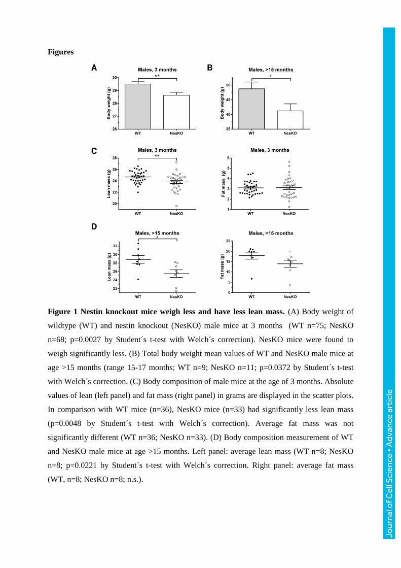
Figure 1 Nestin knockout mice weigh less and have less lean mass. (A) Body weight of
wildtype (WT) and nestin knockout (NesKO) male mice at 3 months (WT n=75; NesKO
n=68; p=0.0027 by Student´s t-test with Welch´s correction). NesKO mice were found to
weigh significantly less. (B) Total body weight mean values of WT and NesKO male mice at
age >15 months (range 15-17 months; WT n=9; NesKO n=11; p=0.0372 by Student´s t-test
with Welch´s correction. (C) Body composition of male mice at the age of 3 months. Absolute
values of lean (left panel) and fat mass (right panel) in grams are displayed in the scatter plots.
In comparison with WT mice (n=36), NesKO mice (n=33) had significantly less lean mass
(p=0.0048 by Student´s t-test with Welch´s correction). Average fat mass was not
significantly different (WT n=36; NesKO n=33). (D) Body composition measurement of WT
and NesKO male mice at age >15 months. Left panel: average lean mass (WT n=8; NesKO
n=8; p=0.0221 by Student´s t-test with Welch´s correction. Right panel: average fat mass
(WT, n=8; NesKO n=8; n.s.).
Jour
nal o
f Cel
l Sci
ence
• A
dvan
ce a
rtic
le
Jour
nal o
f Cel
l Sci
ence
• A
dvan
ce a
rtic
le
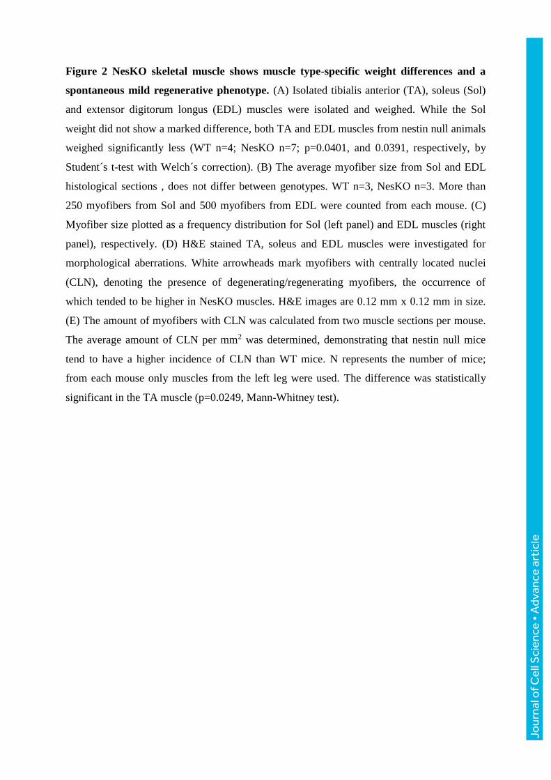
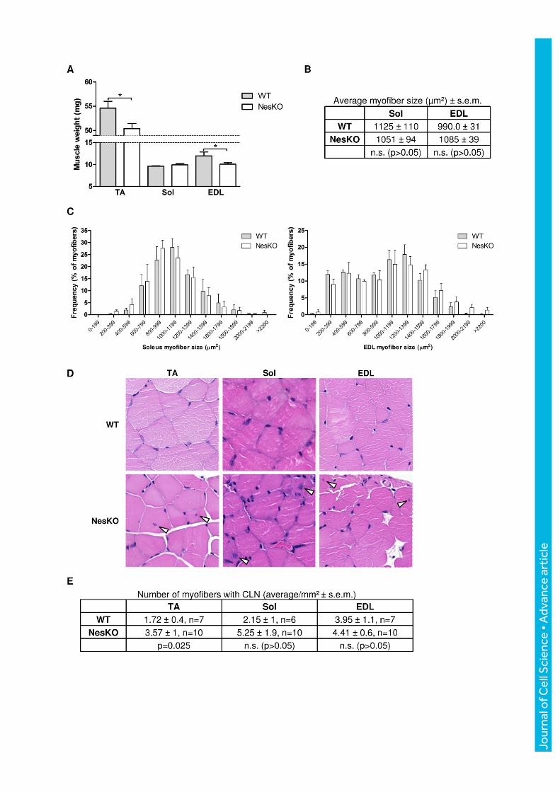
Figure 2 NesKO skeletal muscle shows muscle type-specific weight differences and a
spontaneous mild regenerative phenotype. (A) Isolated tibialis anterior (TA), soleus (Sol)
and extensor digitorum longus (EDL) muscles were isolated and weighed. While the Sol
weight did not show a marked difference, both TA and EDL muscles from nestin null animals
weighed significantly less (WT n=4; NesKO n=7; p=0.0401, and 0.0391, respectively, by
Student´s t-test with Welch´s correction). (B) The average myofiber size from Sol and EDL
histological sections , does not differ between genotypes. WT n=3, NesKO n=3. More than
250 myofibers from Sol and 500 myofibers from EDL were counted from each mouse. (C)
Myofiber size plotted as a frequency distribution for Sol (left panel) and EDL muscles (right
panel), respectively. (D) H&E stained TA, soleus and EDL muscles were investigated for
morphological aberrations. White arrowheads mark myofibers with centrally located nuclei
(CLN), denoting the presence of degenerating/regenerating myofibers, the occurrence of
which tended to be higher in NesKO muscles. H&E images are 0.12 mm x 0.12 mm in size.
(E) The amount of myofibers with CLN was calculated from two muscle sections per mouse.
The average amount of CLN per mm2 was determined, demonstrating that nestin null mice
tend to have a higher incidence of CLN than WT mice. N represents the number of mice;
from each mouse only muscles from the left leg were used. The difference was statistically
significant in the TA muscle (p=0.0249, Mann-Whitney test).
Jour
nal o
f Cel
l Sci
ence
• A
dvan
ce a
rtic
le
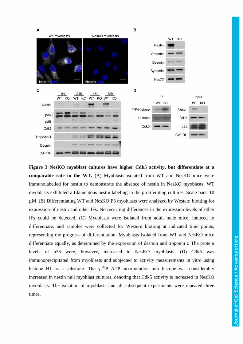
Figure 3 NesKO myoblast cultures have higher Cdk5 activity, but differentiate at a
comparable rate to the WT. (A) Myoblasts isolated from WT and NesKO mice were
immunolabelled for nestin to demonstrate the absence of nestin in NesKO myoblasts. WT
myoblasts exhibited a filamentous nestin labeling in the proliferating cultures. Scale bars=10
µM. (B) Differentiating WT and NesKO P3 myoblasts were analyzed by Western blotting for
expression of nestin and other IFs. No recurring differences in the expression levels of other
IFs could be detected. (C) Myoblasts were isolated from adult male mice, induced to
differentiate, and samples were collected for Western blotting at indicated time points,
representing the progress of differentiation. Myoblasts isolated from WT and NesKO mice
differentiate equally, as determined by the expression of desmin and troponin t. The protein
levels of p35 were, however, increased in NesKO myoblasts. (D) Cdk5 was
immunoprecipitated from myoblasts and subjected to activity measurements in vitro using
histone H1 as a substrate. The γ-32P ATP incorporation into histone was considerably
increased in nestin null myoblast cultures, denoting that Cdk5 activity is increased in NesKO
myoblasts. The isolation of myoblasts and all subsequent experiments were repeated three
times.
Jour
nal o
f Cel
l Sci
ence
• A
dvan
ce a
rtic
le
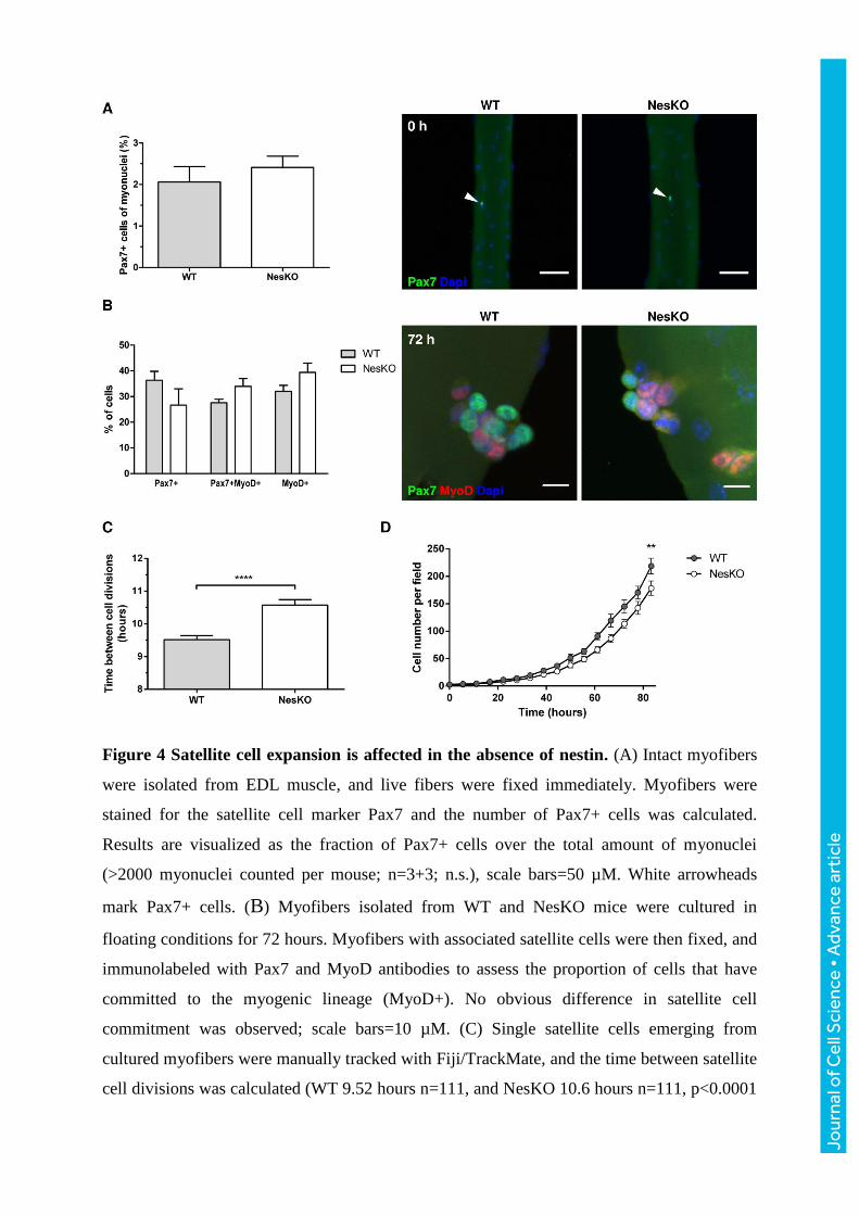
Figure 4 Satellite cell expansion is affected in the absence of nestin. (A) Intact myofibers
were isolated from EDL muscle, and live fibers were fixed immediately. Myofibers were
stained for the satellite cell marker Pax7 and the number of Pax7+ cells was calculated.
Results are visualized as the fraction of Pax7+ cells over the total amount of myonuclei
(>2000 myonuclei counted per mouse; n=3+3; n.s.), scale bars=50 µM. White arrowheads
mark Pax7+ cells. (B) Myofibers isolated from WT and NesKO mice were cultured in
floating conditions for 72 hours. Myofibers with associated satellite cells were then fixed, and
immunolabeled with Pax7 and MyoD antibodies to assess the proportion of cells that have
committed to the myogenic lineage (MyoD+). No obvious difference in satellite cell
commitment was observed; scale bars=10 µM. (C) Single satellite cells emerging from
cultured myofibers were manually tracked with Fiji/TrackMate, and the time between satellite
cell divisions was calculated (WT 9.52 hours n=111, and NesKO 10.6 hours n=111, p<0.0001
Jour
nal o
f Cel
l Sci
ence
• A
dvan
ce a
rtic
le
by Student´s t-test with Welch´s correction). (D) Isolated myofibers were plated on Matrigel-
coated dishes, and emerging satellite cells were imaged continuously with Cell-IQ-imaging
platform. The numbers of proliferating satellite cells was calculated at indicated time points
with Cell-IQ Analyser software. The average number of cells per field at 83 hours was 219 for
WT and 178 for NesKO; p=0.0054 by Student´s t-test, n=3).
Jour
nal o
f Cel
l Sci
ence
• A
dvan
ce a
rtic
le
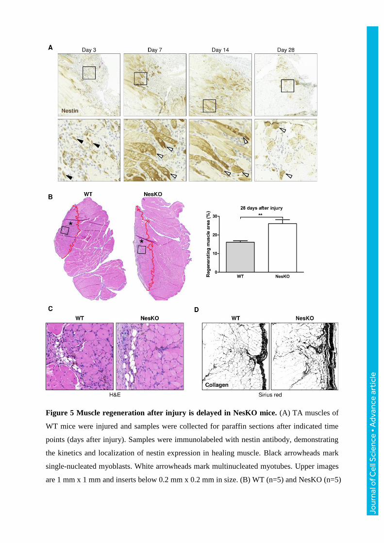
Figure 5 Muscle regeneration after injury is delayed in NesKO mice. (A) TA muscles of
WT mice were injured and samples were collected for paraffin sections after indicated time
points (days after injury). Samples were immunolabeled with nestin antibody, demonstrating
the kinetics and localization of nestin expression in healing muscle. Black arrowheads mark
single-nucleated myoblasts. White arrowheads mark multinucleated myotubes. Upper images
are 1 mm x 1 mm and inserts below 0.2 mm x 0.2 mm in size. (B) WT (n=5) and NesKO (n=5)
Jour
nal o
f Cel
l Sci
ence
• A
dvan
ce a
rtic
le
mice were subjected to muscle injury, and the wound was allowed to heal for 28 days. The
regenerating muscle area (defined by the presence of CLN in myofibers) was calculated and
compared to the total muscle area, revealing that NesKO mice have a significantly larger area
occupied by regenerating myofibers 28 days after injury (WT=16.10% vs. NesKO=26.06%;
p=0.0023 Student´s t-test). The injured area is marked with asterisk and is outlined in red.
Whole muscle sections show representative quantification results of the injured muscle area.
(C) H&E-stained samples from the injured muscles were inspected for histological aberrations,
but showed no obvious difference, except in the area occupied by regenerating fibers. Images
are magnified inserts from the muscle sections in panel B. H&E images are 0.3 mm x 0.3 mm
in size. (D) Fibrotic deposits were studied after injury with Picrosirius red stain against
collagen, imaged with bright field microscope, after which images were transformed to binary
black-and-white images to enhance the visibility of the collagen extracellular matrix
(visualized in black). The images are 0.5 mm x 0.5 mm in size.
Jour
nal o
f Cel
l Sci
ence
• A
dvan
ce a
rtic
le
A
B
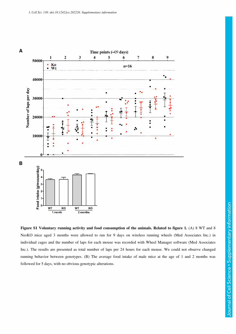
Figure S1 Voluntary running activity and food consumption of the animals. Related to figure 1. (A) 8 WT and 8
NesKO mice aged 3 months were allowed to run for 9 days on wireless running wheels (Med Associates Inc.) in
individual cages and the number of laps for each mouse was recorded with Wheel Manager software (Med Associates
Inc.). The results are presented as total number of laps per 24 hours for each mouse. We could not observe changed
running behavior between genotypes. (B) The average food intake of male mice at the age of 1 and 2 months was
followed for 5 days, with no obvious genotypic alterations.
J. Cell Sci. 130: doi:10.1242/jcs.202226: Supplementary information
Jour
nal o
f Cel
l Sci
ence
• S
uppl
emen
tary
info
rmat
ion
A B
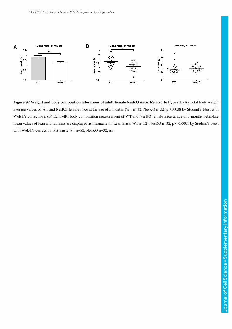
Figure S2 Weight and body composition alterations of adult female NesKO mice. Related to figure 1. (A) Total body weight
average values of WT and NesKO female mice at the age of 3 months (WT n=32; NesKO n=32; p=0.0038 by Student´s t-test with
Welch´s correction). (B) EchoMRI body composition measurement of WT and NesKO female mice at age of 3 months. Absolute
mean values of lean and fat mass are displayed as mean±s.e.m. Lean mass: WT n=32; NesKO n=32, p < 0.0001 by Student´s t-test
with Welch´s correction. Fat mass: WT n=32, NesKO n=32, n.s.
J. Cell Sci. 130: doi:10.1242/jcs.202226: Supplementary information
Jour
nal o
f Cel
l Sci
ence
• S
uppl
emen
tary
info
rmat
ion
Day 14Day 7
Nestin
NesKO
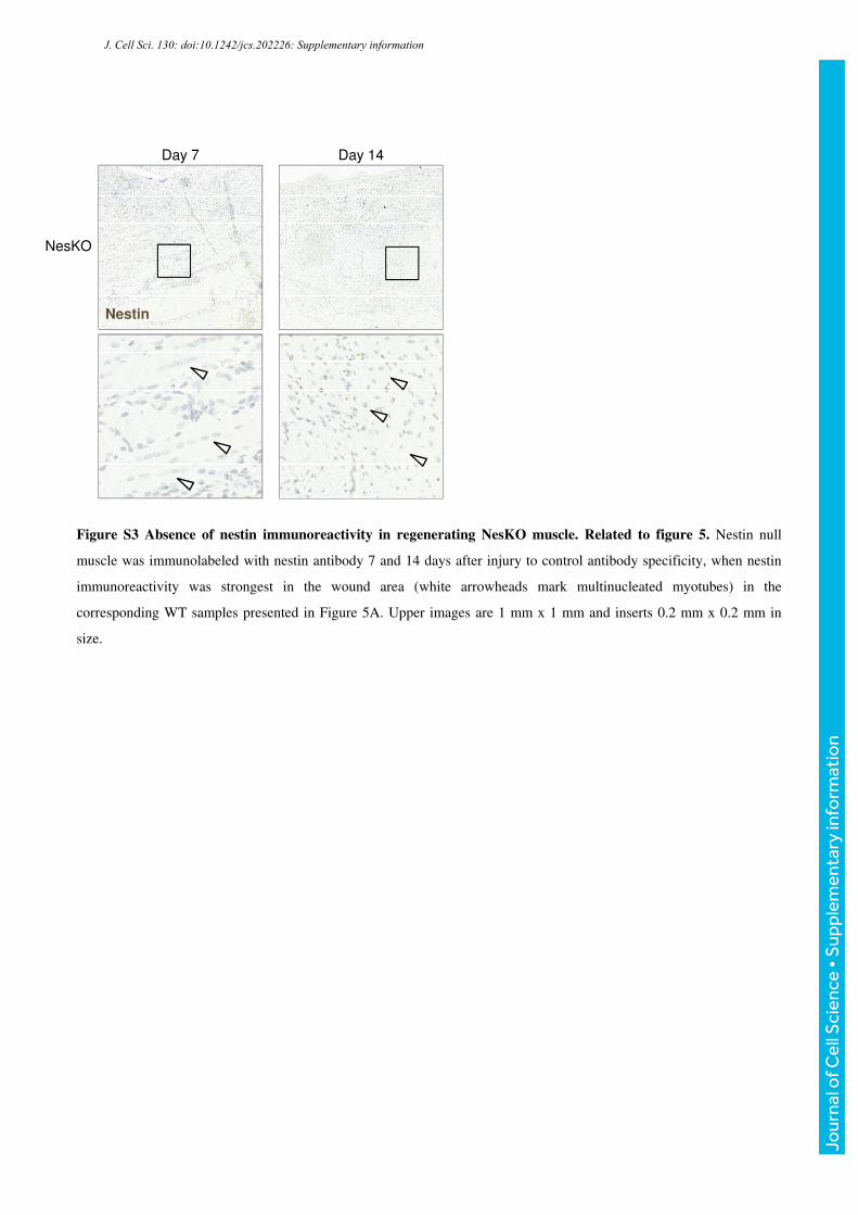
Figure S3 Absence of nestin immunoreactivity in regenerating NesKO muscle. Related to figure 5. Nestin null
muscle was immunolabeled with nestin antibody 7 and 14 days after injury to control antibody specificity, when nestin
immunoreactivity was strongest in the wound area (white arrowheads mark multinucleated myotubes) in the
corresponding WT samples presented in Figure 5A. Upper images are 1 mm x 1 mm and inserts 0.2 mm x 0.2 mm in
size.
J. Cell Sci. 130: doi:10.1242/jcs.202226: Supplementary information
Jour
nal o
f Cel
l Sci
ence
• S
uppl
emen
tary
info
rmat
ion
0 h
40 h
Myofiber explant cultures
Live imaging
Manual lineage tracking
Cell numberquantification
Cell doubling time (Fig. 3C)
Proliferation curve(Fig. 3D)
A
B
C
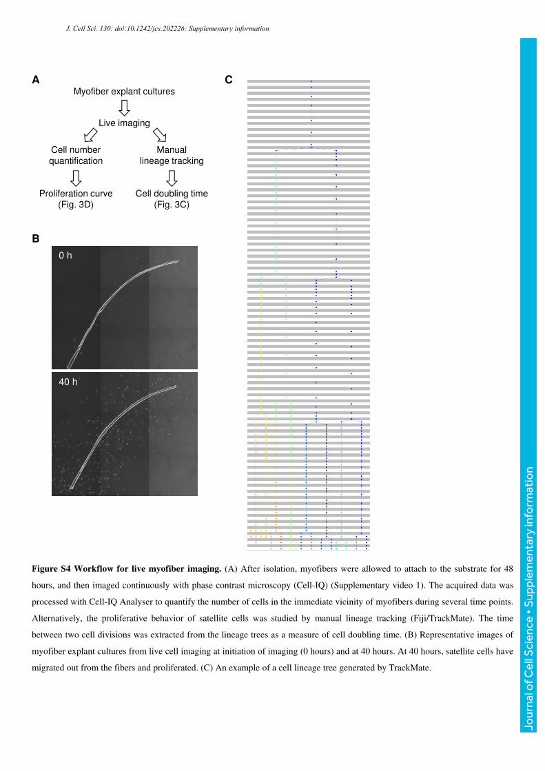
Figure S4 Workflow for live myofiber imaging. (A) After isolation, myofibers were allowed to attach to the substrate for 48
hours, and then imaged continuously with phase contrast microscopy (Cell-IQ) (Supplementary video 1). The acquired data was
processed with Cell-IQ Analyser to quantify the number of cells in the immediate vicinity of myofibers during several time points.
Alternatively, the proliferative behavior of satellite cells was studied by manual lineage tracking (Fiji/TrackMate). The time
between two cell divisions was extracted from the lineage trees as a measure of cell doubling time. (B) Representative images of
myofiber explant cultures from live cell imaging at initiation of imaging (0 hours) and at 40 hours. At 40 hours, satellite cells have
migrated out from the fibers and proliferated. (C) An example of a cell lineage tree generated by TrackMate.
J. Cell Sci. 130: doi:10.1242/jcs.202226: Supplementary information
Jour
nal o
f Cel
l Sci
ence
• S
uppl
emen
tary
info
rmat
ion

Supplementary table 1 Antibodies used in the study
Antibody Clone/cat. number Source Used for Dilution
Actin AC-40 Sigma-Aldrich WB 1:1000
Cdk5 DC-34 Life Technologies WB, IP 1:1000
Cdk5 C-8 Santa Cruz WB 1:200
Desmin #4024 Cell Signaling WB 1:1000
GAPDH 14C10 Cell Signaling WB 1:2000
Hsc70 SPA-810 Stressgen WB 1:2000
MyoD M-318 Santa Cruz ICC 1:100
Nestin 556309 BD Pharmingen IHC, ICC 1:200
Nestin 611659 BD Pharmingen WB 1:1000
p35/p25 C-19 Santa Cruz WB 1:200
Pax7a AB_528428 DSHB ICC 1:20
Syneminb Prof. Omar Skalli WB 1:100
Troponin t JLT-12 Sigma-Aldrich WB 1:200
Vimentin 550513 BD Pharmingen WB 1:1000 aThe Pax 7 antibody developed by A. Kawakami, Tokyo Institute of Technology, was obtained from the Developmental Studies Hybridoma Bank, Created by the NICHD of the NIH and maintained at The University of Iowa, Department of Biology, Iowa City, IA 52242, USA. b The synemin antibody was a kind gift from Professor Omar Skalli, University of Memphis.
J. Cell Sci. 130: doi:10.1242/jcs.202226: Supplementary information
Jour
nal o
f Cel
l Sci
ence
• S
uppl
emen
tary
info
rmat
ion
Movie S1. Live imaging of myofiber explant cultures
J. Cell Sci. 130: doi:10.1242/jcs.202226: Supplementary information
Jour
nal o
f Cel
l Sci
ence
• S
uppl
emen
tary
info
rmat
ion