Embed Size (px)
Citation preview
The cytoskeleton and cell movement
(Actin microfilaments)
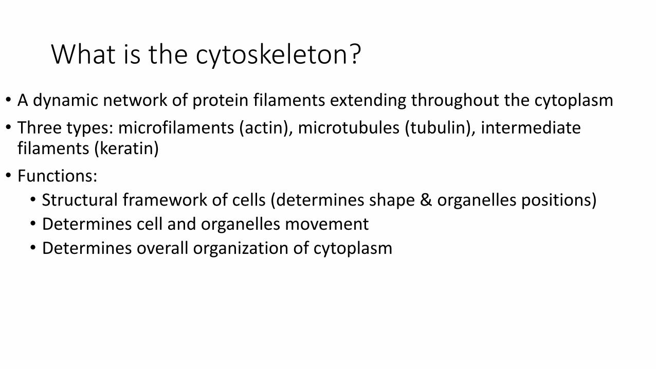
What is the cytoskeleton?
• A dynamic network of protein filaments extending throughout the cytoplasm
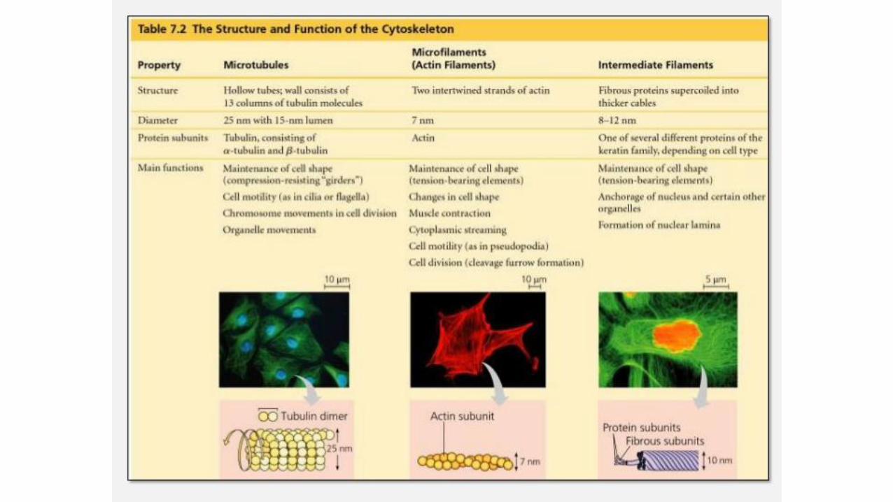
• Three types: microfilaments (actin), microtubules (tubulin), intermediate filaments (keratin)
• Functions:
• Structural framework of cells (determines shape & organelles positions)
• Determines cell and organelles movement
• Determines overall organization of cytoplasm
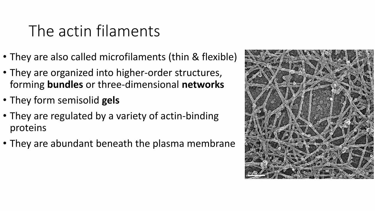
The actin filaments
• They are also called microfilaments (thin & flexible)
• They are organized into higher-order structures, forming bundles or three-dimensional networks
• They form semisolid gels
• They are regulated by a variety of actin-binding proteins
• They are abundant beneath the plasma membrane
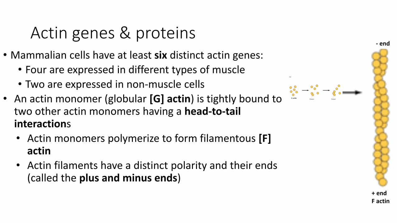
Actin genes & proteins• Mammalian cells have at least six distinct actin genes:
• Four are expressed in different types of muscle
• Two are expressed in non-muscle cells
• An actin monomer (globular [G] actin) is tightly bound to two other actin monomers having a head-to-tail interactions
• Actin monomers polymerize to form filamentous [F] actin
• Actin filaments have a distinct polarity and their ends (called the plus and minus ends)
+ endF actin
- end
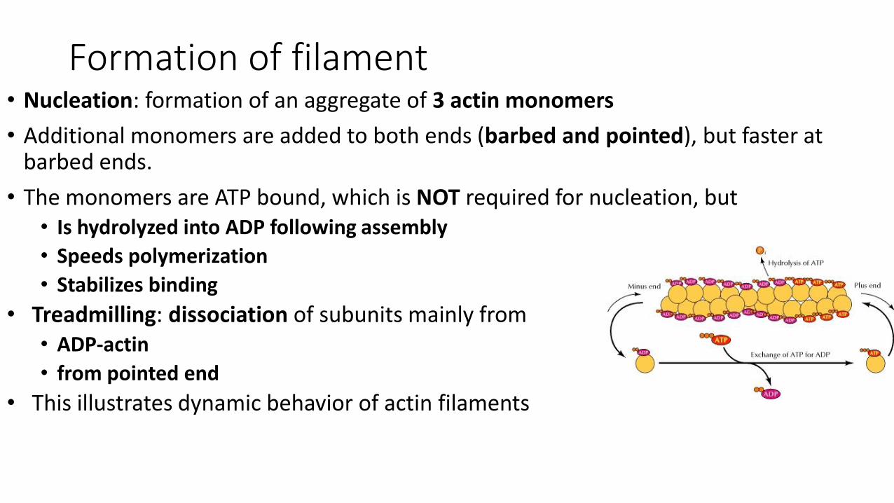
Formation of filament• Nucleation: formation of an aggregate of 3 actin monomers
• Additional monomers are added to both ends (barbed and pointed), but faster at barbed ends.
• The monomers are ATP bound, which is NOT required for nucleation, but • Is hydrolyzed into ADP following assembly
• Speeds polymerization
• Stabilizes binding
• Treadmilling: dissociation of subunits mainly from• ADP-actin
• from pointed end
• This illustrates dynamic behavior of actin filaments
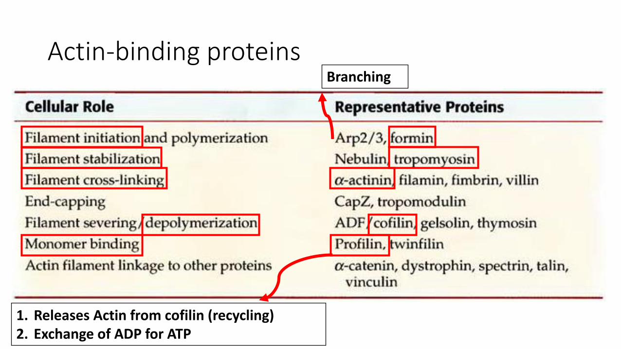
Actin-binding proteins
1. Releases Actin from cofilin (recycling)2. Exchange of ADP for ATP
Branching
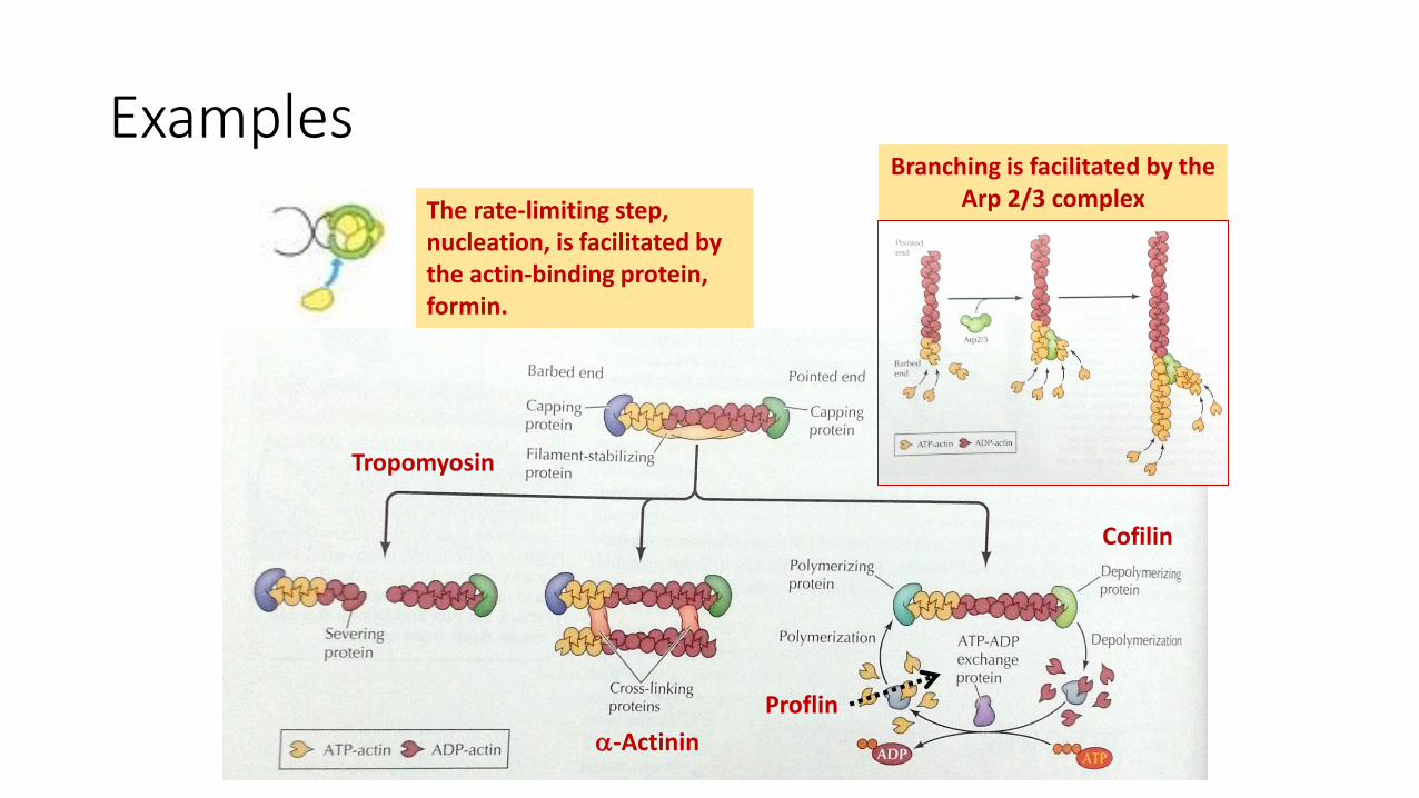
Examples
Tropomyosin
Cofilin
Proflin
-Actinin
The rate-limiting step, nucleation, is facilitated by the actin-binding protein, formin.
Branching is facilitated by the Arp 2/3 complex

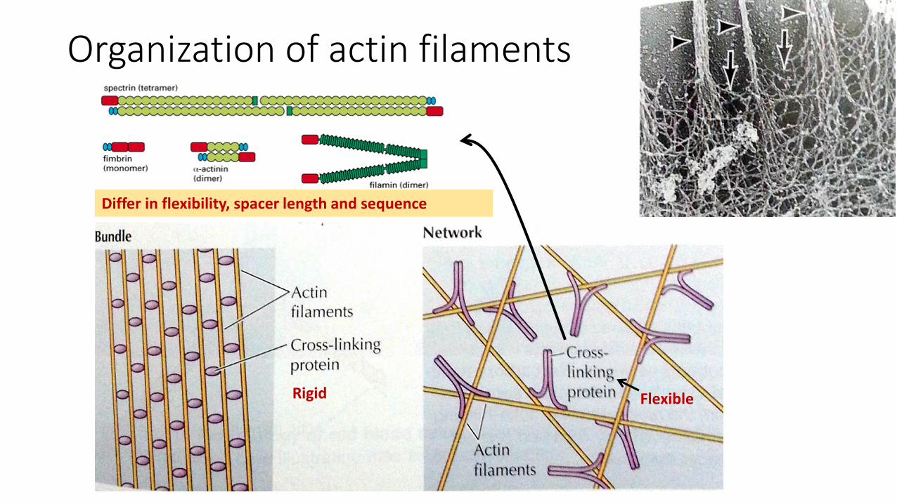
Organization of actin filaments
Rigid Flexible
Differ in flexibility, spacer length and sequence
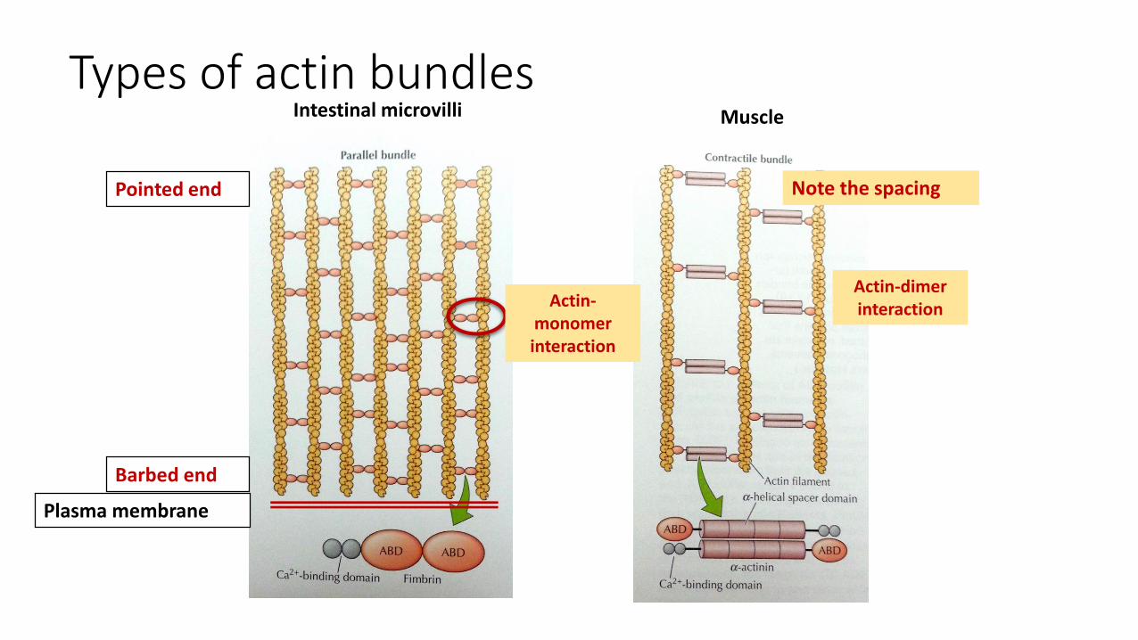
Types of actin bundles
Plasma membrane
Barbed end
Pointed end
Intestinal microvilli
Actin-monomer interaction
Actin-dimerinteraction
Note the spacing
Muscle
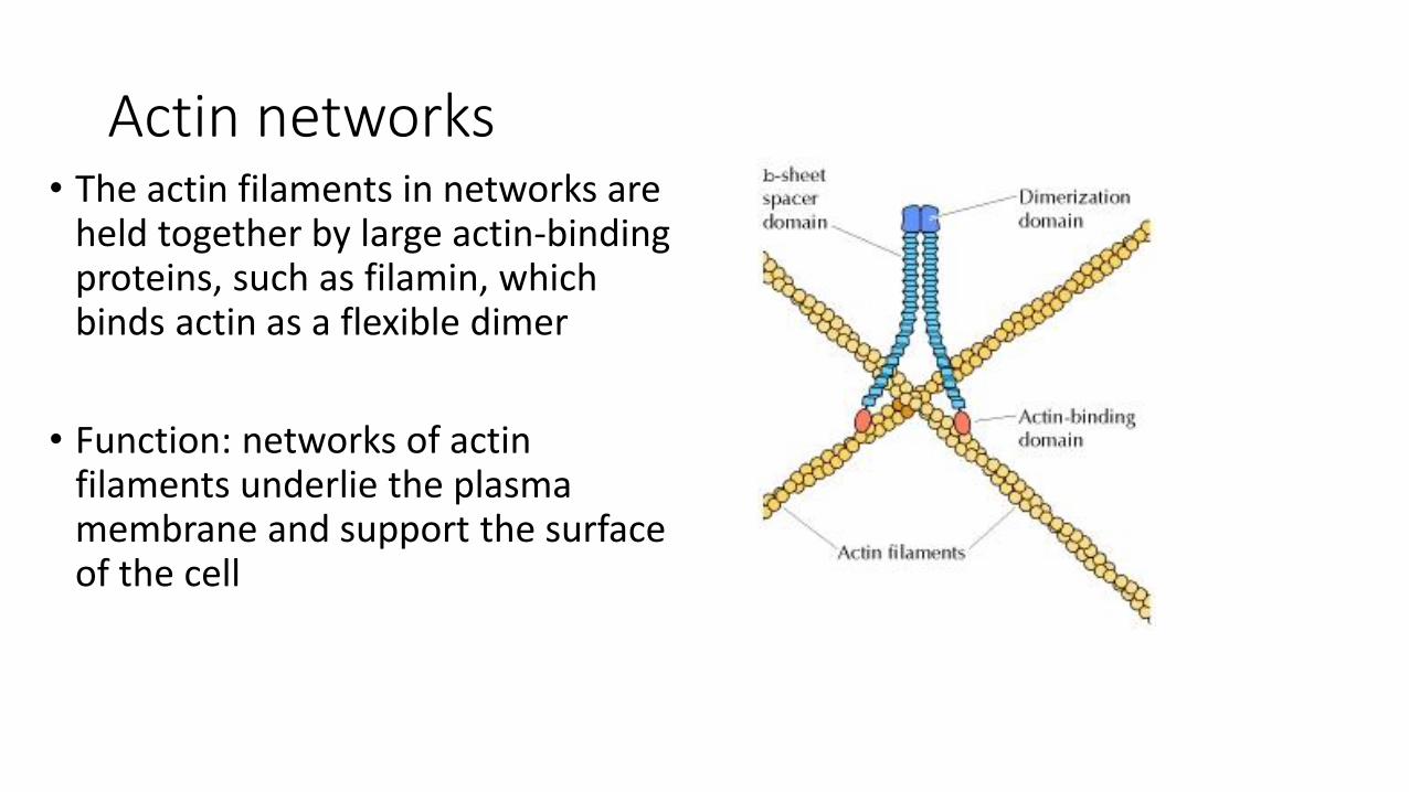
Actin networks• The actin filaments in networks are
held together by large actin-binding proteins, such as filamin, which binds actin as a flexible dimer
• Function: networks of actin filaments underlie the plasma membrane and support the surface of the cell
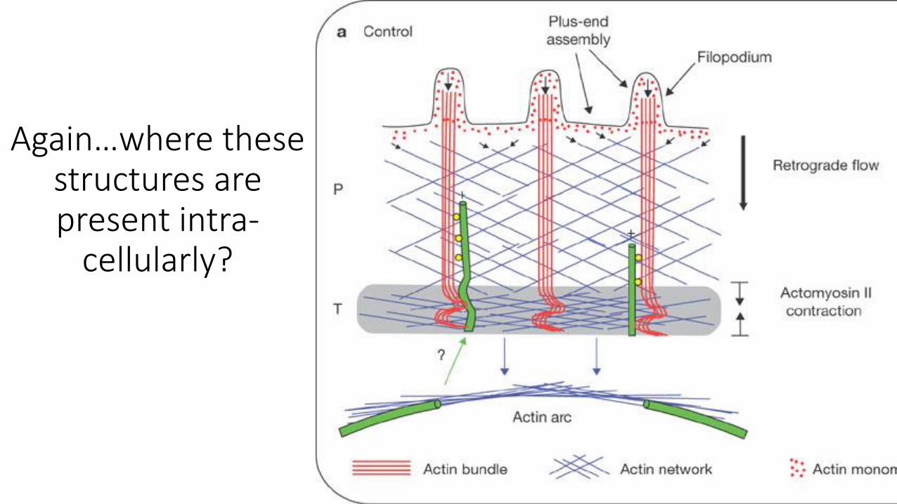
Again…where these structures are present intra-
cellularly?
Actin filaments and plasma membrane
• Cell cortex: The network of actin filaments and associated actin-binding proteins
• Studies in RBC because:
• They do not have other cytoskeletal structures
• They do not have organelles no contamination
• The cytoskeleton is uniform with no specialized regions like in other cells
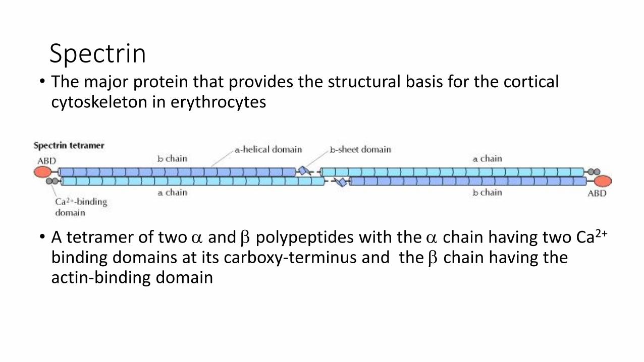
Spectrin• The major protein that provides the structural basis for the cortical
cytoskeleton in erythrocytes
• A tetramer of two and polypeptides with the chain having two Ca2+
binding domains at its carboxy-terminus and the chain having the actin-binding domain
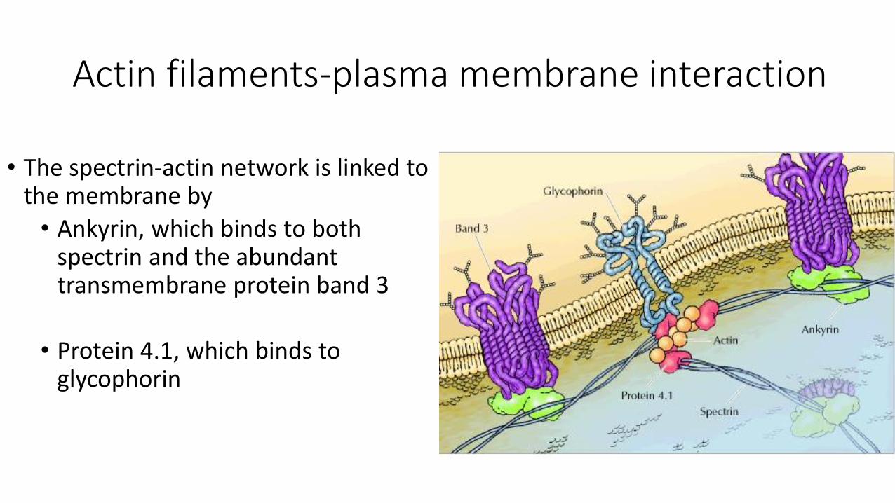
Actin filaments-plasma membrane interaction
• The spectrin-actin network is linked to the membrane by
• Ankyrin, which binds to both spectrin and the abundant transmembrane protein band 3
• Protein 4.1, which binds to glycophorin
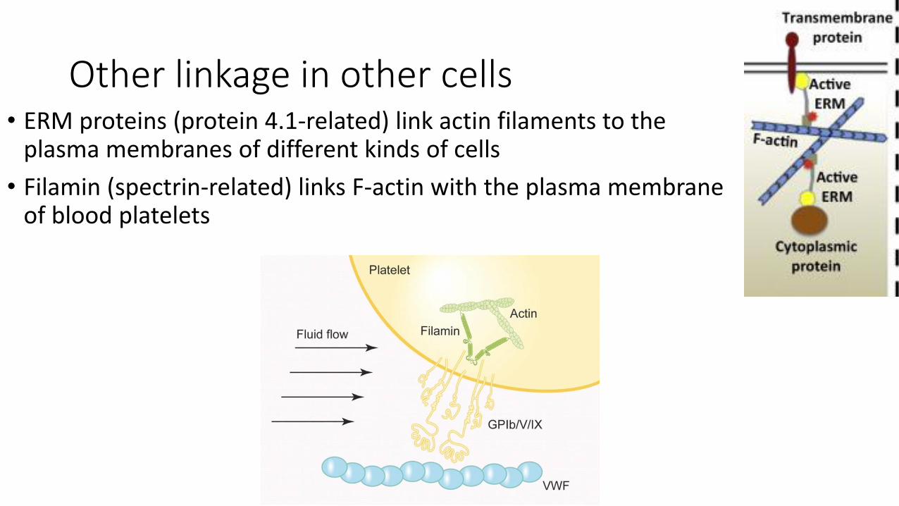
Other linkage in other cells• ERM proteins (protein 4.1-related) link actin filaments to the
plasma membranes of different kinds of cells
• Filamin (spectrin-related) links F-actin with the plasma membrane of blood platelets
Other linkage in other cells
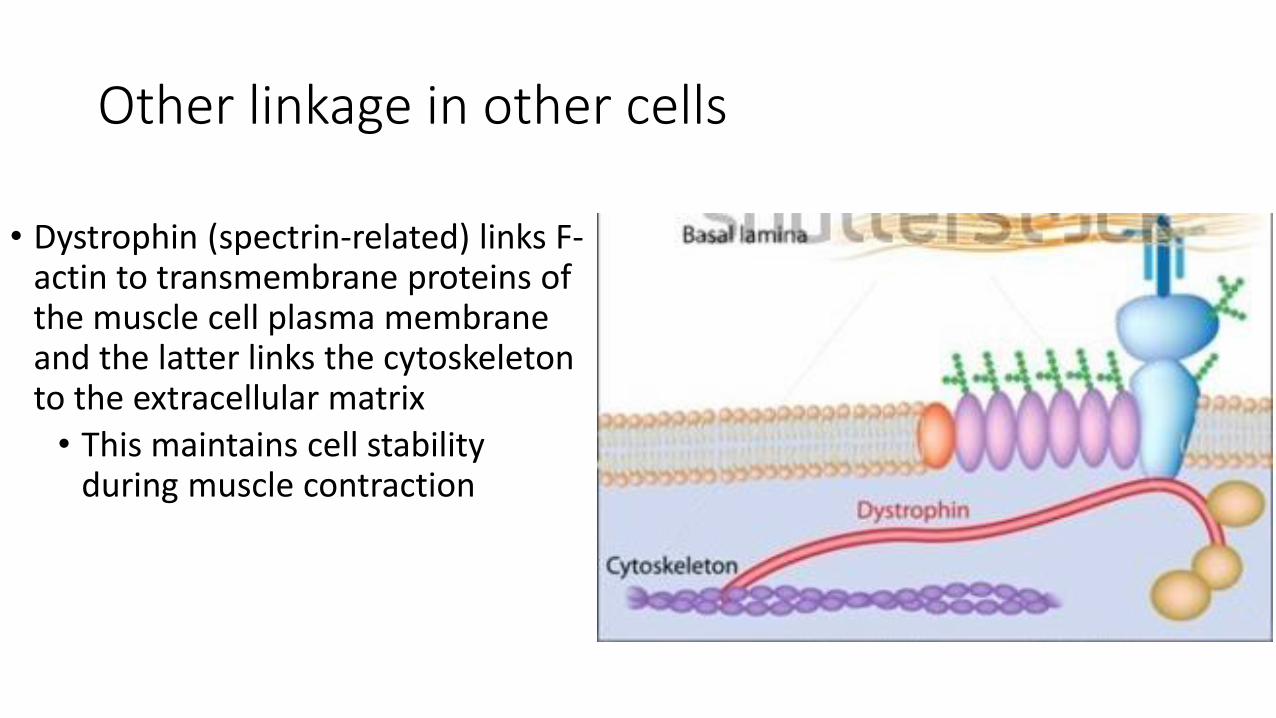
• Dystrophin (spectrin-related) links F-actin to transmembrane proteins of the muscle cell plasma membrane and the latter links the cytoskeleton to the extracellular matrix
• This maintains cell stability during muscle contraction
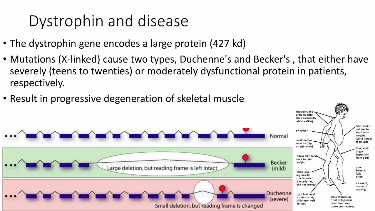
Dystrophin and disease• The dystrophin gene encodes a large protein (427 kd)
• Mutations (X-linked) cause two types, Duchenne's and Becker's , that either have severely (teens to twenties) or moderately dysfunctional protein in patients, respectively.
• Result in progressive degeneration of skeletal muscle
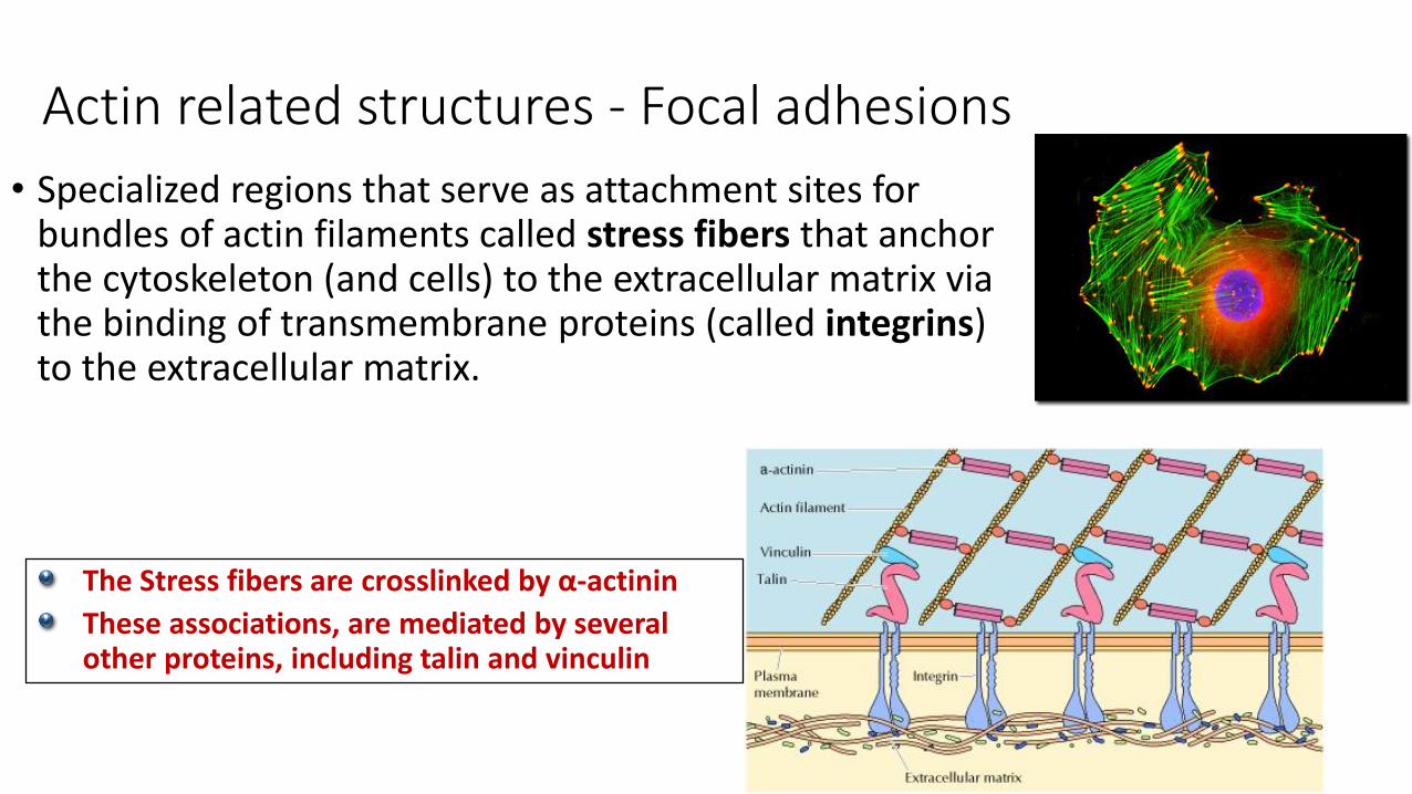
Actin related structures - Focal adhesions
• Specialized regions that serve as attachment sites for bundles of actin filaments called stress fibers that anchor the cytoskeleton (and cells) to the extracellular matrix via the binding of transmembrane proteins (called integrins) to the extracellular matrix.
The Stress fibers are crosslinked by α-actinin
These associations, are mediated by several other proteins, including talin and vinculin
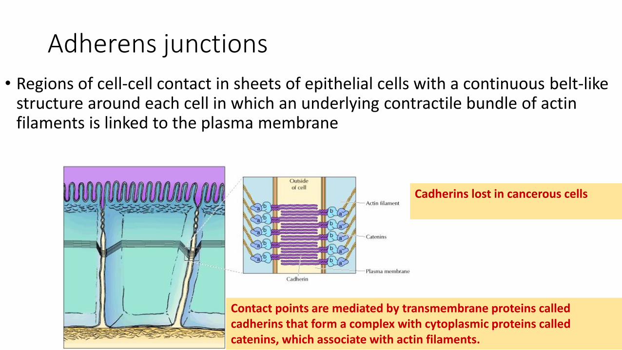
Adherens junctions
• Regions of cell-cell contact in sheets of epithelial cells with a continuous belt-like structure around each cell in which an underlying contractile bundle of actin filaments is linked to the plasma membrane
Contact points are mediated by transmembrane proteins called cadherins that form a complex with cytoplasmic proteins called catenins, which associate with actin filaments.
Cadherins lost in cancerous cells
Protrusions of the cell surface
• The surfaces of most cells have a variety of protrusions or extensions that are involved in cell movement, phagocytosis, or specialized functions such as absorption of nutrients
• Most of these cell surface extensions are based on actin filaments, which are organized into either relatively permanent or rapidly rearranging bundles or networks
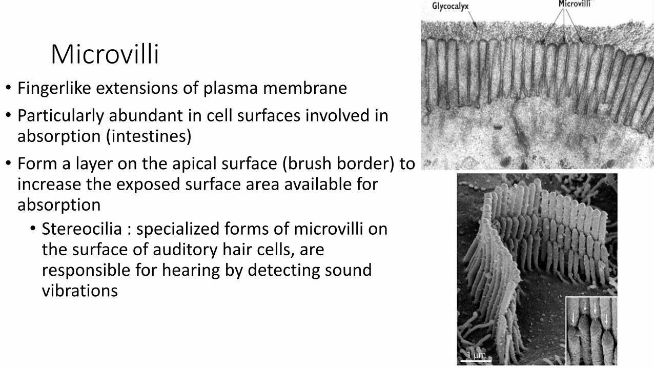
Microvilli• Fingerlike extensions of plasma membrane
• Particularly abundant in cell surfaces involved in absorption (intestines)
• Form a layer on the apical surface (brush border) to increase the exposed surface area available for absorption
• Stereocilia : specialized forms of microvilli on the surface of auditory hair cells, are responsible for hearing by detecting sound vibrations
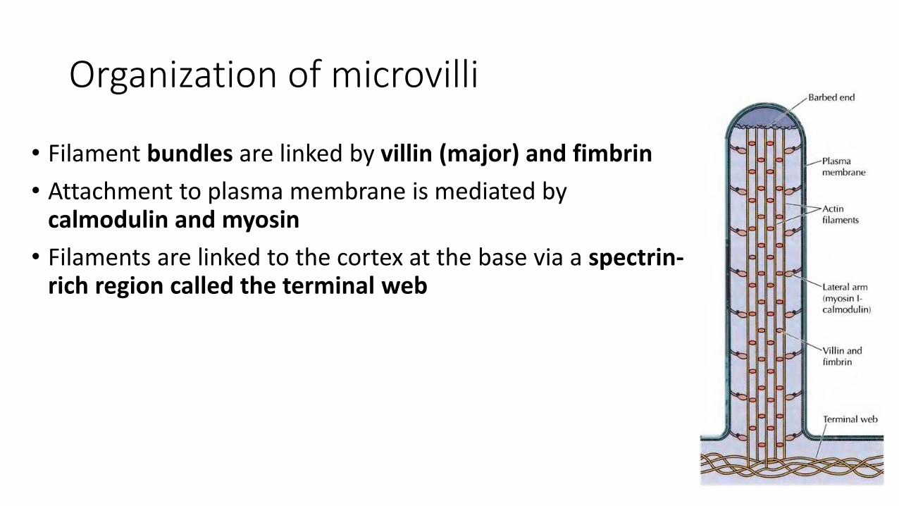
Organization of microvilli
• Filament bundles are linked by villin (major) and fimbrin
• Attachment to plasma membrane is mediated by calmodulin and myosin
• Filaments are linked to the cortex at the base via a spectrin-rich region called the terminal web
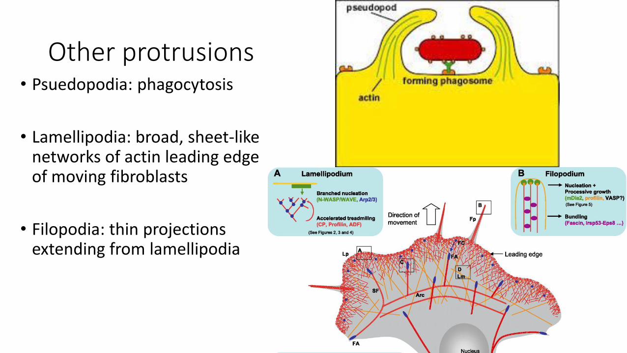
Other protrusions• Psuedopodia: phagocytosis
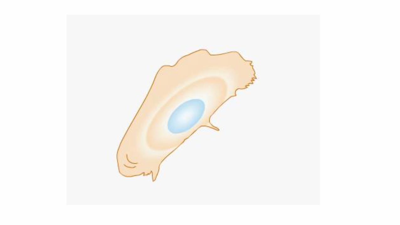
• Lamellipodia: broad, sheet-like networks of actin leading edge of moving fibroblasts
• Filopodia: thin projections extending from lamellipodia
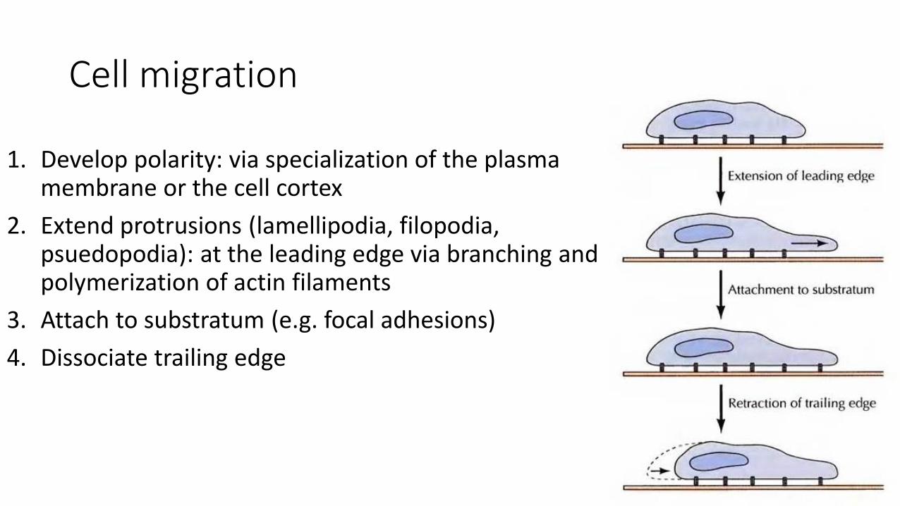
Cell migration
1. Develop polarity: via specialization of the plasma membrane or the cell cortex
2. Extend protrusions (lamellipodia, filopodia, psuedopodia): at the leading edge via branching and polymerization of actin filaments
3. Attach to substratum (e.g. focal adhesions)
4. Dissociate trailing edge
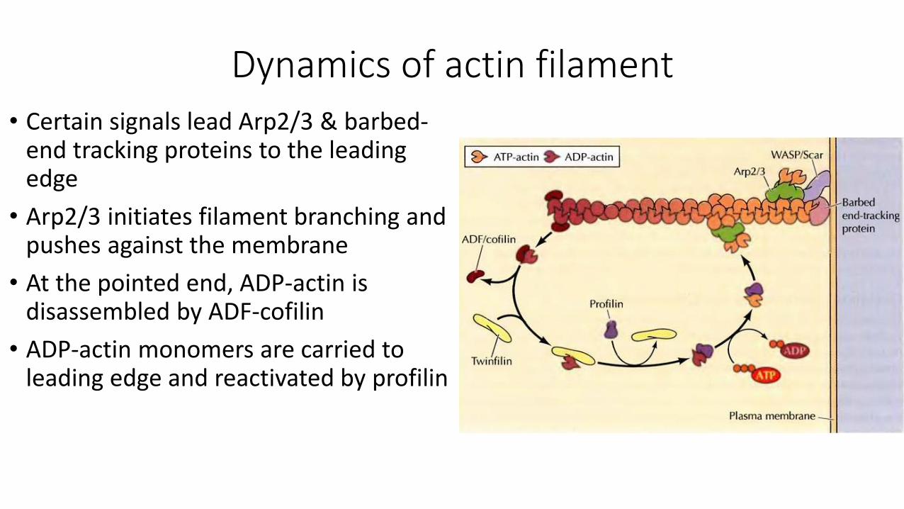
Dynamics of actin filament
• Certain signals lead Arp2/3 & barbed-end tracking proteins to the leading edge
• Arp2/3 initiates filament branching and pushes against the membrane
• At the pointed end, ADP-actin is disassembled by ADF-cofilin
• ADP-actin monomers are carried to leading edge and reactivated by profilin
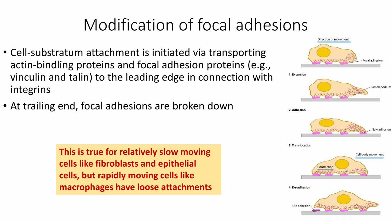
Modification of focal adhesions
• Cell-substratum attachment is initiated via transporting actin-bindling proteins and focal adhesion proteins (e.g., vinculin and talin) to the leading edge in connection with integrins
• At trailing end, focal adhesions are broken down
This is true for relatively slow moving cells like fibroblasts and epithelial cells, but rapidly moving cells like macrophages have loose attachments
The cytoskeleton & cell movement(Microtubules & intermediate filaments)
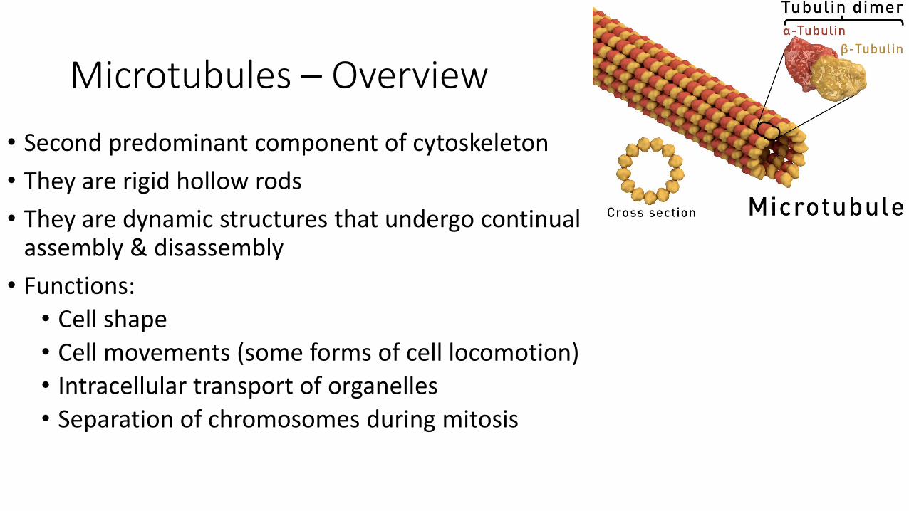
Microtubules – Overview
• Second predominant component of cytoskeleton
• They are rigid hollow rods
• They are dynamic structures that undergo continual assembly & disassembly
• Functions:
• Cell shape
• Cell movements (some forms of cell locomotion)
• Intracellular transport of organelles
• Separation of chromosomes during mitosis
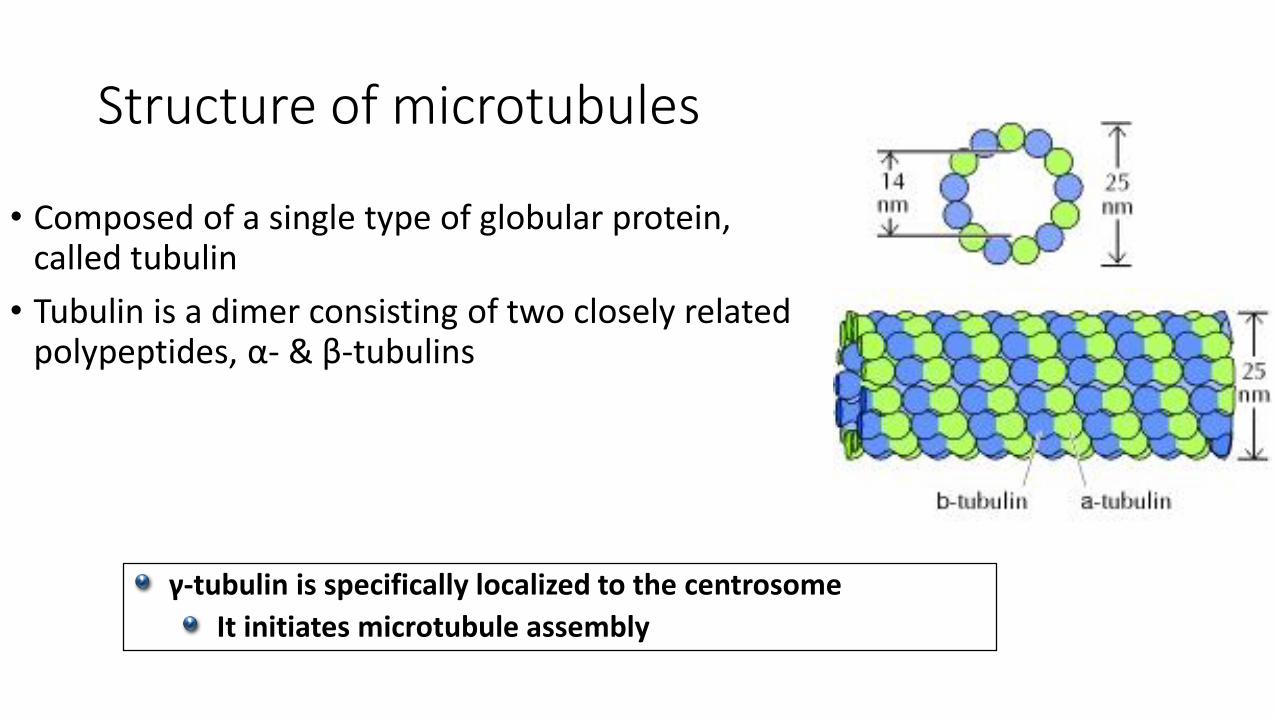
Structure of microtubules
• Composed of a single type of globular protein, called tubulin
• Tubulin is a dimer consisting of two closely related polypeptides, α- & β-tubulins
γ-tubulin is specifically localized to the centrosome
It initiates microtubule assembly
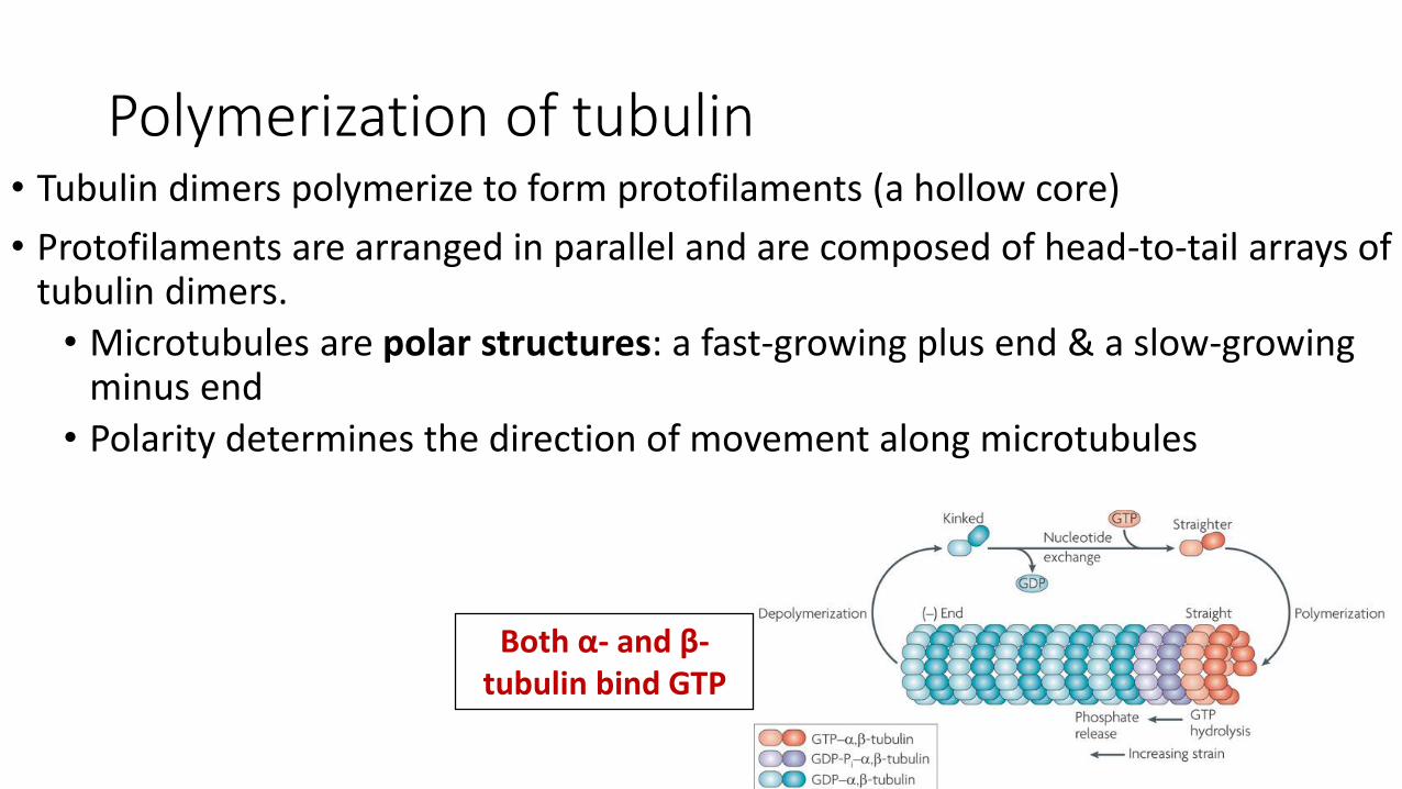
Polymerization of tubulin• Tubulin dimers polymerize to form protofilaments (a hollow core)
• Protofilaments are arranged in parallel and are composed of head-to-tail arrays of tubulin dimers.
• Microtubules are polar structures: a fast-growing plus end & a slow-growing minus end
• Polarity determines the direction of movement along microtubules
Both α- and β-tubulin bind GTP
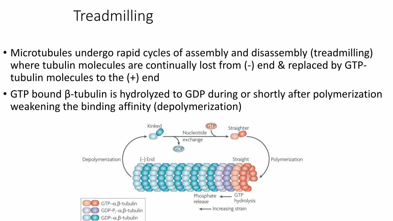
Treadmilling
• Microtubules undergo rapid cycles of assembly and disassembly (treadmilling) where tubulin molecules are continually lost from (-) end & replaced by GTP-tubulin molecules to the (+) end
• GTP bound β-tubulin is hydrolyzed to GDP during or shortly after polymerization weakening the binding affinity (depolymerization)
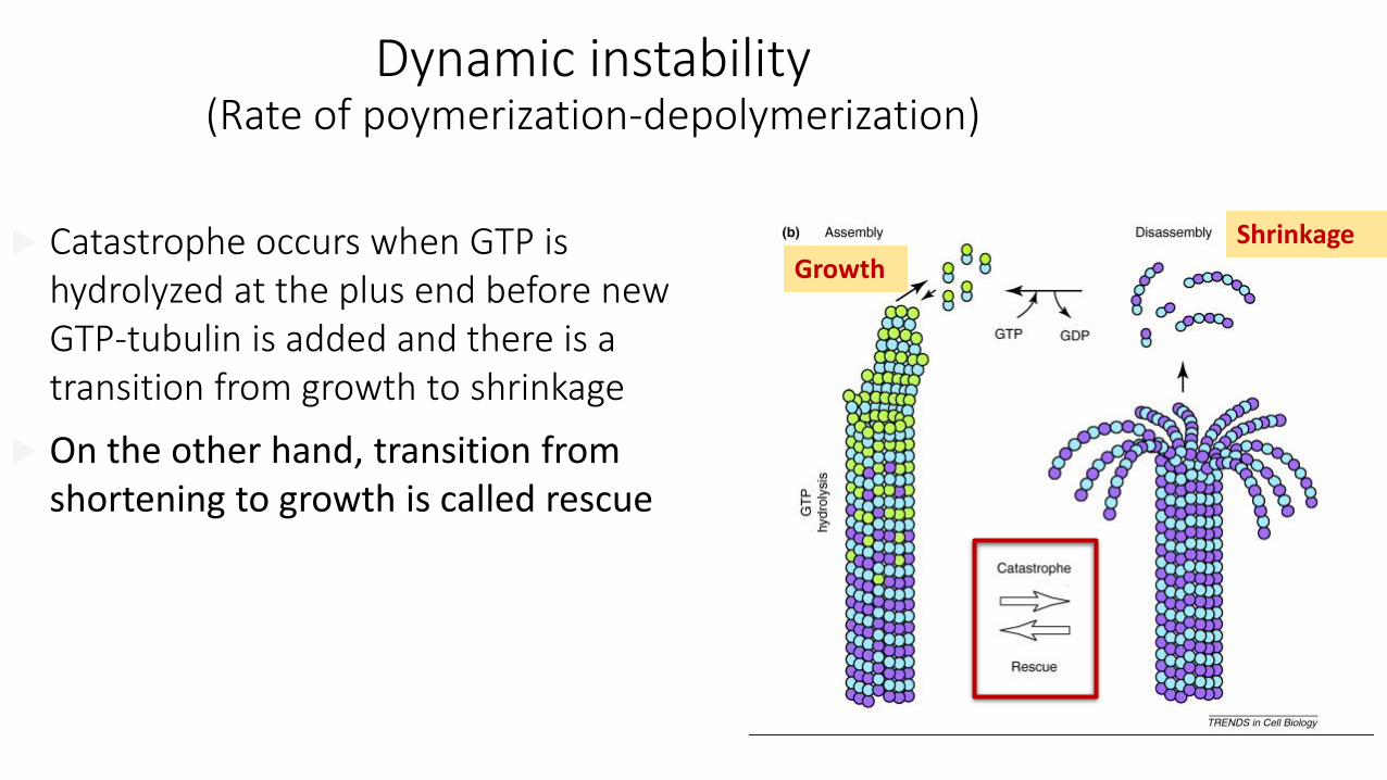
Dynamic instability(Rate of poymerization-depolymerization)
GrowthShrinkage
Catastrophe occurs when GTP is hydrolyzed at the plus end before new GTP-tubulin is added and there is a transition from growth to shrinkage
On the other hand, transition from shortening to growth is called rescue
Drugs
• Colchicine and colcemid bind tubulin, inhibit polymerization, and block mitosis.
• Vinblastine and vincristine bind specifically to tubulin and prevent their polymerization to form microtubules.
• Taxol stabilizes microtubule and blocks cell division.
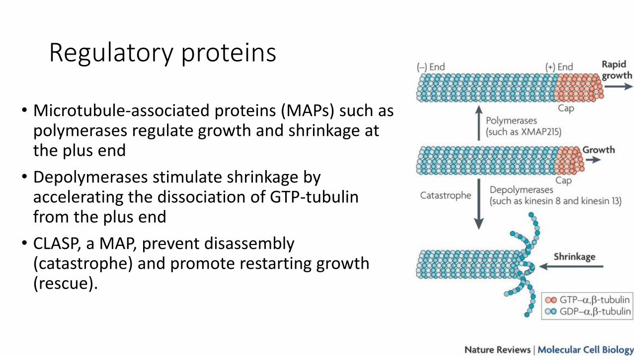
Regulatory proteins
• Microtubule-associated proteins (MAPs) such as polymerases regulate growth and shrinkage at the plus end
• Depolymerases stimulate shrinkage by accelerating the dissociation of GTP-tubulin from the plus end
• CLASP, a MAP, prevent disassembly (catastrophe) and promote restarting growth (rescue).
Organization of microtubules within cellsExample: neuron
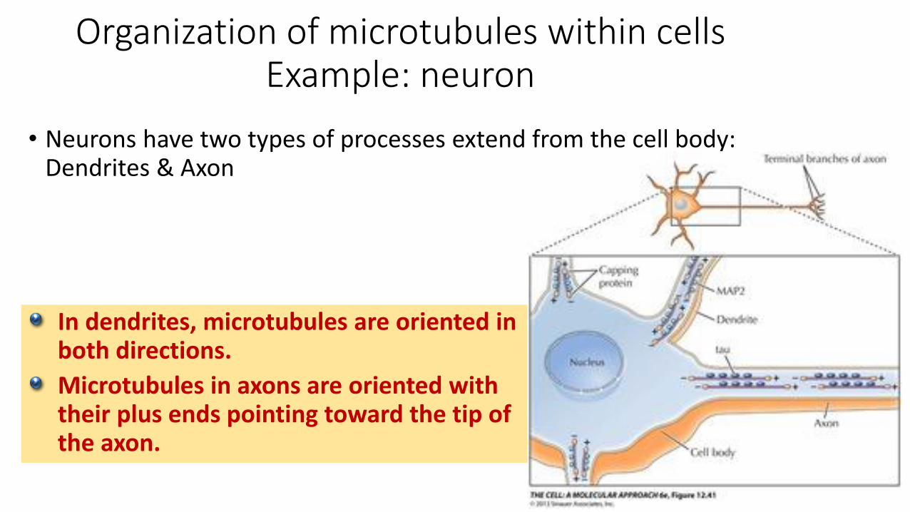
• Neurons have two types of processes extend from the cell body: Dendrites & Axon
In dendrites, microtubules are oriented in both directions.
Microtubules in axons are oriented with their plus ends pointing toward the tip of the axon.
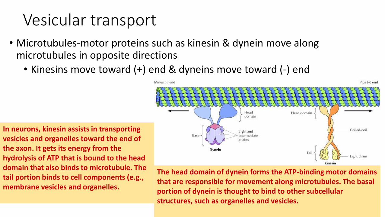
Vesicular transport• Microtubules-motor proteins such as kinesin & dynein move along
microtubules in opposite directions
• Kinesins move toward (+) end & dyneins move toward (-) end
In neurons, kinesin assists in transporting vesicles and organelles toward the end of the axon. It gets its energy from the hydrolysis of ATP that is bound to the head domain that also binds to microtubule. The tail portion binds to cell components (e.g., membrane vesicles and organelles.
The head domain of dynein forms the ATP-binding motor domains that are responsible for movement along microtubules. The basal portion of dynein is thought to bind to other subcellularstructures, such as organelles and vesicles.
Organelle organizations
• kinesin pulls the endoplasmic reticulum toward the cell periphery
• Kinesin positions lysosomes away from the center of the cell
• Members of the kinesin family control the movements of mitochondria
• Cytoplasmic dynein positions the Golgi apparatus in the center of the cell
• Both kinesin and dynein transport selective mRNA molecules in cells
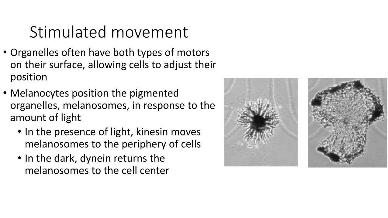
Stimulated movement• Organelles often have both types of motors
on their surface, allowing cells to adjust their position
• Melanocytes position the pigmented organelles, melanosomes, in response to the amount of light
• In the presence of light, kinesin moves melanosomes to the periphery of cells
• In the dark, dynein returns the melanosomes to the cell center
Kinesins and diseases• Mutants in certain kinesin proteins reduce the ability of neurons to move
essential organelles from their cell bodies to their axons leading to neurodegeneration such as amyotrophic lateral sclerosis (ALS)
• Mutations in kinesins lead to peripheral neuropathies such as Charcot-Marie-Tooth disease

What are they?
• Intermediate filaments have a diameter that is intermediate between those of actin filaments and microtubule.
• They provide mechanical strength to cells and tissues.
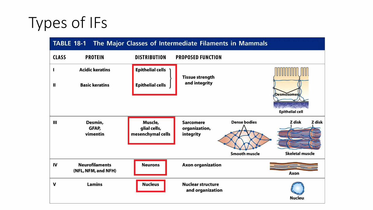
• They are composed of a variety of proteins, which are classified into 5 groups based on similarities between their amino acid sequences.
Types of IFs
• Types I and II are expressed in epithelial cells with each type of cell synthesizing at least one type I (acidic) and one type II (neutral/basic) keratin
• Hard keratins are used for production of structures such as hair, nails, and horns
• Soft keratins are abundant in the cytoplasm of epithelial cells
• Type III:
• Vimentin: found in fibroblasts, smooth muscle cells, and WBCs
• Desmin is specifically expressed in muscle cells
• Type IV: neurofilament (NF) found in the axons of motor neurons
• Type V: nuclear lamins, components of the nuclear envelope
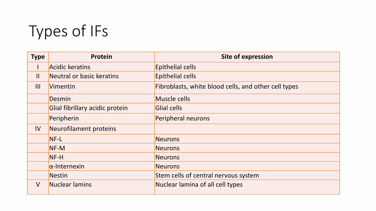
Type Protein Site of expression
I Acidic keratins Epithelial cells
II Neutral or basic keratins Epithelial cells
III Vimentin Fibroblasts, white blood cells, and other cell types
Desmin Muscle cells
Glial fibrillary acidic protein Glial cells
Peripherin Peripheral neurons
IV Neurofilament proteins
NF-L Neurons
NF-M Neurons
NF-H Neurons
α-Internexin Neurons
Nestin Stem cells of central nervous system
V Nuclear lamins Nuclear lamina of all cell types
Types of IFs
Types of IFs
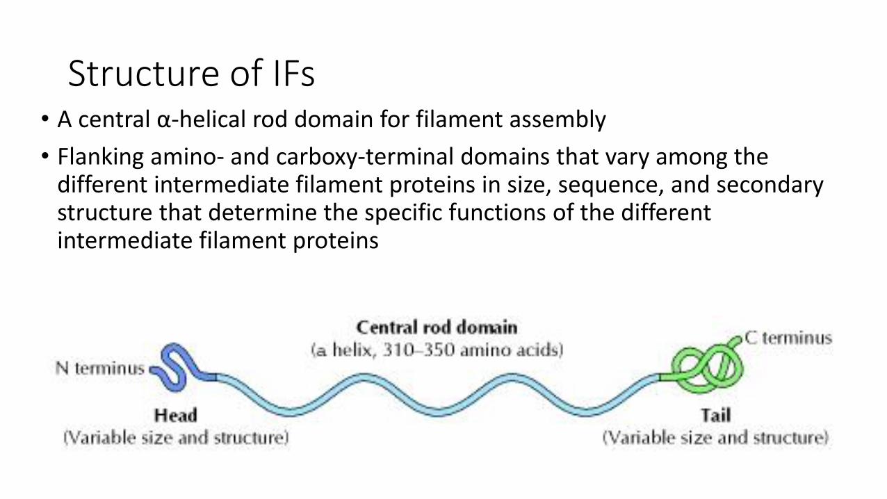
Structure of IFs• A central α-helical rod domain for filament assembly
• Flanking amino- and carboxy-terminal domains that vary among the different intermediate filament proteins in size, sequence, and secondary structure that determine the specific functions of the different intermediate filament proteins
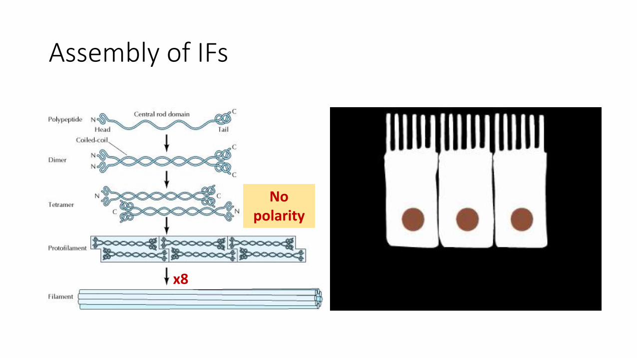
Assembly of IFs
x8
No polarity
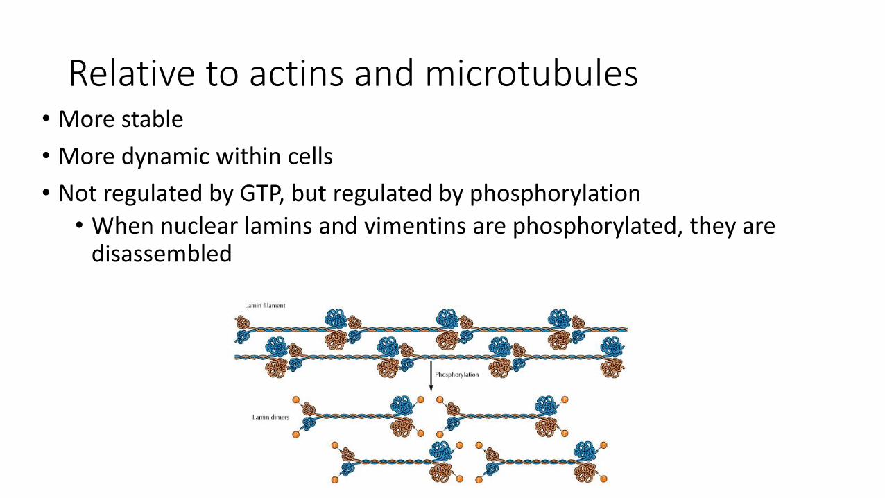
Relative to actins and microtubules• More stable
• More dynamic within cells
• Not regulated by GTP, but regulated by phosphorylation
• When nuclear lamins and vimentins are phosphorylated, they are disassembled
Intracellular Organization of IFs
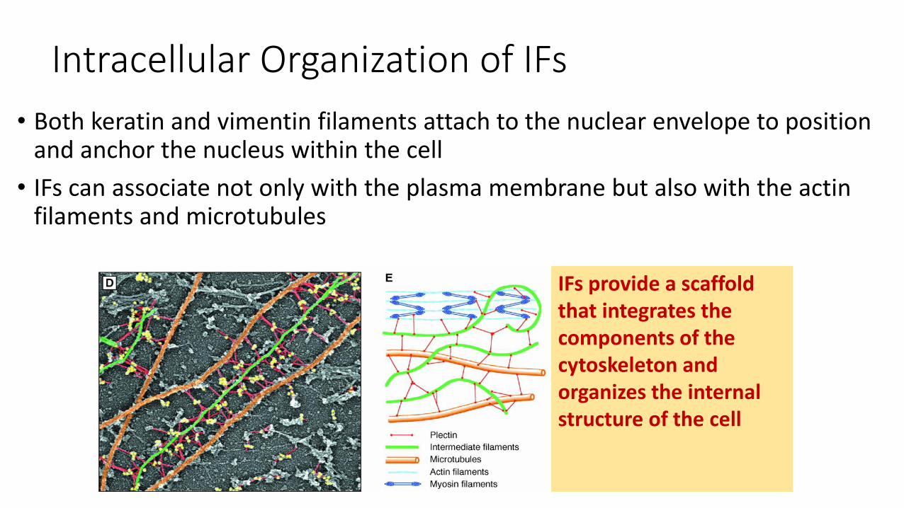
• Both keratin and vimentin filaments attach to the nuclear envelope to position and anchor the nucleus within the cell
• IFs can associate not only with the plasma membrane but also with the actin filaments and microtubules
IFs provide a scaffold that integrates the components of the cytoskeleton and organizes the internal structure of the cell
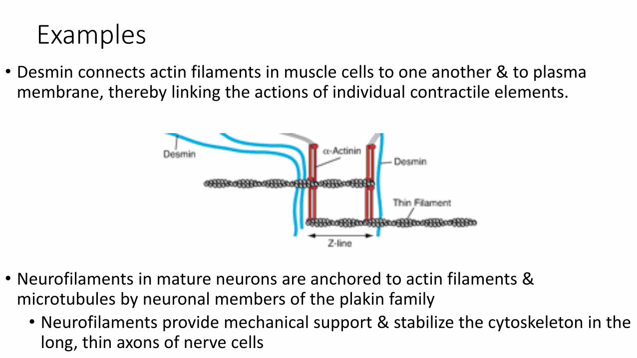
Examples• Desmin connects actin filaments in muscle cells to one another & to plasma
membrane, thereby linking the actions of individual contractile elements.
• Neurofilaments in mature neurons are anchored to actin filaments & microtubules by neuronal members of the plakin family
• Neurofilaments provide mechanical support & stabilize the cytoskeleton in the long, thin axons of nerve cells
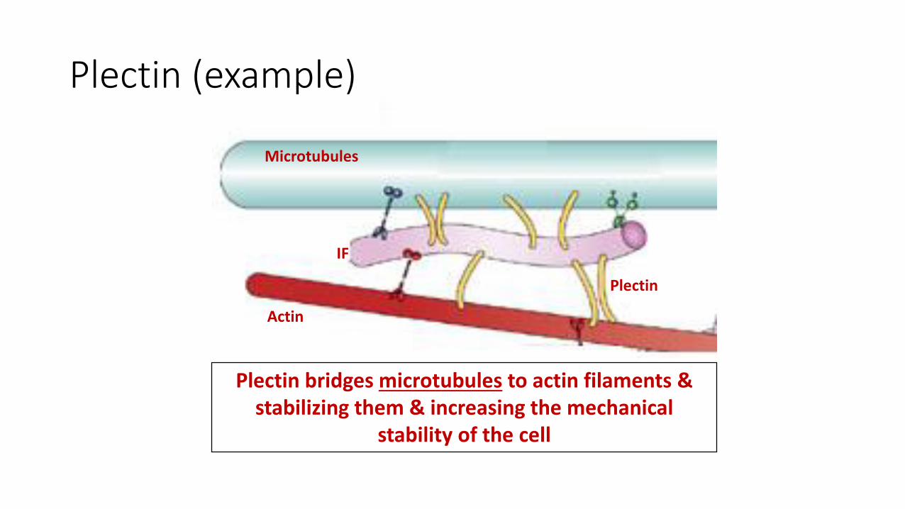
Plectin (example)
Microtubules
Actin
IF
Plectin
Plectin bridges microtubules to actin filaments & stabilizing them & increasing the mechanical
stability of the cell
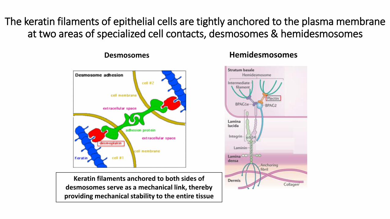
The keratin filaments of epithelial cells are tightly anchored to the plasma membrane at two areas of specialized cell contacts, desmosomes & hemidesmosomes
Desmosomes Hemidesmosomes
Keratin filaments anchored to both sides of desmosomes serve as a mechanical link, thereby providing mechanical stability to the entire tissue
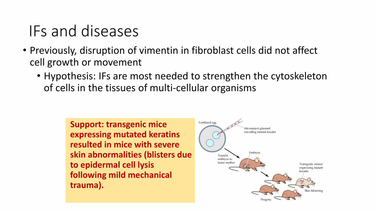
IFs and diseases• Previously, disruption of vimentin in fibroblast cells did not affect
cell growth or movement
• Hypothesis: IFs are most needed to strengthen the cytoskeleton of cells in the tissues of multi-cellular organisms
Support: transgenic mice expressing mutated keratins resulted in mice with severe skin abnormalities (blisters due to epidermal cell lysisfollowing mild mechanical trauma).
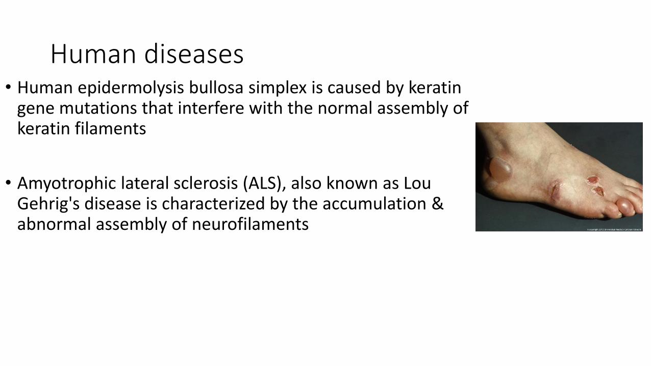
Human diseases• Human epidermolysis bullosa simplex is caused by keratin
gene mutations that interfere with the normal assembly of keratin filaments
• Amyotrophic lateral sclerosis (ALS), also known as Lou Gehrig's disease is characterized by the accumulation & abnormal assembly of neurofilaments


![Review Actin-targeting natural products: structures ... · actin-binding proteins actively break or ‘sever’ actin filaments [e.g. actin-depolymerizing factor (ADF) and cofilin]](https://img.dokumen.tips/doc/110x75/5f0f85bd7e708231d44494d0/review-actin-targeting-natural-products-structures-actin-binding-proteins-actively.jpg)