Embed Size (px)
Citation preview

J Clin Pathol 1983;36:280-288
Nature of the inflammatory cell infiltrate in duodenitisM HASAN, FRANCES HAY, W SIRCUS, ANNE FERGUSON
From the Gastrointestinal Unit, University ofEdinburgh and Western General Hospital, Edinburgh EH42XU
SUMMARY Counts of lamina propria and intraepithelial cells, lymphoid and polymorphonuclear,have been performed on semithin sections of endoscopic biopsies from the duodenum of patientswith ulcer-associated duodenitis, with non-specific duodenitis, and from controls. In both types ofduodenitis there were significant increases in lamina propria counts of plasma cells, lymphocytesand eosinophils, and in intraepithelial lymphocyte counts, when compared with controls. Incontrol specimens, neutrophil polymorphs were very infrequent but a substantial neutrophilpolymorph infiltration of the epithelium and lamina propria was present in both types ofduodenitis. In biopsies from areas of duodenitis scanning electron microscopy showed the pres-ence of celis, which are probably neutrophil polymorphs, on the luminal surface of the mucosa.Abnormalities in cell counts were present only in biopsies taken from visually inflamed areas ofthe duodenal bulb. These values returned to normal after healing of duodenitis with cimetidine.This study highlights the complex nature of the mucosal cellular infiltrate in duodenitis, particu-larly the striking increase in polymorphonuclear leucocytes. Histopathological features of ulcer-associated and non-specific duodenitis are identical.
Inflammation of the proximal duodenum,"duodenitis", can be recognised and defined in vari-ous ways, for example radiologically (coarsemucosal folds), endoscopically (redness, swelling,bleeding), or pathologically (altered mucosalarchitecture, increase in "inflammatory" cells). Wehave defined duodenitis on the basis of visual fea-tures at upper gastrointestinal endoscopy and in anearlier study we have reported that in both ulcer-associated and non-specific duodenitis, there areabnormalities of the duodenal mucosa with reduc-tion in villus length, increase in crypt length andincreased crypt mitotic counts.1 2 We have now com-pared ulcer-associated and non-specific duodenitisin respect of the non-epithelial cells of the duodenalmucosa-the so-called "inflammatory cellularinfiltrate" which is reported to be increased in non-specific duodentis.34 Various methods for the grad-ing of duodenitis, based on subjective assessment ofinflammatory cellular infiltrate, have been attemp-ted.57 However the density of the cell population inthe lamina propria of the duodenum varies consid-erably even in the normal state.8 9 We have thereforeused a quantitative technique to study the nature ofthe inflammatory cellular infiltrate in the duodenal
Accepted for publication 4 October 1982
mucosa, to compare this in non-specific duodenitiswith that in ulcer-associated duodenitis, and toexamine the effects on the infiltrate of successfulclinical treatment with cimetidine.
Patients and methods
PATIENTSThe criteria for selection of patients was asdescribed in an earlier study on intestinal mucosalarchitecture in duodenitis.' Upper gastrointestinalendoscopy was performed as part of the clinicalinvestigation or follow-up of patients with abdomi-nal pain or heartburn. The diagnoses of duodenalulcer and of duodenitis were based on endoscopicfindings and the following groups of patients werestudied.
Controls (C) were subjects in whom the only endo-scopic abnormality was hiatus hernia.
Duodenal ulcer (DU) were patients in whom one ormore ulcer craters, with discrete edge and depth,were present in the duodenal bulb.
Non-specific duodenitis (NSD) was divided into twocategories. Mild NSD comprised hyperaemia andswelling of the mucosa with slight contact bleeding;
280
on April 16, 2020 by guest. P
rotected by copyright.http://jcp.bm
j.com/
J Clin P
athol: first published as 10.1136/jcp.36.3.280 on 1 March 1983. D
ownloaded from

Cellular infitrate in duodenitis
severe NSD comprised intense hyperaemia andmucosal swelling, associated with spontaneousbleeding and multiple erosions.
Post cimetidine (DUPC and NSDPC) These werepatients with duodenal ulcer or with severe non-specific duodenitis previously diagnosed at endos-copy, in whom after six to 12 weeks treatment withcimetidine (1 g/day) there remained no visual evi-dence of inflammation in the duodenal bulb atendoscopy.
For the light microscopic studies, betwen six and 10patients per group were examined; for scanningelectron microscopy biopsies were taken from threepatients each in the control, duodenal ulcer andsevere non-specific duodenitis groups.
METHODS
Biopsy proceduresUpper gastrointestinal endoscopy was performedwith the Olympus GIF K instrument. Perendoscopicbiopsies were taken from specified areas of the firstand second parts of the duodenum under direct vis-ion. In controls, biopsies were taken at random fromthe duodenal bulb and from the second part of theduodenum (D2). In the DU and duodenitis patients,various biopsies were taken from visually inflamedareas and from visually normal areas of theduodenal bulb and of D2. In the patients studiedafter treatment, biopsies were taken from previouslyinflamed sites. The sites of biopsies in the variousgroups of patients are summarised in Table 1.
Preparation ofspecimens for light microscopyBiopsies were fixed in 5% glutaraldehyde in sodiumcacodylate buffer for 4-24 h and post-fixed in 1%osmium tetraoxide. They were embedded in Aral-dite and sections (1 ,m) were cut on an LKB IIIultramicrotome using glass knives. After removal ofAraldite, sections were stained in either 1%toluidine blue or in haematoxylin and eosin.As the study progressed it became obvious that
intestinal mucosal mast cells could not be identifiedin these preparations. A special staining techniquewas applied, in order to demonstrate mast cells inglutaraldehyde fixed mucosal biopsies.'0 Groups ofspecimens were recut, and the sections stained withAzure II, methylene blue and basic fuchsin.
Cell countsCounts of lamina propria and intraepithelial cellswere performed on coded slides, by light micros-copy, with an eyepiece graticule fitted into the mic-roscope dividing the visual field into 100 equal
281
Table 1 Groups ofsubjects and sites ofduodenal biopsies
Diagnosis No ofpatientsTotal Biopsy of bulb Biopsy of
second partVisually VisualYinflamed normal
Control 10 10 6DU 8 8 8 6NSD-severe 8 8 8 6NSD-mild 6 6DUPC 6 6NSDPC 6 6
squares. Lamina propria cell counts were performedin three parts of the section, to give a total laminapropria area studied of 100 squares. Mast cells in theAzure II methylene blue basic fuchsin stained sec-tions were counted in a total of 500 squares oflamina propria for each section. Results were thenconverted to number of cells per square millimetreof lamina propria with a correction factor which hadbeen calculated using the eyepice graticule and acalibration slide. Infiltration of lymphocytes and ofneutrophils within the surface epithelium was meas-ured by counting the lymphocytes and neutrophilsassociated with 200 epithelial cells of the villus sur-face, and the values expressed as counts per 100epithelial cells. The distribution of cells within theepithelial layer was also examined. For this, theepithelium was divided into three levels: apical, mid(including the top half of a nucleus) and lowerzones. In a total of three specimens for the lympho-cyte count, the distribution of 200 of each of thesecells within the epithelium was documented, eachcell being classified according to its location withinthe epithelial layer.
StadsiicsComparison of values between the various groupswas done both by the Kruskal and Wallis test and byWilcoxon's sum of ranks test.
Specimens for scanning electron microscopyFor scanning electron microscopy, specimens wereimmediately fixed in 2-5% glutaraldehyde and laterdehydrated with 100% acetone, critical point driedfrom liquid CO2, sputter coated with gold andexamined under an International Scientific Instru-ment (Model ISI 60) scanning electron microscope.
Resilts
LAMINA PROPRIA CELL COUNTSIt was possible to perform lamina propria cell countsin all but four of the specimens taken. No attemptwas made to assess changes in the sizes of villi and
on April 16, 2020 by guest. P
rotected by copyright.http://jcp.bm
j.com/
J Clin P
athol: first published as 10.1136/jcp.36.3.280 on 1 March 1983. D
ownloaded from

282
crypts since this had been fully documented in theprevious study. Epithelial cell damage and pseudo-stratification were seen frequently in specimensfrom all of the groups studied. Superficial gastricmetaplasia was also seen in specimens from controlsas well as from patients with duodenal ulcer andnon-specific duodenitis.
Results of the counts of total lamina propria cells,plasma cells, lymphocytes and eosinophils (persquare millimetre of lamina propria) are summar-ised in Figs. 1-4. A similar pattern of resultsemerges for all of these categories. In biopsies fromthe duodenal bulb of control patients the meanlamina propria total cell count was 4216/mm2 with2118 plasma cells, 1647 lymphocytes and 83eosinophils. The results of the counts in biopsies ofmucosa from the second part of the duodenum incontrols were similar. In contrast, in biopsies fromareas of ulcer-associated and severe non-specificduodenitis significantly higher counts of all of thesecell types were found. However, in mild duodenitisand in all of the biopsies taken from visually normalduodenum in the duodenal bulb and second part ofthe duodenum, no significant differences from con-
DuodenumEkuodenai bulb second part-
00
10000
E 9000E
CLC. 8000
-7000A*C 6000-E
c5000 T
4
I 4 T
3000-E22000-
-a
* Visually normal * p<0 01o Mild duodenitisX Severe duodenitis
Fig. 1 Total number ofcells per square millimetre (mean± SE) oflamina propria in duodenal mucosa.
Hasan, Hay, Sircus, Ferguson
trols emerged. In the post-treatment biopsies of theduodenal bulb, lamina propria cell counts were alsonormal. Mucosal mast cell counts were performed inspecimens from the duodenal bulb in six controls, sixpatients with ulcer-associated duodenitis and sixwith severe non-specific duodenitis. As shown inFig. 5, no differences were found between thesethree groups. Neutrophil polymorphs were veryrarely present in the lamina propria of control biop-sies (mean 7/mm2 lamina propria). However inulcer-associated and in severe non-specificduodenitis (Fig. 6), values were considerably higherwith 904 + 123 (+ SE) in DU and 421 + 72 inNSD respectively (p < 0X005 in each case). Therewas no difference from controls in the neutrophilpolymorph count in the lamina propria in the othergroups of specimens from the duodenal bulb andsecond part of duodenum.
INTRAEPITHELIAL CELL COUNTSJust as in the case of lamina propria cells,intraepithelial lymphocyte counts were increased ininflamed areas ofDU and severe NSD, as comparedto controls, whereas there were no significant differ-
O
2,o
Ec°
0
E
E
Ea
0
z
* Visuclly normalo Mild duodenitisX Severe duodenitis
*p<0.01
Fig. 2 Number ofplasma ceUls per square milimetre oflamina propria in duodenal mucosa.
on April 16, 2020 by guest. P
rotected by copyright.http://jcp.bm
j.com/
J Clin P
athol: first published as 10.1136/jcp.36.3.280 on 1 March 1983. D
ownloaded from

.3000-
9 2500 *
1000-E
o 500-
o Mild duodenitisX Severe duoderitis
Fig. 3 Number oflymphocytes per square millimetre oflamina propria in duodenal mucosa.
* VisuaCly normal * p<0-01o Mild duodenitis ** p<0-005X Severe duoderitis
Fig. 4 Number ofeosinophils per square millimetre oflamina propria in duodenal mucosa.
Duodeurn-second par
180 -
160
140
a.20. 120
a
100.
E80
'- 60I'Ln
40-
0
S.
00
0
0
0
0
00
S
zu*I*
'-Ii ~~~~0OS
Fig. 5 Number ofmast cells per square millimetre oflamina propria in duodenal mucosa.
1500 -
a
g 1000-00CN
aV
* 500t
z
*;
I,, II
I*0j ii It 3' 1 ' 1I I
* Visually normal * p<0 05o Mild duodenitisX Severe duoderitis
Fig. 6 Number ofneutrophil polymorphs per squaremilimetre oflamina propria in duodenal mucosa.
gn _
on April 16, 2020 by guest. P
rotected by copyright.http://jcp.bm
j.com/
J Clin P
athol: first published as 10.1136/jcp.36.3.280 on 1 March 1983. D
ownloaded from

Hasan, Hay, Sircus, Ferguson
16-
15 -
5 14-u 13--55 12-
a 11-
o 10-
p 9.V 8-
7-
E 6--2~-5 5-
-2 4-4.
2 3
2-
1 -
I I I I
* Visually normal *P<0 01o Mild duodenitis **p<0 005X Severe dcuodenitis
Fig. 7 Intraepithelial lymphocyte count per 100 epithelialcells in duodenal mucosa.
ences from controls in the biopsies taken from visu-ally normal mucosa in the duodenal bulb or secondpart of the duodenum (Fig. 7). With regard to neu-trophil polymorphs, no neutrophil polymorph wasobserved within the epithelium in any of the controlspecimens. In contrast, in specimens from DU andsevere NSD, the figures were 4 + 0*8 (mean + SE)and 4-6 ± 0 9 polymorphs per 100 epithelial cells(Fig. 8) (p < 0 005 for each comparison with con-trol). Intraepithelial neutrophil polymorphs wererarely found in the other groups of specimens fromDU and NSD patients, and for these, values showedno statistically significant differences from controls.An interesting difference emerged when the loca-
tion within the epithelium of lymphocytes and neu-trophils was studied. As shown in Table 2, neu-trophils were evenly distributed within theepithelium, in the apical, mid and basal regions,whereas intraepithelial lymphocytes were basallysituated.
6~~~~5
o Mi 1udeii
~4 I
3-
-51-c~~~~
*Visually normal *P< 00050 Miid dJtodenitisX Severe duodenitis
Fig. 8 Intraepithelial neutrophilpolymorph countper 100epithelial ceUls in duodenal mucosa.
Table 2 Distribution ofintraepithelial lymphocytes andpolymorphs within the epithelium ofthe duodenal mucosa
Base Middle Top
Lymphocytes 69 23 8Neutrophilpolymorphs 28 38 34
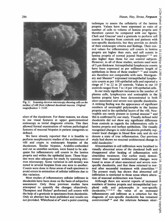
SCANNING ELECTRON MICROSCOP-YExamination of control specimens from theduodenal bulb showed the presence of villi of vari-ous shapes and sizes with no surface exudate and nointraluminal cells adherent to the surface (Fig. 9). Incontrast in the specimens from both DU and severeNSD there were cells on the surfaces of the villiwhich had the scanning EM features of neutrophils,and some red blood cells were also present (Figs. 10and 11).
Discusson
Visual and histological changes of inflammationoften occur in the proximal duodenum in associationwith peptic ulcer and similar changes, when seen inthe absence of frank ulceration, are termed non-specific duodenitis. The overall aim of our work induodenitis has been to elucidate the pathogenesis ofnon-specific duodenitis and its relation to peptic
11
284
on April 16, 2020 by guest. P
rotected by copyright.http://jcp.bm
j.com/
J Clin P
athol: first published as 10.1136/jcp.36.3.280 on 1 March 1983. D
ownloaded from

Cellular infitrate in duodenitis
rigs i (aboveJ ana iUuDeiOW) scanning etectron microscopyshowing surface of villi from control duodenal mucosa.Original magnifration x 500. (Below) Scanning electronmicroscopy showing surface of villi from infpmed duodenalmucosa. Original magnification x 510.
285
on April 16, 2020 by guest. P
rotected by copyright.http://jcp.bm
j.com/
J Clin P
athol: first published as 10.1136/jcp.36.3.280 on 1 March 1983. D
ownloaded from
Hasan, Hay, Sircus, Ferguson
Fig. 11 -canning electron microscopy showing cells on thsurface of villi from inflamed duodenal mucosa. Originalmagnification x 5100.
ulcer of the duodenum. For these reasons, we choseto use visual features at upper gastrointestinalendoscopy as initial diagnostic criteria. This thenallowed formal examination of various pathologicalfeatures of mucosal biopsies in patient categories sodefined.We have already reported that it is feasible to
perform morphometric studies of the architecture ofvilli and crypts in endoscopic biopsies of theduodenum. Similar biopsies, Araldite-embeddedand cut as semithin sections, were found to be ade-quate for inflammatory cell counts in the laminapropria and within the epithelial layer. These biop-sies were also adequate for study by scanning elec-tron microscopy. Some variation in cell density wasnoted in several biopsies from one area to another.Counts were done in three areas of each specimen toavoid errors in estimation of cellular infiltrate due tothis variation.Most studies of inflammatory cellular infiltrate in
duodenitis have been done on the basis of subjectiveassessment. Only three previous studies haveattempted to quantify the changes objectively.Thompson and Holme4 performed cell counts withthe help of a graticule in specimens from duodenitis.Only an abstract has been published and results arenot provided. Whitehead et a15 used a point counting
technique to assess the cellularity of the laminapropria. Values have been expressed as ratio ofnumber of cells to volume of lamina propria andtherefore cannot be compared with our figures.Cheli and Giacosa3 used a graticule to perform cellcounts in biopsies from controls and patients withnon-specific duodenitis, but they provide no detailsof their endoscopic criteria and findings. Their con-trol values for inflammatory cell counts in laminapropria are higher than ours, and cell counts inlamina propria of normal jejunal biopsies"-"' arealso higher than those for our control subjects.However, in all of these studies, sections used wereof 5 ,m thickness. Intraepithelial lymphocyte countshave been expressed as per millimetre length ofepithelium in three of these studies"'-" and resultsare therefore not comparable with ours. Montgom-ery and Shearer'4 expressed intraepithelial lympho-cyte counts as per 100 epithelial cells and reported arange of 7 to 22 in 20 controls. Values in our 10controls ranged from 7 to 14 per 100 epithelial cells.
In our study significant increases in the number ofplasma cells, lymphocytes and eosinophils in thelamina propria have been demonstrated in bothulcer-associated and severe non-specific duodenitis.A striking finding was the appearance of significantnumbers of neutrophil polymorphs in the laminapropria and in the epithelial layers of the vili. Neu-trophils are rarely present in normal mucosa7 9 andthis is confirmed by our study. Visually defined mildduodenitis did not show any significant differencefrom controls as regards the inflammatory cells inlamina propria and surface epithelium. The visuallyrecognised changes in mild duodenitis probably rep-resent local changes in blood flow only and do notdenote a state of true inflammation. Our previousstudy had shown no mucosal architectural change inmild duodenitis.'
Abnormalities in cell infiltration were localised tovisually abnormal areas of the duodenal bulb anddisappeared after healing of inflammation withcimetidine treatment. Our earlier study demon-strated that mucosal architectural changes werefound in areas of ulcer-associated and severe non-specific duodenitis, and that these returned to nor-mal after successful treatment with cimetidine.' 2The present study has shown that abnormal cellinfiltration is restricted to those areas where altera-tion of mucosal architecture was found.Although there are several reports of increased
numbers of lamina propria and intraepithelial lym-phoid cells and polymorphs in non-specificduodenitis,34 1516 the value of an increasedinflammatory cell infiltrate as a criterion for thediagnosis of non-specific duodenitis has remainedcontroversial9 '" and the relations between ulcer-
286
on April 16, 2020 by guest. P
rotected by copyright.http://jcp.bm
j.com/
J Clin P
athol: first published as 10.1136/jcp.36.3.280 on 1 March 1983. D
ownloaded from

Cellular infiltrate in duodenitis
associated and non-specific duodenitis have notbeen clarified.4"9 On subjective assessment, usinghistological sections from surgical specimens,similarities in the histological appearances ofduodenitis and duodenal ulcer have been reported"7and Joffe et all' considered that the histologicalchanges in these two conditions were indistinguish-able. This present study is the first quantitative com-parison of the components of the inflammatory cel-lular infiltrate in ulcer-associated and non-specificduodenitis, and firmly demonstrates that thechanges are identical in these two conditions. Therewas no overlap between results for controls and forthe two types of duodenitis, in the variousinflammatory cells studied. However, one specimenfrom an unaffected area of ulcer-associatedduodenitis and one specimen from an unaffectedarea in a patient with severe non-specific duodenitisshowed increased inflammatory cell counts.A generalised expansion of the mucosal immune
cells in both ulcer-associated and non-specificduodenitis could be due to stimulation of gut-associated lymphoid tissues in these conditions. Thisappears an unlikely explanation, as we have shownthat the changes are very localised. Local factors atsites of inflammation seem a more likelymechanism-these may either attract cells into thetissues, increase the rate of proliferation locally, orimmobilise these cells and prevent them from leav-ing the mucosa in blood or lymph.
Neutrophils are not a normal constituent of thelamina propria or the epithelium of the duodenalbulb in normal subjects. The results of this studynow confirm that the presence of neutrophils withinthe mucosa is a striking and consistent feature, bothin ulcer-associated and in non-specific duodenitis. Asubstantial neutrophil infiltrate was present in everybiopsy taken from a visually abnormal area of theduodenal bulb in severe duodenitis and in duodenalulcer patients; and in none of the control subjectswas there a substantial neutrophil infiltrate in themucosa. The study confirms the presence of neu-trophils as a crucial factor for the histological diag-nosis of duodenitis. These cells appear to be migrat-ing through the epithelial lining and are presenton the mucosal surface, as indicated by scanningelectron microscopy. Mediators of neutrophilchemotaxis in this situation remain unknown. Poss-ible substances include complement components,immunoglobulin fragments, fragments of fibrinogenand collagen, kallikrein and prostaglandin E1. Thesubstances causing chemotaxis may play an impor-tant role in the pathogenesis of duodenal inflamma-tion. The substance or substances involved may bethe same as those responsible for generalised expan-sion of mucosal immune cells and this aspect of
287
duodenal inflammation requires further study. Thelocalised nature of inflammation and return to nor-mality after cimetidine treatment have led us tobelieve that the responsible substance or substancesare most likely to be derived from luminal contents.
We are grateful to all the staff of the EndoscopyTheatre at the Western General Hospital, Edin-burgh, for their help with collection of specimens.Mr A Sutherland, and the staff of the Scanning Elec-tron Microscope Unit, provided valuable technicalassistance. We acknowledge the support of theAssociation of Commonwealth Universities and ofthe Wellcome Trust.
References
Hasan M, Sircus W, Ferguson A. Duodenal mucosal architecturein non-specific and ulcer-associated duodenitis. Gut1981;22:637-41.
2 Hasan M, Ferguson A. Measurements of intestinal villi in non-specific and ulcer-associated duodenitis. J Clin Pathol1981;34:1181-6.
Cheli R, Giacosa A. Inflammatory cell count and identification inchronic non-specific duodenitis. Endoscopy 1977;9:91-5.
4Thompson H, Holme G. The diagnosis of duodenitis. Gut1974;15:842-3.
Whitehead R, Roca M, Meikle DD, Skinner J, Truelove SC. Thehistological classification of duodenitis in fibreoptic biopsyspecimens. Digestion 1975;13:129-36.
6 Gregg JA, Garabedian M. Duodenitis. Am J Gastroenterol1974;61:177-84.
Greenlaw R, Sheahan DG, DeLuca V, Miller D, Myerson D,Myerson P. Gastroduodenitis: a broader concept of pepticulcer disease. Dig Dis Sci 1980;25:660-72.
Roca M, Truelove SC, Whitehead R. The histological state of thegastric and duodenal mucosa in healthy volunteers. Gut1975;16:404.
9 Kom ER, Foroozan P. Endoscopic biopsies of normal duodenalmucosa. Gastrointest Endosc 1974;21:51-4.
Strobel S, Hasan M, Ferguson A. Staining properties of humanintestinal mucosal mast cells after glutaraldehyde fixation. JClin Pathol 1982;35:897-9.
" Holmes GKT, Asquith P, Stokes PL, Cooke WT. Cellularinfiltrate of jejunal biopsies in adult coeliac disease in relationto gluten withdrawal. Gut 1974;15:278-83.
12 Ferguson R, Allan RN, Cooke WT. A study of the cellularinfiltrate of the proximal jejunal mucosa in ulcerative colitisand Crohn's disease. Gut 1975;16:205-8.
13 Ferguson R, Asquith P, Cooke WT. The jejunal cellular infiltratein coeliac disease complicated by lymphoma. Gut1974;15:458-61.
14 Montgomery RD, Shearer ACI. The cell population of the upperjejunal mucosa in tropical sprue and postinfective malabsorp-tion. Gut 1974;15:387-91.
ISGelzayd EA, Biederman MA, Gelfand DW. Changing conceptsof duodenitis. Am J Gastroenterol 1973;64:213-6.
16 Cotton PB, Price AB, Tighe JR, Beales JSM. Preliminary evalua-tion of "duodenitis" by endoscopy and biopsy. Br Med J1973;iii:430-3.
17 Welbrock WLA. Duodenitis and duodenal ulcer. Ann Stg1930;91:533-9.
on April 16, 2020 by guest. P
rotected by copyright.http://jcp.bm
j.com/
J Clin P
athol: first published as 10.1136/jcp.36.3.280 on 1 March 1983. D
ownloaded from

Hasan, Hay, Sircus, Ferguson1 Joffe SN, Thomson WD, Robertson A, Lee F, Blumgart LH. Is
duodenitis a dyspeptic myth? Gut 1977;18:399.9 Joffe SN, Lee FD, Blumgart LH. Duodenitis. Clin Gastroenterol
1978;7:635-50.
Requests for reprints to: Dr Anne Ferguson, Gastrointes-tinal Unit, Western General Hospital, Edinburgh EH42XU, Scotland.
288
on April 16, 2020 by guest. P
rotected by copyright.http://jcp.bm
j.com/
J Clin P
athol: first published as 10.1136/jcp.36.3.280 on 1 March 1983. D
ownloaded from