Embed Size (px)
Citation preview

Muscarinic Receptors Mediated Stimulation and
Intracellular Signaling Pathways Involved in Human Lung Fibroblast Proliferation
Dissertation
zur
Erlangung des Doktorgrades (Dr. rer. nat)
der
Mathemathisch-Naturwissenschaftlichen Fakultät
der
Rheinischen Friedrich-Wilhelms-Universität Bonn
vorgelegt von
Amit Bahulayan
aus
Mumbai
2009
Angefertigt mit der Genehmigung der Mathematisch-Naturwissenschaftlichen Fakultät der Rheinischen
Friedrich-Wilhelms-Universität Bonn
1. Referent: Prof. Dr. med. Kurt Racké
2. Referent: Prof. Dr. med. Klaus Mohr
Tag der Promotion: 13.05.2009

Dedicated to my parents for their support all these years

Table of Contents Abbreviations 1 Introduction 1
1.1 Neuronal and Non Neuronal Acetylcholine 2 1.2 Cholinoceptors 3
1.2.1 Nicotinic Receptors 3 1.2.2 Muscarinic Receptors 4
1.3 Molecular and Pharmacological Classification of Muscarinic Receptor Subtypes 5
1.3.1 Molecular Biology of Muscarinic Receptors 5 1.3.2 Pharmacological Classification of Muscarinic Receptor Subtypes 6
1.4 Muscarinic Coupling to G-proteins and Effector Molecules 8 1.4.1 Adenylyl Cyclase 10 1.4.2 Phospholipase C 11
1.5 Mitogen Activated Protein Kinases 12 1.5.1 MAPKs in Regulation of Cell Cycle 14
1.6 G-protein Coupled Receptor Signaling to MAPK 16 1.7 Intracellular Signaling Pathways Linking G-protein Coupled Receptors to MAPK 17
1.7.1 The Ras Mediated Activation of MAPK Cascade via G-protein Coupled Receptors 18 1.7.2 Rho Mediated Activation of MAPK Cascade via G-protein Coupled Receptors 21
1.7.3 Phosphatidylinositol 3-Kinase (PI3K) Mediated Activation of MAPK Cascade via G-protein Coupled Receptors 23 1.7.4 Phospholipase C (PLC) Mediated Activation of MAPK Cascade via G-protein Coupled Receptors 25 1.8 Transactivation of Classical Receptor Tyrosine Kinases by GPCRs 26 1.9 Apoptosis and G-protein Coupled Receptor Signaling Interaction 27
2 Materials and Methods 29
2.1 Materials 29 2.1.1 Reagents 29 2.1.2 Test Drugs 30
I

2.1.3 Buffers and Reagents for Cell Culture 34 2.1.4 Solution and Buffers for Proliferation Assay 35 2.1.5 Reagents for Protein Determination using the Lowry Method 36 2.1.6 Primers for Polymerase Chain Reaction 37 2.1.7 Buffers and Solutions for RT/PCR 38 2.1.8 Human Lung Fibroblast Cells 40 2.1.9 Buffers and Reagents for Protein Gel Electrophoresis and Immunoblot 40 2.1.10 Antibodies 42 2.1.11 Reagents and Solutions for Caspase-3 Fluorimetric Assay 42 2.1.12 Equipment 44 2.1.13 Tools for Statistical Analysis 44
2.2 Methods 46 2.2.1 Culture of Human Lung Fibroblast 46 2.2.2 RNA Preparation 47 2.2.3 Reverse Transcription 48 2.2.4 Polymerase Chain Reaction (PCR) 49 2.2.5 Agarose Gel Electrophoresis 50 2.2.6 DNA Preparation 50 2.2.7 Protein Preparation 51 2.2.8 Protein Determination 52 2.2.9 Protein Gel Electrophoresis and Immunoblot 52 2.2.9.1 Protein Gel Electrophoresis 52 2.2.9.2 Immunoblot 53 2.2.9.3 Membrane Stripping 55 2.2.10 (3H)-Thymidine Incorporation Proliferation Assay 55 2.2.11 Caspase-3 Fluorimetric Assay 56
3 Results 59
3.1 Muscarinic Receptor Expression in Human Lung Fibroblasts 59 3.1.1 Muscarinic Receptor Expression in MRC-5 Human Lung Fibroblasts 59 3.1.2 Muscarinic Receptor Expression in Primary Human Lung Fibroblasts 62
3.2 Muscarinic Stimulation of Proliferation of Human Lung Fibroblasts 65 3.2.1 Muscarinic Stimulation of Proliferation of MRC-5 Human Lung Fibroblasts 65
3.3 Characterisation of Muscarinic Receptor Sub-Type 68 3.3.1 Inhibition of Muscarnic Proliferation using Receptor Sub-Type Specific Antagonists in MRC-5 Human Lung Fibroblasts 68 3.3.1.2 Effect of Pertussis Toxin on Muscarinic Stimulation of Proliferation of MRC-5 Human Lung Fibroblasts 71
II
3.3.1.3 Muscarinic Stimulation and Effect of Pertussis Toxin on Proliferation in Primary Human Lung Fibroblasts (pHLFb) 72
3.4 Muscarinic Stimulation Mediated Activation of MAPK Pathway in Human Lung Fibroblasts 73
3.4.1 Muscarinic Receptor Stimulation and Activation of p-42/44 MAPK Pathway in proliferation of Human Lung Fibroblast 73 3.4.1.2 Western Blot Analysis of p42/44 MAPK (ERK1, ERK2) Activation in MRC-5 Human Lung Fibroblast 74
3.5 Effect of Kinase Inhibitors on Muscarinic Stimulation of MRC-5 Human Lung Fibroblast Proliferation 76
3.5.1 Effects of the Rho-kinase Inhibitor Y27632 on Muscarinic Stimulation of MRC-5 Human Lung Fibroblast Proliferation 76 3.5.1.2 Effects of the Phospholipase C Inhibitor U73122 on Muscarinic Stimulation of MRC-5 Human Lung Fibroblast Proliferation 78 3.5.1.3 Effects of the PI3-kinase Inhibitor Wortmannin on Muscarinic Stimulation of MRC-5 Human Lung Fibroblast Proliferation 80 3.5.1.4 Effects of the Raf-1 Inhibitor GW5074 on Muscarinic Stimulation of MRC-5 Human Lung Fibroblast Proliferation 82 3.5.1.5 Effects of the Raf-1 Inhibitor GW5074 (GW) on Muscarinic Stimulation of the ERK-MAP Kinase in MRC-5 Human Lung Fibroblasts 84 3.5.1.6 Effects of the Tyrosine-Kinase Inhibitor Tyrphostin AG1295 on Muscarinic Stimulation of MRC-5 Human Lung Fibroblasts 85
3.6 Muscarinic Stimulation of proliferation of MRC-5 Human Lung Fibroblast and Anti-apoptotic effects 87
3.7 Nicotinic Receptor Expression and Function in MRC-5 Human Lung Fibroblasts 89
4 Discussion 92
4.1 Muscarinic Receptor Expression in Human Lung Fibroblasts 92 4.2 Muscarinic Stimulation of Proliferation of Human Lung Fibroblasts 92 4.3 Inhibition of Muscarnic Proliferation using Receptor Sub-Type Specific Antagonists in MRC-5 Human Lung Fibroblasts and Effect of Pertussis Toxin on Muscarinic Stimulation of Proliferation of Human Lung Fibroblasts 94 4.4 Muscarinic Receptor Stimulation and Activation of p-42/44 MAPK Pathway in Proliferation of Human Lung Fibroblast 95
III

4.5 Intracellular Signaling Mechanism Involved in Muscarinic Stimulation Mediated Activation of MAPK Cascade in MRC-5 Human Lung Fibroblast Proliferation 96
4.5.1 Effects of the Rho-kinase Inhibitor Y27632 on Muscarinic Stimulation of MRC-5 Human Lung Fibroblast Proliferation 96 4.5.1.2 Effects of the Phospholipase C Inhibitor U73122 on Muscarinic Stimulation of MRC-5 Human Lung Fibroblast Proliferation 97 4.5.1.3 Effects of the PI3-kinase Inhibitor Wortmannin on Muscarinic Stimulation of MRC-5 Human Lung Fibroblast Proliferation 98 4.5.1.4 Effects of the Raf-1 Inhibitor GW5074 on Muscarinic Stimulation of MRC-5 Human Lung Fibroblast Proliferation 99 4.5.1.5 Effects of the Tyrosine-Kinase Inhibitor Tyrphostin AG1295 on Muscarinic Stimulation of MRC-5 Human Lung Fibroblasts 100
4.6 Muscarinic Stimulation of Proliferation of MRC-5 Human Lung Fibroblast and Anti-apoptotic Effects 101 4.7 Nicotinic Receptor Expression and Function in MRC-5 Human Lung Fibroblasts 102
5 Conclusion 104 6 References 106 7 Seminars and Publications 127 8 Acknowledgements 128 9 Curriculum Vitae 129
IV

Abbreviations ACh Acetylcholine
AC Adenylyl cyclase
ADP Adenosine diphosphate
Akt Protein kinase B
Arg Arginine
Asp Aspartic acid
ATP Adenosine triphosphate
cAMP Cyclic adenosine monophosphate
COPD Chronic obstructive pulmonary disease
cDNA Complementary Deoxyribonucleic acid
CDK Cyclin dependent kinase
CKI Cyclin dependent kinase inhibitor
DAG Diacyl glycerol
DNA Deoxyribonucleic acid
DMSO Dimethylsulfoxide
DEPC Diethylpyrocarbonate
EGF Epidermal growth factor
EGFR Epidermal growth factor receptor
EDTA Ethylenediaminetetraaceticacid
ERK Extracellular signal regulated kinase
FCS Fetal calf serum
GAPs GTPase Activating Proteins
GEFs Guanine nucleotide exchange factors
G-protein Guanine nucleotide binding proteins
GPCR G-protein-coupled receptor
GDP Guanosine diphosphate
GTP Guanosine Triphosphate
GTPase Guanosine triphosphatase
gDNA Genomic DNA
V
VI
IP3 Inositol triphosphate
LPA Lysophosphatidic acid
MAPK Mitogen activated protein kinase
mAChR Muscarinic acetylcholine receptor
mRNA Messenger RNA
PCR Polymerase chain reaction
PBS Phosphate buffered saline
PDGF Platelet derived growth factor
PtdIns Phosphatidylinositol
PIP2 Phosphotidylinositol-4,5-biphosphate
PLC Phospholipase C
PKC Protein kinase C
PI3K Phosphatidylinositol 3 Kinase
PKA Protein kinase A
PTX Pertussis toxin
RNA Ribonucleic acid
RT Reverse transcription
RTK Receptor tyrosine kinase
SEM Standard error of the mean
SAPK Stress activated protein kinase
TNFα Tumor necrosis factor
1 Introduction Asthma and chronic obstructive pulmonary disease (COPD) are two diseases
characterized by airflow limitation due to either reduced driving pressure (loss of
elastic recoil of the lung parenchyma) or an increased resistance (airway
obstruction) which is mostly reversible in asthma and mostly irreversible in COPD
(1, 2). Airway remodeling is a pathological feature observed both in asthma and
COPD. The most important difference between asthma and COPD is the nature
of inflammation, which is primarily eosinophilic and CD-4 driven in asthma, and
neutrophilic and CD-8 driven in COPD (3). In spite of these differences fibrotic
alterations are observed in both these diseases (4). Cholinergic pathway
represents key mechanisms in control of airway smooth muscle tone.
Anticholinergic bronchodilaters reduce vagal cholinergic tone, the main reversible
component in COPD hence constitute an essential element in the therapy of
obstructive airways diseases especially in COPD (5, 6). Furthermore tiotropium
bromide (Spiriva®) a long acting muscarinic antagonist was found to delay the
decline in airway function in COPD (7, 8, 9), suggesting that cholinergic
mechanisms plays a pivotal role in the structural changes associated with airway
remodeling. In studies carried out on human and bovine airway smooth muscle it
was seen that muscarinic agonists enhanced the proliferative response to
epidermal growth factor and platelet-dervied growth factor respectively (10, 11).
Moreover, tiotropium was found to attenuate the increase in airway smooth
muscle mass and myosin expression induced by repeated allergen challenges
(12). Almost every cell type in the airways expresses cholinoreceptor (13, 14)
and hence could be a target for acetylcholine released from neuronal or
nonneuronal sources (13, 15). Expression of mRNA encoding different nicotinic
receptor subunits (16) and M2 receptors in airway fibroblasts has been described,
but a detailed study on expression of muscarinic receptor subtypes in lung and
airway fibroblasts is still lacking. It has been shown that the activation of nicotinic
receptors stimulates collagen gene expression and fibronectin synthesis (17, 18)
whereas the functional role of muscarinic receptors in airway fibroblasts remains
to be illuminated.
1

The purpose of this study was to determine the expression pattern of the
muscarinic receptors in human lung fibroblasts and to investigate whether
muscarinic receptors mediate effects on cell proliferation and if so, to elucidate
the intracellular signaling pathways entailed.
1.1 Neuronal and Non-Neuronal Acetylcholine ACh represents the ‘classical’ neurotransmitter of the parasympathetic nervous
system. As neurotransmitter ACh is synthesized in the nerve endings by the
enzyme choline acetyltransferase using acetyl-CoA and choline as substrates. It
is then stored in the synaptic vesicles. Eventually, depolarization-induced calcium
influx lead to an exocytotic release of the neurotransmitter into the extracellular
space where it may interact with receptors located on the target cells as well as
on the cholinergic nerve terminals themselves (13). In order to limit effector cell
activation, after release, ACh is degraded by acetylcholineesterase to non-active
choline (19).
Recent studies have revealed that choline acetyltransferase and its product ACh
are present in a wide range of non-neuronal cells, including epithelial and
endothelial cells, smooth muscle cells as well as various cells of the immune
system such as lymphoctyes, macrophages, eosinophils and neutrophils (20). In
addition nicotinic and muscarinic receptors have been shown to be widely
expressed on these non-neuronal cells (21, 22, 23, 24). The essential
components of the cholinergic system are present, to a greater or lesser extent,
in many of these non-neuronal inflammatory cells involved in airway physiology
and pathophysiology. However unlike neuronal cells, where ACh is stored in
discrete neurosecretory vesicles, cells of the non-neuronal cholinergic system
appear to release acetylcholine directly upon synthesis via active transport
mediated by members of the organic cation transporter (OCT) family. Although
not much is know about the functional role of non-neuronal ACh it has been
shown to act as a local cell molecule via paracrine and autocrine mechanisms to
control basic cell functions such as proliferation, differentiation, maturation,
migration, secretion, organization of the cytoskeleton and cell-cell contact (23,
2
24, 25, 26, 27). In a recent study it was shown that the dysfunction of the non-
neuronal cholinergic system in the airways could contribute to the deleterious
changes of epithelial ion and water movement involved in the pathogenesis of
cystic fibrosis a hereditary disease that affects mainly the lungs and digestive
system, causing progressive disability (28).
1.2 Cholinoceptors 1.2.1 Nicotinic Receptors
The nicotinic acetylcholine receptors are pentamers of homologous or identical
subunits, symmetrically arranged to form a transmembrane cation channel (29).
There are multiple nicotinic receptor isotypes existing. Muscle nicotinic receptors,
localized at the end plate of the neuromuscular junctions, are assembled of two
α-, one β-, one γ- or one ε- and one δ (30), whereas neuronal nicotinic receptors
appear to be composed either of only two different types of subunits (α and β) or
of five α- subunits. The activation of the nicotinic receptor causes a net influx of
positively charged ions resulting in membrane depolarization, which can be
detected for example as ‘excitatory postsynaptic potential’ at postganglionic
neurons (13). At least 10 different α- and four β- subunits have been identified so
far. In addition to the expression of neuronal nicotinic receptor subunits in
autonomic ganglia, it was observed in almost every other cell type in the airways
and lung tissues. The α-7 subunit was detected in many non-neuronal cells such
as the bronchial epithelial cells, endothelial airway cells and airway fibroblasts
(16). In vitro studies carried out on bronchial epithelial cells and pulmonary
neuroendocrine cells indicated nicotinic receptor mediated activation of cell
proliferation cascade (31, 32). It’s worth mentioning that nicotinic receptors in
non-neuronal cells have also been suggested to be involved in tobacco induced
toxicity in the airways (33). Furthermore, chronic exposure of murine and human
bronchial epithelial cells to nicotine both in vitro and in vivo caused an
upregulation of α-7 and α-3 subunits (34, 35). In another study carried out on
WI38 cells, a human embryonic pulmonary fibroblast cell line expressing α-3 and
α-7 nicotinic receptor subunits, nicotine exposure disrupted the specific epithelial-
3

mesenchymal paracrine signaling pathways and resulted in pulmonary interstitial
lipofibroblast (LIF)-to-myofibroblast (MYF) transdifferentiation, which could result
in altered pulmonary development and function (36).
1.2.2 Muscarinic Receptors
The muscarinic ACh receptors (mAChRs) are members of the superfamily of
hormones and neurotransmitter receptors. The muscarinic acetylcholine receptor
is a seven transmembrane domain receptor that is coupled to a guanosine
triphosphate (GTP) binding protein (G-protein). The muscarinic receptors
regulate the activities of intracellular secondary messenger pathways through ion
channel and enzyme activation/deactivation by interaction with their coupled G-
proteins. Five different subtypes of muscarinic receptors have been identified by
molecular biological techniques (m1-m5) (37, 38, 39), but so far a sufficient
pharmacological and functional characterization has been provided for only four
of them (M1-M4). Nonetheless, there is now significant evidence that the gene
product of the m5 gene forms a functional muscarinic receptor (M5) (39). The ‘odd
numbered’ (M1/M3/M5) muscarinic receptor subtypes couple preferentially to the
pertussis toxin (PTX) insensitive G-proteins of the Gq family, while the ‘even
numbered (M2/M4) muscarinic receptors bind with G-proteins belonging to the Gi/o
family. The former subtypes activates phospholipase C (PLC), phospholipase A2
(PLA2), phospholipase D (PLD), tyrosine kinase and calcium influx but do not
inhibit adenylyl cyclase, while the latter group of receptors inhibit adenylyl
cyclase but do not stimulate PLC. However this coupling specificity of the
mAChRs subtypes is not absolute, for the M2 and the M4 subtypes can weakly
activate PLC when expressed at high levels in certain cell types (40, 41, 42). The
PTX-insensitive coupling to PLC is mediated by Gαq, Gα11, Gα14, and Gα16 , while
the PTX-sensitive coupling to adenylyl cyclase inhibition is mediated by Gαi or
Gαo (43, 44).
4
1.3 Molecular and Pharmacological Classification of Muscarinic Receptor Subtypes: 1.3.1 Molecular Biology of Muscarinic Receptors
Subsequent to early pharmacological findings, molecular cloning studies
revealed five different subtypes of muscarinic receptors (m1-m5) (37). Numa and
colleagues cloned the m1 and the m2 genes (45, 46). The m3, m4, and m5 gene
were thereafter discovered (47, 48, 49). The vertebrate cloned receptor genes
are intronless, and similar across mammalian species (50, 51). These five genes
encode mAChR glycoproteins that display the structural features of the seven
transmembrane helix G-protein-coupled receptor family. Based on the
pharmacological binding studies of each receptor resembling the cloned genes, it
is now recommended that M1, M2, M3, M4 and M5 be used to describe both
pharmacological subtypes and molecular subtypes encoded by the cloned
genes.
Similar to most members of the G-protein-coupled receptor family whose ligand
recognition site binds small molecules, there are several major features of
muscarinic receptor structure. First, the ligand recognition site can be located
within the outer half of the membrane-embedded part of the protein. To bind the
neurotransmitter ACh, all muscarinic receptors have an Asp residue in the distal
N-terminal part of the third transmembrane domain which is thought to interact
with the polar head of the neurotransmitter and other amine ligands (52, 53).
Second, the transmembrane segments are α-helical, whereby three helices are
oriented approximately perpendicular to the membrane, and four helices are
oriented at a more acute angle (54). Furthermore, there are two conserved
cysteine residues that form a disulphide bond between the first and the third
extracellular loops (55, 56). There exists a conserved triplet of amino acids (Asp,
Arg, Tyr) at the cytoplasmic interface of TMIII with the second intracellular loop
that is crucial for both the expression and function of the receptor (57, 58, 59).
Moreover the carboxy terminus of the mAChR is located on the intracellular side
of the membrane, while the amino terminus is located on the extracellular side of
the membrane containing one or more glycosylation sites. On the other hand, the
5

fourth intracellular region plus regions near transmembrane area in the second
and the third internal loops seem to be targeted for phosphorylation by ligand
stimulated negative feedback loop mediated mAChR kinase (60). Yet, to
distinguish between the different mAChR subtypes, a sequence divergence in
the postulated third internal loop (i3) exists between the M1/M3/M5 sequences
compared with the M2/M4 sequences (38, 61, 62). The sequence divergence
probably specifies the different coupling preferences of the two categorized
groups of subtypes. In addition the sequence within the i3 loop differ sufficiently
between each mAChR subtype that allows raising subtype specific antisera (63)
1.3.2 Pharmacological Classification of Muscarinic Receptor Subtypes
The task of pharmacologically characterizing mAChR subtypes has been a
difficult process, especially with the initial lack of agonists with any selectivity in
addition to the lack of any antagonist with high selectivity for any single receptor
subtype. Studies were focused on discovering natural agonists or antagonist plus
attempts to synthesize agonists or antagonists that can bind selectively to each
subtype to distinguish between each of the five mAChRs. Yet the sites for Ach
and other agonists were found to be similar among all five mAChR subtypes.
This was further complicated by the presence of multiple muscarinic binding sites
in most tissues which emphasized the need of the concept for high selective
agonist and antagonist that are needed to be used in clinical application for
disorders resulting from muscarinic abberations.
The definition of antagonist affinities for the five muscarinic receptors has been
aided greatly by the use of radioligand binding techniques, with ligands such as
(3H)pirenzepine and (3H)N-methylscopolamine, in combination with membrane
preparations from cells transfected with the gene for a particular receptor, and
thereby expressing a single receptor subtype. Binding studies using two new
antagonists, hexahydro-sila-difenidiol (HHSiD) and its para-fluoro analogue (p-F-
HHSiD), enabled researchers to distinguish between M2 muscarinic binding sites
in the heart and the glandular tissues (39). The heart M2 receptor had a 70-fold
lower affinity for p-F-HHSiD than for the ‘M2’ glandular tissue receptor, which was
6

then found to be distinct from the M2 subtype, and renamed M3 mAChR. Recently
two new highly selective M2 anatgonists that displays high selectivity between M1
and M4 subtypes has been developed (64).
Further studies have lead to more selective antagonist being synthesized
including the M2 preferential antagonist methoctramine, the M3 antagonist 4-
DAMP and its irreversible analogue 4-DAMP-mustard (65, 66, 67, 68). In addition
two new snake toxins MT3 and MT7 which show high selectivity for muscarinic
subtypes M4 and M1 respectively have been discovered (39). In another
antagonist binding profile study done using a transformed Chinese hamster ovary
cell line (CHO-K1) individually expressing the various muscarinic receptors it was
observed that the ‘M3-selective’ agent p-F-HHSiD (69, 70) showed similar
affinities for M1, M3 and M4 receptors, which were up to 9-fold higher than those
found for M2 and M5 receptors. All five M2-selective muscarinic antagonists
employed in the study (methoctramine, himbacine, AF-DX 384, AQ-RA 741 and
AF-DX 250) bound with high affinities to both M2 and M4 receptors and
intermediate affinities for M1 and M3 receptors, with the exception of himbacine
which showed about a 10-fold higher affinity for M2/M4 than for M1 receptor (71).
7

Antagonist affinity constants (log affinity constants or pKB values) for mammalian muscarinic receptors. Various selective muscarinic acetylcholine receptor antagonists are
shown including their pKB values. Data are from a variety of mammalian species, including
human. Values are adapted from Caulfield and Bridsall (1998). Antagonist M1 M2 M3 M4 M5
Atropine 9.0-9.7 9.0-9.3 8.9-9.8 9.1-9.6 8.9-9.7
Pirenzepine 7.8-8.5 6.3-6.7 6.7-7.1 7.1-8.1 6.2-7.1
Methoctramine 7.1-7.8 7.8-8.3 6.3-6.9 7.4-8.1 6.9-7.2
4-DAMP 8.6-9.2 7.8-8.4 8.9-9.3 8.4-9.4 8.9-9.0
Himbacine 7.0-7.2 8.0-8.3 6.9-7.4 8.0-8.8 6.1-6.3
AF-DX 384 7.3-7.5 8.2-9.0 7.2-7.8 8.0-8.7 6.3
MT3 7.1 <6 <6 8.7 <6
MT7 9.8 <6 <6 <6 <6
1.4 Muscarinic Coupling to G-proteins and Effector Molecules Similar to most seven transmembrane receptors, the mAChRs are coupled to G-
proteins that can transduce the exterior signal in this case the binding of Ach to
the mAChR and the receptor’s subsequent activation, into an intracellular signal
governed by specific second messenger cascade that cause many cellular
responses. The link between the receptors and the effectors is in many cases
mediated by the heterotrimeric G-proteins. G-protein transduced cellular
responses include biochemical activities, metabolic changes, enzyme
activation/deactivation, downstream gene transcription, protein synthesis, cell
division and cell motility. The heterotrimeric G-proteins are composed of one α-,
β- and γ- subunits each and are classified on the basis of their α-subunits. More
than twenty different α-, five β- and ten γ-subunits have been identified at
present, but based on primary sequence homology of the five α-subunits. G-
proteins have been subdivided into four families: Gαs, Gαi/o, Gαq, and Gα12 (72,
73). G-proteins shift between their active and inactive state by binding to GTP or
GDP nucleotide. When bound to the GTP nucleotide, G-protein is in its active
stable state where it can activate directly diverse effector molecules. The G-
proteins possess an intrinsic GTPase function in the α-subunit which hydrolyses
8

GTP to GDP, and thus induces the GDP binding inactive form. At this inactive
stage the α-subunit associates with the βγ-subunit. The stimulation of the
muscarinic recepotor induces the exchange of the GDP to GTP at the α-subunit.
The binding of GTP results in the dissociation of the α-subunit and the βγ-
complex, as a result both subunits can activate their own specific signals and
events. Most cellular effects which have been described to be elicited by
muscarinic receptors involve the α-subunit and the different α-subunits of the G-
proteins are known to mediate distinct cellular signaling by coupling differentially
with the different muscarinic receptors. The odd numbered muscarinic receptor
subtypes, M1/M3/M5, are coupled to the PTX-insensitive Gαq protein, while the
even numbered subtypes of muscarinic receptors M2/M4 coupled to the PTX-
sensitive Gαi/o protein (Figure I). Recently many βγ-subunits have been identified
playing a functional role in the transduction of muscarinic signals eliciting more
investigations in their structure and functions (74). The agonist-induced receptor
phosphorylation by G-protein receptor kinases (75, 76) by this controlling of the
functional state of the receptors, targeting of ion channels, phospholipase Cβ ,
some isoforms of adenylyl cyclase or phosphoinositide-3-kinase and through this
the MAP kinase cascade have been attributed to the functional βγ-subunit of the
G-proteins.
9

Figure I Schematic representation of signal transduction via G-protein coupled receptors
in smooth muscle cells. M1, M2 and M3 muscarinic receptors, PLC phospholipase C, DAG
diacylglycerol, IP3 inositol 1,4,5-triphosphate.( adapted after revision from Eglen RM et al 1994)
1.4.1 Adenylyl Cyclase
The decrease in the adenylyl cyclase activity induced by muscarinic receptor
activation has been well documented (44). The expression of the M2 and M4
receptors in cell lines displayed coupling to adenylyl cyclase inhibition (44, 64,
77, 78, 79, 80). In addition, G-protein reconstitution experiments showed that the
Gi subtype coupled to the muscarinic acetylcholine receptors is responsible for
this response (81). Adenylyl cyclase catalyzes the breakdown of ATP into cAMP,
which in turn can activate cAMP-dependent protein kinases (PKA). Some studies
have shown that the expressed M1 subtype can weakly couple to adenylyl
cyclase inhibition through a PTX-sensitive mechanism like in RAT-1 cells (82),
while on the other hand previous reports have shown that the endogenous M3
subtype can cause an accumulation of cAMP in several other cell types (83, 84,
85). Also the βγ-subunits of the G-proteins coupled to the M1 and the M5
muscarinic receptors can also weakly stimulate adenylyl cylcase (86, 87). The
10

coupling of muscarinic receptors to adenylyl cyclase activation can also be
regulated through calcium and protein kinase mechanisms (88, 89). cAMP
production may occur through calcium/calmodulin sensitive (type I and type III) or
insensitive (type II, IV, V and VI) adenylyl cyclases. The adenylyl cyclase type
coupled to the muscarinic receptors is dependent on the cell line in which they
are expressed. Nevertheless, the accumulation of cAMP may actually be the
result of muscarinic receptors mediated phosphodiesterase inhibition as
observed in a variety of cell types (90).
1.4.2 Phospholipase C
The phospholipid isoenzymes of particular interest with regard to muscarinic
acetylcholine receptor signaling pathways are the phospholipases C, A and D
(PLC, PLA and PLD). The family of PLC enzymes is grouped into three classes
β, γ, and δ, with subtypes within each group (91). The receptor-mediated
activation of the PLC lead to the hydrolysis of the phosphotidylinositol-4,5-
biphosphate (PIP2) to inositol triphosphate (IP3) and diacylglycerol (DAG), which
in turn mediate the activation of intracellular calcium release and protein kinase
C activation respectively. Phosphoinositide breakdown by acetylcholine has been
well studied and linked to M1/M3/M5 muscarinic receptors through the coupling of
Gq/11 protein, while M2 and M4 subtypes have been reported to weakly activate
PLC (92). In the case of M1 subtype, the phosphoinositide breakdown is
mediated by PLCβ1 through Gq/11 alpha subunits (93, 94, 95). On the other hand,
the M5 subtype has been linked to the activation PLCβ and PLCγ. PLCγ
activation is normally stimulated by tyrosine kinase-dependent phosphorylation, a
mechanism induced by M5 mediated calcium influx that activates voltage-
independent calcium channels and subsequent tyrosine kinase phosphorylation
(96). However the M2 subtype did not stimulate the phosphorylation of PLCγ or
mediation of calcium influx. Later studies have shown, though, that the M2 and
M4 receptors can also stimulate with lower efficiency phospholipase C through a
PTX-sensitive G-protein, namely through Gα2 and Gαi3 (86, 97). Also βγ-subunits
were shown to couple M2 receptors and phospholipase Cβ2 (98). In another
11
study it was observed that protein kinase C (PKC) seemed to play a regulatory
role in the muscarinic acetylcholine receptor mediated accumulation of inositol
triphosphate (99). IP3 can act on its respective receptor in the endoplasmic
reticulum (an IP3-sensitive calcium channel), releasing calcium from its
intracellular store, while DAG, can activate with cooperation of calcium and
certain isoenzymes of PKC. PKC consist of three subgroups: the classical,
which include α, βI, βII and γ isoenzymes that are activated by DAG and calcium,
the novel, which include δ, ε, θ, η, μ, and are activated by DAG alone, the
atypical, which include ζ, λ, L, and are independent of both calcium and DAG
(100). It should be noted that certain isoenzymes are involved in stimulating
proliferation (101), while others are involved in negatively regulating muscarinic
acetylcholine receptor activity by phosphorylating sites on the i3 loop (60).
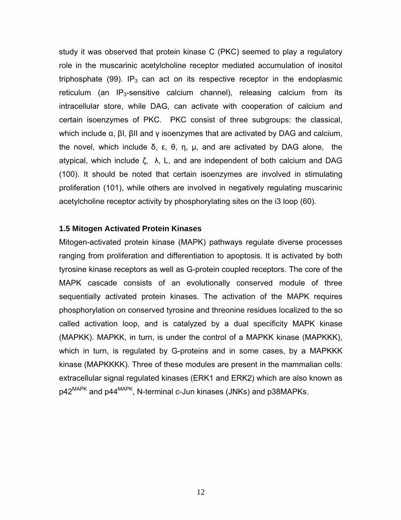
1.5 Mitogen Activated Protein Kinases Mitogen-activated protein kinase (MAPK) pathways regulate diverse processes
ranging from proliferation and differentiation to apoptosis. It is activated by both
tyrosine kinase receptors as well as G-protein coupled receptors. The core of the
MAPK cascade consists of an evolutionally conserved module of three
sequentially activated protein kinases. The activation of the MAPK requires
phosphorylation on conserved tyrosine and threonine residues localized to the so
called activation loop, and is catalyzed by a dual specificity MAPK kinase
(MAPKK). MAPKK, in turn, is under the control of a MAPKK kinase (MAPKKK),
which in turn, is regulated by G-proteins and in some cases, by a MAPKKK
kinase (MAPKKKK). Three of these modules are present in the mammalian cells:
extracellular signal regulated kinases (ERK1 and ERK2) which are also known as
p42MAPK and p44MAPK, N-terminal c-Jun kinases (JNKs) and p38MAPKs.
12

Figure II Schematic view of MAPK signaling pathways in mammalian cells.
ERKs, were the first to be cloned and their actions have been best examined in
the context of growth factor signaling via receptor tyrosine kinases (RTKs). The
archetypal ERK cascade involves the activation of small GTPase Ras upon
agonist stimulation of the RTK receptors. This stimulation leads to the
autophosphorylation of the RTK in tyrosine residues and recruitment of adaptor
proteins bearing SH2 and SH3 motif (Shc and Grbr2) that bind to an exchange
factor for Ras, SOS. SOS allows nucleotide exchange dependent activation of
the MAPKKK Raf-1. Raf-1 is a kinase with a serine/threonine specificity that
catalyzes the activating phosphorylation of MEK1/2, which ultimately
phosphorylates ERK1/2 on its activation loop (Figure II). Amplification via this
signaling cascade is such that it is estimated that activation of solely 5% Ras
molecules is sufficient to induce a full activation of ERKs (102). In resting cells,
ERKs are anchored in the cytoplasm via association with MEK1/MEK2, but
13

following activation they dissociate from the cytoplasmatic anchoring complex
and enter the nucleus, the site for signal termination (103).
The second group of MAPKs, JNK proteins, were first described as kinases that
phosphorylate serine residues on the N-terminus c-Jun transcription factor
following UV exposure. The third subgroup p38, were found to be activated in
response to different forms of stress. Both JNK and p38 are given the more
clarifying name SAPKs (stress activated protein kinases). Rho family members
(Rho, Rac, Cdc42) are thought to be involved in SAPK activation in response to
cytokines or cellular stress (104).
1.5.1 MAPKs in the Regulation of Cell Cycle
The cell cycle is controlled by a class of nuclear enzymes called cyclin-
dependent kinases (CDKs), expressed constitutively but present in inactive form
unless combined with their cyclin partners. The stimulatory effects of cyclins are
counteracted by the inhibitory effects of CDK inhibitory proteins (CKIs), of which
two families are well described: INK4 and Cip/Kip (p21) (105). Progression
through G1 phase is especially affected by extracellular signals, and is regulated
by CDK4/CycD, CDK6/CycD and CDK2/CycE complexes. Most studies indicate
that integrins and growth factors, by activating MAPK pathways, typically control
expression of Cyclin D1 and/or downregulate CKIs. In this regard, the activation
of MAPK/ERK pathway has been linked to the induction of cyclin D1
transcription, since expression of dominant negative mutants of MEK and ERK
prevented growth-factor dependent transcription of the Cyclin D1 gene (106).
The formation of active CyclinD/CDK4 complex is considered rate-limiting for cell
growth (107, 108). Recent studies indicate the critical determinant in the
induction of Cyclin D1 is the duration of the ERK signal. Activation of ERK can be
transient or sustained, depending on the stimulus. For example, serotonin
induced a transient (10 min) stimulation of ERK in the CCL39 cells (Chinese
hamster lung fibroblast cell line); while thrombin induced a far more sustained
activation that peaks 3-4 hours after addition to cells (109). Some studies have
observed a correlation between the strength of the mitogenic signaling and the
14
duration of ERK stimulation. While non-mitogenic factors induce a transient
activation of ERKs (less than 15 mins) that does not lead to cell cycle entry,
mitogens induce cell proliferation and sustained ERK activation (up to 6 hrs)
(110).
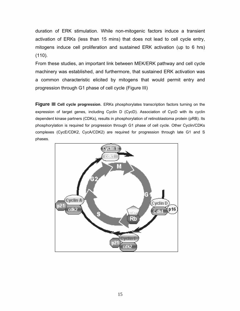
From these studies, an important link between MEK/ERK pathway and cell cycle
machinery was established, and furthermore, that sustained ERK activation was
a common characteristic elicited by mitogens that would permit entry and
progression through G1 phase of cell cycle (Figure III)
Figure III Cell cycle progression. ERKs phosphorylates transcription factors turning on the
expression of target genes, including Cyclin D (CycD). Association of CycD with its cyclin
dependent kinase partners (CDKs), results in phosphorylation of retinoblastoma protein (pRB). Its
phosphorylation is required for progression through G1 phase of cell cycle. Other Cyclin/CDKs
complexes (CycE/CDK2, CycA/CDK2) are required for progression through late G1 and S
phases.
15

1.6 G-protein Coupled Receptor Signaling to MAPK Activation of MAPKs mediated by G-protein-coupled receptors has been
extensively studied, including investigation on bombesin, endothelin-1,
adrenaline, somatostatin, thromboxane A2, and muscarinic receptors. These
receptors have been shown to couple to PTX-sensitive and PTX-insensitive G-
proteins, thus signaling to MAPK using both G-proteins (Gi/o and Gq/11). To study
the mechanism of activation of MAPK by G-protein-coupled receptors such as
muscarinic acetylcholine receptors, transfected cell lines that express p42mapk
(labeled as MAPK in this text) together with muscarinic acetylcholine receptor
subtypes were used (111). In transfected cell lines like COS-7 carbachol
stimulation increased MAPK activity in a concentration dependent manner
through M1 and M2 subtypes (112), whereby the M1 mediated activation was
PTX-insensitive, while the M2 mediated activation displayed PTX-sensitivity. To
elucidate the role of the α-subunits of G-proteins, studies were performed in
transfected cells expressing wild-type or activated G-protein α-subunits, including
Gαq and Gαi2 which couple M1 and M2 subtypes respectively. However, these
transfected cells did not exhibit elevated MAPK phosphorylating activity. As a
result leading to speculations of molecules in addition to α-subunits of G-proteins
playing a role in M1 and M2 mediated MAPK activation.
Recently available evidence has supported an active role for the Gβγ complex in
signal transduction (113). Since α-subunits did not stimulate the activation of
MAPK, studies investigating the role of Gβγ-subunits were performed. Indeed,
transfecting cells with β1 and γ2 subunits and MAPK effectively elevated it
phosphorylating activity demonstrating that the βγ heterodimers directly elicits
biochemical pathways leading to MAPK activation. Also, sequestration studies
performed on βγ complexes by overexpressing Gαi proteins abolished the M2
and βγ-mediated activation of MAPK, and reduced this response when elicited by
M1 stimulation. Therefore, these results indicate that M1 signaling to MAPK
involves βγ-dependent and independent pathways; while M2 mediated activation
of MAPK appears to be strictly dependent on Gβγ.
16

Mutant-Ras studies were performed to investigate whether signaling from M1 and
M2 receptors or βγ dimmers involves p21ras (Ras). These studies have shown
that mutant Ras expression abolished the elevated MAPK activity in response to
M1, M2 and βγ dimmer overexpression (114, 115). Similar studies were done to
elucidate the role of PKC, and the results suggested that signaling from M1
receptors to MAPK involves a PKC-dependent and PKC-independent pathway,
and both pathways converge at Ras. The PKC-independent pathway is mediated
by the Gβγ complex. On the other hand the M2-mediated MAPK activation is PKC-
independent, and involves Giβγ dimmers, acting on a Ras-dependent pathway.
1.7 Intracellular Signaling Pathways Linking G-protein Coupled Receptors to MAPK Activation of a given type of G-protein-coupled receptor (GPCR) triggers a limited
set of signaling events in a very rapid and specific manner. The classical
paradigm of GPCR signaling was rather linear and sequential. Emerging
evidence, however, has revealed that this is only a part of the complex signaling
mediated by GPCR. Propagation of GPCR signaling involves cross-regulation of
many but specific pathways; including cross-talk between different GPCRs as
well as with other signaling pathways and that the MAPK activation appears to be
a common point of convergence for most of these pathways (Figure IV). A few of
these signaling cascades relevant to the present study will be elucidated in this
paper.
17

Figure IV GPCR mediated activation of MAPK regulated by generation of intracellular
second messengers. GPCR activity leads to AC/cAMP (Adenylyl cyclase/cyclic adenosine
monophosphate), and PLCβ/PKC (Phospholipase C/Protein kinase C) second messenger
pathways. cAMP directly, or via PKA (Protein kinase A), activates RAP-1/B-Raf/ERK pathway,
and potentially inhibits Raf-1 activated ERK activity. The Gαq/PLCβ/PKC promotes Ras/Raf-
1/ERK activity, and it is likely that Gq and Gi/o coupled GPCRs can activate JNKs and p38. The
result of the interplay between these pathways is either proliferative or antiproliferative,
depending on the expression of GPCRs and signaling intermediates. Dashed indicators identify
the probable involvement of multiple, unidentified intermediates. (New and Wong 2007)
1.7.1 The Ras Mediated Activation of MAPK Cascade via G-protein Coupled
Receptors
Ras belongs to the family of small GTPases which exhibit high-affinity binding for
GDP and GTP, and possess low intrinsic GTP hydrolysis and GDP/GTP
exchange activities. The GDP/GTP cycling is controlled by two main classes of
regulatory protein. Guanine-nucleotide-exchange factors (GEFs) promote
formation of the active GTP-bound form (116), whereas GTPase-activating
proteins (GAPs) accelerate the intrinsic GTPase activity to promote the formation
of the inactive GDP-bound form. A second important biochemical feature of Ras
proteins is their post-translational modification by lipids. The Ras proteins
18

terminate with a C-terminal CAAX (C=cysteine, A=aliphatic, X=any amino acid)
tetrapeptide sequence (117). This motif, when coupled together with residues
immediately upstream (e.g. cysteine residues modified by the fatty acid
palmitate), comprises the membrane-targeting sequences that dictate
interactions with distinct membrane compartments and subcellular locations. The
CAAX motif is the recognition sequence for franesyltransferase and
geranylgeranyltransferase, which catalyze the covalent addition of a franesyl or
geranylgeranyl isoprenoid, respectively, to the cysteine residue of the
tetrapeptide motif. These modifications are essential for facilitating membrane
association and subcellular localization critical for biological activities. Ras
proteins serve as signaling nodes activated in response to diverse extracellular
stimuli. Activated Ras interacts with multiple, catalytically distinct downstream
effectors, which regulate cytoplasmic signaling networks that control gene
expression and regulation of cell proliferation, differentiation, and survival.
Lysophosphatidic acid (LPA) and thrombin were the first GPCR agonists shown
to rapidly stimulate Ras-GTP accumulation in quiescent mammalian cells (118).
However the best characterized Ras signaling pathway is activation of Ras by
the epidermal growth factor receptor tyrosine kinase through the RasGEF Sos
(119). Activated Ras binds to and promotes the translocation of the Raf
serine/threonine kinase to the plasma membrane, where additional
phosphorylation events promotes full Raf kinase activation. Raf phosphorylates
and activates MEK1/2 dual specificity protein kinase, which phosphorylates and
activates the ERK1/2 mitogen-activated protein (MAP) kinase. Activated ERK
translocates to the nucleus, where it phosphorylates transcription factors leading
to gene transcription and cell cycle progression. In quiescent fibroblast it was
seen that LPA mediated Ras activation via pertussis toxin sensitive Gi protein
(118) and also in certain cell type’s α2 adrenergic receptors could similarly
activate Ras via Gi protein hence suggesting that Ras activation might be a
common event in the action of Gi-coupled receptors (120). Furthermore in Rat 1a
fibroblasts cells expressing the pertussis toxin-sensitive G-protein α subunits αi2
and αi3 but not αo, muscarinic M2 receptor stimulation resulted in activation of
19

Ras-Raf-MAPK, implicating the involvement of the Gi protein in this pathway
(121). Various studies have shown that Gαi directly binds to and inhibits adenylyl
cyclase, thereby lowering cytosolic cAMP levels and since the rise of cAMP
levels inhibits the Ras-MAPK pathway, apparently at the level of Ras-Raf
interaction (122, 123) it is conceivable that a Gαi mediated fall in cAMP levels
may positively influence Ras-Raf coupling.
The classic Gαq-PLC-Ca2+/PKC pathway can also mediate MAPK activation,
either via Ras or independent of Ras. Certain Ras specific GEFs can activate
Ras in response to PLC-generated messengers such as Ca2+ and DAG, which
bind directly to these GEFs (124, 125, 126, 127). In cells expressing these GEFs,
Ras can thus be activated by PLC activation in a Grb2-Sos-independent manner.
Furthermore several PKC isoenzymes can phosphorylate and activate c-Raf
independently of Ras (128, 129). PKC activation by phorbol ester leads to Ras
activation in COS epithelial cells and in neonatal myocytes (130, 131), but this
pathway apparently does not function in fibroblasts (118).
Raf proteins are the conserved signaling module that transducer signals from the
cell surface to the receptor wherein the Ras family of small G-proteins are the
upstream regulators in several cell types. However, it has been shown that
activation of MAPK cascade can occur through Ras-independent mechanisms
involving Raf for example, in rat C6 glioma cells activation of the purinergic
P2YAC-receptor a GPCR, by using specific agonist activated MAPK through a Gi-
RhoA-PKC-Raf-MEK-dependent but a Ras and Ca2+ independent cascade (132).
Another mechanism that has been suggested in the Ras-independent activation
of Raf involves the activation of C-Raf by p21-activated kinases (PAK). PAKs are
serine/threonine specific kinases that bind to and are activated by, the membrane
bound Rho-family GTPases Cdc42 and Rac, and it has been proposed that these
kinases lead to C-Raf activation in a Rac/Cdc42-and phosphatidylinositol-3-
kinase (PI3K)-dependent manner. Although the physiological significance of this
PAK-mediated C-Raf activation is not established, the PAK-dependent Ras-
independent route to C-Raf activation might have an important role in C-Raf
activation by inetgrins or when microtubules are disrupted (133).
20

1.7.2 Rho Mediated Activation of MAPK Cascade via G-protein Coupled
Receptors
Like Ras, Rho proteins also serve as key regulators of extracellular-stimulus-
mediated signaling networks that regulate actin organization, cell cycle
progression and gene expression (134). Twenty members have been identified,
RhoA, Rac and Cdc42 being the best studied. The guanine-nucelotide-exchange
factors (GEFs) and GTPase-activating proteins (GAPs) control the activation of
the small GTPases Rho, Rac and Cdc42. Once activated the GTPases bind to a
spectrum of effectors to stimulate downstream signaling. Rho kinase 1 (ROCK1)
and ROCK2 are key Rho effectors that have multiple substrates. Most GPCR
agonist that regulate actin cytoskeletal responses, smooth muscle contraction,
gene transcription, and cell growth through Rho-dependent pathways can couple
to more than a single class of heterotrimeric G-protein. For example LPA and
thrombin elicit cellular responses through both pertussis toxin-sensitive and
insensitive G-proteins. Furthermore GPCR agonists have been also shown to
mediate Rho activation through the Gαq and Gα12/13 subtypes of the G-protein
family (135).GPCRs including the thrombin receptor in platelets and LPA
receptors in Swiss 3T3 cells have been shown to activate PI3K, and studies
using C3 exoenzyme indicates this requires Rho function (136, 137). It has been
shown that M1 and M2 muscarinic receptor stimulation can lead to
phosphorylation and activation of a serine/threonine protein kinase Akt/PKB
(138). Akt/PKB (protein kinase B) is regulated through PI3K and synthesis of
PIP2 and PIP3 (139). Thus it is possible that GPCRs utilize Rho to regulate this
cell survival pathway. Various studies have demonstrated that Rho-dependent
signaling is required for transformation by oncogenic Ras (140, 141, 142).
However, while Rho activation is known to promote the development of stress
fibers and focal contact, Ras-transformed fibroblast generally exhibit a loss of
these structural elements characteristic of Rho activity. These results have lead
to the interpretation that Rho activity may be reduced in Ras-transformed cells.
Accordingly, activated Rho can restore stress fibers in Ras transformed Rat1
fibroblasts, suggesting that Rho can be inactivated in these Ras-transformed
21

cells (143). By contrast, inhibition of Rho kinase was shown to block Ras
transformation in NIH3T3 cells (144) and to attenuate invasive tumor cells (145).
In another study done on vascular smooth muscle cells (VSMC) from rat aorta it
was seen that thrombin stimulated DNA synthesis and migration were inhibited
by exoenzyme C3 and Y27632 a Rho kinase inhibitor. Although thrombin
activates RhoA, and Rho is necessary for thrombin-stimulated DNA synthesis in
rat aortic VSM, adenoviral transfection of cells with activated RhoA was found not
to be sufficient to induce DNA synthesis. Rather, for mitogeinic signaling in
VSMC, it appears that RhoA collaborates synergistically with Ras, such that DNA
synthesis is activated only by agonists that stimulate both pathways (146). This
supports studies indicating that activation of Rho is critical for Ras-induced cell
cycle progression. Thus the relationship between Ras and Rho dependent
signaling pathways during transformation is still unclear. A role for Rho in
regulating cell proliferation was first suggested by studies in which it was
demonstrated that C3 mediated inhibition of Rho caused fibroblasts to arrest in
the G1 phase of the cell cycle (147). One downstream effect of Rho is to reduce
expression of cell cycle inhbitor such as the cyclin-dependent kinase inhibitors
p21Waf1/Cip1 and p27Kip1 in studies done on rat aortic VSMC it was demonstrated
that adenoviral expression of a constitutively active form of RhoA reduced p27Kip1
protein levels. Direct activation of Rho with cytotoxic necrotizing factor-1 also
decreases p27Kip1 and increases DNA synthesis in human smooth muscle cells
(148). In another study it was shown that serotonin stimulation of bovine
pulmonary artery VSMC leads to translocation of ERK to the nucleus, and this
response is inhibited by treatment with either dominant negative RhoA or the Rho
kinase inhibitor Y27632 (149). Furthermore it was reported that cyclin D1
expression is deregulated through a RhoA dependent mechanism that influences
human lung fibroblast proliferation.
22

1.7.3 Phosphatidylinositol 3-Kinase (PI3K) Mediated Activation of MAPK
Cascade via G-protein Coupled Receptors
Phosphoinositides (PIs) are rare lipids. Phosphoinositide 3-kinases (PI3Ks) are a
family of related enzymes that are capable of phosphorylating the 3 position
hydroxyl group of the inositol ring of phosphatidylinositol (PtdIns). PI3Ks are
structurally diverse enzymes divided into three main classes. Each class exhibits
distinct substrate specificity. Class IA PI3-kinases is a cytosolic heterodimer
composed of a 110-kDa (p110α, -β, or –δ) catalytic subunit and an 85-kDa (p85)
adaptor protein. In response to growth factors stimulation, class IA PI3-kinases
complex with activated receptor protein tyrosine kinases (through SH2 domains
of the p85 subunit) or tyrosine-phosphorylated proteins. The class IB PI3-
kinases-γ does not have an adaptor protein but instead is activated by the Gβγ
subunits of the G-proteins (150). Class II PI3-kinases contain a COOH-terminal
C2 domain that appears involved in regulation of this class by phospholipid and
calcium (151). Recently Class III PI3-kinases have been also identified. It has
been shown that receptor tyrosine kinase (RTK) and GPCR both can activate
class I PI3Ks via phosphorylation steps which leads, through an adaptor
molecule, to the recruitment of PI3K to the membrane. Once activated and
localized to the membrane, PI3K phosphorylates phosphoinositol lipids on the D3
position of the inositol ring generating PtdIns-3-phosphates (PtdIns-3,4-P2 and
PtdIns-3,4,5-P3). These specialized lipids serve to recruit pleckstrin homology
(PH) domain-containing proteins such as the serine-threonine kinase Akt (protein
kinase B) and PDK1 (phosphoinositide-dependent kinase 1) to the plasma
membrane. After recruitment to the membrane Akt is phosphorylated and
consequently activated, by PDK. In turn, Akt phosphorylates multiple proteins on
serine and threonine residues. Through phosphorylation of these targets, Akt
carries out its role as a key regulator of a variety of critical cell functions including
glucose metabolism, cell proliferation and survival. Besides being a downstream
effector of PI3-kinases, Akt may also be activated in a PI3-kinase-independent
manner. Studies have suggested that cAMP-elevating agents could activate AKT
through protein kinase A (PKA), although these studies are disputed and the
23

mechanism of action is unclear. The PI3K dependent Akt activation is regulated
through PTEN which acts as a phosphatase to dephosphorylate PtdIns (3,4,5)P3
back to PtdIns(3,4)P2. This removes the membrane-localization factor from the
Akt signaling pathway thus inhibiting downstream pathways dependent on Akt
activation.
Figure V Activation of PI3K-dependent cell cycle regulation. The expression, stability and
activity of cyclin and CDK inhibitors are regulated by the activity of several PI3K-dependent
pathways. Numerous GPCRs activate PI3K isoforms either through Gβγ subunits or via receptor
tyrosine kinase (RTK) and integrin transactivation. PI3Ks activate ERK and Akt, leading to the
transcriptional regulation of p27Kip1. In addition Akt phosphorylates p27Kip1, thereby affecting its
nuclear localization. Acting through TSC1, TSC2 and mTOR (mammalian target of rapamycin),
Akt can negatively affect the stability of p27Kip1, although GPCR regulation of proliferation through
mTOR has not been established (indicated by dashed line). PI3Ks may also promote proliferation
by promoting cyclin expression (via p70S6K). (New and Wong 2007)
Following direct or indirect GPCR-induced PI3K activation, cell cycle progression
is regulated by the effects of PI3K activated kinases on the expression and
stability of cell cycle proteins, or by the modulation of the activity of other signal
transduction pathways (Figure V). For example, thrombin receptor activation in
vascular smooth muscle cells lead to reduced levels of p27Kip1 and increased
proliferation (152), while in embryonic fibroblast the evidence suggests that
24
thrombin receptor activation of PI3K/Akt pathways promotes cyclin D1
accumulation, cyclin D1-CDK4 activity and cell cycle progression (153, 154).
Furthermore, it has been postulated that thrombin receptor activation of ERK
ultimately leads to enhanced translocation of CDK2 into the nucleus and
fibroblast proliferation (155). In another study overexpression of phosphoinositide
3-kinase γ (PI3Kγ) in COS-7 cells expressing M2 muscarinic receptors, agonist
induced Gβγ-subunit dependent MAPK activation via transactivation of tyrosine
kinase receptors in a Sch-Grb2-Sos-Ras-Raf-MAPK manner. Expression of a
catalytically inactive mutant of PI3Kγ abolished this stimulation of MAPK by the
Gβγ or in response to stimulation of muscarinic M2 G-protein-coupled receptors.
Indicating that, PI3Kγ mediated Gβγ-dependent regulation of the MAPK signaling
pathway (156).
1.7.4 Phospholipase C (PLC) Mediated Activation of MAPK Cascade via G-
protein Coupled Receptors The receptor-mediated activation of the PLC lead to the hydrolysis of the
phosphotidylinositol-4,5-biphosphate (PIP2) to inositol triphosphate (IP3) and
diacylglycerol (DAG), which in turn mediate the activation of intracellular calcium
release and protein kinase C activation respectively. Phospholipase C (PLC) is
the principal effector of Gq-mediated signaling. Eleven different isoforms of PLC
exist and exhibit distinct patterns of regulation; members of the PLCβ subfamily
tend to mediate the actions of activated Gq (157). It has been shown that
activation of PKC isoforms by Gq/11-dependent regulation of phospholipase Cβ
can also lead to MAPK activation through both Ras-dependent and Ras-
independent mechanisms. In another study it was demonstrated that PKCα can
apparently activate Raf-1 by direct phosphorylation thus activating the MAPK
cascade (158). In cells of neuronal origin, Gq/11-dependent activation of the
calcium and focal-adhesion-dependent focal adhesion kinase family member
Pyk2, leads to Ras-dependent ERK activation (159). In this system, intracellular
calcium, released as a result of PLCβ-mediated inositol triphosphate production,
triggers Pyk2 autophosphorylation, recruitment of nonreceptor tyrosine kinase c-
25

Src, tyrosine phosphorylation of Sch, and Ras-dependent ERK activation (160,
161). Activation of ERK1/2 by M1, M3 and M5 muscarinic receptors typically
involves PKC activation following Gq/11 activation of the PLC pathway with
production of DAG. For example, in human neuroblastoma SK-N-BE2(C) cells
activation of ERK1/2 by M3 muscarinic receptors was dependent on PKCε as well
as the guanine nucleotide exchange factor, Ras and the Raf family of serine-
threonine kinases (162).
1.8 Transactivation of Classical Receptor Tyrosine Kinases by GPCRs Receptor transactivation refers to the ability of a primary agonist, via binding to
its receptor, to activate a receptor for another ligand via signaling events.
Transactivation of various growth factor receptors, including the epidermal
growth factor receptor (EGFR), and the platelet derived growth factor (PDGF), by
G-protein-coupled receptors has been documented in multiple cellular model
systems (163, 164, 165, 166, 167). Receptor transactivation can potentially occur
through several different mechanisms. One mechanism is through activation of
intracellular protein tyrosine kinase, such as c-Src and PKC (168, 169), which
can phosphorylate the growth factor receptor and thereby promote its activation.
GPCR-induced transactivation of the EGFRs has been studied in some detail.
GPCR activation leads to cleavage of the pro-EGF (eg pro-heparin-binding-EGF)
ligands from the membrane initiating the transactivation (168, 170, 171).
Proteolysis of the HB-EGF precursor is thought to be mediated by members of
the ADAM family of matrix metalloproteases (MMPs) (172). The step that is
stimulated by GPCRs has not been identified. EGFR transactivation has been
shown to occur primarily through shedding of HB-EGF from the membrane (173,
174). Transactivation of EGFR results in intracellular signaling that leads to
growth, proliferation, and migration. In fibroblasts, both Gi/o-coupled and Gq/11-
coupled receptors have been shown to stimulate HB-EGF release. For Gi/o-
coupled receptor, such as the LPA (112), α2A adrenergic (175) and M2
muscarinic acetylcholine receptors (176), HB-EGF shedding and EGFR
transactivation are mediated by the Gβγ subunits of the heterotrimeric G-proteins
26
(177). HB-EGF shedding in response to stimulation of the Gq/11-coupled
receptors, such as enodthelin-1 and α-thrombin receptors (178), mediated by the
Gq/11α-subunits (179, 180). The G-protein effectors that regulate ectodomain
sheddin remain unidentified, although PI3Ks (156) and Src family nonreceptor
tyrosine kinases have each been proposed as early intermediates in the EGFR
transcativation pathway (177, 181).
1.9 Apoptosis and G-protein Coupled Receptor Signaling Interaction Apoptosis is defined as a genetically controlled mechanism of cell death involved
in the regulation of tissue homeostasis and morphologically characterized by cell
shrinkage, membrane remodeling and blebbing, chromatin condensation and
DNA and cellular fragmentation into apoptotic bodies. Pro-apoptitic singaling may
be mediated by the specific ligands and surface receptors, which are capable of
delivering death signal from the microenvironment and can activate the
excecution of apoptosis in the cell cytoplasm and organelles. Apoptosis can also
be activated from inside the cell through specific cell sensors residing in the cell
nucleus and cytoplasm. Both these pathways of apoptosis signaling converge
into a common pathway causing the activation of effectors termed caspases
(182). Caspases are a family of cysteine proteases that cleave their substrates at
aspartic acid residues (183). They are hierarchically stratified into upstream
initiator caspases, namely procaspases 8, 9, 10 and downstream the effector
caspases 3, 6, 7, caspase-3 activity in particular is the common effector of most
apoptotic pathways. Besides the caspases, members of the Bcl-2 protein family
are also critical for the regulation of apoptosis. Both anti-apoptotic (BCL-2, Bcl-w,
Bcl-xL) and pro-apoptotic (Bax, Bak, Bad) Bcl-2 family members have been
identified (184). Studies have shown that ERK family of the MAPKK may also
have anti-apoptotic effects when activated by the growth factor receptors. The
proposed pathway for ligand binding to growth factor receptors initiating an anti-
apoptotic signaling is via the activation of Raf which in turn activates ERK1/2
which could phosphorylate Bcl-2 in a manner that stabilizes its anti-apoptotic
activity. Another mechanism is via the activation of Rsk, which phosphorylates
27
the pro-apoptotic protein Bad leading to its inactivation thus promoting cell
survival.
The repertoire of signaling pathways controlled by GPCRs has recently been
extended by studies linking GPCRs to the regulation of apoptosis. Depending on
the receptor subtypes and the cell type in which the receptor is expressed,
GPCRs can either induce apoptosis or protect cells from apoptotic stimuli. For
example β1-adrenergic receptors in cardiac myocyctes can induce apoptosis
through a Gs-mediated pathway, whereas β2-adrenergic receptors in the same
cell type can protect cells through a Gi-mediated mechanism (185). Prominent
among the GPCRs that protect cells from apoptotic stimuli are the subtypes of
the muscarinic receptor family (186, 187). Previous studies have also shown that
muscarinic receptors that couple to the Gq/11 protein (e.g. M1, M3 and M5) can
protect cells from apoptosis following DNA damage whereas those coupled to the
Gi-proteins (M2 and M4) have no protective properties (188). In a study done
using CH (Chinese-hamster ovary) cells it was shown that the M3 muscarinic
receptor anti-apoptotic effect is independent of calcium/phospholipase C
signaling but proceeds in a manner that involves both gene transcription and the
up-regulation of the anti-apoptotic Bcl-2 protein.
28
2 Materials and Methods 2.1 Materials 2.1.1 Reagents
Agarose Serva Roth
BSA Bio-Rad
Bromophenol blue Sigma-Aldrich
Chemiluminescence Blotting Substrate (POD) Bio-Rad
Disodium hydrogen orthophosphate (Na2HPO4) Merck
Desoxynucleotide mixture Sigma-Aldrich
DNA Smart-Ladder Eurogene Tech
Developing Solution Sigma-Aldrich
ECL Detection Kit Boehringer Ingelheim
Ethidium Bromide Sigma-Aldrich
Ficoll 400, Type 400 Sigma-Aldrich
Fixing Solution Sigma-Aldrich
Fetal Calf Serum (FCS) Seromed
Kaleidoscope Marker Sigma-Aldrich
Leupeptin Sigma-Aldrich
Lowry Kit Bio-Rad
Lumasafe scintillation cockt Lumac
MEM Non Essential Amino Acids (100 x) = NEAA PAA
β-Mercaptoethanol Sigma-Aldrich
(Methyl-3H)-Thymidine 37 mBq/ml PerkinElmer
Nonidet P-40 Sigma-Aldrich
Oligo dT-Primer MWG-Biotech
Omniscript RT Kit: Qiagen
Penicillin / Streptomycin solution Sigma-Aldrich
(10000 U/ml / 10 mg/ml)
Platelet derived growth factor = PDGF Sigma-Aldrich
Pepstatin A Sigma-Aldrich
29
Phenylmethylsulphonylfluoride (PMSF) Sigma-Aldrich
Ponceau S Boehringer Ingelheim
RNase-Free DNase Set Qiagen
RNase Inhibitor RNasin Plus 40 U/µl Promega
RNeasy Mini Kit Qiagen
RNeasy Tissue Kit Qiagen
Roti-Load 1 Roth
Sodium chloride (NaCl) Roth
Sodium hydroxide pellets (NaOH) Merck
Sodium Pyrovate Solution = SPS (100mM) PAA
Streptomycin Sigma-Aldrich
TaqDNA Polymerase 5 U/µl Invitrogen
Trichloro-acetic acid crystals (TCA) Merck
Tris (Tris-(hydroxymethyl-aminomethane) Roth
Tris-HCl (Tris-(hydroxymethyl)-aminomethane Boehringer Ingelheim
hydrochloride)
Triton x-100 Boehringer Ingelheim
Trypthan Blue (0,4 %) Sigma-Aldrich
Trypsin EDTA 10 Sigma-Aldrich
Tween 20 Sigma-Aldrich
2.1.2 Test Drugs Atropine sulphate (10 mM) Sigma-Aldrich
Stock solution prepared in sterile water, further dilutions were done with sterile
water
Actinomycin D (1 mg/ml) Sigma-Aldrich
Stock solution prepared in DMSO, further dilutions were carried out with sterile
carried
30

AF-DX 384 (10 mM) Boehringer Ingelheim
(5,11-dihydro-11-{[(2-{2-[(dipropylamin)methyl1]-1-
piperidinyl}ethyl)amino]carbonyl1}-6H-pyrido(2,3-ß) (1,4)benzodiazepine-6-one
methanesulfonate)
Stock solution prepared in sterile water, further dilutions were done with sterile
water
AQ-RA 741 (10 mM) Boehringer Ingelheim
(11-({4-[4-(diethylamino)butyl]-1-piperidinyl}acetyl)-5,11-dihydro-6H-pyrido(2,3-ß)
(1, 4)benzodiazepine-6-one hydrochloride)
Stock solution prepared in sterile water, further dilutions were done with sterile
water
Carbachol (10 mM) Sigma-Aldrich
(Carbamylcholine chloride)
Stock solution prepared in sterile water, further dilutions were done with sterile
water
Cycloheximide (1 mg/ml) Sigma-Aldrich
Stock solution prepared in sterile water, further dilutions were done with sterile
water
4-DAMP (10 mM) Sigma-Aldrich
(4-diphenylacetoxy-N-methylpiperidine methoiodide)
Stock solution prepared in sterile water, further dilutions were carried out with
sterile water
31
FTI-277 (10 mM) Calbiochem
(Methyl{N-[2-phenyl-4-N[2(R)-amino-3-mecaptopropylamino]benzoyl]}-
methionate,TFA)
Stock solution prepared in DMSO, further dilutions were performed with sterile
water
GW 5074 (20 mM) Tocris
(3-(3,5-Dibromo-4-hydroxy-benzylidene)-5-iodo-1,3-dihydro-indol-2-one)
Stock solution prepared in DMSO, further dilutions were done with sterile water
Himbacine (10 mM) Sigma-Aldrich
Stock solution prepared in ethanol, further dilutions were performed with sterile
water
Hexamethonium Bromide (10 mM) Sigma-Aldrich
(Hexane-1,6-bis(trimethylammoniumbromide)N,N,N,N’,N’,N’-
Hexamethylhexamethylenediammonium dibromide)
Stock solution prepared in sterile water, further dilutions were done with sterile
water
Manumycin A (10 mM) Calbiochem
Stock solution prepared in DMSO, further dilutions were done with sterile water
Nicotine hydrogen tartrate salt (10 mM) Sigma-Aldrich
Stock solution prepared in sterile water, further dilutions were done with sterile
water
Oxotremorine sesquifumerate (10 mM) Sigma-Aldrich
Stock solution prepared in sterile water, further dilutions were done with sterile
water
32

PD-98059 (10 mM) Sigma-Aldrich
(2-(2’-amino-3’-methoxyphenyl)-oxanaphthalen-4-one)
Stock solution prepared in DMSO, further dilutions were done with serum-free
medium for human lung fibroblast
Pirenzepine (10 mM) Boehringer Ingelheim
Stock solution prepared in sterile water, further dilutions were done with sterile
water
PDGF (1 µg/ml) Sigma-Aldrich
Stock solution prepared with 4 mM HCl in 0.1% BSA further dilutions were done
with the serum-free medium for human lung fibroblast
Pertussis Toxin (10 µg/ml) Sigma-Aldrich
Stock solution prepared in sterile water, further dilutions were done with sterile
water
Tiotropium Bromide monohydrate (10 mM) Boehringer Ingelheim
Stock solution prepared in sterile water, further dilutions were performed with
sterile water
TNF alpha Human recombinant (0.1 mg/ml) Sigma-Aldrich
Stock solution prepared in sterile water, further dilutions were done with sterile
water
Tryphostin AG1295 (5 mM) Sigma-Aldrich
Stock solution prepared in DMSO, further dilutions were done with sterile water
U73122 (10 mM) Sigma-Aldrich
(1-[6-[((17ß)-3-Methoxyestra-1,3,5[10]-trien-17-yl)amino]-1H-pyrrole-2,5-dione)
Stock solution prepared in DMSO, further dilutions were done with sterile water
33
Wortmannin (10 mM) Calbiochem
Stock solution prepared in DMSO, further dilutions were done with sterile water
Y27632 (10 mM) Tocris
(trans-4-[(1R)-1-Aminoethyl]-N-4-pyridinylcyclohexanecarboxamide
dihydrocholiride)
Stock solution prepared in 1X PBS (sterile), further dilutions were done with
sterile water
2.1.3 Buffers and Reagents for Cell Culture Culture medium for human fibroblast without FCS
Basismedium EARLE`S MEM
Penicillin/Streptomycin 100 U/ml / 100 µg/ml
Non Essential Amino Acids = NEAA 1x
Sodium Pyrovate Solution = SPS 1 mM Culture medium for human fibroblast with 10% FCS
Basismedium EARLE`S MEM
FCS 10% (v/v)
Penicillin/Streptomycin 100 U/ml / 100 µg/ml
Non Essential Amino Acids = NEAA 1x
Sodium Pyrovate Solution = SPS 1 mM
10x PBS (Phosphate buffered saline) pH 7.4-7.5
KCl 27 mM
KH2PO4 15 mM
NaCl 1,38 M
Na2HPO4 x H2O 81 mM
Distilled water
34

1x PBS-Buffer pH 7.4-7.5
10x PBS, 1:10 dilution with distilled water
1x Trypsin-EDTA-solution
10x Trypsin-EDTA, 1:10 dilution with1x sterile PBS-Buffer
0.15% (w/v) Trypthan blue staining dye
0.4% Trypthan Blue Stain, 3:8 dilution with 1x PBS-Buffer
2.1.4 Solutions and Buffers for Proliferation Assay 10% Trichloro-acetic acid
Trichloro-acetic acide crystals 10% (w/v)
Distilled water
5% Trichloro-acetic acid
Trichloro-acetic acide crystals 5% (w/v)
Distilled water
0,1 N Sodium hydroxide
Sodium hydroxide pellets 0.1N
Distilled water
1 M Tris-HCl pH 7,4
Tris-HCl 1 M
Distilled water
Adjust to pH 7.4 with HCl
35

2.1.5 Reagents for Protein Determination using the Lowry method BSA-Standard solution
BSA 4 mg/ml
0.1% Tris/Triton X100 solution
1% Tris/TritonX100 solution
1 M Tris-HCl-solution pH 7.4 100 mM
Triton x-100 1% (v/v)
Distilled water
Reagent A’
Reagent S 1% (v/v)
Starting Reagent A 99% (v/v) RIPA Lysis Buffer
50 mM Tris-HCl pH 7.5
150 mM NaCl
0.5% Sodium deoxycholat
1% Nonidet P-40
0.1% SDS
Protein Lysis Buffer with Protease and Phosphatase Inhibitors
RIPA Lysis Buffer
0.1 M EDTA pH 8 2 Mm
125 µg/ml Pepstatin A 0.7 µg/ml (1 µM)
100 mM PMSF 170 µg/ml (1 mM)
10 µg/ml Leupeptin 0.5 µg/ml (1 µM)
1.75 mM NaF 35 mM
20 mM Na3VO4 1 mM
36

2.1.6 Primers for Polymerase Chain Reaction The PCR for ß actin were done using the oligonucleotide pair of rßactin-s
(Sigma-Aldrich) and hßactin-as (MWG Biotech). All the muscarinic and nicotinic
receptor expression PCR’s were carried out using primers provided by MWG
Biotech.
Human
hβactin-s 5´-TTC TAC AAT GAG CTG CGT GTG GC-3´
Human
hβactin-as 5´-CTC GTC ATA CTC CTG CTT GC-3´
Human Muscarinic Receptor primers
hM1-s 5´-CAG GCA ACC TGC TGG TAC TC-3´
hM1-as 5´-CGT GCT CGG TTC TCT GTC TC-3´
hM2-s 5´-CTC CTC TAA CAA TAG CCT GG-3´
hM2-as 5´-GGC TCC TTC TTG TCC TTC TT-3´
hM3-s 5´-GGA CAG AGG CAG AGA CAG AA-3´
hM3-as 5´-GAA GGA CAG AGG TAG AGT GG-3´
hM4-s 5´-ATC GCT ATG AGA CGG TGG AA-3´
hM4-as 5´-GTT GGA CAG GAA CTG GAT GA-3´
hM5-s 5´-ACC ACA ATG CAA CCA CCG TC-3´
hM5-as 5´-ACA GCG CAA GCA GGA TCT GA-3´
37

Human Nicotinic Receptor primers
α1-hnACHR-s 5’-CAT CAA GTA CAT CGC AGA GA-3’
α1-hnACHR-as 5’-TTC TCT GCT CTG GTA GGT TC-3’
α3-hnACHR-s 5’-AGG CCA ACA AGC AAC GAG-3’
α3-hnACHR-as 5’-TTG CAG AAA CAA TCC TGC TG-3’
α5-hnACHR-s 5’-TCA ACA CAT AAT GCC ATG GC-3’
α5-hnACHR-as 5’-CCT CAC GGA CAT CAT TTT CC-3’
α7-hnACHR-s 5’-CTT CAC CAT CAT CTG CAC CAT C-3’
α7-hnACHR-as 5’-GGT ACG GAT GTG CCA AGG ATA T-3’
α9-hnACHR-s 5’-ATC CTG AAA TAC ATG TCC AGC G-3’
α9-hnACHR-as 5’-AAT CGG TCT ATG ACT TTC GCC-3’
ß1-hnACHR-s 5’-TGT ACC TGC GTC TAA AAA GG-3’
ß1-hnACHR-as 5’-TCA ACC CTC CAG TCT TC-3’
ß4-hnACHR-s 5’-TGT GAG CAT TGG CCA TCA AC-3’
ß4-hnACHR-as 5’-AAT GCC AAG CCT CTG AGC TG-3’
2.1.7 Buffers and Solutions for RT/PCR
DEPC-Water DEPC 0.2% (v/v)
Sterile water
38

Oligonucleotide Primers The oligonucleotide primer stocks were prepared with DEPC water to 100 µM
and further diluted in a 1:10 concentration with DEPC water to get a working 10
µM concentration
Gelelectrophoresis 5x TBE-Buffer
Tris 0.45 M
Boric acid 0.44 M
EDTA 0.012 M
Distilled water
Agarose-Gel 1.2%
Agarose 1.2%
0.5x TBE-Buffer
Ethidium bromide solution (10 mg/ml) 5 µl
Ethidiumbromide-solution 1%
Ethidiumbromide 1%
Distilled water
PCR loading Buffer Ficoll 400 15% (w/v)
1% Bromophenolblue-solution 0.25% (w/v)
5x TBE-Buffer 0.5x
Distilled water
39

2.1.8 Human Lung Fibroblast Cells MRC-5 MRC-5 human lung fibroblast cell line was developed from the lung of a 14 week
old human fetus. The cumulative population doublings to senescence is 42-46.
American Type Culture Collection number CCL-171.
Primary Human Lung Fibroblasts Primary human lung fibroblasts (phLFb) were established from histologically
normal areas of surgically resected lung tissue, which was obtained from lung
cancer patients after thoracotomy. The protocol for obtaining human tissue was
approved by the local ethics review board for human studies (Ethics Committee,
Medical Faculty, University of Bonn, Bonn, Germany), and informed consent was
obtained from the patient.
Primary human lung fibroblast cultures (phLFbOH.1, phLFb.4, phLFb.5,
phLFb.6, phLFb.8, phLFb.9, phLFb.11, phLFb.12) were similarly established from
histologically normal areas of surgically resected lung tissue from lung cancer
patients wherein, phLFb.4, phLFb.6, phLFb.8, phLFb.11, phLFb.12 were from the
central and phLFb.5, phLFb.9 were from the peripheral region of the lung.
2.1.9 Buffers and Reagents for Protein Gel Electrophoresis and Immunoblot NuPAGE MOPS SDS Running Buffer 1X
NuPAGE MOPS SDS Running Buffer 20X 5% (v/v)
Distilled water
Laemmli (Running) Buffer 5X
Tris 125 mM
Glycine 7.2%
SDS 5%
Distilled water
40
Transfer Buffer
Tris 25 mM
Glycine 192 mM
Methanol 20% (v/v)
Distilled water
Ponceau S solution
Ponceau S 0.2% (w/v)
Tris-HCl 3% (w/v)
Distilled water
Tris Buffered Saline (TBS) solution pH 7.5
Tris 50 Mm
NaCl 150 mM
Distilled water
TBST (TBS + Tween 20)
Tween 20 0.1% (v/v)
TBS
5% Blocking solution
Dried milk powder 5 gm
Distilled water 100 ml
Chemiluminiscence solution
Starting solution 1% (v/v)
Blotting substrate
0.2% (w/v) Sodium Hydroxide solution
NaOH 0.2% (w/v)
Distilled water
41

Developing solution
Developing stock solution 20% (v/v)
Distilled water
Fixing solution
Fixing stock solution 20% (v/v)
Distilled water
2.1.10 Antibodies Anti-mAChR M2 (H-170) Santa Cruz Biotechnology
Anti-mAChR M3 (H-210) Santa Cruz Biotechnology
Anti-Phospho-p44/42 Cell Signaling Technology
Anti-ERK-2 IgG Santa Cruz Biotechnology
2.1.11 Reagents and Solutions for Caspase-3 Fluorimetric Assay Caspase-3 Fluorimetric Assay was performed using the kit provided by Sigma-
Aldrich, all reagent stocks and working solutions were prepared as per the
manufacturer’s instructions with the materials provided along with the kit unless
otherwise specified.
5X Lysis Buffer
HEPES 250 mM
(4-(2-hydroxyethyl)-1-piperazineethanesulphonicacid)
CHAPS 25 mM
(3-[(3-Cholamidopropyl)dimethylammonio]-1-propanesulphonate)
DTT 25 mM
((D, L)-1, 4-Dithiothreitol)
pH adjusted to 7.4
42

10X Assay Buffer
HEPES 200 mM
CHAPS 1%
DTT 50 mM
Ethylenediaminetetraacetic acid (EDTA) 20 mM
pH adjusted to 7.4
1X Assay Buffer
10X Assay Buffer, 1:10 dilution with 17 megohm water
1X Lysis Buffer
5X Lysis Buffer, 1:5 dilution with 17 megohm water
1X Lysis Buffer with Protease Inhibitors
0.1 M EDTA pH 8 2 mM
125 µg/ml Pepstatin A 0.7 µg/ml
200 µg/ml Aprotinin 2 µg/ml
Volume made up with 1X Lysis Buffer
Caspase-3 Substrate Solution 10 mM
Caspase-3 Substrate (Ac-DEVD-AMC) 2.5 mg
DMSO 370 µl
Caspase-3 Inhibitor Solution 2 Mm
Caspase-3 Inhibitor (Ac-DEVD-CHO) 0.5 mg
DMSO 500 µl
Caspase-3 Inhibitor Assay solution 200 µM
Caspase-3 Inhibitor Solution, 1:10 dilution with 1X Assay Buffer
43

Caspase-3 Positive Control Solution 100 µg/ml
Caspase-3 Positive Control 5 µg
17 Megohm Water 50 µl
Caspase-3 Positive Control Assay Solution 1 µg/ml
Caspase-3 Positive Control Solution, 1:100 dilution with 1X Assay Buffer
Reaction Mixture, 400 µl per reaction for 96 well plate assay
10 mM AC-DEVD-AMC 12.5 µl
1X Assay Buffer 7.5 ml
7-Amino-4-methylcoumarin (AMC) Standard Solution 10 mM
AMC 1 mg
DMSO 0.57 ml
2.1.12 Equipment Mycycler Thermal Cycler System Bio-Rad
Photometer DU-64 spectrophotometer
Liquid Scintillation Analyzer Tri-Carb2100 TR, Packard
Fluorescence Microplate Reader Fluorometer Fluostar OPTIMA, BMG
2.1.13 Tools for Statistical Analysis GraphPad Software
GraphPad InSat 3.01 program was used to calculate statistical significance of
differences using ANOVA, followed by Dunnet’s or Bonferroni’s test. All values
are mean±SEM of n experiments. A P value of < 0.05 was accepted as
significant. All graphical representations were generated using the GraphPad
Prism software.
44

Microsoft Excel 5.0 and Excel 97
This program was used for calculation of means and SEM’s from individual
experimental data and summarizing these values for a particular set.
RFLPscan 2.01
Optical density of bands from PCR and western blot experiments were
semiqauntitatively measured using the RFLPscan software.
45

2.2 Methods 2.2.1 Culture of Human Lung Fibroblast The MRC-5 cell line was derived from normal lung tissue of a 14-week-old male
fetus (189). Cryogenically preserved MRC-5 human lung fibroblast culture was
thawed for 1 minute at room temperature followed by thawing at 37°C on a water
bath. The contents of the vial were then transferred to 75 cm2 pre-incubated
culture flask containing human fibroblast medium supplemented with 10% FCS in
a humidified incubator at 37oC and 5% CO2. After 24 hours of incubation the
medium was changed to remove the cryoprotective agent DMSO, to prevent cell
damage. The cells were then allowed to grow, with the medium being changed
twice per week. Upon confluence cells were passaged further.
For this purpose the cells were washed with warm sterile 1X PBS followed by
addition of warm sterile 1X Trypsin/EDTA solution just enough to cover the cell
surface to facilitate the detachment of the cell from the surface of the flask.
Trypsin is a proteolytic enzyme which helps in breaking cell-cell adhesion
whereas EDTA is a chelator that binds divalent cations such as calcium and
magnesium present between the cells allowing trypsin access to the cell-cell and
cell-substrate bonds. The cells were incubated for 45 seconds after which the
tyrpsin was aspirated out. Then the cells were re-suspended in medium with 10%
FCS and centrifuged at 1000RMP for 5 minutes. The supernatant was discarded
and the cell pellet was re-suspended in the 10% FCS medium. Cell count was
carried out using trypthan blue (20µl of cell suspension plus 80µl of the stain) in a
Neubauer counting chamber. Required amount of cells were seeded further in a
175 cm2 culture flask with medium containing 10% FCS and an aliquot was re-
suspended in a vial containing medium with 10% FCS and 5% DMSO and frozen
to -80oC before being stored in liquid nitrogen.
Primary human lung fibroblasts were established from histologically normal areas
of surgically resected lung tissue, which was obtained from lung cancer patients
after thoracotomy. Tissue was sliced into tiny pieces, treated with pronase
(1mg/ml) at 37oC for 30 minutes, placed in cell culture plates, and incubated in
human fibroblast medium with 15% FCS. After 2 weeks, fibroblasts had grown
46

out from the tissue and were passaged by standard trypsinization protocol as
mentioned in the case of MRC-5 cells.
2.2.2 RNA Preparation Total RNA isolation was done using RNeasy tissue kit provided by Qiagen as per
the manufacturer’s instructions.
The RNeasy procedure for RNA purification combines the selective binding
properties of a silica based membrane with the speed of microspin technology. A
specialized high salt buffer system assists the binding of RNA to the silica based
membrane.
Cells were seeded in a 55mm dish and grown to 85% confluence prior to being
used for RNA isolation. The medium was aspirated out and the cell surface was
washed with 1X PBS followed by addition of the RLT lysis buffer (350µl). The cell
lysate was homogenized by centrifugation with the QIA shredder for 2 minutes at
13000 RPM and the filtrate was collected in 1.5ml Eppendorf tubes. Ethanol
(70%) was added to the filtrate to provide appropriate binding conditions and the
sample was then applied to an RNeasy spin column, centrifuged at 10000 RPM
for 15 seconds. The flow-through was discarded and the spin column was placed
on a new collection tube. The wash buffer RW1 (700µl) was added on the spin
column and centrifuged for 15 seconds at 10000 RPM. The flow-through was
discarded and the spin column was placed on a new collection tube. The
muscarinic receptor genes are intronless, and contamination of RNA preparation
by genomic DNA would cause false-positive PCR results; hence an additional
DNase treatment step was carried out by adding 80µl DNase (10µl DNase + 70µl
RDD buffer) on the spin column and incubating it at room temperature for 15
minutes. After incubation, the spin column was centrifuged and the flow-through
discarded. Buffer RPE was added on the spin column and centrifuged at 10000
RPM for 15 seconds. The RPE buffer wash step was repeated with an extended
centrifugation time for 2 minutes. The flow-through was discarded and the
RNeasy column was spun dry at 10000 RPM for 15 seconds. The elution was
47

done using 40µl of RNase free water at 10000 RPM for 15 seconds twice to get a
total volume of 80µl of eluate collected into a sterile tube.
The RNA concentration in the sample was measured photometrically at a
wavelength of 260nm in a 1:40 dilution with RNase free water and the sample
preparation was stored at -80oC.
Note: - All the steps involved in this protocol were performed in RNase free
conditions using RNase free pipette tips to prevent degradation of the sample.
2.2.3 Reverse Transcription Reverse transcription also known as “first strand reaction,” is a process wherein
complementary DNA (cDNA) is made from an mRNA template using dNTP’s and
an enzyme called reverse transcriptase.
RNA is susceptible to degradation hence care was taken to prevent any
contamination by using RNase free pipette tips and all reactions were carried out
on ice. Total RNA concentration was calculated and amount equivalent to 1µg of
RNA was used as a starting template, making up the volume to 12.5µl with
DEPC water. The RT-Master mix containing oligodT primer in a reverse
transcriptase buffer was added to this probe making up the volume to 20µl/probe
and incubated at 37oC for 1 hr. The reverse transcription was terminated by
incubation at 93oC for 5 minutes. Following which the probes were centrifuged
and 80µl of sterile water were added per probe. The synthesized cDNA were
stored at -20oC or 5µl of this cDNA was used as template for further PCR
reactions.
A260 nm x 40 x Dilution Factor = RNA concentration in µg/ml
48

RT-Master-Mix Reagents Quantity per probe 10 µM Oligo dT-Primer 2 µl
5 mM dNTP-Mix 2 µl
10x RT-Buffer 2 µl
10 U/µl RNase Inhibitor RNasin Plus
(1:4 dilution with 1x RT-Buffer) 0.5 µl
Omniscript Reverse Transcriptase 1 µl
Total volume 7.5µl
2.2.4 Polymerase Chain Reaction (PCR) The polymerase chain reaction (PCR) is a technique for "amplifying" a specific
DNA sequence.
To beware of any contamination all the apparatus used was autoclaved and
sterile pipette tips were used. For the PCR 5µl of cDNA was used as the
template mixed with the PCR-Master mix and run on a PCR thermal cycler set to
the following parameters;
Function Temperature Time Initial Denaturation 94°C 3 mins
23 -35 Cycles of:-
-Denaturation 94°C 45 s
-Annealing 53°C to 60°C 30 s
-Elongation 72°C 1 min
Final Elongation 72°C 10 mins one time
49

PCR-Master-Mix Reagents Quantity per probe
Water 32.5µl
10X PCR Reaction Buffer 5 µl
50 mM MgCl2 1,5 µl
10 mM Desoxynucleotide-Mix 1 µl
10µM sense-Primer 2.5µl
10µM antisense-Primer 2.5µl
5 U/µl TaqDNA Polymerase 0,5 µl
Total volume 45µl
2.2.5 Agarose Gel-Electrophoresis At the end of the PCR reaction 5µl of a PCR buffer was added per probe and
mixed well with a pipette. For the purpose of electrophoresis, a 1.2% agrose gel
was used and 30µl per probe was loaded onto the gel and run at a constant
current of 72 mA for 1 hour. A 100bp DNA ladder was also loaded onto the gel as
a marker. Then the gel was placed under UV light; causing the ethidium bromide
intercalated into the DNA to fluoresce. Gel photos were taken using a digital
camera. Optical density of bands was quantified by RFLPscan software,
corrected for ß-actin, and referred to the respective amplification of genomic DNA
to normalize for variations in PCR effectiveness.
2.2.6 DNA Preparation Genomic DNA from MRC-5 cell line was used as a positive control for the
receptor expression studies carried out on human lung fibroblasts using
polymerase chain reaction. The genomic DNA for this purpose was isolated
using the DNeasy tissue kit provided by Qiagen as per the manufacturer’s
instructions. The basic principle involved here, is that the cells are lysed with
Proteinase K which inactivates the DNases with the buffering conditions being
adjusted to provide optimal DNA binding conditions when the lysate is loaded
onto the DNeasy Mini spin column.
50

Cells in culture medium were centrifuged at 1750RPM for 5 minutes; the pellet
was resuspended in 200µl of 1XPBS, 20µl of Proteinase K and 200µl Buffer AL.
The reaction tube was mixed thoroughly by vortexing and then incubated at 70oC
for 10 minutes. Ethanol 200µl (96%) was added to the sample and homogenized
by vortexing. The homogenized sample was then pipetted into the DNeasy spin
column placed in a 2ml collection tube and centrifuged at 800RPM for 1 minute.
The flow-through was discarded and the spin column was placed on a new
collection tube. Buffer AW1 (500 µl) was added and the tube was centrifuged at
800RPM for 1 minute. Then another wash step with Buffer AW2 was carried out
at 14000RPM for 3 minutes to dry the DNeasy membrane. The DNeasy spin
column was thereafter placed on a new 1.5ml microcentrifuge tube and 200µl of
Buffer AE was added directly onto the DNeasy membrane, incubated for 1
minute at room temperature, and centrifuged at 8000RPM for 1 minute to elute.
The DNA concentration was measured photometrically at 260nm.
2.2.7 Protein Preparation Cells were seeded in 55mm culture dishes and grown in a humidified incubator at
37oC and 5% CO2 with the last 24 hours under serum free conditions. Upon
reaching 80-85% confluence the medium was aspirated out. The cells were then
washed with 1XPBS (cold) and cellular proteins were extracted using 150-300µl
(per dish) of the RIPA buffer (50mM Tris-HCl [pH 7.5], 150 mM NaCl, 0.5%
sodium deoxycholat, 1% Nonidet P-40, 0-1% [wt/vol] SDS, 2mM EDTA [Ph 8.0])
containing protease inhibitors PMSF (1 mM), pepstatin A (0.7 µg/ml), leupeptin
(0.5 µg/ml) and phosphatase inhibitors sodium fluoride (35 mM) and sodium
orthovandate (1 mM). Phosphatase inhibitors were added to lysis buffer in the
case of protein extracts being immunoblotted to test for activated phospho-
proteins. The cell suspension was then transferred to a sterile 1.5ml
microcentrifuge tube and centrifuged at 14000RPM at 4oC for 15 minutes; the
supernatant was used for total protein determination. Care was taken to perform
all the above mentioned steps on ice to prevent sample degradation.
51
2.2.8 Protein Determination Protein determination was carried out using the Protein Assay kit which is a
colorimetric assay for protein concentration following detergent solubilization
based on the reaction of protein with an alkaline copper tartrate solution, and the
subsequent reduction of the Folin reagent by the copper treated protein giving
rise to a characteristic blue colour which can be detected colorimetrically by
absorbance at 750 nm.
For the assay 5 µl of the supernatant previously extracted was mixed with 45 µl
of 0.1% Tris-Triton-X 100. Then 100µl of Working Reagent A’ was added to the
probe followed by 800 µl of Reagent B. Similarly 50 µl BSA standards in the
concentrations of 50, 100, 300, 700, 1100 and 1500 µg/ml in 0.1% Tris-Triton-
X100 were mixed with 100 µl of Working Reagent A’ and 800 µl of Reagent B
was added to each probe. For the blank reading 5 µl of the lysis buffer was
treated in a similar fashion. The probes were later vortexed and incubated at
room temperature for 15 minutes. The absorbance was measured
spectrophotometrically at 750 nm, and a regression analysis was then performed
with the help of the absorbance values obtained for the standards to calculate the
concentration for the sample probe.
2.2.9 Protein Gel-Electrophoresis and Immunoblot Protein gel electrophoresis was performed using the Novex Mini Cell along with
the X Cell Sure Lock while the immunoblot was done using X Cell II Blot Module.
The equipments required were provided by Invitrogen and experiments were
perfomed as per the manufacturer’s instructions.
2.2.9.1 Protein Gel-Electrophoresis
The electrophoretic separation of proteins is based on the principle of the sodium
dodecyl sulphate polyacrylamide gel electrophoresis (SDS-PAGE) wherein the
proteins are separated on the basis of their molecular weight. SDS is an anionic
detergent which denatures secondary and non disulphide linked tertiary
structures of the proteins, and applies a negative charge to each protein in
52

proportion to its mass enabling them to migrate towards the positive pole when
run through a polyacyrlamide gel placed in an electric field.
Cellular proteins were extracted in RIPA buffer as previously described in 3.17.
Totals of 50-100µg protein equivalents for receptor expression studies or 20-
50µg protein equivalents for MAPK activation studies were mixed with Roti-Load
1, a reducing protein loading buffer and heated at 70oC for 10 minutes which
further denatures the proteins by reducing the disulphide linkages, thus
overcoming some forms of tertiary protein folding, and breaking up quaternary
protein structure. The XCell II mini cell apparatus was set up as per instructions
and 500 µl of anti oxidant was added to the running buffer in the upper chamber
which helps keep the sample reduced while the gel runs. Kaleidoscope marker
proteins (5µl) were loaded as molecular size standards and the gel run was
carried out at 200 V constant at approximately 80-90 mA current for 90 minutes.
For the muscarinic receptor expression studies the samples were separated on
10% acrylamide Tris glycine precast gels (Mirador, Montreal, PQ, Canada) using
the Laemmli buffer system (5% SDS, 125 mM Tris, 7.2% glycine whereas, a 4-
12% Nu PAGE Bis-Tris Gel using the 1X MOPS-SDS buffer system was utilized
for cell signaling studies.
2.2.9.2 Immunoblot
Prior to the completion of the gel run, equilibration of the polyvinylidine difluoride
(PVDF) membrane was done by soaking it in methanol for 30 seconds followed
by washing with distilled water for 1-2 minutes and 20 minutes in transfer buffer.
Filter paper and blotting pads were pre soaked in transfer buffer prior to being
used. After the completion of the gel run, the gel was removed from the gel
cassette and equilibrated in transfer buffer for 10-15 minutes. At the end of the
equilibration period a sandwich assembly of the membrane, gel, filter paper and
blotting pads was prepared and transferred to the X Cell II Blot Module apparatus
as instructed in the manual. The inner and outer chambers were then filled with
transfer buffer and the unit was run at a constant current of 250 mA (100 V) for
90 minutes.
53
Once the blotting step was over the PVDF membrane was transferred onto a tray
on a rocker and stained using Ponceau S (0.2% w/v) to ensure the quality of the
blot and the lanes were marked. After the lanes were marked the stain was
removed by washing twice with water followed by a TBS wash step for 3 minutes.
The membrane was then blocked using 10 ml of 5% blocking solution (dried milk
powder in 0.05% TBT) overnight at 4oC.
The blocking solution was then discarded and 10 ml of the primary antibody
solution prepared in 3% blocking solution to appropriate dilution was added onto
the membrane and left to incubate for 90 minutes at room temperature on the
rocker. The membrane was then washed thrice for 10 minutes with 0.1% TBST
and blocked with 3% blocking solution twice for 10 minutes. This was followed by
addition of 10 ml of secondary antibody linked with a reporter enzyme diluted to
optimal concentration in a 3% blocking solution over the membrane and
incubated for 45 minutes on a rocking platform. After the end of the incubation
period with the secondary antibody the membrane was washed for 15 minutes
four times with 0.1% TBST.
During the time of final wash steps of the protocol the chemiluminescene
detection solution was prepared employing BM chemoluminescence blotting
substrate peroxidase (POD) kit and kept at room temperature. At the end of the
final wash step the membrane was taken to the photo laboratory. The excess
buffer from the washed membrane was drained and it was placed on a piece of
two fold transparent film (protein side up). Then the detection solution was added
on the protein side of the membrane for 1 minute following which a sandwich was
prepared of the membrane with a new plastic wrap by closing the membrane with
the second fold of the film. Care was taken to remove any trapped air bubbles by
gently pressing the membrane wrap sandwich. The membrane wrap sandwich
was then transferred to an autoradiographic cassette and exposed to an X-ray
film for anywhere between 15 seconds to 5 minutes depending on the intensity of
the signal. The film was then placed in a developing solution for 3-5 minutes,
washed in water for 3 minutes and finally in a fixing solution for 5 minutes. Based
on the intensity of the signal a second film was developed to optimal exposure
54

time. The developed films were then scanned and RFLPscan software was
employed to quantify the optical density of the protein bands.
2.2.9.3 Membrane Stripping
Stripping of the membrane was done to make sure the loading of the samples
were consistent by re-probing the membrane with another antibody. For this
purpose the membrane was soaked in methanol and washed with distilled water.
Then 0.2% NaOH solution was added onto the membrane for 5 minutes over a
rocking platform followed by washing the membrane for 5 minutes with distilled
water. Finally the membrane was blocked with a 5% blocking solution overnight
at 4oC. This was followed by the addition of another primary antibody in a 3%
blocking solution and the remaining protocol for the immunoblotting was similar
to as described previously.
2.2.10 (3H)-Thymidine Incorporation Proliferation Assay Human lung fibroblasts cells were cultured as previously described in 3.11. Cells
were then trypsinized, harvested, and seeded into 12-well dishes at a density of
7.5 x 10⁴ or 1.5 x 10⁵ cells per well depending on the protocol being
implemented. Four different sets of protocols were tested to find conditions under
which cholinergic effects might be particularly prominent. In the first set, cells
were initially cultured for 24 hours in the presence of 10% FCS, followed by an
additional 48 hours under FCS-free conditions. Test drugs were either added at
the onset of the FCS-free period or 24 hours later. (3H)-Thymidine (37kBq) was
present during the last 24 hours of the 3-d culture period. In another series of
experiments, fibroblasts were cultured for 30 or 48 hours under FCS-free
conditions from the onset. Test drugs were present from the beginning, and (3H)-
Thymidine during the last 24 hours. In experiments of 48 hour duration or longer,
medium was renewed after 24 hours to ensure the presence of active drugs
during the entire culture period. After the end of the incubation period the cells
were washed with ice-cold 1X PBS, followed by a denaturation in 10%
trichloroacetic acid (TCA) for 10 minutes at 37oC. The TCA was then aspirated
55

out and the cells were washed once again with ice cold 1X PBS and DNA was
extracted during incubation for 1 hour in 0.1mol/l NaOH at 37oC. Samples of the
supernatant solution (300 µl portions) were neutralized with 200 µl of Tris HCl
(pH 7.4), combined with the scintillation cocktail, and the radioactivity was
measured by liquid scintillation spectrometry in a Packard 2100 liquid scintillation
analyzer. A blank reading was also perfomed correct for counting efficiency. In a
further set of experiments cells were FCS-deprived and drugs were applied from
the onset of the test culture period and the cells were incubated for 6 hours prior
to the addition of (3H)-Thymidine, for a total of 30 hours. At the end of the
incubation period the cells were washed with ice-cold PBS and denatured with
5% TCA as mentioned before and DNA was extracted by incubating the cells
with 0.1mol/l NaOH at 4oC overnight. The radioactivity was them determined as
described above. These conditions were held for subsequent experiments with
subtype preferring muscarinic receptor anatagonist and for elucidating the
intracellular signaling pathways.
2.2.11 Caspase- 3 Fluorimetric Assay The assay was performed using the Caspase-3 Fluorimetric Kit provided by
SIGMA as per the manufacturer’s instructions.
Caspase-3 is an intracellular cysteine protease that exists as a proenzyme,
becoming activated during the sequence of events associated with apoptosis.
The caspase-3 fluorimetric assay is based on the hydrolysis of the peptide
substrate acetyl-Asp-Glu-Val-Asp-7-amido-4-methylcoumarin (Ac-DEVD-AMC)
by caspase-3, resulting in the release of the fluorescent 7-amino-4-
methylcoumarin (AMC) moiety which when excited by light at 360 nm wavelength
emits fluorescence at 460nm wavelength. The level of caspase enzymatic activity
in the cell lysate being directly proportional to the fluorescence signal detected
with the fluorescent microplate reader.
Stock solutions were prepared and diluted for assay use as per the instructions
provided. Cells were seeded into a 24-well dish at density of 4 x 10⁴ cells per
well in the presence of 10% FCS in a humidified incubator at 37oC and 5% CO2.
56

Upon reaching 80-85% confluence the medium was aspirated out and the cells
were treated with various apoptosis inducers in the presence or absence of a
muscarinic agonist under serum free conditions and incubated a further 4 hours.
At the end of the incubation period the plate was kept on ice and the medium
aspirated out, following which 50 µl of 1X Lysis Buffer was added onto each well
and the plate was left to incubate on ice for 15-20 minutes. Then 400 µl of 1X
Assay Buffer containing the substrate was added to each well and mixed well by
pipetting. Readings were calculated in duplicates for each well by transferring
200 µl to a fluorimeter multi well plate. Two wells for substrate blank, two for
caspase positive control and two for caspase positive in combination with the
caspase inhibitor were also prepared. Parallelly an AMC standard curve analysis
was performed over a concentration range 0.02, 0.1, 0.2, 0.4 and 0.8 nM of AMC
in 200 µl of 1X Assay Buffer. The fluorescence was read in a kinetic mode every
10 minutes over a period of 1 hour using FLUOstar OPTIMA (BMG LABTECH)
microplate reader set to optimal parameters. The enzyme activity was calculated
using the AMC standard curve.
AMC Standard Curve AMC Solution Nmol AMC
in200µl
AMC Working
Solution (10µM)
1X Assay Buffer
100 nM 0.02 3 µl 297 µl
500 nM 0.1 15 µl 287 µl
1 µM 0.2 30 µl 270 µl
2 µM 0.4 60 µl 240 µl
4 µM 0.8 120 µl 180 µl
57

Representation of the Pipetting Scheme followed for the Caspase-3 Assay Sample Apoptosis
Inducers
Muscarinic
Agonist
Caspase
Inhibitor
Caspase
positive
Blank
1 Ii + Iiii
2 Ii + Iiii +
3 Iii + Iiii
4 Iii + Iiii +
5 C
6 Ii + Iiii C
7 Ii + Iiii C +
8 Iii + Iiii C
9 Iii + Iiii C +
10 +
11 + +
Actinomycin D 200 ng/ml (Ii), Cycloheximide 10 µg/ml (Iii) , TNF alpha H 10 ng/ml
(Iiii) , Carbachol 10µM (C), Caspase-3 positive control 0.05 µg/ml, Caspase-3
inhibitor 2 µM,
58

3. Results 3.1 Muscarnic Receptor Expression in Human Lung Fibroblasts 3.1.1 Muscarinic Receptor Expression in MRC-5 Human Lung Fibroblasts Semi-quantitative RT-PCR’s were performed to determine the expression pattern
of the mRNA encoding for the different muscarinic receptors subtypes in MRC-5
fibroblast cell line. Total RNA was isolated as described previously in 3.12 and
first-strand cDNA was synthesized using Omniscript reverse transcriptase. Then
the PCR amplification was carried out using specific oligonucleotide primers for
human muscarinic receptors (M1-M5) or ß-actin in a programmable thermal cycler
set to 23 cycles for ß-actin and 35 cycles for muscarinic receptors.
Positive controls were performed for every PCR using genomic DNA (gDNA)
isolated for MRC-5 cells and one PCR lacking template DNA was regularly
performed as a negative control. Table A, shows the PCR product size and the
specific annealing temperature for the primers pairs employed.
As shown in Figure 1, MRC-5 fibroblasts express mRNA encoding different
muscarinic receptor subtypes. The most prominent expression was found for
mRNA encoding M2 receptor, followed by M3 receptor transcripts. Lower but still
detectable expression for M4 receptor was observed, while only traces of M5
receptor encoding mRNA were detected, and no transcript for M1 receptor was
detected. The expression pattern for the mRNA encoding the muscarinic
receptors was found to be consistent under the culture conditions, as a similar
pattern was found in different passages (passages 1-13).
Western blot analysis using specific commercially available polyclonal antibodies
for M2 and M3 receptors were performed which confirmed the expression of the
M2 and M3 receptors at protein levels (Figure 1.C). A single band somewhat
larger than 80 kDa was observed for the immunoblot for M2 receptor. A band of
similar size was also observed for the M3 receptor, but here two additional
products, with a lower molecular weight, were also detected. These probably
reflect the cellular degradation products which are still recognized by the
antibody. An immunoblot performed with proteins extracted from rat lung tissue is
also shown in Figure 2 for comparison purposes.
59

Table A
Receptor Primer pair Product size (bp) Annealing
temperature oC
hM1 hM1-s/hM1-as 538 58
hM2 hM2-s/hM2-as 654 53
hM3 hM3-s/hM3-as 560 60
hM4 hM4-s/hM4-as 498 58
hM5 hM5-s/hM5-as 751 57
Figure 1. Expression of human muscarinic receptors (M1-M5) in MRC-5 Human Lung
Fibroblasts. A. Samples of RT-PCRs of human muscarinic receptors (M1-M5) on RNA isolated from MRC-5
human lung fibroblasts. Cells were grown in 55-mm culture dishes to confluence, total RNA was
isolated, treated with DNase, and used for RT-PCR with primers specific for the human
muscarinic receptors or ß-actin. To control effectiveness of primer pairs, PCRs were also
performed on genomic DNA (gDNA) isolated from MRC-5 cells. One PCR lacking template DNA
was regularly performed to account for any contamination (negative control, -Ctr.). PCR products
were separated on a 1.2% agarose gel.
B. Densitometric evaluation of a series of experiments. Given are mean values (+SEM) of three
experiments performed with cells of passages 1-13. Values were normalized first for ß-actin to
correct for quality of cDNA preparation and second for the respective amplification products
obtained on gDNA to correct for effectiveness of the respective primer pairs.
C. Immunoblot analysis of protein extracts of MRC-5 cells for comparison of rat lung tissue sing
commercially available antibodies against M2 and M3 receptor protein. Given are the
representative samples of SDS-PAGE.
60

Figure 1. A.
M 1
M 2
M 3
M 4
M 5
- C t r . M R C - 5g D N A
M R C - 5 m R N A
B.
C.
M1 M2 M3 M4 M50
5
10
15
20
25
mR
NA
expr
essi
on(a
rbitr
ary
units
)
61

3.1.2 Muscarinic Receptor Expression in Primary Human Lung Fibroblasts Primary human lung fibroblasts cultures (phLFb) were obtained from patients
with lung cancer and total RNA was isolated, followed by the first-strand DNA
synthesis and amplification using PCR as described previously. As shown in
Figure 2, mRNA encoding muscarinic receptors was detected. The expression of
mRNA encoding the M2 receptor transcript was most dominant, followed by
mRNA encoding M1 and M3 receptors, whereas only traces of mRNA for M4, and
none for M5, were observed at different passages.
To explore the regional expression pattern of mRNA encoding for muscarinic
receptors M2 and M3 a wider pool of primary human lung fibroblasts cultures was
obtained from lung cancer patients wherein, c/phLFb.4, c/phLFb.6, c/phLFb.8,
c/phLFb.11, c/phLFb.12 were from the central and p/phLFb.5, p/phLFb.9 were
from the peripheral region of the lung was obtained. Total RNA was isolated,
followed by the first-strand-DNA synthesis and amplification using PCR as
described previously. As Figure 2C shows, mRNA encoding muscarinic receptors
M2 and M3 were detected, consistent with the results depicted in Figure 2A+B
implicating that; there was no significant difference in the expression pattern of
mRNA encoding for M2 and M3 muscarinic receptors in primary human lung
fibroblast from different regions of the lung.
Figure 2. Muscarinic receptor expression in Primary Human Lung Fibroblasts
A. Samples of RT-PCRs of the five human muscarinic receptors (M1-M5) on RNA isolated from
primary human lung fibroblasts (phLFb). Cells were grown in 55-mm culture dishes to confluence,
total RNA was isolated, treated with DNase, and used for RT-PCR with primers specific for the
human muscarinic receptors or ß-actin. To control effectiveness of primer pairs, PCRs were also
performed on genomic DNA (gDNA) isolated from MRC-5 cells. One PCR lacking template DNA
was regularly performed to account for any contamination (negative control, -Ctr.). PCR products
were separated on a 1.2% agarose gel.
B. Densitometric evaluation of a series of experiments. Data are means (+SEM) of three
experiments performed on cells of passages 1, 5, and 10. Values were normalized first for ß-actin
to correct for quality of cDNA preparation and second for the respective amplification products
obtained on gDNA to correct for effectiveness of the respective primer pairs.
62
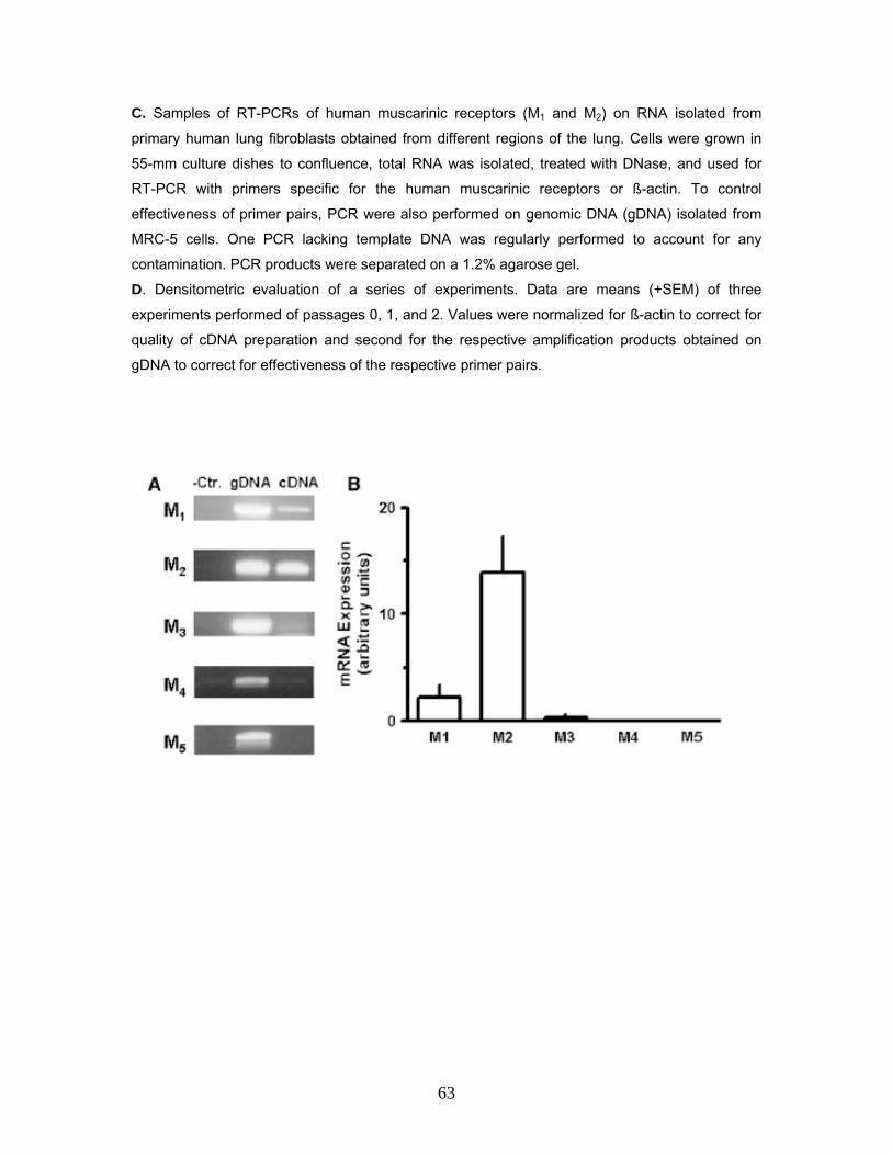
C. Samples of RT-PCRs of human muscarinic receptors (M1 and M2) on RNA isolated from
primary human lung fibroblasts obtained from different regions of the lung. Cells were grown in
55-mm culture dishes to confluence, total RNA was isolated, treated with DNase, and used for
RT-PCR with primers specific for the human muscarinic receptors or ß-actin. To control
effectiveness of primer pairs, PCR were also performed on genomic DNA (gDNA) isolated from
MRC-5 cells. One PCR lacking template DNA was regularly performed to account for any
contamination. PCR products were separated on a 1.2% agarose gel.
D. Densitometric evaluation of a series of experiments. Data are means (+SEM) of three
experiments performed of passages 0, 1, and 2. Values were normalized for ß-actin to correct for
quality of cDNA preparation and second for the respective amplification products obtained on
gDNA to correct for effectiveness of the respective primer pairs.
63
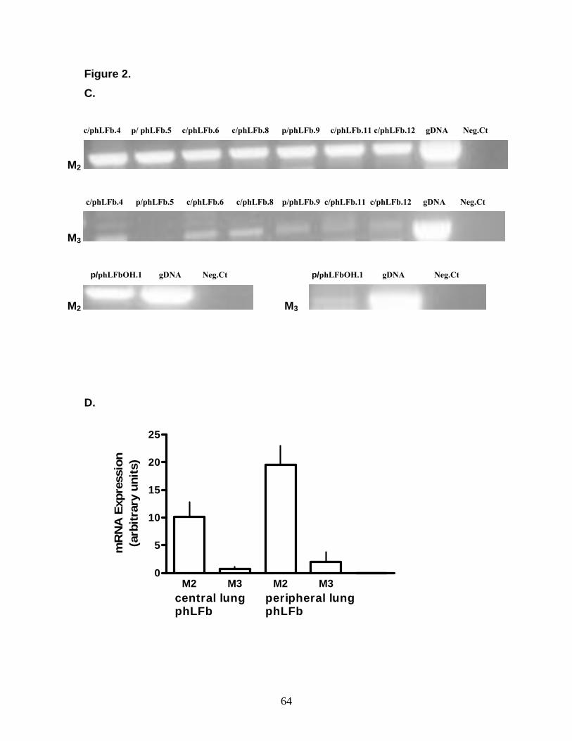
Figure 2. C.
c/phLFb.4 p/ phLFb.5 c/phLFb.6 c/phLFb.8 p/phLFb.9 c/phLFb.11 c/phLFb.12 gDNA Neg.Ct
M2
c/phLFb.4 p/phLFb.5 c/phLFb.6 c/phLFb.8 p/phLFb.9 c/phLFb.11 c/phLFb.12 gDNA Neg.Ct
M3 p/phLFbOH.1 gDNA Neg.Ct p/phLFbOH.1 gDNA Neg.Ct
M2 M3
D.
M2 M3 M2 M30
5
10
15
20
25
central lungphLFb
peripheral lungphLFb
mR
NA
Exp
ress
ion
(arb
itrar
y un
its)
64

3.2 Muscarinic Stimulation of Proliferation of Human Lung Fibroblasts 3.2.1 Muscarinic Stimulation of Proliferation of MRC-5 Human Lung
Fibroblasts
Various experimental conditions were tested in order to determine optimal
parameters for the (3H)-thymidine incorporation assay. The purpose being to test
whether putative muscarinic effects on fibroblast proliferation were dependant on
culture conditions and exposure time. In the first set of experiments, cells were
cultured for 24 hours in the presence of 10% FCS, followed by 24 hours under
FCS-free conditions in the presence of (3H)-thymidine and in the absence or
presence of increasing concentrations of carbachol. Under control conditions
(absence of drugs), (3H)-thymidine incorporation amounted to 7.281 ± 548 d.p.m
(disintegration per minute) (n=52). Carbachol induced an increase in (3H)-
thymidine incorporation by 38 ± 18% at 10 µM in a concentration-dependent
manner (Figure 3A). As shown in Figure 3A, this effect was abolished in the
presence of 1 µM atropine which alone did not have an effect on (3H)-thymidine
incorporation. In spite of the fact that the stimulatory effect of carbachol was
clearly significant, the magnitude of the effect showed considerable variation.
In a second set of experiments, cells were again cultured for 24 hours in the
presence of 10% FCS, followed by 48 hours FCS-free conditions in the absence
or presence of carbachol (10 µM) with (3H)-thymidine present for the last 24 h.
Under these conditions, carbachol caused an increase in (3H)-thymidine
incorporation by 52 ± 10% (Figure 3B). Here again the proliferative effect of
carbachol was inhibited by atropine and also by the long-acting muscarinic
antagonist tiotropium (Figure 3B). Furthermore, oxotremorine also caused a
similar increase (by 61%), an effect also prevented by atropine (Figure 3B). For
comparison, when FCS (1 or 10%) was present instead of muscarinic agonist
during the last 48 hours, (3H)-thymidine incorporation was enhanced by 69 ± 17%
(n = 6) and 216 ± 30% (n = 9), respectively, compared to FCS-free conditions.
In the third protocol, cells were FCS-deprived and drugs were applied from the
onset of the test culture period. Under these conditions, 10 µM carbachol or
oxotremorine increased (3H)-thymidine incorporation by 64 ± 8% and 100 ± 26%,
65

respectively, when present 24 hours before (3H)-thymidine, for a total of 48
hours. These effects were again shown to be atropine sensitive (Figure 3C).
In a further set of experiments carbachol was applied 6 hours before (3H)-
thymidine, for a total of 30 hours, (3H)-thymidine incorporation was increased by
76 ± 4% (n = 80). These conditions were held for the subsequent interaction
experiments with subtype-preferring muscarinic receptor antagonists.
As shown in Figure 4 when 1% FCS was present from the onset of the test
culture period (6 hours before (3H)-thymidine, for a total of 30 hours), (3H)-
thymidine incorporation was increased by 450%. Additional presence of 10 µM
carbachol or oxotremorine induced a further significant increase in (3H)-thymidine
incorporation, by about 300% and 240% respectively, relative to the FCS-free
controls (Figure 4). This corresponds to an increase of 54 and 43% respectively,
when expressed in relation to FCS-containing control experiments.
Figure 3. Effects of muscarinic agonists and antagonists on (3H)-thymidine incorporation
in MRC-5 Human Lung Fibroblasts
7.5 x 10⁴ cells were seeded in 12 well dishes in the absence or presence of 10 % FCS as
indicated. In each series (3H)-thymidine (37 kBq) was present for the last 24 h.
A. Effects of carbachol, at concentrations indicated by the abscissa, in absence (open symbols)
or presence (filled symbols) of 1 µM atropine. Test drugs were added together with (3H)-
thymidine.
B and C. Carbachol (C, 10 µM), oxotremorine (O, 10 µM) and/or atropine (A, 1 µM) and/or
tiotropium (T, 0.1 µM) were present already 24 h prior to (3H)-thymidine until the end of the
protocol. Data are means (±SEM) of n=3-12 (mostly n≥6). ** P < 0.01; *** P< 0.001 vs. controls; +
P < 0.05, ++ P < 0.001, +++ P < 0.001 vs. the respective value in the absence of antagonists.
66

Figure 3. A.
0 .01 0 .1 1 10
100
125
150
24h 10 % F C S 24 h FC S -fre e 24 h F C S -fre e + te st d ru gs
C ar b ach o l [ µ M ]
3 H-T
hym
idin
e in
corp
orat
ion
(% o
f con
trol
s)
* *
B.
C.
0
50
100
150**
**
- C - C - C O O- - A A T T - A
24h 10 % FC S 48 h FC S -fre e ± te st drugs
+ + +
3 H-T
hym
idin
e in
corp
orat
ion
(% o
f con
trol
s)
0
50
100
150
200 *****
- - C C O O- A - A - A
48 h FCS-free ± test drugs
+++++
3 H-T
hym
idin
e in
corp
orat
ion
(% o
f con
trol
s)
67

Figure 4. Comparison of the effects of carbachol and oxotremorine on (3H)-thymidine
incorporation in MRC-5 human lung fibroblasts in absence and presence of FCS. 7.5 x 10⁴ cells
were seeded in 12 well dishes and cultured under FCS-free conditions or in the presence of 1%
FCS. Carbachol (Carb, 10 µM) or oxotremorine (Oxo, 10 µM) was added from the onset of
incubation for a total of 30 h, (3H)-thymidine (37 kBq) was present for the last 24 h. Data are
means (±SEM) of n=3-12. *** P< 0.001 vs. controls (Ctr); + P < 0.01 vs. the respective value in the
presence of FCS alone.
0
100
200
0
250
500
750
1000
Ctr Carb Oxo Ctr - Carb Oxo
1 % FCS
*** ***
***
++
3 H-T
hym
idin
e in
corp
orat
ion
(% o
f con
trol
s)3H
-Thymidine incorporation
(% of controls)
3.3 Characterization of Muscarinic Receptor Sub-Type 3.3.1 Inhibition of Muscarnic Proliferation using Receptor Sub-Type
Specific Antagonists in MRC-5 Human Lung Fibroblasts
Receptor sub-type characterization studies were performed to determine the
functional receptor/receptors involved in the muscarinic stimulation of
proliferation in MRC-5 human lung fibroblasts using different selective muscarinic
antagonists. As summarized in Figure 5, all muscarinic receptor antagonists
tested inhibited the stimulatory effect of carbachol on (3H)-thymidine
incorporation in a concentration dependent manner. Tiotropium was by far the
most potent drug, with an IC50 value of 14 pM. The M2/M4, receptor-preferring
68

antagonists, AQ-RA 741, AF-DX 384, and himbacine, showed IC50 values of 24,
64, and 187 nM, respectively, whereas, for the M3/M1 receptor-preferring
antagonists, 4-DAMP and p-FHHSiD, IC50 values of 127 and 452 nM were
determined. Pirenzepine a M1/M4 receptor-preferring antagonist was found to
have an IC50 value of 1.5 µM. None of the antagonists at any of the
concentrations studied significantly affected (3H)-thymidine incorporation on its
own. When the potencies of these antagonists as determined here were
correlated to their reported affinity constants to cloned human muscarinic
receptors, the best and highly significant correlation was obtained for the M2
receptor. Figures 5B-5D present only the correlations for M1, M2, and M3
receptors, as affinity data of tiotropium for cloned M4 and M5 receptors have not
yet been published. For these two receptors, the respective correlations
calculated on the basis of the remaining six antagonist failed to be statistically
significant, with r = 0.60 and 0.27 for M4 and M5 receptors, respectively.
Furthermore when correlation significance was calculated as described
previously for the various muscarinic receptors in the absence of tiotropium, M2
muscarinic receptor showed the best and highly significant correlation with r =
0.89 as compared to M3 (r = 0.10), M1 (r = 0.17), M4 (r = 0.60) and M5 (r = 0.27).
Figure 5. A. Inhibitory effects of different muscarnic receptor antagonists on carbachol-induced
stimulation of (3H)-thymidine incorporation in MRC-5 human lung fibroblasts. A total of 7.5 x 10⁴
cells were seeded in 12-well dishes under FCS-free conditions. Carbachol (10 µM) was added
from the onset of incubation for a total of 30 h, of which (3H)-thymidine (37 kBq) was present for
the last 24 h. In the absence of antagonists, carbachol enhanced (3H)-thymidine incorporation by
98 ± 10% (n = 53). Antagonists were applied immediately before addition of carbachol at the
concentrations given by the abscissa. Ordinate: stimulatory effect of carbachol in the presence of
antagonist, expressed as percent of mean stimulatory effect of carbachol in the absence of
antagonists, as observed in the respective series of experiment. Data are means of n = 3-12
(mostly n ≥ 6); SEM, 6-12%.
69

Figure 5. B-D. Correlation between the potencies of the antagonists, as determined in the
present experiments (ordinate), and the reported affinity constants, as determined on cloned
human muscarnic receptors (abscissa). Open squares, 4-DAMP; closed triangles, AQ-RA741;
open diamonds, AF-DX384; closed diamonds, p-FHHSiD; closed circles, tiotropium; open circles,
himbacine; open inverted triangles, pirenzepine.
0.001 0.01 0.1 1 10 100 100010000
0
25
50
75
100
1254-DAMPAQ-RA 741AF-DX 384pFHHSiDTiotropium
A MRC-5 cells
HimbacinePirenzepine
antagonist [ nM ]
Car
bach
ol-in
duce
d3 H
-Thy
mid
in in
corp
orat
ion
(%)
6 7 8 9 10 11
6789
1011
r=0.96p=0.0008
C
cloned M2 receptors [-log KB]
MR
C-5
cel
ls [-
log
IC50
]
7 8 9 10 11
6
7
8
9
10
11
r=0.80p=0.03
D
cloned M3 receptors [-log KB]
MR
C-5
cel
ls [-
log
IC50
]
6 7 8 9 10 11
6
7
8
9
10
11
r=0.51p=0.07
B
cloned M1 receptors [-log KB]
MR
C-5
cel
ls [-
log
IC50
]
70

3.3.1.2 Effect of Pertussis Toxin on Muscarinic Stimulation of Proliferation
of MRC-5 Human Lung Fibroblasts
Pertussis toxin (PTX) has been widely used as a reagent to characterize the
involvement of heterotrimeric G-proteins in signalling. This toxin catalyses the
ADP-ribosylation of specific G-protein subunits of the Gi family, and this
modification prevents the occurrence of the receptor–G-protein interaction. As
shown in Figure 6, pretreatment of the fibroblasts with PTX resulted in a slight
reduction in (3H)-thymidine incorporation, and abolished the stimulatory effect of
carbachol. In order to test the specificity of PTX, the effects of this toxin were
also studied on the proliferative effect of PDGF, which is known to act via a
different class of receptors. Pretreatment of the cells with PTX, as described in
Figure 6, did not inhibit the PDGF-induced stimulation of (3H)-thymidine
incorporation (increase by 219 ± 40% [n = 6] and 283 ± 40% [n = 3] in absence
and presence of PTX, respectively).
Figure 6. Effects of carbachol and/or pertussis toxin (PTX) on (3H)-thymidine
incorporation in MRC-5 Human Lung Fibroblasts (A + B) 7.5 x 10⁴ cells were seeded in 12 well dishes and cultured under FCS-free conditions.
PTX (0.1 µg/ml) was present from the onset of incubation. Carbachol (Carb., 10 µM), PDGF 100
ng/ml) were added 3 h after PTX. Finally, (3H)-thymidine (37 kBq) was added 6 h later and
present for 24 h. Data are means (±SEM) of n=3-12 (mostly n≥6). * P< 0.01 and ** P< 0.001 vs.
controls (Ctr); *, + P< 0.01 vs. Ctr, and **P< 0.001 vs. Carb, alone. A. B.
0
50
100
150
200
Ctr. Carb. PTX Carb. +PTX
**
+* *
3 H-T
hym
idin
e in
corp
orat
ion
(% o
f con
trol
s)
0
100
200
300 ****
---- PDGF ---- PT X
3 H-T
hym
idin
e in
corp
orat
ion
(% o
f res
p. c
ontr
ols)
71

3.3.1.3 Muscarinic Stimulation and Effect of Pertussis Toxin on
Proliferation in Primary Human Lung Fibroblasts (pHLFb)
(3H)-thymidine incorporation experiments were performed with primary human
lung fibroblasts to confirm the muscarinic stimulation mediated proliferation using
10 µM carbachol or oxotremorine, in the presence of tiotropium or atropine, as
well as pretreatment with PTX. It was observed that the stimulatory effect in the
presence of the muscarinic agonists carbachol and oxotremorine were abolished
in the presence of tiotropium or atropine and PTX. These results were in line with
those seen for the MRC-5 cell line. Also, in the primary human lung fibroblasts,
PTX pretreatment failed to inhibit the proliferative effect of 100 ng/ml PDGF
(increased by 403 ± 49% [n = 12] and 487 ± 80% [n = 3] in the absence and
presence of PTX, respectively).
Figure 7. Effects of carbachol and/or pertussis toxin (PTX), tiotropium or atropine on (3H)-
thymidine incorporation in Primary Human Lung Fibroblasts. 7.5 x 10⁴ cells were seeded in 12 well dishes and cultured under FCS-free conditions. Cells were
incubated for 30 h with carbachol (Carb, 10 µM) and/or tiotropium (T, 0.1 µM), atropine (A, 1 µM)
or oxotremorine (Oxo, 10 µM) as indicated. (3H)-thymidine (37 kBq) was added 6 h after onset of
exposure to these drugs. In the respective experiments PTX (0.1 µg/ml) was already present 3 h
before carbachol. Data are means (±SEM) of n=3-12. ** P < 0.001 vs. controls; + P < 0.01 vs.
carbachol alone.
0
100
200
300 **
++
- 10 10 10 - 10 - Carb. T A -- PTX -- Oxo
+
**
3 H-T
hym
idin
e in
corp
orat
ion
(% o
f con
trol
s)
72

3.4 Muscarinic Stimulation Mediated Activation of MAPK Pathway in Human Lung Fibroblasts
3.4.1 Muscarinic Receptor Stimulation and Activation of p-42/44 MAPK
Pathway in Proliferation of Human Lung Fibroblast The preceding results confirm the presence of muscarinic receptors and its
involvement in mediating proliferative effects in both the MRC-5 and the primary
human lung fibroblasts. The next aspect to look into was to study the cellular
transduction mechanism involved in the muscarinic stimulation of lung fibroblast
proliferation. Since activation of MAPK cascade has been shown to be an
essential transduction mechanism of many mitogens and growth factors, (3H)-
thymidine incorporation assay was performed. Early passage MRC-5 cells were
used for this purpose and for all experiments; primary cells were used before
their 7th passage. Cells were disseminated in a 12-well dish at a density of 7.5 x
104 per well and cultivated in the absence of FCS for 30 hours in combination
with the test drugs. (3H)-thymidine (37kBq) was applied for the last 24 hours. As
shown in Figure 8, muscarinic receptor agonist carbachol (10 µM) exerted a
marked proliferative effect in both, MRC-5 cells and primary human lung
fibroblasts as indicated by about 2-fold increase in (3H)-thymidine incorporation.
Preceding results show this effect was inhibited by different muscarinic receptor
antagonist like atropine and tiotropium proving that proliferation is mediated via
specific muscarinic receptors. Figure 8, further shows that in both MRC-5
fibroblasts as well as in primary human lung fibroblasts the stimulatory effect of
carbachol could be blocked by PD98059, a specific inhibitor of MAPK-activating
enzyme (MEK), implicating that MAPK cascade is crucially involved in the
proliferative response of human lung fibroblasts to cholinergic stimulation.
73

Figure 8. Effects of carbachol in absence and presence of PD 98059 on the (3H)-
thymidine incorporation in Primary Human Lung Fibroblast (hLFb) and the Human Lung Fibroblast cell line MRC-5. 7.5 x 10⁴ cells were disseminated in 12-well dishes in the absence of
FCS and cultured for 30 h in the absence and presence of test drug as indicated. The cells were
pretreated with the PD 98059 drug for 30 min before the addition of the carbachol. (3H)-thymidine
was applied for the last 24 h. Given are means ± SEM of n = 3-12 (mostly ≥ 6). Significance of
differences; from controls, *P < 0.05; **P < 0.01; from carbachol, +P < 0.05.
0
100
200
- 10 - 10 - 10 - 10 - 10 µM Carbachol- - 3 3 30 30 - - 30 30 µM PD 98059
MRC-5 cells primary hLFb
**
**
*
*+
+
3 H-T
hym
idin
e in
corp
orat
ion
(% o
f con
trol
s)
3.4.1.2 Western Blot Analysis of p42/44 MAPK (ERK1, ERK2) Activation in
MRC-5 Human Lung Fibroblast
Western blot analysis was performed to confirm the activation of the MAPK
cascade in response to the muscarinic stimulation induced proliferationin the
MRC-5 fibroblasts at the protein levels. Western blot analysis conducted after a
two to 120 minute stimulation of serum-starved MRC-5 fibroblasts with carbachol
or oxotremorine (Figure 9) demonstrated activation of p42/44 MAPK (ERK1,
ERK2). The effects of both oxotremorine and carbachol revealed to be
counteractable by the muscarinic receptor antagonists atropine (1 µM, data not
shown) and tiotropium (0.1 µM, exemplary shown for stimulation by carbachol in
74

Figure 9). Finally, similar activation patterns were observed in primary human
lung fibroblasts (10 µM carbachol enhanced p42/44 MAPK to 189 ± 12% of
controls after 10 minutes (n = 6), data not shown).
Figure 9. Western blot analysis of p42/44 MAPK (ERK1, ERK2) activation in MRC-5
Human Lung Fibroblasts. Cells were cultured in a 55 mm2 dishes to nearly confluence, serum-
starved for 24 h and exposed to 10 µM oxotremorine (Oxo), carbachol (C) in absence or
presence of 0.1 µM tiotropium or vehicle (Ctr) for 2-120 min as indicated. Cell lysates were
prepared and 20-50
µg were separated in 10-20% acrylamide Tris-glycine gels, immobilised to PVDF membranes and
detected by antibodies specific for phosphorylated MAPK (P-44/42) and unphosphorylated MAPK
(ERK1/2). Densitometrical quantification of the p-44/42 bands was performed and values
(arbitrary units) in presence of agonists expressed as % of the the respective control. Given are
means ± SEM of n = 3-6 independent Western blots.
Ctr O Ctr O Ctr O Ctr O Ctr C C+T
P-44/42
ERK1/2
2 min 10 min 30 min 120 min 10 min
268 ±12
148 ±14
127 ±7
82 ±39
193 ±28
91 ±14
.
75

3.5 Effect of Kinase Inhibitors on Muscarinic Stimulation of MRC-5 Human Lung Fibroblast Proliferation 3.5.1 Effects of the Rho-kinase Inhibitor Y27632 on Muscarinic Stimulation
of MRC-5 Human Lung Fibroblast Proliferation
The results previously indicated the muscarinic stimulation of proliferation in
human lung fibroblast is mediated via the classical MEK-ERK MAPK cascade
pathway. To elucidate the detailed transduction of how muscarinic receptor
stimulation results in MAPK activation (3H)-thymidine incorporation proliferation
assays were performed with various antagonists specific to signalling molecules,
most likely to be involved in this G protein receptor coupled activation of MAPK
pathway.
Rho a member of the family of small G-proteins has been shown to mediate cell
cycle progression and proliferation via the activation of the MAPK cascade. To
test if Rho was indeed involved in the muscarinic stimulation of proliferation of
MRC-5 human lung fibroblast (3H)-thymidine incorporation assay was performed.
Cells were seeded (1.5 x 10⁵ cells/well) in a 12 well dish in the presence or
absence of FCS and cultured for 30 hours with or without the test drugs.
Passage numbers 1-10 were chosen for these experiments. The Rho-kinase
inhibitor Y27632 (10 µM) was added 30 minutes prior to addition of carbachol (10
µM). As shown in Figure 10 (A + B) using the 30 hours FCS-free or the 6 hours
10% FCS followed by the FCS-free period protocols prior to addition of the drugs,
carbachol enhanced the (3H)-thymidine incorporation to 141 ± 4% and 155 ± 10%
while the Rho-kinase inhibitor Y27632 reduced the (3H)-thymidine incorporation
to 79 ± 2% and 50 ± 4% with respect to the control. However the Y27632 Rho-
kinase inhibitor did not inhibit the carbachol induced stimulation of (3H)-thymidine
incorporation (increase to 136 ± 7% and 160 ± 9% respectively in the presence
of Y27632).
76

Figure 10 A + B. Effects of the Rho-kinase inhibitor Y27632 on muscarinic stimulation of
MRC-5 human lung fibroblast proliferation. MRC-5 cells (1.5 x 10⁵ per well) were disseminated
in 12 well dishes in the absence or presence of FCS and cultured for 30 h with or without test
drugs as indicated. The cells were pre-treated with the Rho-kinase inhibitor Y27632 for 30 min
before the addition of carbachol. (3H)-thymidine was applied for the last 24 h. Given are means ±
SEM of n=9-18. Significance of differences: from respective controls, *P<0.01.
A.
0
50
100
150
- + - - + 10 µ M C arbachol- - + + + 10 µ M Y27632
*
30 hrs FC S -free ± d rugs
* *
[3 H]-T
hym
idin
e in
corp
orat
ion
% o
f res
p. c
ontr
ol
B.
0
50
100
150
- + - - + 10 µ M C arbachol- - + + + 10 µ M Y27632
6 hrs 10 % FC S - 30 hrs FC S-free ± drugs
*
*
*
[3 H]-T
hym
idin
e in
corp
orat
ion
% o
f res
p. c
ontr
ol
77

3.5.1.2 Effects of the Phospholipase C Inhibitor U73122 on Muscarinic
Stimulation of MRC-5 Human Lung Fibroblast Proliferation
Previous studies have demonstrated the muscarinic stimulation induced
activation of the MAPK pathway mediated via the Phospholipase C (PLC) in
pertussis toxin sensitive G-proteins. In order to elucidate the involvement of PLC
on the muscarinic stimulation of MRC-5 human lung fibroblast proliferation (3H)-
thymidine incorporation assays were performed. Cells in early passages were
used in these experiments. Cells were disseminated (1.5 x 10⁵ cells per well) in
12 well dishes in the absence of FCS and cultured for 30 hours with or without
the test drugs. The PLC-inhibitor U73122 was added 30 minutes prior to the
addition of carbachol (10 µM) in concentrations of 0.1, 0.3 and 1 µM. As shown in
Figure 11 (A + B), carbachol caused an increase in the (3H)-thymidine
incorporation by 193 ± 6% whereas the Phospholipase C inhibitor U73122
reduced the (3H)-thymidine incorporation significantly to 80 ± 5% and 84 ± 7% at
0.3 and 1 µM concentrations relative to the control. However the PLC inhibitor
U73122 did not inhibit the carbachol induced stimulation of (3H)-thymidine
incorporation (increase to 224 ± 18% [n = 9] and 183 ± 9% [n = 9] respectively in
the presence of U73122 at 0.3 and 1 µM).
78

Figure 11 A + B. Effects of the Phospholipase C inhibitor U73122 on muscarinic
stimulation of MRC-5 human lung fibroblast proliferation. Cells (1.5 x 10⁵ cell/well) were
disseminated in 12 well dishes in the absence of FCS and cultured for 30 h with or without test
drugs as indicated. The cells were pre-treated with the phospholipase C inhibitor U73122 for 30
min before the addition of carbachol. (3H)-thymidine was applied for the last 24 h. Given are
means ± SEM of n=9-18. Significance of differences: from their respective controls, *P<0.05;
**P<0.01; ***P<0.01.
A.
0
25
50
75
100
- 0.1 0.3 1 µM U73122
[3 H]-T
hym
idin
e in
corp
orat
ion
% o
f con
trol
s
* **
B.
0
100
200
[3 H]-T
hym
idin
e in
corp
orat
ion
% o
f res
p. c
ontr
ol *** ***
***
***
- 10 - 10 - 10 - 10 µM Carbachol- - 0.1 0.1 0.3 0.3 1 1 µM U73122
79
3.5.1.3 Effects of the PI3-kinase Inhibitor Wortmannin on Muscarinic
Stimulation of MRC-5 Human Lung Fibroblast Proliferation
Phosphoinositide 3-kinases (PI3-kinases or PI3Ks) are a family of related
enzymes that are capable of phosphorylating the 3 position hydroxyl group of the
inostol ring of phosphatidylinositol. Several studies have documented the
activation of the MAPK via PI-3 kinase. To investigate the role of PI-3 kinase in
the muscarinic stimulation of proliferation in MRC-5 human lung fibroblasts (3H)-
thymidine incorporation assays were performed using the PI3-kinase inhibitor
wortmannin. Cells in early passages were used in these experiments. Cells were
disseminated (1.5 x 10⁵ cells per well) in 12 well dishes in the absence of FCS
and cultured for 30 hours with or without the test drugs. The PI3-kinase inhibitor
wortmannin was added 30 minutes prior to the addition of carbachol (10 µM) in
concentrations of 30, 100 and 300 nM. As shown in Figure 12 (A + B) carbachol
caused an increase in the (3H)-thymidine incorporation by 191 ± 7% whereas the
PI3-kinase inhibitor wortmannin reduced the (3H)-thymidine incorporation
significantly to 61 ± 1%, 48 ± 3% and 37 ± 2% at 30, 100 and 300 nM
concentrations relative to the control. However the PI3-kinase inhibitor
wortmannin did not inhibit the carbachol induced stimulation of (3H)-thymidine
incorporation (increase to 189 ± 12%) in the presence of U73122 (100 nM). The
effect of wortmannin (300 nM) on the carbachol induced proliferation could not be
considered as a consistent result since the magnitude of the effect showed
considerable variation and was not significantly different from respective value in
the absence of wortmannin.
80

Figure 12 A + B. Effects of the PI3-kinase Inhibitor Wortmannin on Muscarinic
Stimulation of MRC-5 Human Lung Fibroblast Proliferation. Cells (1.5 x 10⁵ cell/well) were
disseminated in 12 well dishes in the absence of FCS and cultured for 30 h with or without test
drugs as indicated. The cells were pre-treated with PI3-kinase inhibitor wortmannin for 30 min
before the addition of carbachol. (3H)-thymidine was applied for the last 24 h. Given are means ±
SEM of n=9-18. Significance of differences: from controls, *P<0.01; n.s., not significantly different
from respective value in absence of wortmannin.
A.
0
25
50
75
100
- 30 100 300 nM Wortmannin
**
*
[3 H]-T
hym
idin
e in
corp
orat
ion
% o
f con
trol
s
B.
0
100
200
- 10 - 10 - 10 µM Carbachol- - 100 100 300 300 nM Wortmannin
* **
n.s.
[3 H]-T
hym
idin
e in
corp
orat
ion
% o
f res
p. c
ontr
ol
81

3.5.1.4 Effects of the Raf-1 Inhibitor GW5074 on Muscarinic Stimulation of
MRC-5 Human Lung Fibroblast Proliferation
The RAS/RAF/MEK/ERK signal transduction pathway is a conserved pathway
that regulates cell growth and signaling. Previous studies have shown the
involvement of Ras and Raf in the Gi-coupled acetylcholine muscarinic M2
receptor activation of MAPK, also Ras-independent activation of MAPK mediated
via the Raf-MEK-MAPK have been reported. To study the role of Raf in the
muscarinic stimulation of proliferation in MRC-5 human lung fibroblasts (3H)-
thymidine incorporation assay was employed using the Raf-1 inhibitor GW5074.
Early passages of MRC-5 cell up to passage 10 were used for the purpose of
these experiments. Cells were disseminated (1.5 x 10⁵ cells per well) in 12 well
dishes in the absence of FCS and cultured for 30 hours with or without the test
drugs. The Raf-1 inhibitor GW5074 was added 30 minutes prior to the addition of
carbachol (10 µM) in concentrations of 0.1, 1 and 3 µM. as indicated in Figure 13
A +B. carbachol induced a proliferative stimuli, increasing the (3H)-thymidine
incorporation to 167 ± 6% with respect to the control. The Raf-1 inhibitor
GW5074 did not affect the basal proliferation rate at 0.1 and 1 µM although at a 3
µM concentration it inhibited the (3H)-thymidine incorporation to 40 ± 7% relative
to the control. It was also observed that the carbachol induced muscarinic
stimulation of proliferation was significantly inhibited by GW5074 at 1 and 3 µM
as shown by the reduction in the (3H)-thymidine incorporation to 151 ± 6% and
132 ± 8% in the presence of the drug respectively.
82
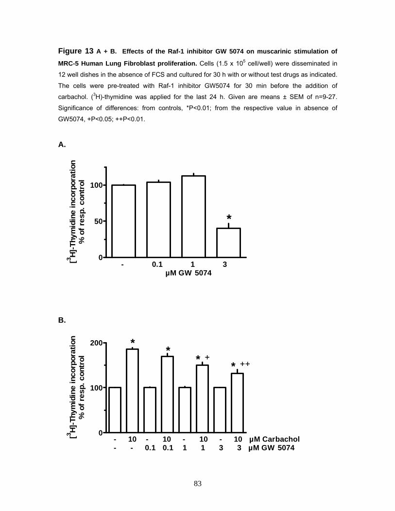
Figure 13 A + B. Effects of the Raf-1 inhibitor GW 5074 on muscarinic stimulation of
MRC-5 Human Lung Fibroblast proliferation. Cells (1.5 x 10⁵ cell/well) were disseminated in
12 well dishes in the absence of FCS and cultured for 30 h with or without test drugs as indicated.
The cells were pre-treated with Raf-1 inhibitor GW5074 for 30 min before the addition of
carbachol. (3H)-thymidine was applied for the last 24 h. Given are means ± SEM of n=9-27.
Significance of differences: from controls, *P<0.01; from the respective value in absence of
GW5074, +P<0.05; ++P<0.01.
A.
0
50
100
- 0.1 1 3 µM GW 5074
*
[3 H]-T
hym
idin
e in
corp
orat
ion
% o
f res
p. c
ontr
ol
B.
0
100
200
- 10 - 10 - 10 - 10 µM Carbachol- - 0.1 0.1 1 1 3 3 µM GW 5074
** *
*+
++
[3 H]-T
hym
idin
e in
corp
orat
ion
% o
f res
p. c
ontr
ol
83

3.5.1.5 Effects of the Raf-1 Inhibitor GW5074 (GW) on Muscarinic
Stimulation of the ERK-MAP Kinase in MRC-5 Human Lung Fibroblasts
Western blot analysis was performed to confirm the inhibitory effect of the Raf-1
inhibitor GW5074 on the activation of the MAPK cascade in response to the
muscarinic stimulation induced proliferation in the MRC-5 fibroblasts at the
protein levels. Western blot analysis conducted after a two minute stimulation of
serum-starved MRC-5 fibroblasts with carbachol (10 µM) demonstrated activation
of p42/44 MAPK (ERK1, ERK2). Densitometrical quantification of the p-44/42
bands was performed and values (arbitrary units) revealed that carbachol (10
µM) enhanced p-44/42 MAPK to 249 ± 61% (n =3) while GW5074 by itself did
not appear to affect the MAPK activation with respect to the control, the
carbachol enhaced p-44/42 MAPK was reduced to 186 ± 36% (n = 3) in the
presence of the Raf-1 inhibitor.
Figure 14. Effects of the Raf-1 inhibitor GW5074 (GW) on muscarinic stimulation of the
ERK-MAP kinase in MRC-5 Human Lung Fibroblasts. Cells were cultured in a 55 mm2 dishes
to nearly confluence, serum-starved for 24 h and exposed to 10 µM carbachol (C) in absence or
presence of 3 µM GW5074 or vehicle (Ctr) for 2 minutes. Cell lysates were prepared and 20-50
µg were separated in 10-20% acrylamide Tris-glycine gels, immobilised to PVDF membranes and
detected by antibodies specific for phosphorylated MAPK (P-44/42) and unphosphorylated MAPK
(ERK1/2).
---- GW ----
P 42/44
ERK-1/2
- c - c
84

3.5.1.6 Effects of the Tyrosine-Kinase Inhibitor Tyrphostin AG1295 on
Muscarinic Stimulation of MRC-5 Human Lung Fibroblasts
Protein tyrosine kinases (PTKs) play a key role in normal cell and tissue
development. Enhanced PTK activity is intimately correlated with cell
proliferation. Numerous studies have shown that GPCRs may utilize receptor
tyrosine kinases (RTKs) such as platelet derived growth factor receptor (PDGF)
or epidermal dervived growth factor receptor (EGFRs) as intermediate signaling
proteins thereby mimicking the signaling pathways downstream leading to
activation of MAPK cascade. To investigate whether muscarinic stimulation of
MRC-5 human lung fibroblast lead to the transactivation of PDGF receptor, (3H)-
thymidine incorporation assays were performed in the presence or absence of
tyrphostin AG1295 a specific tyrosine kinase inhibitor for PDGF receptors. As
shown in Figure 15 A + B. carbachol (10 µM) caused an increase in the (3H)-
thymidine incorporation to 175 ± 5% while tyrphostin AG1295 (5 µM) inhibited
(3H)-thymidine incorporation to 29 ± 4% with respect to the control. However the
tyrphostin AG1295 the tyrosine kinase inhibitor did not inhibit the carbachol
induced stimulation of (3H)-thymidine incorporation (increase to 175 ± 9%) in the
presence of tyrphostin AG1295 (5 µM). In a further set of control experiments
(data not shown [n = 3]) it was observed that the PDGF (10 ng/ml) induced
increase in (3H)-thymidine incorporation in MRC-5 human lung fibroblast was
unimpeded in the presence of tiotropium (1 µM), confirming its selectivity in
inhibition of carbachol induced muscarinic proliferation of MRC-5 human lung
fibroblast.
85
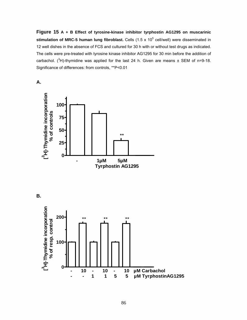
Figure 15 A + B Effect of tyrosine-kinase inhibitor tyrphostin AG1295 on muscarinic
stimulation of MRC-5 human lung fibroblast. Cells (1.5 x 10⁵ cell/well) were disseminated in
12 well dishes in the absence of FCS and cultured for 30 h with or without test drugs as indicated.
The cells were pre-treated with tyrosine kinase inhibitor AG1295 for 30 min before the addition of
carbachol. (3H)-thymidine was applied for the last 24 h. Given are means ± SEM of n=9-18.
Significance of differences: from controls, **P<0.01
A.
0
25
50
75
100
- 1µM 5µM Tyrphostin AG1295
[3 H]-T
hym
idin
e in
corp
orat
ion
% o
f con
trol
s
**
B.
0
100
200
- 10 - 10 - 10 µM Carbachol- - 1 1 5 5 µM TyrphostinAG1295
[3 H]-T
hym
idin
e in
corp
orat
ion
% o
f res
p. c
ontr
ol ** ** **
86

3.6 Muscarinic Stimulation of proliferation of MRC-5 Human Lung Fibroblast and Anti-apoptotic effects Apoptosis represents a multi-step process for elimination and subsequent
removal of cells in response to environment insults or as part of organismal
development. The caspases are a family of proteins that are one of the main
executors of the apoptotic process. They belong to a group of enzymes known as
cysteine proteases which can be activated following the occupation of certain cell
surface ‘death’ receptors (e.g. tumor necrosis factor) or by agents that induce
stress or DNA damage (e.g. actinomycin D, cycloheximide). A member of this
family, caspase-3 has been identified as being a key mediator of apoptosis of
mammalian cells. The previous experiments that were performed clearly
indicated the functional role of the muscarinic receptors in the carbachol induced
proliferation of the MRC-5 human lung fibroblasts cells. Various studies
performed in the past have confirmed the anti-apoptotic effects of the G-protein
coupled muscarinic receptor activation
The next aspect that was explored was whether the proliferative stimuli
mediated via the muscarinic receptors in the MRC-5 fibroblasts were augmented
by an anti-apoptotic effect concomitantly. For this purpose caspase-3 fluorimetric
assays were performed using a combination of apoptosis inducers like TNFα,
actinomycin D and cycloheximide in the presence or absence of the muscarinic
agonist carbachol. MRC-5 cells (4 x 10⁴ cells/well) were disseminated in 24-well
dishes in the presence of 10% FCS, upon confluence the medium was aspirated
out cells were treated with apoptosis inducers in the presence or absence of
carbachol (10 µM) as indicated in Figure 16, and incubated further for 4 hours
under serum-free conditions followed by lysis and treatment with the substrate.
Two wells for substrate blank, two for caspase positive control and two for
caspase positive in combination with the caspase inhibitor were also prepared for
every experiment. As shown in Figure 16, actinomycin D in presence of TNFα
and cycloheximide in combination with TNFα enhanced capase-3 activity to 119
± 2%, 176 ± 40% respectively relative to the control. The positive control showed
an increased caspase- 3 activity to 203 ± 11% which was reduced to 127 ± 10%
87

with respect to the control in the presence of the caspase inhibitor. Nonetheless it
was observed that carbachol (10 µM) did not significantly affect the level of
caspase-3 activity in the presence of the either of the two combinations of the
apoptosis inducers. Although these results seem to suggest that the carbachol
induced muscarinic proliferation of MRC-5 human lung fibroblasts is unaided by
anti-apoptotic mechanisms the magnitude of the effect showed considerable
variations hence could not be considered conclusive.
Figure 16. Effects of various apoptosis inducers on muscarinic stimulation of MRC-5
Human Lung Fibroblast using caspase-3 fluorimetric assay. Cells (4 x 10⁴ cell/well, passages 14, 15 and 16)) were disseminated in 24 well dishes in the
presence of 10% FCS upon confluence the medium was changed to serum free with the addition
of TNFα (TNFalphaH) 10 ng/ml, Actnomycin D 200 ng/ml, Cycloheximide 10 µg/ml, Carbachol 10
µM, Caspase positive 0.05 µg/ml and Caspase inhibitor 2 µM as indicated and incubated for 4 h
followed by lysis and substrate addition. Given are means ± SEM of n = 6 for 3 sets of
experiments. Significance of differences: from controls, *P<0.05, **P<0.01 (paired t-test).
0
100
200
+ - - - - - - - - - - - Reagent Blank - + + - - - + + - - - - 200 ng/ml ActinomycinD - + + + + - + + + + - - 10 ng/ml TNFalphaH - - - + + - - - + + - - 10 µg/ml Cycloheximide - - - - - + + + + + - - 10 µM Carbachol - - - - - - - - - - + + 0.05 µg/ml Caspasepositive - - + - + - - + - + - + 2 µM Caspase inhibitor
% o
f Cas
pas
3 ac
tivity
*
* **
**
88

3.7 Nicotinic Receptor Expression and Function in MRC-5 Human Lung Fibroblasts Semi-quantitative RT-PCR’s were performed to determine the expression pattern
of the mRNA encoding for the different nicotinic receptors subunit subtypes in
MRC-5 fibroblast cell line. Total RNA was isolated as described previously and
first-strand cDNA was synthesized using Omniscript reverse transcriptase. Then
the PCR amplification was carried out using specific oligonucleotide primers for
human nicotinic receptors (α1-α7, α9, α10 and ß1-ß4) or ß-actin in a programmable
thermal cycler set to 23 cycles for ß-actin and 35 cycles for nicotinic receptors.
Positive controls were performed for every PCR using genomic DNA (gDNA)
isolated for MRC-5 cells and one PCR lacking template DNA was regularly
performed as a negative control. Table B, shows the PCR product size and the
specific annealing temperature for the primers pairs employed.
As shown in Figure 17 A + B, MRC-5 fibroblasts express mRNA encoding
different nicotinic receptor subunit subtypes. The most prominent expression was
found for mRNA encoding α1 subunit, followed by α7, α5 subunit transcripts
respectively. Slightly lower expression of mRNA encoding for α3 and ß1 subunit
was also observed. The expression pattern for the mRNA encoding the nicotinic
receptors subunit subtypes was found to be consistent under the culture
conditions, as a similar pattern was found in different passages (passages 5-7).
Studies conducted previously have implicated α7 nicotinic receptors subunits
increased expression associated with lung fibroblast proliferation. To explore the
possibility of a cross talk between the muscarinic and nicotinic receptors
expressed in the MRC-5 fibroblast cells (3H)-thymidine incorporation assays were
performed using nicotinic agonist and antagonist in the presence or absence of
carbachol. Nicotine did not appear to either affect proliferation by itself nor in the
presence of carbachol (data not shown) although there was some indication that
the muscarinic agonist carbachol induced increase in (3H)-thymidine
incorporation was supplemented in the presence of hexamethonium a nicotinic
antagonist, but the extent of the effect showed considerable variation over a
range of experiments making it impossible to be considered as a true effect.
89

Table B
Receptor Primer pair Product size (bp) Annealing
temperature oC
hα1 α1-hnACHR-s
α1-hnACHR-as
233 55
hα3 α3-hnACHR-s
α3-hnACHR-as
419 52
hα5 α5-hnACHR-s
α5-hnACHR-as
324 64
hα7 α7-hnACHR-s
α7-hnACHR-as
308 55
hß1 ß1-hnACHR-s
ß1-hnACHR-as
455 51.9
Figure 17. Expression of human nicotinic receptors in MRC-5 Human Lung Fibroblasts.
A. Samples of RT-PCRs of human nicotinic receptors on RNA isolated from MRC-5 human lung
fibroblasts. Cells were grown in 55-mm culture dishes to confluence, total RNA was isolated,
treated with DNase, and used for RT-PCR with primers specific for the human nicotinic receptors
or ß-actin. To control effectiveness of primer pairs, PCRs were also performed on genomic DNA
(gDNA) isolated from MRC-5 cells. One PCR lacking template DNA was regularly performed to
account for any contamination (negative control, -Ctr.). PCR products were separated on a 1.2%
agarose gel.
B. Densitometric evaluation of a series of experiments. Given are mean values (+SEM) of three
experiments performed with cells of passages 5-7. Values were normalized for ß-actin to correct
for quality of cDNA preparation
90
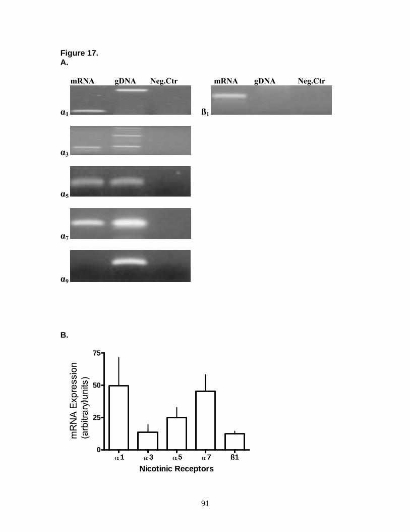
Figure 17. A. mRNA gDNA Neg.Ctr mRNA gDNA Neg.Ctr
α1 ß1
α3
α5
α7
α9 B.
α1 α3 α5 α7 ß10
25
50
75
mR
NA
Exp
ress
ion
(arb
itrar
y un
its)
Nicotinic Receptors
)
91

4. Discussion 4.1 Muscarinic Receptor Expression in Human Lung Fibroblasts Almost every cell type in the lung and airways expresses cholinoceptors, but
expression of muscarinic receptors in lung fibroblasts has not been characterized
yet. For this purpose semi-quantitative RT-PCR’s were performed using both the
MRC-5 as well as the primary human lung fibroblasts. Our results demonstrated
that muscarinic receptors were expressed in both MRC-5 and primary human
lung fibroblasts. In both, the cell line and primary cells the strongest mRNA
expression was observed for M2 receptor, whereas some gradual differences
were found with regard to the other muscarinic receptors. In MRC-5 cells the
levels of mRNA encoding M3 receptor appeared to be higher than in the primary
cells, which on the other hand seem to express a clear message for M1 receptor.
As mentioned before the genes of muscarinic receptors are intronless. Therefore,
the RNA preparations were treated with DNase prior to the RT reaction to
exclude any contamination with genomic DNA. The differential expression
pattern observed in the present study, in particular the lack of PCR products
accounting for M1 and M5 receptors in MRC-5 and primary lung fibroblasts,
respectively, argues against an artefact. Moreover, in MRC-5 cells the
expression of M2 and M3 receptors could be confirmed at the protein level by
Western blot analysis. Finally to confirm the regional expression pattern of mRNA
encoding for muscarinic receptors M2 and M3 semi-quantitative RT-PCRs were
performed using primary fibroblast cultures obtained from peripheral and distal
region of the human lung and our results showed that again in this case mRNA
encoding M2 receptor subtype was dominantly expressed followed by a slightly
lower expression of mRNA encoding for M3 receptor subtype.
4.2 Muscarinic Stimulation of Proliferation of Human Lung Fibroblasts Subsequent to our findings that human lung fibroblasts expressed muscarinic
receptors we investigated further to see if these receptors did play a part in
mediating proliferative response. For this purpose proliferation studies were
performed using a range of protocols to determine the optimal parameters which
92
then could be set as a standard for the remaining proliferation studies that would
ensue. The proliferation of lung fibroblasts in vitro largely depends on the
presence of FCS which obviously contains a cocktail of diverse factors promoting
fibroblast growth. Consequently, the effects of muscarinic agonists were first
tested in FCS-deprived conditions, i.e. in absence of other mitogenic stimuli.
Under these conditions carbachol and oxotremorine caused an increase in (3H)-
thymidine incorporation in MRC-5 cells and primary human lung fibroblasts. In
both cell types, the proliferative effects of carbachol or oxotremorine were
blocked by the muscarinic antagonist atropine and the long acting muscarinic
antagonist tiotropium at concentrations at least 10- and 100-fold lower than those
of the agonists, proving the involvement of specific muscarinic receptors. The
antagonists had no effect on their own, which excludes significant effects of non-
neuronal acetylcholine under the present culture conditions.
In MRC-5 cells the muscarinic stimulation of proliferation was studied in more
detail. The stimulatory effect of carbachol appeared to be somewhat more
pronounced when the drug was added after dissemination isochronal to FCS-
deprivation and when it was present already prior to the (3H)-thymidine exposure
compared to conditions where the agonist was applied at the same time with
(3H)-thymidine only for the last part of the incubation period. This may suggest
that the muscarinic mitogenic stimulus is slowly developing, but long lasting. A
mitogenic effect of muscarinic agonists has also been demonstrated on airway
smooth muscle (10, 11), but in contrast to the present observations on
fibroblasts, muscarinic agonists alone showed no proliferative effect in airway
smooth muscle, but augmented the effect of other mitogenic stimuli such as
platelet-derived growth factor (PDGF) or epidermal growth factor (EGF). The
present experiments showing that the proliferative effects of carbachol and
oxotremorine were basically the same in absence or presence of 1% FCS,
indicate that in human lung fibroblasts the proliferative effects induced by
muscarinic receptor activation may be additive to proliferative effects caused by
other mitogenic stimuli.
93
4.3 Inhibition of Muscarnic Proliferation using Receptor Sub-Type Specific Antagonists in MRC-5 Human Lung Fibroblasts and Effect of Pertussis Toxin on Muscarinic Stimulation of Proliferation of Human Lung Fibroblasts Our previous results indicate that human lung fibroblasts do express muscarinic
receptors and that they mediated proliferative effects. Both, M2 and/or M3
receptors have been shown to mediate activation of the mitogen-activated
protein kinase/extracellular signal–regulated kinase 1/2 pathway (188, 190, 191),
which is known to mediate proliferative responses in various cell types (192, 193,
194). In order to characterize the muscarinic receptor subtype mediating the
proliferative effect in MRC-5 fibroblasts the M2/M4 receptor preferring antagonists
AF-DX 384 , AQ-RA 741 and himbacine, the M3/M1 receptor preferring
antagonists 4-DAMP and p-FHHSiD and the M1/M4 receptor preferring antagonist
pirenzepine were tested in addition to tiotropium. It should be emphasized that
the subtype selectivity of the currently available “selective” muscarinic receptor
antagonists is rather limited. Usually they show a relatively high potency at least
for two of the five muscarinic receptors and the potency difference to the other
muscarinic receptors is only about one order of magnitude. Nonetheless,
comparing the potency of a series of subtype preferring antagonists is an
approach to deal with this problem (195). When the antagonist potencies as
observed in the present experiments were correlated to their reported affinity
constants on cloned human muscarinic receptors (71, 196), by far the best
correlation was obtained for the M2 receptor, supporting the assumption that this
muscarinic receptor subtype may be essential in mediating the proliferative
effects. This correlates well with the observation that at the mRNA level M2
receptors represented the major muscarinic receptor subtype in these cells. In
line with this conclusion is also the observation, that pertussis toxin abolished the
effect of carbachol, since pertussis toxin is known to inhibit Gi/o, G proteins to
which only M2 and M4 receptors couple (196). An unspecific antiproliferative
action of the PTX may be excluded, as PTX did not inhibit the proliferative effect
of PDGF, which is known to exert its effects via activation of a tyrosine kinase
94
receptor, a class of receptors different from G-protein-coupled receptors.
Therefore, the PTX-induced reduction of baseline proliferation may reflect a
background activation of Gi-mediated effects, possibly caused by paracrine
mediators (as, for example, chemokines) released by the fibroblasts themselves.
The long-acting muscarinic antagonist tiotropium was by far the most potent
antagonist. The affinity constants of tiotropium are similar for M1, M2 and M3
receptors, but the dissociation of the respective receptor complexes shows
different kinetics, with the longest half-life for the M3 receptor (196). This kinetic
subtype selectivity of tiotropium may not be of significance in the present
experiments, because they were performed under equilibrium conditions.
Whether the kinetical subtype selectivity of tiotropium is of clinical importance is
still debatable (197). Because the half-life of the tiotropium-M2 receptor complex,
although shorter than that of the M3 receptor complex, is still 3.6 hours (196), it
may be expected that tiotropium causes a significant blockade of M2 receptors in
the lung under clinical conditions.
Although detailed pharmacological experiments have only been performed on
MRC-5 cells, the observations that pertussis toxin abolished the proliferative
effect of carbachol also in primary human lung fibroblasts and that the M2
receptor mRNA represented the predominant muscarinic receptor mRNA in
these cells as well suggest that also in primary human lung fibroblasts M2
receptors might mediate the proliferative stimulus.
4.4 Muscarinic Receptor Stimulation and Activation of p-42/44 MAPK Pathway in Proliferation of Human Lung Fibroblast Our findings from the present study indicate that human lung fibroblasts do
indeed express muscarinic receptors and that they mediate proliferation in
pertussis toxin-sensitive manner. Mitogen-activated protein kinase (MAP kinase)
/ extracellular signal–regulated kinase 1/2 (ERK-1/2) is one of the classical
pathways mediating proliferative responses induced by various stimuli including
activation of G-protein coupled receptor (192, 193, 194, 198), and also
muscarinic receptors (both, M2 and M3) have been shown to be able to activate
95

this pathway (188, 190, 191). Therefore to probe if muscarinic receptor
stimulation in human lung fibroblasts leads to the activation of MAPK pathway
functional studies were performed using PD98059 a specific inhibitor for MAPK-
activating enzyme (MEK). Our results demonstrated that the carbachol induced
muscarinic proliferation in human lung fibroblasts was completely inhibited by
PD98059, moreover in Western blot analysis carried out with MRC-5 human lung
fibroblasts it was observed that the carbachol and oxotremorine induced
activation of p-42/44 MAPK was blocked by muscarinic receptor antagonists
tiotropium and atropine. These observations strongly support the conclusion that
muscarinic stimulation of proliferation in human lung fibroblasts is mediated via
the classical MEK-ERK MAPK cascade.
4.5 Intracellular Signaling Mechanism Involved in Muscarinic Stimulation Mediated Activation of MAPK Cascade in MRC-5 Human Lung Fibroblast Proliferation 4.5.1 Effects of the Rho-kinase Inhibitor Y27632 on Muscarinic Stimulation
of MRC-5 Human Lung Fibroblast Proliferation
Our previous results demonstrated that the muscarinic stimulation of proliferation
in human lung fibroblasts is mediated via the classical MEK-ERK MAPK cascade
in a pertussis toxin sensitive manner. Rho a member of the family of small G-
proteins has been shown to mediate cell cycle progression and proliferation via
the activation of the MAPK cascade. Rho kinase, a key Rho effector has been
implicated in the activation of MAPK pathway. In a previous study it was shown
that inhibition of Rho kinase blocked the Ras transformation in NIH3T3 cells
(144). Furthermore serotonin stimulation of bovine pulmonary artery VSMC lead
to the translocation of ERK to the nucleus, and this response was inhibited by
treatment with either dominant negative RhoA or the Rho kinase inhibitor Y27632
(149). Therefore to investigate the role of Rho in muscarinic stimulation mediated
proliferation in MRC-5 human lung fibroblasts we performed (3H)-thymidine
proliferation assay using the selective Rho kinase inhibitor Y27632. In both the
protocols that were tested be it the 6 hours 10% FCS period prior to addition of
96

drugs or the one with the FCS free conditions for the duration of the assay it was
observed that carbachol induced more than 2-fold increase in (3H)-thymidine
incorporation and the Rho kinase inhibitor Y27632 by itself inhibited the basal
proliferation level but failed to inhibit the carbachol induced (3H)-thymidine
incorporation. These results indicate that although Rho does mediate proliferative
signals in these cells, it is not involved in the muscarinic stimulation mediated
activation of MAPK in MRC-5 human lung fibroblast cells.
4.5.1.2 Effects of the Phospholipase C Inhibitor U73122 on Muscarinic
Stimulation of MRC-5 Human Lung Fibroblast Proliferation
The receptor mediated activation of PLC leads to the generation of two second
messengers, inositol1,4,5-triphosphate (IP3) and diacylglycerol (DAG). It has
been well established that IP3 can cause an increase in the concentration of
intracellular calcium as a result of intracellular storage mobilization and DAG can
activate the protein kinase C (PKC). The activation of the ERK1/2 pathway
involving PKC activation following the activation of PLC pathway with production
of DAG has been previously reported. Therefore we speculated a role for PLC
and its generated second messengers in linking the activation of muscarinic
acetylcholine receptors to downstream activation of MAPK in MRC-5 human lung
fibroblast cells. To investigate the role of PLC, in the present study we performed
(3H)-thymidine incorporation assays using U73122 a selective PLC inhibitor. Our
results demonstrate that although the PLC inhibitor U73122 inhibited the basal
level of (3H)-thymidine incorporation it failed to inhibit the carbachol induced
increase in the (3H)-thymidine incorporation. Various studies have reported that
PLC is the principal effector of Gq-mediated signaling. Activation of ERK1/2 by
the odd numbered muscarinic receptors has been shown to typically involve PKC
activation following Gq/11 activation of PLC pathway with production of DAG. On
the other hand M2 and M4 muscarinic receptors have been shown to produce
only weak activation of PLC pathway (40, 41, 42, 199). Our previous results have
indicated that the muscarinic stimulation mediated activation of MAPK in human
lung fibroblasts follows a pertussis toxin sensitive mechanism via the Gi/o protein
97

which correlates well with our finding that PLC inhibition did not inhibit the
carbachol induced (3H)-thyimidine incorporation in MRC-5 human lung fibroblast
cells. The decrease in the basal level of (3H)-thymidine incorporation in the
presence of the PLC inhibitor U73122 could be explained possibly by the fact
that U73122 could act on other sites in addition to PLC inhibition. Such
mechanisms include the inhibition of both the intracellular calcium channels,
which are activated by formation of IP3 by PLC, and plasma membrane bound
calcium channels (200). In the absence of carbachol stimulation, muscarinic
receptor coupled PLC is inactive and thus intracellular calcium channels
activated by IP3 are closed. On the other hand plasma membrane bound calcium
channels are independent of PLC activation and may play a role in transient
calcium currents. Therefore, U73122 may antagonise plasma membrane calcium
channels and as a result decrease the basal levels of (3H)-thymidine
incorporation.
4.5.1.3 Effects of the PI3-kinase Inhibitor Wortmannin on Muscarinic
Stimulation of MRC-5 Human Lung Fibroblast Proliferation
Phosphoinositide 3-kinases (PI3-kinases or PI3Ks) are a family of related
enzymes that are capable of phosphorylating the 3 position hydroxyl group of the
inositol ring of phosphatidylinositol. Previously, Lopez-IIasaca et al. (1998) have
demonstrated that the p42 MAPK activation by G-protein-coupled receptors can
be mediated by PI3K through a process that requires G-proteins βγ-subunits.
The wortmannin-sensitive PI3K isoenzyme, PI3Kγ, can be activated by both the
α and the βγ subunits of the heterotrimeric G-proteins (150). Several studies
have reported on the regulation of different phosphatidylinositol kinases and
phosphoinositide pools by muscarinic acetylcholine receptors. Hence to
investigate whether the PI-3 kinase was crucially involved in the activation of the
MAPK pathway, in the muscarinic stimulation of proliferation in MRC-5 human
lung fibroblasts we performed (3H)-thymidine incorporation assays using the PI3-
kinase inhibitor wortmannin. Our results demonstrate that although PI3K is
involved in mediating proliferative signals in these cells the carbachol induced
98

(3H)-thymidine incorporation is unaffected by the PI3K inhibitor wortmannin thus
suggesting that PI3K may not be implicated in the carbachol mediated process.
4.5.1.4 Effects of the Raf-1 Inhibitor GW5074 on Muscarinic Stimulation of
MRC-5 Human Lung Fibroblast Proliferation
The RAS/RAF/MEK/ERK signal transduction pathway is a conserved pathway
that regulates cell growth and signaling. Previous studies have shown the
involvement of Ras and Raf in the Gi-coupled acetylcholine muscarinic M2
receptor activation of MAPK, also Ras-independent activation of MAPK mediated
via the Raf-MEK-MAPK have been reported. Hence to elucidate the role of Raf in
the muscarinic stimulation of proliferation in MRC-5 human lung fibroblasts we
performed (3H)-thymidine incorporation using the Raf-1 inhibitor GW5074. Our
results demonstrated Raf-1 inhibitor GW5074 did not affect the basal proliferation
rate at 0.1 and 1 µM although at a 3 µM concentration it inhibited the (3H)-
thymidine incorporation to 40 ± 7% relative to the control. It was also observed
that the carbachol induced muscarinic stimulation of proliferation was significantly
inhibited by GW5074 at 1 and 3 µM as shown by the reduction in the (3H)-
thymidine incorporation to 151 ± 6% and 132 ± 8% in the presence of the drug
respectively. Furthermore Western blot analysis carried out to confirm the
inhibitory effect of the Raf-1 inhibitor on the activation of the MAPK cascade in
response to the muscarinic stimulation induced proliferation in the MRC-5
fibroblasts at the protein levels showed that while GW5074 by itself did not
appear to affect the MAPK activation, the carbachol induced increase in MAPK
activity was inhibited in the presence of the Raf-1 inhibitor. These findings show
that Raf-1 is indeed involved in the muscarinic stimulation mediated activation of
MAPK cascade in MRC-5 human lung fibroblast cells and is a signal upstream of
the MEK-ERK-MAPK cascade. Raf proteins are the conserved signaling module
that transducer signals from the cell surface to the receptor wherein the Ras
family of small G-proteins have been shown to be the upstream regulators in
several cell types. One of the critical steps in the Ras-Raf-MEK cascade is the
post translational lipid modification of Ras which aids the Ras protein to attach to
99

the cell membrane; this process involves the enzyme farnesyl transferase. Hence
we performed (3H)-thymidine incorporation assays using farnesly transferase
inhibitors, however the inhibitor of Ras farnesyl transferase FTI-277 (up to 3 µM)
did not affect carbachol-stimulated (3H)-thymidine incorporation, whereas
manumycin, a chemically different Ras inhibitor, exerted large cytotoxic effects.
Therefore making it impossible to draw any conclusions about the role of Ras in
the muscarinic stimulation mediated activation of MAPK in MRC-5 human lung
fibroblast cells.
4.5.1.5 Effects of the Tyrosine-Kinase Inhibitor Tyrphostin AG1295 on
Muscarinic Stimulation of MRC-5 Human Lung Fibroblasts
Receptor transactivation refers to the ability of a primary agonist, via binding to
its receptor, to activate a receptor for another ligand via signaling events.
Transactivation of various growth factor receptors, including the epidermal
growth factor receptor (EGFR), and the platelet derived growth factor (PDGF), by
G-protein-coupled receptors has been documented in multiple cellular model
systems (163, 164, 165, 166, 167). Studies have shown that GPCRs may utilize
receptor tyrosine kinases (RTKs) such as platelet derived growth factor receptor
(PDGF) or epidermail dervived growth factor receptor (EGFRs) as intermediate
signaling proteins thereby mimicking the signaling pathways downstream leading
to activation of MAPK cascade. To explore the possibility of whether muscarinic
stimulation of MRC-5 human lung fibroblast lead to the transactivation of PDGF
receptor, (3H)-thymidine incorporation assays were performed using tryphostin
AG1295 a specific tyrosine kinase inhibitor for the PDGF receptors. Our findings
show that although tryphostin AG1295 inhibited the basal level of (3H)-thymidine
incorporation it did not inhibit the carbachol induced (3H)-thymidine incorporation
excluding transactivation of receptor tyrosine kinase as a signaling pathway
mediated via the carbachol stimulated G-protein coupled stimulation of MRC-5
human lung fibroblast cells. Studies performed by other groups in our laboratory
using human lung fibroblasts showed that PDGF (10 ng/ml) induced almost a
one fold increase in the (3H)-thymidine incorporation confirming its strong
100

stimulatory effect on proliferation and this effect was potently inhibited by the
tyrosine kinase inhibitor tyrphostin AG1295 (5 µM). In a further set of control
experiments it was observed that the PDGF induced increase in (3H)-thymidine
incorporation in MRC-5 human lung fibroblast was unimpeded in the presence of
tiotropium confirming its selectivity in inhibition of carbachol induced muscarinic
proliferation of MRC-5 human lung fibroblast.
4.6 Muscarinic Stimulation of Proliferation of MRC-5 Human Lung Fibroblast and Anti-apoptotic Effects Apoptosis represents a multi-step process for elimination and subsequent
removal of cells in response to environment insults or as part of organismal
development. Pro-apoptitic singaling may be mediated by the specific ligands
and surface receptors or can also be activated from inside the cell through
specific cell sensors residing in the cell nucleus and cytoplasm. The caspases
are a family of proteins that are one of the main executors of the apoptotic
process. They belong to a group of enzymes known as cysteine proteases which
can be activated following the occupation of certain cell surface ‘death’ receptors
(e.g. tumor necrosis factor) or by agents that induce stress or DNA damage (e.g.
actinomycin D, cycloheximide). A member of this family, caspase-3 has been
identified as being a key mediator of apoptosis of mammalian cells. The
repertoire of signaling pathways controlled by GPCRs has recently been
extended by studies linking GPCRs to the regulation of apoptosis. Depending on
the receptor subtypes and the cell type in which the receptor is expressed,
GPCRs can either induce apoptosis or protect cells from apoptotic stimuli.
Prominent among the GPCRs that protect cells from apoptotic stimuli are the
subtypes of the muscarinic receptor family (186, 187). Previous studies have also
shown that muscarinic receptors that couple to the Gq/11 protein (e.g. M1, M3 and
M5) can protect cells from apoptosis following DNA damage whereas those
coupled to the Gi-proteins (M2 and M4) have no protective properties (186). The
previous experiments that were performed clearly indicated the functional role of
the muscarinic receptors in the carbachol induced proliferation of the MRC-5
101

human lung fibroblasts cells. Hence we explored the possibility whether the
proliferative stimuli mediated via the muscarinic receptors in the MRC-5
fibroblasts were augmented by an anti-apoptotic effect concomitantly. Our
studies performed by means of the caspase-3 activity as a marker for the level of
apoptotic signaling using a combination of apoptosis inducers in the presence or
absence of muscarinic stimuli in MRC-5 human lung fibroblasts suggests that the
muscarinic mediation of proliferation in these cells is unaided by anti-proliferative
effects mediated via the muscarinic stimulation. These findings appear to
correlate well with our previous findings that in MRC-5 human lung fibroblasts the
muscarinic stimulation mediated proliferation is via the M2 muscarinic receptor
subtype and also literature which has attributed the anti-proliferative effects
mediated via muscarinic receptors to the odd numbered muscarinic subtypes.
However because of the considerably large variation in the magnitude of effects
over a range of experiments it makes it difficult to positively conclude the
relationship between the muscarinic stimuli mediated proliferation and its anti-
apoptotic effects in MRC-5 human lung fibroblasts. We speculate that a study
done on the regulation of pro/anti-apoptotic genes during muscarinc stimulation
mediated proliferation in MRC-5 human lung fibroblasts might probably helps
shed more light on this interaction.
4.7 Nicotinic Receptor Expression and Function in MRC-5 Human Lung Fibroblasts The α-7 nicotinic receptor subunit has been detected in many non-neuronal cells
such as the bronchial epithelial cells, endothelial airway cells and airway
fibroblasts (16). In vitro studies carried out on bronchial epithelial cells and
pulmonary neuroendocrine cells indicated nicotinic receptor mediated activation
of cell proliferation cascade (31, 32). Furthermore studies carried out on WI38
cells, a human embryonic pulmonary fibroblast cell line showed an expression of
α-3 and α-7 nicotinic receptor subunits. Hence we performed semi-quantitative
RT-PCRs to explore the expression pattern of nicotinic receptor subunits in
MRC-5 human lung fibroblasts. Our results demonstrated the expression of α1,
102

α3, α5, α7 and β1 nicotinic receptor subunits in MRC-5 human lung fibroblasts with
the most dominant expression of α1 subunit followed by α7 subunit. Studies
conducted previously have implicated an increased expression of α7 subunit of nicotinic receptors with lung fibroblast proliferation. To explore the possibility of a
cross talk between the muscarinic and nicotinic receptors expressed in the MRC-
5 fibroblast cells we performed (3H)-thymidine incorporation assays using
nicotinic agonist and antagonist in the presence or absence of carbachol. Our
results demonstrated that nicotine did not appear to either affect proliferation by
itself nor in the presence of carbachol although there was some indication that
the muscarinic agonist carbachol induced increase in (3H)-thymidine
incorporation was supplemented in the presence of hexamethonium a nicotinic
antagonist, but the extent of the effect showed considerable variation over a
range of experiments making it impossible to be considered as a true effect.
103

5. Conclusion Fibrotic alteration and airway remodeling are pathological features associated
with asthma and COPD and almost every cell type in the airways expresses
cholinoceptors. However, only fragmentary knowledge exists with regards to the
expression and function of muscarinic receptors in pulmonary fibroblasts. To
characterize the expression pattern of muscarinic receptors we performed semi-
quantitative RT-PCR studies which showed that human lung fibroblasts indeed
expressed muscarinic receptors with the most dominantly expressed subtype
being the M2 followed by the M3 muscarinic receptor. Since confirming the
expression of muscarinic receptors in human lung fibroblasts we explored its
functional role in mediating proliferation using muscarinic agonists such as
carbachol and oxotremorine and it was observed that the muscarinic stimulation
indeed mediated proliferation in human lung fibroblast cells which was
completely blocked in the presence muscarinic antagonist such as atropine or
tiotropium. To further explore the muscarinic receptor subtype involved in
mediating this proliferative response, receptor subtype characterization studies
were performed using various muscarinic receptor subtype preferring
antagonists. The pharmacological profile obtained by comparing the potency of
several subtype preferring antagonists to inhibit the proliferative effect of
carbachol with reported affinity constants of these antagonists at cloned
muscarinic receptors indicated that muscarinic stimulation of proliferation of
human lung fibroblasts was mediated via M2 receptors. In line with this
conclusion was the observation that pertussis toxin, known to inactivate Gi/o
proteins prevented the stimulatory effect of carbachol, both in MRC-5 cells and
primary human lung fibroblasts. Mitogen-activated protein kinase (MAP kinase) /
extracellular signal–regulated kinase 1/2 (ERK-1/2) is one of the classical
pathways mediating proliferative responses induced by various stimuli including
activation of G-protein coupled receptor and muscarinic receptors (both, M2 and
M3) have been shown to be able to activate this pathway so we looked into this
possibility. In our present study we demonstrated that in pulmonary human lung
fibroblasts activation of muscarinic receptors indeed caused an increase in the
104

phosphorylated, i.e. activated form of the ERK-MAP kinase which was blocked
by PD 98059 a specific inhibitor of the MAPK-activating enzymes (MEK) These
observations strongly support the conclusion that muscarinic stimulation of
proliferation in human lung fibroblasts is mediated via the classical MEK-ERK
MAPK cascade. To illuminate further the signalling pathways, linking the
muscarinic receptor stimulation and activation of MAPK cascade we explored
several putative pathways that might be involved in this process and
demonstrated the role of Raf-1 using GW5074 a specific Raf-1 inhibitor in
activation of MAPK pathway in response to muscarinic stimulation in MRC-5
fiobroblasts at a functional and protein level. Recently studies were carried out in
our laboratory on pulmonary fibroblasts investigating whether muscarinic
mechanisms were involved in collagen synthesis in turn contributing to the airway
remodelling process. These studies confirmed that muscarinic stimulation indeed
lead to increased collagen synthesis and activation of the MAPK cascade in a
pertussiss toxin sensitive manner thus implicating a pivotal role for the M2
muscarinic receptors in this process (201).
In conclusion, human lung fibroblasts express muscarinic receptors, which
obviously mediate proliferative stimulus via a Gi/o-Raf-1-MEK-MAPK/ERK
pathway, an effect which might contribute to structural changes known to occur in
chronic obstructive airway diseases. Prolonged blockade of these receptors may
contribute to the long term beneficial effects of anticholinergics, as observed for
example, for the long-acting muscarinic antagonist, tiotropium, in COPD.
105

6. References 1. Saetta M, Finkelstein R, Cosio MG. Morpholigical and cellular basis for
airflow limitation in smokers. Eur Respir J 1994; 7: 1505-1515.
2. Saetta M, Turato G. Airway pathology in asthma. Eur Respir J 2001; 18:
Suppl. 34, 18s–23s.
3. Buist AS. Similarities and differences between asthma and chronic
obstructive pulmonary disease: treatment and early outcomes. Eur Respir J
2003; 21: Suppl. 39, 30s–35s.
4. Jeffery PK. Remodeling in asthma and chronic obstructive lung disease. Am
J Respir Crit Care Med 2001; 164: 28s–38s.
5. Barnes PJ. The role of anticholinergics in chronic obstructive pulmonary
disease. Am J Med 2004; 20(117, Suppl. 12A): 24s–32s.
6. Kai-Michael Beeh, Tobais Welte, Roland Buhl. Anticholinergics in the
treatment of chronic obstructive pulmonary disease. Respiration 2002; 69:
372–379.
7. Vincken W, van Noord JA, Greefhorst AP, Bantje TA, Kesten S, Korducki L,
Cornelissen PJ. Improved health outcomes in patients with COPD during 1
yr’s treatment with tiotropium. Eur Respir J 2002; 19: 209–216.
8. Anzuetto A, Tashkin D, Menjoge S, Kesten S. One-year analysis of
longitudinal changes in spirometry in patients with COPD receiving
tiotropium. Pulm Pharmacol Ther 2005; 18: 75–81.
9. Keam SJ, Keating GM. Tiotropium bromide. A review of its use as
maintenance therapy in patients with COPD. Treat Respir Med 2004; 3(4):
247–68.
10. Krymskaya VP, Orsini MJ, Eszterhas AJ, Brodbeck KC, Benovic JL,
Panettieri RA, Penn RB. Mechanisms of proliferation synergy by receptor
tyrosine kinase and G protein-coupled receptor activation in human airway
smooth muscle. Am J Respir Cell Mol Biol 2000; 23: 546–554.
11. Gosens R, Nelemans SA, Grootee Bromhaar MM, McKay S, Zaagsma J,
Meurs H. Muscarinic M3-receptors mediate cholinergic synergism of
106

mitogenesis in airway smooth muscle. Am J Respir Cell Mol Biol 2003; 28:
257–262.
12. Gosens R, Bos IS, Zaagsma J, Meurs H. Protective effects of tiotropium
bromide in the progression of airway smooth muscle remodeling. Am J
Respir Crit Care Med 2005; 171: 1096–1102.
13. Racké K, Matthiesen S. The airway cholinergic system: physiology and
pharmacology. Pulm Pharmacol Ther 2004; 17: 181–198.
14. Racké K, Juergens UR, Matthiesen S. Control by cholinergic mechanisms.
Eur J Pharmacol 2006; 533: 57–68.
15. Wessler I, Kirkpatrick CJ, Racké K. Non-neuronal acetylcholine, a locally
acting molecule, widespreadly distributed in biological systems: expression
and function in man. Pharmacol Ther 1998; 77: 59–79.
16. Carlisle DL, Hopkins TM, Gaither-Davis A, Silhanek MJ, Luketich JD,
Christie NA, Siegfried JM. Nicotine signals through muscle-type and
neuronal nicotinic acetylcholine receptors in both human bronchial epithelial
cells and airway fibroblasts. Respir Res 2004; 5:27.
17. Sekhon HS, Keller JA, Proskocil BJ, Martin EL, Spindel ER. Maternal
nicotine exposure upregulates collagen gene expression in fetal moneky
lung: association with α7 nicotinic acetylcholine receptors. Am J Respir Cell
Mol Biol 2002; 26:31–41.
18. Roman J, Ritzenthaler JD, Gil-Acosta A, Rivera HN, Roser-Page S. Nicotine
and fibronectin expression in lung fibroblasts: implications for tobacco-
related lung tissue remodeling. FASEB J 2004; 18:1436–1438.
19. Catherine R, Gwilt, Louise E. Donnelly, Duncan F. Rogers. The non-
neruonal cholinergic system in the airways: An unappreciated regulatory
role in pulmonary inflammation? Pharmacol Ther (2007), doi:
10.1016/j.pharmthera.2007.05.007.
20. Wessler I, Kirkpatrick CJ, Racké K. The cholinergic ‘pitfall’: acetylcholine, a
universal cell molecule in biological systems, including humans. Clin Exp
Pharmacol Physiol 1999; 26: 19196–19199.
107

21. Wessler I, Kilbinger H, Bittinger F, Kirkpatrick CJ. The biological role of non-
neuronal acetylcholine in plants and humans. Japanese Journal of
Pharmacology 2001a; 85, 2–10.
22. Wessler I, Panter H, Bittinger F, Kriegsmann J, Kirkpatrick CJ, Kilbinger H.
Subcellular location of choline acetyltransferase (ChAT) and acetylcholine
(Ach) in human placenta. Naunyn-Schmiedeberg’s Archives of
Pharmacology 2001b; 363, R23.
23. Kawashima K, Fujii T. The lymphocytic cholinergic system and its
contribution to the regulation of immune activity. Life Sciences 2003; 74,
675–696.
24. Grando SA, Pittelkow MR, Schallreuter KU. Adrenergic and cholinergic
control in the biology of epidermis: physiology and clinical significance.
Journal of Investigative Dermatology 2006; 126 (9), 1948–1965.
25. Grando SA. Biological functions of keratinocyte cholinergic receptors.
Journal of Invetsigative Dermatology Symposium Proceeding 1997; 2, 41–
48.
26. Wessler IK, Kirkpatrick CJ. The non-neuronal cholinergic system: an
emerging drug target in the airways. Pulmonary, Pharmacology and
Therapeutics 2001; 14, 423–434.
27. Kawashima K, Fujii T. Extraneuronal cholinergic sytem in lymphocytes.
Pharmacology and Therapeutics 2000; 86, 29–48.
28. Wessler I et al. Dysfunction of the non-neuronal cholinergic system in the
airways and blood cells of patients with cystic fibrosis. Life Sciences 2007;
doi: 10.1016/j.lfs.2007.01.042.
29. Conti-Tronconi BM, McLane KE, Raftery MA, Grando SA, Protti MP. The
nicotinic acetylcholine receptor: structure and autoimmune pathology. Crit
Rev Biochem Mol Biol 1994; 29:69–123.
30. Lukas RJ, Changeux JP, Le Novere N, Albuquerque EX, Balfour DJ, Berg
DK, Bertand D, Chiappinelli VA, Clarke PB, Collins AC, Dani JA, Grady SR,
Kellar KJ, Lindstorm JM, Marks MJ, Quik M, Taylor PW, Wonnacott S.
International Union of Pharmacology. XX. Current status of nomenclature
108
for nicotinic acetylcholine receptors and their subunits. Pharmacol Rev
1999; 51:397–401.
31. West KA, Brognard J, Clark AS, Linnoila IR, Yang X, Swain SM, Harris C,
Belinsky S, Dennis PA. Rapid Akt activation by nicotine and a tobacco
carcinogen modulates the phenotype of normal human airway epithelial
cells. J Clin Invest 2003; 111: 81–90.
32. Jull 3rd BA, Plummer HK, Schuller HM. Nicotinic receptor mediated
activation by the tobacco-specific nitrosamine NNK of a Raf-1/MAP kinase
pathway, resulting in phosphorylation of c-myc in human small cell lung
carcinoma cells and pulmonary neuroendocrine cells. J Cancer Res Clin
Oncol 2001; 127: 707–17.
33. Conti-Fine BM, Navaneetham D, Lei S, Maus AD. Neuronal nicotinic
receptors in non-neuronal cells: new mediators of tobacco toxicity? Eur J
Pharmacol 2000; 393: 279–94.
34. Zia S, Ndoye A, Nguyen VT, Grando SA. Nicotine enhances expression of
α3, α4, α5, and α7 nicotinic receptors modulating calcium metabolism and
regulating adhesion and motility of respiratory epithelial cells. Res Commun
Mol Pathol Pharmacol 1997; 97: 243–62.
35. Wang Y, Pereira EF, Maus AD, Ostlie NS, Navaneetham D, Lei S,
Albuquerque EX, Conti-Fine BM. Human bronchial epithelial and endothelial
cells express α7 nicotinic acetylcholine receptors. Mol Pharmacol 2001; 60:
1201–9.
36. Rehan VK, Wang Y, Sugano S, Romero S, Chen X, Santos J, Khazanchi A,
Torday JS. Mechanism of nicotine-induced pulmonary fibroblast
transdiffrentiation. Am J Physiol Lung Cell Mol Physiol 2005; 289: L667–
L676.
37. Bonner TI. The molecular basis of muscarinic receptor diversity. Trends
Neurosci 1989; 12: 148–51.
38. Hulme EC, Birdsall NJ, Buckley NJ. Muscarinic receptor subtypes. Ann Rev
Pharmacol Toxicol 1990; 30: 633–73.
109

39. Caulfield MP, Birdsall NJM. International Union of Pharmacology. XVII.
Classification of muscarinic acetylcholine receptors. Pharmacol Rev 1998;
50: 279–90.
40. Ashkenazi A, Peralta EG, Winslow JW, Ramachandran J, Capon DJ.
Functionally distinct G proteins selectively couple different receptors to PI
hydrolysis in the same cell. Cell 1989; Vol 56, 487–493.
41. Tietje KM, Goldman PS, Nathanson NM. Cloning and functional analysis of
a gene encoding a novel muscarinic acetylcholine receptor expressed in
chick heart and brain. J Biol Chem 1990; Vol 265. Issue 5, 2828–2834.
42. Tietje KM, Nathanson NM. Embryonic chick heart expresses multiple
acetylcholine receptor subtypes. Isolation and characterization of a gene
encoding a novel m2 muscarinic acetylcholine receptor with high affinity for
pirenzepine. J Biol Chem 1991; Vol 266, Issue 26, 17382–17387.
43. Katz A, Wu D, Simon MI. Subunits βγ of heterotrimeric G protein activate β2
isoform of phospholipase C. Nature 1992; 360: 686–689.
44. Felder CC. Muscarinic acetylcholine receptors: signal transduction through
multiple effectors. FASEB J 1995; 9: 619–25.
45. Kubo T, Fukuda K, Mikami A, Maeda A, Takahashi H, Mishina M, Haga T,
Haga K, Ichiyama A, Kangawa K, Matsuo H, Hirose T, Numa S. Cloning,
sequencing and expression of complementary DNA encoding the
muscarinic acetylcholine receptor. Nature 1986a; 323: 411–416.
46. Kubo T, Maeda A, Sugimoto K, Akiba I, Mikami A, Takahashi H, Haga T,
Haga K, Ichiyama A, Kangawa K, Matsuo H, Hirose T, Numa S. Primary
structure of procine primary muscarinic acetylcholine receptor deduced from
the cDNA sequence. FEBS Lett 1986b; 209: 367–372.
47. Bonner TI, Buckley NJ, Young AC, Brann MR. Identification of a family of
muscarinic acetylcholine receptor genes. Science 1987; 237: 527–532.
48. Bonner TI, Young AC, Brann MR, Buckley NJ. Cloning and expression of
the human and rat m5 muscarinicacetylcholine receptor genes. Neuron
1988; 1: 403 – 410.
110

49. Peralta EG, Ashkenazi A, Winslow JW, Smith DH, Ramachandran J, Capon
DJ. Distinct primary structures, ligand binding properties and tissue-specific
expression of four human muscarinic acetylcholine receptors. EMBO J
1987; 6: 3923–3929.
50. Hall JM, Caulfield MP, Watson SP, Guard S. Receptor subtypes or species
homologues: Relevance to drug discovery. Trends Pharmacol Sci 1993; 14:
376–383.
51. Eglen RM, Hedge SS, Watson N. Muscarinic receptor subtypes and smooth
muscle function. Pharmacol Rev 1996; 48: 531–565.
52. Curtis CA, Wheatly M, Bansal S, Birdsall NJM, Eveleigh P, Pedder EK,
Poyner D, Hume EC. Propylbenzilylcholine mustard labels an acidic residue
in transmembrane helix 3 of the muscarinic receptor. J Biol Chem 1989;
264: 489–495.
53. Spalding TA, Birdsall NJM, Curtis CAM, Hulme EC. Acetylcholine mustard
labels the binding site aspartate in muscarinic acetylcholine receptors. J Biol
Chem 1994; 269: 4092–4097.
54. Baldwin JM, Schertler GF, Unger VM. An alpha-carbon template for the
transmembrane helices in the rhodopsin family of G-protein-coupled
receptors. J Mol Biol 1997; 272: 144–164.
55. Kurtenbach E, Curtis CA, Pedder EK, Aitken A, Harris AC, Hulme EC.
Muscarinic acetylcholine receptors: Peptide sequencing identifies residues
involved in antagonist binding and disulphide bond formation. J Biol Chem
1990; 265: 13702–13708.
56. Savarese TM, Wang CD, Fraser CM. Site-directed mutagenesis of the rat
m1 muscarinic acetylcholine receptor: Role of conserved cysteines in
receptor function. J Biol Chem 1992; 267: 11439–11448.
57. Zhu SZ, Wang SZ, Hu J, El-Fakahany EE. An arginine residue conserved in
most G-protein-coupled receptors is essential for the function of the m1
muscarinic receptor. Mol Pharmacol 1994; 45: 517–525.
111

58. Jones PG, Curtis CAM, Hulme EC. The function of a highly-conserved
arginine residue in activation of the muscarinic M1 receptor. Eur J
Pharmacol 1995; 288: 251 – 257.
59. Lu ZL, Curtis CA, Jones PG, Pavia J, Hulme EC. The role of aspartate-
arginine-tyrosine triad in the m1 muscarinic receptor: Mutations of aspartate
122 and tyrosine 124 decreases receptor expression but do not abolish
signaling. Mol Pharmacol 1997; 51: 234–241.
60. Haga T, Haga K, Kameyama K, Nakata H. Phosphorylation of muscarinic
receptors: Regulation of G-proteins. Life Sci 1993; 52: 421–428.
61. Wess J. Molecular biology of muscarinic acetylcholine receptors. Crit Rev
Neurobiol 1996; 10: 69–99.
62. Wess J, Liu J, Blin N, Yun J, Lerche C, Kostenis E. Structural basis of
receptor/G-protein coupling selectivity studied with muscarinic receptors as
model systems. Life Sci 1997; 60: 1007–1014.
63. Levey AI. Immunological localization of m1-m5 muscarinic receptor subtypes
in the peripherial tissues and brain. Life Sci 1993; 52: 441–448.
64. Grimm U, Moser U, Mutschler E, Lambrecht G. Muscarinic receptors: Focus
on presynaptic mechanisms and recently developed novel agonist and
anatgonists. Pharmazie 1994b; 49: 711–726.
65. Michel AD, Stefanich E, Whiting RL. Direct labeling of rat M3 muscarinic
receptors by (3H)4DAMP. Eur J Pharmacol 1989; 166: 459–466.
66. Lazareno S, Buckley NJ, Roberts FF. Characterization of muscarinic M4
binding sites in rabbit lung, chicken heart and NG108-15 cells. Mol
Pharmacol 1990; 53: 573–589. 38: 805–815.
67. Caulfield MP. Muscarinic receptors: Characterization, coupling and function.
Pharmacol Ther 1993; 58: 319–379.
68. Kondou H, Inagaki N, Fukui H. Formation of inositol phosphates mediated
by M3 muscarinic receptors in type-1 and type-2 astrocytes from neonatal
rat cerebral cortex. Neurosci Lett 1994; 180: 131–134.
112

69. Lambrecht G, Feifel R, Forth B, Strohmann C, Tacke R, Mutschler E. p-
Fluoro-hexahydro-sila-difenidol: The first M2β- selective muscarinic
antagonist. Eur J Pharmacol 1988a; 152: 193 – 194.
70. Lambrecht G, Feifel R, Wagner-Röder M, Strohmann C, Zilch H, Tacke R,
Waelbroeck M, Christophe J, Boddeke H, Mutschler E. Affinity profiles of
hexahydro-sila-difenidol analogues at muscarinic receptor subtypes. Eur J
Pharmacol 1989b; 168: 71–80.
71. Dörje F, Wess J, Lambrecht G, Tacke R, Mutschler E, Brann MR.
Antagonist binding profiles of five cloned human muscarinic receptor
subtypes. The Journal of Pharmacology and Experimental Therapeutics
1991; 256: 727–733.
72. Simon MI, Strathmann MP, Gautam N. Diversity of G proteins in signal
transduction. Science 1991; 252: 802 – 8.
73. Gudermann T, Kalkbrenner F, Schultz G. Diversity and selectivity of
receptor-G protein interaction. Annu Rev Pharmacol Toxicol 1996; 36: 429 –
59.
74. Clapham DE, Neer EJ. G protein βγ subunits. Annu Rev Pharmacol Toxicol
1997; 37: 167 – 203.
75. Wu G, Benovic JL, Hildebrandt JD, Lanier SM. Receptor docking sites for G
protein βγ subunits. Implications for signal regulation. J Biol Chem 1998;
273: 7197 – 200.
76. Wu G, Bogatkevich GS, Mukhin YV, Benovic JL, Hildebrandt JD, Lanier SM.
Identification of G βγ binding sites in the third intracellular loop of the M3-
muscarinic receptor and their role in receptor regulation. J Biol Chem 2000;
275: 9026 – 25.
77. Buckley NJ. Molecular pharmacology of cloned muscarinic receptors. In: SR
Nahorski, ed Transmembrane Signalling, Intracellular Messengers and
Implications for Drug Development. Wiley and Sons 1990; 11 – 30.
78. Ashkenazi A, Ramachandran J, Capon DJ. Acetylcholine analogue
stimulates DNA synthesis in brain-derived cells via specific muscarinic
subtypes. Nature 1989; 340: 146 – 150.
113

79. Hulme EC, Birdsall NJM, Buckley NJ. Muscarinic receptor subtypes. Annu
Rev Pharmacol Toxicol 1990; 30: 633 – 673.
80. Baumgold J. Muscarinic receptor mediated stimulation of adenylyl cyclase.
Trends Pharmacol Sci 1992; 13: 339 – 340.
81. Parker EM, Kameyama K, Higashijima T, Ross EM. Reconstitutively active
G protein-coupled receptors purified from baculovirus-infected insect cells. J
Biol Chem 1991; 266(1): 519 – 27.
82. Stein R, Pinkas-Kramarski R, Sokolovsky M. Cloned M1 muscarinic
receptors mediates both adenylate cyclase inhibition and phosphoinositide
turnover. EMBO J 1988; 7: 3031 – 3035.
83. Baumgold J, Fishman PH. Muscarinic receptor-mediated increase in cAMP
levels in SK-N-SH human neuroblastoma cells. Biochem Biophys Ress
Commun 1988; 154: 1137 – 1143.
84. Olianas MC, Onali P. Properties of muscarinic –stimulated adenylate
cyclase in rat olfactory bulb. J Neurochem 1992; 58: 1723 – 1729.
85. Watson EL, Singh JC, McPhee C, Beavo J, Jacobson KL. Regulation of
cAMP metabolism in mouse parotid gland by cGMP and calcium. Mol
Pharmacol 1990; 38(4): 547 – 553.
86. Ashkenazi A, Peralta EG, Winslow JW, Ramachandran J, Capon DJ.
Functionally distinct G proteins selectively couple different receptors to PI
hydrolysis in the same cell. Cell 1989; 56: 487 – 493.
87. Pieroni JP, Jacobowitz O, Chen J, Iyengar R. Signal recognition and
integration by Gs stimulated adenylyl cyclases. Curr Opin Neurobiol 1993;
3: 345 – 351.
88. Jansonn CC, Kukonnen J, Akerman KE. Muscarinic receptor-linked
elevation of cAMP in SH-SY5Y neuroblastoma cells is mediated by Ca2+
and protein kinase C. Biochem Biophys Acta 1991; 1095 (3): 255 – 260.
89. Baumgold J. Muscarinic receptor-mediated stimulation of adenylyl cyclase.
Trends Pharmacol Sci 1992; 13: 339 – 340.
90. Meeker RB, Harden TK. Muscarinic cholinergic receptor-mediated activation
of phosphodiesterase. Mol Pharmacol 1982; 22: 310 – 319.
114

91. Rhee SG, Choi KD. Regulation of inositol phospholipid-specific
phospholipase C isozymes. J Biol Chem 1992; 267: 12393 – 12396.
92. Lambert DG, Burford NT, Nahorski SR. Muscarinic receptor subtypes:
inositol phosphates and intracellular calcium. Biochem Soc Trans 1992; 20:
130 – 135.
93. Berstein G, Blank JL, Smrcka A, Higashijima T, Sternweis PC, Exton JH,
Ross BJ, De Jong P, Reed RR, Simon MI. G protein α subunit multi gene
family. Nat Genet 1992; 1: 85 – 91.
94. Sawaki K, Hiramatsu Y, Baum BJ, Ambudkar IS. Involvement of Gαq/11 in
m3-muscarinic receptor stimulation of phosphotidylinositol 4,5 bisphosphate-
specific phospholipase C in rat parotid gland membranes. Arch Biochem
Biophys 1993; 305: 546 – 550.
95. Hildebrandt JP, Shuttleworth TJ. A Gq-type G protein couples muscarinic
receptors to inositol phosphate and calcium signaling in exocrine cells from
avian salt glands. J Membr Biol 1993; 133: 183 – 190.
96. Gusovsky F, Lueders JE, Kohn EC, Felder CC. Muscarinic receptor-
mediated tyrosine phosphorylation of phospholipase C-gamma. J Biol Chem
1993; 268: 7768 – 7772.
97. Dell’Acqua ML, Carroll RC, Peralta EG. Transfected m2 muscarinic
acetylcholine receptors couple to G alpha i2 and G alpha i3 in Chinese
hamster ovary cells. Activation and desensitization of the phospholipase C
signaling pathway. J Biol Chem 1993; 268: 5676 – 5685.
98. Katz A, Wu D, Simon MI. Subunits beta gamma of heterotrimeric G protein
activate beta isoform of phospholipase C. Nature 1992; 360: 686 – 689.
99. Cohen RI, Almazan G. Rat oligodendrocytes express muscarinic receptors
coupled to phsophoinositide hydrolysis and adenylyl cyclase. Eur J Neurosci
1994; 6: 1213 – 1224.
100. Moller R, Parker PJ. The extended protein kinase C superfamily. Biochem J
1998; 332: 281 – 292.
101. Stephens EV, Kalinec G, Brann MR, Gutkind JS. Transforming G protein
coupled receptors transducer potent mitogenic signals in NIH 3T3 cells
115

102. Hallberg B, Rayter SI, Downward J. Iteraction of Ras and Raf in intact
mammalian cells upon extracellular stimulation. J Biol Chem 1994; 269:
3913 – 3916.
103. Volmat V, Camps M, Arkinstall S, Pouyssegur J, Leonormand P. The
nucleus, the site for signal termination by sequestration and inactivation of
p42/p44 MAP kinases. J Cell Sci 2001; 114: 3433 – 3443.
104. Shaeffer HJ, Weber MJ. Mitogen-activated protein kinases: specific
messages for ubiquitous messengers. Mol Cell Biol 1999; 19: 2435 – 2444.
105. Nakayama K. Cip/Kip cyclin-dependent kinase inhibitors: brakes of the cell
cycle engine during development. Bioessays 1998; 20: 1020 – 1029.
106. Lavoie JN, L’allemain G, Brunet A, Müller R, Pouyssegur J. Cyclin D1
expression is regulated positively by p42/p44 MAPK and negatively by the
p38/HOGMAPK pathway. J Biol Chem 1996; 271: 20608 – 20616.
107. Lavoie JN, Rivard N, L’allemain G, Pouyssegur J. A temporal and
biochemical link between growth factor-activated MAP-kinases, cyclin D1
induction and cell cycle entry. Prog Cell Cycle Res 1996; 2: 49 – 58.
108. Whitmarsh AJ, Davis RJ. A central control for cell growth. Nature 2000; 403:
255 – 256.
109. Meloche S, Seuwen K, Pages G, Pouyssegur J. Biphasic and synergistic
activation of p44 MAPK (ERK1) by growth factors: correlation between late
phase activation. Mol Endocrinol 1992; 6: 845 – 854.
110. Kahan C, Seuwen K, Meloche S, Pouyssegur J. Coordinate biphasic
activation of p44 mitogen activated protein kinase and S6 kinase by growth
factors in hamster fibroblasts. J Biol Chem 1992; 267: 13369 – 13375.
111. Gutkind JS, Crespo P, Xu N, Teramoto H, Coso OA. The pathway
connecting the m2 receptors to the nucleus involves small GTP-binding
proteins acting on divergent MAP kinase cascades. Life Sci 1997; 60: 999 –
1006.
116
112. Crespo P, Xu N, Simonds WF, Gutkind JS. Ras-dependent activation of
MAPK pathway mediated by G-protein beta gamma subunits. Nature 1994;
369: 418 – 420.
113. Clapham DE, Neer EJ. New roles for G-protein beta gamma-dimers in
transmembrane signaling. Nature 1993; 365: 403 – 406.
114. Gutkind JS. Cell growth control by G protein-coupled receptors: from signal
transduction to signal integration. Oncogene 1998; 17(11): 1331 – 1342.
115. Lewis TS, Shapiro PS, Ahn NG. Signal transduction through MAP kinase
cascades. Adv Cancer Res 1998; 74: 49 – 139.
116. Schmidt A, Hall A. Guanine nucleotide exchange factors for Rho GTPases:
turning on the switch. Genes Dev 2002; 16: 1587 – 1609.
117. Cox AD, Der CJ. Ras family signaling: therapeutic targeting. Cancer Biol
Ther 2002; 1: 599 – 606.
118. Van Corven EJ, Hordijk PL, Medema RH, Bos JL, Moolenaar WH. Pertussis
toxin-sensitive activation of p21ras by G protein-coupled receptor agonist in
fibroblasts. Proc Natl Acad Sci USA 1993; 90: 1257 – 1261.
119. Repasky GA, Chenette EJ, Der CJ. Renewing the conspiracy theory debate:
does Raf function alone to mediate Ras oncogenesis? Trends Cell Biol
2004; 14: 639 – 647.
120. Alblas J, Van Corven EJ, Hordijk PL, Milligan G, Moolenaar WH. Gi-
mediated activation of the p21ras mitogen-activated protein kinase pathway
by α2 adrenergic receptors expressed in fibroblasts. J Biol Chem 1993; 268:
22235 – 22238.
121. Winitz S, Russell M, Qian XN, Gardner A, Dwyer L, Johnson GL.
Involvement of Ras and Raf in the Gi-coupled acetylcholine muscarinic M2
receptor activation of mitogen-activated protein (MAP) kinase kinase and
MAP kinase. J Biol Chem 1993; 268: 19196 – 19199.
122. Burgering BM, Pronk GJ, Van Weeren PC, Chardin P, Bose JL. cAMP
antagonizes p21ras-directed activation of extracellular signal-regulated
kinase 2 and phosphorylation of mSOS nucleotide exchange factor. EMBO
J 1993; 12: 4211 – 4220.
117

123. Wu J, Dent P, Jelinek K, Wolfman A, Weber MJ, Sturgill TW. Inhibition of
the EGF-activated MAP kinase signaling pathway by adenosine 3’,5’-
monophosphate. Science 1993; 262: 1065 – 1069.
124. Buchsbaum R, Telliez JB, Goonesekera S, Feig LA. The N-terminal
pleckstrin, coiled-coil, and IQ domains of the exchange factor Ras-GRF act
cooperatively to facilitate activation by calcium. Mol Cell Biol 1996; 16: 4888
– 4896.
125. Farnsworth CL, Freshney NW, Rosen LB, Ghosh A, Greenberg ME, Feig
LA. Calcium activation of Ras mediated by neuronal exchange factor Ras-
GRF. Nature 1995; 376: 524 – 527.
126. Ebinu JO, Bottorff DA, Chan EY, Stang SL, Dunn RJ, Stone JC. RasGRP, a
Ras guanyl nucleotide-releasing protein with calcium-and diacylglycerol-
binding motifs. Science 1998; 280: 1982 – 1086.
127. Mattingly RR, Saini V, Macara IG. Activation of the Ras-GRF/CDC25Mm
exchange factor by lysophosphatidic acid. Cell Signal 1999; 11: 603 – 610.
128. Schonwasser DC, Marais RM, Marshall CJ, Parker PJ. Activation of the
mitogen activated protein kinase/extracellular signal –regulated kinase
pathway by conventional, novel, and atypical protein kinase C isotypes. Mol
Cell Biol 1998; 18: 790 – 798.
129. Ueda Y, Hirai S, Osada S, Suzuki A, Mizuno K, Ohno S. Protein kinase C
activates the MEK-ERK pathway in a manner independent of Ras and
dependent on Raf. J Biol Chem 1996; 271: 23512 – 23519.
130. Marais RM, Light Y, Mason C, Paterson H, Olson MF, Marshall CJ.
Requirement of Ras-GTP-Raf complexes for activation of Raf-1 by protein
kinase C. Science 1998; 280: 109 – 112.
131. Chiloeches A, Paterson HF, Marais R, Clerk A, Marshall CJ, Sugden PH.
Regulation of RasGTP loading and Ras-Raf association in neonatal rat
ventricular myocytes by G protein-coupled receptor agonist and phorbol
ester. Activation of the extracellular signal-regulated kinase cascade by
phorbol ester is mediated by Ras. J Biol Chem 1999; 274: 19762 – 19770.
118

132. Grobben B, Claes P, Kolen KV, Roymans D, Frnasen P, Sys SU, Slegers H.
Agonists of the P2YAC-receptor activate MAP kinase by a Ras independent
pathway in rat C6 glioma. Journal of Neurochemistry 2001; 78: 1325 –
1338.
133. Wellbrock C, Karasarides M, Marais R. The Raf protein takes centre stage.
Nature Reviews Molecular Cell Biology 2004; 5: 875 – 885.
134. Etienne-Manneville S, Hall A. Rho GTPases in cell biology. Nature 2002;
420: 629 – 635.
135. Sah VP, Seasholtz TM, Sagi SA, Heller Brown J. The role of Rho in G
protein coupled receptor signal transduction. Annu Rev Pharmacol Toxicol
2000; 40: 459 – 489.
136. Kumagi N, Morii N, Fujisawa K, Nemoto Y, Narumiya S. ADP-ribosylation of
Rho p21 inhibits lysophosphatidic acid-induced protein tyrosine
phosphorylation and phosphatidylinositol 3-kinase activation in cultured swis
3T3 cells. J Biol Chem 1993; 268: 24535 – 38.
137. Zhang J, King W, Dillon S, Hall A, Feig L, Rittenhouse SE. Activation of
platelet phosphotidylinositide 3-kinase requires the small GTP-binding
protein Rho. J Biol Chem 1993; 268: 22251 – 54.
138. Murga C, Laguinge L, Wetzker R, Cuadrado A, Gutkind JS. Activation of
Akt/protein kinase B by G protein-coupled receptors. A role for α and βγ
subunits of heterotrimeric G proteins acting through phosphatidylinositol-3-
OH kinaseγ. J Biol Chem 1998; 273: 19080 – 85.
139. Toker A, Cantley LC. Signaling through the lipid products of
phosphoinositide-3-OH kinase. Nature 1997; 387: 673 – 76.
140. Khosravi-Far R, Solski PA, Clark GJ, Kinch MS, Der CJ. Activation of Rac1,
RhoA and mitogen-activated protein kinases is required for Ras
transformation. Mol Cell Biol 1995; 15: 6443 – 6453.
141. Qiu RG, Chen J, McCormick F, Symons M. A role for Rho in Ras
transformation. Proc Natl Acad Sci USA 1995; 92: 11781 – 11785.
119

142. Zhong C, Kinch MS, Burridge K. Rho-stimulated contractility contributes to
the fibroblastic phenotype of Ras-transformed epithelial cells. Mol Cell Biol
1997; 8: 2329 – 2344.
143. Izawa I, Amano M, Chihara K, Yamamoto T, Kaibuchi K. Possible
involvement of the inactivation of the Rho-Rho-Kinase pathway in oncogenic
Ras-induced transformation. Oncogene 1998; 17: 2863 – 2871.
144. Sahai E, Ishizaki T, Narumiya S, Treisman R. Transformation mediated by
RhoA requires activity of ROCK kinases. Curr Biol 1999; 9: 136 – 145.
145. Itoh K, Yoshioka K, Akedo H, Uehata M, Ishizaki T, Narumiya S. An
essential part for Rho associated kinase in the transcellular invasion of the
tumor cells. Nature Med 1999; 5: 221 – 225.
146. Seasholtz TM, Brown JH. Rho signaling in vascular diseases. Molecular
interventions 2004; 4: 348 – 357.
147. Yamamoto M, Marui N, Sakai T, Morii N, Kozaki S. ADP ribosylation of the
RhoA gene product by botulinum C3 exoenzyme causes Swiss 3T3 cells to
accumulate in the G1 phase of the cell cycle. Oncogene 1993; 8: 1449 –
1455.
148. Kureishi Y, Kobayashi S, Amano M, Kimura K, Kanaide H, Naekano T,
Kailbuchi K, Ito M. Rho-associated kinase directly induces smooth muscle
contraction through myosin light chain phosphorylation. J Biol Chem 1997;
272: 12257 – 12260.
149. Liu Y, Suzuki YJ, Day RM, Fanburg BL. Rho-kinase induced nuclear
translocation of ERK1/ERK2 in smooth muscle cell mitogenesis caused by
serotonin. Circ Res 2004; 95: 579 – 586.
150. Stoyanov B, Volonia S, Hanck T, Rubio I, Loubtchenkov M, Malek D,
Stoyanova S, Vanhaesebroeck B, Dhand R, Nurnberg B, Gierschik P,
Seedorf K, Hsun JJ, Waterfield MD, Wetzker R. Cloning and
characterization of a G protein-activated human phosphoinositide 3-kinase.
Science 1995; 269: 690 – 693.
151. Domin J, Pages F, Volina S, Rittenhouse SE, Zvelebil MJ, Stein RC,
Waterfield MD. Cloning of human phosphoinositide 3-kinase with C2 domain
120
that displays reduced sensitivity to the inhibitor wortmannin. J Biochem
1997; 326; 139 – 147.
152. Seasholtz TM, Zhang T, Morissette MR, Howes AL, Yang AH, Brown JH.
Increased expression and activity of RhoA are associated with increased
DNA synthesis and reduced p27Kip1 expression in the vasculature of
hypertensive rats. Circulation Res 2001; 89: 488 – 495.
153. Phillips-Mason PJ, Raben DM, Baldassare JJ. Phosphatidylinositol 3-kinase
activity regulates α-thrombin-stimulated G1 progression by its effects on
cyclin D1 expression and cyclin dependent kinase 4 activity. J Biol Chem
2000; 275: 18046 – 18053.
154. Goel R, Phillips-Mason PJ, Raben DM, Baldassare JJ. α-Thrombin induces
rapid and sustained Akt phosphorylation by β-arrestin 1-dependent and
independent mechanisms, and only the suatined Akt phosphorylation is
essential for G1 phase progression. J Biol Chem 2002; 277: 18640 – 18648.
155. Keenan SM, Bellone C, Baldassare JJ. Cyclin dependent kinase 2
nucleocytoplasmic translocation is regulated by extracellular regulated
kinase. J Biol Chem 2001; 276: 22404 – 22409.
156. IIasaca LM, Crespo P, Giuseppe P, Gutkind JS, Wetzker R. Linkage of G
protein-coupled receptors to the MAPK signaling pathway through PI 3-
kinaseγ. Science 1997; 275: 394 – 396.
157. Rhee SG. Regulation of phsphoinositide-specific phospholipase C. Annu
Rev Biochem 2001; 70: 281 – 312.
158. Kolch W, Heldecker G, Kochs G, Hummel R, Vahidi H, Mischak H,
Finkenzeller G, Marme D, Rapp U. Protein kinase Cα activates Raf-1 by
direct phosphorylation. Nature 1993; 364: 249 – 255.
159. Lev S, Moreno H, Martinez R, Canoll P, Peles E, Musacchio JM, Plowman
GD, Rudy B, Schlessinger J. Protein tyrosine kinase PYK2 involved in
Ca(2+)-induced regulation of ion channel and MAP kinase functions. Nature
1995; 376: 737 – 745.
121

160. Dikic I, Tokiwa G, Lev S, Courtneidge SA, Schlessinger J. A role for PYK2
and Src in linking G-protein-coupled receptors with MAP kinase activation.
Nature 1996; 383: 547 – 550.
161. Della Rocca GJ, Maudsley S, Daaka Y, Lefkowitz RJ, Luttrell LM.
Pleiotropic coupling of G-protein-coupled receptors to the MAP kinase
cascade: role of focal adhesions and receptor tyrosine kinases. J Biol Chem
1999; 274: 13978 – 13984.
162. Kim JY, Yang MS, Oh CD, Kim KT, Ha MJ, Kang SS, Chun JS. Signaling
pathway leading to an activation of mitogen-activated protein kinase by
stimulating M3 muscarinic receptors. J Biochem 1999; 337: 275 – 280.
163. Arthur DB, Akassoglou K, Insel PA. P2Y(2) and TrkA receptors inzeract
with the Src family kinase for neuronal differentiation. Biochem Biophys Res
Commun, epub ahead of print, 2006.
164. Filardo EJ. Epidermal growth factor transactivation by estrogen via the G-
protein-coupled receptor, GPR30: a novel signaling pathway with potential
significance for breast cancer. J Steroid Biochem Mol Biol 2002; 80: 231 –
238.
165. Jeanneteau F, Chao MV. Promoting neurotrophic effects by GPCR ligands.
Novartis Found Symp 2006; 276: 181 – 189.
166. Kanno H, Norikawa Y, Hodges RR, Zoukhri D, Shatos MA, Rios JD, Dartt
DA. Cholinergic agonists transactivate EGFR and stimulate MAPK to induce
goblet cell secretion. Am J Physiol Cell Physiol 2003; 284: C988 – 998.
167. Mori K, Kitayama J, Shida D, Yamashita H, Watanabe T, Nagawa H.
Lysophosphatidic acid induced effects in human colon carcinoma DLD1
cells are partially dependent on transactivation of epidermal growth factor
receptor. J Surg Res 2006; 132: 56 – 61.
168. Amos S, Martin PM, Polar GA, Parsons SJ, Hussaini IM. Phorbol 12-
myristate 13-acetate induces epidermal growth factor receptor (EGFR) and
c-erbB-2 protein overexpression in adenocarcinoma of uterine cervix. Eur J
Gynaecol Oncol 2005; 17: 267 – 270.
122
169. Roelle S, Grosse R, Aigner A, Krell HW, Czubayko F, Gudermann T. Matrix
metalloproteinases 2 and 9 mediate epidermal growth factor receptor
transactivation by gonadotropin-releasing hormone. J Biol Chem 2003; 278:
47307 – 47318.
170. Kue PF, Taub JS, Harrington LB, Polakiewicz RD, Ullrich A, Daaka Y.
Lysophosphatidic acid regulated mitogenic ERK signaling in androgen-
insensitive prostate cancer PC-3 cells. Int J Cancer 2002; 102: 572 – 579.
171. Pandiella A, Bosenberg MW, Huang EJ, Besmer P, Massague J. Clevage of
membrane-anchored growth factors involves distinct protease activities
regulated through common mechanisms. J Biol Chem 1992; 267: 24028 –
24033.
172. Schlondorff J, Blobel CP. Metalloprotease-disintegrins: modular proteins
capable of promoting cell-cell interactions and trigerring signals by protein-
ectodomain shedding. J Cell Sci 1999; 112: 3603 – 3617.
173. Lesser M, Gschwind A, Ullrich A. Epidermal growth factor receptor signal
transactivation. IUBMB Life 2000; 49: 405 – 409.
174. Lucchesi PA, Sabri A, Belmadani S, Matrougui K. Involvement of
metalloproteinases 2/9 in epidermal growth factor receptor transactivation in
pressure-induced myogenic tone in mouse mesenteric resistance arteries.
Circulation 2004; 110: 3587 – 3593.
175. Koch WJ, Hawes BE, Allen LF, Lefkowitz RJ. Direct evidence that Gi-
coupled receptor stimulation of mitogen-activated protein kinase is mediated
by the Gβγ activation of the p21ras. Proc Natl Acad Sci USA 1994; 91:
12706 – 12710.
176. Faure M, Voyno-Yasenetskaya TA, bourne HR. cAMP and βγ subunits of
heterotrimeric G proteins stimulate the mitogen-activated protein kinase
pathway in COS-7 cells. J Biol Chem 1994; 269: 7851 – 7854.
177. Luttrell LM, Della Rocca GJ, Van Biesen T, Luttrell DK, Lefkowitz RJ. Gβγ
subunits mediate Src-dependent phosphorylation of the epidermal growth
factor receptor. J Biol Chem 1997; 272: 4637 – 4644.
123

178. Prenzel N, Zwick E, Daub H, Leserer M, Abraham R, Wallasch C, Ullrich A.
EGF receptor transactivation by G protein-coupled receptors requires
metalloproteinase cleavage of proHB-EGF. Nature 1999; 402: 884 – 888.
179. Hawes BE, Van Biesen T, Koch WJ, Luttrell LM, Lefkowitz RJ. Distinct
pathways of Gi and Gq mediated mitogen activated protein kinase
activation. J Biol Chem 1995; 270: 17148 – 17153.
180. Seo B, Choy EW, Maudsley S, Miller WE, Wilson BE, Luttrell LM. Pasturella
multocida toxin stimulates MAP kinase via Gq/11-dependent transactivation
of epidermal growth factor receptors. J Biol Chem 2000; 275: 2239 – 2245.
181. Eguchi S, Numaguchi K, Iwasaki H, Matsumoto T, Yamakawa T,
Utsunomiya H, Motley ED, Owada KM, Hirata Y, Marumo F, Inagami T.
Calcium dependent epidermal growth factor receptor transactivation
mediates the angiotensin II-induced mitogen-activated protein kinase
activation in vascular smooth muscle cells. J Biol Chem 1998; 273: 8890 –
8896.
182. Herr I, Debatin KM. Cellular stress response and apoptosis in cancer
therapy. Blood 2001; 98: 2603 – 2614.
183. Fadeel B, Orrenius S, Zhivotovsky B. The most unkindest cut of all: on the
multiple roles of mammalian caspases. Leukemia 2000; 14: 1514 – 1525.
184. Martinou JC, Green DR. Breaking the mitochondrial barrier. Nat Rev Mol
Cell Biol 2001; 2: 63 – 67.
185. Zhu WZ, Zheng M, Koch WJ, Lefkowitz RJ, Kobilka BK, Xia RP. Dual
modulation of cell survival and cell death by β2 adrenergic signaling in adult
mouse cardiac myocyctes. Proc Natl Acad Sci USA 2001; 98: 1607 – 1612.
186. Budd DC, Mcdonald J, Emsley N, Cain K, Tobin AB. The C-terminal tail of
the M3 muscarinic receptor possesses anti-apoptotic properties. J Biol
Chem 2003; 278: 19565 – 19573.
187. Tobin AB, Budd DC. The anti-apoptotic response of the Gq/11-coupled
muscarinic receptor. Biochem Soc Trans 2003; 31: 1182 – 1185.
188. Budd DC, Willars GB, Mcdonalnd JE, Tobin AB. Phosphorylation of the
Gq/11-coupled M3-muscarinic receptor is involved in receptor activation of
124

the ERK-1/2 mitogen-activated protein kinase pathway. J Biol Chem 2001;
276: 4581 – 4587.
189. Jacobs. Characteristics of a human diploid cell designated MRC-5. Nature
1970; 277: 168.
190. Cook AK, Carty M, Singer CA, Yamboliev IA, Gerthoffer WT. Coupling of
M(2) muscarinic receptors to ERK MAP kinases and caldesmon
phosphorylation in colonic smooth muscle. Am J Physiol Gastrointest Liver
Physiol 2000; 278: G429 – G437.
191. Wylie PG, Challiss RA, Blank JL. Regulation of extracellular-signal
regulated kinase and c-Jun N-terminal kinase by G protein-linked
muscarinic acetylcholine receptors. Biochem J 1999; 338: 619 – 628.
192. Zhou L, Hershenson MB. Mitogen signaling pathways in airways smooth
muscle. Respir Physiol Neurobiol 2003; 137: 295 – 308.
193. Zhang W, Liu HT. MAPK signal pathways in regulation of cell proliferation in
mammalian cells. Cell Res 2002; 12: 9 – 18.
194. Fang JY, Richardson BC. The MAPK signaling pathways and colorectal
cancer. Lancet Oncol 2005; 6: 322 – 327.
195. Hey C, Wessler I, Racke K. Muscarinic inhibition of endogenous
noradrenaline release from rabbit isolated trachea: receptor subtype and
receptor reserve. Naunyn Schmiedebergs Arch Pharmacol 1994; 350: 464 –
472.
196. Disse B, Speck GA, Rominger KL Jr, Hammer R. Tiptropium (Spiriva):
mechanistical considerations and clinical profile in obstructive lung disease.
Life Sci 1999; 64: 457 – 464.
197. Gosen R, Zaagsma J, Meurs H, Halayko AJ. Muscarinic receptor signalling
in the pathophysiology of asthma and COPD. Respir Res 2006; 7: 73.
198. Marinissen MJ, Gutkind JS. G-protein-coupled receptors and signaling
networks: emerging paradigms. Trends in Pharmacological Sciences 2001;
22: 368 – 376.
199. Ashkenazi A, Winslow JW, Peralta EG, Peterson GL, Schimerlik MI, Capon
DJ, Ramachandran J. An M2 muscarinic receptor subtype coupled to both
125

adenylyl cyclase and phosphoinositide turnover. Science 1987; 238: 672 –
675.
200. Pulcinelli FM, Gresele P, Bonuglia M, Gazzaniga PP. Evidence for separate
effects of U73122 on phospholipase C and calcium channels in human
platelets. Biochem Pharmacol 1998; 56: 1481 – 1484.
201. Racké K, Haag S, Bahulayan A, Warnken M. Pulmonary fibroblasts, an
emerging target for anti-obstructive drugs. Naunyn-Schmiedeberg’s Arch
Pharmacol 2008; 378: 193 – 201.
126

7. Seminars and Publications Matthiesen S, Kempkens S, Bahulayan A, Juergens UR, Racké K. Muscarinic
stimulation of human lung fibroblast proliferation. Proceeding of British
Pharmacological Society 2006; 3: A37P
‘Muscarinic receptor stimulation of human lung fibroblast’ [Conference
Presentation] Second International Symposium on Non-neuronal Acetylcholine
August 31-September 2, 2006 Academy of Science and Literature, Mainz,
Germany
Matthiesen S, Bahulayan A, Kempkens S, Haag S, Fuhrmann R, Stichnote C,
Jurgens UR, Racké K. Muscarinic Receptors Mediate Stimulation of Human Lung
Fibroblast Proliferation. American Journal of Respiratory Cell and Molecular
Biology 2006; 35: 621 – 627.
Matthiesen S, Bahulayan A, Holz O, Racké K. MAPK pathway mediates
muscarinic receptor-induced human lung fibroblast proliferation. Life Science
2007; 80: 24 – 25.
Racké K, Haag S, Bahulayan A, Warnken M. Pulmonary fibroblasts, an emerging
target for anti-obstructive drugs. Naunyn-Schmiedeberg’s Arch Pharmacol 2008;
378: 193 – 201.
127

8. Acknowledgements I would first and foremost like to thank my Professor, Dr. Kurt Racké for
developing a scientist out of me; I will always be indebted for his guidance and
support contributing to my work and for his compassion and understanding as an
individual.
My special thanks go to my supervisor Dr: Sonja Matthiesen and my colleagues
Marielle Warnken and Susanne Haag for their valuable contribution and
assistance in my project, and for providing a very amicable working environment.
I would also like to thank Rita Fuhrmann for excellent technical assistance.
Last but not the least I would like to expresses my gratitude and thank the
Department of Pharmacology and Toxicology for offering me the opportunity to
complete my project in a stimulating scientific environment.
This project was funded by Boehringer Ingelheim Germany.
128

129
9. Curriculum Vitae Name: Amit Bahulayan Date of Birth: 27.1.1978 Nationality: Indian
Marital Status: Single
Educational Background:
Name of institute Degree obtained Duration of course Score/Grade
Griffith university Brisbane, Australia
Masters of Health Services Management
Feb2002-Sept2003
5GPA
Bharati Vidyapeeth’s college of Pharmacy Mumbai, India
Bachelor of Pharmaceutical Sciences
May1995-May1999
Passed with First Class
Mithibai college of Science Mumbai, India
Higher Secondary State Board Examination
March1993-March1995
First Class
St.Xaviers High School Mumbai, India
Secondary School Certificate Examination
March1983-March1993
Distinction
Current Status: July 2005 to date: Pursuing a doctorate degree at the Institute for Pharmacology
and Toxicology, University of Bonn.