Embed Size (px)
Citation preview
Muscarinic Receptors Control Frequency Tuning Through the Downregulationof an A-Type Potassium Current
Lee D. Ellis,1 Rudiger Krahe,2 Charles W. Bourque,1 Robert J. Dunn,1 and Maurice J. Chacron3
1Center for Research in Neuroscience, 2Department of Biology, McGill University; and 3 Departments of Physiology and Physics,Center for Non-Linear Dynamics, McGill University, Montreal, Canada
Submitted 21 May 2007; accepted in final form 2 July 2007
Ellis LD, Krahe R, Bourque CW, Dunn RJ, Chacron MJ. Mus-carinic receptors control frequency tuning through the downregulationof an A-type potassium current. J Neurophysiol 98: 1526–1537, 2007.First published July 5, 2007; doi:10.1152/jn.00564.2007. The func-tional role of cholinergic input in the modulation of sensory responseswas studied using a combination of in vivo and in vitro electrophys-iology supplemented by mathematical modeling. The electrosensorysystem of weakly electric fish recognizes different environmentalstimuli by their unique alteration of a self-generated electric field.Variations in the patterns of stimuli are primarily distinguished basedon their frequency. Pyramidal neurons in the electrosensory lateralline lobe (ELL) are often tuned to respond to specific input frequen-cies. Alterations in the tuning of the pyramidal neurons may allowweakly electric fish to preferentially select for certain stimuli. Here weshow that muscarinic receptor activation in vivo enhances the excit-ability, burst firing, and subsequently the response of pyramidal cellsto naturalistic sensory input. Through a combination of in vitroelectrophysiology and mathematical modeling, we reveal that thisenhanced excitability and bursting likely results from the down-regulation of an A-type potassium current. Further, we provide anexplanation of the mechanism by which these currents can mediatefrequency tuning.
I N T R O D U C T I O N
It is well known that sensory neurons can give vastlydifferent responses to the same sensory stimulus based on thebehavioral context, and there is great interest in understandingthe mechanisms by which this occurs (Abbott 2005).
Cholinergic pathways can regulate information processing inseveral brain areas (reviewed in Everitt and Robbins 1997;Sarter et al. 2005), and studies have shown that cholinergicinput can enhance a neuron’s response to specific types ofsensory input (Bakin and Weinberger 1996; Gu 2003; Kilgardand Merzenich 1998; Sarter et al. 2005; Tang et al. 1997;Weinberger 2003). Cholinergic input can control a cell’s firingproperties in a number of different ways, possibly through theregulation of several ion channels or through the modulation ofsynaptic properties (review; Lucas-Meunier et al. 2003). How-ever, direct links between the cholinergic modulation of anindividual neuron and the effects that such changes would haveon responses to external sensory stimuli at the systems levelhave been more difficult to make.
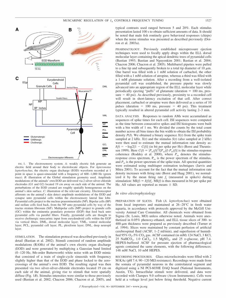
Weakly electric fish sense distortions in their self-generatedelectric field caused by nearby objects (Fig. 1, A and B).Electroreceptive neurons on their skin encode these perturba-
tions through changes in firing rate (Bastian 1981), and thisinformation is then relayed to the pyramidal cells within theelectrosensory lateral line lobe (ELL) (for a review of theelectrosensory system, see Turner and Maler 1999) (Fig. 1C).The electrosensory system displays many similarities withmammalian sensory systems (Berman and Maler 1999; Kraheand Gabbiani 2004; Sadeghi et al. 2007) and benefits fromdetailed neuroanatomy (Maler et al. 1991), well-characterizedphysiology (both at the systems and single neuron level), anddistinct response patterns to natural sensory input (Bastian etal. 2002; Berman and Maler 1998a–c; Chacron 2006; Chacronet al. 2005). Previous studies have shown that descendingglutamatergic pathways can control pyramidal cell responses tosensory input (Chacron et al. 2005; Mehaffey et al. 2005). Herewe focus on descending cholinergic input, which likely ema-nates from a unique pathway (Fig. 1C) (Maler et al. 1981). Weshow that activation of muscarinic acetylcholine receptors canincrease a neuron’s response to low-frequency input through anincrease in firing rate and burst fraction that is the result of thedownregulation of an A-type potassium current.
M E T H O D S
In vivo electrophysiology
EXPERIMENTAL SETUP. The weakly electric fish Apteronotus lepto-rhynchus was used exclusively in this study. Fish were housed ingroups of 3–10 in 150-l tanks, water temperature was maintainedbetween 26 and 28°C, and water resistivity varied between 2,000 and5,000 ��cm. Experiments were performed in a 39 � 44 � 12-cm-deepPlexiglass aquarium with water recirculated from the animal’s hometank. Animals were artificially respirated with a continuous water flowof 10 ml/min. Surgical techniques were the same as those describedpreviously (Bastian 1996a,b), and all procedures were in accordancewith animal care and use guidelines of McGill University.
Recording
Extracellular recordings from pyramidal neurons were made withmetal-filled micropipettes (Frank and Becker 1964). Recording sitesas determined from surface landmarks and recording depth werelimited to the centrolateral and lateral segments of the ELL only.Extracellular signals were recorded at 10 kHz using a CED 1401amplifier with spike2 software (Cambridge Electronic Design, Cam-bridge, UK). Spikes were detected with custom-written software inMatlab (Mathworks, Natick, MA).
Address for reprint requests and other correspondence: M. J. Chacron, Dept.of Physiology, McGill University, McIntyre Medical Sciences Bldg., Rm.1137, 3655 Promenade Sir William Osler, Montreal, Quebec H3G 1Y6,Canada (E-mail: [email protected]).
The costs of publication of this article were defrayed in part by the paymentof page charges. The article must therefore be hereby marked “advertisement”in accordance with 18 U.S.C. Section 1734 solely to indicate this fact.
J Neurophysiol 98: 1526–1537, 2007.First published July 5, 2007; doi:10.1152/jn.00564.2007.
1526 0022-3077/07 $8.00 Copyright © 2007 The American Physiological Society www.jn.org
STIMULATION. The stimulation protocol was described previously indetail (Bastian et al. 2002). Stimuli consisted of random amplitudemodulations (RAMs) of the animal’s own electric organ discharge(EOD) and were generated by multiplying a Gaussian band limited(0–120 Hz, 8th-order Butterworth) white noise with an EOD mimicthat consisted of a train of single-cycle sinusoids with frequencyslightly higher than that of the EOD and phase locked to the zero-crossings of the animal’s own EOD. The resulting signal was thenpresented via two silver-silver-chloride electrodes located 19 cm oneach side of the animal, giving rise to stimuli that were spatiallydiffuse (Fig. 1B). Stimulus intensities were similar to those previouslyused (Bastian et al. 2002; Chacron 2006; Chacron et al. 2005), and
typical contrasts used ranged between 5 and 20%. Each stimuluspresentation lasted 100 s to obtain sufficient amounts of data. It shouldbe noted that male fish routinely gave behavioral responses (chirps)when the noise stimulus was presented as described previously (Doi-ron et al. 2003a).
PHARMACOLOGY. Previously established micropressure ejectiontechniques were used to focally apply drugs within the ELL dorsalmolecular layer containing the apical dendritic trees of pyramidal cells(Bastian 1993; Bastian and Nguyenkim 2001; Bastian et al. 2004;Chacron 2006; Chacron et al. 2005). Multibarrel pipettes were pulledto a fine tip and subsequently broken to a total tip diameter of 10 �m.One barrel was filled with a 1 mM solution of carbachol, the otherfilled with a 1 mM solution of atropine, whereas a third was filled witha 1 mM glutamate solution. After a recording from a well-isolatedpyramidal cell was established, the pressure pipette was slowlyadvanced into an appropriate region of the ELL molecular layer whileperiodically ejecting “puffs” of glutamate (duration � 100 ms, pres-sure � 40 psi). As described previously, proximity to a recorded cellwill result in short-latency excitation of that cell. After correctplacement, carbachol or atropine were then delivered as a series of 10pulses (duration � 100 ms, pressure � 40 psi). This treatmenttypically resulted in altered pyramidal cell activity lasting 2–3 min.
DATA ANALYSIS. Responses to random AMs were accumulated assequences of spike times for each cell. ISI sequences were computedas the time between consecutive spikes and ISI histograms were builtwith a bin width of 1 ms. We divided the counts by the total countnumber across all bins times the bin width to obtain the ISI probabilitydensity P(I). We obtained a binary sequence X(t) from the spike trainsampled at 2 kHz. X(t) and the stimulus S(t) (also sampled at 2 kHz)were then used to estimate the mutual information rate density as:I(f) � �log2[1 � C(f)] (in bit per spike per Hz) (Borst and Theunis-sen 1999). Here C(f) � �Prs(f)�
2/[Pss(f) Prr(f)] is the stimulus-responsecoherence (Roddey et al. 2000), where Prs denotes the stimulus-response cross spectrum, Pss is the power spectrum of the stimulus,and Prr is the power spectrum of the spike train. All spectral quantitieswere estimated using multitaper estimation techniques (Jarvis andMitra 2001). To account for the fact that the mutual information ratedensity increases with firing rate (Borst and Haag 2001), we normal-ized it by the mean firing rate fr (measured in spike/s) duringstimulation: MI(f) � I(f)/fr. MI(f) is thus measured in bit per spike perHz. All values are reported as means � SD.
In vitro electrophysiology
PREPARATION OF SLICES. Fish (A. leptorhynchus) were obtainedfrom local importers and maintained at 26–28°C in fresh wateraquaria, in accordance with protocols approved by the McGill Uni-versity Animal Care Committee. All chemicals were obtained fromSigma (St. Louis, MO) unless otherwise noted. Animals were anes-thetized in 0.05% phenoxy-ethanol, and ELL tissue slices of 300- to400-�m thickness were prepared as previously described (Turner etal. 1994). Slices were maintained by constant perfusion of artificialcerebrospinal fluid (ACSF, 1–2 ml/min), and superfusion of humidi-fied 95% O2-5% CO2 gas. ACSF contained (in mM) 124 NaCl, 3 KCl,25 NaHCO3, 1.0 CaCl2, 1.5 MgSO4, and 25 D-glucose, pH 7.4.HEPES-buffered ACSF for pressure ejection of pharmacologicalagents contained the same elements, with the following differences:148 mM NaCl, 10 mM HEPES.
RECORDING PROCEDURES. Glass microelectrodes were filled with 2M KAc (pH 7.4; 90–120 M� resistance). Recordings were made fromthe somata of pyramidal neurons in the centrolateral segment anddigitized using a NI PCI-6030E DAQ board (National Instruments,Austin, TX). Intracellular stimuli were delivered, and data wererecorded with Clampex 9.0 software (Axon Instruments). Cells wereheld at a voltage level just below firing threshold. Negative current
NP
EGp
BP
MP
Stellate
Granule Cells
EurydendroidCells
Parallel Fibers
Vertical Fibres
Electroreceptor Afferents
GlutamatergicGABAergicCholinergic
DML
VML
StF
PCL
PL
GCL
DNL
DFL
EOD Waveform
10 ms
A B
C
Muscarinic??
PyramidalCell
FIG. 1. The electrosensory system. A: weakly electric fish generate anelectric field around their body to electrolocate objects. For Apteronotusleptorhynchus, the electric organ discharge (EOD) waveform recorded at 1point in space is quasi-sinusoidal with a frequency of 600–1,000 Hz (greentrace). B: illustration of the Global stimulation geometry used. Amplitudemodulations of the animals’ own EOD are delivered via 2 silver-silver-chlorideelectrodes (G1 and G2) located 19 cm away on each side of the animal. Theperturbations of the EOD created are roughly spatially homogeneous on theanimal’s skin surface. C: illustration of the relevant circuitry. Electroreceptorafferents on the animal’s skin detect amplitude modulations of the EOD andsynapse unto pyramidal cells within the electrosensory lateral line lobe.Pyramidal cells project to the nucleus praeeminentialis (NP). Bipolar cells (BP)and stellate cells feed back, from the NP unto pyramidal cells by way of thetractus stratum fibrosum (StF). Multipolar cells (MP) project to granule cells(GC) within the eminentia granularis posterior (EGP) that feed back untopyramidal cells via parallel fibers. Finally, pyramidal cells are thought toreceive cholinergic muscarinic input from eurydendroid cells within the EGPvia vertical fibers. DML, dorsal molecular layer; VML, ventral molecularlayer; PCL, pyramidal cell layer; PL, plexiform layer; DNL, deep neuropillayer.
1527MUSCARINIC REGULATION OF IA CONTROLS FREQUENCY TUNING
J Neurophysiol • VOL 98 • SEPTEMBER 2007 • www.jn.org

injections (0.3 nA) were given every 5 s to measure resistance changesduring drug application. All drugs [carbachol (1 mM), atropine (1mM), 4-aminopyridine (4-AP, 1 mM), and TEA (1 mM)] were focallyejected into the dorsal molecular layer (DML) using electrodes of 1-to 2-�m tip diameter and 7–15 psi pressure ejection. Previous studieshave estimated a �10 times dilution factor for ejected toxins (Turneret al. 1994). Pharmacological agents were dissolved in HEPES-buffered ACSF.
DATA ANALYSIS. Data were analyzed using Clampfit 9.0 software(Axon Instruments). Baseline depolarizations were measured as thedifference between 1 s membrane potential averages before drugapplication and after a depolarization plateau had been reached. Burstfractions were the proportion of ISIs that were �10 ms, representinga previously defined burst threshold (Oswald et al. 2004). The ISIhistogram (ISIH) was created from the sum of ISI events (bin width �0.5 ms) from a 1-s depolarization (0.3 nA) for three cells in whichcarbachol was washed out. The ISIH values were normalized as afraction of the total event number to account for the increased firingrate after carbachol. Resistance was measured as the average hyper-polarization value 40–50 ms after a negative current injection (0.3nA). Significance was evaluated using the Student’s t-test (P � 0.05).ISI sequences were computed as the time between consecutive spikes,and ISI histograms were built with a bin width of 1 ms. We dividedthe counts by the total count number across all bins times the binwidth to obtain the ISI probability density P(I). All values are reportedas means � SD.
Model description
We used a previously described two-compartment model of an ELLpyramidal cell (Doiron et al. 2002; Oswald et al. 2004) that containsall the essential elements to reproduce bursting seen experimentally(Doiron et al. 2001; Lemon and Turner 2000). The model neuron iscomprised of an isopotential soma (s) and a single dendritic compart-ment that are joined through an axial resistance of 1/gc (gc: couplingconductance) allowing for the electrotonic diffusion of currents fromthe soma to dendrite (d) and vice versa. Both compartments containthe essential spiking currents; fast inward Na� (INa,s, INa,d) andoutward delayed rectifying (Dr) K� (IDr,s, IDr,d), and passive leakcurrents (Ileak). The presence of spiking currents in the dendriteenables the active backpropagation of somatic action potentials re-quired for bursting. The membrane potentials at the soma, Vs, and thedendrite, Vd, are determined using a Hodgkin–Huxley like formalism(Koch 1999). The original model (Doiron et al. 2002) comprised sixnonlinear differential equations. This study expands on this model byincorporating an A-type K� current, Ia,d, into the dendritic compart-ment. We note that an A-type potassium current was previouslyincorporated into a multicompartmental model of pyramidal neuronactivity on which the reduced model used here is based (Doiron et al.2001). The kinetics for the A-type potassium current are qualitativelysimilar to those used previously. Vs and Vd are described by thefollowing equations
Cm
dVs
dt� I0 � gNos � m
2 � 1 � ns� � VNa � Vs� � �1t� � gDr,s � ns2 � VK � VS�
�gc
K� Vd � Vs� � gleak � Vt � Vs� � St� (1)
Cm
dVd
dt� gNa,d � md
2 � hd � VNa � Vd� � ga,d � ma,d2 � ha � VK � Vd�
� gDr,d � nd2 � Pd � VK � Vd� �
gc
1 � K� Vs � Vd� � gleak � Vt � Vd� � �2t� (2)
�1(t) and �2(t) are Ornstein-Uhlenbeck processes (Gardiner 1985)to account for sources of intrinsic noise described by
d�1.2
dt� �
�1.2
��
� �D�1.2
where �1,2(t) are independent Gaussian random variables with zeromean and unit SD. Here S(t) is a time varying stimulus which is eitherlow-pass filtered (8th-order Butterworth, 120-Hz cutoff frequency)Gaussian white noise or a sinusoid with a given frequency. Theparameter g is a maximal conductance (gmax, mS/cm2), whereas m ands are activation parameters and h, n, and p are inactivation parameters.Each is described by the following equation
dx
dt�
xV� � x
�(3)
where x(V) is the infinite conductance curve and � is the timeconstant. The infinite conductance curve is modeled as a sigmoid
xV� �1
1 � e�V�V1.2�k (4)
and the values for �, V1/2, and k for each current are given in Table 1.Other parameter values are the ratio of somatic to total area: � � 0.8;the reversal potentials: VNa � 40 mV, VK � �88.5 mV, Vleak � �70mV; membrane capacitance: Cm � 1 �F/cm2; gleak � 0.18 mS/cm2,and gc � 0.21 mS/cm2; time constant of the noise: �� � 20 ms;intensity of the noise: D � 0.01 (ms)�2. The model equations wereintegrated using an Euler-Maruyama algorithm with a time step of 0.5�s.
R E S U L T S
Carbachol increases burst firing in vivo
We obtained extracellular recordings from ELL pyramidalneurons in vivo. The stimulation consisted of random ampli-tude modulations of the animal’s own electric field that werespatially diffuse, mimicking distortions caused by conspecifics(see Fig. 1B) (Bastian et al. 2002; Chacron 2006; Chacron et al.2005). To look at the effects of cholinergic input onto pyra-midal neurons, we used previously established pharmacologi-cal techniques (Bastian 1993; Bastian et al. 2004; Chacron2006; Chacron et al. 2005) to apply carbachol, a cholinergicreceptor agonist, within the ELL dorsal molecular layer (DML)where cholinergic binding sites are located (Phan and Maler1983). In all cases, this led to an increased firing rate thatrecovered to values similar to those seen under control condi-tions (Fig. 2; control: 15.57 � 6.42 Hz; carbachol: 20.12 �9.34 Hz; recovery: 13.56 � 6.62 Hz). The firing rate undercarbachol was significantly higher than the firing rate undercontrol conditions (P � 0.005, paired t-test, n � 14). For thecells that were followed to recovery, the firing rate afterrecovery was not significantly different from the firing rateunder control conditions (P � 0.35, paired t-test, n � 6).
The increased firing rate was accompanied by an increasedfraction of high-frequency ISIs (�10 ms; Fig. 3A). It has beenshown previously that ISIs with a value �10 ms were primarily
TABLE 1. Model parameters
Current gmax, mS/cm2 V1/2, mV k �, m
INas[m(Vg)] 35 �50 3 N/AIDr.s[ng(Vs)] 15 �50 3 0.19INa,d[md(Vd)/hd(Vd)] 4 �40/�52 5/�5 0.3/1.0Ia,d[ma(Vd)/ha(Vd)] 2 �69/�69 4/1 1/10IDr,d[nd(Vd)/Pd(Vd)] 15 �40/�65 5/�6 0.9/3
1528 L. D. ELLIS, R. KRAHE, C. W. BOURQUE, R. J. DUNN, AND M. J. CHACRON
J Neurophysiol • VOL 98 • SEPTEMBER 2007 • www.jn.org
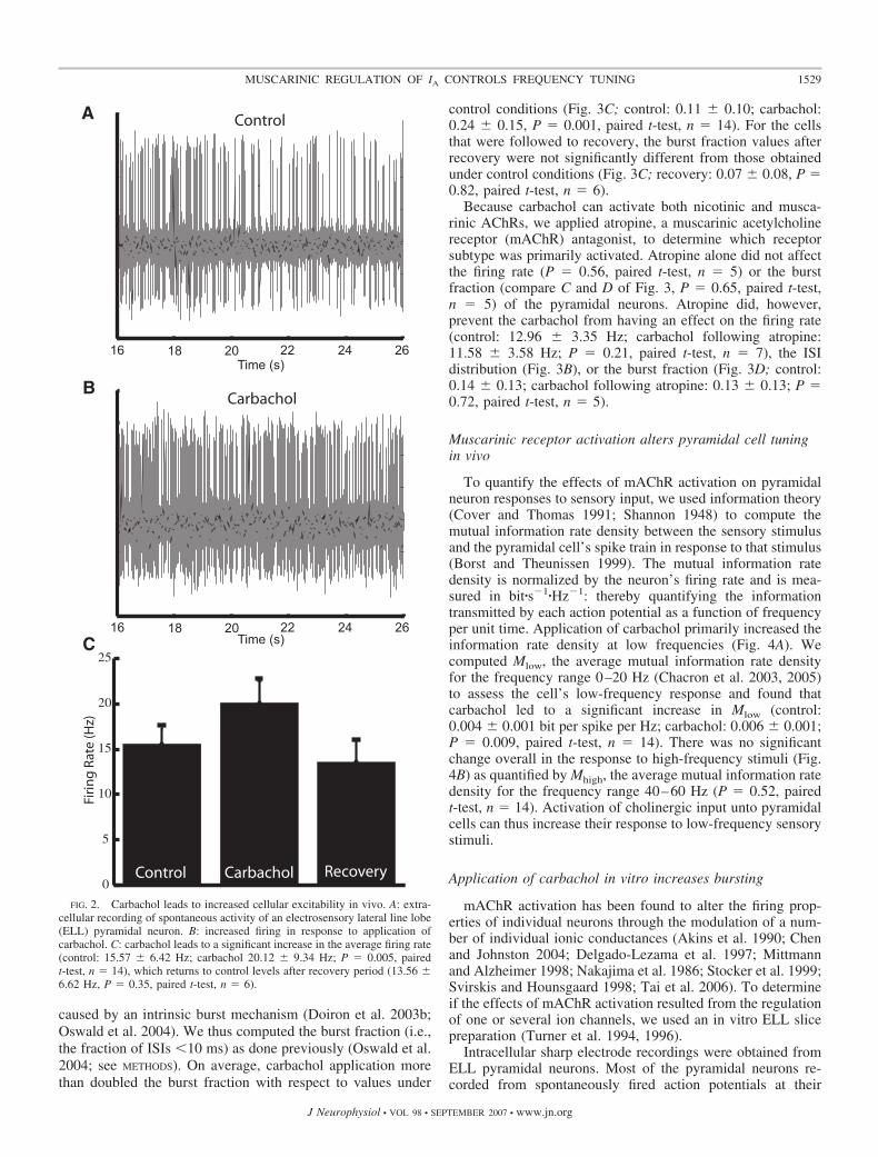
caused by an intrinsic burst mechanism (Doiron et al. 2003b;Oswald et al. 2004). We thus computed the burst fraction (i.e.,the fraction of ISIs �10 ms) as done previously (Oswald et al.2004; see METHODS). On average, carbachol application morethan doubled the burst fraction with respect to values under
control conditions (Fig. 3C; control: 0.11 � 0.10; carbachol:0.24 � 0.15, P � 0.001, paired t-test, n � 14). For the cellsthat were followed to recovery, the burst fraction values afterrecovery were not significantly different from those obtainedunder control conditions (Fig. 3C; recovery: 0.07 � 0.08, P �0.82, paired t-test, n � 6).
Because carbachol can activate both nicotinic and musca-rinic AChRs, we applied atropine, a muscarinic acetylcholinereceptor (mAChR) antagonist, to determine which receptorsubtype was primarily activated. Atropine alone did not affectthe firing rate (P � 0.56, paired t-test, n � 5) or the burstfraction (compare C and D of Fig. 3, P � 0.65, paired t-test,n � 5) of the pyramidal neurons. Atropine did, however,prevent the carbachol from having an effect on the firing rate(control: 12.96 � 3.35 Hz; carbachol following atropine:11.58 � 3.58 Hz; P � 0.21, paired t-test, n � 7), the ISIdistribution (Fig. 3B), or the burst fraction (Fig. 3D; control:0.14 � 0.13; carbachol following atropine: 0.13 � 0.13; P �0.72, paired t-test, n � 5).
Muscarinic receptor activation alters pyramidal cell tuningin vivo
To quantify the effects of mAChR activation on pyramidalneuron responses to sensory input, we used information theory(Cover and Thomas 1991; Shannon 1948) to compute themutual information rate density between the sensory stimulusand the pyramidal cell’s spike train in response to that stimulus(Borst and Theunissen 1999). The mutual information ratedensity is normalized by the neuron’s firing rate and is mea-sured in bit�s�1�Hz�1: thereby quantifying the informationtransmitted by each action potential as a function of frequencyper unit time. Application of carbachol primarily increased theinformation rate density at low frequencies (Fig. 4A). Wecomputed Mlow, the average mutual information rate densityfor the frequency range 0–20 Hz (Chacron et al. 2003, 2005)to assess the cell’s low-frequency response and found thatcarbachol led to a significant increase in Mlow (control:0.004 � 0.001 bit per spike per Hz; carbachol: 0.006 � 0.001;P � 0.009, paired t-test, n � 14). There was no significantchange overall in the response to high-frequency stimuli (Fig.4B) as quantified by Mhigh, the average mutual information ratedensity for the frequency range 40–60 Hz (P � 0.52, pairedt-test, n � 14). Activation of cholinergic input unto pyramidalcells can thus increase their response to low-frequency sensorystimuli.
Application of carbachol in vitro increases bursting
mAChR activation has been found to alter the firing prop-erties of individual neurons through the modulation of a num-ber of individual ionic conductances (Akins et al. 1990; Chenand Johnston 2004; Delgado-Lezama et al. 1997; Mittmannand Alzheimer 1998; Nakajima et al. 1986; Stocker et al. 1999;Svirskis and Hounsgaard 1998; Tai et al. 2006). To determineif the effects of mAChR activation resulted from the regulationof one or several ion channels, we used an in vitro ELL slicepreparation (Turner et al. 1994, 1996).
Intracellular sharp electrode recordings were obtained fromELL pyramidal neurons. Most of the pyramidal neurons re-corded from spontaneously fired action potentials at their
0
5
10
15
20
25
A
B
C
Control Carbachol Recovery
16 18 20 22 24 26
16 18 20 22 24 26
Time (s)
Time (s)
Firi
ng
Rat
e (H
z)
Control
Carbachol
FIG. 2. Carbachol leads to increased cellular excitability in vivo. A: extra-cellular recording of spontaneous activity of an electrosensory lateral line lobe(ELL) pyramidal neuron. B: increased firing in response to application ofcarbachol. C: carbachol leads to a significant increase in the average firing rate(control: 15.57 � 6.42 Hz; carbachol 20.12 � 9.34 Hz; P � 0.005, pairedt-test, n � 14), which returns to control levels after recovery period (13.56 �6.62 Hz, P � 0.35, paired t-test, n � 6).
1529MUSCARINIC REGULATION OF IA CONTROLS FREQUENCY TUNING
J Neurophysiol • VOL 98 • SEPTEMBER 2007 • www.jn.org

resting membrane potential. To silence cell firing, negativecurrent injections were used to hold the membrane potentialbelow the firing threshold (average holding potential: �73 � 6mV; n � 13). Carbachol (1 mM) was applied to the DML andthis resulted on average in a 3.8 � 1.8-mV depolarization ofthe membrane potential (P � 0.001; paired t-test; n � 20; Fig.5A), which often caused the action potential threshold to bereached. Application of atropine (1 mM) prior to carbacholprevented this depolarization (Fig. 5B; �76.5 � 4.4 to –77.2 �4.2 mV; P � 0.11; paired t-test; n � 4), suggesting the effectis due in large part to mAChR activation. When applied onits own atropine had no effect on the membrane properties(data not shown). Step current injections revealed an in-crease in firing rate following carbachol (Fig. 5C, control:39 � 14; carbachol: 67 � 23 Hz; P � 0.001; paired t-test;n � 13) as well as an increase in burst fraction (Fig. 5D;0.26 � 0.29 to 0.39 � 0.26; P � 0.01; paired t-test; n � 13).The effects of carbachol on the in vitro slice preparationswere thus found to be qualitatively similar to those obtainedin vivo as, in both cases, carbachol lead to increased spikingand bursting activity and its effects were prevented byatropine.
The membrane depolarization caused by carbachol applica-tion in vitro suggests an effect on subthreshold membrane
properties, which could be mediated by regulation of one ormultiple ion channels. The effects of carbachol were mostlikely not due to voltage-gated Ca2�, high-threshold K�, orCa2�-activated K� channels because these channels are nottypically active at these potentials. To further characterize thechanges caused by carbachol, negative current pulses (0.3 nA)were applied at regular intervals to measure subthresholdmembrane resistance changes in response to drug application.Carbachol increased the membrane resistance on average by37% (Fig. 5E; control: 15.7 � 7.1 M�; carbachol: 20.1 � 7.7M�; P � 0.001; paired t-test; n � 12), suggesting the down-regulation of an outward current. The carbachol effect is thenunlikely to be mediated by subthreshold inward currents suchas persistent sodium.
It was then hypothesized that subthreshold K� currents wereresponsible for the effects of carbachol. The negative currentinjections showed no evidence of a depolarizing rectificationcharacteristic of H-type currents (Ih) (Maccaferri et al. 1993),and previous studies have been unable to demonstrate thepresence of H-type potassium currents in pyramidal neurons.However, evidence does exist for an A-type K� current (IA) inpyramidal neurons (Berman and Maler 1999; Mathieson andMaler 1988), which lead us to speculate that a downregulationof IA was responsible for the effects observed.
0 025 50ISI (msec)
25 50ISI (msec)
0 0
10
20
30
10
20
30 control
atropineatropine+carbachol
control
carbachol
recovery
P(I
SI)
P(I
SI)
A B
0
0.05
0.1
0.15
0.2
0.25
0.3
0.35
Baseline Carbachol AtropineAtropine +Carbachol Recovery
Bur
st F
ract
ion
C D
Recovery0
0.05
0.1
0.15
0.2
0.25
0.3
0.35
Bur
st F
ract
ion
FIG. 3. Muscarinic receptor activation leads to an increase in burst firing in vivo. A: interspike interval (ISI) histograms before (blue), during (red), and after(green) application of carbachol. Carbachol leads to an increase in ISIs �10 ms. B: ISI histograms before (blue), during (red), and after (green) application ofcarbachol in the presence of atropine. Atropine prevents the effect of carbachol revealing muscarinic receptor activation by carbachol. C: bar graph representingthe average increase of the burst fraction (fraction of ISIs �10 ms; control: 0.11 � 0.10; carbachol: 0.24 � 0.15, P � 0.001, paired t-test, n � 14) after carbacholand return to control levels after recovery period. (D) Atropine prevents an increase in burst fraction for the population of cells tested.
1530 L. D. ELLIS, R. KRAHE, C. W. BOURQUE, R. J. DUNN, AND M. J. CHACRON
J Neurophysiol • VOL 98 • SEPTEMBER 2007 • www.jn.org
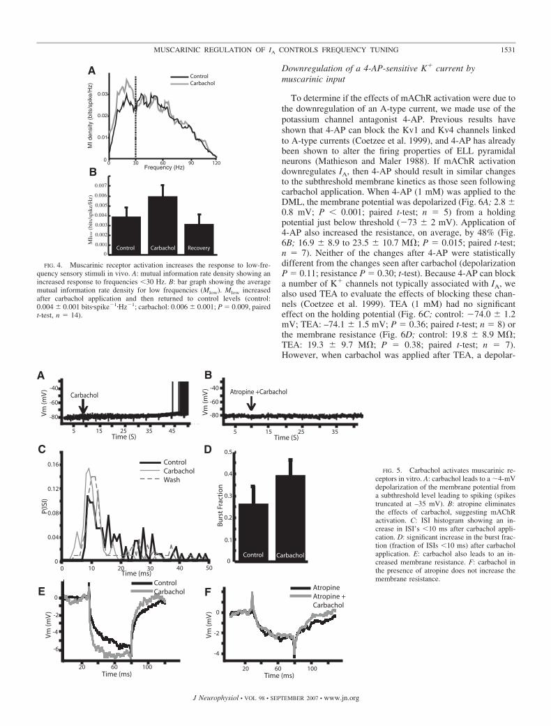
Downregulation of a 4-AP-sensitive K� current bymuscarinic input
To determine if the effects of mAChR activation were due tothe downregulation of an A-type current, we made use of thepotassium channel antagonist 4-AP. Previous results haveshown that 4-AP can block the Kv1 and Kv4 channels linkedto A-type currents (Coetzee et al. 1999), and 4-AP has alreadybeen shown to alter the firing properties of ELL pyramidalneurons (Mathieson and Maler 1988). If mAChR activationdownregulates IA, then 4-AP should result in similar changesto the subthreshold membrane kinetics as those seen followingcarbachol application. When 4-AP (1 mM) was applied to theDML, the membrane potential was depolarized (Fig. 6A; 2.8 �0.8 mV; P � 0.001; paired t-test; n � 5) from a holdingpotential just below threshold (�73 � 2 mV). Application of4-AP also increased the resistance, on average, by 48% (Fig.6B; 16.9 � 8.9 to 23.5 � 10.7 M�; P � 0.015; paired t-test;n � 7). Neither of the changes after 4-AP were statisticallydifferent from the changes seen after carbachol (depolarizationP � 0.11; resistance P � 0.30; t-test). Because 4-AP can blocka number of K� channels not typically associated with IA, wealso used TEA to evaluate the effects of blocking these chan-nels (Coetzee et al. 1999). TEA (1 mM) had no significanteffect on the holding potential (Fig. 6C; control: �74.0 � 1.2mV; TEA: –74.1 � 1.5 mV; P � 0.36; paired t-test; n � 8) orthe membrane resistance (Fig. 6D; control: 19.8 � 8.9 M�;TEA: 19.3 � 9.7 M�; P � 0.38; paired t-test; n � 7).However, when carbachol was applied after TEA, a depolar-
B
0.01
0.02
0.03M
I den
sity
(bi
ts/s
pike
/Hz)
0
Frequency (Hz)
0
0.001
0.002
0.003
0.004
0.005
0.006
0.007
A
Control Carbachol Recovery
MIlo
w (
bits
/spi
ke/H
z)
ControlCarbachol
0 30 60 90 120
FIG. 4. Muscarinic receptor activation increases the response to low-fre-quency sensory stimuli in vivo. A: mutual information rate density showing anincreased response to frequencies �30 Hz. B: bar graph showing the averagemutual information rate density for low frequencies (Mlow). Mlow increasedafter carbachol application and then returned to control levels (control:0.004 � 0.001 bits�spike�1�Hz�1; carbachol: 0.006 � 0.001; P � 0.009, pairedt-test, n � 14).
-6
420
-6
Carbachol Atropine +Carbachol
Bu
rst
Frac
tio
n
Control Carbachol
0.5
0.4
0.3
0.2
0.1
0
0.04
0.08
0.12
0.16
00
10 20 30 40 50Time (ms)
P(IS
I)
Time (S)5 15 25 35
Time (S)
-80
-60
-40
-80
-60
-40
5 15 25 35 45
Vm
(mV
)
-4
0
-4
0
-2
-4
20 60 100 20 60 100 Time (ms) Time (ms)
A B
C D
E FControlCarbachol
ControlCarbacholWash
AtropineAtropine + Carbachol
Vm
(mV
)V
m (m
V)
Vm
(mV
) -2
-6
FIG. 5. Carbachol activates muscarinic re-ceptors in vitro. A: carbachol leads to a �4-mVdepolarization of the membrane potential froma subthreshold level leading to spiking (spikestruncated at –35 mV). B: atropine eliminatesthe effects of carbachol, suggesting mAChRactivation. C: ISI histogram showing an in-crease in ISI’s �10 ms after carbachol appli-cation. D: significant increase in the burst frac-tion (fraction of ISIs �10 ms) after carbacholapplication. E: carbachol also leads to an in-creased membrane resistance. F: carbachol inthe presence of atropine does not increase themembrane resistance.
1531MUSCARINIC REGULATION OF IA CONTROLS FREQUENCY TUNING
J Neurophysiol • VOL 98 • SEPTEMBER 2007 • www.jn.org
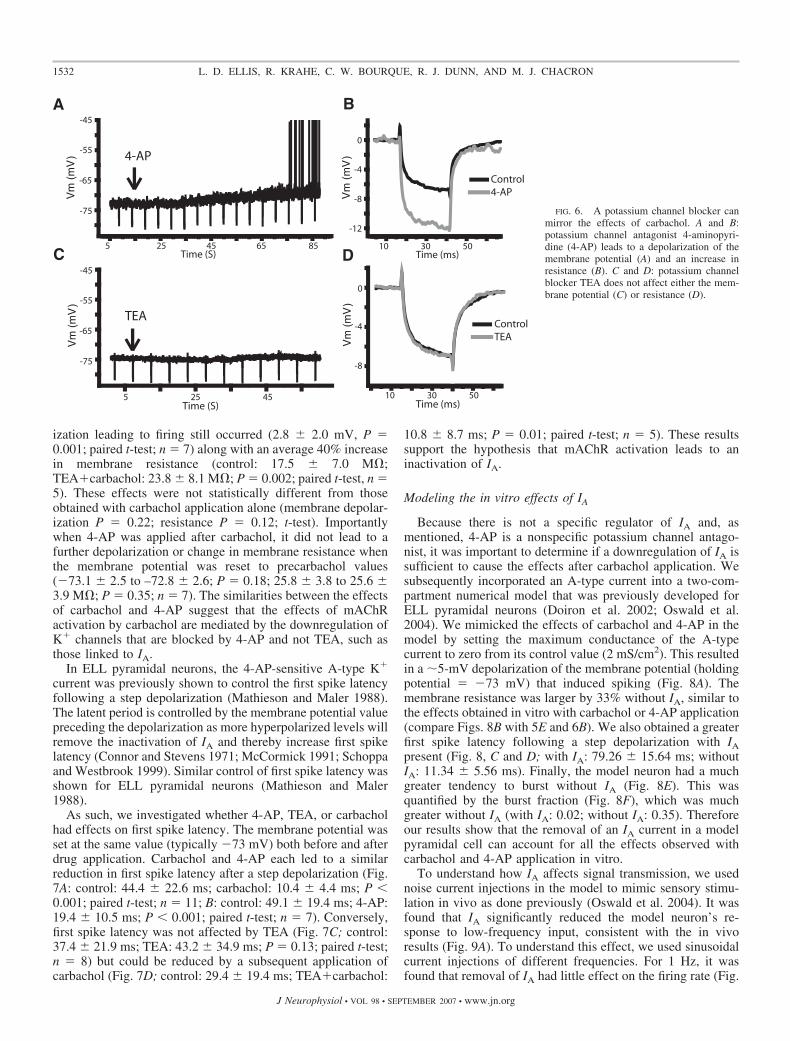
ization leading to firing still occurred (2.8 � 2.0 mV, P �0.001; paired t-test; n � 7) along with an average 40% increasein membrane resistance (control: 17.5 � 7.0 M�;TEA�carbachol: 23.8 � 8.1 M�; P � 0.002; paired t-test, n �5). These effects were not statistically different from thoseobtained with carbachol application alone (membrane depolar-ization P � 0.22; resistance P � 0.12; t-test). Importantlywhen 4-AP was applied after carbachol, it did not lead to afurther depolarization or change in membrane resistance whenthe membrane potential was reset to precarbachol values(�73.1 � 2.5 to –72.8 � 2.6; P � 0.18; 25.8 � 3.8 to 25.6 �3.9 M�; P � 0.35; n � 7). The similarities between the effectsof carbachol and 4-AP suggest that the effects of mAChRactivation by carbachol are mediated by the downregulation ofK� channels that are blocked by 4-AP and not TEA, such asthose linked to IA.
In ELL pyramidal neurons, the 4-AP-sensitive A-type K�
current was previously shown to control the first spike latencyfollowing a step depolarization (Mathieson and Maler 1988).The latent period is controlled by the membrane potential valuepreceding the depolarization as more hyperpolarized levels willremove the inactivation of IA and thereby increase first spikelatency (Connor and Stevens 1971; McCormick 1991; Schoppaand Westbrook 1999). Similar control of first spike latency wasshown for ELL pyramidal neurons (Mathieson and Maler1988).
As such, we investigated whether 4-AP, TEA, or carbacholhad effects on first spike latency. The membrane potential wasset at the same value (typically �73 mV) both before and afterdrug application. Carbachol and 4-AP each led to a similarreduction in first spike latency after a step depolarization (Fig.7A: control: 44.4 � 22.6 ms; carbachol: 10.4 � 4.4 ms; P �0.001; paired t-test; n � 11; B: control: 49.1 � 19.4 ms; 4-AP:19.4 � 10.5 ms; P � 0.001; paired t-test; n � 7). Conversely,first spike latency was not affected by TEA (Fig. 7C; control:37.4 � 21.9 ms; TEA: 43.2 � 34.9 ms; P � 0.13; paired t-test;n � 8) but could be reduced by a subsequent application ofcarbachol (Fig. 7D; control: 29.4 � 19.4 ms; TEA�carbachol:
10.8 � 8.7 ms; P � 0.01; paired t-test; n � 5). These resultssupport the hypothesis that mAChR activation leads to aninactivation of IA.
Modeling the in vitro effects of IA
Because there is not a specific regulator of IA and, asmentioned, 4-AP is a nonspecific potassium channel antago-nist, it was important to determine if a downregulation of IA issufficient to cause the effects after carbachol application. Wesubsequently incorporated an A-type current into a two-com-partment numerical model that was previously developed forELL pyramidal neurons (Doiron et al. 2002; Oswald et al.2004). We mimicked the effects of carbachol and 4-AP in themodel by setting the maximum conductance of the A-typecurrent to zero from its control value (2 mS/cm2). This resultedin a �5-mV depolarization of the membrane potential (holdingpotential � �73 mV) that induced spiking (Fig. 8A). Themembrane resistance was larger by 33% without IA, similar tothe effects obtained in vitro with carbachol or 4-AP application(compare Figs. 8B with 5E and 6B). We also obtained a greaterfirst spike latency following a step depolarization with IApresent (Fig. 8, C and D; with IA: 79.26 � 15.64 ms; withoutIA: 11.34 � 5.56 ms). Finally, the model neuron had a muchgreater tendency to burst without IA (Fig. 8E). This wasquantified by the burst fraction (Fig. 8F), which was muchgreater without IA (with IA: 0.02; without IA: 0.35). Thereforeour results show that the removal of an IA current in a modelpyramidal cell can account for all the effects observed withcarbachol and 4-AP application in vitro.
To understand how IA affects signal transmission, we usednoise current injections in the model to mimic sensory stimu-lation in vivo as done previously (Oswald et al. 2004). It wasfound that IA significantly reduced the model neuron’s re-sponse to low-frequency input, consistent with the in vivoresults (Fig. 9A). To understand this effect, we used sinusoidalcurrent injections of different frequencies. For 1 Hz, it wasfound that removal of IA had little effect on the firing rate (Fig.
TEA
4-AP
-45
-55
-65
-75
-45
-55
-65
-75
5 25 45 65 85
5 25 45
0
-4
-12
-8
0
-4
-8
10 30 50
10 30 50
Time (ms)
Time (ms)
Time (S)
Time (S)
Control4-AP
ControlTEA
A
C
B
D
Vm
(m
V)
Vm
(m
V)
Vm
(m
V)
Vm
(m
V)
FIG. 6. A potassium channel blocker canmirror the effects of carbachol. A and B:potassium channel antagonist 4-aminopyri-dine (4-AP) leads to a depolarization of themembrane potential (A) and an increase inresistance (B). C and D: potassium channelblocker TEA does not affect either the mem-brane potential (C) or resistance (D).
1532 L. D. ELLIS, R. KRAHE, C. W. BOURQUE, R. J. DUNN, AND M. J. CHACRON
J Neurophysiol • VOL 98 • SEPTEMBER 2007 • www.jn.org

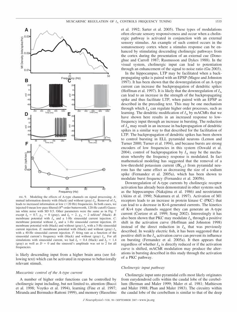
9, B and C). This can be understood as follows: during thenegative portion of the sinusoid, the membrane is hyperpolar-ized, and this will tend to de-inactivate IA; however, because ofthe large period (1 s), IA will subsequently inactivate before theonset of spiking, resulting in no net effect. In contrast, removalof IA significantly increased the firing rate for a 5-Hz sinusoid(Fig. 9D). In this case, IA does not fully inactivate during theshorter depolarizing portion of the sinusoid and thus can havea significant effect on firing rate. Finally, removal of IA hadlittle effect on the firing rate for a 40-Hz sinusoid (Fig. 9E). Inthis case, the short hyperpolarization is unlikely to de-inacti-vate the A-type channels. A plot of the model neuron’s firingrate with and without IA as a function of frequency revealed aneffect for frequencies contained between 1 and 40 Hz (Fig. 9F).
Our modeling results have thus shown that A-type currentscan have significant effects on the tuning of neurons in thelow-frequency range. As such, our modeling results provide anexplanation for why mAChR activation in vivo increased thecell’s response to low-frequency input only.
D I S C U S S I O N
We have shown that activation of muscarinic receptors cansignificantly alter the response of ELL pyramidal neurons tosensory input. In vivo application of the cholinergic agonistcarbachol increased pyramidal neuron excitability, leading toan increase in spiking and bursting activity. Moreover, it wasfound that carbachol application onto pyramidal neurons in-creased the transmission of the low-frequency components ofan external electrosensory signal. The effects of carbacholcould be prevented by the muscarinic antagonist atropine,suggesting carbachol was primarily activating muscarinic ace-tylcholine receptors (mAChR).
To understand the cellular mechanisms that mediate thesealtered responses, we investigated the consequences of cholin-ergic receptor activation in vitro. We found that carbacholapplication also increased pyramidal cell excitability and burstfiring and that these effects were also prevented by atropine.Activation of mAChRs in vitro led to a �4-mV membranedepolarization from a level below threshold accompanied by anincrease in the subthreshold membrane resistance. These re-sults suggested that cholinergic receptor activation downregu-lates an outward current that is active in the subthresholdregime. Because the presence of an A-type current was previ-ously shown in ELL pyramidal neurons (Mathieson and Maler1988), we set out to determine if this current was being downregulated by mAChR activation. It was found that the K�
channel antagonist 4-AP had effects on the subthreshold mem-brane kinetics similar to those of carbachol. Furthermore,carbachol could prevent the subthreshold effects of 4-AP,suggesting that mAChR activation downregulates 4-AP-sensi-tive channels. Because 4-AP is a nonspecific potassium chan-nel blocker, we also used the K� channel blocker TEA toconfirm that the effects of carbachol were confined to the K�
channels that are known to mediate A-type currents. TEA hadno significant effect on either the holding potential or themembrane resistance, whereas application of carbachol in thepresence of TEA led to changes in holding potential andmembrane resistance that were comparable to those seen withcarbachol application alone. Because previous studies hadshown that blocking A-type currents with 4-AP could have asignificant effect on first spike latency (Mathieson and Maler1988), we measured the effects of carbachol and 4-AP on firstspike latency. We confirmed the effect of 4-AP previouslyshown and additionally demonstrated that carbachol could leadto similar decreases in first spike latency, whereas TEA had nosignificant effect.
Control of a neuron’s frequency tuning by A-type currents
We used a two-compartmental mathematical model to assessif the removal of an A-type current could replicate the effectsof carbachol. The model showed that the removal of an A-typecurrent can result in similar changes in burst firing, holdingpotential, membrane resistance, and first spike latency as thoseseen under experimental activation of mAChRs, supporting thein vitro findings (Fig. 8). Additionally it was shown that whenthe model was presented with a noise current injection tomimic sensory stimulation in vivo, removal of IA resulted in anincreased response to low-frequency input, which was similarto that seen after carbachol application in vivo. To understandthis effect, we used sinusoidal current injections. It was shown
Control Carbachol Control 4-AP TEAControl
Time (ms)
20
0
-20
-40
-60
-80
20
0
-20
-40
-60
-80
20
0
-20
-40
-60
-80
20
0
-20
-40
-60
-80
-20 0 20 40 60 -20 0 20 40 60
0 20 40 60 0 20 40 60
Time (ms) Time (ms)
Time (ms)
A B
C D
E
Late
ncy
(ms)
10
20
30
40
50
60
Carbachol 4-AP
TEA TEA+Carbachol
Vm
(mV
)
Vm
(mV
)V
m (m
V)
Vm
(mV
)
FIG. 7. Carbachol and 4-AP lead to a decreased 1st spike latency, suggest-ing the involvement of an A-type current. A: carbachol leads to a reduced spikelatency after a step current injection (0.2 nA). B: 4-AP mirrors the effect ofcarbachol reducing 1st spike latency in a representative cell. C: TEA does notalter the spike latency. D: carbachol decreases latency even when applied afterTEA. E: bar graphs showing a significant decrease in latency after carbacholand 4-AP (control: 44.4 � 22.6 ms; carbachol: 10.4 � 4.4 ms; P � 0.001;paired t-test; n � 11: control: 49.1 � 19.4 ms; 4-AP: 19.4 � 10.5 ms; P �0.001; paired t-test; n � 7), whereas TEA did not show a significant decreasein latency (control: 37.4 � 21.9 ms; TEA: 43.2 � 34.9 ms; P � 0.13; pairedt-test; n � 8).
1533MUSCARINIC REGULATION OF IA CONTROLS FREQUENCY TUNING
J Neurophysiol • VOL 98 • SEPTEMBER 2007 • www.jn.org

that for very low frequencies (�1 Hz) removal of IA had noeffect on neuronal response properties. We propose that this isdue to the length of the depolarizing phase of the sine wave,which should result in a complete inactivation of IA by the timespike threshold is reached, thus eliminating its effect. Further,it was shown that at frequencies �40 Hz, the removal of IAagain had no effect because the period of hyperpolarization isinsufficient to de-inactivate the A-type channels. As such, ourmodeling results suggest that the downregulation of A-typepotassium channels is sufficient to explain the increase in thelow-frequency tuning, in the range between 1 and 40 Hz, ofpyramidal neurons found in vivo after pharmacological activa-tion of mAChRs.
Pyramidal neuron’s response to low frequencies isbehaviorally relevant
We have shown that mAChR activation leads to an increasedpyramidal neuron response to the low temporal frequencycomponents of sensory input. A number of natural sensorystimuli contain low temporal frequencies. The jamming avoid-
ance response (JAR) (reviewed in Heiligenberg 1991) is awell-described behavior that is triggered by low temporalfrequency, spatially diffuse electrosensory stimuli. Specifi-cally, the JAR occurs when a fish encounters a conspecific withan EOD frequency close to its own. These signals can interferewith electrolocation if the frequency difference is �8 Hz(Heiligenberg 1991). The fish with the highest EOD frequencyincreases its EOD frequency until the signals will interfere lesswith electrolocation. However, previous in vivo studies haveshown that pyramidal neurons generally display poor responsesto low-frequency global stimuli (Bastian et al. 2002, 2004;Chacron 2006; Chacron et al. 2003, 2005). Our results showthat cholinergic input has the capacity to make pyramidalneurons more responsive these stimuli.
Alternatively, prey can also cause low-frequency stimuli.However, these signals are more spatially localized (Nelsonand Maciver 1999). It is therefore also possible that thecholinergic pathway can increase the response of pyramidalneurons to prey stimuli, thereby improving the animal’s preydetection abilities. Although further studies are needed tounderstand how and when cholinergic input becomes active, it
Bu
rst
Frac
tio
nFi
rst
Spik
e La
ten
cy (m
s)
-80
-60
-40
-20
0
20
40
0 20 40 60 80 100Time (ms)
Vm
(m
V)
Vm
(m
V)
Vm
(m
V)
Time (ms)
-70
-69.5
-69
-68.5
-68
-67.5
0 50 100 150 200 250
0 50 100 150Time (ms)
-80
-60
-40
-20
0
20
40
0
20
40
60
80
100
0 20Time (ms)
400
0.1
0.2
P(IS
I)
0
0.2
0.4
without IA
with IA
A B
C D
E F
IA Removed
without IA
with IAwithout IA
with IA
without IA
with IA
60
FIG. 8. Modeling the effects of blockingA-type channels. A: removal of IA leads to a�5-mV depolarization and spiking. At t � 30ms, we set gA from 2 mS/cm2 to 0. We hadI0 � 1.3. B: step hyperpolarization with gA �2 mS/cm2 (black) and gA � 0 (gray) showing a33% increase in resistance with D � 0. Thiswas obtained by setting I0 from 1.375 to 0.875.C: step depolarizations with (black, gA � 2mS/cm2) and without (gray, gA � 0) IA. At t �130 ms, we increased I0 from 0.5 to 1.7 (gray)and from 1 to 2.3 (black). D: mean latenciesobtained under repeated step depolarizations asdescribed in C. E: ISI probability densitiesobtained under repeated step depolarizations asdescribed in C. F: burst fraction obtained fromthe ISI probability. Note that S(t) � 0 for allpanels.
1534 L. D. ELLIS, R. KRAHE, C. W. BOURQUE, R. J. DUNN, AND M. J. CHACRON
J Neurophysiol • VOL 98 • SEPTEMBER 2007 • www.jn.org
is likely descending input from a higher brain area (see fol-lowing text) which can be activated in response to behaviorallyrelevant stimuli.
Muscarinic control of the A-type current
A number of higher order functions can be controlled bycholinergic input including, but not limited to, attention (Bucciet al. 1998; Voytko et al. 1994), learning (Fine et al. 1997;Miranda and Bermudez-Rattoni 1999), and memory (Hasselmo
et al. 1992; Sarter et al. 2005). These types of modulationsoften elevate sensory responsiveness and occur when a cholin-ergic pathway is activated in conjunction with an externalsensory stimulus. An example of such control occurs in thesomatosensory cortex where a stimulus response can be en-hanced by stimulating descending cholinergic pathways fromthe cortex during the presentation of an external cue (Dono-ghue and Carroll 1987; Rasmusson and Dykes 1988). In thevisual system, cholinergic input can lead to potentiationthrough an enhancement of the signal to noise ratio (Gu 2003).
In the hippocampus, LTP may be facilitated when a back-propagating spike is paired with an EPSP (Magee and Johnston1997). It has been shown that the downregulation of an A-typecurrent can increase the backpropagation of dendritic spikes(Hoffman et al. 1997). It is likely that the downregulation of IAcan lead to an increase in the strength of the backpropagatingspike and thus facilitate LTP, when paired with an EPSP asdescribed in the preceding text. This may be one mechanismthrough which IA can regulate higher order processes, such aslearning. The dendritic modification of IA by mAChRs that wehave shown here results in an increased response to low-frequency input through an increase in bursting. The reductionof IA may result in an increase in backpropagation of dendriticspikes in a similar way to that described for the facilitation ofLTP. The backpropagation of dendritic spikes has been shownto control bursting in ELL pyramidal neurons (Lemon andTurner 2000; Turner et al. 1994), and because bursts are strongencoders of low frequencies in this system (Oswald et al.2004), control of backpropagation by IA may be the mecha-nism whereby the frequency response is modulated. In factmathematical modeling has suggested that the removal of alow-threshold potassium current (IKLT) from pyramidal neu-rons has the same effect as decreasing the size of a sodiumspike (Fernandez et al. 2005a), which has been shown tomodulate burst frequency (Fernandez et al. 2005b).
Downregulation of A-type currents by cholinergic receptoractivation has already been demonstrated in other systems suchas the hippocampus (Nakajima et al. 1986) and neostriatum(Akins et al. 1990; Nakamura et al. 1997). Activation of M1receptors leads to an increase in protein kinase C (PKC) thatcan lead to a decrease in Kv4-generated currents. The kineticsof Kv4 type channels suggest they can generate an A-typecurrent (Coetzee et al. 1999; Song 2002). Interestingly it hasalso been shown that PKC may modulate IA through a positiveshift in the activation curve (Hoffman and Johnston 1998)instead of the direct reduction in IA that was previouslydescribed. In weakly electric fish, it has been suggested that apositive shift in the IA activation curve can prevent its influenceon bursting (Fernandez et al. 2005a). It then appears thatregardless of whether IA is directly reduced or if the activationcurve is shifted, mAChR modulation may produce the alter-ations in bursting described in this study through the activationof a PKC pathway.
Cholinergic input pathway
Cholinergic input unto pyramidal cells most likely originatesfrom eurydendroid cells within the caudal lobe of the cerebel-lum (Berman and Maler 1999; Maler et al. 1981; Mathiesonand Maler 1988; Phan and Maler 1983). The circuitry withinthe caudal lobe of the cerebellum is similar to that of the deep
FIG. 9. Modeling the effects of A-type channels on signal processing. A:mutual information density with (black) and without (gray) IA. Removal of IA
leads to increased information at low (�20 Hz) frequencies. In both cases, weinjected 0 mean low-pass filtered (8th-order butterworth, 120-Hz cutoff) Gauss-ian white noise with SD 0.5. Other parameters were the same as in Fig. 7except I0 � 0.7, gA � 0 (gray), and I0 � 2, gA � 2 mS/cm2 (black). B:membrane potential with IA and a 1-Hz sinusoidal current injection. C:membrane potential without IA and a 1-Hz sinusoidal current injection. D:membrane potential with (black) and without (gray) IA with a 5-Hz sinusoidalcurrent injection. E: membrane potential with (black) and without (gray) IA
with a 40-Hz sinusoidal current injection. F: firing rate as a function of thesinusoidal current’s frequency with (black) and without (gray) IA. For allsimulations with sinusoidal current, we had I0 � 0.4 (black) and I0 � 1.4(gray) as well as D � 0 and the sinusoid’s amplitude was set to 2 for allfrequencies.
1535MUSCARINIC REGULATION OF IA CONTROLS FREQUENCY TUNING
J Neurophysiol • VOL 98 • SEPTEMBER 2007 • www.jn.org
cerebellar nuclei in mammalian systems (Finger 1978). Al-though the general morphology (Guest 1983) and some of thecellular projections (Carr et al. 1986) of eurydendroid cellshave been investigated, the sensory responsiveness of this celltype is unknown and may be activated by input from higherbrain centers similar to the cholinergic pathways in mammaliansystems. The results presented here make the analysis of thisinput pathway a critical component required to understand itseffects on sensory processing.
A recent review has highlighted the similarities betweenELL pyramidal neurons and LGN relay cells in terms of burstfiring (Krahe and Gabbiani 2004). LGN relay cells have awell-characterized burst mechanism (Sherman 2001) and in theLGN bursts transmit information about low-frequency stimuli(Lesica and Stanley 2004). The results presented here alongwith previous work showing an increase in burst firing canincrease a neurons response to low frequencies (Oswald et al.2004) are consistent with the results from the LGN. BecauseA-type potassium currents are present in LGN relay cells(Budde et al. 1992) and because acetylcholine has been shownto increase their excitability (Kemp and Sillito 1982; Sillito etal. 1983), it is possible that regulation of burst firing by A-typechannels regulates information transmission in the LGN in amanner similar to that observed in ELL.
Overall, control of frequency tuning through the regulationof A-type currents is likely to be found in other systems andmay be a general mechanism by which neural responses tosensory input are regulated. Enhanced processing of specificsensory information through increases in excitability mediatedby the downregulation of an IA current may thus be a generalfeature of sensory processing regulating higher cognitive func-tions such as attention in mammals.
A C K N O W L E D G M E N T S
We thank J. Bastian for useful discussions.
G R A N T S
This research was supported by funding from the National Science andEngineering Research Council to R. Krahe and Canadian Institute of HealthResearch to C. W. Bourque, R. J. Dunn, and M. J. Chacron.
R E F E R E N C E S
Abbott LF. Where are the switches on this thing? In: 23 Problems in SystemsNeuroscience, edited by van Hemmen JL, Sejnowski TJ. New York: Oxford,2005, p. 423–431.
Akins PT, Surmeier DJ, Kitai ST. Muscarinic modulation of a transient K�
conductance in rat neostriatal neurons. Nature 344: 240–242, 1990.Bakin JS, Weinberger NM. Induction of a physiological memory in the
cerebral cortex by stimulation of the nucleus basalis. Proc Natl Acad SciUSA 93: 11219–11224, 1996.
Bastian J. Electrolocation. I. How the electroreceptors of Apteronotus albi-frons code for moving objects and other electrical stimuli. 144: 465–479,1981.
Bastian J. The role of amino acid neurotransmitters in the descending controlof electroreception. J Comp Physiol [A] 172: 409–423, 1993.
Bastian J. Plasticity in an electrosensory system. I. General features of adynamic sensory filter. J Neurophysiol 76: 2483–2496, 1996a.
Bastian J. Plasticity in an electrosensory system. II. Postsynaptic eventsassociated with a dynamic sensory filter. J Neurophysiol 76: 2497–2507,1996b.
Bastian J, Chacron MJ, Maler L. Receptive field organization determinespyramidal cell stimulus-encoding capability and spatial stimulus selectivity.J Neurosci 22: 4577–4590, 2002.
Bastian J, Chacron MJ, Maler L. Plastic and nonplastic pyramidal cellsperform unique roles in a network capable of adaptive redundancy reduc-tion. Neuron 41: 767–779, 2004.
Bastian J, Nguyenkim J. Dendritic modulation of burst-like firing in sensoryneurons. J Neurophysiol 85: 10–22, 2001.
Berman NJ, Maler L. Distal versus proximal inhibitory shaping of feedbackexcitation in the electrosensory lateral line lobe: implications for sensoryfiltering. J Neurophysiol 80: 3214–3232, 1998a.
Berman NJ, Maler L. Inhibition evoked from primary afferents in theelectrosensory lateral line lobe of the weakly electric fish (Apteronotusleptorhynchus). J Neurophysiol 80: 3173–3196, 1998b.
Berman NJ, Maler L. Interaction of GABAB-mediated inhibition with volt-age-gated currents of pyramidal cells: computational mechanism of a sen-sory searchlight. J Neurophysiol 80: 3197–3213, 1998c.
Berman NJ, Maler L. Neural architecture of the electrosensory lateral linelobe: adaptations for coincidence detection, a sensory searchlight andfrequency-dependent adaptive filtering. J Exp Biol 202: 1243–1253, 1999.
Borst A, Haag J. Effects of mean firing on neural information rate. J ComputNeurosci 10: 213–221, 2001.
Borst A, Theunissen FE. Information theory and neural coding. Nat Neurosci2: 947–957, 1999.
Bucci DJ, Holland PC, Gallagher M. Removal of cholinergic input to ratposterior parietal cortex disrupts incremental processing of conditionedstimuli. J Neurosci 18: 8038–8046, 1998.
Budde T, Mager R, Pape HC. Different types of potassium outward currentin relay neurons acutely isolated from the rat lateral geniculate nucleus. EurJ Neurosci 4: 708–722, 1992.
Carr CE, Maler L, Taylor B. A time-comparison circuit in the electric fishmidbrain. II. Functional morphology. J Neurosci 6: 1372–1383, 1986.
Chacron MJ. Nonlinear information processing in a model sensory system.J Neurophysiol 95: 2933–2946, 2006.
Chacron MJ, Doiron B, Maler L, Longtin A, Bastian J. Non-classicalreceptive field mediates switch in a sensory neuron’s frequency tuning.Nature 423: 77–81, 2003.
Chacron MJ, Maler L, Bastian J. Feedback and feedforward control offrequency tuning to naturalistic stimuli. J Neurosci 25: 5521–5532, 2005.
Chen X, Johnston D. Properties of single voltage-dependent K� channels indendrites of CA1 pyramidal neurones of rat hippocampus. J Physiol 559:187–203, 2004.
Coetzee WA, Amarillo Y, Chiu J, Chow A, Lau D, McCormack T, MorenoH, Nadal MS, Ozaita A, Pountney D, Saganich M, Vega-Saenz dM,Rudy B. Molecular diversity of K� channels. Ann NY Acad Sci 868:233–285, 1999.
Connor JA, Stevens CF. Voltage clamp studies of a transient outwardmembrane current in gastropod neural somata. J Physiol 213: 21–30, 1971.
Cover T, Thomas J. Elements of Information Theory. New York: Wiley,2001.
Delgado-Lezama R, Perrier JF, Nedergaard S, Svirskis G, Hounsgaard J.Metabotropic synaptic regulation of intrinsic response properties of turtlespinal motoneurons. J Physiol 504: 97–102, 1997.
Doiron B, Chacron MJ, Maler L, Longtin A, Bastian J. Inhibitory feedbackrequired for network oscillatory responses to communication but not preystimuli. Nature 421: 539–543, 2003a.
Doiron B, Laing C, Longtin A, Maler L. Ghostbursting: a novel neuronalburst mechanism. J Comput Neurosci 12: 5–25, 2002.
Doiron B, Longtin A, Turner RW, Maler L. Model of gamma frequencyburst discharge generated by conditional backpropagation. J Neurophysiol86: 1523–1545, 2001.
Doiron B, Noonan L, Lemon N, Turner RW. Persistent Na� currentmodifies burst discharge by regulating conditional backpropagation ofdendritic spikes. J Neurophysiol 89: 324–337, 2003b.
Donoghue JP, Carroll KL. Cholinergic modulation of sensory responses inrat primary somatic sensory cortex. Brain Res 408: 367–371, 1987.
Everitt BJ, Robbins TW. Central cholinergic systems and cognition. AnnuRev Psychol 48: 649–684, 1997.
Fernandez FR, Mehaffey WH, Molineux ML, Turner RW. High-thresholdK� current increases gain by offsetting a frequency-dependent increase inlow-threshold K� current. J Neurosci 25: 363–371, 2005a.
Fernandez FR, Mehaffey WH, Turner RW. Dendritic Na� current inacti-vation can increase cell excitability by delaying a somatic depolarizingafterpotential. J Neurophysiol 94: 3836–3848, 2005b.
Fine A, Hoyle C, Maclean CJ, Levatte TL, Baker HF, Ridley RM. Learningimpairments following injection of a selective cholinergic immunotoxin,ME20.4 IgG-saporin, into the basal nucleus of Meynert in monkeys. Neu-roscience 81: 331–343, 1997.
Finger TE. Efferent neurons of the teleost cerebellum. Brain Res 153:608–614, 1978.
1536 L. D. ELLIS, R. KRAHE, C. W. BOURQUE, R. J. DUNN, AND M. J. CHACRON
J Neurophysiol • VOL 98 • SEPTEMBER 2007 • www.jn.org
Frank K, Becker MC. Microelectrodes for recording and stimulation. In:Physical Techniques in Biological Research. New York: Academic, 1964,part A. p. 23–84.
Gardiner CW. Handbook of Stochiastic Methods. Berlin: Springer, 1985.Gu Q. Contribution of acetylcholine to visual cortex plasticity. Neurobiol
Learn Mem 80: 291–301, 2003.Guest RM. Alternate Cerebellar Circuitry—The Morphology of the Caudal
Cerebellar Lobe of the Weakly Electric Fish (Phd thesis). Ottawa, Ontario,Canada: University of Ottawa, 1983.
Hasselmo ME, Anderson BP, Bower JM. Cholinergic modulation of corticalassociative memory function. J Neurophysiol 67: 1230–1246, 1992.
Heiligenberg W. Sensory control of behavior in electric fish. Curr OpinNeurobiol 1: 633–637, 1991.
Hoffman DA, Johnston D. Downregulation of transient K� channels indendrites of hippocampal CA1 pyramidal neurons by activation of PKA andPKC. J Neurosci 18: 3521–3528, 1998.
Hoffman DA, Magee JC, Colbert CM, Johnston D. K� channel regulationof signal propagation in dendrites of hippocampal pyramidal neurons.Nature 387: 869–875, 1997.
Jarvis MR, Mitra PP. Sampling properties of the spectrum and coherency ofsequences of action potentials. Neural Comput 13: 717–749, 2001.
Kemp JA, Sillito AM. The nature of the excitatory transmitter mediating Xand Y cell inputs to the cat dorsal lateral geniculate nucleus. J Physiol 323:377–391, 1982.
Kilgard MP, Merzenich MM. Cortical map reorganization enabled by nu-cleus basalis activity. Science 279: 1714–1718, 1998.
Koch C. Biophysics of Computation. New York: University Press, 1999.Krahe R, Gabbiani F. Burst firing in sensory systems. Nat Rev Neurosci 5:
13–23, 2004.Lemon N, Turner RW. Conditional spike backpropagation generates burst
discharge in a sensory neuron. J Neurophysiol 84: 1519–1530, 2000.Lesica NA, Stanley GB. Encoding of natural scene movies by tonic and burst
spikes in the lateral geniculate nucleus. J Neurosci 24: 10731–10740, 2004.Lucas-Meunier E, Fossier P, Baux G, Amar M. Cholinergic modulation of
the cortical neuronal network. Pfluegers 446: 17–29, 2003.Maccaferri G, Mangoni M, Lazzari A, DiFrancesco D. Properties of the
hyperpolarization-activated current in rat hippocampal CA1 pyramidal cells.J Neurophysiol 69: 2129–2136, 1993.
Magee JC, Johnston D. A synaptically controlled, associative signal forHebbian plasticity in hippocampal neurons. Science 275: 209–213, 1997.
Maler L, Collins M, Mathieson WB. The distribution of acetylcholinesteraseand choline acetyl transferase in the cerebellum and posterior lateral linelobe of weakly electric fish (Gymnotidae). Brain Res 226: 320–325, 1981.
Maler L, Sas E, Johnston S, Ellis W. An atlas of the brain of the electric fishApteronotus leptorhynchus. J Chem Neuroanat 4: 1–38, 1991.
Mathieson WB, Maler L. Morphological and electrophysiological propertiesof a novel in vitro preparation: the electrosensory lateral line lobe brain slice.J Comp Physiol [A] 163: 489–506, 1988.
McCormick DA. Functional properties of a slowly inactivating potassiumcurrent in guinea pig dorsal lateral geniculate relay neurons. J Neurophysiol66: 1176–1189, 1991.
Mehaffey WH, Doiron B, Maler L, Turner RW. Deterministic multiplica-tive gain control with active dendrites. J Neurosci 25: 9968–9977, 2005.
Miranda MI, Bermudez-Rattoni F. Reversible inactivation of the nucleusbasalis magnocellularis induces disruption of cortical acetylcholine releaseand acquisition, but not retrieval, of aversive memories. Proc Natl Acad SciUSA 96: 6478–6482, 1999.
Mittmann T, Alzheimer C. Muscarinic inhibition of persistent Na� current inrat neocortical pyramidal neurons. J Neurophysiol 79: 1579–1582, 1998.
Nakajima Y, Nakajima S, Leonard RJ, Yamaguchi K. Acetylcholine raisesexcitability by inhibiting the fast transient potassium current in culturedhippocampal neurons. Proc Natl Acad Sci USA 83: 3022–3026, 1986.
Nakamura TY, Coetzee WA, Vega-Saenz dM, Artman M, Rudy B.Modulation of Kv4 channels, key components of rat ventricular transientoutward K� current, by PKC. Am J Physiol Heart Circ Physiol 273:H1775–H1786, 1997.
Nelson ME, Maciver MA. Prey capture in the weakly electric fish Apterono-tus albifrons: sensory acquisition strategies and electrosensory conse-quences. J Exp Biol 202: 1195–1203, 1999.
Oswald AM, Chacron MJ, Doiron B, Bastian J, Maler L. Parallel process-ing of sensory input by bursts and isolated spikes. J Neurosci 24: 4351–4362, 2004.
Phan M, Maler L. Distribution of muscarinic receptors in the caudal cere-bellum and electrosensory lateral line lobe of gymnotiform fish. NeurosciLett 42: 137–143, 1983.
Rasmusson DD, Dykes RW. Long-term enhancement of evoked potentials incat somatosensory cortex produced by co-activation of the basal forebrainand cutaneous receptors. Exp Brain Res 70: 276–286, 1988.
Roddey JC, Girish B, Miller JP. Assessing the performance of neuralencoding models in the presence of noise. J Comput Neurosci 8: 95–112,2000.
Sadeghi SG, Chacron MJ, Taylor MC, Cullen KE. Neural variability,detection thresholds, and information transmission in the vestibular system.J Neurosci 27: 771–781, 2007.
Sarter M, Hasselmo ME, Bruno JP, Givens B. Unraveling the attentionalfunctions of cortical cholinergic inputs: interactions between signal-drivenand cognitive modulation of signal detection. Brain Res Brain Res Rev 48:98–111, 2005.
Schoppa NE, Westbrook GL. Regulation of synaptic timing in the olfactorybulb by an A-type potassium current. Nat Neurosci 2: 1106–1113, 1999.
Shannon CE. The mathematical theory of communication. Bell SystemsTechnical Journal, 1948, p 379–423.
Sherman SM. Tonic and burst firing: dual modes of thalamocortical relay.Trends Neurosci 24: 122–126, 2001.
Sillito AM, Kemp JA, Berardi N. The cholinergic influence on the functionof the cat dorsal lateral geniculate nucleus (dLGN). Brain Res 280: 299–307, 1983.
Song WJ. Genes responsible for native depolarization-activated K� currentsin neurons. Neurosci Res 42: 7–14, 2002.
Stocker M, Krause M, Pedarzani P. An apamin-sensitive Ca2�-activated K�
current in hippocampal pyramidal neurons. Proc Natl Acad Sci USA 96:4662–4667, 1999.
Svirskis G, Hounsgaard J. Transmitter regulation of plateau properties inturtle motoneurons. J Neurophysiol 79: 45–50, 1998.
Tai C, Kuzmiski JB, MacVicar BA. Muscarinic enhancement of R-typecalcium currents in hippocampal CA1 pyramidal neurons. J Neurosci 26:6249–6258, 2006.
Tang Y, Mishkin M, Aigner TG. Effects of muscarinic blockade in perirhinalcortex during visual recognition. Proc Natl Acad Sci USA 94: 12667–12669,1997.
Turner RW, Maler L. Oscillatory and burst discharge in the apteronotidelectrosensory lateral line lobe. J Exp Biol 202: 1255–1265, 1999.
Turner RW, Maler L, Deerinck T, Levinson SR, Ellisman MH. TTX-sensitive dendritic sodium channels underlie oscillatory discharge in avertebrate sensory neuron. J Neurosci 14: 6453–6471, 1994.
Turner RW, Plant JR, Maler L. Oscillatory and burst discharge acrosselectrosensory topographic maps. J Neurophysiol 76: 2364–2382, 1996.
Voytko ML, Olton DS, Richardson RT, Gorman LK, Tobin JR, Price DL.Basal forebrain lesions in monkeys disrupt attention but not learning andmemory. J Neurosci 14: 167–186, 1994.
Weinberger NM. The nucleus basalis and memory codes: auditory corticalplasticity and the induction of specific, associative behavioral memory.Neurobiol Learn Mem 80: 268–284, 2003.
1537MUSCARINIC REGULATION OF IA CONTROLS FREQUENCY TUNING
J Neurophysiol • VOL 98 • SEPTEMBER 2007 • www.jn.org