Embed Size (px)
Citation preview

ORIGINAL ARTICLE
Microalgae as a safe food source for animals: nutritionalcharacteristics of the acidophilic microalga Coccomyxa onubensis
Francisco Navarro1,2, Eduardo Forjan2, Marıa Vazquez2, Zaida Montero1,2,Elisabeth Bermejo2, Miguel Angel Castano3, Alberto Toimil1, Enrique Chaguaceda4,Miguel Angel Garcıa-Sevillano5, Marisa Sanchez6, Marıa Jose Domınguez2,Rosario Pasaro7, Ines Garbayo2, Carlos Vılchez2* and Jose Marıa Vega8
1Department of Environmental Biology and Public Health, Cell Biology, Faculty of Experimental Sciences, University ofHuelva, Huelva, Spain; 2Algal Biotechnology Group, CIDERTA and Faculty of Sciences, University of Huelva and MarineInternational Campus of Excellence (CEIMAR), Huelva, Spain; 3University Hospital Complex of Huelva Juan RamonJimenez, Huelva, Spain; 4CIDERTA Investigation Center, University of Huelva, Huelva, Spain; 5Department of Chemistryand Materials Science, Faculty of Experimental Sciences, University of Huelva, Huelva, Spain; 6Riotinto Hospital, Huelva,Spain; 7Department of Physiology, Faculty of Biology, University of Seville, Seville, Spain; 8Department of PlantBiochemistry and Molecular Biology, Faculty of Chemistry, University of Seville, Seville, Spain
Abstract
Background: Edible microalgae are marine or fresh water mesophilic species. Although the harvesting of
microalgae offers an abundance of opportunities to the food and pharmaceutical industries, the possibility to
use extremophilic microalgae as a food source for animals is not well-documented.
Objective: We studied the effects of dietary supplementation of a powdered form of the acidophilic microalga
Coccomyxa onubensis on growth and health parameters of laboratory rats.
Method: Four randomly organized groups of rats (n�6) were fed a standard diet (Diet 1, control) or with
a diet in which 0.4% (Diet 2), 1.25% (Diet 3), or 6.25% (Diet 4) (w/w) of the standard diet weight was
substituted with dried microalgae powder, respectively. The four groups of animals were provided ad libitum
access to feed for 45 days.
Results: C. onubensis biomass is rich in protein (44.60% of dry weight) and dietary fiber (15.73%), and has a
moderate carbohydrate content (24.8%) and a low lipid content (5.4%) in which polyunsaturated fatty acids
represent 65% of the total fatty acid. Nucleic acids are present at 4.8%. No significant difference was found in
growth rates or feed efficiency ratios of the four groups of rats. Histological studies of liver and kidney tissue
revealed healthy organs in control and C. onubensis-fed animals, while plasma hematological and biochemical
parameters were within healthy ranges for all animals. Furthermore, animals fed a microalgae-enriched
diet exhibited a statistically significant decrease in both blood cholesterol and triglyceride levels. The blood
triglyceride content and very low density lipoprotein-cholesterol levels decreased by about 50% in rats
fed Diet 4.
Conclusions: These data suggest that C. onubensis may be useful as a food supplement for laboratory animals
and may also serve as a nutraceutical in functional foods. In addition, microalgae powder-supplemented diets
exerted a significant hypocholesterolemic and hypotriglyceridemic effect in animals.
Keywords: Coccomyxa onubensis; rats; safe food; nutraceuticals; hypolipidemic induction
Received: 20 November 2015; Revised: 12 September 2016; Accepted: 22 September 2016; Published: 17 October 2016
The health of people is linked directly to lifestyle,
of which a balanced diet plays a fundamental role.
Within the concept of what is meant by such a diet,
nutraceuticals and functional foods have become pro-
gressively relevant. From the middle of the 20th century,
large-scale microalgae cultivation has been carried out in
regions with harsh climatic conditions where other crops
cannot be grown, such as desert and coastal areas, and the
obtained biomass used for biotechnological applications (1).
Microalgae such as Chlorella spp., Dunaliella spp.,
Scenedesmus spp., Nannochloropsis spp., Tetraselmis spp.,
Spirulina spp., and Aphanizomenon flos-aquae have been
researchfood & nutrition�
Food & Nutrition Research 2016. # 2016 Francisco Navarro et al. This is an Open Access article distributed under the terms of the Creative Commons Attribution 4.0 International License (http://
creativecommons.org/licenses/by/4.0/), allowing third parties to copy and redistribute the material in any medium or format and to remix, transform, and build upon the material for any purpose, even
commercially, provided the original work is properly cited and states its license.
1
Citation: Food & Nutrition Research 2016, 60: 30472 - http://dx.doi.org/10.3402/fnr.v60.30472(page number not for citation purpose)

used as nutrient-dense foods and sources of nutraceuticals
for functional foods (2�4). The use of microalgae for animal
feed and aquaculture is of particular interest (5). While
edible microalgae have been obtained from marine or
freshwater mesophilic species, just a few attempts have
been made to the present time to introduce the produc-
tion of extremophilic microalgae, including Dunaliella,
Spirulina, and Galdieria species (6, 7), to be used as a feed
or food source, probably due to low growth rates and
poor biomass productivity. However, recent studies have
shown that acidophilic Coccomyxa onubensis can be photo-
trophically cultivated at pH 2.5 in minimum mineral
medium, thereby reaching a moderate growth rate (8).
Issues associated with the use of microalgal biomass as
a direct food source for animals include its high nucleic
acid content and possible contamination, which raise con-
cerns regarding potential toxicity and long-term effects
on human health (3). Some authors indicate that the
future use of microalgae biomass in the food industry
will be as a source of nutraceuticals for functional foods
rather than the direct use of such biomass. Microalgal
biomass contains variable amounts of biological com-
pounds that are beneficial to human and animal health,
including lipids, polysaccharides, antioxidants, vitamins,
minerals, and biomolecules with pharmaceutical activities
(9�11). Some of these compounds are considered as func-
tional ingredients in traditional foods, such as breakfast
cereals, spreads, breads, cookies, brownies, energy bars,
mayonnaises, gelled dessert, pastas, emulsions, ice creams,
and beverages (12).
Several polyhydroxysterols from microalgae have also
been found to display cytotoxic and anticancer activities
(13). Polysaccharides belong to a large family of highly
diverse chemical compounds, some of which have been
described to stimulate the human immune system and/or
have potential biomedical applications (14). Therefore,
there is no doubt concerning the high potential value of
microalgal biomass as a possible source of nutraceuticals
for functional foods.
Because the natural habitat of C. onubensis supports
high irradiation and oxidative conditions, physicochemical
and nutritional parameters can be adequately fixed to
produce biomass with a high carotenoid (mainly lutein)
and polyunsaturated fatty acid (PUFA) content (15). As
such, C. onubensis might be a good model to investigate the
potential role of an acidophilic photosynthetic microalga
to be used in animal feed. This study was undertaken to
elucidate the effects of microalgae-supplemented diets on
the health of laboratory rats. Hematological and biochem-
ical parameters were analyzed in parallel with histological
studies, with results demonstrating no adverse effects on
rat health. In addition, C. onubensis�supplemented diets
exhibited a potent hypocholesterolemic and hypotrigly-
ceridemic effect in experimental animals.
Materials and methods
Microalgal biomass production
The microalga C. onubensis (SAG 2510) (16), deposited at
the Culture Collection of Algae at Goettingen University,
was bulk produced in 400 L transparent plastic bags of
60 cm diameter and 2.1 m height. The bags were main-
tained indoors at 208C, filled with K9 culture medium
(adjusted to pH 2.5), and bubbled with CO2-enriched
air (5% v/v) via air diffusers placed at the bottom of the
bags. The cultures were illuminated with white fluorescent
light at 200-mmol photons/m2s at the bag surface. The
cells were harvested by continuous flow centrifugation at
8,400 rpm using an industrial centrifuge (GEA Westfalia
Separator model KA-6, Oelde, Germany), and the col-
lected pellet was washed twice with deionized water. The
biomass was dried in an oven with fan-assisted circulation
(JP Selecta DRY-BIG 2002972, Barcelona, Spain) and con-
verted into a powder of grain size B100 mm using a vibra-
tory disc mill (Retsch GmbH RS100, Haan, Germany).
The powder was then vacuum packed and stored at
�808C until use.
Chemical analysis of the C. onubensis biomass
The main components of the algal biomass were analyzed
by the Central Services Unit of the University Pablo de
Olavide (Seville, Spain). To determine the fatty acid com-
position of the biomass, the acid catalyzed transester-
ification of extracted glycerides was carried out in flasks
according to the following procedure: the standard re-
action mixture containing glycerides, methanol, and con-
centrated sulfuric acid (5% v/v) was heated at 708C for
3 h, then cooled and treated with hexane and water.
The mixture was separated by centrifugation (2,000 rpm,
10 min) into two layers, the upper lipidic layer of which
was washed with water until the washing water was pH
neutral. Analysis of the resulting fatty acid monoester
(FAME) composition of this hexane layer was carried
out using an Agilent 7890A gas chromatography unit
(Agilent Technologies, Wilmington, DE, USA) equipped
with flame ionization detector. Samples (1 mL) were in-
jected into an OMEGAWax-fused silica capillary column
(30 m, 0.32 mm id, and 0.25 mm film thickness) in which
the flow rate of the carrier gas helium was constant at
1.5 mL/min and a split ratio of 20:1 was used. A solvent
delay period of 1.5 min was assigned. The injector tem-
perature was 1008C, and the detector temperature was
maintained at 2008C. The oven temperature was raised
from 80 to 1408C at 58C/min, then increased up to 1708Cat 48C/min, and maintained for 2 min at that temperature.
It was then raised to 1908C at 18C/min and maintained
for 2 min, and finally, the oven temperature program was
increased to 2008C. Individual FAMEs were identified by
comparing their retention times with those of a mixed
FAME standard (FAMEs MIX C4-C24 SUPELCO
Francisco Navarro et al.
2(page number not for citation purpose)
Citation: Food & Nutrition Research 2016, 60: 30472 - http://dx.doi.org/10.3402/fnr.v60.30472

Analytical, Bellefonte, PA, USA). Concentrations of
FAMEs in the injected hexane solution were quantified
by comparing their peak areas with those obtained from
the standards of known concentration. Fatty acid com-
position was calculated as the percentage of total fatty
acids in the volume of hexane.
Experimental diet preparation
The experimental diets used in this work were based on
conventional rodent chow pellets from Harlan Labora-
tories, Inc. (Indianapolis, IN, USA), which were grounded
into powder using a jaw crusher (Retsch GmbH BB200)
and a vibratory disc mill until the grain size was B100 mm.
The four diets were prepared as follows: Diet 1, control:
composed of standard diet powder only; Diet 2: 4 mg of
microalgae powder were mixed with 996 mg of standard
diet powder; Diet 3: composed of 12.5 mg microalgae
powder plus 987.5 mg standard diet powder; and Diet 4:
62.5 mg microalgae powder plus 937 mg standard diet
powder. In all cases, the powders were mixed homoge-
neously, reconstituted with distilled water in a kneader
(Fimar AM1, Rimini, Italy), and then made into pellets
again with an extruder (Fimar MPF4). The four recon-
stituted pellet diets were dried in an oven with fan-assisted
circulation to obtain the same degree of humidity as the
original standard rodent diet. Dried pellets were stored at
48C under vacuum until used.
According to equivalences shown in Table 1, the amount
of microalga used in these experimental diets was com-
parable to a human consumption of 20.4�255 g/day micro-
algae powder, which would be sufficient quantities to
enable information about toxicity to be obtained.
Animal handling
Experiments were performed on 4-week-old Long Evans
male rats (n�24) weighing 130�140 g, obtained from the
Charles River Laboratories (St. Germain-Nuelles, France).
Animals were handled in accordance with Directive 8609/
CEE of the European Community Council and Spanish
Legislation (R.D. 53/2013). The protocols used in this
work were approved by the Ethics Committee of the
University of Huelva (Spain). Animals were allowed to
acclimatize for 5 days with free access to food (Diet 1)
and water under controlled conditions of temperature
(22.091.38C) and a 12 h light�dark cycle prior to the
start of experiments. They were then randomly distrib-
uted into four groups of six rats each, with similar mean
weights, housed individually, and fed with one of the
described diets (Diet 1, Diet 2, Diet 3, and Diet 4). They
were weighed every second day, and biochemical and
histological analyses were performed on all animals after
a period of 45 days of ad libitum feeding.
Hematological and biochemical analyses
Rats were fasted for 8 h and anesthetized with inhaled
isoflurane prior to sacrifice. Following a cardiac punc-
ture, blood was collected from the left ventricle in a 2 mL
glass BD Vacutainer K3 EDTA tube (Becton, Dickinson
and Company, Franklin Lakes, NJ, USA) and processed
immediately according to the manufacturer’s instructions
for hematological studies. The hematological analysis was
carried out on a Sysmex XT-4000i automated hematology
analyzer (Sysmex America, Inc., Lincolnshire, IL, USA).
To study the erythrocyte morphology, blood smears were
prepared following the procedure of Bain et al. (17),
stained with Hemacolor† staining kit (Merck Millipore,
Darmstadt, Germany), and examined under a Nikon
Eclipse E400 microscope (Nikon, Tokyo, Japan).
Samples for serum biochemistry determinations were
collected in 2 mL tubes with the Advanced BD Vacutainer
SST II gel separator and suction system. Blood samples
were first cooled in a refrigerator and protected from
light for 60 min to allow clot retraction, and serum
obtained after centrifugation at 4,000 rpm for 30 min at
48C. Parameter determinations were carried out on a
Sysmex XT-4000i automated hematology analyzer (Sysmex
America, Inc.). Enzyme activities were determined on
a Cobas 8000 modular analyzer (Roche Diagnostics, Basel,
Switzerland) according to the manufacturer’s instructions.
Plasma total cholesterol (TC) and triglyceride concentra-
tions were determined enzymatically on the Cobas 8000
modular analyzer according to the manufacturer’s instruc-
tions. The same instrument was also used to analyze high
density lipoprotein (HDL)- and very low density lipopro-
tein (VLDL)-cholesterol contents following the manufac-
turer’s instructions in each case.
Histological analysis
Liver and kidney tissues from animals in the different
group were extracted, weighed, cleaned with 0.9% (w/v)
NaCl solution, and fixed in 4% neutral buffered formalin.
This was followed by dehydration in ascending grades of
alcohol for 51 h, xylene for 1 h, and finally embedding in
paraffin wax. Liver and kidney sections (4 mm thickness)
were obtained with a Leica Leitz 1512 precision rotary
microtome (Leitz, Wetzlar, Germany) and stained with
hematoxylin and eosin for histological examination.
Table 1. Relationship of microalga powder in rat diets and its
corresponding equivalence in human diets
Diet
Alga amount in
rat diet (%)
Alga consumption by
rats (mg/day)
Equivalent alga
consumption by
68 kg person (g/day)
1 0 0 0
2 0.4 60 20.4
3 1.25 187.5 63.8
4 6 750 255
Calculations were made for an average size (200 g) rat consuming
15 g/day of feed (26).
Microalgae as a safe food source for animals
Citation: Food & Nutrition Research 2016, 60: 30472 - http://dx.doi.org/10.3402/fnr.v60.30472 3(page number not for citation purpose)

Slide-mounted sections were observed under light micro-
scope (Nikon Eclipse E400 microscope) for the evalua-
tion of tissue integrity.
Statistical analyses
Statistical analyses were conducted by using the SPSS
version 19 statistical analysis package. The data of the
four dietary groups were analyzed by a non-parametric
test (Kruskal�Wallis). Differences were accepted as sig-
nificant for values of pB0.05.
Results
Effect of C. onubensis biomass on rat body weights
The composition of C. onubensis biomass is indicated in
Table 2. Of particular interest from a nutritional point of
view was the high protein (44.60% of dry weight) and
dietary fiber (15.73%) content of the material, together
with a moderate carbohydrate content (24.80%), and
a low lipid (5.40%) and nucleic acids (4.8%) content.
An additional dietary advantage of this biomass is its
low monosaccharide and disaccharide content (0.1%),
and high PUFA content (65% of total fatty acid content).
The four groups of rats exhibited constant weekly weight
gain, which at the end of the experiment ranged from
21098.08 g for control animals (Diet 1) to 20598.46 g
for the Diet 4 group. Rats did not show any visible physio-
logical or behavioral alterations over the experimental
period. The ratio of food consumed (g)/weight gained (g)
(feeding efficiency) was between 5.0490.20 (Diet 1, control)
and 4.4090.11 (Diet 4) (Table 3), which is within the normal
range for rats.
Histological studies of liver and kidney
At the end of the experiment, each rat was sacrificed,
and its liver and kidneys extracted and studied. Freshly
excised organs showed a normal aspect in all groups and
weighed 10.4890.55 g for liver and 2.1390.28 g for
kidneys (Table 3). The relative weights of the liver and
kidney tissues (g/100 g body weight) were similar for all
groups of animals; at around 3.00 for liver and 0.60 for
kidney in the control and Diet 4 groups (Table 3).
The thickness and configuration of the trabeculae, the
presence of regenerative changes, and an initial assess-
ment of fibrosis in the liver were carried out, with no
pathogenic changes observed for any groups (Fig. 1a�d).
In addition, parameters indicative of degeneration and
substance deposition, such as ballooning degeneration
(swelling), presence of cholestasis, and of macro and
micro vesicular steatosis, were found to be normal in all
groups. All of the sections studied showed a healthy
hepatocyte architecture in the parenchyma and central
vein, with similar degrees of steatosis in the different
groups of rats. However, striking alterations were identi-
fied in the chromatin pattern of hepatocyte nuclei from
the Diet 4 group (Fig. 1d), which did not affect the
health of animals. Likewise, the cytoplasm presented an
Table 2. Chemical composition of dry biomass from C. onubensis
Parameter
Fraction
composition (%)
Sub-fraction
composition (%)
Proteins 44.60 �
Carbohydrates 24.80 �
Soluble sugars � 4.11
Dietary fiber 15.73 �
Triglycerides 5.40 �
Saturated FA � 17.55
Monounsaturated FA � 17.45
Polyunsaturated FA � 65.00
Nucleic acids 4.8 �
The dry weight represents 24.35% of the biomass. Numbers are
per cent of dry weight. Parameters were determined as indicated in
Materials and Methods. FA, fatty acids.
Table 3. Anthropometric data for control rats and rats fed powdered C. onubensis in the diet
Anthropometric data Diet 1 Diet 2 Diet 3 Diet 4
Initial weight (g) 13494.64 13792.38 12594.64 12393.35
Final weight (g) 34496.62 33897.72 33698.76 32897.77
Weight gain (g) 21098.08 20198.07 21199.91 20598.46
Feed consumption (g) 1,058943.38 1,016940.65 939.5943.22 902937.88
Feed efficiency ratio 5.0490.20 5.0690.22 4.4590.17 4.4090.11
Liver weight (g) 10.4890.55 9.6890.36 9.7890.24 9.8590.64
Relative weight (g/100 g bw) 3.00 2.86 2.90 3.00
Kidney weight (g) 2.1390.28 2.0490.35 1.9790.42 2.0190.38
Relative weight (g/100 g bw) 0.60 0.60 0.50 0.60
Each value is expressed as the mean9SE (n �6) in all groups. Results were statistically analyzed with Kruskal�Wallis test. No significant differences
were observed at the p B0.05 level among the different diets. bw, body weight.
Francisco Navarro et al.
4(page number not for citation purpose)
Citation: Food & Nutrition Research 2016, 60: 30472 - http://dx.doi.org/10.3402/fnr.v60.30472
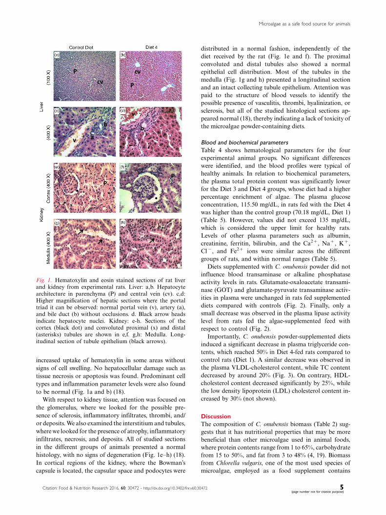
increased uptake of hematoxylin in some areas without
signs of cell swelling. No hepatocellular damage such as
tissue necrosis or apoptosis was found. Predominant cell
types and inflammation parameter levels were also found
to be normal (Fig. 1a and b) (18).
With respect to kidney tissue, attention was focused on
the glomerulus, where we looked for the possible pre-
sence of sclerosis, inflammatory infiltrates, thrombi, and/
or deposits. We also examined the interstitium and tubules,
where we looked for the presence of atrophy, inflammatory
infiltrates, necrosis, and deposits. All of studied sections
in the different groups of animals presented a normal
histology, with no signs of degeneration (Fig. 1e�h) (18).
In cortical regions of the kidney, where the Bowman’s
capsule is located, the capsular space and podocytes were
distributed in a normal fashion, independently of the
diet received by the rat (Fig. 1e and f). The proximal
convoluted and distal tubules also showed a normal
epithelial cell distribution. Most of the tubules in the
medulla (Fig. 1g and h) presented a longitudinal section
and an intact collecting tubule epithelium. Attention was
paid to the structure of blood vessels to identify the
possible presence of vasculitis, thrombi, hyalinization, or
sclerosis, but all of the studied histological sections ap-
peared normal (18), thereby indicating a lack of toxicity of
the microalgae powder-containing diets.
Blood and biochemical parameters
Table 4 shows hematological parameters for the four
experimental animal groups. No significant differences
were identified, and the blood profiles were typical of
healthy animals. In relation to biochemical parameters,
the plasma total protein content was significantly lower
for the Diet 3 and Diet 4 groups, whose diet had a higher
percentage enrichment of algae. The plasma glucose
concentration, 115.50 mg/dL, in rats fed with the Diet 4
was higher than the control group (70.18 mg/dL, Diet 1)
(Table 5). However, values did not exceed 135 mg/dL,
which is considered the upper limit for healthy rats.
Levels of other plasma parameters such as albumin,
creatinine, ferritin, bilirubin, and the Ca2�, Na�, K�,
Cl�, and Fe2� ions were similar across the different
groups of rats, and within normal ranges (Table 5).
Diets supplemented with C. onubensis powder did not
influence blood transaminase or alkaline phosphatase
activity levels in rats. Glutamate-oxaloacetate transami-
nase (GOT) and glutamate-pyruvate transaminase activ-
ities in plasma were unchanged in rats fed supplemented
diets compared with controls (Fig. 2). Finally, only a
small decrease was observed in the plasma lipase activity
level from rats fed the algae-supplemented feed with
respect to control (Fig. 2).
Importantly, C. onubensis powder-supplemented diets
induced a significant decrease in plasma triglyceride con-
tents, which reached 50% in Diet 4-fed rats compared to
control rats (Diet 1). A similar decrease was observed in
the plasma VLDL-cholesterol content, while TC content
decreased by around 20% (Fig. 3). On contrary, HDL-
cholesterol content decreased significantly by 25%, while
the low density lipoprotein (LDL) cholesterol content in-
creased by 30% (not shown).
Discussion
The composition of C. onubensis biomass (Table 2) sug-
gests that it has nutritional properties that may be more
beneficial than other microalgae used in animal foods,
where protein contents range from 1 to 65%, carbohydrate
from 15 to 50%, and fat from 3 to 48% (4, 19). Biomass
from Chlorella vulgaris, one of the most used species of
microalgae, employed as a food supplement contains
Fig. 1. Hematoxylin and eosin stained sections of rat liverand kidney from experimental rats. Liver: a,b. Hepatocytearchitecture in parenchyma (P) and central vein (cv). c,d:Higher magnification of hepatic sections where the portaltriad it can be observed: normal portal vein (v), artery (a),and bile duct (b) without occlusions. d. Black arrow headsindicate hepatocyte nuclei. Kidney: e-h. Sections of thecortex (black dot) and convoluted proximal (x) and distal(asterisks) tubules are shown in e,f. g,h: Medulla. Long-itudinal section of tubule epithelium (black arrows).
Microalgae as a safe food source for animals
Citation: Food & Nutrition Research 2016, 60: 30472 - http://dx.doi.org/10.3402/fnr.v60.30472 5(page number not for citation purpose)

28.0% protein, 49.5% carbohydrate, 17.5% lipid and 4.5%
nucleic acids (20). Microalgal lipids are particularly
interesting because they contain high levels of PUFAs
such as docosahexaenoic (DHA) and eicosapentaenoic
(EPA) acids, as well as carotenoids, such as astaxanthin
and lutein, and other antioxidants (21). C. onubensis
biomass is particularly rich in lutein and b-carotene, which
have high antioxidant activities and are considered very
useful as a source of nutraceuticals (8). Antioxidant
phenolic compounds and vitamins (A precursor, E, and
B group) are also produced by microalgae and accumulate
at different levels depending on the cultivation conditions
(4, 9). Further studies are necessary to examine ways to
increase the antioxidant phenolic compounds and vitamin
contents in C. onubensis biomass, and thereby to improve
the properties of this material. The content in nucleic acids
of C. onubensis is 4.8%, which is within the average nucleic
acid content for most of microalgae, including those which
are used as food for humans (22). This value should be
compared with 1.5 and 2.2% of nucleic acids content found
in beef and beef liver, respectively. It is generally considered
that the long-term maximum acceptable daily intake of
nucleic acid is about 4.0 g/day for an adult (23).
In general, the use of powdered microalgae in diets
here was well tolerated by rats, as indicated by the similar
weight gains in the four experimental groups over the
course of the experiment (Table 3). In this context, a
decrease in the body weight of animals has been observed
with other microalgae-supplemented diets such as Isochrysis
galbana and Nannocholopsis oculata (24), and the red micro-
alga Porphyridium sp. (25). In contrast, rats fed a powdered
form of the cyanobacteria A. flos-aquae showed a body
weight increase of around 10% with respect to the control
group (26).
Histological studies, along with hematological and
biochemical analyses demonstrated that the C. onubensis
powder-supplemented diets were not associated with any
toxic side-effects to animals. The observed plasma levels
of albumin and creatinine suggested that the four groups
of animals had a good nutritional status and good renal
Table 4. Hematological parameters of rats fed control or C. onubensis�supplemented diets
Parameter Diet 1 Diet 2 Diet 3 Diet 4
Hemoglobin (g/dL) 14.5890.99 14.7891.1 15.8890.11 16.0890.05
Hematocrit (%) 44.4392.93 44.2593.12 47.3890.46 48.0090.36
Erythrocytes (cells�10�6/mL) 8.6690.60 8.2490.57 8.7690.13 8.6190.03
Leucocytes (cells�10�3/mL) 5.5790.89 5.3591.04 8.2391.89 6.6591.02
Lymphocytes (%) 84.7591.89 90.0092.64 85.7592.06 89.7590.63
Platelets (cells�10�3/mL) 584.67918.46 618.33929.80 665.00910.13 779.00914.12
Data obtained at the end of the experiment (45 days). Each value is expressed as the mean9SD (n�6 per group). Results were statistically analyzed
with Kruskal�Wallis test. No significant differences were observed at the p B0.05 level among the different diets. More details in Materials and
Methods section.
Table 5. Biochemical parameters of rats fed control or C. onubensis�supplemented diets
Parameter Diet 1 Diet 2 Diet 3 Diet 4
Total protein (g/dL) 6.0590.05 6.1890.02 5.4590.31*# 5.6590.35*#
Albumin (g/dL) 4.4090.11 4.4890.11 4.2390.25 4.2890.23
Ca2 � (mg/dL) 10.4090.50 10.4090.5 10.1990.19 10.4090.32
Na2 � (mEq/L) 145.0090.82 146.0091.78 144.2591.65 147.0090.91
K� (mEq/L) 4.4390.19 4.4390.19 5.2690.69 4.2290.38
Cl� (mEq/L) 90.1391.07 98.1391.07 99.1590.65 99.8891.62
Glucose (mg/dL) 70.1894.97 99.6897.63 115.50914.47 103.03915.44
Creatinine (mg/dL) 0.3290.02 0.3290.02 0.3290.02 0.2790.03
Fe 2 � (mg/dL) 128.9096.44 136.3397.16 137.4597.89 139.58911.89
Ferritin (ng/mL) 185.3095.52 177.9392.81 174.38 99.97 152.70925.70
Bilirubin (mg/dL) 0.0490.01 0.0790.01 0.1090.02 0.0890.03
Data obtained at the end of the experiment (45 days). Each value is expressed as the mean9SD (n�6 per group). Results were statistically analyzed with
Kruskal�Wallis test. The only significant difference observed was for total protein. *pB0.05 compared with the control group. #pB0.05 compared with
other groups. No significant difference was observed at the pB0.05 for other parameters. More details in Materials and Methods section.
Francisco Navarro et al.
6(page number not for citation purpose)
Citation: Food & Nutrition Research 2016, 60: 30472 - http://dx.doi.org/10.3402/fnr.v60.30472

function, while plasma transaminase activity levels de-
monstrated that the liver status of animals was good. In
line with these observations, Ekmay et al. (27) observed a
lack of toxicity in laying hens fed diets supplemented with
biomass from the green microalga Desmodesmus spp. or
the diatom Staurospira spp. The high phenolic compound
content in Spirulina platensis biomass seems to exert a
hepatoprotective effect in rats (28).
Previous findings concerning the plasma glucose con-
centration in animals fed on microalgal biomass are
contentious. For example, I. galbana induced a decrease
in plasma glucose and cholesterol values, whereas con-
sumption of the microalga N. oculata showed no benefit
for normal and diabetic rats (24). The moderate hyper-
glycemic effect induced by C. onubensis powder in rats in
this study (Table 5) had no apparent consequences on
animal health status. This effect cannot be directly attrib-
uted to the microalgae powder because soluble carbohy-
drates found in this biomass were very low (Table 2).
The C. onubensis biomass induced a potent hypolipi-
demic activity in rats, which suggests that this material may
have been used as a source of nutraceuticals in functional
foods and/or have pharmacologic applications. It is im-
portant to note that, in humans and rodents, the liver is
the primary site for de novo lipogenesis, where glucose is
the main carbon precursor for this process. However, a
principle difference between these species is that carry
out the majority of cholesterol in the plasma of rodents
is bound to HDL-lipoproteins, whereas in humans it is
bound to LDL-lipoproteins (29). In animals, ProAlgaZyme,
an infusion of fermented algae biomass, induces a sig-
nificant reduction in body weight, body fat, TC, LDL-
cholesterol, and triglycerides and is accompanied by
a significant increase in HDL-cholesterol levels. This
infusion is well tolerated by animals and does not cause
significant side effects (30). Results obtained with C.
pyrenoidosa are particularly interesting, as this microalga
was shown to prevent hyperlipidemia and atherosclerosis
in rats and hamsters fed chronically with a high-fat diet
(31). A study performed on chickens demonstrated that
both 5 and 10% levels of Porphyridium sp. biomass
supplemented in the diet were effective in lowering
cholesterol levels, while a trend toward lower cholesterol
in eggs was also observed (32). The next step in our
research will be to examine the hypolipidemic effects of
C. onubensis on rats fed a cholesterol-rich diet.
Identification of the active constituents that give rise to
the hypolipidemic activity of C. onubensis must be further
investigated. In this context, PUFAs have been reported
to reduce blood LDL-cholesterol and HDL-cholesterol
levels (33). Van Beelen et al. (34) suggested that omega-3
fatty acids lowered blood LDL-cholesterol levels, producing
Fig. 2. Enzyme activities in plasma from rats fed diets supplemented with C. onubensis biomass. Experimental conditions aredescribed in the Materials and Methods. GOT, glutamate-oxaloacetate transaminase; GPT, glutamate-pyruvate transaminase.Each value is expressed as the mean9SD (n �6 per group). Results were statistically analyzed with Kruskal�Wallis test. Noneof the diets (Diet 2, Diet 3, and Diet 4) showed significant differences at the pB0.05 level with respect to each other, or relativeto the control (Diet 1).
Microalgae as a safe food source for animals
Citation: Food & Nutrition Research 2016, 60: 30472 - http://dx.doi.org/10.3402/fnr.v60.30472 7(page number not for citation purpose)

similar protective effects to those of fish oil against heart
disease, atherosclerosis, cancer, and diabetes. The fatty acid
content of C. onubensis is rich in PUFAs (see Table 2), which
could be responsible for the observed lipoprotein profile
in rats, although it is possible that the presence of other
components could also be involved. Chen et al. (35)
concluded that microalgal lipids are the active compounds
responsible for the triglyceride- and cholesterol-lowering
activity induced in hamsters. Algal-DHA acid induced a
significant dose-related decrease of triglycerides and cho-
lesterol in rats fed a high fructose diet (36). In this context,
it is important to note that algae rich in n-3 fatty acids
have been used in feed supplements for dairy cattle (37).
Our study shows that the acidophilic microalga
C. onubensis provides a good source of dietary fiber,
which may help to promote hypocholesterolemic and
hypotriglyceridemic outcomes in rats due to its high
PUFA content. Polysaccharides and dietary fiber have
also been demonstrated to be involved in the hypolipi-
demic effect of Porphyridium (25). Animal studies have
shown that the consumption of dietary fiber is associated
with changes in lipid metabolism (9). In addition, soluble
polysaccharides and biomass obtained from the red micro-
alga Porphyridium sp. altered the intestinal morphology
and reduced serum cholesterol in rats (25). Several health
benefits derived from the presence of algal polysaccharides
in the diet have also been reported in humans (38, 39). On
contrary, sterol extracts from the alga Schizochytrium sp.
were as effective as b-sitosterol in reducing plasma
cholesterol concentrations (40).
Conclusions
Based on histological, hematological, and biochemical
analyses, this study shows that a diet enriched with
C. onubensis biomass is non-toxic to rats. No negative
effects were observed on body weight, physiological or
morphological parameters over the course of the experi-
mental period. C. onubensis biomass also had significant
hypocholesterolemic and hypotriglyceridemic effects in
rats. As such, C. onubensis biomass may serve directly as
a functional food for animals or as source of nutraceutical
and pharmacological compounds.
Acknowledgements
The authors acknowledge the financial support received from the
University of Huelva and Andalusian Government (Research
project. BIO-214).
Fig. 3. Lipid profiles of rats fed with diets supplemented with C. onubensis biomass. Experimental conditions are described inthe Materials and Methods section. Each value is expressed as the mean9SD (n�6 per group). Results were statisticallyanalyzed with Kruskal�Wallis test. *pB0.05 compared with the control group. None of the diets (Diet 2, Diet 3, and Diet 4)showed significant differences at the pB0.05 level with respect to each other. HDL�High density lipoprotein. VLDL�Verylow density lipoprotein.
Francisco Navarro et al.
8(page number not for citation purpose)
Citation: Food & Nutrition Research 2016, 60: 30472 - http://dx.doi.org/10.3402/fnr.v60.30472

Conflict of interest and funding
The authors declare no conflict of interest.
References
1. Forjan E, Navarro F, Cuaresma M, Vaquero I, Ruiz-Domınguez
MC, Gojkovic Z, et al. Microalgae: fast-growth sustainable
green factories. Crit Rev Environ Sci Technol 2015; 45:
1705�55.
2. Kay RA. Microalgae as food and supplement. Crit Rev Food
Sci Nutr 1991; 30: 555�73.
3. Gantar M, Svircev Z. Microalgae and cyanobacteria: food for
thought. J Phycol 2008; 44: 60�8.
4. Bishop WM, Zubeck HM. Evaluation of microalgae for use as
nutraceuticals and nutritional supplements. J Nutr Food Sci
2012; 2: 147�52.
5. Yaajob Z, Ali E, Zainai A, Mohamad M, Takriff MS. An
overview: biomolecules from microalgae for animal feed and
aquaculture. J Biol Res 2014; 21: 6.
6. Varshney P, Mikulic P, Vonshak A, Beardall J, Wangikar PP.
Extremophilic micro-algae and their potential contribution in
biotechnology. Bioresour Technol 2015; 184: 363�72.
7. Graziani G, Schiavo S, Nicolai MA, Buono S, Fogliano V,
Pinto G, et al. Microalgae as human food: chemical and
nutritional characteristics of the thermo-acidophilic microalga
Galdieria sulphuraria. Food Funct 2013; 4: 144�52.
8. Vaquero I, Mogedas B, Ruiz-Domınguez MC, Vega JM,
Vılchez C. Light-mediated lutein enrichment of an acid en-
vironment microalga. Algal Res 2014; 6: 70�7.
9. Spolaore P, Joannis-Cassan C, Duran E, Isambert A. Commer-
cial applications of Microalgae. J Bios Bioeng 2006; 101: 87�96.
10. Vılchez C, Forjan E, Cuaresma M, Bedmar F, Garbayo I,
Vega JM. Marine carotenoids: biological functions and com-
mercial applications. Mar Drugs 2011; 9: 319�33.
11. Plaza M, Santoyo S, Jaime L, Garcıa-Blairsy G, Herrero M,
Senorans FJ, et al. Screening for bioactive compounds from
algae. J Pharm Biomed Anal 2010; 51: 450�5.
12. Buono S, Langellotti AL, Martello A, Rinna F, Fogliano V.
Functional ingredients from microalgae. Food Funct 2014; 5:
1669�85.
13. Gouveia L, Marques AE, Sousa JM, Moura P, Bandarra NM.
Microalgae-source of natural bioactive molecules as functional
ingredients. Food Sci Technol Bull Funct Foods 2010; 7:
21�37.
14. De Jesus-Raposo MF, de Morais AM, de Morais RM. Marine
polysaccharides from algae with potential biomedical applica-
tions. Mar Drugs 2015; 13: 2967�3028.
15. Ruiz-Domınguez MC, Vaquero I, Obregon V, de la Morena B,
Vılchez C, Vega JM. Lipid accumulation and antioxidant activity
in the eukaryotic acidophilic microalga Coccomyxa sp. (strain
onubensis) under nutrient starvation. J Appl Phycol 2015; 27:
1099�108.
16. Fuentes JL, Huss VAR, Montero Z, Torronteras R, Cuaresma M,
Garbayo I, et al. Phylogenetic characterization and morphologi-
cal and physiological aspects of a novel acidotolerant and
halotolerant microalga Coccomyxa onubensis sp. nov. (Chloro-
phyta, Trebouxiophyceae). J Appl Phycol 2016; doi: http://dx.
doi.org/10.1007/s10811-016-0887-3
17. Bain BJ, Lewis SM. Preparation and staining methods for blood
and bone marrow film. In: Lewis SM, Bain B, Bates I, eds.
Dacie and Lewis Practical Haematology, 10th ed. Philadelphia:
Churchill Livingston; 2006, pp. 59�78.
18. Kierszenbaum AL. Histologıa y Biologıa Celular. Barcelona:
Elsevier Espana; 2008.
19. Tibetts SM, Milley JE, Lall SP. Chemical composition and
nutritional properties of fresh water and marine microalgal
biomass cultured in photobioreactors. J Appl Phycol 2015; 27:
1109�19.
20. Lardon L, Helias A, Sialve B, Steyer J-P, Bernard O. Life-cycle
assessment of biodiesel production from microalgae. Environ
Sci Technol 2015; 3: 386�95.
21. Christaki E, Florou-Paneri P, Bonos E. Microalgae: a
novel ingredient in nutrition. Int J Food Sci Nutr 2011; 62:
794�9.
22. Becker EW. Microalgae: biotechnology and microbiology.
Cambridge: Cambridge University Press; 1994.
23. Zepka LQ, Jacob-Lopes E, Goldbeck R, Souza-Soares LA,
Queiroz MI. Nutritonal evaluation of single-cell protein pro-
duced by Aphanothece microscopica Nageli. Bioresourc Technol
2010; 101: 7107�11.
24. Nuno K, Villarruel-Lopez A, Puebla-Perez AM, Romero-
Velarde E, Puebla-Mora AG, Ascencio F. Effects of the marine
microalgae Isochrysis galbana and Nanochloropsis oculata in
diabetic rats. J Funct Foods 2013; 5: 106�15.
25. Dvir I, Chayoth R, Sod-Moriah U, Shany S, Nyska A, Stark A,
et al. Soluble polysaccharide and biomass of red microalga
Pophyridium sp. alter intestinal morphology and reduce serum
cholesterol in rats. Br J Nutr 2000; 84: 469�76.
26. Kushak RI, Drapeau C, Winter HS. The effect of blue-green
algae Aphanizomenon flos-aquae on nutrient assimilation in rats.
J Nutrac Nutr 2001; 3: 35�9.
27. Ekmay RD, Chou K, Lei XG. Continual feeding of two types of
microalgal biomass affected protein digestion and metabolism
in laying hens. J Anim Sci 2015; 93: 287�97.
28. Kepekci RA, Polat S, Celik A, Bayat N, Saygideger SD.
Protective effect of Spirulina platensis enriched in phenolic
compounds against hepatotoxicity induced by CCl4. Food
Chem 2013; 141: 1972�9.
29. Bergen WG, Mersmann HJ. Comparative aspects of lipid
metabolism: impact on contemporary research and use of
animal models. J Nutr 2005; 135: 2499�502.
30. Geamanu A, Goja A, Saadat N, Khosla P, Gupta SV.
ProAlgaZyme subfraction improves the lipoprotein profile of
hypercholesterolemic hamsters, while inhibiting production of
betaine, carnitine, and choline metabolites. Nutr Metabol 2013;
10: 55�64.
31. Cherng J-Y, Shih M-F. Preventing dyslipidemia by Chlorella
pyrenoidosa in rats and hamsters after chronic high fat diet
treatment. Life Sci 2005; 76: 3001�13.
32. Ginzberg A, Cohen M, Sod-Moriah UA, Shany S,
Rosenshtrauch A, Arad S. Chickens fed with biomass of the
red microalga Porphyridium sp. have reduced blood cholesterol
level and modified fatty acid composition in egg yolk. J Appl
Phycol 2000; 12: 325�30.
33. Komprda T, Zornikova G, Knoll A, Vykoukalova Z,
Rozikova V, Skultety O, et al. Effect of dietary eicosapentaenoic
and docosahexaenoic acid on expression of rat liver genes
controlling cholesterol homeostasis and on plasma cholesterol
level. Czech J Anim Sci 2014; 59: 391�8.
34. Van Beelen VA, Spenkelink B, Mooibroek H, Sijtsma L,
Bosch D, Rietjens IM, et al. An n-3 PUFA-rich microalgal oil
diet protects to a similar extent as a fish oil-rich diet against
AOM-induced colonic aberrant cryp foci in F344 rats. Food
Chem Toxicol 2009; 47: 316�20.
35. Chen J, Jiang Y, Ma K-Y, Chen F, Chen Z-Y. Microalga
decreases plasma cholesterol by down-regulation of intestinal
NPC1L1, hepatic LDL receptor, and HMG-CoA reductase. J
Agric Food Chem 2011; 59: 6790�7.
Microalgae as a safe food source for animals
Citation: Food & Nutrition Research 2016, 60: 30472 - http://dx.doi.org/10.3402/fnr.v60.30472 9(page number not for citation purpose)

36. Ryan AS, Bailey-Hall E, Nelson EB, Salem N Jr. The
hypolipidemic effect of an ethyl ester of algal-docosahexaenoic
acid in rats fed a high-fructose diet. Lipids 2009; 44: 817�26.37. Stamey JA, Shepherd DM, de Veth MJ, Corl BA. Use of alga or
algal oil rich in n-3 fatty acids as a feed supplement for dairy
cattle. J Dairy Sci 2012; 95: 5269�412.
38. Werman MJ, Sukenik A, Mokady S. Effect of the marine
unicellular alga Nanochloropsis sp. to reduce the plasma and
liver cholesterol levels in male rats fed on diets with cholesterol.
Biosci Biotechnol Biochem 2003; 67: 2266�8.
39. Misurcova L, Skrovankova S, Samek D, Ambrozova J,
Machu L. Health benefits of algal polysaccharides in human
nutrition. Adv Food Nutr Res 2012; 66: 75�145.
40. Chen J, Jiao R, Jiang Y, Bi Y, Chen Z-Y. Algal sterols are as
effective as b-sitosterol in reducing plasma cholesterol concen-
tration. J Agric Food Chem 2014; 62: 675�81.
*Carlos Vılchez
Algal Biotechnology Group
CIDERTA and Faculty of Sciences
University of Huelva and
Marine International Campus of Excellence (CEIMAR)
ES-21007 Huelva, Spain
Email: [email protected]
Francisco Navarro et al.
10(page number not for citation purpose)
Citation: Food & Nutrition Research 2016, 60: 30472 - http://dx.doi.org/10.3402/fnr.v60.30472

![Industrial application of microalgae in the circular ... · Industrial application of microalgae in the circular bioeconomy Dorinde Kleinegris [Applied Biotechnology / Microalgae]](https://img.dokumen.tips/doc/110x75/5ead3c152d0239422909016e/industrial-application-of-microalgae-in-the-circular-industrial-application.jpg)