Embed Size (px)
Citation preview

Mice diseases

Bacterial diseases
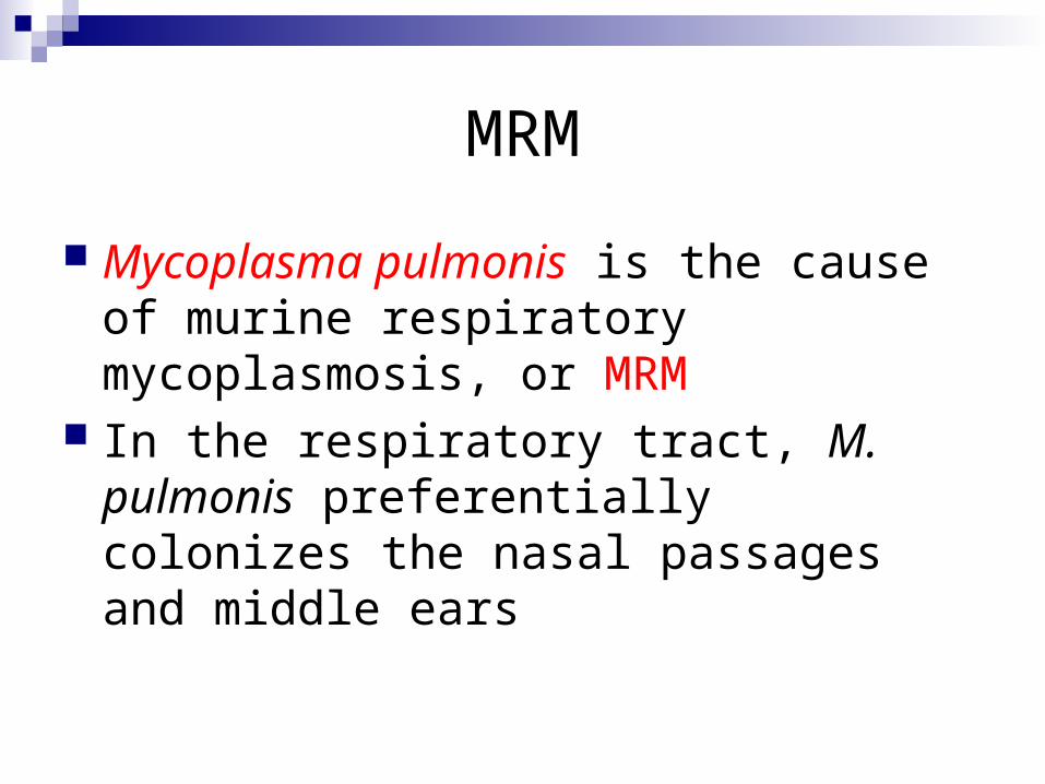
MRM
Mycoplasma pulmonis is the cause of murine respiratory mycoplasmosis, or MRM
In the respiratory tract, M. pulmonis preferentially colonizes the nasal passages and middle ears

Etiology - Mycoplasma pulmonis
Most infections are subclinical- carried in nasopharynx
Disease induced by high cage ammonia or pathogens
Slow onset, chronic condition Aerosol and direct contact “Proximal Airway Disease”

Mycoplasma pulmonis Early signs: purulent nasal discharge, otitis
media, sneezing, “sniffling”, porphyrin staining
Progresses to labored breathing, anorexia, lethargy, hunched posture
Bronchopneumonia

Gross Lesions
In advanced disease, the lungs can have gray-purple patches that often have a "cobblestone" appearance.
Pictures…

The left lobe of these lungs from a mouse with experimental MRM has a gray-purple "cobblestone" appearance due to accumulation of inflammatory cells in
and around airways,

Diagnosis
Commercial ELISA and IFA tests are useful for health monitoring, but serologic testing can be complicated by potential cross reactions with other rodent mycoplasmas.

Treatment
Tetracycline limits losses Enrofloxacin Dosages p.58

Tyzzer’s disease
Clostridium piliforme is the cause of Tyzzer’s disease. It is a gram-negative, motile, spore-forming bacillus. It has been grown in embryonated eggs and cell cultures
Epizootiology
C. piliforme has a worldwide distribution and a wide host range, including rats, mice, gerbils, hamsters, guinea pigs, rabbits, dogs, cats, horses, nonhuman primates, and various wild species. Mice appear to be affected more often than rats.

Clinical signs
Many affected rats and mice are simply found dead without prior signs being observed. If present, clinical signs include lethargy, ruffled fur, and diarrhea, as indicated by fecal soiling of the fur.

Transmission
Transmission is presumably fecal-oral. Possibilities include vertical transmission
and introduction of spores via vermin*; contaminated feed; or incompletely sterilized food, bedding, or water. Morbidity and mortality are highly variable.
*term applied to various animal species regarded as pests or nuisances and especially to those associated with the carrying of disease.

The most consistent lesions in mice are multiple, pale, slightly depressed foci of necrosis in the liver, as seen here. Thickening and hyperemia of the intestine and pale areas in the myocardium also can be seen in some cases, but are not consistently present.

Pathogenesis
C. piliforme becomes established in the intestine as a primary infection, usually in the ileum, cecum, or both. It spreads via the portal circulation to the liver, and from there via the blood to other organs, chiefly the heart.

Diagnosis:
Since the organism cannot be propagated on artificial media, histopathologic diagnoses are made by demonstration of the bacillus in the enterocytes, hepatocytes or cardiacmyocytes bordering necrotic foci in tissues stained with silver stains

Treatment:
Oxytetracycline at 0.1 mg/ml water for 30 days was reported to abate mortality of an epizootic in mice. Treatment is usually not warranted

Salmonella
Salmonella enteritidis is a gram-negative bacillus. Salmonellae are identified according to serotype, of which over 1500 exist.
Virulence varies widely among serotypes; enteritidis and typhimurium are most commonly associated with disease in rodents

Epizootiology
Salmonellosis was a major epizootic and enzootic disease of laboratory mice prior to the 1950s, but clinical salmonellosis in mice is now rare. Transmission is fecal-oral. Salmonellae in general are not very host specific, and carriers are common among exposed populations. Therefore, potential sources of infection for laboratory mice include vermin; contaminated food, water, bedding, and fomites

Clinical signs
Most infections are subclinical. Signs in mice infected with weakly virulent
serotypes vary from none to occasional losses among sucklings and weanlings

Gross lesions
This mouse with salmonellosis has pale foci of necrotizing and suppurative hepatitis at the margins of the liver lobes (arrows). The spleen is greatly enlarged due to production of increased numbers of neutrophils, or myeloid hyperplasia.

Diagnosis
Diagnosis of salmonellosis is by culture and serotyping. Several sites should be cultured, including liver, spleen, mesenteric lymph nodes, blood, and intestinal contents.

Control
Special attention should be given to vermin control and procurement and storage of bedding materials and food, which are easily contaminated by wild rodents. If salmonellosis is diagnosed in a research facility, the facility should be immediately quarantined, the affected populations destroyed and their environment decontaminated.
ZOONOTIC disease!!!
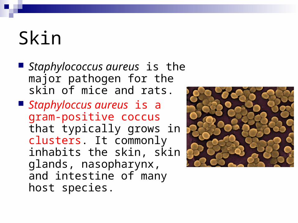
Skin Staphylococcus aureus is the
major pathogen for the skin of mice and rats.
Staphyloccus aureus is a gram-positive coccus that typically grows in clusters. It commonly inhabits the skin, skin glands, nasopharynx, and intestine of many host species.

Epizootiology
Staph. aureus is a common commensal of many species. It also is common in the environment.
Human carriers can be an important source of infection for rodent colonies

Staph. aureus is associated mostly with dermatitis in the form of pyoderma or ulcerative dermatitis. The face, shoulders, neck, and ears are most commonly affected.

Staphylococcus aureus
Abscesses and granulomas, most commonly affecting the face and tissues around the base of the tail, also occur and can be associated with fight wounds

Staphylococcus aureus
The best methods of control are improved sanitation, frequent sanitizing of cages and other equipment, and elimination of equipment that could cause skin injury
Topical treatment with Nolvasan BID

Viral diseases
SDAV (Sendai Virus) SV
Sialodacryoadenitis virus (SDAV), another coronavirus (Parainfluenza 1), is one of the most common viruses found in laboratory rats and mice. It is highly contagious, and is spread by direct contact with infected animals or by respiratory aerosol

SDAV
The incubation period for SDAV less than 1 week. In naive populations, a sudden high incidence of overt disease with sneezing, porphyrin-stained nasal and ocular discharges (as seen in this image), cervical edema, corneal ulceration, and kerato conus may be the first indications of a problem

Sendai Virus
This image shows swollen submandibular salivary glands (arrows) in a mouse with SDAV. SDAV has tissue tropism for the submaxillary and parotid salivary, exorbital, Harderian, and intraorbital lacrimal glands

In rats and mice, few gross morphologic lesions are seen in uncomplicated Sendai infections. The lungs can be focally reddened and atelectatic with serous fluid visible in the pleural and pericardial cavities.

Mouse Hepatitis Virus (MHV)
Mouse hepatitis virus (MHV) and sialodacryoadenitis virus (SDAV) are frequently encountered coronaviruses of mice and rats respectively
Some primarily infect the gastrointestinal system; some the respiratory tract; some the brain

MHV
Mice (Mus musculus) are the only natural hosts. MHV is extremely contagious with prevalence rates exceeding 80% in outbreaks. Active infection lasts 2-3 weeks, during which mice shed the virus in gastrointestinal and respiratory excretions. Direct contact with shedding mice, contaminated cell lines, fomites, or airborne particles are the important routes of viral transmission.

MHV
An epizootic can produce nonspecific clinical signs in naïve*, juvenile mice, such as runting, as shown here, or failure to thrive
not previously subjected to experimentation or a particular experimental situation <made the test with naive mouse>

MHV
This image shows large coalescing cream-colored friable foci (arrow) of necrosis that result when acute multifocal hepatitis progresses to chronic active hepatitis in nude mice. Gross pathology in immune-incompetent mice is more generalized and pro gressive than in immune-competent mice.

Mouse Hepatitis Virus
Enterotropic versus respiratory strains Extremely contagious by many routes Nursing pups: diarrhea and mortality Weanlings: Obstipation Adults: Hunched posture, weight loss, rough hair
coat, variable mortality No latent infections- stop breeding ELISA

Mouse Hepatitis Virus
Diagnosis of latent infections is dependent on the histologic demonstration of large, multinucleate syncytial cells (arrow) in the liver, brain, or mucosal epithelium of the intestine

Rotavirus
Rotavirus, another genus of the family Reoviridae, is associated with clinical disease. Rotaviruses affecting mice and rats respectively are mouse rotavirus, a group-A rotavirus associated with the syndrome, epizootic diarrhea of infant mice (EDIM)

Rotavirus
Epizootic Diarrhea of Infant Mice (EDIM) Most susceptible: birth to 17 days of age Fecal-oral transmission Yellow, watery diarrhea in 14-17 day old pups Death with a full stomach (versus reovirus),
shortened intestinal villi Unapparent viral carriers ELISA

EDIM
Neonatal diarrhea is the most prominent sign. Watery yellow stool accumulates around the anus and tailbase, soiling the coats of neonatal pups and their dams. Pups appear stunted and lethargic and have distended abdomens. Mortality rates are low

Rotavirus

Parasites

Myobia musculi, Radfordia affinis, and Myocoptes musculinus
Common Transmitted by direct contact May be subclinical, or develop pruritus, scruffy
coat, patchy alopecia, self-trauma, pyoderma Typically found on back and head Mist with ivermectin (0.1%) for 3 weekly
treatments

Myobia musculi
Myobia musculi lesions in a pet white mouse, characteristic of hypersensitivity to the mites. Intense pruritus, often directed at the neck and ears, leads to self-mutilation. Early lesions consist of subtle hair thinning on the dorsal neck and shoulders
Acariasis???

Myobia musculi
Single claw

Radfordia affinis
Two claws

Myocoptes musculinus

Pinworms
Common Fecal-oral transmission Usually subclinical, but may cause rectal
prolapse Fecal floatation (Aspiculuris tetraptera) or tape
test (Syphacia obvelata) Ivermectin misting (others more labor intensive) Clean environment well
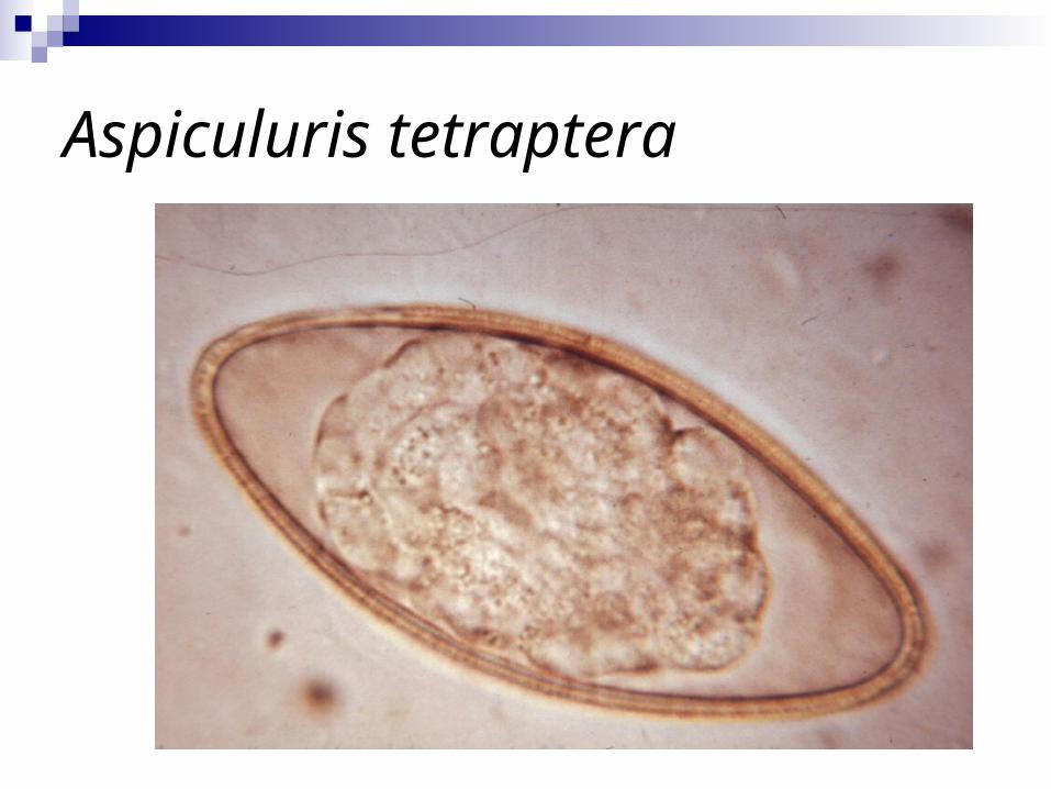
Aspiculuris tetraptera

Syphacia obvelata

Rodentolepis nana and Hymenolepis diminuta
Common tapeworms among pet mice Roaches, beetles, & fleas = intermediate
hosts R. nana: also transmitted directly, or by
autoinfection (retroinfection) Usually subclinical infection

Rodentolepis nana and Hymenolepis diminuta
Often find proglottids in feces instead of individual eggs
Zoonotic (more commonly R. nana because of direct transmission)
Praziquantel is effective
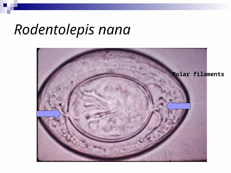
Rodentolepis nana
Polar filaments

Tumors

Mammary Adenocarcinoma
Most common tumor of mice Mice have 3 pair of thoracic and 2 pair
of abdominal mammary glands- glandular tissue may be found up around the body to the dorsum
Poor prognosis- anaplastic and very invasive

Mammary Adenocarcinoma

Tissue: Mouse: mammary gland; adenocarcinoma

MiscellaneousDiseases

Barbering
Animal model of trichotillomania MOBS (“Move Over Buddy” Syndrome) Alopecia (R.O. ectoparasites, dermatophytes,
endocrinopathy) Well demarcated area of alopecia without
dermatitis- exposed skin appears normal Commonly involves hair over the nasal and
orbital regions, or over the dorsal cervical area Separate out barber

Barbering

Bite Wounds
Males fight and abuse females Bites often found on face, back, and
genital area May abscess Nolvasan (+ lance abscesses) Separate offenders Provide enrichment

Malocclusion
Genetic predisposition (autosomal recessive)
Incisors hypsodont Inanition, starvation Trim teeth with nail clippers (no “scissor”
action) Do not breed these mice

Malocclusion