Embed Size (px)
Citation preview
RESEARCH Open Access
Mesothelin is a target of chimeric antigenreceptor T cells for treating gastric cancerJiang Lv1,2,3†, Ruocong Zhao1,2,3†, Di Wu1,2,4†, Diwei Zheng1,2,3, Zhiping Wu1,2,3, Jingxuan Shi1,2,3, Xinru Wei1,2,Qiting Wu1,2, Youguo Long1,2, Simiao Lin1,2, Suna Wang1,2, Zhi Wang5, Yang Li6, Yantao Chen7, Qing He8,Suimin Chen9, Huihui Yao10, Zixia Liu11, Zhaoyang Tang12, Yao Yao1,2, Duanqing Pei1,2, Pentao Liu13,Xuchao Zhang14, Zhenfeng Zhang15, Shuzhong Cui16, Ren Chen17 and Peng Li1,2,11,18*
Abstract
Background: Gastric cancer (GC) is a common cancer in Asia and currently lacks a targeted therapy approach.Mesothelin (MSLN) has been reported to be expressed in GC tissue and could be targeted by chimeric antigenreceptor (CAR) T cells. Mesothelin targeting CAR-T has been reported in mesothelioma, lung cancer, breast cancer,and pancreas cancer. However, the feasibility of using anti-MSLN CAR T cells to treat GC remains to be studied.
Methods: We verified MSLN expression in primary human GC tissues and GC cell lines and then redirected T cellswith a CAR containing the MSLN scFv (single-chain variable fragment), CD3ζ, CD28, and DAP10 intracellular signalingdomain (M28z10) to target MSLN. We evaluated the function of these CAR T cells in vitro in terms of cytotoxicity, cytokinesecretion, and surface phenotype changes when they encountered MSLN+ GC cells. We also established four differentxenograft GC mouse models to assess in vivo antitumor activity.
Results: M28z10 T cells exhibited strong cytotoxicity and cytokine-secreting ability against GC cells in vitro. In addition,cell surface phenotyping suggested significant activation of M28z10 T cells upon target cell stimulation. M28z10 T cellsinduced GC regression in different xenograft mouse models and prolonged the survival of these mice compared withGFP-transduced T cells in the intraperitoneal and pulmonary metastatic GC models. Importantly, peritumoral deliverystrategy can lead to improved CAR-T cells infiltration into tumor tissue and significantly suppress the growth of GC in asubcutaneous GC model.
Conclusion: These results demonstrate that M28z10 T cells possess strong antitumor activity and represent a promisingtherapeutic approach to GC.
Keywords: Gastric cancer, Mesothelin, Chimeric antigen receptor T cells, Immunotherapy, Immunodeficient mice
BackgroundGastric cancer (GC) is one of the most common cancertypes worldwide and is the second leading cause ofcancer-related death in Asia [1]. Currently, the 5-yearsurvival rate of GC patients is approximately 20–30%.Most patients are diagnosed with advanced stage or
metastatic disease and therefore are not candidates forsurgery [2–4]. Although overall survival and quality oflife are improved by chemo- and radiotherapeutic ap-proaches, the median overall survival remains less than12 months for patients diagnosed with advanced stageGC [3, 5]. Recently, targeted strategies, including HER-2and VEGFR-2 monoclonal antibodies, have achievedsubstantial responses in GC patients [5–10]. Meanwhile,immune checkpoint inhibitors, such as antibodiesagainst CTLA-4, PD-1, or PD-L1, are being investigatedin different phase clinical trials [11–14]. Nonetheless,the response rate and progression-free survival remainpoor for patients with advanced GC.
* Correspondence: [email protected]†Jiang Lv, Rooking Zhao and Di Wu contributed equally to this work.1Key Laboratory of Regenerative Biology, South China Institute for Stem CellBiology and Regenerative Medicine, Guangzhou Institutes of Biomedicineand Health, Chinese Academy of Sciences, Guangzhou, China2Guangdong Provincial Key Laboratory of Stem Cell and RegenerativeMedicine, South China Institute for Stem Cell Biology and RegenerativeMedicine, Guangzhou Institutes of Biomedicine and Health, ChineseAcademy of Sciences, Guangzhou, ChinaFull list of author information is available at the end of the article
© The Author(s). 2019 Open Access This article is distributed under the terms of the Creative Commons Attribution 4.0International License (http://creativecommons.org/licenses/by/4.0/), which permits unrestricted use, distribution, andreproduction in any medium, provided you give appropriate credit to the original author(s) and the source, provide a link tothe Creative Commons license, and indicate if changes were made. The Creative Commons Public Domain Dedication waiver(http://creativecommons.org/publicdomain/zero/1.0/) applies to the data made available in this article, unless otherwise stated.
Lv et al. Journal of Hematology & Oncology (2019) 12:18 https://doi.org/10.1186/s13045-019-0704-y
Chimeric antigen receptor (CAR) T cell therapy hasbeen developed for the treatment of different types ofcancer, including acute lymphoblastic leukemia and va-rious solid cancers [15–20]. CARs are composed of anextracellular single-chain variable fragment (scFv) thatrecognizes diverse tumor-associated antigens (TAAs), atransmembrane fragment, a CD3ζ T cell activatingdomain, and a costimulatory domain, such as the intracel-lular domains of CD28 and 4-1BB [21–24]. Upon specificbinding of CAR to a TAA on target tumor cells, the CD3ζand costimulatory domains are activated, and the phos-phorylation cascade is triggered in T cells, leading to therelease of cytotoxic granules, the transcription of genesencoding cytokines, and cell proliferation [25].Mesothelin (MSLN) is a 40-kDa membrane protein
that has been reported to be expressed on normal meso-thelial tissue and highly expressed in mesothelioma andlung, pancreas, breast, ovarian, and gastric cancer [26–28]. To date, MSLN has been utilized as a target of CART cells against solid cancers, including mesothelioma,lung cancer, breast cancer, and pancreas cancer [29–33].However, the feasibility of using anti-MSLN CAR T cellsto treat GC remains to be explored. In the previousstudies, we constructed third-generation CAR M28z10and demonstrated the improved anti-tumor activitiescompared with the second-generation CAR M28zagainst lung cancer [34]. In the present study, we firstlyidentified MSLN as an available target on GC tissue andcells and then characterized the efficacy of anti-MSLNCAR (M28z10) T cells against GC in multiple in vitrofunctional assays and in vivo xenograft mouse models,and we proved the improved CAR-T cell infiltration andefficacy by regional peritumoral delivery compared withsystemic intravenous delivery approach. Our findingsdemonstrate that M28z10 T cells possess strong antitu-mor activity against GC and pave the way for the futureclinical application of this treatment for GC patients.
Materials and methodsChimeric antigen receptor vector designThe third-generation anti-MSLN CARs containing bothCD28 and DAP10 cytoplasmic sequences were previouslyreported [34, 35]. Briefly, human DAP10 cytoplasmic do-main sequence was obtained from the UniProt database(ID: Q9UBK5). The third-generation anti-MSLN contain-ing the DAP10 cytoplasmic sequence were synthesized byGenscript (Nanjing) Co., Ltd. (Nanjing, China), and clonedinto the second-generation lentiviral vector pWPXLd-2A-eGFP through Pme1 and Spe1 cloning sites.
Lentivirus productionLentivirus particles were produced in HEK-293 T cellsvia polyethyleneimine (Sigma-Aldrich, St Louis, MO,USA) transfection. The pWPXLd-based lentiviral plasmid
and two packaging plasmids, psPAX2 and pMD.2G, werecotransduced into HEK-293 T cells in 10 cm dish at a ratioof 3:1:4, with a total amount of 24 μg. Lentivirus-contain-ing supernatants were harvested at 24, 48, and 72 h post-transfection and filtered through a 0.45-μm filter.
Isolation, transduction, and expansion of primary humanT lymphocytesPeripheral blood mononuclear cells (PBMCs) were iso-lated from the buffy coats of healthy donors using Lym-phoprep (Fresenius Kabi Norge, AS, Berg i Østfold,Norway). T cells were negatively selected from PBMCsusing a MACS Pan T Cell Isolation Kit (Miltenyi Biotec,Bergish Gladbach, Germany) and activated usingmicrobeads coated with anti-human CD3, anti-humanCD2, and anti-human CD28 antibodies (Miltenyi Biotec)at a 1:1 bead:cell ratio for 24 h in RPMI-1640 supple-mented with 10% heat-inactivated fetal bovine serum(FBS), 40 IU/ml interleukin-2 (IL-2), 10 mM HEPES,2 mM glutamine, and 1% penicillin/streptomycin. Every1*106 T cells were transduced with 5–10 ml CAR lenti-viral supernatants in the presence of 8 μg/ml polybrene(Sigma) for 5 h with 1 ml 10% FBS containingRPMI1640, and a continuous two rounds of transductionwere conducted. After transduction, T cells were cul-tured in fresh media containing IL-2 (300 IU/ml). Subse-quently, fresh media were added every 2–3 days tomaintain cell density within the range of 0.5–1 × 10⁶/ml.Healthy PBMC donors who provided primary specimensgave informed consent for the use of their samples forresearch purposes, and all procedures were approved bythe Research Ethics Board of Guangzhou Institutes ofBiomedicine and Health (GIBH).
Cells and culture conditionsHEK-293 T cells were maintained in Dulbecco’s modifiedEagle medium (Gibco, Grand Island, NY, USA). K562(human myelogenous leukemia cell line), AGS (humangastric adenocarcinoma), BGC-823 (human gastricadenocarcinoma), KATO III (human gastric carcinoma),and MKN-28 (human gastric carcinoma) cell lines wereobtained from ATCC (Manassas, VA, USA) and main-tained in RPMI-1640. Luciferase/GFP-expressing celllines (K562-GL, AGS-GL, BGC-823-GL, KATO III-GL,MKN-28-GL) were generated by transfection of the par-ental cell line with lentiviral supernatant containingluciferase-2A-GFP and were sorted for GFP expressionon a FACS AriaTM cell sorter (BD Biosciences, San Jose,CA, USA). DMEM and RPMI-1640 media were supple-mented with 10% heat-inactivated FBS (Gibco/Life Tech-nologies), 10 mM HEPES, 2 mM glutamine (Gibco/LifeTechnologies), and 1% penicillin/streptomycin. Allcells were cultured at 37 °C in an atmosphere of 5%carbon dioxide.
Lv et al. Journal of Hematology & Oncology (2019) 12:18 Page 2 of 14
Flow cytometryAll samples were analyzed using a NovoCyteTM (ACEABiosciences), LSR Fortessa, or C6 flow cytometer (BD Bio-sciences), and data were analyzed using FlowJo software(FlowJo, LLC, Ashland, OR, USA). The antibodies usedincluded anti-MSLN-biotin (clone MB), Streptavidin-APC, anti-human CCR7-APC (clone 3D12), anti-humanCD62L-PE (clone DREG-56), anti-human CD45RA-APC(clone HI100), anti-human CD45RO-PE (clone UCHL1),anti-human TIM3-PE (clone F38-2E2), anti-humanLAG3-PerCP/Cy5.5 (clone 11C3C65), anti-humanPD-1-APC (clone NAT105), anti-human CD27-PE (cloneM-T271), anti-human CD28-APC (clone CD28.2),anti-human CD25-PE (clone BC96), anti-human CD69-APC (clone FN50), anti-human CD107a-APC (cloneH4A3), anti-human CD3-APC (clone UCHT1), anti->hu->human <?A3B2 thyc=CD4-PerCP/Cy5.5 (clone OKT4),anti-human CD8-PE (clone OKT8), mouse IgG2a isotypecontrol-APC (clone RMG2a-62), mouse IgG1kappaisotype control-PE, mouse IgG1kappa isotype control-PerCP/Cy5.5, and mouse IgG1kappa isotype control-APC(clone MOPC-21) (Biolegend, San Diego, CA, USA). AllFACS-related staining procedures were performed on icefor 30 min, and cells were then washed with PBS con-taining 1% FBS before cytometry analysis. PB, spleen(SP), and tumor samples from mouse xenografts weretreated with red blood cell lysis buffer (Biolegend),and the cells were stained with the correspondingantibodies.
In vitro tumor killing assaysAGS-GL, BGC-823-GL, KATO III-GL, and MKN-28-GLtarget cells were incubated with GFP or CAR-MSLN Tcells at the indicated ratio in triplicate wells of white96-well plates. Target cell viability was monitored 18 hlater by adding 100 μl/well D-luciferin (potassium salt)(Cayman Chemical, USA) at 150 μg/ml. Backgroundluminescence was negligible (< 1% of the signal fromwells containing only target cells). The percent via-bility (%) was calculated as experimental signal/ma-ximal signal × 100, and the percent lysis was equal to100% viability.
Cytokine release assaysEnzyme-linked immune absorbance assay (ELISA) kits forIL-2, interferon-γ (IFN-γ), granzyme B, and granulocyte-#macrophage colony-stimulating factor (GM-CSF) werepurchased from eBioscience (San Diego, CA, USA), and allELISAs were performed according to the manufacturer’sprotocols. T cells were cocultured with target cells at anE:T ratio of 1:2 for 18 h. The culture supernatants werethen collected and analyzed for the secretion of IL-2, IFN-γ,GM-CSF, and granzyme B using ELISA kits.
CDX models for CAR T cell treatmentAnimal experiments were performed in the LaboratoryAnimal Center of GIBH, and all animal procedures wereapproved by the Animal Welfare Committee of GIBH.All protocols were approved by the relevant InstitutionalAnimal Care and Use Committee (IACUC). All micewere maintained in specific pathogen-free (SPF)-gradecages and were provided autoclaved food and water.For the intraperitoneal GC models, 1 × 10⁶ BGC-823-GL
cells in 100 μl PBS were injected into the peritoneal cavityof NSI mice aged 6–8 weeks. Two weeks after tumor cellinjection, the mice were subjected to bioluminescenceimaging (BLI) and randomly divided into three groups:NC, GFP-T, and M28z10-T. Mice received 5 × 10⁶
M28z10-T cells or equivalent number of GFP-T cellssuspended in 100 μl PBS by tail vein injection. On day 21and 33, mice were subjected to the BLI analysis again.For the pulmonary metastatic GC models, 1 × 10⁶
BGC-823-GL cells in 100 μl PBS were injected into NSImice aged 6–8 weeks through tail vein. Two weeks aftertumor cells injection, the mice were subjected to BLIand randomly divided into two groups: GFP-T andM28z10-T. Mice received 5 × 10⁶ M28z10-T cells andequivalent number of GFP-T cells suspended in 100 μlPBS and injected into the mice through tail vein. On day23 and 35, mice were subjected to BLI analysis again.For the cell line-based GC subcutaneous (s.c.) xeno-
graft models, 1 × 10⁶ BGC-823 or MKN-28 cells in100 μl PBS were injected subcutaneously into the rightflanks of NSI mice aged 6–8 weeks. When tumor nodeswere palpable, the mice were divided into five groups(NC, GFP-T i.v., GFP-T p.t., M28z10-T i.v., andM28z10-T p.t.) and received 5 × 10⁶ M28z10-T cells orthe equivalent number of GFP-T cells in 100 μl PBSintravenously or peritumorally. Tumor volume weremeasured twice a week with a caliper and calculated bythe following equation: tumor volume = (length ×width2)/2. For the CAR-T cell infiltration experiment,1 × 106 BGC-823 cells in 100 μl PBS were injected s.c.into the right flanks of NSI mice aged 6–8 weeks.When tumor nodes were palpable, mice were ran-domly divided into two groups (i.v. and p.t., n = 18)and received 5 × 10⁶ M28z10-T cells in 100 μl PBSintravenously or peritumorally. On day 3, 9, and 15after M28z10-T cells injection, six mice were sacri-ficed and tumor tissues was dissected to examine thestatus of CAR-T cells infiltration by FACS and im-munofluorescence imaging respectively.
Bioluminescence imagingIn vivo whole-body imaging of luciferase-labeled cellswas performed using a cooled CCD camera system (IVIS100 Series Imaging System, Xenogen, Alameda, CA,USA). D-Luciferin firefly potassium salt was injected at
Lv et al. Journal of Hematology & Oncology (2019) 12:18 Page 3 of 14
75 mg/kg. Mice were anesthetized by isoflurane and sub-ject to imaging 5 min after the injection of substrate.Quantification of total and average emissions was per-formed using Living Image software (Xenogen).
Immunohistochemistry and immunofluorescence imagingTumor tissue sections were fixed with 10% paraformal-dehyde, embedded in paraffin, sectioned at a thicknessof 4 μm, and stained using a standard hematoxylin andeosin technique. Paraffin sections were also immuno-stained with antibodies specific for MSLN (ZSGB-BIO,Beijing, China) overnight at 4 °C, followed by secondarystaining with goat anti-rabbit Ig (PV-9000) (ZSGB-BIO,Beijing, China). Images of all sections were obtainedwith a microscope (DMI6000B; Leica Microsystems,Wetzlar, Germany).For the immunofluorescence imaging, paraffin sections
were incubated with antibodies specific for CD3(ZSGB-BIO, Beijing, China) overnight at 4 °C, followedby secondary staining with goat anti-rabbit IgG (H + L)(Beyotime, Shanghai, China). Images of sections wereobtained with a laser scanning confocal microscopy(LSM 800, Carl Zeiss AG, Oberkochen, Germany).
StatisticsData are presented as the means ± standard errors of themeans. Student’s t test was used to determine the sta-tistical significance of differences between samples, anda P value < 0.05 indicated a significant difference. Allstatistical analyses were performed using Prism software,version 7.0 (GraphPad, Inc., San Diego, CA, USA).
Results
1. MSLN expression in primary GC tissue and celllines
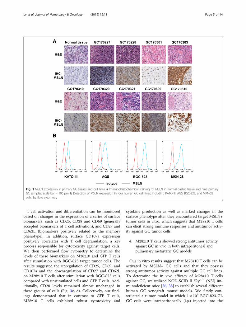
Tumor targeting by CAR T cells requires the expres-sion of certain TAAs on the surface of tumor cells. Toevaluate MSLN expression in primary GC tissue, we per-formed immunohistochemical staining for MSLN innine primary GC samples and found robust expressionin most of these samples compared with normal gastrictissue (Fig. 1a). We examined MSLN expression in fourhuman GC cell lines, including BGC-823, AGS, KATOIII, and MKN-28 cells, by flow cytometry. All four celllines expressed MSLN, but BGC-823 and MKN-28 cellsexpressed higher levels than did AGS and KATO IIIcells (Fig. 1b). Collectively, these results indicate thatMSLN expression is upregulated in both GC primarycells and cell lines.
2. Generation of third-generation CAR T cells target-ing MSLN
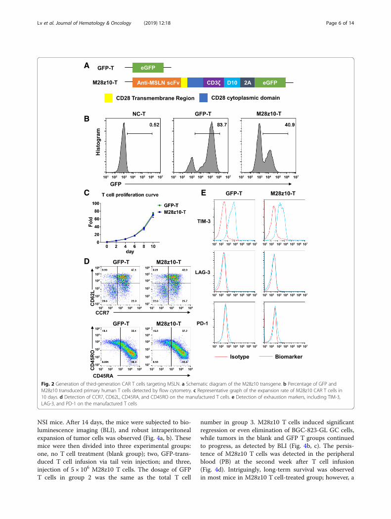
To redirect human T cells to the MSLN antigenexpressed by GC tumor cells, we constructed the third-generation M28z10 vector containing the scFv thatrecognizes MSLN, CD28 transmembrane domain, CD3ζT cell activating domain, and the costimulatory domainsfrom both CD28 and DAP10 as previously described[23, 36]. CAR was coexpressed with eGFP separated by a2A sequence (Fig. 2a). Primary human T lymphocytesisolated from peripheral blood mononuclear cells(PBMCs) by magnetic selection were activated withanti-CD3/CD28/CD2-coated beads for 24 h beforetransduction with the M28z10 transgene. Transductionefficiency was determined after 72 h by the percentageof GFP+ cells detected by flow cytometry (Fig. 2b). Thetransduced T cells were cultured for 10 days, achieving agreater than 60-fold expansion with the addition of300 IU of exogenous interleukin-2 (IL-2) (Fig. 2c).GFP-transduced T cells were used as a control group. Asubstantial fraction of manufactured CAR T cells showeda CD45RA+CCR7+CD62Lhigh phenotype. Most of thecells express TIM-3, but expression levels of PD-1 andLAG-3 are pretty low as detected by FACS (Fig. 2d, e).
3. M28z10 T cells showed strong antitumor activityagainst GC cell lines in vitro
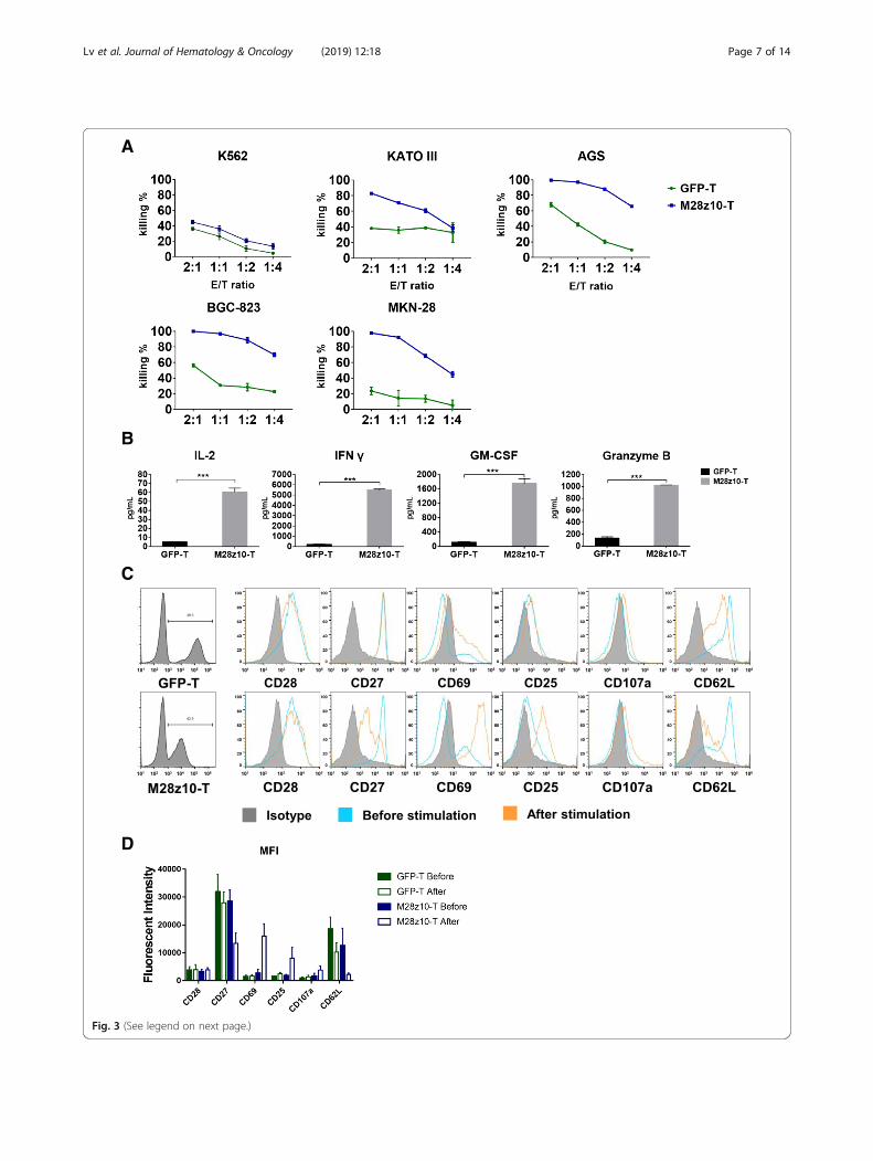
To determine the cytotoxicity of M28z10 T cellsagainst MSLN+ GC cell lines in vitro, we lentivirallytransduced 5 GC cell lines with a GFP-2A-luciferasetransgene and thereby constructed the KATO III-GL,AGS-GL, BGC-823-GL, and MKN-28-GL cell lines. Cellviability was determined by a luciferase reporter systemand an illuminator [37]. We performed an 18-h killingassay of M28z10 T and GFP T cells on the four GC celllines along with K562-GL cells which were used as amesothelin negative control. The results showed thatM28z10 T cells exerted stronger cytotoxicity than GFPT cells after coculture with all four GC cell lines at theindicated effector to target (E:T) ratio in vitro, while formesothelin negative K562-GL cells, cytotoxicity remainalmost same between M28z10 and GFP T cells (Fig. 3a).To analyze cytokine secretion by M28z10 T cells aftertarget cell stimulation, we collected the culture super-natant of BGC-823-GL tumor cells in a killing assay andthen detected cytokines and effector molecules that aregenerally secreted by activated T cells, including IL-2,interferon-γ (IFN-γ), granulocyte-macrophage colony-stimulating factor (GM-CSF), and granzyme B, byenzyme-linked immune absorbance assay (ELISA). Therewas a significant increase in the secretion of these 4 pro-teins by M28z10 T cells compared with GFP T cells(Fig. 3b), indicating the strong cytokine-secreting ca-pabilities of M28z10 T cells upon encountering MSLN+GC cells.
Lv et al. Journal of Hematology & Oncology (2019) 12:18 Page 4 of 14
T cell activation and differentiation can be monitoredbased on changes in the expression of a series of surfacebiomarkers, such as CD25, CD28 and CD69 (generallyaccepted biomarkers of T cell activation), and CD27 andCD62L (biomarkers positively related to the memoryphenotype). In addition, surface CD107a expressionpositively correlates with T cell degranulation, a keyprocess responsible for cytotoxicity against target cells.We then performed flow cytometry to determine thelevels of these biomarkers on M28z10 and GFP T cellsafter stimulation with BGC-823 target tumor cells. Theresults suggested the upregulation of CD25, CD69, andCD107a and the downregulation of CD27 and CD62Lon M28z10 T cells after stimulation with BGC-823 cellscompared with unstimulated cells and GFP T cells. Add-itionally, CD28 levels remained almost unchanged inthese groups of cells (Fig. 3c, d). Collectively, our find-ings demonstrated that in contrast to GFP T cells,M28z10 T cells exhibited robust cytotoxicity and
cytokine production as well as marked changes in thesurface phenotype after they encountered target MSLN+tumor cells in vitro, which suggests that M28z10 T cellscan elicit strong immune responses and antitumor activ-ity against GC tumor cells.
4. M28z10 T cells showed strong antitumor activityagainst GC in vivo in both intraperitoneal andpulmonary metastatic GC models
Our in vitro results suggest that M28z10 T cells can beactivated by MSLN+ GC cells and that they possessstrong antitumor activity against multiple GC cell lines.To determine the in vivo efficacy of M28z10 T cellsagainst GC, we utilized NOD-SCID IL2Rγ−/− (NSI) im-munodeficient mice [36, 38] to establish several differenthuman GC xenograft mouse models. We firstly con-structed a tumor model in which 1 × 10⁶ BGC-823-GLGC cells were intraperitoneally (i.p.) injected into the
A
B
Fig. 1 MSLN expression in primary GC tissues and cell lines. a Immunohistochemical staining for MSLN in normal gastric tissue and nine primaryGC samples, scale bar = 100 μm. b Detection of MSLN expression in four human GC cell lines, including KATO III, AGS, BGC-823, and MKN-28cells, by flow cytometry
Lv et al. Journal of Hematology & Oncology (2019) 12:18 Page 5 of 14
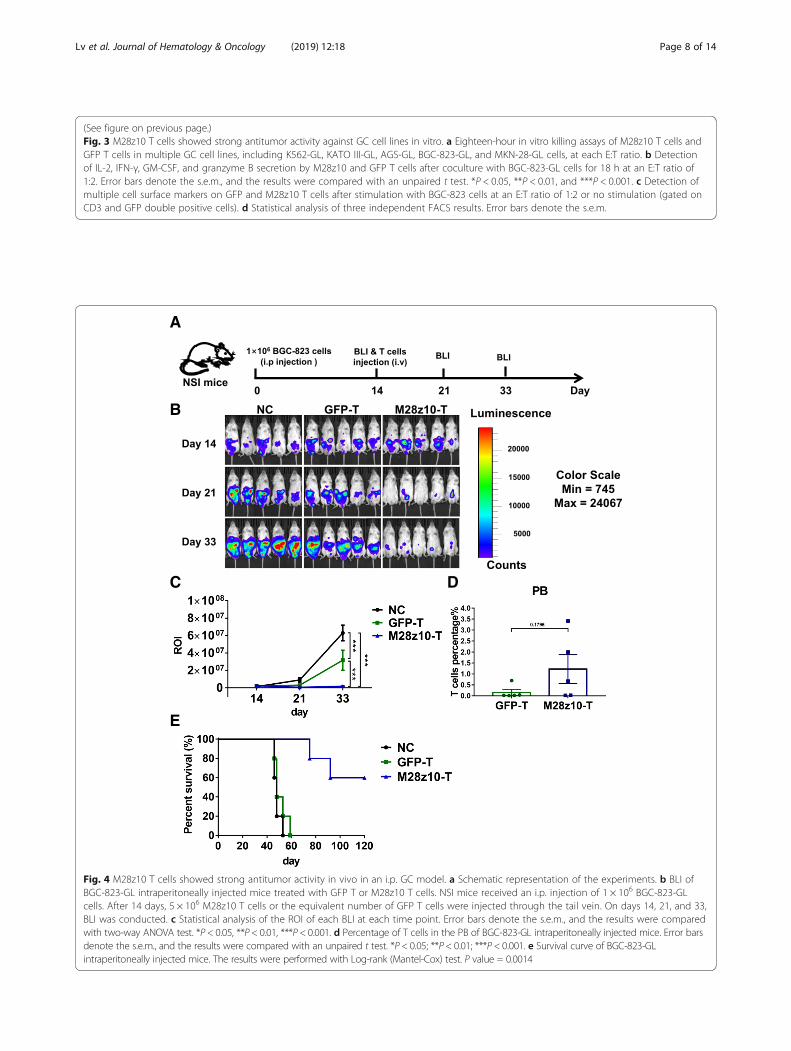
NSI mice. After 14 days, the mice were subjected to bio-luminescence imaging (BLI), and robust intraperitonealexpansion of tumor cells was observed (Fig. 4a, b). Thesemice were then divided into three experimental groups:one, no T cell treatment (blank group); two, GFP-trans-duced T cell infusion via tail vein injection; and three,injection of 5 × 10⁶ M28z10 T cells. The dosage of GFPT cells in group 2 was the same as the total T cell
number in group 3. M28z10 T cells induced significantregression or even elimination of BGC-823-GL GC cells,while tumors in the blank and GFP T groups continuedto progress, as detected by BLI (Fig. 4b, c). The persis-tence of M28z10 T cells was detected in the peripheralblood (PB) at the second week after T cell infusion(Fig. 4d). Intriguingly, long-term survival was observedin most mice in M28z10 T cell-treated group; however, a
A
B
C E
D
Fig. 2 Generation of third-generation CAR T cells targeting MSLN. a Schematic diagram of the M28z10 transgene. b Percentage of GFP andM28z10 transduced primary human T cells detected by flow cytometry. c Representative graph of the expansion rate of M28z10 CAR T cells in10 days. d Detection of CCR7, CD62L, CD45RA, and CD45RO on the manufactured T cells. e Detection of exhaustion markers, including TIM-3,LAG-3, and PD-1 on the manufactured T cells
Lv et al. Journal of Hematology & Oncology (2019) 12:18 Page 6 of 14
A
B
C
D
Fig. 3 (See legend on next page.)
Lv et al. Journal of Hematology & Oncology (2019) 12:18 Page 7 of 14
(See figure on previous page.)Fig. 3 M28z10 T cells showed strong antitumor activity against GC cell lines in vitro. a Eighteen-hour in vitro killing assays of M28z10 T cells andGFP T cells in multiple GC cell lines, including K562-GL, KATO III-GL, AGS-GL, BGC-823-GL, and MKN-28-GL cells, at each E:T ratio. b Detectionof IL-2, IFN-γ, GM-CSF, and granzyme B secretion by M28z10 and GFP T cells after coculture with BGC-823-GL cells for 18 h at an E:T ratio of1:2. Error bars denote the s.e.m., and the results were compared with an unpaired t test. *P < 0.05, **P < 0.01, and ***P < 0.001. c Detection ofmultiple cell surface markers on GFP and M28z10 T cells after stimulation with BGC-823 cells at an E:T ratio of 1:2 or no stimulation (gated onCD3 and GFP double positive cells). d Statistical analysis of three independent FACS results. Error bars denote the s.e.m.
A
B
C D
E
Fig. 4 M28z10 T cells showed strong antitumor activity in vivo in an i.p. GC model. a Schematic representation of the experiments. b BLI ofBGC-823-GL intraperitoneally injected mice treated with GFP T or M28z10 T cells. NSI mice received an i.p. injection of 1 × 10⁶ BGC-823-GLcells. After 14 days, 5 × 10⁶ M28z10 T cells or the equivalent number of GFP T cells were injected through the tail vein. On days 14, 21, and 33,BLI was conducted. c Statistical analysis of the ROI of each BLI at each time point. Error bars denote the s.e.m., and the results were comparedwith two-way ANOVA test. *P < 0.05, **P < 0.01, ***P < 0.001. d Percentage of T cells in the PB of BGC-823-GL intraperitoneally injected mice. Error barsdenote the s.e.m., and the results were compared with an unpaired t test. *P < 0.05; **P < 0.01; ***P < 0.001. e Survival curve of BGC-823-GLintraperitoneally injected mice. The results were performed with Log-rank (Mantel-Cox) test. P value = 0.0014
Lv et al. Journal of Hematology & Oncology (2019) 12:18 Page 8 of 14
majority of the mice in the blank and GFP T-treatedgroups died between 45 and 60 days after the start ofthe experiment, suggesting that treatment with M28z10T cells can significantly prolong the survival of tumor-bearing mice (Fig. 4e). Xenogenic graft versus hostdisease (GVHD) was observed in some of the mice inM28z10 T cell group as they showed significant hairloss, which should be the reason for the decrease oftheir survival.
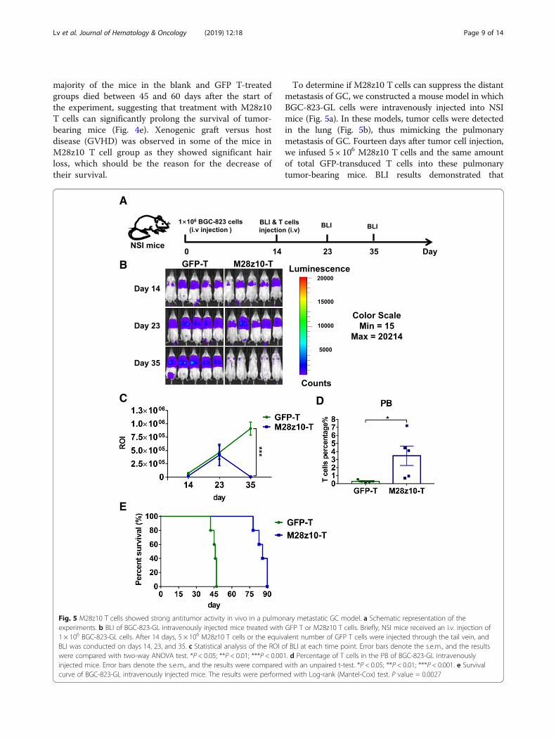
To determine if M28z10 T cells can suppress the distantmetastasis of GC, we constructed a mouse model in whichBGC-823-GL cells were intravenously injected into NSImice (Fig. 5a). In these models, tumor cells were detectedin the lung (Fig. 5b), thus mimicking the pulmonarymetastasis of GC. Fourteen days after tumor cell injection,we infused 5 × 10⁶ M28z10 T cells and the same amountof total GFP-transduced T cells into these pulmonarytumor-bearing mice. BLI results demonstrated that
A
B
C D
E
Fig. 5 M28z10 T cells showed strong antitumor activity in vivo in a pulmonary metastatic GC model. a Schematic representation of theexperiments. b BLI of BGC-823-GL intravenously injected mice treated with GFP T or M28z10 T cells. Briefly, NSI mice received an i.v. injection of1 × 106 BGC-823-GL cells. After 14 days, 5 × 10⁶ M28z10 T cells or the equivalent number of GFP T cells were injected through the tail vein, andBLI was conducted on days 14, 23, and 35. c Statistical analysis of the ROI of BLI at each time point. Error bars denote the s.e.m., and the resultswere compared with two-way ANOVA test. *P < 0.05; **P < 0.01; ***P < 0.001. d Percentage of T cells in the PB of BGC-823-GL intravenouslyinjected mice. Error bars denote the s.e.m., and the results were compared with an unpaired t-test. *P < 0.05; **P < 0.01; ***P < 0.001. e Survivalcurve of BGC-823-GL intravenously injected mice. The results were performed with Log-rank (Mantel-Cox) test. P value = 0.0027
Lv et al. Journal of Hematology & Oncology (2019) 12:18 Page 9 of 14
M28z10 T cells almost eliminated pulmonary tumor cellsin most of the mice in day 35, while GFP-transduced Tcells could not control tumor cell progression (Fig. 5b, c).Significantly, higher percentage of T cells was detected inthe M28z10 T group at the second week after T cell infu-sion, indicating the expansion of M28z10 T cells in vivo(Fig. 4d). Finally, pulmonary-colonized tumor cellscaused mouse death within 60 days in the GFP T group.In contrast, a majority of the mice in the M28z10 groupcontinued to survive until 80 days (Fig. 5e) and finallydied of xenogenic GVHD. Collectively, our data de-monstrated that M28z10 T cells exert strong antitumoractivity against MSLN+ GC cells in vivo.
5. Peritumoral delivery of M28z10 T cells showedsignificantly improved tumor infiltration andefficacy compared with intravenous deliveryapproach in subcutaneous GC mouse models
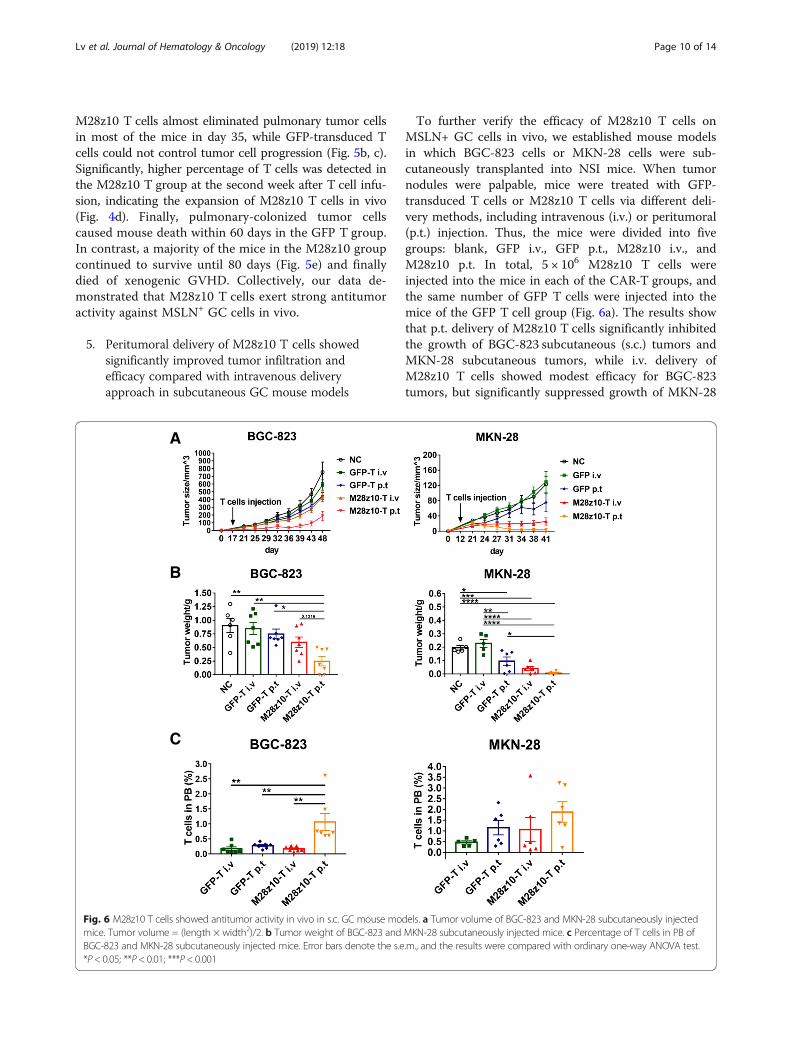
To further verify the efficacy of M28z10 T cells onMSLN+ GC cells in vivo, we established mouse modelsin which BGC-823 cells or MKN-28 cells were sub-cutaneously transplanted into NSI mice. When tumornodules were palpable, mice were treated with GFP-transduced T cells or M28z10 T cells via different deli-very methods, including intravenous (i.v.) or peritumoral(p.t.) injection. Thus, the mice were divided into fivegroups: blank, GFP i.v., GFP p.t., M28z10 i.v., andM28z10 p.t. In total, 5 × 10⁶ M28z10 T cells wereinjected into the mice in each of the CAR-T groups, andthe same number of GFP T cells were injected into themice of the GFP T cell group (Fig. 6a). The results showthat p.t. delivery of M28z10 T cells significantly inhibitedthe growth of BGC-823 subcutaneous (s.c.) tumors andMKN-28 subcutaneous tumors, while i.v. delivery ofM28z10 T cells showed modest efficacy for BGC-823tumors, but significantly suppressed growth of MKN-28
A
B
C
Fig. 6 M28z10 T cells showed antitumor activity in vivo in s.c. GC mouse models. a Tumor volume of BGC-823 and MKN-28 subcutaneously injectedmice. Tumor volume = (length × width2)/2. b Tumor weight of BGC-823 and MKN-28 subcutaneously injected mice. c Percentage of T cells in PB ofBGC-823 and MKN-28 subcutaneously injected mice. Error bars denote the s.e.m., and the results were compared with ordinary one-way ANOVA test.*P < 0.05; **P < 0.01; ***P < 0.001
Lv et al. Journal of Hematology & Oncology (2019) 12:18 Page 10 of 14
tumors (Fig. 6a, b). Persistence of M28z10 T cells wasdetected in PB (Fig. 6c).The improved efficacy of peritumoral delivery of
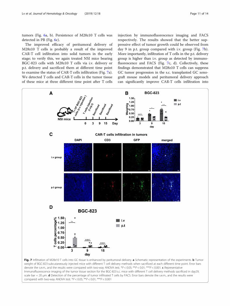
M28z10 T cells is probably a result of the improvedCAR-T cell infiltration into solid tumors in the earlystage; to verify this, we again treated NSI mice bearingBGC-823 cells with M28z10 T cells via i.v. delivery orp.t. delivery and sacrificed them at different time pointto examine the status of CAR-T cells infiltration (Fig. 7a).We detected T cells and CAR-T cells in the tumor tissueof these mice at three different time point after T cells
injection by immunofluorescence imaging and FACSrespectively. The results showed that the better sup-pressive effect of tumor growth could be observed fromday 9 in p.t. group compared with i.v. group (Fig. 7b).More importantly, infiltration of T cells in the p.t. deliverygroup is higher than i.v. group as detected by immuno-fluorescence and FACS (Fig. 7c, d). Collectively, thesefindings demonstrated that M28z10 T cells can suppressGC tumor progression in the s.c. transplanted GC xeno-graft mouse models and peritumoral delivery approachcan significantly improve CAR-T cells infiltration into
A B
C
D
Fig. 7 Infiltration of M28z10 T cells into GC tissue is enhanced by peritumoral delivery. a Schematic representation of the experiments. b Tumorweight of BGC-823 subcutaneously injected mice with different T cell delivery methods when sacrificed at each different time point. Error barsdenote the s.e.m., and the results were compared with two-way ANOVA test. *P < 0.05; **P < 0.01; ***P < 0.001. c RepresentativeImmunofluorescence imaging of the tumor tissue section for the BGC-823 s.c. mice with different T cell delivery methods sacrificed in day29,scale bar = 20 μm. d Detection of the percentage of tumor infiltrated T cells by FACS. Error bars denote the s.e.m., and the results werecompared with two-way ANOVA test. *P < 0.05; **P < 0.01; ***P < 0.001
Lv et al. Journal of Hematology & Oncology (2019) 12:18 Page 11 of 14
solid tumor tissue in the early stage compared with intra-venous delivery.
DiscussionTargeted therapies, including small molecules, monoclo-nal antibodies, and adoptive cell transfer, have opened anew era of cancer treatment and significantly improvedthe prognosis of patients [39]. However, for a majority ofGC patients, there is a lack of available targetedapproaches, and the 5-year survival rate is far from satis-factory. Therefore, the development of targeted strategiesis urgently needed to improve the clinical outcome ofadvanced GC patients and prolong their survival.The applications of CAR T cells have achieved re-
markable success in acute lymphoblastic leukemia.Moreover, many preclinical and clinical studies havesuggested the potential of CAR T cells to treat solidcancer [33, 40]. Previously, we have showed the improvedanti-tumor activities of the third-generation mesothelintargeting CAR T cells M28z10 compared with thesecond-generation M28z CAR T cells. Nonetheless, therehave been few studies of CAR T cells targeting GC, whichhas limited their clinical usage. In the present study, wecharacterized MSLN as a target of human GC in multipleprimary GC samples and cell lines and then generatedthird-generation CAR (M28z10) T cells to target MSLN+GC cells. Our in vitro functional assays demonstrated thatM28z10 T cells have significant antitumor activity againstmultiple MSLN+ GC cell lines and suppress GC progres-sion in vivo, as evaluated in GC xenograft mouse models.Other groups have reported MSLN as a target of CAR Tcells against multiple types of solid cancers. Zhao Y et al.reported that multiple injections of electroporated autolo-gous mesothelin-targeted T cells can mediate regressionof human ovarian cancer in NSG mice [41]. Beatty GL etal. reported the result of a phase 1 trial of mRNA electro-porated mesothelin-specific chimeric antigen receptor TCells to treat patients with pancreatic carcinoma metasta-ses. This strategy was proved to be safe, and was shown toelicit anti-tumor responses [32]. However, mRNA electro-porated CAR was only transiently expressed in T cells, sothat the anti-tumor responses may not be sufficiently dur-able to elicit a remission. Interestingly, antigen spreadingwas observed in the case report published by the samegroup, suggesting the potential of CAR-mesothelin T cellsto trigger the host intrinsic immune responses [42]. Thismay prompt us to combine CAR-T therapy with other im-mune stimulating adjuvants to promote tumor antigenpresentation and recognition by host immune cells,thereby helping to overcome tumor antigen heterogeneitywhich is an obstacle for the current single target CAR-Timmune therapy.Although many studies have reported the efficacy of
second- or third-generation CAR T cells against multiple
types of solid cancer based on mouse models, clinical re-ports of complete remission in patients are very limited.This may be due to the T cell suppressive microenviron-ment in solid cancers and the large tumor burden in ad-vanced patients, which limit the efficacy of current CART cell therapy [43, 44]. In our study, systemic i.v. admin-istration of M28z10 T cells induced the regression ofi.p.- and i.v.-transplanted diffuse GC tumors, but for theestablished BGC-823 s.c. tumors, the efficacy of i.v.M28z10 T cell treatment was modest at the same dos-age, and peritumoral delivery strategy can result in im-proved CAR-T cells infiltration into tumor tissue in theearly stage and cause tumor regression. Regional deli-very of CAR-T cells can not only enhance theanti-tumor efficacy for injected tumors but also is be-lieved to avoid the adverse effect caused by systemicdelivery method [45–50]. Therefore, regional delivery issuitable to be used to inject anti-MSLN CAR T cells aclinical trial to minimize the risk of adverse effects inthe future. Moreover, these data suggest that the antitu-mor activity of CAR T cells should be further enhanced byincorporating other functional elements into the CAR vec-tors to promote CAR T cell infiltration, to sustain T celleffector activities and to enhance cooperation with by-stander T cells or innate immune cells [51, 52]. Currentmethods for implementing this strategy include the co-expression of immune-promoting cytokines [53–55], thesecretion of scFv that blocks the PD-1 receptor [56, 57],and the introduction of dominant negative forms ofinhibitory receptors with the CAR [52]. These modifi-cations to our CAR T cells will be tested in futureworks to further improve the efficacy against largeestablished gastric tumors.Overall, we have demonstrated the feasibility and effi-
cacy of M28z10 T cells against GC in vitro and in vivoin multiple xenograft mouse models. Our results suggestthat mesothelin may be a potential target of CAR T cellsfor treating gastric cancer.
ConclusionsIn summary, we characterized mesothelin as an antigenof chimeric antigen receptor T cells in human gastriccancer, and utilized third-generation anti-MesothelinCAR-T cell M28z10 to target human gastric cancer. Ourdata demonstrated that M28z10 T cells can exert potentanti-tumor activities in vitro and in vivo against GC asevaluated by several xenograft GC mouse models. Thesedata suggest the potential value of employing M28z10 Tcells to treat GC patients in the clinic.
AbbreviationsBLI: Bioluminescence imaging; CAR: Chimeric antigen receptor; E:T: Effectorto target; ELISA: Enzyme-linked immune absorbance assay; GC: Gastriccancer; GM-CSF: Granulocyte-macrophage colony-stimulating factor;GVHD: Graft versus host disease; i.p.: Intraperitoneally; i.v.: Intravenous; IFN-
Lv et al. Journal of Hematology & Oncology (2019) 12:18 Page 12 of 14
γ: Interferon-γ; IL-2: Interleukin-2; M28z10: a CAR containing the MSLN scFv,CD3ζ, CD28, and DAP10 intracellular signaling domain; MSLN: Mesothelin;NSI: NOD-SCID IL2Rγ−/−; p.t.: Peritumoral; PBMCs: Peripheral bloodmononuclear cells; s.c.: Subcutaneous; scFv: Single-chain variable fragment;TAAs: Tumor-associated antigens
AcknowledgmentsWe thank all the members in list and their lab members for experimentalmaterials, technical assistance, helpful discussions, and comments.
FundingThis study is supported by the National Natural Science Foundation of China(NSFC)—81522002, 81773301; the Strategic Priority Research Program of theChinese Academy of Sciences, Grant No. XDB19030205, No. XDA12050305;Guangdong Special Support Program, No. 2017TX04R102; the NaturalScience Fund of Guangdong Province: Distinguished Young Scholars (GrantNo. 2014A030306028), Doctoral Foundation (Grant No. 2017A030310381); theNational Major Scientific and Technological Special Project for “SignificantNew Drugs Development” (Grant No. SQ2018ZX090201); the GuangdongProvincial Applied Science and Technology Research & DevelopmentProgram (Grant No. 2016B020237006); the Frontier and key technologyinnovation special grant from the Department of Science and Technology ofGuangdong province, (2015B020227003), (2014B020225005),(2016B030229006); Science and Technology Planning Project of GuangdongProvince, China (2017B030314056); and Guangdong Laboratory ofRegenerative Medicine and Health-Guangzhou Frontier Exploration Project,No. 2018GZR110105003.
Availability of data and materialsAll data generated or analyzed during this study are included in this article.
Authors’ contributionsConception and design: JL, RZ, DW, PL. Development of methodology: JL,RZ, DW, DZ, ZW, JS, XW. Acquisition of data (provided animals, acquired andmanaged patients, provided facilities, etc.): JL, RZ, XW, QW, YL, SL, SW, ZW,YL, YC, QH, SC, HY, ZL, ZT. Analysis and interpretation of data (e.g., statisticalanalysis, biostatistics, computational analysis): JL, RZ, DW, YL, YC, QH, HY, ZL.Writing, review, and/or revision of the manuscript: JL, RZ, DW, YY, DP, PL, XZ,ZZ, SC, RC, PL. Administrative, technical, or material support (i.e., reporting ororganizing data, constructing databases): ZT, YY, DP, PL, XZ, ZZ, SC, RC, PL.Study supervision: XZ, ZZ, SC, RC, PL. All authors read and approved the finalmanuscript.
Ethics approval and consent to participateHuman whole peripheral blood mononuclear cells were collected fromhealthy volunteers in the Department of Hematology, Guangdong GeneralHospital, Guangzhou, China. Primary gastric tumor tissues and normal gastrictissues were provided by the Affiliated Cancer Hospital & Institute ofGuangzhou Medical University, Guangzhou, China. Donors who providedprimary specimens gave informed consent for the use of their samples forresearch purposes, and all procedures were approved by the Research EthicsBoard of Guangzhou Institutes of Biomedicine and Health (GIBH).All animal experiments were performed at the Laboratory Animal Center ofthe Guangzhou Institutes of Biomedicine and Health (GIBH), and allprocedures were approved by the Animal Welfare Committee of GIBH. NOD-scid-IL2Rg−/− (NSI) mice were derived at GIBH. All mice were maintained inspecific pathogen-free cages and provided autoclaved food and water. Ani-mal protocols were approved by the institutional animal care and use com-mittee (IACUC).
Consent for publicationNot applicable.
Competing interestsThe authors declare that they have no competing interests.
Publisher’s NoteSpringer Nature remains neutral with regard to jurisdictional claims inpublished maps and institutional affiliations.
Author details1Key Laboratory of Regenerative Biology, South China Institute for Stem CellBiology and Regenerative Medicine, Guangzhou Institutes of Biomedicineand Health, Chinese Academy of Sciences, Guangzhou, China. 2GuangdongProvincial Key Laboratory of Stem Cell and Regenerative Medicine, SouthChina Institute for Stem Cell Biology and Regenerative Medicine, GuangzhouInstitutes of Biomedicine and Health, Chinese Academy of Sciences,Guangzhou, China. 3University of Chinese Academy of Sciences, ShijingshanDistrict, Beijing, China. 4School of Life Sciences, University of Science andTechnology of China, Hefei, China. 5The Center of Research Animal,Guangzhou Institutes of Biomedicine and Health, Chinese Academy ofSciences, Guangzhou 510530, China. 6Department of Pediatric Hematology/Oncology, Sun Yat-Sen Memorial Hospital, Sun Yat-Sen University,Guangzhou, China. 7Orthopaedics Department, Sun Yat-Sen MemorialHospital, Sun Yat-Sen University, Guangzhou 510120, China. 8SICUDepartment, Sun Yat-Sen Memorial Hospital, Sun Yat-Sen University,Guangzhou 510120, China. 9Huangpu Hospital of Guangdong SecondTraditional Chinese Medicine Hospital, Guangzhou 510120, China.10Department of Outpatient, The 91th Military Hospital, Jiaozuo, China.11Division of Reproductive Endocrinology, The 91th Military Hospital, Jiaozuo,China. 12Guangdong Zhaotai InVivo Biomedicine Co. Ltd., Guangzhou, China.13School of Biomedical Sciences, Li Ka Shing Faculty of Medicine, Stem Celland Regenerative Medicine Centre, University of Hong Kong, Hong Kong,China. 14Guangdong Lung Cancer Institute, Medical Research Center,Guangdong General Hospital, Guangdong Academy of Medical Sciences,Guangzhou, China. 15Department of Radiology, The Second AffiliatedHospital of Guangzhou Medical University, Guangzhou, China. 16AffiliatedCancer Hospital & Institute of Guangzhou Medical University, Guangzhou,China. 17Department of Infectious Disease, Guangdong General Hospital,Guangdong Academy of Medical Sciences, Guangzhou, China. 18HefeiInstitute of Stem Cell and Regenerative Medicine, Guangzhou Institutes ofBiomedicine and Health, Chinese Academy of Sciences, Guangzhou 510530,China.
Received: 20 December 2018 Accepted: 6 February 2019
References1. Karimi P, et al. Gastric cancer: descriptive epidemiology, risk factors, screening,
and prevention. Cancer Epidemiol Biomark Prev. 2014;23(5):700–13.2. Duraes C, et al. Biomarkers for gastric cancer: prognostic, predictive or
targets of therapy? Virchows Arch. 2014;464(3):367–78.3. Orditura M, et al. Treatment of gastric cancer. World J Gastroenterol.
2014;20(7):1635–49.4. Correction: mesothelin-specific chimeric antigen receptor mRNA-engineered
T cells induce antitumor activity in solid malignancies. Cancer Immunol Res,2015. 3(2): p. 217.
5. Magalhaes H, Fontes-Sousa M, Machado M. Immunotherapy in advancedgastric Cancer: an overview of the emerging strategies. Can J GastroenterolHepatol. 2018;2018:2732408.
6. Liu L, Wu N, Li J. Novel targeted agents for gastric cancer. J Hematol Oncol.2012;5:31.
7. Bang Y-J, et al. Trastuzumab in combination with chemotherapy versuschemotherapy alone for treatment of HER2-positive advanced gastric orgastro-oesophageal junction cancer (ToGA): a phase 3, open-label,randomised controlled trial. Lancet. 2010;376(9742):687–97.
8. Thiel A, Ristimaki A. Targeted therapy in gastric cancer. APMIS. 2015;123(5):365–72.9. Yuan DD, et al. Targeted therapy for gastric cancer: current status and
future directions (review). Oncol Rep. 2016;35(3):1245–54.10. Yu J, et al. Efficacy and safety of angiogenesis inhibitors in advanced gastric
cancer: a systematic review and meta-analysis. J Hematol Oncol. 2016;9(1):111.11. Muro K, et al. Pembrolizumab for patients with PD-L1-positive advanced
gastric cancer (KEYNOTE-012): a multicentre, open-label, phase 1b trial. TheLancet Oncology. 2016;17(6):717–26.
12. Fuchs CS, et al. KEYNOTE-059 cohort 1: efficacy and safety ofpembrolizumab (pembro) monotherapy in patients with previously treatedadvanced gastric cancer. J Clin Oncol. 2017;35
13. Ralph C, et al. Modulation of lymphocyte regulation for cancer therapy: aphase II trial of tremelimumab in advanced gastric and esophagealadenocarcinoma. Clin Cancer Res. 2010;16(5):1662–72.
Lv et al. Journal of Hematology & Oncology (2019) 12:18 Page 13 of 14
14. Long J, et al. PD-1/PD-L blockade in gastrointestinal cancers: lessonslearned and the road toward precision immunotherapy. J HematolOncol. 2017;10(1):146.
15. June CH, et al. CAR T cell immunotherapy for human cancer. Science. 2018;359(6382):1361–5.
16. Zhang Q, et al. CAR-T cell therapy in gastrointestinal tumors and hepaticcarcinoma: from bench to bedside. Oncoimmunology. 2016;5(12):e1251539.
17. Maude SL, et al. Chimeric antigen receptor T cells for sustained remissionsin leukemia. N Engl J Med. 2014;371(16):1507–17.
18. Zhang C, et al. Engineering CAR-T cells. Biomark Res. 2017;5:22.19. Yu S, et al. Chimeric antigen receptor T cells: a novel therapy for solid
tumors. J Hematol Oncol. 2017;10(1):78.20. Liu B, Song Y, Liu D. Clinical trials of CAR-T cells in China. J Hematol Oncol.
2017;10(1):166.21. Fesnak AD, June CH, Levine BL. Engineered T cells: the promise and
challenges of cancer immunotherapy. Nat Rev Cancer. 2016;16(9):566–81.22. Savoldo B, et al. CD28 costimulation improves expansion and persistence of
chimeric antigen receptor-modified T cells in lymphoma patients. J ClinInvest. 2011;121(5):1822–6.
23. Zhong XS, et al. Chimeric antigen receptors combining 4-1BB and CD28signaling domains augment PI3kinase/AKT/Bcl-XL activation and CD8+ Tcell-mediated tumor eradication. Mol Ther. 2010;18(2):413–20.
24. Song DG, et al. In vivo persistence, tumor localization, and antitumoractivity of CAR-engineered T cells is enhanced by costimulatory signalingthrough CD137 (4-1BB). Cancer Res. 2011;71(13):4617–27.
25. Johnson LA, June CH. Driving gene-engineered T cell immunotherapy ofcancer. Cell Res. 2017;27(1):38–58.
26. Chang K, Pastan I. Molecular cloning of mesothelin, a differentiation antigenpresent on mesothelium, mesotheliomas, and ovarian cancers. Proc NatlAcad Sci U S A. 1996;93(1):136–40.
27. Ito T, et al. ERC/mesothelin is expressed in human gastric cancer tissues andcell lines. Oncol Rep. 2014;31(1):27–33.
28. Sotoudeh M, et al. MSLN (Mesothelin), ANTXR1 (TEM8), and MUC3A are thepotent antigenic targets for CAR T cell therapy of gastric adenocarcinoma. JCell Biochem. 2018;
29. Morello A, Sadelain M, Adusumilli PS. Mesothelin-targeted CARs: driving Tcells to solid tumors. Cancer Discov. 2016;6(2):133–46.
30. Beatty GL, O'Hara M. Chimeric antigen receptor-modified T cells for thetreatment of solid tumors: defining the challenges and next steps.Pharmacol Ther. 2016;166:30–9.
31. Watanabe K, et al. Pancreatic cancer therapy with combined mesothelin-redirected chimeric antigen receptor T cells and cytokine-armed oncolyticadenoviruses. JCI Insight. 2018;3(7)
32. Beatty GL, et al. Activity of Mesothelin-specific chimeric antigen receptor Tcells against pancreatic carcinoma metastases in a phase 1 trial.Gastroenterology. 2018;155(1):29–32.
33. Beatty GL, et al. Mesothelin-specific chimeric antigen receptor mRNA-engineered T cells induce anti-tumor activity in solid malignancies. CancerImmunol Res. 2014;2(2):112–20.
34. Zhao, R., et al., DNAX-activating protein 10 co-stimulation enhances the anti-tumor efficacy of chimeric antigen receptor T cells OncoImmunology (accepted).
35. Lai Y, et al. Toll-like receptor 2 costimulation potentiates the antitumorefficacy of CAR T cells. Leukemia. 2018;32(3):801–8.
36. Xiao Y, et al. ANGPTL7 regulates the expansion and repopulation of humanhematopoietic stem and progenitor cells. Haematologica. 2015;100(5):585–94.
37. Wei X, et al. PSCA and MUC1 in non-small-cell lung cancer as targets ofchimeric antigen receptor T cells. Oncoimmunology. 2017;6(3):e1284722.
38. Ye W, et al. Quantitative evaluation of the immunodeficiency of a mousestrain by tumor engraftments. J Hematol Oncol. 2015;8:59.
39. Vanneman M, Dranoff G. Combining immunotherapy and targetedtherapies in cancer treatment. Nat Rev Cancer. 2012;12(4):237–51.
40. Guo Y, Wang Y, Han W. Chimeric antigen receptor-modified T cells for solidtumors: challenges and prospects. J Immunol Res. 2016;2016:3850839.
41. Zhao Y, et al. Multiple injections of electroporated autologous T cellsexpressing a chimeric antigen receptor mediate regression of humandisseminated tumor. Cancer Res. 2010;70(22):9053–61.
42. Beatty GL, et al. Mesothelin-specific chimeric antigen receptor mRNA-engineered T cells induce antitumor activity in solid malignancies. CancerImmunology Research. 2014;2(2):112–20.
43. Abken H. Adoptive therapy with CAR redirected T cells: the challenges intargeting solid tumors. Immunotherapy. 2015;7(5):535–44.
44. Kakarla S, Gottschalk S. CAR T cells for solid tumors: armed and ready to go?Cancer J. 2014;20(2):151–5.
45. Adusumilli PS, et al. Regional delivery of mesothelin-targeted CAR T celltherapy generates potent and long-lasting CD4-dependent tumorimmunity. Sci Transl Med. 2014;6(261):261ra151.
46. Yu J, et al. Anti-GD2/4-1BB chimeric antigen receptor T cell therapy for thetreatment of Chinese melanoma patients. J Hematol Oncol. 2018;11(1):1.
47. Hardaway JC, et al. Regional infusion of chimeric antigen receptor T cells toovercome barriers for solid tumor immunotherapy. J Vasc Interv Radiol.2018;29(7):1017–21. e1
48. Tchou J, et al. Safety and efficacy of Intratumoral injections of chimericantigen receptor (CAR) T cells in metastatic breast Cancer. Cancer ImmunolRes. 2017;5(12):1152–61.
49. Priceman SJ, et al. Regional delivery of chimeric antigen receptor-engineered T cells effectively targets HER2(+) breast Cancer metastasis tothe brain. Clin Cancer Res. 2018;24(1):95–105.
50. Katz SC, et al. Regional CAR-T cell infusions for peritoneal carcinomatosis aresuperior to systemic delivery. Cancer Gene Ther. 2016;23(5):142–8.
51. Li J, et al. Chimeric antigen receptor T cell (CAR-T) immunotherapy for solidtumors: lessons learned and strategies for moving forward. J HematolOncol. 2018;11(1):22.
52. Qin L, Zhao R, Li P. Incorporation of functional elements enhances theantitumor capacity of CAR T cells. Exp Hematol Oncol. 2017;6:28.
53. Huang Y, et al. Interleukin-armed chimeric antigen receptor-modified T cellsfor cancer immunotherapy. Gene Ther. 2018;25(3):192–7.
54. Krenciute G, et al. Transgenic expression of IL15 improves antigliomaactivity of IL13Ralpha2-CAR T cells but results in antigen loss variants.Cancer Immunol Res. 2017;5(7):571–81.
55. Hu B, et al. Augmentation of antitumor immunity by human and mouseCAR T cells secreting IL-18. Cell Rep. 2017;20(13):3025–33.
56. Li S, et al. Enhanced cancer immunotherapy by chimeric antigen receptor-modified T cells engineered to secrete checkpoint inhibitors. Clin CancerRes. 2017;23(22):6982–92.
57. Rafiq S, et al. Targeted delivery of a PD-1-blocking scFv by CAR-T cellsenhances anti-tumor efficacy in vivo. Nat Biotechnol. 2018;
Lv et al. Journal of Hematology & Oncology (2019) 12:18 Page 14 of 14