Embed Size (px)
Citation preview

Raven Smith-Parris
8/6/2013
The
Conception of
GCC- Specific
Chimeric Antigen Receptors

Abstract
Colorectal Cancer is an aggressive disease that claims the lives of
both men and women every year. It is the second leading cause of
cancer-related death in the U.S. The overwhelming mortality rate it
amasses begs for the discovery of alternative forms of treatment.
The nature of cancer requires immunotherapeutic approach that is
tumor specific in nature. Adoptive T-cell therapy is an alternative
that satisfies these conditions. Guanylyl Cyclase C is a receptor
found on the luminal side of the gut that is tissue-specific for the
intestinal epithelium. Further, its expression is maintained
throughout colorectal tumorigenesis making in an excellent marker
of metastatic disease. By employing the use of chimeric antigen
receptors we have created twelve different CAR’s containing
GCC-specificity which we will use to target tumors expressing

Guanyl Cyclase C. We hypothesize that these new CAR’s will
recognize GCC and induce T cell effector responses.

Colon Cancer is an aggressive form of cancer that develops in the
rectum. Colorectal cancer is the fourth most commonly diagnosed
cancer and the second leading cause of cancer death in both men and
women combined within the United States. It is estimated that in 2013
over 50,000 people will die from colorectal cancer. As colorectal cancer
progresses it becomes metastatic and travels to the lymph nodes and
various parts of the body. The abysmal death rate in regards to colorectal
cancer begs for alternative forms of cancer treatment.
Adoptive T-Cell therapy is a form of cancer treatment that can
prove to be very successful. Guanylyl Cylclase C is a gene that codes for
a protein found in the intestines and cancers arising from the intestine.
Guanylyl Cyclase C has limited expression in extra-intestinal tissues
making it an ideal marker for the detection of colorectal cancer
metastasis in non-intestinal tissues. Recently, we have engineered T cells
to express GCC specific chimeric antigen receptors in the hope that they
will attack GCC expressing tumors.

Figure #1: pMA plasmid containing 5F9.
Our research began with the use of circular plasmid entitled pMA.
The pMA contained a CAR sequence with an antibody sequence derived
from the 5F9 antibody. 5F9 is a single chain fv that recognizes human

GCC. A digestion reaction was performed in order to remove the 5F9
antibody sequence from the CAR pMA plasmid.
Figure 2: Digestion reaction containing 5F9 & pMA CAR
After completing the digestion, a gel extraction was performed to
remove the PMA CAR from within the gel itself. After isolating the
CAR containing pma plasmid GeneArt assembly was used to connect
individual antibodies to the CAR pMA construct. There were twelve
antibody sequences used to create twelve new CARS. The antibody

sequences used for the benefit of this experiment are Abx012, Abx020,
Abx106, Abx198, Abx221, Abx229, Abx338, Abx393, 6H8, 8C2,
10C10 and 10D3. These antibodies were chosen due to their capabilities
to recognize human GCC with hopes that at least some would be cross
reactive for mouse GCC. Seeing as any results obtained would be done
from a mouse model, the outcome, if favorable, would need to be
recreated using a human model.

Next, a transformation was preformed inserting the GeneArt reactions
into bacteria. This allowed the assembled CAR pMA plasmids to get
into the bacteria. This was made possible by heat shocking the bacteria
cells. Heat shocking allows for the mild disruption of the cell which
permits the DNA to enter. The bacteria was then plated out on agarose
Figure 3: pMA plasmid containing new antibody sequence
(outlined in red).

plates for an overnight incubation. Following incubation, colonies were
picked and grown in order to test for positive insertions.
Figure 4: Model depicting a transformation
reaction.

After identifying positive clones, 10ML cultures of the cells were
grown and placed through the plasmid purification process. Next, the
purified plasmids were sent out for sequencing to confirm correct
assembly of the new CARs. Once the plasmids were confirmed to be
correct, the CARs were then digested out of the pMA plasmid. This was
done with the help of two enzymes entitled XhoI and EcoRI. These
enzymes used were chosen specifically because they can be found
Figure 5: PCR screen of clones

within the new retroviral plasmid that would be used to further the
experiments. The use of these enzymes would ensure a smooth transition
of the various CARS from one plasmid to the other.
Figure #6: pMA plasmid containing XhoI & EcoRI sites.

A separate digestion reaction was competed in order to disrupt
pMIG, our retroviral plasmid. This disruption allowed for the integration
of the CAR DNA into the retroviral plasmid. Next, using a ligation
reaction the various CAR’s were attached to the pMIG plasmid.
Following the ligation, bacteria was transformed with the ligation
reactions and plated out on agarose plates. Colonies were then grown;
Figure 7: pMA CAR digestion

PCR screened to identify positives, and sequenced to confirm correct
insertion into pMIG before proceeding to the next step.
Next, we made retrovirus from the CAR-pMIG plasmids. In order
to achieve this, the retroviral packaging cell line referred to as Phoenix
were used. Phoenix cells are easy to use where DNA integration and
virus collection is concerned. Phoenix Cells create retrovirus when the
retrovirus buds off of the Phoenix cell membrane and into the easily
accessed cell culture supernatant. 48 hours after the transfection,
supernatant containing the virus was collected.
Next we purified CD8+ T cells from mice. The purification
process began with the removal of spleens from mice. The removal was
followed by a disruption. The disruption allowed for the isolation of
white blood cells (B cells, T cells, dendritic cells, monocytes,
macrophages, etc.). The cells were then labeled with an antibody
cocktail containing biotin-labeled antibodies specific for all white blood
cell types excluding CD8+ T cells.
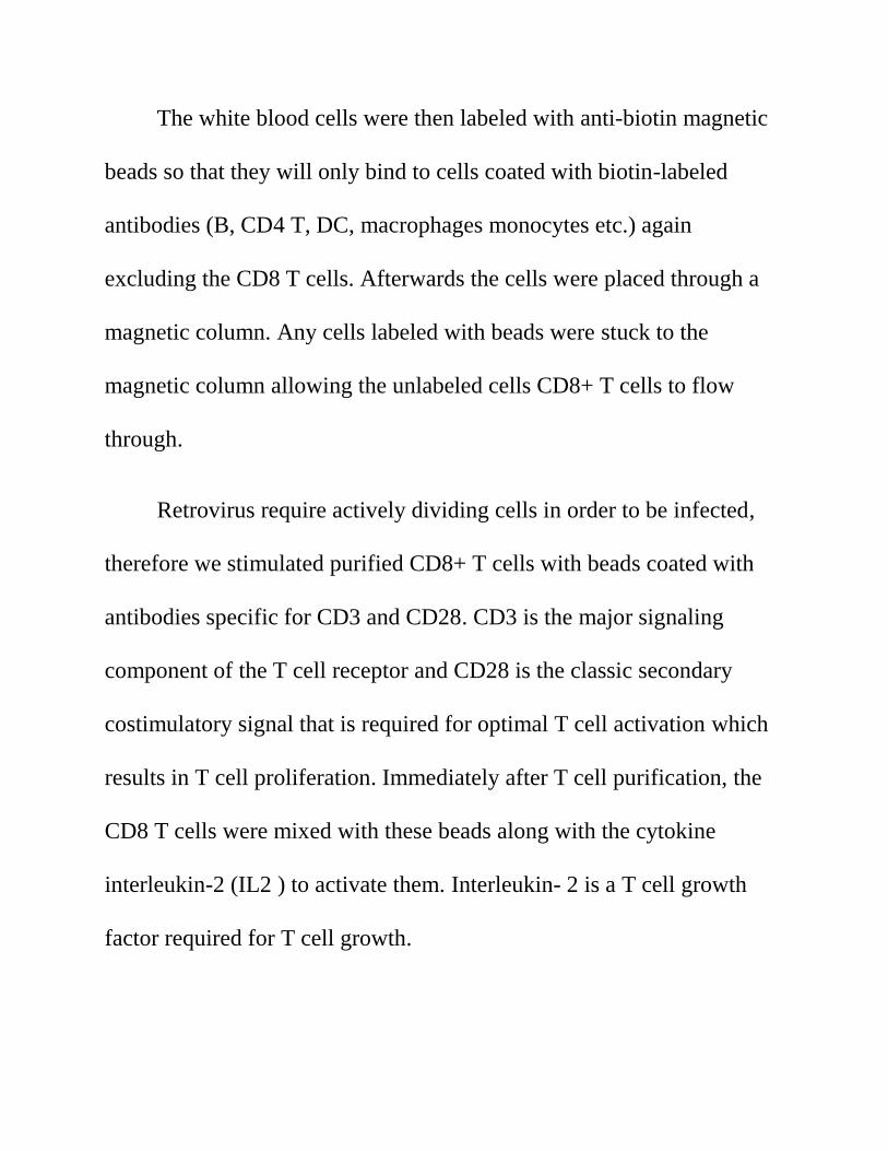
The white blood cells were then labeled with anti-biotin magnetic
beads so that they will only bind to cells coated with biotin-labeled
antibodies (B, CD4 T, DC, macrophages monocytes etc.) again
excluding the CD8 T cells. Afterwards the cells were placed through a
magnetic column. Any cells labeled with beads were stuck to the
magnetic column allowing the unlabeled cells CD8+ T cells to flow
through.
Retrovirus require actively dividing cells in order to be infected,
therefore we stimulated purified CD8+ T cells with beads coated with
antibodies specific for CD3 and CD28. CD3 is the major signaling
component of the T cell receptor and CD28 is the classic secondary
costimulatory signal that is required for optimal T cell activation which
results in T cell proliferation. Immediately after T cell purification, the
CD8 T cells were mixed with these beads along with the cytokine
interleukin-2 (IL2 ) to activate them. Interleukin- 2 is a T cell growth
factor required for T cell growth.

They day after T cell purification and stimulation (with anti-
CD3/CD28 beads), the T cells are actively dividing. They are mixed in
with the retroviral supernatant. Afterwards the retrovirus then infects
the T cells and the CAR DNA is then stably inserted into the genome of
T cells. The T cells were monitored and counted on a daily basis after
this point and constantly fed with more media and IL2 as they continue
to expand.
The graph pictured above shows the results garnered when we
tested one out of twelve of our CAR’s. Our finding were based on the

increased expression of CD25.The CAR entitled ABX338 showed an
increase in CD25 expression where human GCC was concern but did not
mirror the same affinity for mouse GCC. Our findings have led us to
believe that further testing is required for a CAR satisfying our
requirements may lie within the remaining eleven untested CARS.

REFERENCES
Bandi, Dorothy . "Colon Cancer Alliance." Colon Cancer Alliance. N.p.,
n.d. Web. 29 July 2013. <http://www.ccalliance.org/>.
"Cancer - National Library of Medicine - PubMed Health." National
Center for Biotechnology Information. National Center for
Biotechnology Information at the U.S. National Library of Medicine ,
n.d. Web. 29 July 2013.
<http://www.ncbi.nlm.nih.gov/pubmedhealth/PMH0002267/#adam_001
289.disease.causes>.

"Colorectal Cancer - DiagnoCure, Colorectal cancer staging, Colorectal
cancer recurrence." DiagnoCure - Cancer diagnostic tests, Cancer
biomarkers, Molecular marker, Clinical confidence. N.p., n.d. Web. 29
July 2013. <http://www.diagnocure.com/en/products-projects/colorectal-
cancer/gcc-marker.php>.
Rosen, Leo , and Gloria Rosen. "What are the key statistics about
colorectal cancer?." American Cancer Society | Information and
Resources for Cancer: Breast, Colon, Lung, Prostate, Skin. N.p., n.d.
Web. 29 July 2013.
<http://www.cancer.org/cancer/colonandrectumcancer/detailedguide/col
orectal-cancer-key-statistics>.
Sadelain, Michel , Renier Brentjens, and Isabelle Rivière. "The
Basic Principles of Chimeric Antigen Receptor Design ." Cancer
Discovery 3 (2013): n. pag. Cancer Discovery. Web. 9 July 2013.