Embed Size (px)
Citation preview
Texas Medical Center LibraryDigitalCommons@The Texas Medical Center
UT GSBS Dissertations and Theses (Open Access) Graduate School of Biomedical Sciences
12-2015
REDIRECTING T CELLS WITH CHIMERICANTIGEN RECEPTORS TO TARGETCD123+ LEUKEMIARadhika Thokala
Follow this and additional works at: http://digitalcommons.library.tmc.edu/utgsbs_dissertations
Part of the Translational Medical Research Commons
This Dissertation (PhD) is brought to you for free and open access by theGraduate School of Biomedical Sciences at DigitalCommons@The TexasMedical Center. It has been accepted for inclusion in UT GSBSDissertations and Theses (Open Access) by an authorized administrator ofDigitalCommons@The Texas Medical Center. For more information,please contact [email protected].
Recommended CitationThokala, Radhika, "REDIRECTING T CELLS WITH CHIMERIC ANTIGEN RECEPTORS TO TARGET CD123+ LEUKEMIA"(2015). UT GSBS Dissertations and Theses (Open Access). Paper 644.
REDIRECTING T CELLS WITH CHIMERIC ANTIGEN
RECEPTORS TO TARGET CD123+ LEUKEMIA
by
Radhika Thokala, M.S.
APPROVED:
Dean Anthony Lee, M.D., Ph.D.,
Supervisory Professor
Richard Eric Davis, M.D
Dat Tran M.D.
Kenneth Tsai, M.D., Ph.D
Elizabeth Shpall, M.D.
APPROVED:
Dean, The University of Texas
Graduate School of Biomedical Sciences at Houston
i
REDIRECTING T CELLS WITH CHIMERIC ANTIGEN
RECEPTORS TO TARGET CD123+ LEUKEMIA
A
DISSERTAT ION
Presented to the Faculty of
The University of Texas
Health Science Center at Houston and
The University of Texas MD Anderson Cancer Center
Graduate School of Biomedical Sciences
in Partial Fulfillment of the Requirements for the Degree of
DOCTOR OF PHILOSOPHY
by
Radhika Thokala M.S.
Houston, Texas
December 2015
ii

DEDICATION
JESUS ALMIGHTY
MY BELOVED PARENTS
iii

ACKNOWLEDGEMENTS
This work could not have been accomplished without the help of many people. I would like to
thank my mentor Dr. Laurence Cooper with utmost gratitude for the opportunity to work in his
laboratory. His guidance and supervision throughout my graduate studies have been
instrumental for my development as an independent researcher. My sincere thanks to Dr. Lee for
being my mentor for the last few months and his valuable guidance and support for m y
dissertation, when Dr. Cooper moved to Ziopharm as C.E.O. I would also like to extend my
thankfulness to my advisory and examination committee members Dr. Richard Eric Davis,
Dr. Dat Tran, Dr. Kenneth Tsai, Dr. Elizabeth Shpall, Dr. Francois Claret for their important
contribution, guidance and support for my PhD training.
I would like to thank my lab members in the past and present who taught me many different
scientific techniques making this work possible. In particular Helen Huls who was always there
for me in the lab, Simon Olivares who taught me molecular biology, Tiejuan Mi for mice
experiments. I would like to thank Drew Deniger a senior graduate student and good friend
helped me to choose right experiments which saved me lot of time. Thanks to my fellow
students Lenka Hurton, Denise Crossland, Hillary Caruso, David Rushworth and Janani
Krishnamurthy for their help and fun time we had as graduate students. Thanks to my other lab
members and numerous technicians without whose help this dissertation is possible. My deepest
gratitude and thanks to Sanat Dave at histopathology tissue bank for providing primary samples
needed for this study.
I am especially grateful to my family for giving me the opportunity to follow my
dreams and the love to make them a reality. Most of all I would like to thank my lord Jesus
Christ whose perfect love, patience, and gift are the real strength behind all my work and
accomplishments.
iv
ABSTRACT
REDIRECTING T CELLS WITH CHIMERIC ANTIGEN
RECEPTORS TO TARGET CD123+ LEUKEMIA
Radhika Thokala, Ph.D*
Advisory Professor: Dean Anthony Lee, M.D, Ph.D
CD123 or interleukin receptor alpha (IL-3Rα) is expressed on hematological
malignancies such as acute myeloid leukemia (AML) and some acute lymphoblastic
leukemia (ALL). Significantly, CD123 is over-expressed on leukemic stem cells
(LSCs) compared to normal hematopoietic stem cells and thus targeting this tumor-
associated antigen (TAA) provides the potential to prevent relapse. The prototyical
chimeric antigen receptor (CAR) is fashioned by combining the variable light (V L)
and heavy (VH) as a scFv derived from a single monoclonal antibody (mAb)
specific for the TAA. We describe a new approach for generating CD123-specific
CARs generating a chimeric scFv that is made up of the VL and VH harvested from
two mAbs that are each specific for CD123. The hypothesis is VL and VH from
different antibodies to the same TAA can be recombined to form unique binding
domains that retain antigen specificity but may have altered binding
characteristics. This non-homologous recombination of antibody binding domain
may be used to select CAR for optimal anti-tumor characteristics, such as
increasing the therapeutic index. The chimeric scFvs were derived by fusing the
VL a n d V H chains derived from mAbs 26292, 32701, 32703, 32716 specific to
v
CD123. Sleeping Beauty (SB) was employed as a non-viral gene transfer s ystem
to stably express 2nd
generation CARs in T cells derived from peripheral blood
mononuclear cells (PBMC). The CARs were co-expressed with inducible Caspase
9 (iCaspase9) for conditional ablation of T cells in case of off-target toxicities. The SB
plasmids coding for two CARs (transposons) activated T cells via chimeric CD28
with CD3-zeta and CD137 with CD3-zeta were electroporated into PBMC.
Following electrotransfer of the SB system the genetically modified T cells were
preferentially propagated on activating and propagating cells (AaPC) designated as
Clone 1-CD123. The AaPC were derived from K562 cells genetically modified to
co-express co-stimulatory molecules (CD86 and CD137L), a membrane bound
cytokine (IL-15 fused to IL-15Rα), and the TAAs CD123 and CD19. CAR+ T
cells specifically produced IFN-γ and lysed CD123+ leukemic cell lines and
primary AML patient samples, but did not lyse D123neg tumor cells. The addition
of a chemical dimerizer to activate iCaspase9 resulted in destruction of genetically
modified T cells. Both populations of CAR+ T cells produced and eliminated
leukemic tumors in vivo. We observed no difference in the anti-tumor effects
whether the CARs triggered T cells via CD28 or CD137. These studies suggest
that CD123 can be targeted by CAR+ T cells and that the hybrid arrangement
of VL and VH maintained specificity for CD123.
vi
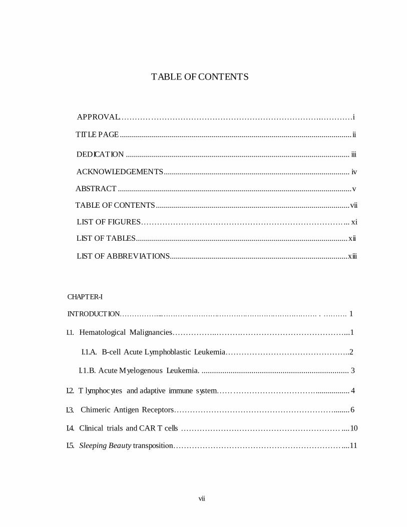
TABLE OF CONTENTS
APPROVAL………………………………………………………………….…………i
TITLE PAGE .................................................................................................................... ii
DEDICATION ................................................................................................................ iii
ACKNOWLEDGEMENTS ............................................................................................. iv
ABSTRACT ..................................................................................................................... v
TABLE OF CONTENTS ................................................................................................. vii
LIST OF FIGURES…………………………………………………………………. ... xi
LIST OF TABLES .......................................................................................................... xii
LIST OF ABBREVIATIONS......................................................................................... xiii
CHAPTER-I
INTRODUCT ION……………...…………………………………………………………. . ………. 1
I.1. Hematological Malignancies……………..…………………………………………...1
I.1.A. B-cell Acute Lymphoblastic Leukemia………………………………………..2
I.1.B. Acute Myelogenous Leukemia. ....................................................................... 3
I.2. T lymphocytes and adaptive immune system………………………………. ................. 4
I.3. Chimeric Antigen Receptors……………………………………………………........ 6
I.4. Clinical trials and CAR T cells …………………………………………………… .... 10
I.5. Sleeping Beauty transposition……………………………………………………… .... 11
vii
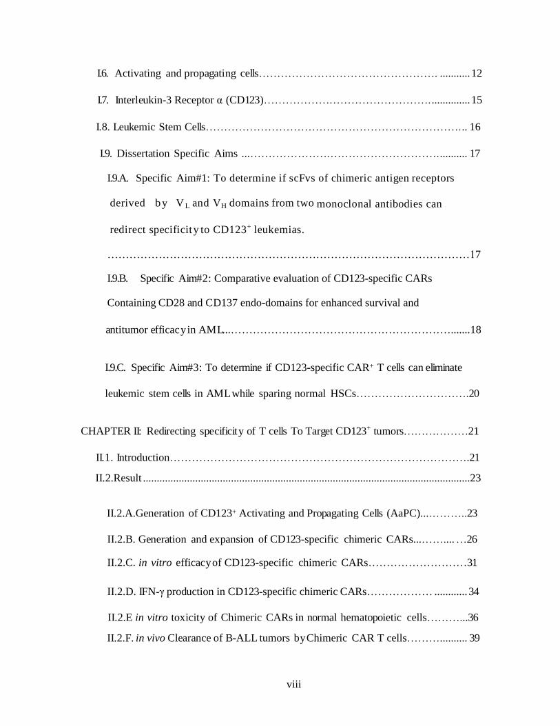
I.6. Activating and propagating cells…………………………………………. ........... 12
I.7. Interleukin-3 Receptor α (CD123)………………………………………. .............. 15
I.8. Leukemic Stem Cells……………………………………………………………. .. 16
I.9. Dissertation Specific Aims ...……………………………………………. .......... 17
I.9.A. Specific Aim#1: To determine if scFvs of chimeric antigen receptors
derived b y V L and VH domains from two monoclonal antibodies can
redirect specificit y to CD123+ leukemias.
………………………………………………………………………………………17
I.9.B. Specific Aim#2: Comparative evaluation of CD123-specific CARs
Containing CD28 and CD137 endo-domains for enhanced survival and
antitumor efficacy in AML...……………………………………………………....... 18
I.9.C. Specific Aim#3: To determine if CD123-specific CAR+ T cells can eliminate
leukemic stem cells in AML while sparing normal HSCs………………………….20
CHAPTER II: Redirecting specificity of T cells To Target CD123+ tumors………………21
II.1. Introduction……………………………………………………………………….21
II.2.Result .......................................................................................................................23
II.2.A.Generation of CD123+ Activating and Propagating Cells (AaPC)...………..23
II.2.B. Generation and expansion of CD123-specific chimeric CARs...…….... …26
II.2.C. in vitro efficacy of CD123-specific chimeric CARs………………………31
II.2.D. IFN-γ production in CD123-specific chimeric CARs……………… ............ 34
II.2.E in vitro toxicity of Chimeric CARs in normal hematopoietic cells………...36
II.2.F. in vivo Clearance of B-ALL tumors by Chimeric CAR T cells……… .......... 39
viii
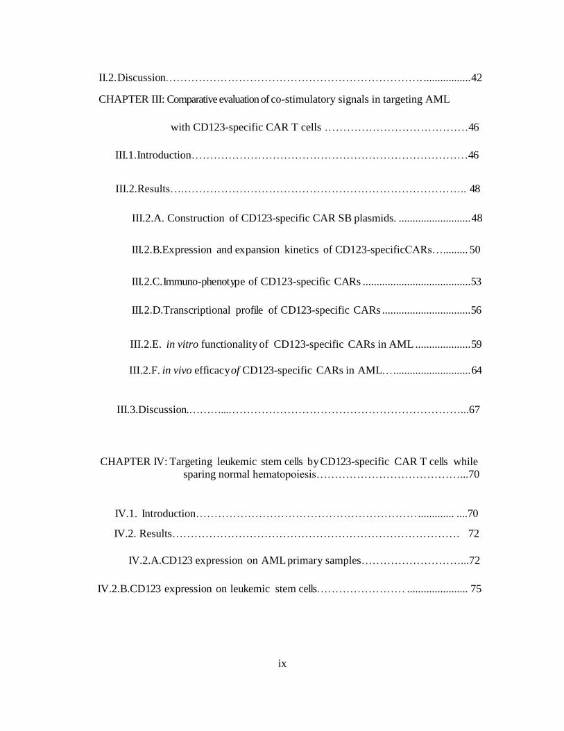
II.2.Discussion……………………………………………………………. ................. 42
CHAPTER III: Comparative evaluation of co-stimulatory signals in targeting AML
with CD123-specific CAR T cells …………………………………46
III.1.Introduction…………………………………………………………………46
III.2.Results….………………………………………………………………….. 48
III.2.A. Construction of CD123-specific CAR SB plasmids. .......................... 48
III.2.B.Expression and expansion kinetics of CD123-specificCARs…......... 50
III.2.C.Immuno-phenotype of CD123-specific CARs ....................................... 53
III.2.D.Transcriptional profile of CD123-specific CARs ................................ 56
III.2.E. in vitro functionality of CD123-specific CARs in AML .................... 59
III.2.F. in vivo efficacy of CD123-specific CARs in AML… ............................ 64
III.3.Discussion.………...………………………………………………………...67
CHAPTER IV: Targeting leukemic stem cells by CD123-specific CAR T cells while
sparing normal hematopoiesis…………………………………...70
IV.1. Introduction…………………………………………………… ............. ....70
IV.2. Results…………………………………………………………………… 72
IV.2.A.CD123 expression on AML primary samples………………………...72
IV.2.B.CD123 expression on leukemic stem cells…………………… ...................... 75
ix
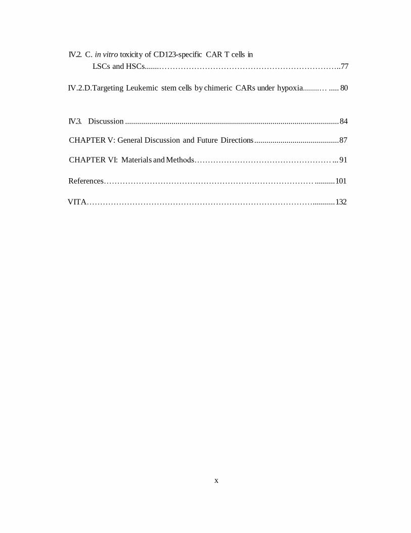
IV.2. C. in vitro toxicity of CD123-specific CAR T cells in
LSCs and HSCs.......…………………………………………………………..77
IV.2.D.Targeting Leukemic stem cells by chimeric CARs under hypoxia........… ..... 80
IV.3. Discussion .......................................................................................................... 84
CHAPTER V: General Discussion and Future Directions .......................................... 87
CHAPTER VI: Materials and Methods…………………………………………… ... 91
References…………………………………………………………………… .......... 101
VITA………………………………………………………………………… ........... 132
x
LIST OF FIGURES
Page
Figure 1. Schematic representation of 2nd generation CAR………………...8
Figure 2. Schematic of three generations of CARs…………………………..9
Figure 3. Schematic of CAR T cells Expansion on AaPC……………………..14
Figure 4. Surface phenotype of AaPC Clone1…………………………… 24
Figure 5. Generation of CD123+ Clone1 …………………………………….25
Figure 6. CD123-specific CARs with chimeric scFvs…………………….28
Figure 7. Expression and expansion kinetics of chimeric CARs .………..30
Figure 8. CD123 expression on leukemic cell lines and 293T cells………….32
Figure 9. Specific cytolysis of chimeric CAR T cells……………………..33
Figure10. IFN-γ production in CAR T cells with chimeric scFvs
................................................................................................................................. 35
.
Figure 11. Anti-tumor efficacy of CD123-chimeric CAR (CAR-10)…… ...38
Figure 12. Expressing firefly luciferase on RCH-ACV………….................40
Figure 13. in vivo efficacy of CD123-chimeric CAR (CAR10)………… ..... 41
Figure 14. CD123-specific CAR plasmids ………… ............................................... 49
Figure 15. CAR Expression in CD123-specific CARs .................................. 51
Figure 16. Expansion kinetics of CD123-specific CARs. ........................... 52
Figure 17.Immuno-phenotype of CD123-specific CARs. .............................. 55
Figure 18. Transcriptional profile of CD123-specific CARs. ...................... 58
xi
Figure 19. in vitro lysis of CD123-specific CARs in AML…… ................. 61
Figure 20. in vitro lysis of CD123-specific CARs in AML
Primary samples……………………………………… ............. 62
Figure 21. in vitro functionality of iCaspase9 in CD123-specific CARs……63
Figure 22. in vivo efficacy of CD123-specific CARs. .................................. 66
Figure 23. CD123 expression analysis in primary AML samples… ............. 73
Figure 24. CD123 expression on AML isolated leukemic stem cells. ........ 76
Figure 25. in vitro efficacy of chimeric CARs in hematopoietic cells………78
Figure 26. in vitro efficacy of chimeric CARs in freshly isolated AML-
LSCs………………………………………… .............................. 79
Figure 27. Expansion of AML-LSCs under hypoxic conditions……………82
Figure 28. in vitro lysis of hypoxia-expanded LSCs by chimeric CARs…...83
xii

LIST OF TABLES
Page
Table1. CD123 expression assessment on Primary AML samples 74
xiii
ABBREVIATIONS
aAPC: Artificial antigen presenting cell
AaPC: Activating and propagating Cells
APC: Antigen Presenting Cell
Ab: Antibody
Ag: Antigen
ALL: Acute Lymphoblastic Leukemia
AML: Acute Myeloid Leukemia
ATCC: American Type Culture Collection
BLI: Bioluminescence Imaging
CAR: Chimeric Antigen Receptor
CCL: CC Chemokine ligands
CCR: CC Chemokine Receptors
CD: Cluster of Differentiation
CDR: Complementarity Determining Regions
cGMP: Current Good Manufacturing Practices
CLL: Chronic Lymphocytic Leukemia
CML: Chronic Myeloid Leukemia
CMV: Cytomegalovirus
CRA: Chromium Release Assay
DC: Dendritic Cell
eGFP: enhanced Green Fluorescent Protein
xiv
EGFR: Epidermal Growth Factor Receptor
FACS: Fluorescence Activated Cell sorting
FBS: Fetal Bovine Serum
FDA: Food and Drug Administration
ffLuc: Firefly Luciferase
GvHD: Graft-versus-Host Disease
HLA: Human Leukocyte Antigen
HIV: Human Immunodeficiency Virus
HSC: Hematopoietic Stem Cell
ICOS: Inducible T-cell Co-Stimulator
ICS: Intracellular Cytokine Staining
IFNγ: Interferon-γ
Ig: Immunoglobulin
IL: Interleukin
IRB: Institutional Review Board
LCA: Lymphocyte Code-set Array
mAb: monoclonal Antibody
MDACC: MD Anderson Cancer Center
MHC: Major H istocompatibility Complex
MRD: Minimal Residual Disease
NIH: National Institutes of Health
NKT cells: Natural Killer T cells
xv
PBMC: Peripheral Blood Mononuclear Cells
PCR: Polymerase Chain Reaction
PD1: Programmed Death-1
PI3K: Phosphoinositide 3-Kinase
PKC: Protein Kinase C
polyA: polyadenylation tail for mRNA transcripts
pSBSO: Sleeping Beauty transposon plasmid
ROR1: Receptor tyrosine kinase-like Orphan Receptor-1
RPMI: Roswell Park Memorial Institute medium
SB: Sleeping Beauty
scFv: single-chain variable fragment
SCID: Severe Combined Immunodeficiency
STAT: Signal Transducer and Activator of Transcription
TCM: Central memory T cell
TEFF: Effector T cell
TEM: Effector memory T cell
TEMRA: Effector memory RA T cell
TM: Memory T cell
TN: Naïve T cell
TAA: Tumor-associated antigen
TCR: T-cell Receptor
UCB: Umbilical Cord Blood
UPenn: University of Pennsylvania
WBC: White Blood Cell
xvi
CHAPTER-I
INTRODUCTION
I.1. Hematological malignancies
Hematological malignancies affects blood, bone marrow (BM) and
lymphatic system. They originate from BM or the cells of immune system and
are the fifth most commonly occurring cancers and the second leading cause
of cancer death. Based on the type of white blood cells affected hematological
malignancies are broadly classified as i) Lymphoma: affects the lymphatic
system, produces uncontrolled growth of white blood cells (WBCs) in lymph
nodes. Lymphoma can be further classified as Hodgkin’s lymphoma (HL) and
Non-Hodgkin’s lymphoma (NHL) ii) Myeloma: also known as plasma cell
myeloma, myelomatosis, or Kahler's disease a type of cancer affecting plasma
cells that produces antibodies. It begins in the BM by accumulation of abnormal
plasma cells iii) Leukemia: leukemia is the most common type of cancer in
children younger than 15 years and adults older than 55 years. Leukemia begins
with the abnormal accumulation of lymphocytes or myeloid cells in the BM.
The four major types of leukemia are acute myelogenous leukemia (AML),
chronic myelogenous leukemia (CML) acute lymphocytic leukemia (ALL) and
chronic lymphocytic leukemia (CLL). Approximately 75% of leukemias
affecting children are ALL, whereas AML and CLL are the most common
1
among adults followed by ALL and CML (1-3). Immunotherapies targeting
tumor associated antigens (TAAs) e.g CD19 by adoptive transfer of
genetically engineered T cells resulted in drastic regression of tumors and
complete remission in CLL patients in clinical setting (4-8). The focus of
this dissertation is on developing adoptive immunotherapies by targeting surface
proteins expressed on for B-ALL and AML through genetic modification of T
cells.
I.1.A. B-cell Acute Lymphoblastic Leukemia
ALL originates from B or T lymphocytes in the BM. B-cell acute
lymphoblastic leukemia (B-ALL) is clonal accumulation of B cell blasts
resulting in suppression of normal hematopoiesis. More than 80% of ALLs in
children and 70% ALLs in adults belong to B-ALL lymphoid group (9, 10).
Key tools to diagnose B-ALL include c ytogenetic studies to identify genetic
alterations in B cell blasts, molecular studies to detect translocations, genome-
wide associations to detect genetic changes where routine techniques are
unavailable, flow cytometry to analyze surface phenotype and monitoring
minimal residual disease (MRD) (11). Improved chemotherapeutic approaches
and radiation followed by allogeneic hematopoietic stem cell transplantation
(HSCT) with cord-blood and haplo-identical approaches over the past decade
enhanced the long-term survival in 90% of children.
2

Although transplant related mortality (TRM) has decreased markedly over
the past 15 years, relapse remains a concern in high risk group children.
Several groups reported that presence of MRD pre and post HSCT is a
predictable tool to detect relapse. Rate of relapse can be decreased by
monitoring MRD and occurrence of Graft versus Host Disease (GvHD) in
First 2 months after the transplant. Employing novel agents and
immunotherapies before and after HSCT will lower MRD and improve Graft
versus Leukemic effect (GvL) and survival in children and adults (12-16).
I.1.B. Acute Myelogenous Leukemia
AML is the most common form of leukemia mostly affecting adults over
55 years. AML is a clonal proliferation of malignant myeloid blast cells in the
BM with impaired normal hematopoiesis. Despite many advances in treatments
AML still remains a lethal disease. Standard chemotherapy and radiation
regimens ensure long-term remission only in 30 to 50% of patients with a low
survival probability resulting in resistance and relapse (17-19). The relapse in
AML is due to MRD caused by small population of Leukemic stem cells
(LSCs) resistant to drugs and radiation. Initial treatment strategy for AML
patients include induction chemotherapy to eliminate blast cells, followed by
consolidation therapy to target the leukemic stem cells. Because of abundant
availability of AML samples, relative simplicity of acquiring them from BM, recent
advances in the understanding of molecular aspects such as role of
3
Chromosomal translocations, easy to analyze AML subsets by flow cytometry enable
to progress the studies on AML. Introducing advanced treatment options beyond
or in addition to current standard treatments will radically change the survival
rates of people diagnosed with AML. Antigen specific based adoptive
immunotherapy will play a complimentary role in eradicating MRD by targeting
leukemia associated antigens expressed on LSCs and leukemic cells (20-22).
I.2. T lymphocytes and adaptive immune system
Immune system protects organisms from infection and disease and broadly
classified as innate immune system and adaptive immune system. The innate
immune system serves as first line of defence in case of infection and has broad
range of specificity for different pathogens. The blood cell types that mediate
innate immune system include i.e macrophages, natural killer (NK) cells. In
contrast adaptive immune system is specific to part of pathogen (tumor
associated antigens and peptides) resulting in long-lasting response through
formation of immunological memory (78-82). T lymphocytes are a type of
white blood cells that plays a major role in adaptive immune system by cell-
mediated-response. Based on TCR structure, T cells can be classified into two
types, i) alpha/beta (αβ) T cells: TCR is a heterodimer composed of an alpha
and beta chains. Each chain has a variable (V) region and a constant
(C) region. The V regions each contain 3 hyper variable regions that make up
4
the antigen-binding site. αβ T cells comprises up to 95-99% of circulating T
cells ii) gamma/delta (γδ) T cells : TCR is a heterodimer composed of
gamma and delta chains. The TCR of αβ T cells binds to a bimolecular
complex consisting of peptide of antigen lying within the groove of MHC
displayed at the surface of antigen presenting cell (APC) i.e. dendritic cells
(DCs), B-cells, macrophages (83). Αβ T cells are distinguished from other
lymphocytes such as NK cells and B cells by the presence of T cell receptor
(TCR) on their surface and recognize its antigens in the context of major
histocompatibility complex (MHC). Most of T cells in the body belong to sub
sets CD4 or CD8. CD8+ T cells bind to epitopes that are part of major MHC
class I and CD4+ T cells bind to epitopes that are part of MHC class II molecules.
All most all the cells in the body express MHC-class I and professional
antigen APCs DCs, B cells and macrophages express MHC-class II molecules.
The best understood CD8+ T cells cytotoxic lymphocytes (CTLs) whose main
function is to destroy infected or a tumor cell by binding to its specific peptide
or antigen. CD4+ T cells are essential for both cell mediated and antibody
mediated (Humoral) immunity. In cell mediated immunity CD4+ T cells binds
to antigen presented by APCs by releasing lymphokines that attract other
immune cells to the area resulting in inflammation. Humoral immunity is
mediated by B cells primarily through
5
production of antibodies. CD4+ cells, called helper T cells binds to antigen
presented by B cells resulting development of clones of plasma cells secreting
antibodies (84-85).
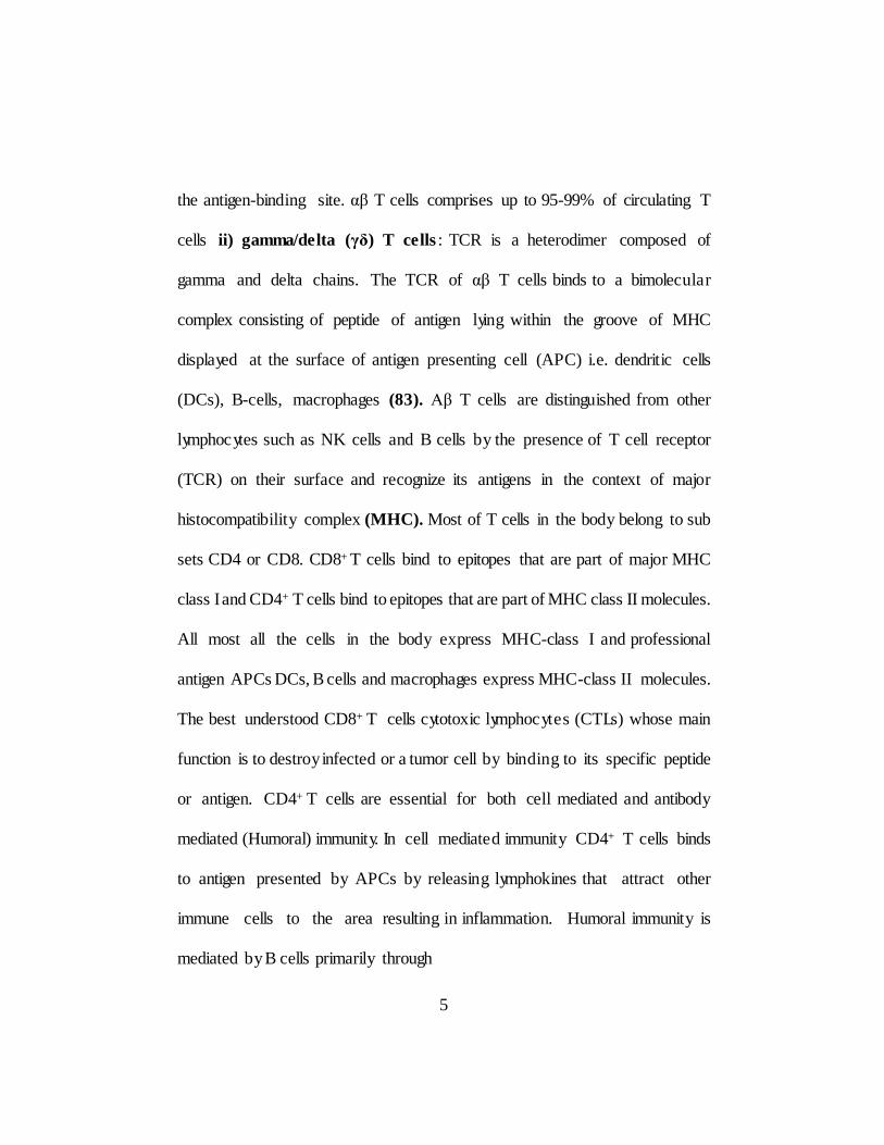
I.3. Chimeric Antigen Receptors
The concept of redirecting T-cells to TAAs by genetic modification
was first developed by Prof. Zelig Eshhar and colleagues at the Weizmann
Institute of Science in Rehovot, Israel in 1980s. By 1989, the same group had
created the first functional CAR T cells (25). Chimeric antigen receptors
(CARs) are recombinant receptors derived by fusing single chain fragment
variable (scFv) region of a monoclonal antibodies (mAbs) specific to TAAs to
T cell signaling domains (i.e. CD3ζ, CD28) via a transmembrane domain
CD8α, CD28) and a hinge (i.e IgG4, CD8α, CD28) (Figure 1). Generally
the scFvs used in making CARs are derived from well characterized murine
mAbs or fully humanized mAbs (hmAbs). CARs recognize targeted antigen in
its native form independent of major MHC compatability. The moieties used to
recognize antigens by CARs can be broadly fall into three categories
i) scFv derived from mAbs specific to targeted antigen ii) fragment antigen-
binding (Fab) selected from libraries iii) nature ligands that binds to their
cognate receptors. The “generation” in the CAR refer to the intracellular
signaling domains. First generation CARs include only CD3ζ as signaling
domain and showed limited T cell activation and short term T cell expansion
6
but enabled cytotoxicity. Second generation CARs include one co- stimulatory
domain such as CD28 or 41BB exhibited improved T cell expansion, cytokine
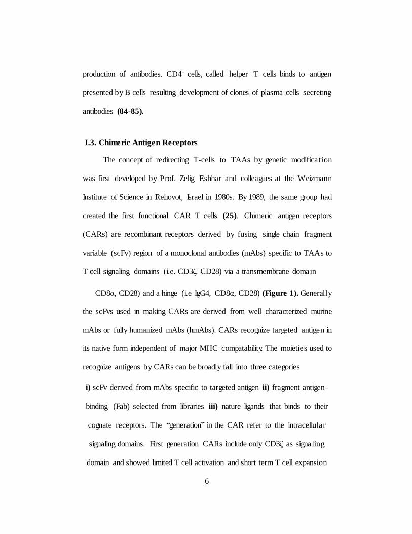
production and T cell persistence. Third generation CARs include three
intracellular endo-domains the most common combination has been CD28,
CD137 (4-1BB), and CD3ζ (26-29) (Figure 2). The efficacy of CAR T cells
targeting its TAAs depends on various factors such as i) position and distance
of epitope from cell surface and formation of optimal T cell synapse ii) length
and flexibility of hinge region between scFv and transmembrane domain iii)
antigen density on tumor cells iv) Activation of endo-domains (30-33).
7
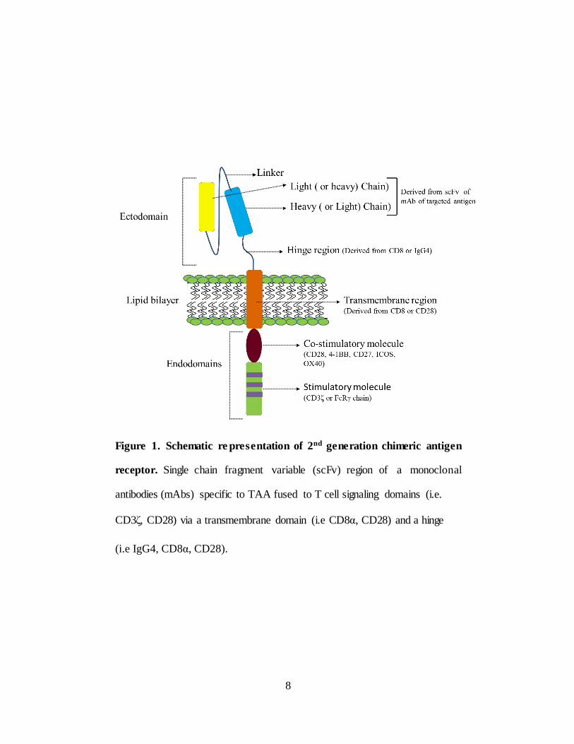
Figure 1. Schematic re presentation of 2nd generation chimeric antigen
receptor. Single chain fragment variable (scFv) region of a monoclonal
antibodies (mAbs) specific to TAA fused to T cell signaling domains (i.e.
CD3ζ, CD28) via a transmembrane domain (i.e CD8α, CD28) and a hinge
(i.e IgG4, CD8α, CD28).
8
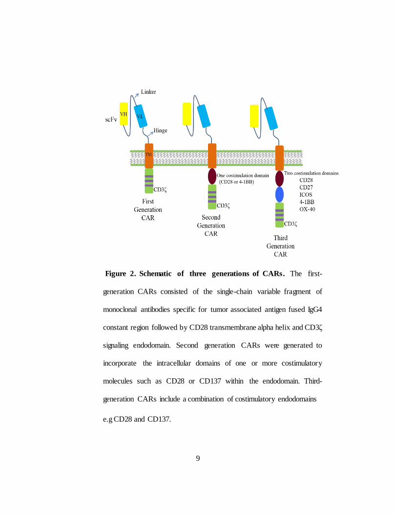
Figure 2. Schematic of three generations of CARs. The first-
generation CARs consisted of the single-chain variable fragment of
monoclonal antibodies specific for tumor associated antigen fused IgG4
constant region followed by CD28 transmembrane alpha helix and CD3ζ
signaling endodomain. Second generation CARs were generated to
incorporate the intracellular domains of one or more costimulatory
molecules such as CD28 or CD137 within the endodomain. Third-
generation CARs include a combination of costimulatory endodomains
e.g CD28 and CD137.
9
I.4. Clinical trials and CAR T cells
CD19 was the first antigen targeted by CAR engineered T cells since it is
expressed by most of B-cell leukemias and lymphomas but not on tissue other
than normal B lineage cells (34, 35). Successful eradication of tumors with
different CD19 directed CARs resulted in multiple clinical studies targeting
large number of surface molecules expressing on hematological malignancies
as well as solid tumors such as HER2, GD2, prostate-specific membrane
antigen (PSMA) and mesothelin (36). To date the most promising clinic a l
outcome including complete remission have been reported with second
generation CARs targeting CD19 expressed by B-cell leukemia and lymphoma
(37-39). In July, 2014, CD19-specific CAR T cell therapy (CTL019)
developed at University of Pennsylvania (UPenn) was granted “breakthrough
therapy” status by Food and Drug Administration (FDA) (40). Second
generation CARs with CD3ζ and CD137 signaling domains out- performed the
ones signaling through CD28 and CD3ζ in terms of therapeutic efficacy though
the preclinical models have not shown any difference between them (41). The
reasons for better efficacy of CD137 CARs over CD28 CARs not known at
present, chapter III of this dissertation will describe the comparative evaluation
of efficacy CD123-specific CARs with CD28 and CD137 co-stimulatory
domains.
10
I.5. Sleeping beauty transposition
Stable integration of transgenes can be accomplished by viral and non-
viral methods. Most of the clinical trials currently use retroviral or lentiviral
vectors for CAR transgene transfer (42). Viral vectors are efficient in gene
transfer but often associated with genotoxic effects and immunological
complications (43-44). DNA transposons have been developed as an alternative
method for gene transfer. Sleeping beauty (SB) transposon system is a
molecular reconstruction from evolutionarily decayed sequences in salmonid
genomes (45). Unlike lentiviral and retroviral vectors, SB gene transfer requires
less production cost for manufacturing clinical grade T cells and does not
integrate at sites of active transcription. It has been shown SB transposons do
not activate oncogenes though the mode of integration into genome by random
method. The SB system has a two DNA plasmids a transposon with the gene
of interest (e.g CAR) flanked by Inverted repeats/Direct repeats (IR/DR) and
a transposase that catalyzes excision and integration of gene of interest into TA
dinucleotide site of recipient genome (46). TA nucleotides are randomly
distributed in the genome enabling random integration of transgenes through SB
s ystem and has been shown to be safe in preclinical studies (47- 49). Electro-
transfer of two transposons into peripheral blood mononuclear cells (PBMC)
results in transient expression of SB transposase and stable expression and
integration of CAR transgene into the genome. The major safety concern for
CAR T cells is genotoxicity and
11
the risk of insertional mutagenesis associated with introduced genetic material.
The risk of insertional mutagenesis can be alleviated by transiently expressing
CAR by mRNA electroporation. This would require multiple infusions of CAR
T cells to generate effective anti-tumor effect but it may reduce the
cytotoxicity to normal tissues (50).
I.6. Activating and Propagating Cells
Activating and Propagating Cells (AaPCs) are a group of immune cells that
mediate immune response by presenting antigens complexed with MHC to
certain lymphocytes such as T cells. Classical APCs include dendritic cells
(DCs), macrophages and B cells among which DCs are the most efficient and
equipped with MHC I and MHC II molecules on their surface (51). Adoptive
transfer of mature DCs augment T-cell responses in humans, hence DC
immunization is considerably important in immunotherapy of cancer (52).
However development of DCs as T cell expanding platform is expensive and
laborious and sometimes dysfunctional in cancer patients (53, 54). Since CARs
activate T cells independent of MHC and TCR specificity, a method of
propagation avoiding TCR/MHC interactions is also needed for ex vivo
propagation. Different platforms do exist to achieve this most popular are
CD3/CD28 coated beads and artificial antigen presenting cells (aAPC) (2).
This dissertation uses an approach that focuses on expanding Sleeping beauty
modified T cells on Activating and Propagating Cells (AaPCs) a K562 is a CML
12
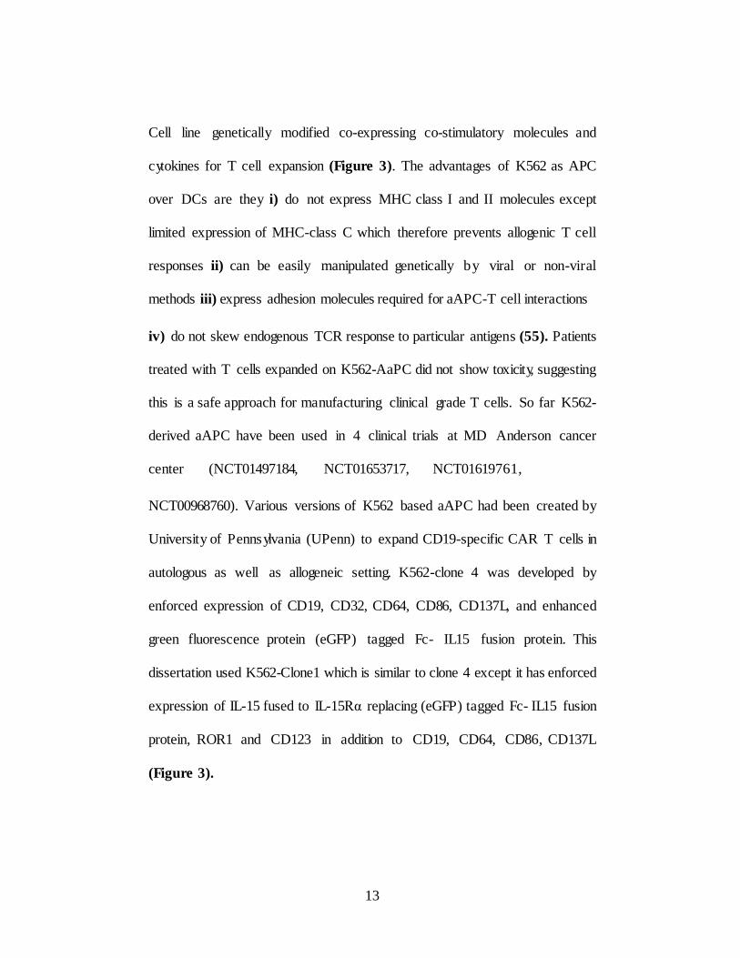
Cell line genetically modified co-expressing co-stimulatory molecules and
cytokines for T cell expansion (Figure 3). The advantages of K562 as APC
over DCs are they i) do not express MHC class I and II molecules except
limited expression of MHC-class C which therefore prevents allogenic T cell
responses ii) can be easily manipulated genetically by viral or non-viral
methods iii) express adhesion molecules required for aAPC-T cell interactions
iv) do not skew endogenous TCR response to particular antigens (55). Patients
treated with T cells expanded on K562-AaPC did not show toxicity, suggesting
this is a safe approach for manufacturing clinical grade T cells. So far K562-
derived aAPC have been used in 4 clinical trials at MD Anderson cancer
center (NCT01497184, NCT01653717, NCT01619761,
NCT00968760). Various versions of K562 based aAPC had been created by
University of Pennsylvania (UPenn) to expand CD19-specific CAR T cells in
autologous as well as allogeneic setting. K562-clone 4 was developed by
enforced expression of CD19, CD32, CD64, CD86, CD137L, and enhanced
green fluorescence protein (eGFP) tagged Fc- IL15 fusion protein. This
dissertation used K562-Clone1 which is similar to clone 4 except it has enforced
expression of IL-15 fused to IL-15Rα replacing (eGFP) tagged Fc- IL15 fusion
protein, ROR1 and CD123 in addition to CD19, CD64, CD86, CD137L
(Figure 3).
13
Figure 3. Sche matic of CAR T ce lls Expansion on AaPC. PBMC are isolated from whole
blood by density gradient centrifugation using Ficoll-Hypaque and are electroporated with
plasmids encoding either (i) Sleeping Beauty transposase or (ii) Sleeping Beauty transposon
containing CAR. Transfected cells were phenotyped for CAR expression next day and cells
stimulated with γ-irradiated K562-derived AaPC every7 days supplementing with IL2 and IL-
21. Following 3-4 weeks of co-culture, CAR T cells expanded to clinically relevant numbers are
ready for cr yopreservation and then infusion into cancer patients. Figure includes K562- Clone1-
CD123 that express CD19, ROR1, CD64, CD86, CD137L, mIL15 (1IL15 fused to IL15Rα)
and CD123.
14
I.7. Interleukin-3 Receptor α (CD123)
CD123 is the α subunit of Interleukin-3 cytokine receptor (IL-3Rα) which
forms high-affinity functional hetero-dimeric receptor along with its β subunit
CD131. Binding of IL-3 to IL-3R activates the receptor leading to cell survival
and proliferation (56, 57). IL-3 stimulated activation of spontaneous signal
transducer and activator of transcription 5 (STAT5) is correlated with over
expression of CD123 on AML cells resulting in proliferation of tumor cells (58-
60). CD123 has been reported to be overexpressed on up to 95% of leukemic
blasts and leukemic stem cells (LSCs) in AML, majority of B- ALL blasts,
but not on normal hematopoietic stem cells (HSC) and no cells outside
hematopoietic lineage (61-66). Clinically, high CD123 expression in AML
patients at diagnosis is associated with higher blast counts and a lower complete
remissions resulting in reduced survival (58-60). Collectively, these findings
point to the significance of CD123 expression in leukemia cell stimulation and
AML patient outcome. Phase1 clinical trials targeting CD123 in AML using
neutralizing mAbs and cytotoxic protein fused to IL-3 cytokine showed limited
therapeutic efficacy pressing the need for more novel efficacious treatments
(67-68). Thus CD123 is a viable target in AML through chimeric antigen
receptors in AML give n its wide expression on leukemic blasts, progenitors,
LSCs and weak or no detectable expression on hematopoietic stem cells. The
main goal of this
15
dissertation is, to redirect T –cell specificity to CD123 through chimeric
antigen receptors (CARs) to target AML and to generate preclinical data in
support of an adoptive immunotherapy trial.
I.8. Leukemic Stem cells and minimal residual disease
The majority of treated AML patients deemed to be in complete remission
by chemo and radiation therapies resulting in relapse. The relapse is due to
MRD attributed to LSCs (74). LSCs are pre-leukemic clonal population of
HSC by genetic and molecular alterations capable of self- renewal, able to
initiate leukemia when transplanted in SCID mice by generating rapidly
proliferating progenitors and leukemic blasts (69-71). HSCs and LSCs have
common features such as basic phenotype (Lineageneg CD34+CD38neg), slow
division, self-renewal capacity (69, 72). To our knowledge most of the antigens
that are expressed in AML are also present on hematopoietic stem cells and
progenitors. However certain AML markers are over expressed on LSC while
there is weak or no detectable expression on normal HSCs. CD123 is highly
expressed on the CD34+CD38neg fraction, leukemic blasts and bulk of AML
cells when compared to normal hematopoietic cells (73). CD123 and C- type
lectin-like molecule1 (CLL-1) are robust markers for MRD and highly
expressed on LSC (75, 76). Employing CAR T cells specific to CD123 after
hematopoietic
16
transplantation eradicate MRD which contain residual leukemic stem cells.
Moreover some of the B-ALL patients treated with CD19 CAR T resulted in
relapse with the residual population of leukemic cells negative for CD19 and
positive for CD123+ (77). These patients can be treated using CAR T cells
specific to CD123. The increased expression of CD123 on LSCs compared
with weak or no detectable expression on HSC presents an opportunity for
selectively targeting LSCs on AML with CD123-specific CAR+ T cells.
I.9. Dissertation specific aims
This dissertation focuses on three specific aims described as follows
I.9.A. Specific aim #1. To determine if scFvs of chimeric antigen
receptors derived by “Mix -and-Matching” VL and VH domains from
two monoclonal antibodies can redirect specificity to CD123+
leukemias. The VL and VH of scFvs of CARs usuall y derived from
single monoclonal antibody specific to targeted antigen. However this
dissertation describe a new approach for generating CD123-specific CARs
generating a chimeric scFv that is made up of the VL and VH harvested from
two mAbs that are each specific for CD123. The major hypothesis for this
specific aim is that CARs generated by combining VL and VH chains from
two different mAbs for CD123 will retain specificity for CD123. We
hypothesize that the CARs can be selected for targeting CD123
overexpressing leukemia while sparing normal hematopoietic cells
expressing CD123 at low levels for improved therapeutics. Rationale for this
specific aim is i) TAAs are not specific to tumors but also may be
17
expressed at low levels on normal cells, potentially resulting in on-target,
off-tumor toxicities i i) CD123 is expressed on hematopoietic progenitors
and weakly expressed on monocytes, neutrophils, basophils and
megakaryocytes iii) The affinity of the scFv for TAA also affects the
density of TAA required for efficient killing. iv) CAR T cells preferably
target tumors with high antigen density, while cells with lower density are
more resistant to CAR T cells (97, 98). A panel of CARs have been
generated by mix-and-matching VL and VH of four mAbs specific to CD123
and tested their cytolytic efficacy in B-ALL and normal BM cells.
I.9.B.Specific Aim#2: Comparative evaluation of CD123-specific chimeric
CARs containing CD28 or CD137 endo-domains for enhanced survival and
anti-tumor efficacy in AML. The hypothesis of this aim is that CAR T cells
containing CD137 endo-domain will be superior to those signaling through
CD28 in therapeutic efficacy. The rationale is i) Optimal CAR design
enhances the persistence of CAR T cells ii) studies showed that CARs that
incorporates CD137 has enhanced survival and anti- tumor efficacy
compared to CARs with CD28 endo-domain iii) the clinical outcome of
complete remission of CAR T cells correlated with long-term persistence
of CAR T cells iv) CD123, the IL-3 receptor α- subunit has been reported to
be overexpressed in AML. Two second-generation CD123- specific CARs were
generated from chimeric scFv by fusing VH and VL from two mAbs specific
to CD123 to CD3ζ and CD28 signaling domains
18
(designated CD123-CD28 CAR) and by fusing the same scFv to CD3ζ and
CD137 signaling domains (designated CD123-CD137 CAR). Each CAR
connect the scFv region to the endodomains via a modified hinge and Fc region
from IgG4. The Sleeping Beaut y (SB) system was used for non- viral gene
transfer to stably express CARs into T cells derived from peripheral blood
mononuclear cells (PBMC). Two SB plasmids coding for transposons
(CARs co-expressed with iCaspase9) and transposase (SB11) were
electroporated into PBMC and numerically expanded on designer AaPCs
(designated Clone 1-CD123) a genetically modified K562 cells co-
expressing co-stimulatory molecules (CD86 and CD137L), a membrane
bound cytokine mIL15 (IL-15 fused to IL-15R) and the TAAs ROR1 CD19
and CD123 supplemented with cytokines IL-2 and IL-21. Expanded T cells
were monitored for CAR expression, counted to determine expansion kinetic
s over a period of 4 to 5 weeks. At the end of 4 weeks of co-culture the
surface and memory phenotype were determined. The effector function of
CAR+ T cells were determined by assessing in vitro lysis of
CD123+leukemic cell lines and primary AML patient samples. To evaluate
in vivo tumor clearance CD123+ leukemia xenografts were established in
NSG mice and treated with CAR T cells.
19
I.9.C Specific Aim#3: in vitro targeting of AML leukemic stem cells by CAR T
cells specific to CD123. The hypothesis for this specific aim is to determine if
CD123-specific CAR T cells can eliminate leukemic stem cells in AML. The
rationale is i) The relapse in AML is due to minimal residual disease caused
by small population of LSCs resistant to drugs and radiation ii) high
expression of CD123 on LSCs compared with weak or no detectable
expression on HSCs presents an opportunity for selectively targeting AML-
LSCs iii) Antigen specific based adoptive immunotherapy will play a
complimentary role in eradicating MRD by targeting TAAs expressed on
LSCs. CD123 expression levels were determined in AML primary samples
and phenotypically defined LSCs. The efficacy of CAR cells in elimination
of LSCs and HSCs were determined in vitro by co-culture killing assays.
20
CHAPTER-II
Redirecting specificity of T cells To Target CD123+ B-ALL
Tumors
II.1. Introduction
CARs can empower T cells with an antibody-like specificity and is able to
transmit signals leading to T cell activation, proliferation and its effector functions
upon binding its specific antigen. The binding chemistry of CAR’s scFv with
its cognate antigen is not well studied at present. Eshhar et.al demonstrated
that the antigen binding site and idiotope for anti-2, 4, 6- trinitrophenyl (TNP)
antibody (SP6) reside exclusively in VH region. In general, T cells expressing
chimeric antigen receptors (CARs) are generated by combining the variable
light (VL) and heavy (VH) chains of scFv derived from single mAb specific
to targeted antigen (86). Examination of the contribution of VH and VL chains
of scFvs specific to targeted antigen ma y help us to better understand the
functionalit y of CARs and to derive CARs with different affinities to targeted
antigen. One of the limiting factors in CAR T cell therapy is TAAs are not
tumor “specific” but also expressed at low levels on normal cells and often
associated with off tumor toxicities. Recent preclinical studies targeting EGFR
and erbB2 with affinity lowered CAR T cells have demonstrated potent
antitumor effect on tumors with high antigen density while sparing normal cells
(87, 88). The present chapter describes a new approach for
21
generating CD123-specific CARs derived from a chimeric scFv that is made
up of the VL and VH harvested from two mAbs that are each specific for
CD123. The major hypothesis for this specific aim is that CARs generated
by combining VL and VH chains from two different mAbs for CD123 will
retain specificity for CD123. We hypothesize that the CARs can be selected
for targeting CD123 overexpressing leukemia while sparing normal
hematopoietic cells expressing CD123 at low levels for improved
therapeutics. To test this hypothesis we have generated six CARs with
chimeric scFvs by mix and matching VH and VL of four mAbs specific to
CD123. CARs derived from VH and VL of original mAbs without mix and
matching were used as control. We have chosen the one with least killing and
effector functions in normal hematopoietic cells carried forward to target
B-ALL (described in present chapter) and AML (described in chapter III).
22
II.2.Results
II.2.A. Generation of CD123+ Activating and Propagating Cells (AaPC)
Activating and Propagating cells (AaPC) has been successfully shown to
expand antigen specific CAR T cells ex vivo (45-49). Binding of T cells to its
cognate antigen on APC cell surface results in CAR+ T cell clustering,
phosphorylation of immune-receptor tyrosine-based activation motifs (ITAMs)
there by activating T cells (89). K562 based AaPC-Clone 1 was previously
made to expand CAR T cells co-express TAAs (CD19 and ROR1) co-
stimulatory molecules (CD86 and CD137L), Fc receptors (endogenous CD32
and transfected CD64) for loading of agonistic anti-CD3 antibod y OKT3 and
IL-15 fusion protein (IL-15 fused to IL-15Rα) (Figure 4). However AaPC-
Clone 1 do not express CD123. Therefore a new AaPC has been derived to
expand CD123-specific CAR T cells by enforced expression of CD123 on
AaPC-Clone 1 (designated as Clone1-CD123). The CD123 DNA sequence
was synthesized and codon optimized by Gene Art (Regensburg, Germany)
fused to hygromycin resistance gene through F2A peptide and sub cloned into
a SB transposon plasmid (Figure 5A). AaPC-Clone 1 cells were co-
electroporated with CD123 transposon and transposase SB11 and CD123+
positive cells were selected by hygromycin selection. Within 9 days after
electroporation more than 98% of cells express CD123 (Figure 5B)
23
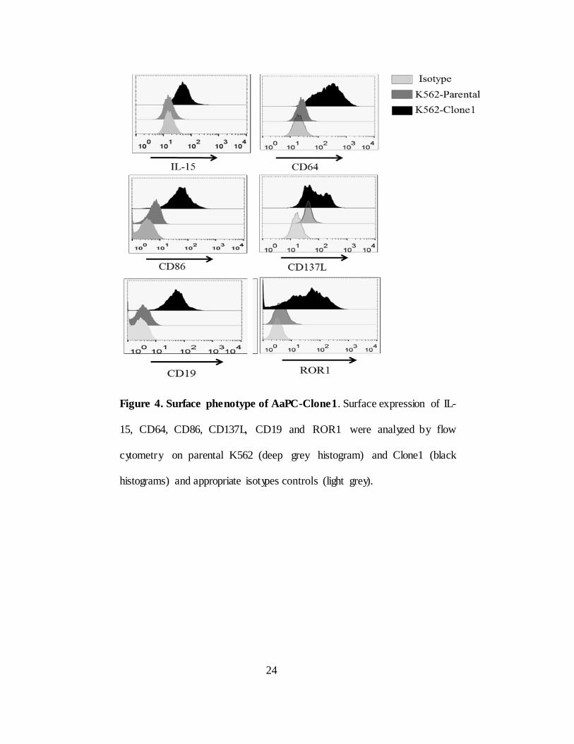
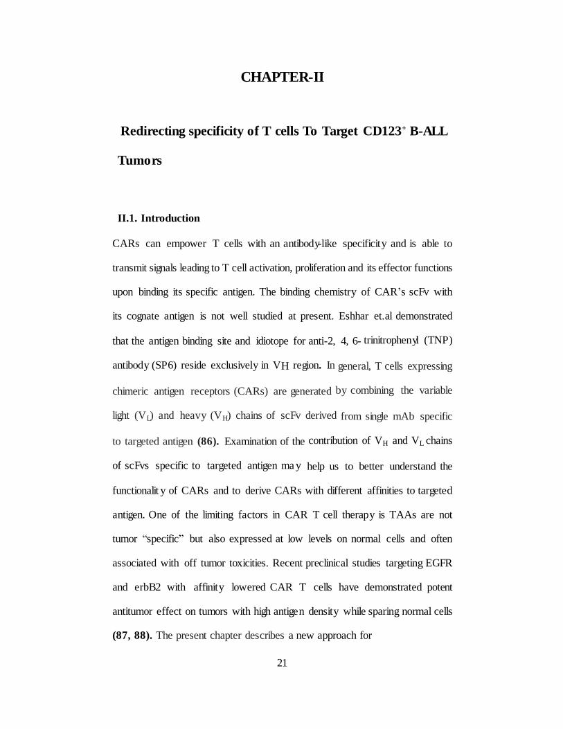
Figure 4. Surface phenotype of AaPC-Clone1. Surface expression of IL-
15, CD64, CD86, CD137L, CD19 and ROR1 were analyzed by flow
cytometry on parental K562 (deep grey histogram) and Clone1 (black
histograms) and appropriate isotypes controls (light grey).
24
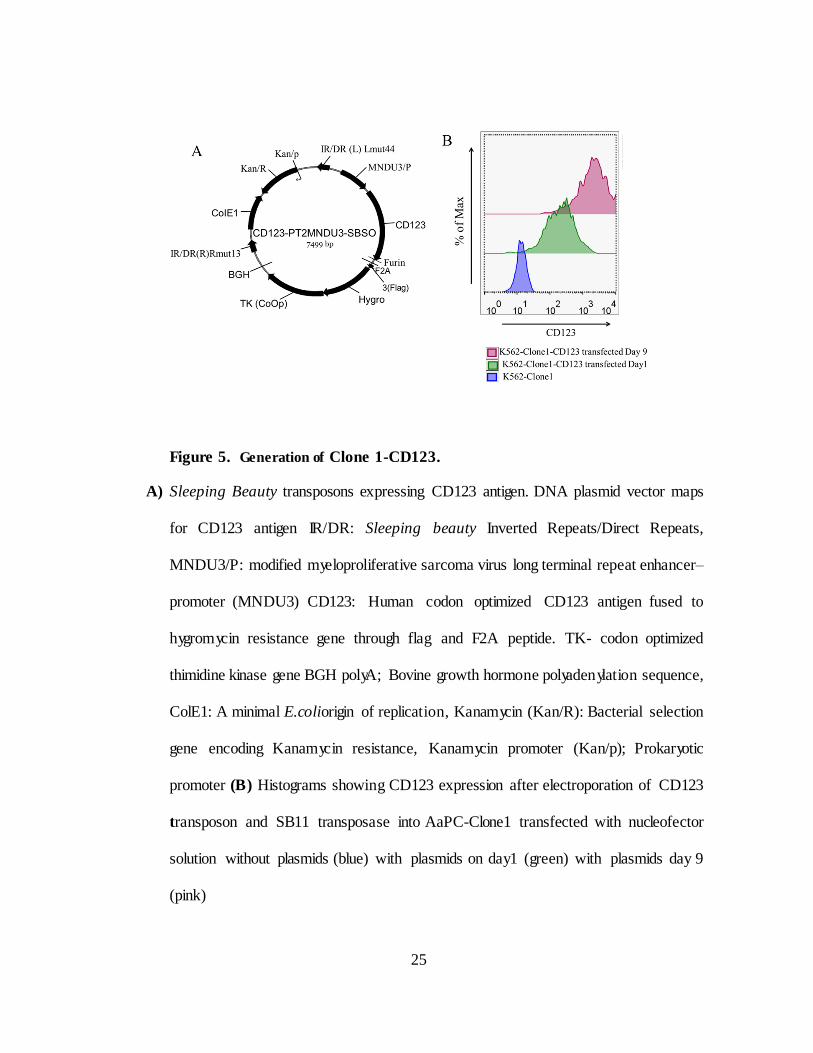
Figure 5. Generation of Clone 1-CD123.
A) Sleeping Beauty transposons expressing CD123 antigen. DNA plasmid vector maps
for CD123 antigen IR/DR: Sleeping beauty Inverted Repeats/Direct Repeats,
MNDU3/P: modified myeloproliferative sarcoma virus long terminal repeat enhancer–
promoter (MNDU3) CD123: Human codon optimized CD123 antigen fused to
hygromycin resistance gene through flag and F2A peptide. TK- codon optimized
thimidine kinase gene BGH polyA; Bovine growth hormone polyadenylation sequence,
ColE1: A minimal E.coliorigin of replication, Kanamycin (Kan/R): Bacterial selection
gene encoding Kanamycin resistance, Kanamycin promoter (Kan/p); Prokaryotic
promoter (B) Histograms showing CD123 expression after electroporation of CD123
transposon and SB11 transposase into AaPC-Clone1 transfected with nucleofector
solution without plasmids (blue) with plasmids on day 1 (green) with plasmids day 9
(pink)
25
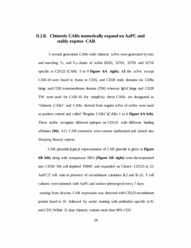
II.2.B. Chimeric CARs numerically expand on AaPC and
stably express CAR
5 second generation CARs with chimeric scFvs were generated by mix
and matching VL and VH chains of mAbs 26292, 32701, 32703 and 32716
specific to CD123 (CARs 5 to 9 Figure 6A right). All the scFvs except
CAR-10 were fused in frame to CD3ζ and CD28 endo domains via CD8α
hinge and CD8 transmembrane domain (TM) whereas IgG4 hinge and CD28
TM were used for CAR-10. For simplicity these CARs are designated as
“chimeric CARs” and CARs derived from regular scFvs of mAbs were used
as positive control and called “Regular CARs” (CARs 1 to 4 Figure 6A left).
These mAbs recognize different epitopes on CD123 with different binding
affinities (96). A l l CAR constructs were custom synthesized and cloned into
Sleeping Beauty system.
CAR plasmids (typical representation of CAR plasmid is given in Figure
6B left) along with transposase SB11 (Figure 6B right) were electroporated
into CD56+ NK cell depleted PBMC and expanded on Clone1- CD123 at 2:1
AaPC:T cell ratio in presence of recombinant cytokines IL2 and IL-21. T cell
cultures were stimmed with AaPC and surface phenotyped every 7 days
starting from day one. CAR expression was detected with CD123 recombinant
protein fused to Fc followed by serial staining with antibodies specific to Fc
and CD3. Within 21 days chimeric contain more than 90% CD3
26
and CAR double positive cells like regular CAR cultures (Figure 7A).
Cultures were devoid of NK cells though a small proportion of T cells express
CD56, they do not express CD3 (data not shown). Chimeric CARs expanded
at similar rates as regular CARs in sufficient amounts for clinic (Figure 7B
and 7C).
27
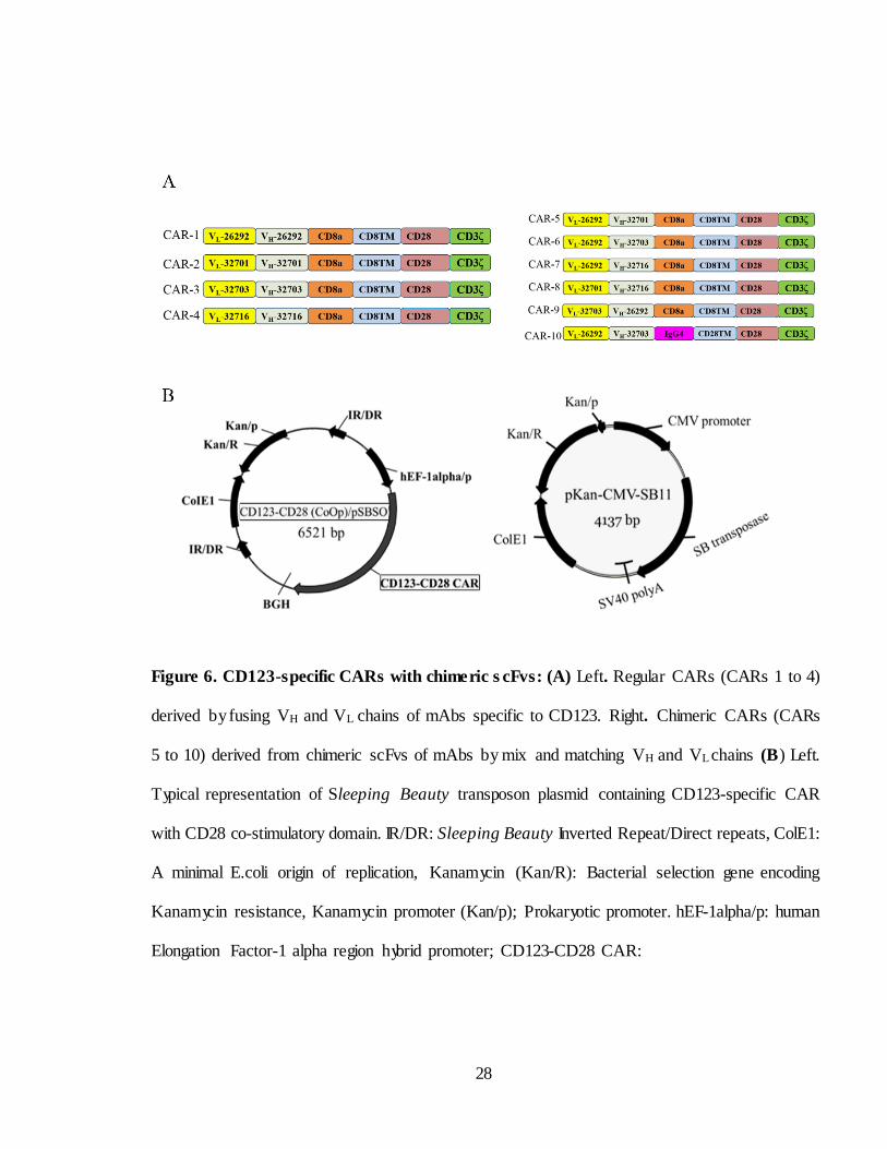
Figure 6. CD123-specific CARs with chime ric s cFvs: (A) Left. Regular CARs (CARs 1 to 4)
derived by fusing VH and VL chains of mAbs specific to CD123. Right. Chimeric CARs (CARs
5 to 10) derived from chimeric scFvs of mAbs by mix and matching VH and VL chains (B) Left.
Typical representation of Sleeping Beauty transposon plasmid containing CD123-specific CAR
with CD28 co-stimulatory domain. IR/DR: Sleeping Beauty Inverted Repeat/Direct repeats, ColE1:
A minimal E.coli origin of replication, Kanamycin (Kan/R): Bacterial selection gene encoding
Kanamycin resistance, Kanamycin promoter (Kan/p); Prokaryotic promoter. hEF-1alpha/p: human
Elongation Factor-1 alpha region hybrid promoter; CD123-CD28 CAR:
28
Human codon optimized CD123-specific CAR with CD28 co-stimulatory domain; BGH polyA;
Bovine growth hormone poly adenylation sequence, (right) SB11 transposase; CMV promoter
(Cytomegalovirus promoter) SV40 PolyA (Simian Virus 40 PolyA).
29
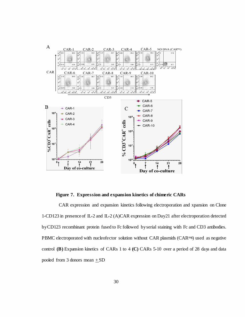
Figure 7. Expression and expansion kinetics of chimeric CARs
CAR expression and expansion kinetics following electroporation and xpansion on Clone
1-CD123 in presence of IL-2 and IL-2 (A)CAR expression on Day21 after electroporation detected
by CD123 recombinant protein fused to Fc followed by serial staining with Fc and CD3 antibodies.
PBMC electroporated with nucleofector solution without CAR plasmids (CARneg) used as negative
control (B) Expansion kinetics of CARs 1 to 4 (C) CARs 5-10 over a period of 28 days and data
pooled from 3 donors mean + SD
30
II.2.C. Chimeric CARs maintain specificity to CD123
Before testing in vitro efficac y of chimeric CARs several leukemic cell
lines including pre-B-ALL cell line Nalm6 and AML cell line TF1 and human
embryonic kidney cell line 293T (Figure 8). To test chimeric CAR T cells
demonstrate effective specific lysis of CD123+
tumor cells in vitro,
a chromium-51 labeled target cell lines were co-cultured with CAR T cells in
a standard 4 hour chromium release assay effector: target (E:T) ratio 20:1 .
CD123+ pre B-ALL cell line Nalm6, and AML cell line TF1 were used as
positive controls and 293T human embryonic kidney cell line used as
negative control. CAR T cells able to lyse CD123+ B-ALL tumor cell lines
(Figure 9A) but not CD123neg cell line 293T (Figure 9B). To further verify
killing by CAR T cells we co-cultured CARneg with target cell lines in 20:1
they fail to kill CD123+ ALL cell lines. To test antigen-specific lysis 293T cells
CAR T cells were co-cultured with 293T cells CAR T cells and 293T cells
transfected with CD123. CAR T cells lysed transfected cells but not CD123neg
293T (Figure 9A). This data suggests that chimeric CARs recognize the CD123
antigen and execute antigen specific killing.
31
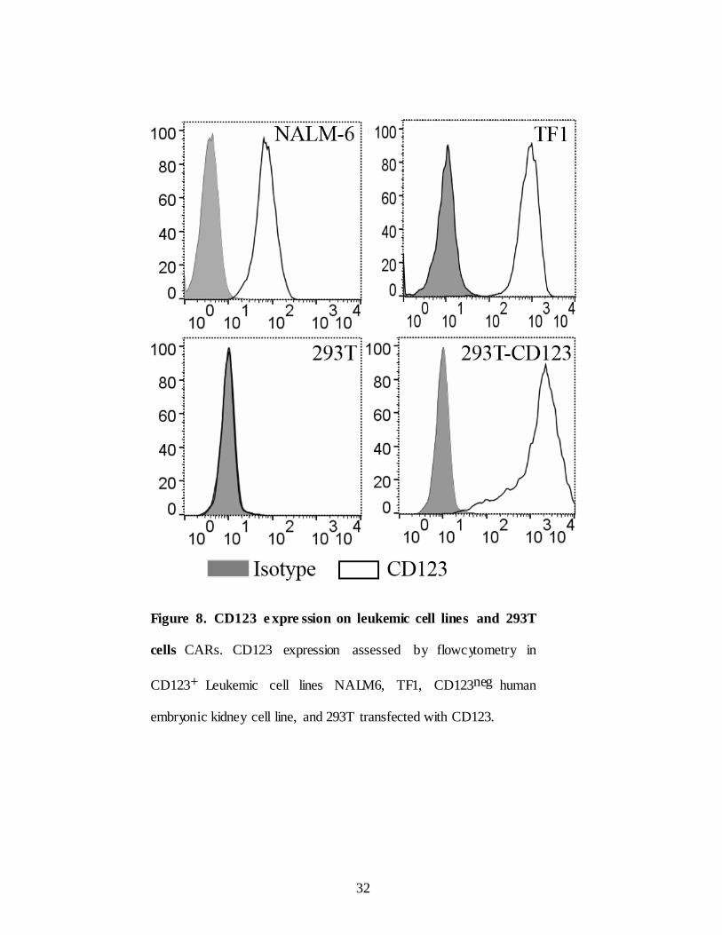
Figure 8. CD123 e xpre ssion on leukemic cell lines and 293T
cells CARs. CD123 expression assessed by flowcytometry in
CD123+ Leukemic cell lines NALM6, TF1, CD123neg human
embryonic kidney cell line, and 293T transfected with CD123.
32
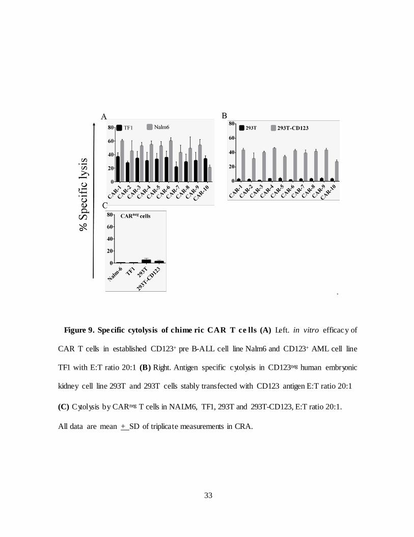
Figure 9. Spe cific cytolysis of chime ric CAR T ce lls (A) Left. in vitro efficacy of
CAR T cells in established CD123+ pre B-ALL cell line Nalm6 and CD123+ AML cell line
TF1 with E:T ratio 20:1 (B) Right. Antigen specific cytolysis in CD123neg human embryonic
kidney cell line 293T and 293T cells stably transfected with CD123 antigen E:T ratio 20:1
(C) Cytolysis by CARneg T cells in NALM6, TF1, 293T and 293T-CD123, E:T ratio 20:1.
All data are mean + SD of triplicate measurements in CRA.
33
II.2.D. IFN-γ production by chimeric CARs in response to
CD123 antigen
In order to assess antigen-specific effector function of chimeric CARs
IFN-γ production was assessed in CD123+ Nalm6 cells. 293T cells used as
negative control. T cells on Day 21 after electroporation were incubated with
Nalm6 and 293T cells in E:T ratio 2:1 for 48 hours. T cells without targets
used to see the difference with and without targets. Nalm6 stimulated chimeric
CAR T cells produced IFN-γ in significant amounts compared to CAR T cells
treated with 293T and T cells alone (Figure 10). These data established the
effector function and functionality of chimeric CARs in response to antigen.
34
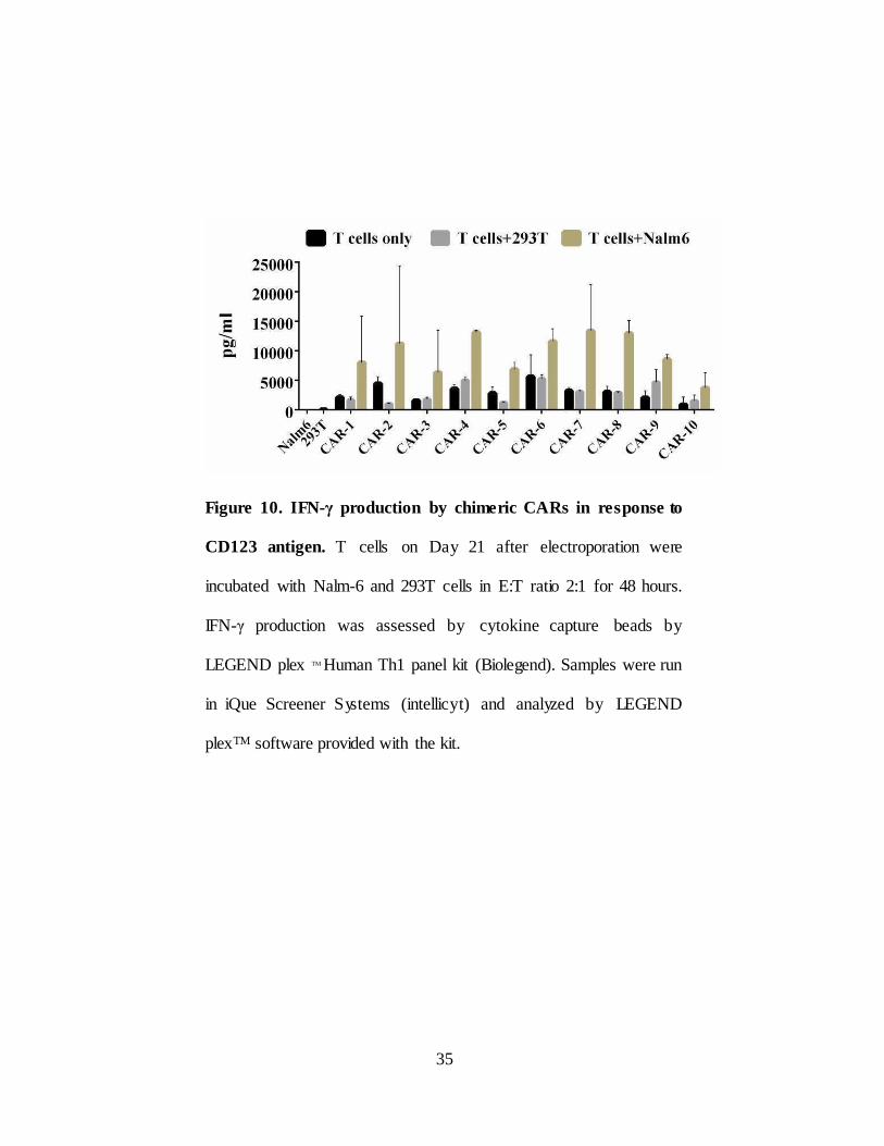
Figure 10. IFN-γ production by chimeric CARs in response to
CD123 antigen. T cells on Day 21 after electroporation were
incubated with Nalm-6 and 293T cells in E:T ratio 2:1 for 48 hours.
IFN-γ production was assessed by cytokine capture beads by
LEGEND plex TM Human Th1 panel kit (Biolegend). Samples were run
in iQue Screener Systems (intellicyt) and analyzed by LEGEND
plexTM software provided with the kit.
35
II.2.E. in vitro toxicity of chimeric CAR T cells in normal
hematopoietic cells
Many studies explored the expression of CD123 indicate that part of
hematopoietic progenitors from human cord blood, bone marrow, peripheral
blood and fetal liver express CD123 while primitive population of HSCs
express at low levels or absent (157). Though the antibody based CD123-
targeting therapies in AML reported to be well tolerated sparing normal
hematopoietic cells, recent pre-clinical studies employing CD123-specific CAR
T cells resulted in eradication of normal human myelopoiesis (161).
To test the in vitro toxicity of chimeric CARs for normal hematopoietic
cells, we isolated lineage+ and HSCs enriched lineageneg fractions from normal BM
cells and labeled with PKH-26. CAR T cells co-cultured with PKH-26 labeled
cells for 48 hours with E: T ratio 2:1.CD19 CAR T cells used as control. Cells were
stained with 7AAD and live/dead cells were enumerated by 7AAD exclusion.
CAR T cells are apparently lysed both lineage+ and lineageneg hematopoietic cells
(Figure 11A). CD19 is expressed on differentiated cells but not expressed on
HSCs. This is apparent by minimal lysis in lineageneg population than lineage+
population. These data raises concern that CD123- specific CAR therapy can be
detrimental to normal hematopoiesis. However IgG4 hinge based CAR-10
showed less cytotoxicity to normal hematopoietic cells when compared to its
counterparts with CD8α hinge (CARs 5-9) (Figure 11B).
36
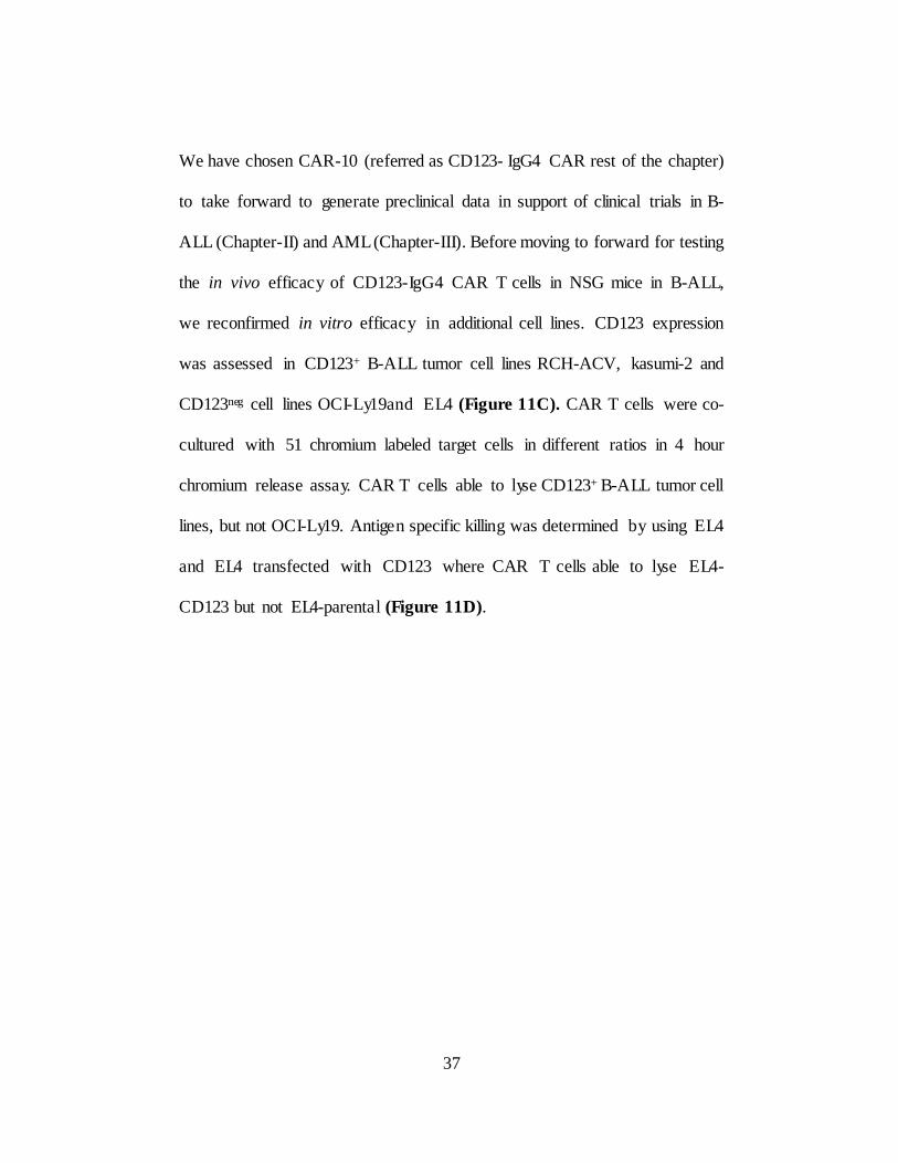
We have chosen CAR-10 (referred as CD123- IgG4 CAR rest of the chapter)
to take forward to generate preclinical data in support of clinical trials in B-
ALL (Chapter-II) and AML (Chapter-III). Before moving to forward for testing
the in vivo efficacy of CD123-IgG4 CAR T cells in NSG mice in B-ALL,
we reconfirmed in vitro efficacy in additional cell lines. CD123 expression
was assessed in CD123+ B-ALL tumor cell lines RCH-ACV, kasumi-2 and
CD123neg cell lines OCI-Ly19and EL4 (Figure 11C). CAR T cells were co-
cultured with 51 chromium labeled target cells in different ratios in 4 hour
chromium release assay. CAR T cells able to lyse CD123+ B-ALL tumor cell
lines, but not OCI-Ly19. Antigen specific killing was determined by using EL4
and EL4 transfected with CD123 where CAR T cells able to lyse EL4-
CD123 but not EL4-parental (Figure 11D).
37
Figure 11. Anti-tumor efficacy of chimeric CARs (CAR-10)
A) in vitro lysis of normal hematopoietic cells by chimeric CARs. (A)Mononuclear
cells isolated from normal BM samples and separated into lineage+ and lineageneg
cells labeled with PKH-26 and co-cultured with CAR T cells at E:T ratio 1:1
for 48 hours. 7-AAD added to distinguish live and dead cells to assess killing.
B) In vitro lysis of TF1 tumor cells vs Normal BM cells by chimeric CARs
Reduced cytolytic activity of CAR-10 compared to CARs 1-9 shown inbox
C) Flow analysis of CD123 expression on B-ALL cell lines RCH-ACV, KASUMI-2,
Nalm6 and B-cell lymphoma OCI-Ly19. D) in vitro efficacy of CD123-chimeric
CAR (CAR-10) specific CAR+ T cells in B-ALL cell lines in a standard 4 hour
chromium release assay. CD123neg mouse T cell lymphoma cell line EL4 was
transfected with CD123 antigen to determine antigen specific killing. Data was
reported as mean ± SD
38
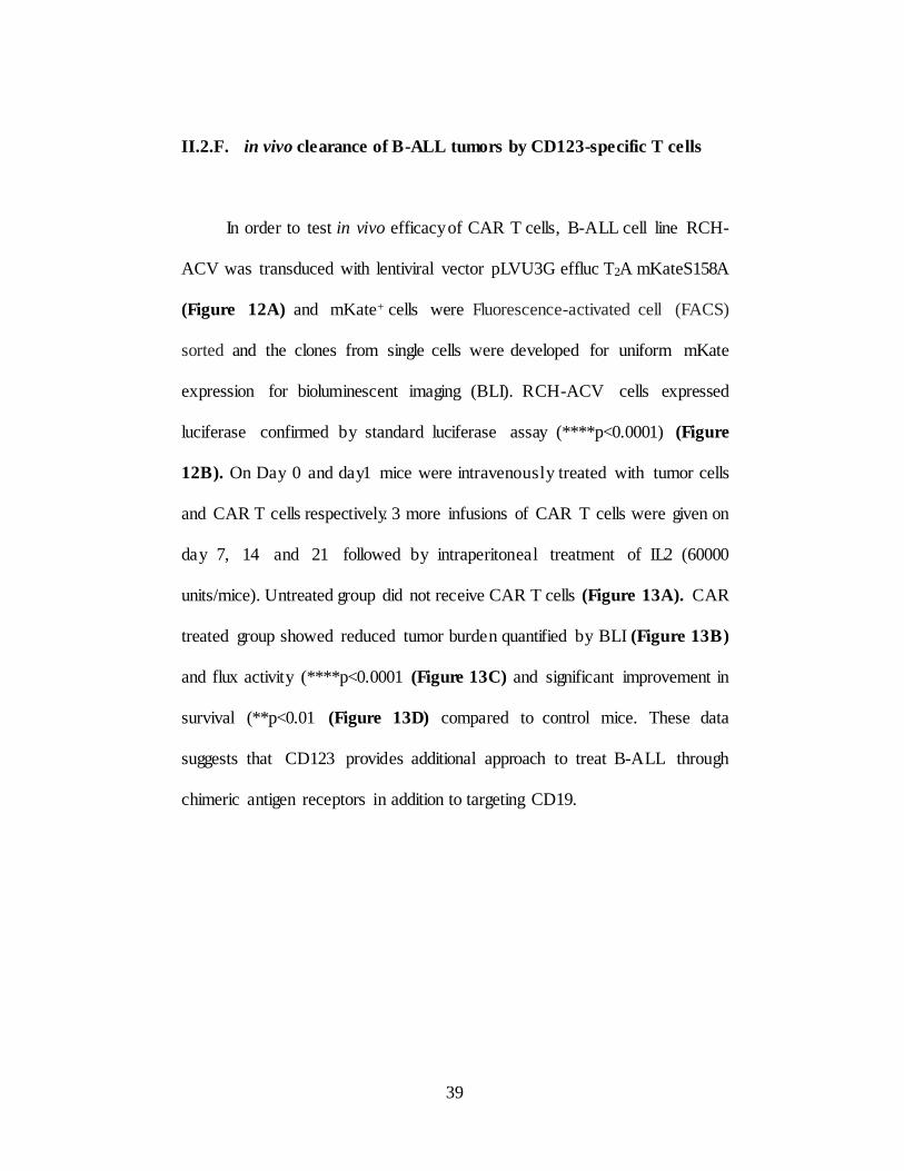
II.2.F. in vivo clearance of B-ALL tumors by CD123-specific T cells
In order to test in vivo efficacy of CAR T cells, B-ALL cell line RCH-
ACV was transduced with lentiviral vector pLVU3G effluc T2A mKateS158A
(Figure 12A) and mKate+ cells were Fluorescence-activated cell (FACS)
sorted and the clones from single cells were developed for uniform mKate
expression for bioluminescent imaging (BLI). RCH-ACV cells expressed
luciferase confirmed by standard luciferase assay (****p<0.0001) (Figure
12B). On Day 0 and day1 mice were intravenously treated with tumor cells
and CAR T cells respectively. 3 more infusions of CAR T cells were given on
day 7, 14 and 21 followed by intraperitoneal treatment of IL2 (60000
units/mice). Untreated group did not receive CAR T cells (Figure 13A). CAR
treated group showed reduced tumor burden quantified by BLI (Figure 13B)
and flux activity (****p<0.0001 (Figure 13C) and significant improvement in
survival (**p<0.01 (Figure 13D) compared to control mice. These data
suggests that CD123 provides additional approach to treat B-ALL through
chimeric antigen receptors in addition to targeting CD19.
39
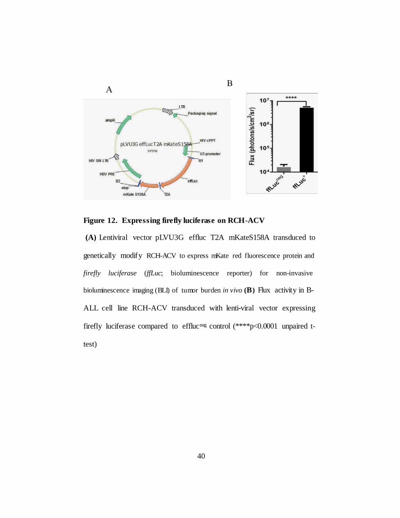
Figure 12. Expressing firefly luciferase on RCH-ACV
(A) Lentiviral vector pLVU3G effluc T2A mKateS158A transduced to
genetically modify RCH-ACV to express mKate red fluorescence protein and
firefly luciferase (ffLuc; bioluminescence reporter) for non-invasive
bioluminescence imaging (BLI) of tumor burden in vivo (B) Flux activity in B-
ALL cell line RCH-ACV transduced with lenti-viral vector expressing
firefly luciferase compared to efflucneg control (****p<0.0001 unpaired t-
test)
40
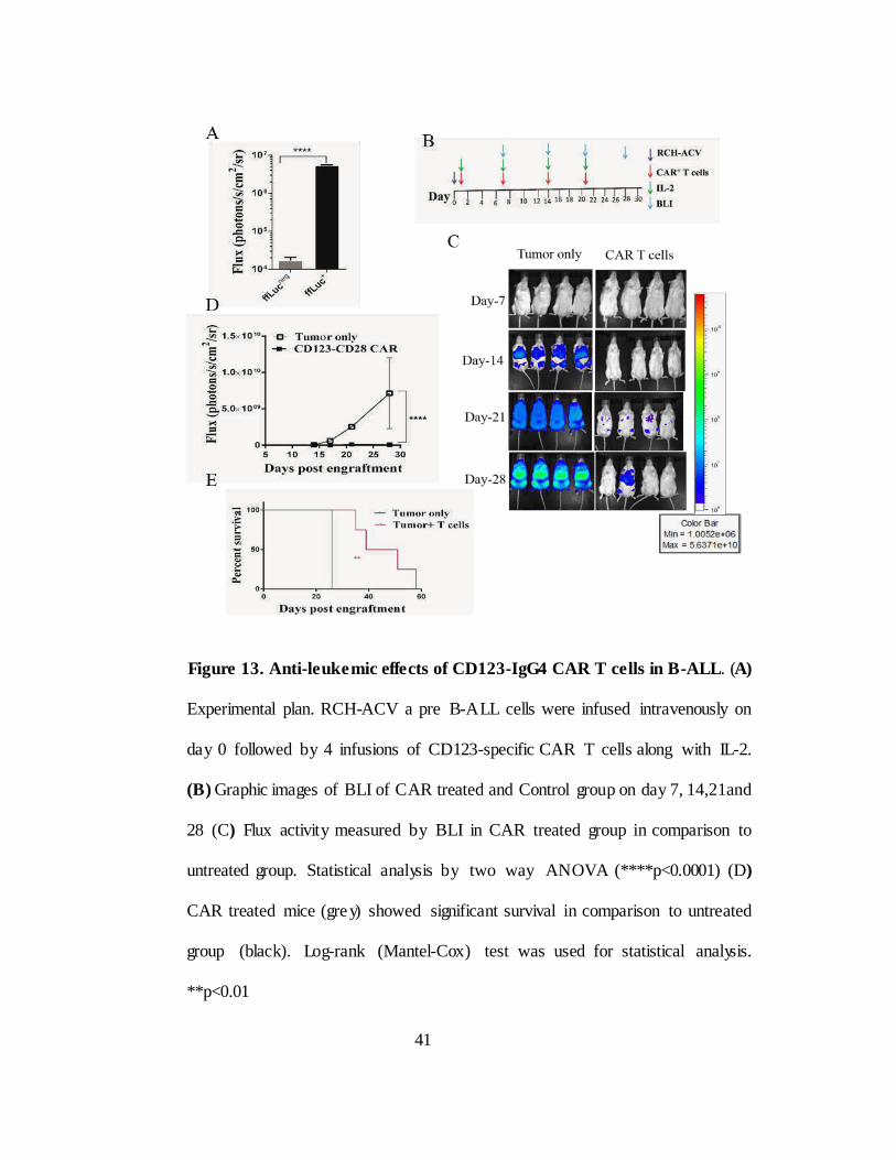
Figure 13. Anti-leuke mic effects of CD123-IgG4 CAR T cells in B-ALL. (A)
Experimental plan. RCH-ACV a pre B-ALL cells were infused intravenously on
day 0 followed by 4 infusions of CD123-specific CAR T cells along with IL-2.
(B) Graphic images of BLI of CAR treated and Control group on day 7, 14,21and
28 (C) Flux activity measured by BLI in CAR treated group in comparison to
untreated group. Statistical analysis by two way ANOVA (****p<0.0001) (D)
CAR treated mice (gre y) showed significant survival in comparison to untreated
group (black). Log-rank (Mantel-Cox) test was used for statistical analysis.
**p<0.01
41
II.3. Discusssion
Cell-based immunotherapies have demonstrated efficacious results in cancer
treatment modalities. This dissertation aimed to develop pre-clinical data to
support a clinical trial of CD123-specific CAR T cell treatments for CD123+
B-A LL and AML malignancies. We used existing platforms, Sleeping Beauty
system non-viral gene transfer and AaPC for expanding genetically modified
T cells with CARs (45-49, 55).
Traditional CARs have been generated using single-chain variable
fragments (scFv), often derived from a single mAb. Here we described a novel
approach for making CARs using chimeric scFvs deriving by assembling VH
and VL chains from two mAbs specific to CD123. Six CARs were generated
by mix and matching of VH and VL chains mAbs 26292, 32701, 32703 and
32716 specific to CD123. The CARs with chimeric scFvs were expressed,
expanded and mediated target cell lysis in vitro in similar fashion as CARs
derived from regular scFvs of mAbs. This approach may allow us to design
affinity tuned CARs with chimeric scFvs by mix and matching of VL and VH
chains of mAbs of various affinities. Clinical outcome of CAR T cells attributes
to several factors including CAR design, affinity of scFv to targeted antigen,
density of targeted molecule on tumor cells age and strength of immune system
of blood donor used for manufacturing T cells.
42
CD123 is over expressed in more than 95% of B-ALL patients while it is absent
in normal early B-cell precursors and weakly expressed on intermediate and
mature normal B cells. CD123 expression is correlated with hyper diploid
genotype a frequent genetic abnormality in childhood ALLs. In contrast B-
ALLs associated with other genetic abnormalities such as chromosomal
translocations or normal karyotype do not express CD123 (99, 100). The
overexpression of CD123 expression on B-ALL compared to normal B cells
and correlated expression in hyper diploid B-ALL, provide s an opportunity to
therapeutically target B-ALL through chimeric antigen receptors.
Relapse is the main reason for treatment failure in ALL patients,
minimal residual disease (MRD) has significant prognostic value in pediatric
and adult ALL (101-107). Leukemic stem cells are well documented in AML,
their existence and relevance in ALL is less clear. However, several reports
suggested that, a majority of leukemic populations with primitive stem-like
phenotype can propagate leukemia in the appropriate experimental setting and
their hierarchial organization is less strict like LSCs in AML (158). As reported
by several groups TEL/AML1-positive CD34+ cells that carried no lineage
markers specific to lymphoid differentiation (CD19 or CD10) were capable of
engraftment and propagating leukemia and even engraft secondary recipients
(159-160). These findings corroborate recent clinical findings by June et.al
while targeting B-ALL by CAR T cells specific to CD19. In their studies though
CD19 CAR+ T cells have been shown to induce potent anti-
43
tumor activity against B-ALL tumors, some of the CD19 CAR treated B-ALL
relapsed patients exhibited phenotype that were negative for CD19 but
expressed D123 (108). It appears that, employing CD123-specific CAR T cells
for relapsed patients after CD19 CAR therapy feasible strategy to prevent
relapse and improved survival.
One of the limiting factors in CAR T cell therapy is TAAs are not tumor
“specific” but also expressed at low levels on normal cells often associated with
off tumor toxicities. This is a considerable concern since CD123+B-ALL antigen
targeted therapies results in elimination of HSCs along with leukemic cells.
Though the effect of antigen density for CAR therapy is not well defined yet it
appears that CAR T cells preferably target tumors with high antigen density
while the ones with lower density are resistant to therapy (97, 98). Recent
preclinical studies with CAR T cells with lowered affinity targeting EGFR and
erbB2 showed potent antitumor effect on tumors with high antigen density while
sparing normal cells (87, 88).
Though CARs typically are identified by their endo-domains and scFv,
the other components of CARs, including the hinge/spacer region, also play
a crucial role in their function and clinical efficacy. The constant region of
IgG4 and CD8α re frequently used extracellular (stalk) hinge regions, though
the Fc region has been reported to engage Fc receptors and activate innate
immune cells (137). To avoid off target activation of CARs and unwanted
immune responses we have generated a CD123 specific CAR construct by
introducing L235E and N297Q mutations in the CH2 region of IgG4-Fc spacer.
44
We showed that CAR constructs using CD8α-derived hinge provide
highly effective cytolysis in our CD123-specific constructs. Interestingly,
the choice of spacer had a much greater impact on target cytolysis than
expected, with a CAR utilizing a CD8-derived spacer achieving much
better cytolysis than the same scFv using an IgG4-derived spacer.
Importantly CAR-10 with IgG4 hinge showed minimal lysis in normal
BM cells compared to CAR-6 with same scFv with CD8α hinge (Figure
6A). This observation requires further investigation in future models.
By choosing different sources of VH and VL chains and perhaps
different hinge regions, we may be able to tune the activation threshold
for CAR T cells further, especially if a wider range of antibody
affinities is used than was chosen for these studies. This finding may allow
us to generate CARs with low affinity to selectively target high antigen
density tumors while sparing normal hematopoiesis.
45
CHPTER-III
Comparative evaluation of co-stimulatory signals in targeting AML
with CD123-specific T cells
III.1.Introduction
Acute myeloid leukemia (AML) is a clonal proliferation of
malignant myeloid blast cells in the BM with impaired normal hematopoiesis.
Despite many advances AML remains a lethal disease. Standard treatment
regimens chemotherapy and radiation ensure long-term remission only in 30 to
50% of patients with a low survival probability resulting in resistance and
relapse (109-111). CARs have demonstrated clinical efficacy in treating
leukemia in preclinical models and are being tested in several clinical trials and
emerging as powerful tools for adoptive immunotherapy (115). CD123, the IL-
3 receptor α- Sub unit has been reported to be overexpressed on up to 95%
of leukemic in AML with weak on normal HSCs and absent on cells outside
hematopoietic lineage (120-124). Phage1 clinical trials targeting CD123 in
AML using neutralizing mAbs and cytotoxic protein fused to IL-3 cytokine
showed limited therapeutic efficacy pressing the need for more novel
efficacious treatments (125, 126). Several pre-clinical and animal models
have demonstrated that CAR T cells including CD28 or CD137 co-
stimulatory domains as a built in source of signal 2 have improved persistence
compared with those containing the CD3ζ signaling domain alone
(119,196,197). However, the anti-tumor efficacy of one over the other
costimulatory domain has not been investigated in depth.
46
An additional challenge in developing CAR T cells for immunotherapy is the
management of toxicities, especially those related to excess activation of
infused cells or targeting of TAA expressed on normal tissues (195). To
address these questions, we engineered constructs in which the CAR10
CD123-specific second generation CAR was fused to either the CD28
(designated as CD123-CD28 CAR) or CD137 (designated as CD123- CD137
CAR) co-stimulatory domains. To reduce off-target toxicities, the utility of
the inducible suicide switch iCaspase9 has been evaluated in this context.
The main goal of this study is comparative functional evaluation of two
CD123- specific CARs with CD28 or CD137 co-stimulatory domains. The
hypothesis of this aim is that T cells expressing CD123-specific CARs will
re-direct the specificity of T cells to target CD123+ AML and CARs
containing CD137 endo-domain will be superior to those signaling through
CD28 in therapeutic efficacy. The rationale is i) Optimal CAR design
enhances the persistence of CAR T cells ii) studies showed
that CARs that incorporates CD137 has enhanced survival and
anti-tumor efficacy compared to CARs with CD28 endo-domain
iii) the clinical outcome of complete remission of CAR T cells correlated
with long-term persistence of CAR T iv) CD1 23, the IL-3 receptor α -
subunit has has reported to be overexpressed in AML v) evidence that
complete remissions (CR) were observed in B-ALL and CLL patients treated
with CD19 CAR T cells.
47
III.2. RESULTS
III.2.A. Construction of CD123-specific CAR SB plasmids
Two codon-optimized Sleeping Beauty transposons encoding CD123-
specific second generation CARs fused to suicide gene iCaspase9 with CD28
(Figure 14A) or CD137 (Figure 14B) co-stimulator y domains were swapped
into previously made iCaspase 9 co-expressing CD19 CARs in SB system.
Swapping replaces CD19-specific scFv sequence with CD123-specific scFv
keeping rest of the plasmid intact. The CAR plasmids were constructed in the
following order: human elongation factor-α (hEF-α) promoter was used to drive
expression of CARs. Following promoter, 5’ to3’ CAR open reading frame
(ORF) consisting of signal peptide, scFv, whitlow linker, modified IgG4 hinge,
CD28 transmembrane domain, CD28 or C D 137 endo-domain and CD3ζ
signaling domain. The scFv is derived from V L of mAb 26292 and VH of mAb
32703 specific to CD123 (Figure 6A, CAR- 10 Chapter II). To distinguish
CARs with CD28 and CD137 endo-domains by PCR in cells isolated from
in vivo studies a unique oligonucleotides SIM for CD123- CD28 CAR and
FRA for CD123-CD137 CAR were interspersed between stop codon of
CAR and BGH polyA tail. Upon electroporation the indirect repeats (IR) of
SB system flanking 5’ end of hEF-α promoter and 3’ end of Poly A tail is
cut by SB11 transposase and integrates within the TA repeats in human T cell
genome. Kanamycin resistance gene will allow to amplify the SB plasmids in
large numbers in bacteria.
48
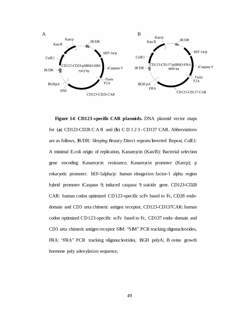
Figure 14: CD123-specific CAR plasmids. DNA plasmid vector maps
for (a) CD123-CD28 C A R and (b) C D 1 2 3 - CD137 CAR. Abbreviations
are as follows, IR/DR: Sleeping Beaut y Direct repeats/Inverted Repeat, ColE1:
A minimal E.coli origin of replication, Kanamycin (Kan/R): Bacterial selection
gene encoding Kanamycin resistance, Kanamycin promoter (Kan/p); p
rokaryotic promoter. hEF-1alpha/p: human elongation factor-1 alpha region
hybrid promoter iCaspase 9; induced caspase 9 suicide gene. CD123-CD28
CAR: human codon optimized CD123-specific scFv fused to Fc, CD28 endo-
domain and CD3 zeta chimeric antigen receptor, CD123-CD137CAR: human
codon optimized CD123-specific scFv fused to Fc, CD137 endo- domain and
CD3 zeta chimeric antigen receptor SIM: “SIM” PCR tracking oligonucleotides,
FRA: “FRA” PCR tracking oligonucleotides, BGH poly A; B ovine growth
hormone poly adeny lation sequence,
49
B A
III.2.B. SB modified T cells stably co-express CD123-specific CAR and
iCaspase9
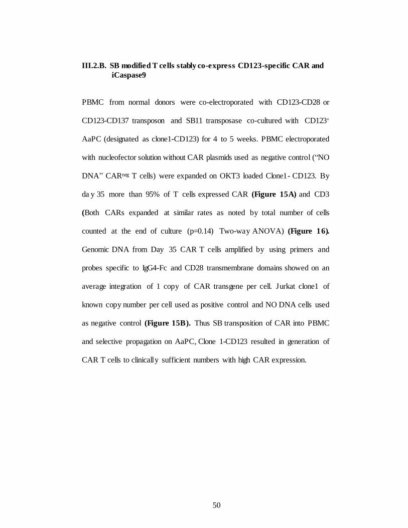
PBMC from normal donors were co-electroporated with CD123-CD28 or
CD123-CD137 transposon and SB11 transposase co-cultured with CD123+
AaPC (designated as clone1-CD123) for 4 to 5 weeks. PBMC electroporated
with nucleofector solution without CAR plasmids used as negative control (“NO
DNA” CARneg T cells) were expanded on OKT3 loaded Clone1- CD123. By
da y 35 more than 95% of T cells expressed CAR (Figure 15A) and CD3
(Both CARs expanded at similar rates as noted by total number of cells
counted at the end of culture (p=0.14) Two-way ANOVA) (Figure 16).
Genomic DNA from Day 35 CAR T cells amplified by using primers and
probes specific to IgG4-Fc and CD28 transmembrane domains showed on an
average integration of 1 copy of CAR transgene per cell. Jurkat clone1 of
known copy number per cell used as positive control and NO DNA cells used
as negative control (Figure 15B). Thus SB transposition of CAR into PBMC
and selective propagation on AaPC, Clone 1-CD123 resulted in generation of
CAR T cells to clinically sufficient numbers with high CAR expression.
50
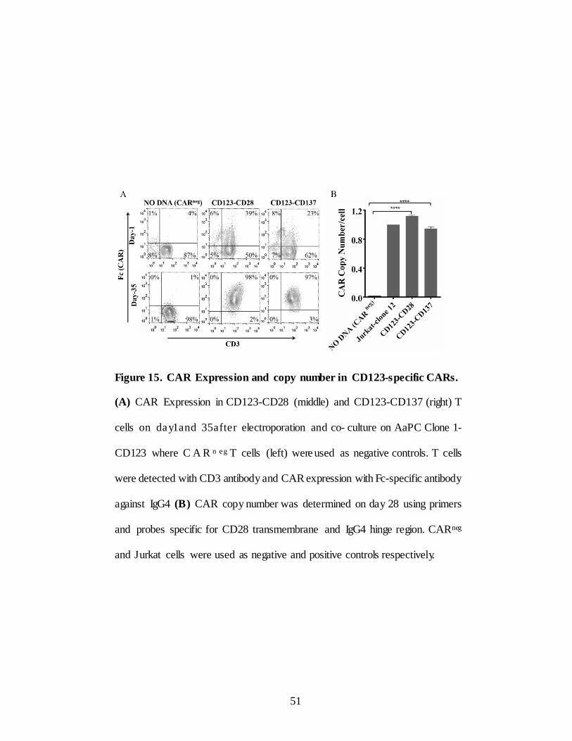
Figure 15. CAR Expression and copy number in CD123-specific CARs.
(A) CAR Expression in CD123-CD28 (middle) and CD123-CD137 (right) T
cells on da y1and 35after electroporation and co- culture on AaPC Clone 1-
CD123 where C A R n e g T cells (left) were used as negative controls. T cells
were detected with CD3 antibody and CAR expression with Fc-specific antibody
against IgG4 (B) CAR copy number was determined on day 28 using primers
and probes specific for CD28 transmembrane and IgG4 hinge region. CARneg
and Jurkat cells were used as negative and positive controls respectively.
51
Figure 16. Expansion kinetics of CD123-specific CARs.
Expansion of CD3+ and CD3+CAR+ T cells over a period of 35
days after electroporation in CD123-CD28 and CD123-CD137
CAR T cells as noted by total number of cells counted at the end
of culture (p=0.14) Two-way ANOVA (CD3+ and CD3+ CAR+
T cells).
52
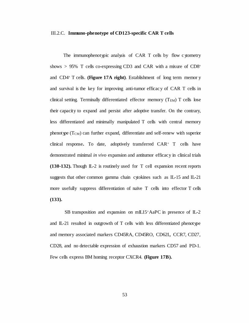
III.2.C. Immuno-phenotype of CD123-specific CAR T cells
The immunophenotypic analysis of CAR T cells by flow c ytometry
shows > 95% T cells co-expressing CD3 and CAR with a mixure of CD8+
and CD4+ T cells. (Figure 17A right). Establishment of long term memor y
and survival is the key for improving anti-tumor efficac y of CAR T cells in
clinical setting. Terminally differentiated effector memory (TEM) T cells lose
their capacity to expand and persist after adoptive transfer. On the contrary,
less differentiated and minimally manipulated T cells with central memory
phenotype (TCM) can further expand, differentiate and self-renew with superior
clinical response. To date, adoptively transferred CAR+ T cells have
demonstrated minimal in vivo expansion and antitumor efficacy in clinical trials
(130-132). Though IL-2 is routinely used for T cell expansion recent reports
suggests that other common gamma chain cytokines such as IL-15 and IL-21
more usefully suppress differentiation of naïve T cells into effector T cells
(133).
SB transposition and expansion on mIL15+AaPC in presence of IL-2
and IL-21 resulted in outgrowth of T cells with less differentiated phenotype
and memory associated markers CD45RA, CD45RO, CD62L, CCR7, CD27,
CD28, and no detectable expression of exhaustion markers CD57 and PD-1.
Few cells express BM homing receptor CXCR4. (Figure 17B).
53
CAR+ T cells belonged to less differentiated phenotype primarily compose d
of few naïve (TN) defined by CD45RA+CD62L+CD95neg CCR7+, TEMRA
(CD45RA+CD62LnegCD95neg CCR7neg), TEM (CD45RAnegCD62LnegCD95+
CCR7neg) and TCM (CD45RAnegCD62L+CD95+ CCR7+) and co-express CD27
and CD28 to engage co-stimulatory ligands for long term survival (Figure 17A
and 17B).
54
Figure 17. Immunophenotype of CD123-specific CAR T cells (A) Flow analysis
of memory markers on CD3+Fc+ gated T cells. Representation of one donor of total 3 donors
actually used in the experiment (left) and selective surface markers CD4, CD8, and CD56
(right) (B) memory and exhaustion markers CD57 and PD1 expressed (n=3) on CD123-
CD28 and CD123-CD137 CAR+T cells. Paired Student’s two-tailed t- test was used
*p<0.05 (C) T cell differentiation subsets gated on CD3+Fc+ population , histograms
depicting cell percentage in each subset, TNaive CD45RA+CD62L+CD95neg CCR7+,
TEMRA (CD45RA+CD62LnegCD95negCCR7neg), TEM
(CD45RAnegCD62LnegCD95+ CCR7neg) and TCM (CD45RAnegCD62L+CD95+
CCR7+) in CD123-CD28 CAR+ T cells (Black bars) and CD123-CD137 CAR+ T cells
(Grey bars) (n=3). Statistical analysis by Student’s t test or nonparametric Mann–whitney
Method.
55
III.2.D. Transcriptional profile of CD123-specific CAR T cells
Transcriptional profile of CAR T cells was assessed by nanostring digital
multiplex array of mRNA showed expression of T cell activation markers
CD69, CD44, TIM3, co-stimulatory molecules CD40L, CD27 CD28 and no
expression of exhaustion and terminal differentiation markers above detectable
levels B3GAT1 (Beta-1, 3-Glucuronyltransferase-1; CD57) and KLRG1
(KLRG1) by CAR T cells shows they are fully activated and has the potential
for persistence after adoptive transfer (Figure 18A). Concurrent expression
of transcription factors associated with less differentiate d phenotype i.e ID2
(Inhibitor of DNA Binding-2), KLF2 (Kruppel-like Factor-2), FOXO1 (Forkhead
Box-O1), CTNNB1 (β-Catenin), BACH2 (BTB and CNC Homology-2), GFI1
(Growth Factor Independence-1), LEF1 (Lymphoid Enhancer Binding Factor-
1) and later memory stages, i.e BCL6 (B-cell Lymohoma-6), PRDM1
(BLIMP-1), and TBX21 (T-bet), suggests that the expanded CAR+ T cells
were heterogeneous in memory regulation (Figure 18B). Expression of
cytokine receptors e.g., IL2RA (IL-2-Receptor- α; CD25), IL2RB (IL-2-
Receptor-β; CD122), IL2RG (IL-2-Receptor-γ;CD132), IL7R (IL-7-Receptor-
α; CD127), and IL15RA (IL-15-Receptor-α), suggests that CAR T cells has
potential for continuous survival and persistence after adoptive transfer. CAR
T cells express molecules associated with T cell effector (Granzyme A,
Granzyme B, Perforin 1, Granulysin, IFN- γ and TNF) memory and trafficking
(SELL (L-Selectin; CD62L), CD95,
56
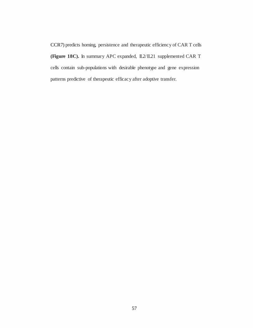
CCR7) predicts homing, persistence and therapeutic efficiency of CAR T cells
(Figure 18C). In summary APC expanded, IL2/IL21 supplemented CAR T
cells contain sub-populations with desirable phenotype and gene expression
patterns predictive of therapeutic efficacy after adoptive transfer.
57
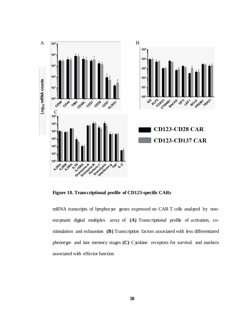
Figure 18. Transcriptional profile of CD123-specific CARs
mRNA transcripts of lymphocyte genes expressed on CAR T cells analyzed by non-
enzymatic digital multiplex array of (A) Transcriptional profile of activation, co-
stimulation and exhaustion (B) Transcription factors associated with less differentiated
phenotype and late memory stages (C) Cytokine receptors for survival and markers
associated with effector function
58
III.2.E. in vitro functionality of CD123-specific CAR T cells
Before testing functionality of CAR T cells, CD123 expression was evaluated
on AML cell lines MV4-11, TF1, Molm-13, OCI-AML3 and mouse T cell
lymphoma cell line CD123neg EL4-parental (EL4-P) and EL4-P transfected
with CD123 antigen. All the cell lines tested were positive for CD123 except
EL4-P cells and OCI-Ly19 (Figure 19A). To evaluate functionality of CD123-
specific CAR+ T cells in vitro, we used 4 hour chromium release assay for
AML cell lines and flow-cytometry based killing assay for AML primary
cells. CD123- specific T cells were able to lyse CD123+ AML cell lines but
did not kill CD123neg B-cell lymphoma cell line OCI-Ly19. To provide further
evidence that CD123-specific C A R T cells specifically target CD123+ tumors
we genetically modified EL4 parental cell line to enforce CD123 expression.
CD123-specific T cells efficiently killed EL4-CD123 but not EL4 parental cells
(Figure 19B).
In order to assess killing efficacy in primary patient samples, CD123
expression was analyzed on primary samples by flow cytometr y (Figure 20A).
All 4 primary samples do not express CD19 (data not shown). CAR T cells
were co-cultured with PKH-26 labeled CD123+ primary AML cells in E:T
ratio 2:1 for 72 hours and CD19 CAR T cells used as negative control. CD123-
specific T cells recognized and killed CD123+ AML primary cells but not in
CD19neg AML primary cells co-cultured with CD19 CAR T cells
59
(Figure 20B). iCaspase 9 expression on CAR T cells was assessed by flow
cytometry (Figure 21A) and in vitro functionalit y of iCasp9 was assessed by
treating CAR T cells with 1 µM chemical inducer of dimerization (CID) a
synthetic homo-dimerizer AP20187 for 24 hours. Untreated CAR T cells used
as negative control. Within 24 hours the dimerizer drug rapidly eliminate d
CAR T cells in CID treated group (Figure 21B) compared to untreated
control. In summary CD123-specific CAR T cells demonstrated anti-tumor
efficac y in CD123+ cell lines and primary tumors, and conditionally ablated
CAR T cells.
60
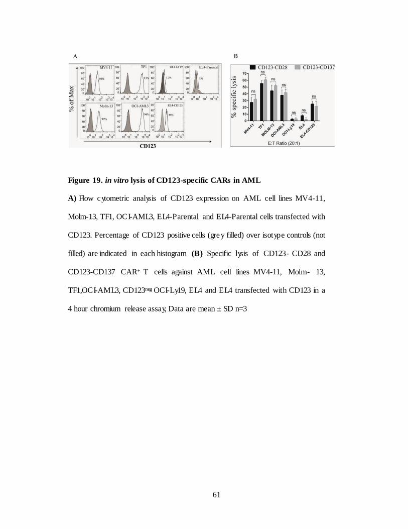
Figure 19. in vitro lysis of CD123-specific CARs in AML
A) Flow cytometric analysis of CD123 expression on AML cell lines MV4-11,
Molm-13, TF1, OCI-AML3, EL4-Parental and EL4-Parental cells transfected with
CD123. Percentage of CD123 positive cells (gre y filled) over isotype controls (not
filled) are indicated in each histogram (B) Specific lysis of CD123- CD28 and
CD123-CD137 CAR+ T cells against AML cell lines MV4-11, Molm- 13,
TF1,OCI-AML3, CD123neg OCI-Ly19, EL4 and EL4 transfected with CD123 in a
4 hour chromium release assay, Data are mean ± SD n=3
61
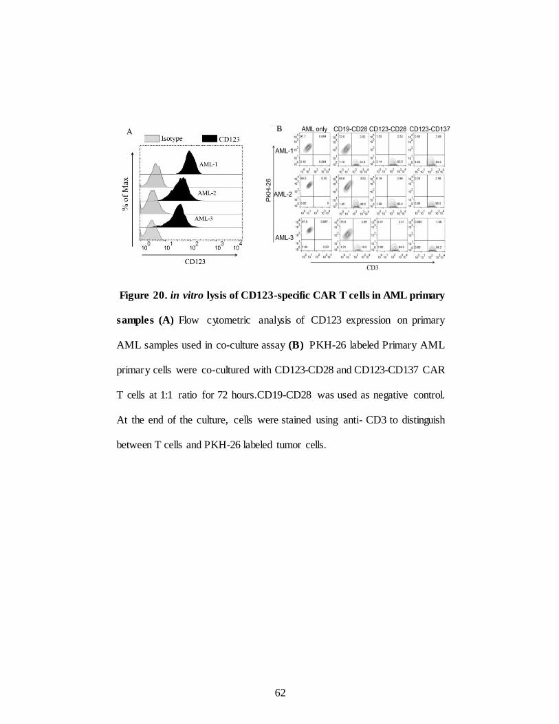
Figure 20. in vitro lysis of CD123-specific CAR T cells in AML primary
samples (A) Flow cytometric analysis of CD123 expression on primary
AML samples used in co-culture assay (B) PKH-26 labeled Primary AML
primary cells were co-cultured with CD123-CD28 and CD123-CD137 CAR
T cells at 1:1 ratio for 72 hours.CD19-CD28 was used as negative control.
At the end of the culture, cells were stained using anti- CD3 to distinguish
between T cells and PKH-26 labeled tumor cells.
62
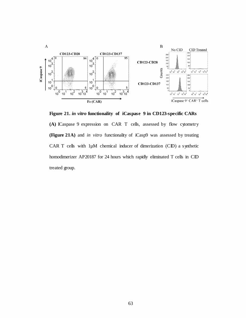
Figure 21. in vitro functionality of iCaspase 9 in CD123-specific CARs
(A) ICaspase 9 expression on CAR T cells, assessed by flow cytometry
(Figure 21A) and in vitro functionality of iCasp9 was assessed by treating
CAR T cells with 1µM chemical inducer of dimerization (CID) a synthetic
homodimerizer AP20187 for 24 hours which rapidly eliminated T cells in CID
treated group.
63
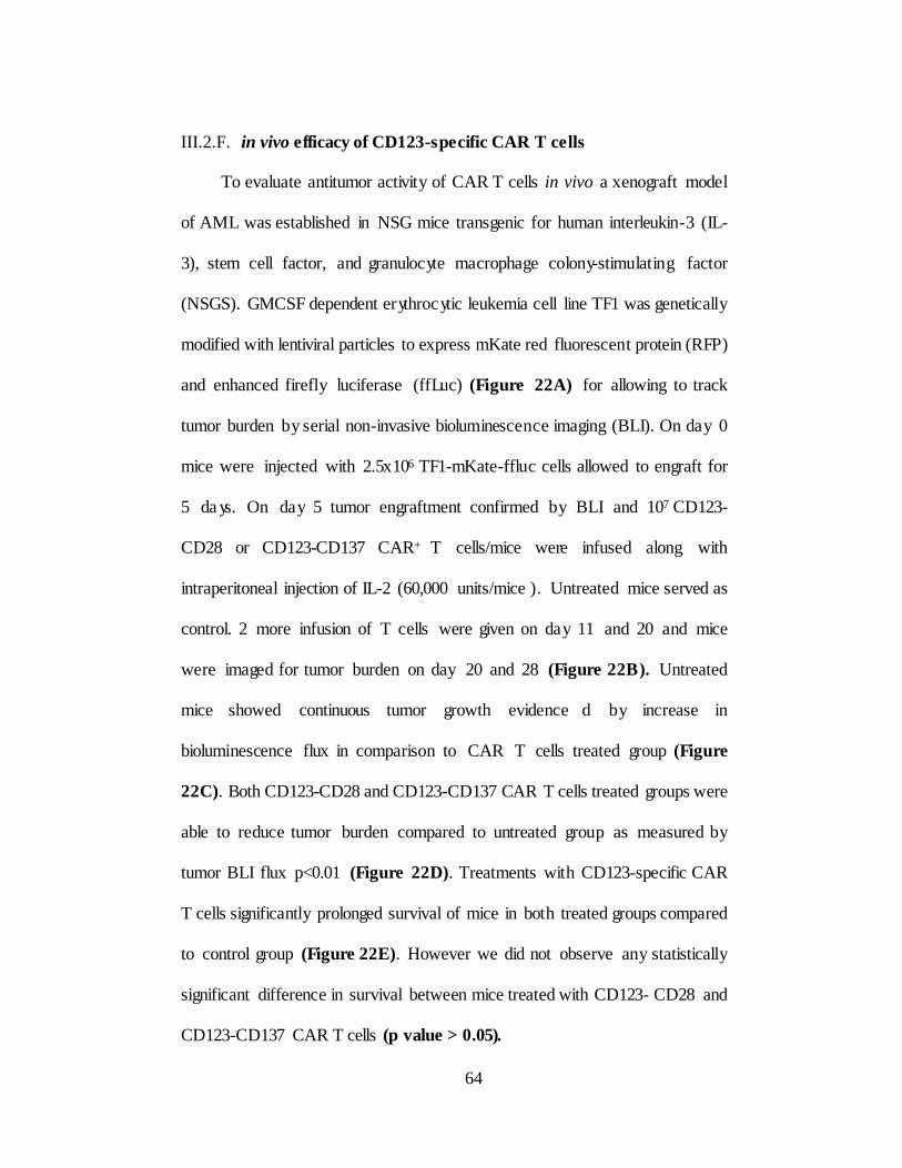
III.2.F. in vivo efficacy of CD123-specific CAR T cells
To evaluate antitumor activity of CAR T cells in vivo a xenograft model
of AML was established in NSG mice transgenic for human interleukin-3 (IL-
3), stem cell factor, and granulocyte macrophage colony-stimulating factor
(NSGS). GMCSF dependent erythrocytic leukemia cell line TF1 was genetically
modified with lentiviral particles to express mKate red fluorescent protein (RFP)
and enhanced firefly luciferase (ffLuc) (Figure 22A) for allowing to track
tumor burden by serial non-invasive bioluminescence imaging (BLI). On day 0
mice were injected with 2.5x10⁶ TF1-mKate-ffluc cells allowed to engraft for
5 da ys. On day 5 tumor engraftment confirmed by BLI and 107 CD123-
CD28 or CD123-CD137 CAR+ T cells/mice were infused along with
intraperitoneal injection of IL-2 (60,000 units/mice ). Untreated mice served as
control. 2 more infusion of T cells were given on day 11 and 20 and mice
were imaged for tumor burden on day 20 and 28 (Figure 22B). Untreated
mice showed continuous tumor growth evidence d by increase in
bioluminescence flux in comparison to CAR T cells treated group (Figure
22C). Both CD123-CD28 and CD123-CD137 CAR T cells treated groups were
able to reduce tumor burden compared to untreated group as measured by
tumor BLI flux p<0.01 (Figure 22D). Treatments with CD123-specific CAR
T cells significantly prolonged survival of mice in both treated groups compared
to control group (Figure 22E). However we did not observe any statistically
significant difference in survival between mice treated with CD123- CD28 and
CD123-CD137 CAR T cells (p value > 0.05).
64

In summary, preclinical data thus generated so far will allow us to test
CD123-specific T cells in clinical setting to treat CD123+ malignancies in
patients.
65
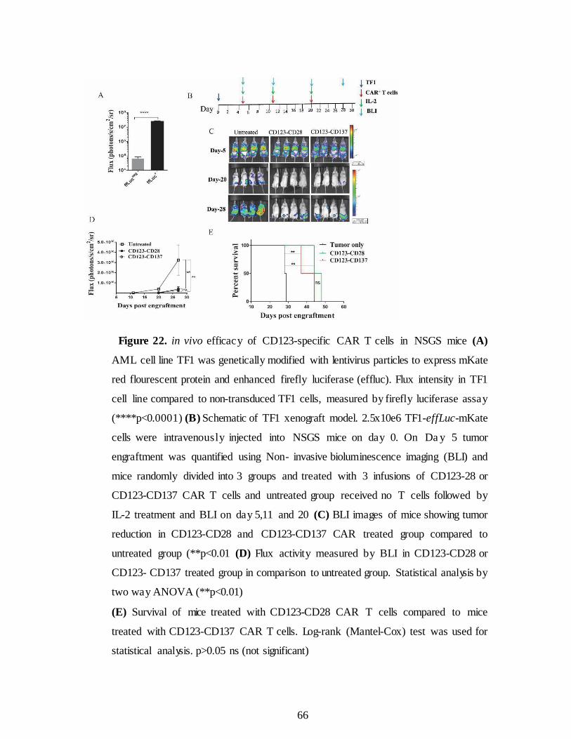
Figure 22. in vivo efficacy of CD123-specific CAR T cells in NSGS mice (A)
AML cell line TF1 was genetically modified with lentivirus particles to express mKate
red flourescent protein and enhanced firefly luciferase (effluc). Flux intensity in TF1
cell line compared to non-transduced TF1 cells, measured by firefly luciferase assay
(****p<0.0001) (B) Schematic of TF1 xenograft model. 2.5x10e6 TF1-effLuc-mKate
cells were intravenously injected into NSGS mice on day 0. On Da y 5 tumor
engraftment was quantified using Non- invasive bioluminescence imaging (BLI) and
mice randomly divided into 3 groups and treated with 3 infusions of CD123-28 or
CD123-CD137 CAR T cells and untreated group received no T cells followed by
IL-2 treatment and BLI on day 5,11 and 20 (C) BLI images of mice showing tumor
reduction in CD123-CD28 and CD123-CD137 CAR treated group compared to
untreated group (**p<0.01 (D) Flux activity measured by BLI in CD123-CD28 or
CD123- CD137 treated group in comparison to untreated group. Statistical analysis by
two way ANOVA (**p<0.01)
(E) Survival of mice treated with CD123-CD28 CAR T cells compared to mice
treated with CD123-CD137 CAR T cells. Log-rank (Mantel-Cox) test was used for
statistical analysis. p>0.05 ns (not significant)
66
III.3. Discussion
CARs that activate through chimeric CD28 or CD137 endo-domains have
anti-tumor activity and durable remissions in clinical trials with pros and cons
for each design. However the improved efficacy over the other is unknown at
present. Preclinical data that supports targeting CD123 on AML using CARs
with CD28 and CD137 co-stimulatory domains have been reported by two
groups respectively (134, 135). In this study we described the head to head
comparison of CD123-specific CARs with co-stimulator y domains CD28 or
CD137 and have been evaluated in the lines of CD19- specific CAR T cells
currently in clinical trials (NCT00968760).
We have redirected the specificity of T cells using Sleeping Beauty
system to stably express CARs and selectively propagated on Clone 1-CD123
AaPC, co-expressing CD123, co-stimulatory molecules CD86 and CD137L and
a membrane bound IL-15. Trans-presentation of mIL-15/IL-15Rα fusion
protein expressed on Clone 1-CD123 support enhanced proliferation and
survival of CAR T cells without altering T-cell activation patterns and global
T-cell receptor (TCR) repertoire (136, 137). Unlike lentiviral and retroviral
vectors SB transposition is cost effective gene transfer system requires less
production cost for manufacturing clinical grade T cells. SB system doesn’t
integrate at sites of active transcription, has been shown not to activate
oncogenes (127-129). Establishment of long term memory and survival is the
key for improving anti-tumor efficacy of CAR T cells in clinical setting.
67
Terminally differentiated effector memory (TEM) T cells lose their capacity
to expand and persist after adoptive transfer. On the contrary, less
differentiated and minimally manipulated T cells with central memory
phenotype (TCM) can further expand differentiate and self-renew with superior
clinical response. To date, adoptively transferred CAR T cells have
demonstrated minimal in vivo expansion and antitumor efficac y in clinical
trials (130-132). Though IL-2 is routinely used for T cell expansion recent
reports suggests that other common gamma chain cytokines such as IL-15 and
IL-21 suppress differentiation of naïve T cells into effector T cells (133).
SB transposition and expansion on mIL15+AaPC in presence of IL-2
and IL-21 resulted in outgrowth of T cells with less differentiated phenotype
and memory associated markers CD45RA, CD45RO, CD62L, CCR7, CD27,
CD28, with no detectable expression of exhaustion markers CD57 and PD1.
Few cells express BM homing receptor CXCR4 (Figure 17B) which helps T
cells to migrate to BM regions and clear leukemic cells. More than 95% of T
cells express CAR and expanded to clinically relevant numbers with
heterogeneous phenotype consistent with central memory T cells (TCM) and
effector memory (TEM). Redirected specificity was established based on CAR-
dependent T-cell effector function such as specific lysis of CD123+ leukemic
cell lines and primary AML samples, but not CD123neg targets. Most of the
tumor associated myeloid antigens are also expressed on normal hematopoietic
cells. Recent studies by Gill et al reported that CD123-specific CAR T cells
treated mice showed marked reduction in myelo-ablation in a preclinical
xenograft model suggests that new approaches needed to mitigate
68
off-target toxicities (134). This raises concern for targeting CD123 in
AML as it is expressed on normal hematopoietic cells. Our approach of using
chimeric CARs with the combination of changing CAR hinge to IgG4 may
enable us to generate low affinity CARs by minimal lysis of normal
hematopoietic cells by CAR 10 (Figure 11A Chapte r II). Recent data have
shown that mRNA modified T cells with transient CAR expression specific
to CD19 and CD33 resulted in target specific killing (139). Identification of
unique molecular abnormalities helps to develop targeted and personalized
treatment options for AML patients. Flow cytometry and immuno-
histochemical studies showed CD123-positive AML is most frequently
encountered within the intermediate cytogenetic risk group and is associated
with FLT3-ITD and NPM1 Mutations (140). These patients with FLT3-ITD
and NPM1 Mutations can be benefited by CAR based therapy targeting
CD123. Though CARs generated by viral vectors exhibit significant anti- tumor
efficac y and in vivo persistence sometimes resulted in on-target and off-target
cytotoxicities. Introduction of suicide genes such as iCapase 9 may mitigate the
risks by conditional ablation of T cells off target toxicities evidenced by our
in vitro data that addition of CID rapidly eliminated T cells in 24 hours at 1µM
concentration. In summary, our data also shows that CARs activated through
CD28 or CD137 showed similar efficacy in vitro and in vivo, and that inclusion
of an iCasp9 domain in frame with a Furin/F2A domain does not impair
CAR function and generates an effective suicide switch in CAR+ T cells.
69
CHAPTER IV
Targeting leuke mic stem cells by CD123-specific CAR T cells
while sparing normal hematopoiesis
IV.1. Introduction
Leukemic stem cells (LSCs) are a rare population of cells resistant to conventional
treatments and responsible for relapse and therapy failure. LSCs are pre-leukemic clonal
population of HSCs arise by genetic and molecular alterations. This is evidenced by
common features that LSCs share with HSCs including self- renewal, engraftment
potential and are enriched in Lineageneg (Linneg) fraction of blood cells with surface
phenotype of CD34+CD38ne g. LSCs are capable of self- renewal and able to initiate
leukemia when transplanted in SCID mice. CD123 is overexpressed on AML blasts,
hematopoietic progenitors and LSCs compared to normal hematopoietic stem cells and
confers growth advantage in AML (147-150). Overexpression of CD123 is associated
with higher blast counts, poor prognosis and reduced survival in AML patients (147-
150). Phase-I clinical trials targeting CD123 by monoclonal antibodies had limited
efficacy pressing the need for alternative and more potent treatments. (151, 152).
However most of the tumor associated myeloid antigens that are expressed on LSCs
are also expressed on normal hematopoietic stem cells and its progenitors. Tumor
targeted immune therapies that damage normal hematopoietic stem cells are often
associated with irreversible and reversible side effects. Clinical trials targeting CD33
antigen in AML using gemtuzumab ozogamicin (GO) antibody conjugated to a
cytotoxic agent have been shown to have proplonged cytopenia though exhibited potent
anti-tumor effect (139). Recent report by Casucci et al suggests though CARs targeting
CD44v6+ AML effective in eliminating AML, but associated with reversible
monocytopenia upon contraction of
70
T cells in vivo (145). Gill et.al reported off-target cytotoxicity to myeloid progenitors
in CAR directed immunotherapy for CD123+ AML (146). Hence targeting
AML can be myelo-toxic, careful study of possible off-target cytotoxicity is an
important concern while designing CAR therapies. In the present chapter we evaluated
in vitro killing efficacy of CD123-specific chimeric CAR T cells targeting leukemic
stem cells in AML and normal hematopoietic cells.
71
IV.2. Results
IV.2.A. CD123 is frequently expressed in AML and Leukemic stem cells
In order to decide whether CD123 is a suitable target for CAR therapy
in AML we determined CD123 expression levels in 30 random primary AML
patient samples. Peripheral blood samples of 30 patients were processed for
mononuclear cells (MNCs) established protocols. Samples include treated non-
treated and relapsed patients. FAB classification is not available for some of the
patients. MNCs from each patient were stained with CD34, CD38 and CD123
antibodies. CD123 expression levels were assessed on LSC enriched fraction
(CD34+CD38neg) fraction (Figure 23A) and blasts (CD38+) population (Figure
23B). In AML patients the percentages of total CD34+ cells, CD34+ CD38+
cells, and CD34+ CD38neg CD34negCD38+ cells within the MNC fraction was
highly heterogeneous. However Consistent with previous reports CD123 is
frequently expressed on more than 95% of AML samples (Figure 24). List of
patients samples used in the study and total % of CD123 on each sample
listed in table1.
72
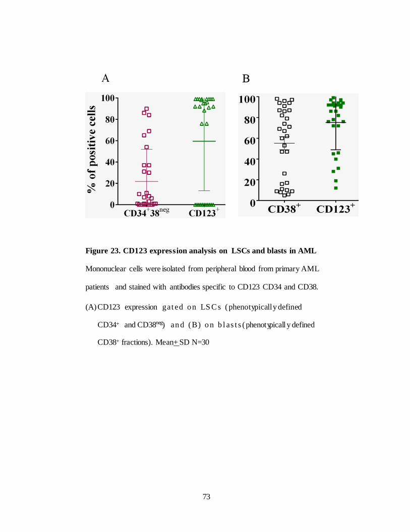
Figure 23. CD123 expression analysis on LSCs and blasts in AML
Mononuclear cells were isolated from peripheral blood from primary AML
patients and stained with antibodies specific to CD123 CD34 and CD38.
(A) CD123 expression g a t e d o n LS C s ( phenotypically defined
CD34+ and CD38neg) a n d ( B ) o n b l a s t s ( phenotypicall y defined
CD38+ fractions). Mean+ SD N=30
73
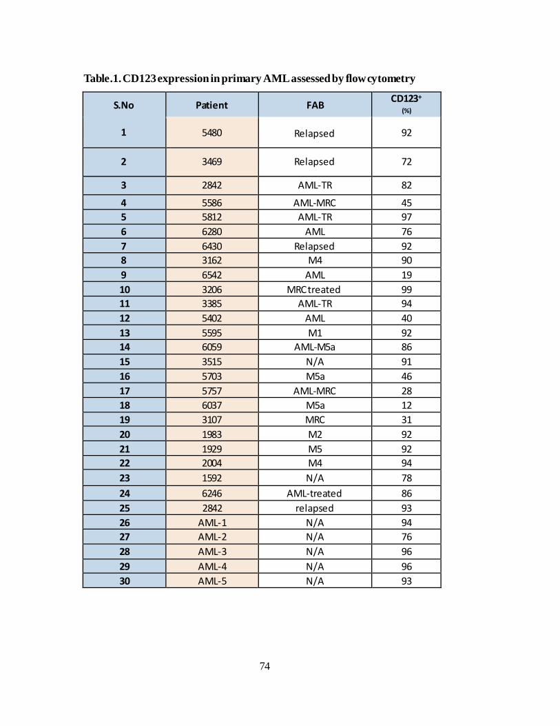
Table.1. CD123 expression in primary AML assessed by flowcytometry
S.No
1
Patient
FAB CD123+
(%)
5480
Relapsed
92
2
3469
Relapsed
72
3 2842 AML-TR 82
4 5586 AML-MRC 45
5 5812 AML-TR 97
6 6280 AML 76
7 6430 Relapsed 92
8 3162 M4 90
9 6542 AML 19
10 3206 MRC treated 99
11 3385 AML-TR 94
12 5402 AML 40
13 5595 M1 92
14 6059 AML-M5a 86
15 3515 N/A 91
16 5703 M5a 46
17 5757 AML-MRC 28
18 6037 M5a 12
19 3107 MRC 31
20 1983 M2 92
21 1929 M5 92
22 2004 M4 94
23 1592 N/A 78
24 6246 AML-treated 86
25 2842 relapsed 93
26 AML-1 N/A 94
27 AML-2 N/A 76
28 AML-3 N/A 96
29 AML-4 N/A 96
30 AML-5 N/A 93
74
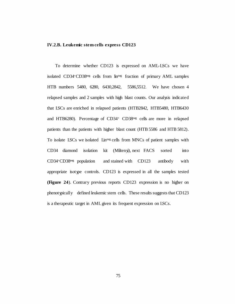
IV.2.B. Leukemic ste m cells express CD123
To determine whether CD123 is expressed on AML-LSCs we have
isolated CD34+CD38neg cells from linneg fraction of primary AML samples
HTB numbers 5480, 6280, 6430,2842, 5586,5512. We have chosen 4
relapsed samples and 2 samples with high blast counts. Our analysis indicated
that LSCs are enriched in relapsed patients (HTB2842, HTB5480, HTB6430
and HTB6280). Percentage of CD34+ CD38neg cells are more in relapsed
patients than the patients with higher blast count (HTB 5586 and HTB 5812).
To isolate LSCs we isolated Linneg cells from MNCs of patient samples with
CD34 diamond isolation kit (Miltenyi), next FACS sorted into
CD34+CD38neg population and stained with CD123 antibody with
appropriate isotype controls. CD123 is expressed in all the samples tested
(Figure 24). Contrary previous reports CD123 expression is no higher on
phenotypically defined leukemic stem cells. These results suggests that CD123
is a therapeutic target in AML given its frequent expression on LSCs.
75
Figure 24. CD123 expression on AML isolated leukemic stemcells
linneg cells from MNCs of patient samples were isolated and FACS sorted
into CD34+CD38neg population and stained with CD123 antibody with
appropriate isotype controls.
76
IV.2.C. in vitro cytotoxicity of chimeric CAR T cells against
AML-LSCs and normal hematopoietic cells
Human HSCs express lineage associated genes during their differentiation
into blood cells. However, HSCs are generally regarded as being devoid of
lineage specific markers expressed by differentiated blood cells. Studies in mice
indicate that well established myeloid lineage associated markers CD33, CD13,
CD123 are expressed on long-te rm repopulating HSCs from cord blood and
BM. This finding raises the concern that myeloid antigen targeted therapies
has the potential of killing HSCs (155). To determine whether chimeric CARs
target normal hematopoietic stem cells and progenitors we have isolated lineage
positive and negative cells from normal BM samples, Lineage+ and HSCs
(linneg CD34+CD38neg) from cord blood MNCs and co- cultured with chimeric
CAR T cells in E:T ratio 1:1 for 48 hours. in vitro toxicity by CAR T cells was
observed in lineage positive and lineage negative cells from BM (Figure 25A).
However HSCs and lineage positive cells from cord blood showed minimal lysis
by CAR T cells. (Figure 25B). Next we determined anti-tumor efficacy in
freshly isolate d phenotypically defined linneg CD34+CD38neg AML-LSCs with
similar co- culture conditions used for hematopoietic cells.
77
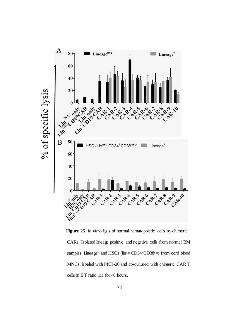
Figure 25. in vitro lysis of normal hematopoietic cells by chimeric
CARs. Isolated lineage positive and negative cells from normal BM
samples, Lineage+ and HSCs (linneg CD34+CD38neg) from cord blood
MNCs, labeled with PKH-26 and co-cultured with chimeric CAR T
cells in E:T ratio 1:1 for 48 hours.
78
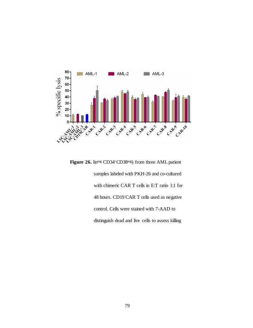
Figure 26. linneg CD34+CD38neg) from three AML patient
samples labeled with PKH-26 and co-cultured
with chimeric CAR T cells in E:T ratio 1:1 for
48 hours. CD19 CAR T cells used as negative
control. Cells were stained with 7-AAD to
distinguish dead and live cells to assess killing
79
IV.2.D. Expansion of LSC under hypoxia
Reports suggests that LSCs reside in hypoxic regions of bonemarrow
micro environment in quiescent stage and resistant to conventional treatments.
It has been demonstrated that intravenously injected AML-LSCs home to BM
engraft and subsequently reside in endosteal regions. Therefore novel
approached are needed to target LSCs in hypoxic regions of BM niche thereby
preventing relapse and therapy failure (167). LSCs are rare and few in number
in AML which limits the feasibility of cell-based assays. Current culture
conditions do not prevent LSCs and HSCs from differentiation. It has been
shown that Aryl hydrocarbon receptor (AHR) pathway is inactive in vivo and
rapidly activated in vitro in HSCs and LSCs. Stem regenin1 (SR1) is an
antagonist of the aryl hydrocarbon receptor that promotes the self- renewal of
human HSCs and LSCs in culture supplemented by cytokines and prevents their
differentiation (168,169).
To expand LSCs under hypoxic conditions we isolated linneg
CD34+CD38neg fraction from relapsed AML patients cultured at 1% oxygen
and 5% CO2. Cells were cultured in serum free stemspan II media (stem cell
technologies) in presence SR1 1µM/ml supplemented by cytokines stem cell
factor (SCF), human FLT3 ligand, interleukin-3 for 7 days. SR1 non-treated
cells used as control. All AML-LSCs treated with SR1 showed higher
percentages of CD34+CD38neg fraction with relative CD123 expression after
80
a 7-day culture period compared to SR1 non treated controls (Figure 27).
On day 7, LSCs were labeled with PKH26 and co-cultured with CD123-
specific chimeric CAR T cells in 1:1 ratio for 48 hours under hypox ic
conditions. CD19 CAR T cells used as negative control. CD123-specific
CAR T cells lysed LSCs expanded under hypoxia compared to CD19 CAR
which exhibited minimal lysis (Figure 28).
81
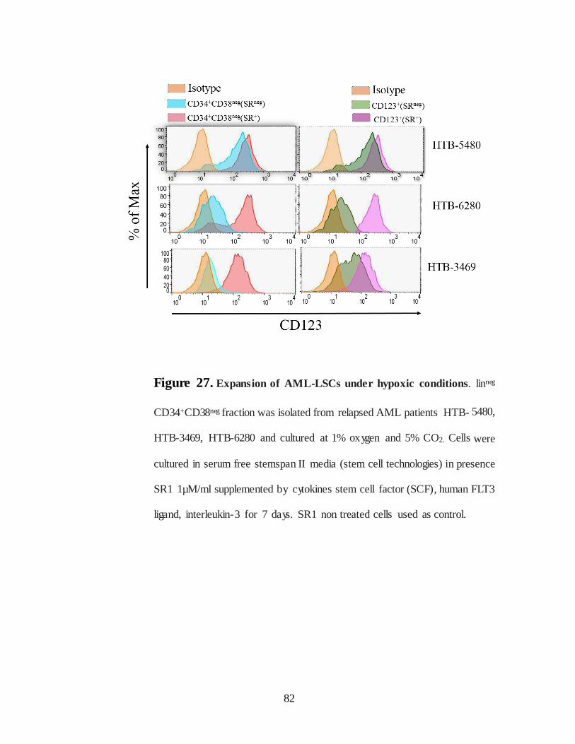
Figure 27. Expansion of AML-LSCs under hypoxic conditions. linneg
CD34+CD38neg fraction was isolated from relapsed AML patients HTB- 5480,
HTB-3469, HTB-6280 and cultured at 1% oxygen and 5% CO2. Cells were
cultured in serum free stemspan II media (stem cell technologies) in presence
SR1 1µM/ml supplemented by cytokines stem cell factor (SCF), human FLT3
ligand, interleukin-3 for 7 days. SR1 non treated cells used as control.
82
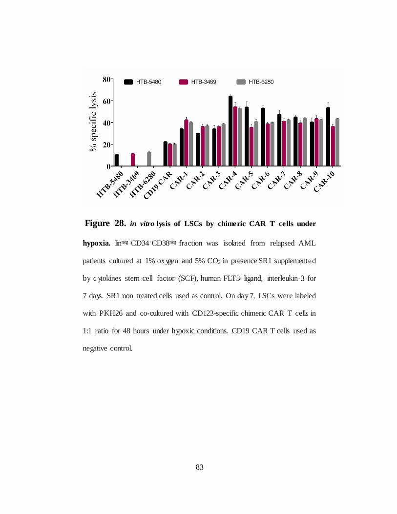
Figure 28. in vitro lysis of LSCs by chimeric CAR T cells under
hypoxia. linneg CD34+CD38neg fraction was isolated from relapsed AML
patients cultured at 1% oxygen and 5% CO2 in presence SR1 supplemented
by c ytokines stem cell factor (SCF), human FLT3 ligand, interleukin-3 for
7 days. SR1 non treated cells used as control. On day 7, LSCs were labeled
with PKH26 and co-cultured with CD123-specific chimeric CAR T cells in
1:1 ratio for 48 hours under hypoxic conditions. CD19 CAR T cells used as
negative control.
83
IV.3. Discussion
LSCs has many features in common with HSCs including self- renewal and
engraftment potential and are enriched in linneg fraction of blood cells. Initial studies
suggests that LSC activity was restricted to LinnegCD34+CD38ne g fraction in
NOD/SCID mice models (171). Later studies using refined mice models have
demonstrated LSC activity is confined to more than one compartment and even more
mature linnegCD34+CD38+ progenitor population of some of AML patients able to
initiate leukemia (172). LSCs were well documented in AML though they are reported
in other leuekemic groups. Studies in ALL suggests that, greater degree of plasticity
is observed in LSC compartment with phenotype CD34ne gCD19+ and able to
regenerate CD34+ progeny within transplanted leukemia. However the frequency of
LSC is more in linneg CD34+CD38neg fraction than linneg CD34+CD38+ progenitors
(173). More recent data indicate that LSCs has phenotypically distinct subpopulations
and may vary from patient to patient (190, 191).
Ideally, therapeutic targets that specifically expressed on LSC but not on
HSC need to be identified to spare HSCs from being targeted and to protect normal
hematopoiesis in the patient. Therefore functionally defining LSCs and HSCs is crucial
in identifying therapeutic targets that are specific to LSC (177-180). A detailed LSC
phenotype have been described as LinnegCD34+CD38+CD90ne gCD96+CD45RAneg
which distinguish them from normal myeloid stem cells and progenitors (174, 175).
LSC have been shown to overexpress other myeloid markers such as CD13, CD33
and CD123 which are weakly expressed on HSCs
84
(181). Recently several other markers have been shown to be expressed exclusively
on LSC than normal HSC which include CLL1, CD32, CD45RA, TIM3, CD47 and
CD25 (182-186). HSCs are generally regarded as being devoid of expressing markers
specific to blood cell lineages. However recent studies suggests that HSCs
promiscuously express lineage-specific genes prior to commitment including myeloid
markers (171-175). This aspect has significant clinical importance since number of
therapies targeting myeloid markers aim to selectively kill leukemic blasts while sparing
normal HSCs. HSCs will be targeted along with leukemic cells if they also express
myeloid markers along with leukemic blasts (177-181). For example, clinical trials of
AML therapy targeting CD33 by gemtuzumab ozogamicin (GO) antibody conjugated
to a cytotoxic agent have been shown to have prolonged cytopenia despite successful
tumor clearance (177,182,183). This may be due to targeted killing of HSCs by GO
and many patients have relapses since LSCs are resistant to the toxins (184). CD123
expression was assessed on HSCs and progenitors in 3 normal bone marrow samples
as described (185, 186).
Our in vitro data indicate that CAR T cells showed toxicity to lineage positive
and negative cells from however the HSCs from cord blood are minimally lysed.
CAR T cells able to lyse LSCs which are phenotypically similar to HSCs shows the
potential CAR therapy to eradicate leukemic stem cells which are responsible for
relapse and therapy failure. Previous trials targeting CD123 with mAbs cytokine
modalities did not report BM suppression. However pre-clinical studies targeting
CD123 by CAR T cells showed myeloablation in mice. This may be the result of
ability of CAR T cells to eliminate CD123dim population compared
85
to antibody therapies. These observations prompt us to develop careful risk free
therapy for AML and using low affinity CARs may rescue hematopoiesis while
successfully eliminating AML tumors. These in vitro data suggests that CAR
therapy can be detrimental to normal hematopoiesis and CD123-specific CAR
T cells need to be employed with rescue strategies such as myeloablation as
conditioning regimen for HSC transplantation.
86
CHAPTER-V
General Discussion and Future Directions
The main aim of this dissertation is to develop novel adaptive
immunotherapies for the treatment of AML and B-ALL patients by
redirecting specificity of T cells by genetic modification through chimeric
antigen receptors. This was tested in three different specific aims. In first
specific aim we have shown that CARs (chimeric CARs) can be generated by
mix and matching VH and VL chains from different mAbs, which is a novel
finding because traditional CARs derive their scFv from a single mAb. Chimeric
CARs successfully expanded on AaPC, Clone 1-CD123 to large numbers
needed for the clinic and stably co-expressed CAR and suicide gene iCaspase 9.
Chimeric CARs executed effector functions, antigen-specific killing and
IFN-γ production. Second specific aim enabled us head to head comparison of
CD123- specific CARs with co-stimulatory domains CD28 and CD137. It was
reported that CD19-specific CARs containing CD137 endo domain mediated
enhanced survival of T cells and increased anti-leukemic activity in vivo
compared to CARs with CD28 in clearing B-ALL tumors (41). Contrary to this
in our head-to-head comparison of CD123-specific CARs with co-stimulatory
domains CD28 and CD137, we observed similar rates of target lysis with both
constructs in vitro, though there was a trend
87
towards better survival with the CD28-containing construct in our in vivo
AML model. However it is difficult to compare CARs across research groups
since each group has different protocols, CARs vary in their design, expression
on the T cells, culture conditions for propagating T cells, antigen density on
tumor, affinity of scFv, CD4:CD8 ratio in T cell cultures, cytokine support for
the infused T cells, lympho depleting strategy, disease targeted, and timing of
CAR T-cell infusion with regard to standard therapy such as bone marrow
transplantation (162). These data supports the translation of preclinical methods
into clinical trials for AML by targeting CD123. In the third specific aim we
showed that in vitro targeting of LSCs, and normal hematopoietic cells by CAR
T cells. Since CD123 is expressed on normal hematopoietic cells, careful
planning is needed in clinical studies t o prevent off target toxicities. The hinge
is the least commented aspect of CARs though they make important
contributions to the interaction of CAR with its cognate antigen, formation of
immunological synapse and necessary interaction of CAR with other proteins for
activation signal (162). Preclinical data suggests that the spacial location of
epitope length and binding, flexibility and origin of the hinge domain are
important variables in the design of CARs and has bigger impact on CAR
activity than variation affinity to scFv (163-166). The affinity of the scFv
for antigen also affects the density of antigen required for efficient killing (87).
Though the effect of antigen
88
density for CAR therapy is not yet well-defined, it appears that CAR T cells
preferably target tumors with high antigen density, while cells with lower
density are more resistant to CAR T cells (97,98). Importantly our studies
CAR-10 (Figure 6A) with IgG4 hinge showed minimal lysis in normal BM
cells compared to CARs 6 with CD8α hinge with same scFv (Figure 11A). This
finding has to be validated further in chimeric CARs other than CAR-10 in vitro
and in vivo to generate low affinity CARs with various combination of hinge and
chimeric scFvs to rescue normal hematopoiesis. Thus by choosing different
source of VH and VL chains and perhaps different hinge regions, we may be
able to tune the activation threshold for CAR T cells further, especially if a
wider range of antibody affinities is used than was chosen for these studies.
89
Future Directions
Though the CARs typically identified by their endo-domains, the other components
of CAR also has crucial role in their function and clinical efficacy including hinge
portion. Constant region of IgG4 and CD8α are frequently used hinge regions,
however Fc region have been reported to engage Fc receptors and activate innate
immune cells (137). To avoid off target activation of CARs and unwanted immune
responses we have generated CD123 specific chimeric CARs by introducing L235E
and N297Q mutations in the CH2 region of IgG4-Fc spacer or replacing IgG4-Fc
hinge with CD8α. In our future studies these CARs will be evaluated in vivo for
enhanced anti-tumor activity and persistence. Additional modifications to the ex vivo
culture by reducing the number of stimulations (addition of AaPC) of γ- irradiated
AaPC further improved persistence and anti-tumor effect in preclinical studies
targeting CD19 (data not shown). Furthermore, clinical data involving CAR T cells
have highlighted that the persistence of genetically modified T cells can be
compromised in human applications due to recognition of the recipient’s immune system
to mouse elements of the scFv used to derive specificit through the CAR architecture
which can be resolved by using humanized scFvs. CARs generated by lentiviral or
retroviral vectors exhibit significant anti-tumor efficacy and in vivo persistence but
sometimes results in on-target and off- target cyto-toxicities. Unlike lentiviral and
retroviral vectors SB transposition is cost effective gene transfer system requires less
production cost for manufacturing clinical grade T cells. SB transfected genes doesn’t
integrate at sites of active transcription, has been shown not to activate oncogenes
90
(144-149). The introduction of suicide genes such as iCapase9 may mitigate the risks
by conditional ablation of T cells. However the efficacy of these strategies are limited
by incomplete elimination of transferred T cells (138). In summary our data supports
CARs can be derived from two or three mAbs specific to an antigen by combining
VH and VL chains. This approach may allow us to derive affinity tuned CARs to
target tumors with differential antigen density sparing normal cells expressing
antigen at low levels. Recent studies demonstrated enhanced persistence for CARs
with CD137 compared to those with CD28 endodomain, however our data shows that
CARs activated through CD28 or CD137 showed similar efficacy in vitro and in
vivo.
91
CHAPTER-VI
Materials and Methods
Primary samples and animal use
All patient samples used for this study were obtained after written informed
consent was granted in accordance with protocols established and approve d
by the MD Anderson Cancer Center (MDACC) and Internal Review Board
(IRB). The identities of all samples were kept private. Animals were handle d
in accordance with the strict guidelines established by the MDACC Institutional
Animal Care and Use Committee (IACUC).
Generation of CD123 specific CARs with scFvs derived from two
monoclonal antibodies
To generate CARs specific for CD123, we generated scFv by assembling VH
and VL chains from four monoclonal antibodies 26292, 32701, 32703 and
32716 specific to CD123 (96) and then fused in frame to the human CD8
spacer and transmembrane domain, then the CD3ζ and CD28 endodomains.
Of the 12 possible scFvs that could have been made, we chose five at random
for further testing. (Figure 6A). These five mix-and-match scFvs were
spliced into the existing anti-CD19 CAR construct above to generate CARs
5-9 (Figure 6A). CAR 10 has the same scFv as CAR 6, but uses the IgG4
spacer and CD28 TM. CARs 1-4 has scFvs derived from
92
VH and VL single MAb. CAR constructs were custom synthesized and cloned
into SB system constructs, as described previously for CD19 CARs (46).
Construction of iCaspase 9 co-expressing CARs in SB transposons
For experiments testing the relative contributions of CD28 vs. CD137
signaling as the costimulation signal for CAR T cells, we chose the CAR10
scFv described above (Figure 6A), since we have previously engineered CAR
constructs using these costimulatory domains fused to the IgG4
transmembrane domain. On the 5’ side of the resulting CAR sequence, there
is an in-frame inducible caspase 9 sequence (iCasp9), followed by a Furin
element and F2A peptide sequence, which together make an auto-cleavage
site within the protein, resulting in two mature proteins from the single
polypeptide sequence. The iCasp9 element creates a chemically inducible
suicide switch in CAR+ T cells. The CAR constructs were custom
synthesized and codon optimized by Geneart, (Invitrogen, Grand Island, NY)
and cloned into SB vectors (Figure14). The sequence for both plasmids was
verified by Sanger sequencing (DNA Sequencing Core, MD Anderson cancer
center (MDACC).
93
Primary cells and cell lines.
The TF1 cell line was obtained from European collection cell cultures
(ECACC). Molm13, MV411 and OCI-AML3 were kind gifts from Dr. Dean
A. Lee (MDACC). EL4 cells were obtained from ATCC. RCH-ACV and
Kasumi-2 were kind gifts from Jeffrey Tyner (Oregon Health Sciences
University). OCI-Ly19 was a kind gift from Dr. Richard Eric Davis (MD
ACC). K562-derived aAPC were obtained from Dr. Carl H. June (University
of Pennsylvania) and further modified with mIL15 and TAAs ROR1 and
CD123 (see below). The Nalm-6 cell line was obtained from Deutsche
Sammlung von Mikroorganismen und Zellkulturen (DSMZ). PBMC for T
cell transfections were obtained from healthy donors after informed consent
and isolated by density gradient centrifugation using Ficoll-PaqueTM PLUS
(GE Healthcare). All cell lines were maintained in complete RPMI media,
10% FBS and 1X Glutamax-100. STR DNA Fingerprinting was done to
confirm the identity of all cell lines at MD Anderson’s Cancer Center Support
Grant (CCSG) supported facility “Characterized Cell Line Core.”
94
Generation of CD123+ Clone1-APC and EL4
To generate activating and propagating cells (AaPC) to support expansion of
CD123-specific CAR T cells, we modified K562-based AaPC originally
obtained from Carl June (Clone 9) which express CD19, CD64, CD86, and
CD137L, to express an IL15/IL15Rα fusion protein ROR1 and CD123 using
the Sleeping Beauty (SB) gene transfer according to our published methods
(46). This new AaPC line we termed Clone1-CD123. The same process was
used to create EL4 cells expressing CD123. CD123+ cells were selected by
hygromycin selection.
Electroporation and propagation of CAR T cells.
On day 0, 20 million PBMC were resuspended in 100 µL of Amaxa human T
cell Nucleofector solution (Cat. no. VPA-1002; Lonza, Basel, Switzerla nd)
mixed with 15 µg of transposon and 5 µg of transposase (pKan-CMV-SB11)
and electroporated using Program U-14. The following day (da y1) cells were
counted, surface stained for CAR expression by Fc antibody and stimulated
with γ-irradiated (100 Gy) Clone 1-CD123 at 1:2 ratio of T cells to AaPCs.
Cultures were supplemented with recombinant human IL-2 50 units/ml and 30
ng/ml of recombinant human IL-21 (Pepro Tech). AaPCs added ever y 7 days
and IL-2, IL-21 added monday-wednesday and friday schedule beginning of
day1 of each 7 day T cell expansion cycle. T cell cultures were phenotyped
every week to monitor CAR expression and outgrowth of NK cells
(CD3negCD56+ population) usually occurred between
95
10 to 14 days after electroporation. If the percentage of NK cells exceeded
approximately 10% total population, depletion of NK cells was carried out with
CD56 beads (cat.no.130-050-401, Miltenyi Biotech) according to
manufacture instructions. As a control, 5x10⁶ PBMC were mock transfected
wit h nucleofector solution without CAR plasmid and were co-cultured
on γ-irradiated (100 Gy) anti-CD3 (OKT3) loaded K562-aAPC clone #1 at a
ratio of 1:1 in a 7-day stimulation cycle along with similar concentrations
of IL-2 and IL-21 as CAR T cells.
Real time PCR to determine integrated CAR copy number
The number of integrated copies of CD123-specific CAR transgene was
determined by isolating genomic DNA using AllPrep DNA/RNA Mini Kit,
(Qiagen cat # 80204) as described in (25 ). Briefly about 50-100 ng of DNA
amplified using Steponeplus Real-time PCR system (Applied Biosystems),
forward primer (5’-CAGCGACGGCAGCTTCTT-3’), reverse primer (5’-
TGCATCACGGAGCTAAA-3’) and carboxyfluorescein (FAM)-labeled
probe and ( 5’- AGAGCCGGTGGCAGG-3’). These primers hybridize to
the CAR in IgG4 and CD28 transmembrane region. Genomic DNA from a
genetically modified Jurkat T-cell (clone #12) containing 1 copy of CAR per
cell from CoOpCD19RCD28/pSBSO DNA plasmid was used as positive
control. No DNA (CARneg) T cells were used as negative control. Results
were analyzed using GraphPad Prism software.
96
Immunophenotype of CAR+ T cells
T cells were immunophenotyped using appropriate antibodies and isotype
controls. Cells were stained for 30 minutes at 40C followed by 2 washes with
FACS buffer (PBS, 2% FBS, 0.1% sodium azide). For intracellular staining
cells were fixed and permeabilized for 20 minutes at 4oC with BD
Cytofix/Cytoperm (BD Biosciences, San Diego, CA) followed by staining with
appropriate antibodies. All samples were acquired on FACS Calibur (BD
Bioscience) and analyzed with FlowJo software (version 7.6.3).
Multiplex Ge ne Expression Analysis of CAR T cells
On day 35 of co-culture of CAR T cells on AaPC about 105 were lysed in 17
μl of RLT buffer (Qiagen) and frozen at -800C. Cell lysates were thawed and
analyzed immediately using nCounter analysis System (NanoString
Technologies, Seattle, WA) with “lymphoc yte codeset array” as described (26).
Data was normalized to spike positive control RNA and housekeeping genes
(ACTB, G6PD, OAZ1, POLR1B, POLR2A, RPL27, Rps13, and TBP)
Where 2 normalization factors were calculated and applied to the raw counts.
Each normalization factor was calculated from the average of sum of all
samples divided by the sum of counts for an individual sample. Total counts
for LCA genes described in CD123-specific CAR+ T cells were directly
reported as normalized mRNA counts. Limit-of-detection (LOD) was calculated
from the negative control counts and reported as the mean plus
97
two times the standard deviation (mean+2xSD) and shown as dashed lines in
graphs of mRNA data.
iCaspase 9 functional assay.
CAR+ T cells with and without icaspase 9 were seeded in 24 well plate @ 106
cells/well. 1µM of chemical inducer of dimerization (CID) (AP20187; Clontech)
was added, cells were harvested after 24 hours and surface stained with CD3,
FC followed by annexin-V and 7-amino-actinomycin D (7-AAD) for 15
minutes according to the manufacturer's instructions (BD Pharmingen). Within
1 hour after staining, cells were analyzed by flow cytometry using BD FACS
caliber
Chromium release assay.
The cytolytic efficac y of CAR+ T cells with target cell lines was evaluated by
4-hour chromium release assay as described in (20). Briefly 5x10³ 51Cr-
labeled target cells were incubated with CD123 specific CAR+ T cells in
complete medium or 0.1% Triton X-100 (company) to determine spontaneous
and maximum 51Cr release, in a V-bottomed 96-well plate. The mean
percentage of specific cytolysis of triplicate wells was calculated from the
release of 51Cr using a Top Count NXT (Perkin-Elmer Life and Analytic al
Sciences, Inc.) as 100 × (experimental release - spontaneous release)/(maximal
release - spontaneous release). Data was reported as mean ± SD
98
Flow cytometric killing assay
For T cell killing assays, target cells were labeled with PKH-26 (Sigma,
cat.no PKH26PCL) according to manufacture instructions and co-cultured with
CAR+T cells at E:T ratio of 1:1 for 3 days without exogenous cytokines. 7-
AAD was added prior to flow cytometric analysis to exclude dead cells, viable
cells phenotyped by CD3 and PKH-26.
Cytokine production by CAR+ T cells.
Effector cells were incubated with target cells at T cell to target ratio of 1:1
for 24 hours. C ytokine production from CAR+ T cells in response to antige n
was determined using LEGENDplex™ multi analyte flow assay kit (Biolegend,
cat.no 790004) according to manufacture instructions.
Mice studies
The in vivo antitumor efficacy of CAR T cells in TF1 cells was assessed in
NOD/SCID/IL- -/- (NSG) mice transgenic for human interleukin-3 (IL-3),
stem cell factor (SCF), and granulocyte macrophage colony-stimulating
factor (GM-CSF) obtained from Jackson Laboratories. For bioluminescent
xenograft models, the TF1 cell line was genetically modified to express
enhanced firefly luciferase (effLuc) (Figure S7) by transduction with a
pLVU3G effLuc-T2A-mKateS158A lentivirus construct and sorting cells for
uniform mKate expression. On day 0, 12 mice were injected intravenously
(i.v) with 2.5 million TF1-effLuc cells, then divided into three groups of 4
mice each. On day 5, mice were injected with 107 cells CD123-
99
CD28 (group 2), or CD123-41BB CAR T cells (group 3) per mouse, or were
given no cells (group 1). Tumor engraftment was confirmed by
bioluminescent imaging (BLI) before T cell infusion. Additional T cell
infusions were administered on days 11 and 20, and the tumor burden was
assessed serially by BLI. To test in vivo efficacy of CAR T cells in B-ALL
model pre B-ALL cell line RCH-ACV was modified with enhanced firefly
luciferase same way as TF1 and infused into NSG mice on day 0 and CAR T
cells were infused on day 1, 7, 14 and 21 followed by BLI and IL-2 (60,000
units/mice) infusion. The experiments were performed twice; one
representative experiment is shown.
Isolation of Lineage+ and Lineageneg cells from BM cells
To determine CD123 expression on normal BM cells we have isolate d Lineage
positive cells using biotin conjugated lineage antibody cocktail followed by
positive selection with anti-biotin microbeads using LD Column unlabeled
fraction collected lineage negative and labeled fraction lineage positive.
Ethics statement
All patient samples used for this study were obtained after written informed
consent was obtained in accordance with protocols established and approved
by the MD Anderson Cancer Center (MD Anderson) and Internal Review
Board (IRB). The all samples were de-identified. Animals were handled in
accordance with the strict guidelines established by the MD Anderson
Institutional Animal Care and Use Committee (IACUC).
100
References
1. http://www.cancer.gov/publications/patient-education/wyntk-non-hodgkin-
lymphoma. U.S. Department of Health And Human Services National Institutes of
Health
2. http://www.lls.org/disease-information/myeloma. Leukemia and Lymphoma Society
3. http://www.cancer.gov/publications/patient-education/leukemia.pdf
What You Need to Know About leukemia. U.S. Department of Health and Human
Services National Institutes of Health
4. Porter, D. L., Levine, B. L., Kalos, M., Bagg, A. & June, C. H. Chimeric antigen
receptor-modified T cells in chronic lymphoid leukemia. N. Engl. J. Med. 365,
725–733 (2011).
5. Kalos, M. et al. T cells with chimeric antigen receptors have potent antitumor
effects and can established memory in patients with advanced leukemia. Sci.
Transl. Med. 3, 95ra73 (2011).
6. Kochenderfer, J. N. et al. Chemotherapy-refractory diffuse large B-cell lymphoma
and indolent B-cell malignancies can be effectively treated with autologous T
cells expressing an anti-CD19 chimeric antigen receptor. J. Clin. Oncol. 33, 540–
549(2015).
7. Maude, S. L. et al. Chimeric antigen receptor T cells for sustained remissions in
leukemia. N. Engl. J. Med. 371, 1507–1517 (2014)
8. Davila, M. L. et al. Efficacy and toxicity management of 19-28z CAR T cell
Therapy in B cell acute lymphoblastic leukemia. Sci. Transl. Med. 6, 224ra25
(2014).
101
9. Hjalgrim, L. L., Rostgaard, K., Schmiegelow, K., So derha ll, S., Kolmannskog,
S., et al. Age-and sex-specific incidence of childhoodleukemia by
immuneimmunophenotype in the Nordic countries. J Natl CancerInst 2003. 95:
1539–1544.
10. Pui, C. H., Relling, M. V. and Downing, J. R., Mechanisms of Disease:
acute lymphoblastic leukemia. N Engl J Med 2004. 350: 1535–1548.
11. Campana D. Role of minimal residual disease monitoring in adult and pediatric
Acute lymphoblastic leukemia. Hematol Oncol Clin North Am. Oct 2009;23
(5):1083-98,
12. Hunger SP, Lu X, Devidas M, Camitta BM, Gaynon PS, Winick NJ, Reaman
GH,Carroll WL. Improved survival for children and adolescents with acute
lympholymphoblastic leukemia between 1990 and 2005: a report from the
children's oncology group.
13. MacMillan ML, Davies SM, Nelson GO, Chitphakdithai P, Confer DL, King RJ
et al.Twenty years of unrelated donor bone marrow transplantation for pediatric
acut leukemia facilitated by the National Marrow Donor Program. Biol Blood
Marrow Transplant 2008; 14(9 Suppl): 16–22.
14. Hahn T, McCarthy PL Jr, Hassebroek A, Bredeson C, Gajewski JL, Hale GA et al.
Significant improvement in survival after allogeneic hematopoietic cell trans-
plantation during a period of significantly increased use, older recipient age, anduse
of unrelated donors. J Clin Oncol 2013; 31: 2437–2449.
102
15. Leung W, Campana D, Yang J, Pei D, Coustan-Smith E, Gan K et al. High success
rate of hematopoietic cell transplantation regardless of donor source in children
with very high-risk leukemia. Blood 2011; 118: 223–230.
16. Publisher MA, Bader P,Klingebiel T, Cooper LJ. Allogeneic transplantation for
Pediatric acute lymphoblastic leukemia: the emerging role of
peritransplantation minimal residual disease/chimerism monitoring and
novel chemotherapeutic,molecular, and immune approaches aimed at
preventing relapse. Biol Blood Marrow Transplant 2008; 15(1 Suppl): 62–
71.
17. National Cancer Institute. SEER Stat Fact Sheets: Acute Myeloid Leukemia,
1975–2009. Available at http://seer.cancer.gov/statfacts/html/amyl.html#
incidence-mortality. Accessed Apr 16, 2013.
18. Dohner H, Estey EH, Amadori S, et al. Diagnosis and management of acute
myeloid leukemia in adults: recommendations from an international expert
panel, on behalf of the European LeukemiaNet. Blood 2010; 115:453-74.
19. Fuad El Rassi and Martha Arellano. Update on Optimal Management of Acute
Myeloid Leukemia. Clinical Medicine Insights: Oncology 2013:7 181–197
20.Norimitsu Kadowaki and Toshio Kitawaki. Recent Advance in Antigen-Specific
Immunotherapy for Acute Myeloid Leukemia. Clinical and Developmenta l
Immunology Volume 2011, Article ID 104926, 7 pages
21.Francesco Buccisano, Luca Maurillo, Maria Ilaria Del Principe, Giovanni Del
Poeta, Giuseppe Sconocchia, Francesco Lo-Coco, William Arcese, Sergio
Amadori, and Adriano Venditti Prognostic and therapeutic implications of
minimal residual disease detection in acute myeloid leukemia. Blood, 2012
volume (112), Number (2) P.332-341
103
22. Joseph G. Jurcic. Immunotherapy for Acute Myeloid Leukemia. Current Oncology
Reports 2005, 7:339–346
23. Frankel AE, Konopleva M, Hogge D, Rizzieri D, Brooks C, Cirrito T, et al:
Activity and tolerability of SL-401, a targeted therapy directed to the interleukin- 3
receptor on cancer stem cells and tumor bulk, as a single agent in patients with
advanced hematologic malignancies. J Clin Oncol 2013, 31(suppl): abstract nr 7029.
24. Roberts AW, He S, Ritchie D, Hertzberg MS, Kerridge I, et al: A phase I study
of anti-CD123 monoclonal antibody (CD123) CSL360 targeting leukemia stem
cells (LSC) in AML. J Clin Oncol 2010, 28: abstract nre13012.
25. Gideon Gross, Tova Waks, and Zelig Eshhar. Expression of immunoglobulin-T-
cell receptor chimeric molecules as functional receptors with antibody-t ype
specificity. Proc. Natl. Acad. Sci. USA Vol. 86, pp. 10024-10028, December 1989
26. Michel Sadelain, Renier Brentjens, and Isabelle Rivière. The Basic Principles of
ChimericAntigen Receptor Design.Cancer discovery 2013. P.388-398
27. Zhao, Y., Q. J. Wang, S. Yang, J. N. Kochenderfer, Z. Zheng, X. Zhong, M.
Sadelain, Z. Eshhar, S. A. Rosenberg, and R. A. Morgan. 2009. A herceptin-
Based chimeric antigen receptor with modified signaling domains leads to enhanced
survival of transduced T lymphoc ytes and antitumor activity. Journal of Immunology
183:5563-5574.
104
28. Wang, J., M. Jensen, Y. Lin, X. Sui, E. Chen, C. G. Lindgren, B. Till,
A.Raubitschek, S. J. Forman, X. Qian, S. James, P. Greenberg, S. Riddell, and
O.W. press. 2007. Optimizing adoptive polyclonal T cell immunotherapy of
lymphomas, using a chimeric T cell receptor possessing CD28 and CD137
costimulatorydomains.Human GeneTherapy18:712-725.
29. Carpenito, C., M. C. Milone, R. Hassan, J. C. Simonet, M. Lakhal, M.
M.Suhoski, A. Varela-Rohena, K. M. Haines, D. F. Heitjan, S. M. Albelda, R.
G. Carroll, J. L. Riley, I. Pastan, and C. H. June. 2009. Control of large, established
tumor xenografts with genetically retargeted human T cells containing CD28
and CD137 domains. Proceedings of the National Academy of Sciences of the
United States of America 106:3360-3365.
30. Dustin ML, Depoil D. New insights into the T cell synapse from
single molecule techniques. Nat Rev Immunol 2011; 11: 672–84.
31. Bridgeman JS, Hawkins RE, Hombach AA, Abken H, Gilham DE .
Building better chimeric antigen receptors for adoptive T cell therapy
Curr Gene Ther 2010; 10: 77 – 90
32. Stone JD, Chervin AS , Kranz DM . T-cell receptor binding affinities and kinetics:
impact on T-cell activity and specificity. Immunology 2009;126 :165-76
33. Edwards LJ, Evavold BD . T cell recognition of weak ligands: roles of signaling,
receptor number, and affi nity. Immunol Res 2011; 50:39 – 48.
34. Li YS1, Wasserman R, Hayakawa K, Hardy RR. Identification of the earliest B
lineage stage in mouse bone marrow. Immunity. 1996 Dec;5 (6):527-35.
105
35. Li YS1, Ha yakawa K, Hardy RR. The regulated expression of B lineage associated
genes during B cell differentiation in bone marrow and fetal liver. J Exp Med.
1993 Sep 1;178 (3):951-60
36. www.clinical trials.gov
37. Grupp, S. A., M. Kalos, D. Barrett, R. Aplenc, D. L. Porter, S. R. Rheingold,
D.T. Teachey, A. Chew, B. Hauck, J. F. Wright, M. C. Milone, B.
L. Levine, and C. H. June. 2013. Chimeric antigen receptor- modified T cells for
acute lymphoid leukemia. The New England Journal of Medicine 368:1509- 1518.
38. Kochenderfer, J. N., M. E. Dudley, S. A. Feldman, W. H. Wilson, D. E. Spaner,
Maric, M. Stetler-Stevenson, G. Q. Phan, M. S. Hughes, R. M. Sherry, J. C.
Yang, U. S. Kammula, L. Devillier, R. Carpenter, D. A. Nathan, R. A. Morgan
C.Laurencot, and S. A. Rosenberg. 2012. B-cell depletion and remissions of
malignancy along with cytokine-associated toxicity in a clinical trial of anti-
CD19 chimeric-antigen-receptor-transduced T cells. Blood 119:2709-2720.
39. Brentjens, R. J., I. Riviere, J. H. Park, M. L. Davila, X. Wang, J. Stefanski, C.
Taylor, R. Yeh, S. Bartido, O. Borquez-Ojeda, M. Olszewska, Y. Bernal,
H.Pegram, M. Przybylowski, D. Hollyman, Y. Usachenko, D. Pirraglia, J.
Hosey,E. Santos, E. Halton, P. Maslak, D. Scheinberg, J. Jurcic, M. Heaney,
G.Heller,M. Frattini, and M. Sadelain. 2011. Safety and persistence of
adoptively transferred autologous CD19-targeted T cells in patients with relapsed
or chemotherapy refractory B-cell leukemias. Blood 118:4817-4
106
40. https://www.novartis.com/news/media-releases/novartis-personalized-cell-therap y-
ctl019-receives-fda-breakthrough-therap y
41. Michael C. Milone, Jonathan D. Fish, Carmine Carpenito, Richard G. Carroll,
Gwendolyn K. Binder, David Teachey, Minu Samanta, Mehdi Lakhal, Brian
Gloss, Gwenn Danet-Desnoyers, Dario Campana, James L. Riley, Stephan A.
Grupp and CarlH. June Chimeric Receptors Containing CD137 Signal
Transduction Domains Mediate Enhanced Survival of T Cells and Increased
Antileukemic efficacy in vivo. Molecular Therap y vol. 17 no. 8, 1453–1464
42. Marcela V. Maus, Stephan A. Grupp, David L. Porter and Carl H. June. Antibod y-
modified T cells: CARs take the front seat for hematologic malignancies Blood
2014 123: 2625-2635
43. Scholler J, Brady TL, Binder-Scholl G, et al. Decade-long safety and function of
retroviralmodified chimeric antigen receptor T cells. Sci Transl Med. 2012;4
(132):132ra153.
44. Biffi A, Bartolomae CC, Cesana D, et al. Lentiviral vector common integration
sites in preclinical models and a clinical trial reflect a benign integration bias and
not oncogenic selection. Blood. 2011; 117 (20):5332-5339.
45. Zolta n Ivics, and Zsuzsanna Izsva k. Nonviral Gene Delivery with the Sleeping
Beauty Transposon System. Human gene therapy 22:1043–1051 (September 2011)
107
46. Singh, H., P. R. Manuri, S. Olivares, N. Dara, M. J. Dawson, H. Huls, P. B.
Hackett, D. B. Kohn, E. J. Shpall, R. E. Champlin, and L. J. Cooper. 2008.
Redirecting specificity of T-cell populations for CD19 using the Sleepin
Beauty s ystem. Cancer Research 68:2961-2971.
47. Hackett, P. B., D. A. Largaespada, K. C. Switzer, and L. J. Cooper. 2013.
Evaluating risks of insertional mutagenesis by DNA transposons in gene therapy.
Translational Research: The Journal of Laboratory and Clinical Medicine 161:265-
283.
48. Hackett, P. B., Jr., E. L. Aronovich, D. Hunter, M. Urness, J. B. Bell, S. J. Kass,L.
J. Cooper, and S. McIvor. 2011. Efficacy and safety of Sleeping Beauty transposon-
mediated gene transfer in preclinical animal studies. Current Gene Therapy 11:341-349.
49. Liu, G., A. M. Geurts, K. Yae, A. R. Srinivasan, S. C. Fahrenkrug, D.
Largaespada, J. Takeda,K. Horie, W. K. Olson, and P. B. Hackett. 2005 Target-
site preferences of Sleeping Beauty transposons. Journal of Molecular Biology
346:161-173.
50. Boissel L, Betancur M, Wels WS, et al. Transfection with mRNA for CD19 specific
chimeric antigen receptor restores NK cell mediated killing of CLL cells. Leuk Res.
2009;33 (9):1255-1259.
51. Antigen presenting cells. Hamilos DL. Immunol Res.1989;8(2):98-117. Review
108
52. Dhodapkar MV, Steinman RM, Sapp M, Desai H, Fossella C, Krasovsky J, et
al. Rapid generation of broad T-cell immunity in humans after a single injection
of mature dendritic cells. J Clin Invest.1999; 104:173–180. [PubMed: 10411546]
53. Nestle FO, Banchereau J, Hart D. Dendritic cells: on the move from bench to
bedside. Nat Med.2001; 7:761–765.
54. Almand B, Resser JR, Lindman B, Nadaf S, Clark JI, Kwon ED, et al. Clinical
significance of defective dendritic cell differentiation in cancer. Clin Cancer Res.
2000; 6:1755–1766.
55. Maiti, S. N., H. Huls, H. Singh, M. Dawson, M. Figliola, S. Olivares, P. Rao, Y.
J. Zhao, A. Multani, G. Yang, L. Zhang, D. Crossland, S. Ang, H. Torikai, B
Rabinovich, D. A. Lee, P. Kebriaei, P. Hackett, R. E. Champlin, and L. J Cooper.
2013. Sleeping beauty s ystem to redirect T-cell specificity for human applications.
Journal of Immunotherapy 36:112-123.
56. Bagle y, C.J., Woodcock, J.M., Stomski, F.C., and Lopez, A.F. (1997). The
structural and functional basis of cytokine receptor activation: lessons from
the common beta subunit of the granulocyte-macrophage colony-stimulating
factor, interleukin-3 (IL-3), and IL-5 receptors. Blood 89, 1471–148257.
57. Miyajima, A., Mui, A.L., Ogorochi, T., and Sakamaki, K. (1993). Receptors for
granulocyte-macrophage colony-stimulating factor, interleukin-3, and interleukin- 5.
Blood 82, 1960–1974.
58. Graf, M., Hecht, K., Reif, S., Pelka-Fleischer, R., Pfister, K., and Schmetzer, H
(2004). Expression and prognostic value of hematopoietic cytokine receptors in
acute myeloid leukemia (AML): implications for future therapeutical strategies.
Eur. J. Haematol. 72, 89–106.
109
59. Testa, U., Riccioni, R., Militi, S., Coccia, E., Stellacci, E., Samoggia, P.,
Latagliata, R., Mariani, G., Rossini, A., Battistini, A.(2002). Elevated
expression of IL-3Ralpha in acute myelogenous leukemia is associated with
enhanced blast proliferation, increased cellularity, and poor prognosis. Blood 100,
2980–2988.
60. Testa, U., Riccioni, R., Diverio, D., Rossini, A., Lo Coco, F., and Peschle, C.
(2004). Interleukin-3 receptor in acute leukemia. Leukemia 18, 219–226.
61. Moretti S, Lanza F, Dabusti M. CD123 (interleukin 3 receptor alpha chain).
J Biol Regul Homeost Agents. 2001; 15:98–100.
62. C T Jordan, D Upchurch, S J Szilvassy, M L Guzman, D S Howard, A L
Pettigrew, T Meyerrose, R Rossi, B Grimes, D A Rizzieri, S M Luger and G L
Phillips. The interleukin-3 receptor alpha chain is a unique marker for human acute
myelogenous leukemia stem cells. Leukemia. 2000; 14: 1777–1784
63. L Munoz, JF Nomdedeu, O Lopez, MJ Carnicer, M Bellido, Aventin, S Brune t,
J Sierra. Interleukin-3 receptor alpha chain (CD123) is widely expressed in
hematologic malignancies Haematologica. 2001; 86: 1261–1269.
64. Testa U, Riccioni R, Militi S, Coccia E, Stellacci E, Samoggia P, Latagliata R,
Mariani G, Rossini A, Battistini A, Lo-Coco F, Peschle C. .Elevated
expression of IL-3R alpha in acute myelogenous leukemia is associated with
enhanced blast proliferation,increased cellularity, and poor prognosis. Blood.
2002; 100: 2980–2988.
110
65. Graf M, Hecht K, Reif S, Pelka-Fleischer R, Pfister K, Schmetzer H.Expression
and prognostic value of hemopoietic cytokine receptors in acute myeloid leukemia
(AML): implications for future therapeutical strategies. Eur J Haematol. 2004;
72:89–106.
66. Muñoz L, Nomdedéu JF, López O, Carnicer MJ, Bellido M, Aventín A, Brunet
S, Sierra J.Interleukin-3 receptor in acute leukemia.Leukemia. 2004;18: 219–22
67. Black JH1, McCubrey JA, Willingham MC, Ramage J, Hogge DE, Franke AE.
Diphtheria toxin-interleukin-3 fusion protein (DT (388) IL3) prolongs disease- free
survival of leukemic immunocompromised mice.
68. Feuring-Buske M, Frankel AE, Alexander RL, Gerhard B, Hogge DE. A
diphtheria toxin-interleukin 3 fusion protein is cytotoxic to primitive acute
myeloid leukemia progenitors but spares normal progenitors
Cancer Res. 2002 Mar 15; 62 (6):1730-6.
69. Lapidot T, Sirard C, Vormoor J, Murdoch B, Hoang T, Caceres-Cortes J, Minden
M, Paterson B, Caligiuri MA, Dick JE.A cell initiating human acute myeloid
leukaemia after transplantation into SCID mice. Nature. 1994 Feb 17;
367(6464):645-8.
70. Bhatia, M., Wang, J.C., Kapp, U., Bonnet, D., and Dick, J.E. (1997). Purification
of primitive human hematopoietic cells capable of repopulating immune - deficient
mice. Proc. Natl. Acad. Sci. USA 94, 5320–5325.
71. Bonnet, D., and Dick, J.E. (1997). Human acute myeloid leukemia is organized
as a hierarchy that originates from a primitive hematopoietic cell. Nat. Med.3, 730–
737
111
72. Florian, S., Sonneck, K., Hauswirth, A.W., Krauth, M.T., Schernthaner, G.H.,
Sperr, W.R., and Valent, P. (2006). Detection of molecular targets on the
surface of CD34+/CD38– stem cells in various myeloid malignancies. Leuk.
Lymphoma 47, 207–222
73. Jordan CT, Upchurch D, Szilvassy SJ, Guzman ML, Howard DS, et al. (2000)
The interleukin-3 receptor alpha chain is a unique marker for human acute
myelogenous leukemia stem cells. Leukemia 14: 1777-1784.
74. Bachas C, Schuurhuis GJ, Assaraf YG, Kwidama ZJ, Kelder A, Wouters F, Snel
AN, Kaspers GJ, Cloos J.The role of minor subpopulations within the leukemic blast
compartment of AML patients at initial diagnosis in the development of relapse.
Leukemia. 2012 Jun; 26 (6):1313-20.
75. Estey EH.Acute myeloid leukemia: 2012 update on diagnosis, risk stratification,
and management. Am J Hematol. 2012 Jan; 87(1):89-99.
76. Roug AS, Larsen H, Nederby L, Just T, Brown G, Nyvold CG, Ommen HB,
Hokland P.hMICL and CD123 in combination with a CD45/CD34/CD117
backbone – a universal marker combination for the detection of minimal residua l
disease in acute myeloid leukemia. Br J Haematol. 2014 Jan;164(2):212-22
77. M Ruella, O Shestova, S Kenderian, D Barrett, S Grupp, J Scholler,S Lacey, M
Kalos, CH June, S GillAnti-CD123 chimeric antigen receptors redirected T cells
for relapsed B-cell acute lymphoblstic leukemia. Cytotherapy April Cytotherapy
April April 2014Volume 16, Issue 4, Supplement, Page S8
78. Medzhitov, R., and C. Janeway, Jr. 2000. Innate immunity. The New England
Journal of Medicine 343:338-344.
79. Hoebe, K., E. Janssen, and B. Beutler. 2004. The interface between innate and
adaptive immunit y. Nature Immunology 5:971-974.
112
80. Schenten, D., and R. Medzhitov. 2011. The control of adaptive immune responses
by the innate immune system. Advances in Immunology 109:87-124.
81. Vesely, M. D., M. H. Kershaw, R. D. Schreiber, and M. J. Smyth. 2011. Natural
Innate and adaptive immunit y to cancer. Annual Review of Immunology 29:235-
271.
82. Janeway, C. A., Jr., and R. Medzhitov. 2002. Innate immune recognition.
Annual review of Immunology 20:197-216.
83. Janeways immunology 5th edition chapter 5
84. Janeways immunology 5th edition chapter 5
85. Janeways immunology 5th edition chapter 9
86. Gideon Gross, Tova Waks, and Zelig Eshar. Expression of immunoglobulin-T-cell
receptor chimeric molecules as functional receptors with antibody-t ype
specificity. Proc. Natl. Acad. Sci. USA Vol. 86, pp. 10024-10028, December1989
Immunology
87. Hillary G. Caruso, Lenka V. Hurton, Amer Najjar, David Rushworth, Sonny Ang,
Simon Olivares, Tiejuan Mi, Kirsten Switzer, Harjeet Singh, Helen Huls, Dean
A.Lee, Amy B. Heimberger, Richard E. Champlin, and Laurence J.N. Cooper
Tuning sensitivity of CAR to EGFR density limits recognition of normal tissue while
maintaining potent antitumor activity Cancer Res; 75(17) 3505-3518
88. Xiaojun Liu, Shuguang Jiang, Chongyun Fang, Shiyu Yang, Devvora Olalere,
Edward C. Pequignot, Alexandria P. Cogdill , Na Li , Melissa Ramones, Brian
Granda , Li Zhou , Andreas Loew, Regina M. Young, Carl H. June, and Yangbing
Zhao Affinity-Tuned ErbB2 or EGFR Chimeric Antigen Receptor T Cells Exhibit
an Increased Therapeutic Index against Tumors in Mice. Cancer Research 75 (17)
3596-3607, 201
113
89. Matthias Gunzer, Carsten Weishaupt, Anja Hillmer, Yasmin Basoglu, Peter Friedl,
Kurt E. Dittmar, Waldemar Kolanus, Georg Varga, andStephan Grabbe A
biophysical interaction modes between T cells and different antigen- presenting
cells during priming in 3-D collagen and in vivo Blood, 2004 vol.104 (9) p. 2801-2809
90. Acuto, O and Michel, F (2003). CD28-mediated co-stimulation: a
quantitative support for TCR signalling. Nat Rev Immunol 3: 939–951.
91. Zhe Shao and Herbert Schwarz1. CD137 ligand, a member of the tumor
necrosis factor family, regulates immune responses via reverse signal
transduction. Journal of Leukocyte Biology 2 Volume 89, J 2011 p.21-29
92. Fella Tamzalit, Isabelle Barbieux Ariane Plet Julie Heim, Steven Nedellec
Sébastien Morisseau, Yannick Jacques, and Erwan Mortier. IL-15.IL- 15Rα
complex shedding following trans-presentation is essential for the survival of
IL- 15 responding NK and T cells. Proc Natl Acad Sci U S A. 2014 Jun 10;
111(23): 8565–8570.
93. Nicholas D. Huntington, Nuno L. Alves, Nicolas Legrand, Annick Lim,Helene
Strick- Marchand, Jean-Jacques Mention, Ariane Plet, Kees Weijer, Yannick
Jacques,Pablo D. Becker, Carlos Guzman, Patrick Soussan, Dina Kremsdorf, Hergen
Spits, James P. Di Santo. IL-15 transpresentation promotes both human T- cell
reconstitution and T-cell– dependent antibody responses in vivo) PNAS 2011 vol.
108 no. 15 6217–6222
94. Stonier, S. W., and K. S. Schluns. 2010. Trans-presentation: a novel
mechanism regulating IL-15 delivery and responses. Immunology Letters
127:85-92.
114
95. Stonier, S. W., L. J. Ma, E. F. Castillo, and K. S. Schluns. 2008. Dendritic
cells drive memory CD8 T-cell homeostasis via IL-15 transpresentation.
Blood 112:4546-4554.
96. Xing Du, Mitchell Ho, and Ira Pastan. New Immunotoxins Targeting CD123,
a Stem Cell Antigen on Acute Myeloid Leukemia Cells. J Immunother 2007;
30:607– 613)
97. Hudecek M, Lupo-Stanghellini M-T, Kosasih PL, Sommermeyer D, Jensen MC,
Rader C, Riddell SR: Receptor affinity and extracellular domain modifications
affect tumor recognition by ROR1-specific chimeric antigen receptor T cells. Clin
Cancer Res 2013, 19:3153–3164.
98. James SE, Greenberg PD, Jensen MC, Lin Y, Wang J, Till BG, Raubitschek AA,
Forman SJ, Press OW: Antigen sensitivity of CD22-specific chimeric TCR is
modulated by target epitope distance from the cell membrane. J Immunol 2008,
180:7028–7038.
99. Miroslav Djokic, Elisabet Björklund, Elisabeth Blennow, Joanna Mazur, Stefan
Söderhäll, and Anna Porwit. Overexpression of CD123 correlates with the
hyperdiploid genotype in acute lymphoblastic leukemia. Haematologica. 2009 Jul;
94(7): 1016–1019.
100. Hassanein NM1, Alcancia F, Perkinson KR, Buckley PJ, Lagoo AS. Distinct
expression patterns of CD123 and CD34 on normal bone marrow B-cell precursors
("hematogones") and B lymphoblastic leukemia blasts.Am J Clin Pathol. 2009
Oct;132(4):573-80
115
101. Renato Bassan, Orietta Spinelli, Elena Oldani, Tamara Intermesoli, Manuela
Tosi, Barbara Peruta, Giuseppe Rossi, Erika Borlenghi, Enrico M. Pogliani,
Elisabetta Terruzzi, Pietro Fabris, Vincenzo Cassibba, Giorgio Lambertenghi-
Deliliers, Agostino Cortelezzi, Alberto Bosi, Giacomo Gianfaldoni, Fabio Ciceri,
Massimo Bernardi, Andrea Gallamini, Daniele Mattei, Eros Di Bona, Claudio
Romani, Anna Maria Scattolin, Tiziano Barbui, and Alessandro
Rambaldi1Improved risk classification for risk-specific therapy based on the
molecular study of minimal residual disease (MRD) in adult acute lymphoblastic
leukemia (ALL), Blood 113 (18) (2009) 4153–4162.
102. Holowiecki J, Krawcz yk-Kulis M, Giebel S, Jagoda K, Stella-Holowiecka B,
Piatkowska-Jakubas B, Paluszewska M, Seferynska I, Lewandowski K,
Kielbinski M, Czyz A, Balana-Nowak A, Król M, Skotnicki AB, Jedrzejczak
WW, Warzocha K, Lange A, Hellmann A.Status of minimal residual disease after
induction predicts outcome in both standard and high-risk Ph-negative adult acute
lymphoblast ic leukaemia. The Polish Adult Leukemia Group ALL 4-2002 MRD
study, Br. J. Haematol. 142 (2) (2008) 227–237.
103. Krampera M, Vitale A, Vincenzi C, Perbellini O, Guarini A, Annino L,
Todeschini G, Camera A, Fabbiano F, Fioritoni G, Nobile F, Szydlo R, Mandelli
F, Foà R,Pizzolo G. Outcome prediction by immunophenot ypic minimal residual
disease detection in adult T-cell acute lymphoblastic leukaemia, Br. J. Haematol.
120 (1) (2003) 74–79.
116
104. T. Raff, et al., Molecular relapse in adult standard-risk ALL patients detected by
prospective MRD monitoring during and after maintenance treatment: data from
the GMALL 06/99 and 07/03 trials, Blood 109 (3) (2007) 910–915.
105. P. Stow, et al., Clinical significance of low levels of minimal residual disease at
the end of remission induction therapy in childhood acute lymphoblast ic leukemia,
Blood 115 (23) (2010) 4657–4663.
106. M.B. Vidriales, et al., Minimal residual disease in adolescent (older than 14
years) and adult acute lymphoblastic leukemias: early immunophenotypic evaluation
has high clinical value, Blood 101 (12) (2003) 4695–4700.
107. Vidriales MB, Pérez JJ, López-Berges MC, Gutiérrez N, Ciudad J, Lucio
P, Vazquez L, García-Sanz R, del Cañizo MC, Fernández-Calvo J, Ramos
F,Rodríguez MJ, Calmuntia MJ, Porwith A, Orfao A, San-Miguel JF. Minimal
residual disease in adolescent (older than 14 years) and adult acute lymphoblastic
leukemias: early immunophenot ypic evaluation has high clinical value, Blood 101
(12) (2003) 4695–4700.
108. Marco Ruella, David Barrett, Saad S. Kenderian, Olga Shestova, Ted J. Hofmann,
John Scholler, Simon F. Lace yJ an J Melenhorst, Farzana Nazimuddin, MS,
Michael Kalos, David L. Porter, Carl H. June, Stephan A. Grupp and Saar
I. Gill, Novel Chimeric Antigen Receptor T Cells for the Treatment of CD19-
Negative Relapses Occurring after CD19-Targeted Immunotherapies. Oral and
Poster Abstracts ASH 2014
109. National Cancer Institute. SEER Stat Fact Sheets: Acute Myeloid Leukemia, 1975-
2009. Available at http://seer.cancer.gov/statfacts/html/amyl.ht m l#
incidence-mortality. Accessed Apr 16, 2013.
117
110. Dohner H, Estey EH, Amadori S, et al. Diagnosis and management of acute myeloid
leukemia in adults: recommendations from an international expert panel, on behalf of
the European LeukemiaNet. Blood 2010; 115:453-74.
111. Fuad El Rassi and Martha Arellano. Update on Optimal Management of Acute
Myeloid Leukemia. Clinical Medicine Insights: Oncology 2013:7 181– 197
112. Norimitsu Kadowaki and Toshio Kitawaki. Recent Advance in Antigen-
SpecificImmunotherapy for Acute Myeloid Leukemia. Clinical and
Developmental Immunolog yVolume 2011, Article ID 104926, 7 pages
113. Francesco Buccisano,1 Luca Maurillo,1 Maria Ilaria Del Principe,1 Giovanni Del
Poeta,1 Giuseppe Sconocchia,2 Francesco Lo-Coco,1,3 William Arcese,1 Sergio
Amadori,1 and Adriano Venditti1 Prognostic and therapeutic implications of
minimal residual disease detection in acute myeloid leukemia. Blood, 2012 volume
(112), Number (2) P.332-341
114. Joseph G. Jurcic. Immunotherapy for Acute Myeloid Leukemia. Current
Oncology Reports 2005, 7:339–346
115. Saar Gill and Carl H.June. Going viral: chimeric antigen receptor T-cell
therapy for hematological malignancies. Immunological reviews 2015
Vol.263: 68-89
116. Jena B, Dotti G, Cooper LJ (2010) Redirecting T-cell specificity by
introducing a tumor-specific chimeric antigen receptor. Blood 116: 1035-
1044.
118
117. Goverman J, Gomez SM, Segesman KD,Hunkapiller T, Laug WE, Hood L.
Chimeric immunoglobulin-T cell receptor proteins form functional receptors:
implications for T cell receptor complex formation and activation. Cell 1990;
60:929–939.
118. Milone MC, et al. Chimeric receptors containing CD137 signal transduction
domains mediate enhanced survival of T cells and increased antileukemic
efficac y in vivo. Mol Ther 2009; 17:1453–1464.
119. Savoldo B, et al. Brief report CD28 costimulation improves expansion and
persistence of chimeric antigen receptor-modified T cells in lymphoma patients.
J Clin Invest 2011; 121:1822–1826.
120. Ugo Testa1, Elvira Pelosi1 and Arthur Frankel. CD 123 is a membrane
biomarker and a therapeutic target in hematologic malignancies. Biomarker
Research 2014 2:4 p.1-11
121. Jordan CT, Upchurch D, Szilvassy SJ, Guzman ML, Howard DS, Pettigrew
AL, Me yerrose T, Rossi R, Grimes B, Rizzieri DA, Luger SM, Phillips GL:
The interleukin-3 receptor alpha is a unique marker for human acute
myelogenous leukemis stem cells. Leukemia 2000, 14(10):1777–1784
122. Testa U, Riccioni R, Coccia E, Stellacci E, Samoggia P, Latagliata R,
Latagliata R, Mariani G, Rossini A, Battistini A, Lo-Coco F, Peschle C:
Elevated expression of IL-3Ralpha in acute myelogenous leukemia is
associated with enhanced bast proliferation, increased cellularity and poor
prognosis. Blood 2002, 100 (8):2980–2988.
119
123. Hassanein N, Alcancia F, Perkinson K, Buckley P, Lagoo A: Distinct
expression patterns of CD123 and CD 34 on normal bone marrow B-cell
precursors (“hematogenes”) and B lymphoblastic leukemia blasts. Am J Clin
Pathol 2009, 132(4):573–580
124. Munoz L, Nomdedeu JF, Lopez O, Cornier MJ, Bellido M, Aventin A, Brunet
S, Sierra J: Interleukin-3 receptor alpha chain (CD123) is widely expressed in
hematologic malignancies. Haematologica 2001, 86(12):1261– 1269.
125. Frankel AE, Konopleva M, Hogge D, Rizzieri D, Brooks C, Cirrito T, et al:
Activity and tolerability of SL-401, a targeted therapy directed to the interleukin-3
receptor on cancer stem cells and tumor bulk, as a single agent in patients with
advanced hematologic malignancies. J Clin Oncol 2013, 31(suppl): abstract nr 7029.
126. Roberts AW, He S, Ritchie D, Hertzberg MS, Kerridge I, et al: A phase I study
of anti-CD123 monoclonal antibody (CD123) CSL360 targeting leukemia stem cells
(LSC) in AML. J Clin Oncol 2010, 28: abstract nr e13012.
127. Singh H, Manuri PR, Olivares S, Dara N, Dawson MJ, et al. (2008)
Redirecting specificity of T-cell populations for CD19 using the Sleeping
Beauty s ystem. Cancer Research, 68: 2961-2971
128. Singh H, Figliola MJ, Dawson MJ, Huls H, Olivares S, et al. (2011)
Reprogramming CD19-Specific T Cells with IL-21 Signaling Can Improve
Adoptive Immunotherapy of BLineage Malignancies. Cancer Res 71: 3516-
3527
120
129. Singh H, Figliola MJ, Dawson MJ, Huls H, Olivares S, et al. (2011)
ReprogrammingCD19-Specific T Cells with IL-21 Signaling Can Improve
Adoptive Immunotherapy of BLineage Malignancies. Cancer Res 71: 3516-
3527.
130. Pule MA, et al. Virus-specific T cells engineered to coexpress tumor-specific
receptors: persistence and antitumor activity in individuals with neuroblastoma.Nat
Med. 2008;14(11):1264–1270.
131. Till BG, et al. Adoptive immunotherapy for indolent non-Hodgkin lymphoma and
mantle cell lymphoma using genetically modified autologous CD20-specific T
cells. Blood. 2008;112 (6):2261–2271.
132. Kershaw MH, et al. A phase I studyon adoptive immunotherapy using gene-
modified T cells for ovarian cancer. Clin Cancer Res. 2006;12 (20 pt 1):6106–
6115.
133. Hinrichs CS, Spolski R, Paulos CM, Gattinoni L, Kerstann KW, Palmer DC,
Klebanoff CA, Rosenberg SA, Leonard WJ, Restifo NP. IL-2 and IL- 21
confer opposing differentiation programs to CD8+ T cells for adoptive
immunotherapy. Blood. 2008 Jun 1;111(11):5326-33.
134. Armen Mardiros, Cedric Dos Santos, Tinisha McDonald, Christine E. Brown,Xiuli
Wang, L. Elizabeth Budde, Lauren Hoffman, Brenda Aguila r, Wen-Chung Chang,
William Bretzlaff, Brenda Chang, Mahesh Jonnalagadda, Renate Starr, Julie R.
Ostberg, Michael C. Jensen, Ravi Bhatia, and Stephen J. Forman. T cells
1 2 1
135. Expressing CD123-specific chimeric antigen receptors exhibit specific cytolytic
effector functions and antitumor effects against human acute myeloid leukemia.
Blood. 2013; 122 (18):3138- 3148
136. Saar Gill, Sarah K. Tasian, Marco Ruella, Olga Shestova, Yong Li, David L.
Porter, Martin Carroll, Gwenn Danet-Desnoyers, John Scholler, Stephan A.
Grupp, Carl H. June and Michael Kalos Preclinical targeting of human acute
myeloid leukemia and myeloablation using chimeric antigen receptor−modified T
cells. 2014 123: 2343-2354
137. Huntington ND, Alves NL, Legrand N, Lim A, Strick-Marchand H, Mention
JJ,Plet A, Weijer K, Jacques Y, Becker PD, Guzman C,Soussan P, Kremsdorf D,
Spits H, Di Santo JP. IL-15 transpresentation promotes both human T-cell
reconstitution and T-cell–dependent antibody responses in vivo Proc Natl Acad
Sci U S A. 2011 Apr 12;108(15):6217-22
138. Jonnalagadda M, Mardiros A, Urak R, Wang X, Hoffman LJ, Bernanke A. Chang
WC, Bretzlaff W, Starr R, Priceman S,Ostberg JR, Forman SJ, Brown
CE.Chimeric Antigen Receptors with Mutated IgG4 Fc Spacer Avoid Fc Receptor
Binding and Improve T cell Persistence and Anti-Tumor Efficacy. Mol Ther. 2015
Apr;23(4):757-68
139. Karin C. Straathof, Martin A. Pule` , Patricia Yotnda, Gianpietro Dotti, Elio
F. Vanin, Malcolm K. Brenner Helen E. Heslop, David M. Spencer, and Cliona
M. Rooney. An inducible caspase 9 safety switch for T-cell therapy. Blood.2005;
105:4247-4254
140. S S Kenderian, M Ruella, O Shestova, M Klichinsky, V Aikawa, J J D
Morrissette, J Scholler, D Song, D L Porter, M Carroll, C H June and S Gill
CD33 specific chimeric antigen receptor t cells exhibit potent preclinical activity
against human acute myeloid leukemia Leuk emia advance online publication 22
May 2015
122
141. Lina Han, PhD Jeffrey L. Jorgensen, MD, PhD, Sa A. Wang, MD Xuelin
Huang, Graciela M Nogueras González, Christopher Brooks, PhD, Eric Rowinsky,
MD, Mark Levis, MD, PhD, Jin Zhou, Stefan O. Ciurea, MD, Gheath Alatrash, DO,
PhD, Jorge E. Cortes, MD, Hagop M. Kantarjian, MD, Michael Andreeff, MD, PhD,
Farhad Ravandi, MD, and Marina Konopleva, MD, PhDLeukemia Stem Cell Marker
CD123 (IL-3R alpha) Predicts Minimal Residual Disease and Relapse, Providing a
Valid Target For SL-101 In Acute Myeloid Leukemia With FLT3-ITD
MutationsBachas C, Schuurhuis GJ, Assaraf YG, Kwidama ZJ, Kelder A, Wouters F,
Snel AN, Kaspers GJ, Cloos J.The role of minor subpopulations within the leukemic
blast compartment of AML patients at initial diagnosis in the development of relapse.
Leukemia. 2012 Jun;26(6):1313-20
142. Estey EH. Am J Hematol. 2012 Jan; 87(1):89-99.Acute myeloid leukemia: 2012 update
on diagnosis, risk stratification, and management
143. Roug AS, Larsen H, Nederby L, Just T, Brown G, Nyvold CG, Ommen HB, Hokland
P.hMICL and CD123 in combination with a CD45/CD34/CD117 backbone
– a universal marker combination for the detection of minimal residual disease in acute
myeloid leukaemia. Br J Haematol. 2014 Jan; 164(2):212-22.
144. Marco Ruella, David Barrett, Saad S. Kenderian, Olga Shestova, Ted J. Hofmann, John
Scholler, Simon F. Lace y Jan J Melenhorst, Farzana Nazimuddin, MS, Michael Kalos,
David L. Porter, Carl H. June, Stephan A. Grupp and Saar
I. Gill, Novel Chimeric Antigen Receptor T Cells for the Treatment of CD19- Negative
Relapses Occurring after CD19-Targeted Immunotherapies. Oral and Poster Abstracts
ASH 2014
123
145. T cells mediate potent antitumor effects against acute myeloid leukemia and
multiple myeloma. Blood. 2013 Nov 14;122 (20):3461-7 2.
146. Gill S1, Tasian SK, Ruella M, Shestova O, Li Y, Porter DL, Carroll M, Danet-
Desnovers, Scholler J, Grupp SA, June CH, Kalos M.Preclinical targeting of
human acute myeloid leukemia and myeloablation using chimeric antigen
receptor–modified T cells. Blood. 2014 Apr 10;123(15):2343-54
147. Liran I. Shlush1 , Sasan Zandi , Amanda Mitchell , Weihsu Claire Chen , Joseph
M. Brandwein, Vikas Gupta, J ames A. Kennedy, Aaron D. Schimmer Andre C. Schuh
Karen W. Yee, Jessica L. McLeod, Monica Doedens , Jessie J. F. Medeiros
, Rene Marke, Hyeoung Joon Kim , Kwon Lee , John D. McPherson, Thomas J.
Hudson, The HALT Pan-Leukemia Gene Panel Consortium{, Andrew M. K. Brown ,
Fouad Yousif , Quang M. Trinh , Lincoln D. Stein, Mark D. Minden, Jean
C. Y. Wang & J ohn E. Dick1, Identification of pre-leukaemic haematopoietic stem
cellsin acuteleukaemia. Nature 506 328-333.
148. Jordan CT , Upchurch D, Szilvassy SJ, Guzman ML, Howard DS, et al. (2000) The
interleukin-3 receptor alpha chain is a unique marker for human acute myelogenous
leukemia stem cells. Leukemia 14: 1777-1784.
149. Testa U, Riccioni R, Militi S, Coccia E, Stellacci E, et al. (2002) Elevated
expression of IL-3Ralpha in acute myelogenous leukemia is associated with
enhanced blast proliferation, increased cellularity, and poor prognosis. Blood
100: 2980-2988.
150. Vergez F1, Green AS, Tamburini J, Sarry JE, Gaillard B, Cornillet-
Lefebvre P, Pannetier M, Neyret A, Chapuis N, Ifrah N, Dreyfus
124
F, Manenti S, Demur C,Delabesse E, Lacombe C, Mayeux P, Bouscary D,
Recher C, Bardet V. High levels of CD34+CD38low/-CD123+ blasts are
predictive of an adverse outcome in acute myeloid leukemia: a Groupe
Ouest-Est des Leucemies Aigues et Maladies du Sang (GOELAMS) study
Haematologica. 2011 Dec; 96(12):1792
151. Roberts AW, He S, Ritchie D, Hertzberg MS, Kerridge I, et al. (2010) A phase
I study of anti-CD123 monoclonal antibody (mAb) CSL360 targeting leukemia
stem cells (LSC) in AML. J Clin Oncol 28: Abstract nr e13012.
152. Jin L, Lee EM, Ramshaw HS, Busfeld SJ , Peoppl AG, et al. (2009) Monoclonal
antibody-mediated targeting of CD123, IL-3 receptor a chain, eliminates
human acute myeloid leukemic stem cells. Cell Stem Cells 5: 31-42.
153 Rongvaux A, Takizawa H, Strowig T, et al. Human hemato-
lymphoid system mice: current use and future potential for medicine. Annu Rev
Immunol. 2013;31:635-674.
154. fusion protein in patients with acute myeloid leukemia and myelodysplas ia.
Leuk Lymphoma. 2008; 49 (3):543-553.
155. David C. Taussig, Daniel J. Pearce, Catherine Simpson, Ama Z. Rohatiner,
T. Andrew Lister, Gavin Kelly, Jennifer L. Luongo, Gwenn-ae¨ l H. Danet-
Desnoyers, and Dominique Bonnet. Hematopoietic stem cells express multiple
myeloid markers: implication for the origin and targeted therapy of acute
myeloid leukemia
156. Ravindra Majeti, Christopher Y. Park, and Irving L. Weissman.
Identification of a Hierarchy of Multipotent Hematopoietic Progenitors in Human
Cord Blood Cell Stem Cell 1, 635–645, December 2007
125
157. Bhatia M1, Wang JC, Kapp U, Bonnet D, Dick JE. Purification of primitive
human hematopoietic cells capable of repopulating immune deficient mice.
Proc Natl Acad Sci U S A. 1997 May 13; 94 (10):5320-5.
158. Bernt KM1, Armstrong SA.Kathrin M. Bernt and Scott A. Armstrong.
Leukemia Stem Cells and Human Acute Lymphoblastic Leukemia. Semin
Hematol 2009 Jan;46 (1):33-8
159. Cox CV, Evely RS, Oakhill A, Pamphilon DH, Goulden NJ, Blair A.
Characterization of acute lymphoblastic leu-kemia progenitor cells. Blood.
2004; 104:2919-25.
160. Hotfilder M, Rottgers S, Rosemann A, Jurgens H, Harbott J, Vormoor J.
Immature CD34+CD19- progenitor/stem cells in TEL/AML1-positive acute
lymphoblastic leukemia are genetically and functionally normal. Blood. 2002;
100:640-6.
161. Saar Gill, Sarah K. Tasian, Marco Ruella, Olga Shestova,Yong Li,3,4 David
L. Porter, Martin Carroll, Gwenn Danet-Desnoyers, John Scholler, Stephan
A. Grupp, Carl H. June, and Michael Kalos. Preclinical targeting of human acute
myeloid leukemia and myeloablation using chimeric antigen receptor– modified T
cells. Blood. 2014;123 (15):2343-2354
162. Marcela V. Maus, Stephan A. Grupp, David L. Porter and Carl H.
JuneAntibody-modified T cells: CARs take the front seat for hematologic
malignancies. Blood 2014 123: 2625-2635
126
163. Haso W, Lee DW, Shah NN, et al. Anti-CD22-chimeric antigen receptors
targeting B-cell precursor acute lymphoblastic leukemia. Blood. 2013;
121(7):1165-1174.
164. Hudecek M, Lupo-Stanghellini MT, Kosasih PL, et al. Receptor affinity and
extracellular domain modifications affect tumor recognition by ROR1-specific
chimeric antigen receptor T cells. Clin Cancer Res. 2013; 19(12):3153-3164.
165. Hombach A, Hombach AA, Abken H. Adoptive immunotherapy with genetically
engineered T cells: modification of the IgG1 Fc ‘spacer’ domain in the extracellular
moiety of chimeric antigen receptors avoids ‘off-target’ activation
166. Mbach A, Heuser C, Gerken M, et al.T cell activation by recombinant FcepsilonRI
gamma-chain immune receptors: an extracellular spacer domain impairs antigen-
dependent T cell activation but not antigen recognition. Gene Ther. 2000; 7
(12):1067-1075.
167. Marina Y. Konopleva and Craig T. JordanLeukemia Stem Cells and
Microenvironment: Biology and Therapeutic Targeting. J Clin Oncol. 2011 Feb 10;
29 (5):591-9.
168. Anthony E. Boitano, Jian Wang, Russell Romeo, Laure C. Bouchez, Albert E.
Parker2 Sue E. Sutton, John R. Walker, Colin A. Flaveny, Gar y H. Perdew, Michael
S. Denison Peter G. Schultz, Michael P. Cooke Aryl H ydrocarbon Receptor
Antagonists Promote the Expansion of Human Hematopoietic Stem Cells.
SCIENCE VOL 329 1345- 1348.
169. Caroline Pabst, Jana Krosl, Iman Fares, Geneviève Boucher, Réjean Ruel, Anne
Marinier Sébastien Lemieux, Josée Hébert& Guy Sauvageau. Identification of
small molecules that support human leukemia stem cell activity ex vivo Nature
methods VOL.11 NO.4 2014 4 3 6 - 4 4 2
127
170. Jordan CT. Targeting myeloid leukemia stem cells.Sci.Transl
Med 2010:2(31):31ps2
171. Andrews RG, Takahashi M, Segal GM, et al. The L4F3 antigen is expressed by
unipotent and multipotent colony-forming cells but not by their precursors. Blood.
1986; 68:1030-1035.
172. Andrews RG, Singer JW, Bernstein ID. Human hematopoietic precursors in
long-term culture: single CD34 cells that lack detectable T cell, B cell, and
myeloid cell antigens produce multiple colony-forming cells when cultured
with marrow stromal cells. J Exp Med. 1990; 172:355-358.
173. Andrews RG, Singer JW, Bernstein ID. Precursors of colony-forming cells in
humans can be distinguished from colony-forming cells by expression of the
CD33 and CD34 antigens and light scatter properties. J Exp Med. 1989; 169:
1721-1731. 4. Hu M, Krause D, Greaves M, et al. Multilineage gene expression
precedes commitment in the hemopoietic system. Genes Dev. 1997; 11:774-78 5.
174. Hu M, Krause D, Greaves M, et al. Multilineage gene expression precedes
commitment in the hemopoietic system. Genes Dev. 1997; 11:774-785.
175. Orkin SH. Priming the hematopoietic pump. Immunity. 2003; 19:633-634
176. Orkin SH. Priming the hematopoietic pump. Immunity. 2003; 19:633-634
177. Sievers EL, Appelbaum FR, Spielberger RT, et al. Selective ablation of acute
myeloid leukemia using antibody-targeted chemotherapy: a phase I study of an
anti-CD33 calicheamicin immunoconjugate. Blood. 1999; 93:3678-3684.
178. Jurcic JG, Larson SM, Sgouros G, et al. Targeted alpha particle immunotherap y
for myeloid leukemia. Blood. 2002; 100:1233-1239.
128
179. Feuring-Buske M, Frankel AE, Alexander RL, Gerhard B, Hogge DE. A
diphtheria toxin-interleukin 3 fusion protein is cytotoxic to primitive acute myeloid
leukemia progenitors but spares normal progenitors. Cancer Res. 2002; 62:1730-
1736.
180. Bae J, Martinson JA, Klingemann HG. Heteroclitic CD33 peptide with enhanced
anti-acute myeloid leukemic immunogenicity. Clin Cancer Res. 2004; 10:7043-
7052.
181. Griffin JD, Linch D, Sabbath K, Larcom P, Schlossman SF. A monoclonal
antibody reactive with normal and leukemic human myeloid progenitor cells.
Leuk Res. 1984;8 (4):521-34.
182. Sievers EL, Larson RA, Stadtmauer EA, et al. Efficac y and safet y of gemtuzumab
ozogamicin in patients with CD33-positive acute myeloid leukemia in first relapse.
J Clin Oncol. 2001; 19:3244- 3254.
183. Kell WJ, Burnett AK, Chopra R, et al. A feasibility study of simultane ous
administration of gemtuzumab ozogamicin with intensive chemotherapy in induction
and consolidation in younger patients with acute myeloid leukemia . Blood. 2003;
102: 4277-4283
184. Linenberger ML, Hong T, Flowers D, et al. Multidrug-resistance phenotype
and clinical responses to gemtuzumab ozogamicin. Blood. 2001; 98:988- 994
185. Majeti R, Park CY, Weissman IL. Identification of a Hierarchyof Multipotent
Hematopoietic Progenitors in Human Cord Blood. Cell Stem Cell. 2007 Dec 13;1
(6):635-45.
129
186. Gill S, Tasian SK, Ruella M, Shestova O, Li Y, Porter DL, Carroll M, Danet-
Desnoyers G, Scholler J, Grupp SA, June CH, Kalos M. Preclinical targeting of
human acute myeloid leukemia and myeloablation using chimeric antige n
receptor−modified T cells. Blood. 2014 Apr 10; 123 (15):2343-54.
187. Taussig DC1, Pearce DJ, Simpson C, Rohatiner AZ, Lister TA, Kelly G,
Luongo J L, Danet-Desnoyers GA, Bonnet D.Hematopoietic stem cells express
multiple myeloid markers: implications for the origin and targeted therapy of acute
myeloid leukemia. Blood. 2005 Dec 15;106(13):4086-92
188. Taussig DC, Vargaftig J, Miraki-Moud F, et al: Leukemia-initiating cells from
some acute myeloid leukemia patients with mutated nucleophosmin reside in the
CD34 (-) fraction. Blood 115:1976-1984, 2010
189. Taussig DC, Miraki-Moud F, Anjos-Afonso F, et al: Anti-CD38 antibody-
mediated clearance of human repopulating cells masks the heterogeneity of
leukemia-initiating cells. Blood 112:568-575, 2008
190. Leukemia-initiating cells from some acute myeloid leukemia patients with
mutated nucleophosmin re-side in the CD34 (-) fraction. Blood 115:1976-
1984 2010
191. Anti-CD38 antibody-mediated clearance of human repopulating cells masks
the heterogeneity of leukemia-initiating cells. Blood 112:568-575, 2008
192. Brentjens R, Yeh R, Bernal Y, Riviere I, Sadelain M. Treatment of Chronic
Lymphocytic Leukemia With Genetically Targeted Autologous T Cells: Case Report of
an Unforeseen Adverse Event in a Phase I Clinical Trial. Mol Ther 2010;18(4):666-68.
130
193. Minagawa K, Zhou X, Mineishi S, Di Stasi A. Seatbelts in CAR therapy: How
Safe Are CARS? Pharmaceuticals 2015;8(2):230
194. Morgan RA, Yang JC, Kitano M, Dudley ME, Laurencot CM, Rosenberg SA. Case
Report of a Serious Adverse Event Following the Administration of T Cells
Transduced With a Chimeric Antigen Receptor Recognizing ERBB2. Mol Ther
2010;18(4):843-51.
195. Gill S, Tasian SK, Ruella M, Shestova O, Li Y, Porter DL, et al. Preclinical
targeting of human acute myeloid leukemia and myeloablation using chimeric
antigen receptor-modified T cells. Blood 2014;123 (15):2343-54.
196. Kowolik CM, Topp MS, Gonzalez S, Pfeiffer T, Olivares S, Gonzalez N, et al.
CD28 costimulation provided through a CD19-specific chimeric antigen receptor
enhances in vivo persistence and antitumor efficacy of adoptively transferred T
cells. Cancer Res 2006;66(22):10995-1004.
197. Zhang H, Snyder KM, Suhoski MM, Maus MV, Kapoor V, June CH, et al. 4- 1BB
is superior to CD28 costimulation for generating CD8+ cytotoxic lymphocytes for
adoptive immunotherapy. J Immunol 2007;179 (7):4910-8.
131
VITA
Radhika Thokala was born on February 1st, at her grandparents place Guntur,
Andhrapradesh state, India to Rama Koteswara Rao Thokala and Rangavardhani Thokala.
She Received her Bachelors in Horticulture from Andhrapradesh Agricultural university,
Hyderabad, Andhrapradesh moved to United states in 2001 to attend Texas A&M University
from 2001-2003 to do her masters in Agriculture. She then moved to Northern Illinois
University Dekalb, IL and obtained another Masters in Cancer Biology under the mentor
ship of Dr.John LA Mitchell. She then enrolled in the Ph.D. program in 2009 at The
University of Texas Graduate School of Biomedical Sciences and joined the Immunology
Program and the laboratory of Laurence J.N. Cooper, M.D., Ph.D. in the Division of
Pediatrics at The University of Texas M.D. Anderson Cancer Center.
132