Embed Size (px)
Citation preview
GASTROENTEROLOGY 2013;145:456–465
BASIC
AND
TRANSLA
TIONAL
LIVER
T Cells Expressing a Chimeric Antigen Receptor That Binds HepatitisB Virus Envelope Proteins Control Virus Replication in MiceKARIN KREBS,1,* NINA BÖTTINGER,1,* LI–RUNG HUANG,2 MARKUS CHMIELEWSKI,3 SILKE ARZBERGER,1
GEORG GASTEIGER,1 CLEMENS JÄGER,1 EDGAR SCHMITT,4 FELIX BOHNE,1 MICHAELA AICHLER,5 WOLFGANG UCKERT,6
HINRICH ABKEN,3 MATHIAS HEIKENWALDER,1 PERCY KNOLLE,2 and ULRIKE PROTZER1
1Institute of Virology, Technische Universität München/Helmholtz Zentrum München, München; 2Institute of Molecular Medicine, University of Bonn, Bonn; 3Departmentof Internal Medicine I, University Hospital Cologne, Köln; 4Institute for Immunology, University of Mainz, Mainz; 5Institute of Pathology, Helmholtz Zentrum München,Neuherberg; and 6Max Delbrück Center for Molecular Medicine Berlin-Buch, Berlin, Germany
*Authors share co-first authorship.
Abbreviations used in this paper: ALT, alanine aminotransferase; CAR,chimeric antigen receptor; cccDNA, covalently closed circular DNA; CEA,carcinoembryonic antigen; DC, dendritic cells; HBsAg, hepatitis B sur-face antigen; HBV, hepatitis B virus; HBVtg, hepatitis B virus transgenic;HCC, hepatocellular carcinoma; IFN, interferon; IL, interleukin; S-CAR,hepatitis B virus–specific chimeric antigen receptor; scFv, single-chainfragment variable; TNF, tumor necrosis factor.
BACKGROUND & AIMS: Antiviral agents suppresshepatitis B virus (HBV) replication but do not clear theinfection. A strong effector T-cell response is required toeradicate HBV, but this does not occur in patients withchronic infection. T cells might be directed toward virus-infected cells by expressing HBV-specific receptors andthereby clear HBV and help to prevent development ofliver cancer. In mice, we studied whether redirected T cellscan engraft after adoptive transfer, without prior T-celldepletion, and whether the large amounts of circulatingviral antigens inactivate the transferred T cells or leadto uncontrolled immune-mediated damage. METHODS:CD8þ T cells were isolated from mice and stimulatedusing an optimized protocol. Chimeric antigen receptors(CARs) that bind HBV envelope proteins (S-CAR) andactivate T cells were expressed on the surface of cells usingretroviral vectors. S-CAR–expressing CD8þ T cells, whichcarried the marker CD45.1, were injected into CD45.2þ
HBV transgenic mice. We compared these mice with micethat received CD8þ T cells induced by vaccination, cellsthat express a CAR without a proper signaling domain, orcells that express a CAR that does not bind HBV proteins(controls). RESULTS: CD8þ T cells that expressed HBV-specific CARs recognized different HBV subtypes andwere able to engraft and expand in immune-competentHBV transgenic mice. After adoptive transfer, theS-CAR–expressing T cells localized to and functioned inthe liver and rapidly and efficiently controlled HBVreplication compared with controls, causing only transientliver damage. The large amount of circulating viral antigendid not impair or overactivate the S-CAR–grafted T cells.CONCLUSIONS: T cells with a CAR specific for HBVenvelope proteins localize to the liver in mice to reduceHBV replication, causing only transient liver damage.This immune cell therapy might be developed forpatients with chronic hepatitis B, regardless of theirHLA type.
Keywords: Immunotherapy; Chronic Hepatitis B; Hepato-cellular Carcinoma; Adoptive T-Cell Therapy.
atients chronically infected with hepatitis B virus
© 2013 by the AGA Institute0016-5085/$36.00
http://dx.doi.org/10.1053/j.gastro.2013.04.047
P(HBV) are at high risk for developing cirrhosis andhepatocellular carcinoma (HCC), which lead to more than0.5 million deaths per year.1 Antiviral nucleos(t)ide
analogues control but do not eradicate the virus becausethey do not target the nuclear persistence form of thevirus, the covalently closed circular DNA (cccDNA).2 Theepisomal HBV cccDNA serves as a transcription templateand can cause a relapse of hepatitis B when pharmaco-logical treatment ends.3,4 During acute, self-limited hep-atitis B, patients mount a strong T-cell response againstmultiple viral antigens5–8 that is required to eliminatecccDNA-positive hepatocytes and to clear the virus.9 Sucha T-cell response is lacking in chronic infection.
The aim of immunotherapy against chronic hepatitisB is to restore efficient antiviral immune responses andcomplement pharmacological antiviral therapy to elimi-nate remaining infected cells. A promising immunother-apeutic approach is the adoptive transfer of geneticallymodified HBV-specific T cells. In infected cells, HBV en-velope proteins are incorporated into endoplasmic retic-ulum membranes, where they either form (sub)viralparticles or may reach the cell surface by physiologicalmembrane exchange.10 These (sub)viral particles can bedetected in large amounts in sera of infected patientsas hepatitis B surface antigen (HBsAg) and very likelycontribute to induction of immune tolerance.11 Becausethe expression of HBV surface proteins is not controlledby available antiviral agents and is usually maintained inHCC with integrated viral genomes, HBsAg remains pos-itive under antiviral therapy, even in late stages of chronichepatitis B in which HCC has developed. Targeting HBVsurface proteins therefore seems most promising.
We have previously shown that expression of a chimericantigen receptor (CAR) directed against the HBV surfaceproteins enables human T cells to kill HBV-infectedhuman hepatocytes and to eliminate viral cccDNAin vitro.12 On this basis, we here addressed the question

August 2013 T–CELL THERAPY FOR CHRONIC HEPATITIS B 457
whether CAR-grafted, adoptively transferred T cells wouldretain their function in vivo and control virus replicationwithout significant T cell–related toxicity in a model ofpersistent HBV infection in HBV transgenic (HBVtg) micewith a functional immune system.
Materials and MethodsMice
C57BL/6 (CD45.1þ) and HBVtg HBV1.3xfs mice (HBVgenotype D, serotype ayw13, CD45.2þ) were bred in specificpathogen–free animal facilities. The study was conducted ac-cording to the German Law for the Protection of Animals.
Retroviral Transduction
BASICAND
TRANSL
ATIONAL
LIVER
CAR transgenes12 were cloned into the retroviral vectorMP71.14 Plasmids were amplified using Stbl3 bacteria (LifeTechnologies, Darmstadt, Germany) and purified with a Mid-iprep Plasmid DNA Endotoxin-free Kit (Sigma-Aldrich, Tauf-kirchen, Germany). The packaging cell line Platinum-E15 wastransfected in a 6-well plate with 4 mg of plasmid DNA and 10 mLof Lipofectamine 2000 (Life Technologies). After 16 hours, themedium was replaced with 1.5 mL of T-cell medium. After 24and 48 hours, the retrovirus supernatant was collected andfiltered through a 0.45-mm filter. Splenocytes were isolated fromCD45.1þ C57BL/6 mice after lysis of red blood cells.
For in vitro assays, splenocytes were stimulated overnight at adensity of 3 � 106 cells/mL with 10 ng/mL interleukin (IL)-2(R&D Biosystems, Wiesbaden, Germany), 2 mg/mL anti-CD3,and 0.1 mg/mL anti-CD28 antibody (kindly provided byE. Kremmer, Helmholtz Zentrum München) and spinoculatedon RetroNectin-coated plates (12.5 mg/mL; TaKaRa Bio EuropeSAS, St. Germain en Laye, France) at 850g for 90 minutes at 32�Cwith retrovirus supernatant supplemented with IL-2 and4 mg/mL protamine sulfate (Sigma-Aldrich).
For in vivo studies, CD8þ T cells were positively selected withmagnetic beads (MACS CD8a [Ly2] Microbeads; Miltenyi Biotec,Bergisch-Gladbach, Germany). A total of 1� 106 CD8þT cells/wellwere stimulated overnight with 5 ng/mL IL-12 (see SupplementaryMaterials and Methods) on 24-well plates pre-coated with anti-CD3 and anti-CD28 antibodies at room temperature overnight(10 mg/mL phosphate-buffered saline [PBS]; eBioscience, Frank-furt, Germany). Fresh retrovirus supernatant was twice spinocu-lated onto CD8þ T cells supplemented with protamine sulfate.
Isolation of Liver-Associated Lymphocytes
Livers were perfused with PBS to remove circulatingleukocytes. Approximately two-thirds of the liver was mashedwith 3 mL medium through a 100-mm cell strainer. Cells thatpassed were pulled through a 20-gauge needle and collected. Theprocedure was repeated twice, and then mononuclear cells wereseparated from other cells using a Ficoll gradient according tothe manufacturer’s instructions (Lymphoprep; PAA, Pasching,Austria). For cell type analysis, perfused livers were digested with4500 U collagenase (Worthington, Lakewood, NJ) for 20 minutesat 37�C. Leukocytes were purified in an 80%/40% Percoll gradient(GE Healthcare, Uppsala, Sweden) at 1400g for 20 minutes.
Flow Cytometry
Staining was performed for 30 minutes on ice in the darkusing primary antibodies (eBioscience) diluted in 0.1% bovine
serum albumin/PBS. Transduction efficiency was assessed 1 dayafter the second transduction by staining the CAR with anti-human immunoglobulin G/fluorescein isothiocyanate antibody(Sigma-Aldrich). To assess cytotoxic degranulation, anti–CD107a-APC was added for 4 hours during incubation of T cellson HBsAg-coated or uncoated plates. For intracellular cytokinestaining, Brefeldin A (1 mg/mL; Sigma-Aldrich) was added for 5hours during antigen stimulation. Before phenotyping, cellswere incubated with Fc-Block for 15 minutes (BD Biosciences,Heidelberg, Germany). After staining of dead cells with EMA(Life Technologies) and cell surface molecules, intracellular cy-tokines were stained using the Cytofix/Cytoperm Kit (BD Bio-sciences). Cells were analyzed using a FACSCanto II flowcytometer (BD Biosciences), and data were analyzed using FlowJo9.2 software (Tree Star, Inc, Ashland, OR).
Adoptive Transfer
T cells were isolated, stimulated, and transduced for3 days before transfer. The cells were then harvested and washed2 times with ice-cold PBS (180g, 4�C, 8 minutes). CAR expressionwas determined by flow cytometry. The cell number was adjustedto 4 � 106 CARþ cells per animal dissolved in PBS and injectedintraperitoneally. Mice were bled at indicated time points.Recipient mice were 16- to 24-week-old male animals. Groups ofmice were matched for age and hepatitis B e antigen titers.
Statistical Analysis
Data are reported as mean values � SEM. Groups werecompared with the nonparametric Kruskal–Wallis test usingPrism 5.0 (GraphPad Software, Inc, La Jolla, CA). A P value lessthan .05 was considered statistically significant. Additionalmethods are described in Supplementary Materials and Methods.
ResultsMurine T Cells Redirected by an HBV-SpecificCAR Acquire Properties of Fully ActivatedEffector T Cells
The HBV-specific chimeric antigen receptor(S-CAR) used in this study to redirect T cells contains asingle-chain antibody fragment (scFv) that binds to theS domain of all 3 HBV envelope proteins (S, M, and Lprotein, combined as HBsAg). The scFv is linked to theCD3z and costimulatory CD28 signaling domains(Figure 1A), providing combined activation signals toT cells when recognizing cell surface–bound HBsAg. Theaim of this is to overcome local hepatic coinhibitory sig-nals.11 A human carcinoembryonic antigen (CEA)-specificCAR served as a control for antigen-independent activa-tion of grafted T cells. After transduction of T cells withCARs using retroviral vectors (Figure 1B), only S-CAR–transduced T cells produced high amounts ofinterferon (IFN)-g and proliferated in an antigen-specificmanner, that is, when cocultured with HBV-replicatinghuman hepatoma cells but not with HBV-negativeparental cells (Figure 1C and D). We observed mobiliza-tion of the lysosomal-associated membrane protein 1 onbinding of S-CAR–grafted T cells to plate-bound HBsAg(Figure 1E), indicating release of cytotoxic granules.Notably, S-CAR–redirected T cells recognized surface

Figure 1. In vitro activation of CAR-grafted primary murine T cells. (A) Schematic representation of a CAR construct consisting of a k leader (Lk),heavy (VH) and light (VL) variable chain of an scFv, the fragment crystallizable (Fc) region of human immunoglobulin G1 (hIgG1), and transmembrane(tm) and intracellular (ic) signaling domains of CD28 and CD3z. (B) Cell surface expression of CARs on primary murine CD4þ and CD8þ T cellsdetected by anti-hIgG staining and analyzed by flow cytometry. Splenocytes were gated on CD4þ and CD8þ T cells. Percentages of cells detected ineach quadrant are given. CEA-CAR, scFv directed against CEA; S-CAR, scFv directed against the S domain of HBV envelope proteins. (C) IFN-gproduction (effector to target cell ratio, n ¼ 3) and (D) proliferation of CAR-grafted T cells seeded onto HBV-replicating (right panel) and HBV-negative(left panel) HepG2 hepatoma cells. Proliferation was assayed by flow cytometry of carboxyfluorescein succinimidyl ester (CFSE)-stainedS-CAR–grafted T cells. (E) Staining for lysosomal-associated membrane protein 1 (LAMP-1) in CAR-grafted CD8þ T cells after incubation with plate-bound recombinant HBsAg. (F) Exposure of S-CAR–grafted T cells to recombinant HBsAg of subtype adw and ayw (n ¼ 3). All data are presented asmean values � SEM.
458 KREBS ET AL GASTROENTEROLOGY Vol. 145, No. 2
BASIC
AND
TRANSLA
TIONAL
LIVER
antigen of the 2 most prevalent subtypes of HBV: adw andayw (Figure 1F).
Adoptively Transferred S-CAR–Grafted T CellsAre Attracted to the Site of HBV Replicationand Proliferate in the Liver of Immune-Competent Mice
Critical for the success of adoptive cell therapyis the proper functionality of transferred T cells,ensuring that these cells survive and accumulate at thesite of antigen expression.16 We compared classic IL-2stimulation with IL-12 stimulation of T cells duringin vitro expansion and retroviral CAR transduction. IL-12 conditioning led to a 6-fold increase in the fre-quency of T cells within 6 days after adoptive transfer,which was a significant improvement compared withthe established IL-2 protocol (Supplementary Figure 1Aand B). Therefore, in all further experiments, trans-duction of CD8þ T cells was performed in the presenceof IL-12.
To study targeting and antiviral properties in vivo, wetransferred CARþ CD8þ T cells (4 � 106) carrying thecongenic marker CD45.1 into CD45.2þ HBVtg mice. Toexclude that a mere capture of virus particles byS-CAR–grafted T cells may contribute to or even initiateantiviral effects, we grafted T cells with an S-decoy(D)-CARthat uses the scFv binding site of the S-CAR but lacksfunctional signaling domains (Supplementary Figure 2Aand B). Whereas numbers of T cells grafted with eitherCEA-CAR or SD-CAR decreased rapidly after adoptivetransfer, S-CAR–grafted cells expanded to up to 40% oftotal circulating CD8þ T cells on day 8 (Figure 2A). Becauseall cells were pretreated with IL-12 in vitro, this indicatedantigen-triggered T-cell proliferation in vivo. Quantifica-tion of transferred cells on day 12 after transfer revealedpreferential T-cell accumulation (Figure 2B) and prolifera-tion (Supplementary Figure 2) in the liver of animals thathad received S-CAR–grafted T cells. Immunohistochem-istry confirmed hepatic infiltration of lymphocytes(Figure 2C), which showed cell surface expression of the
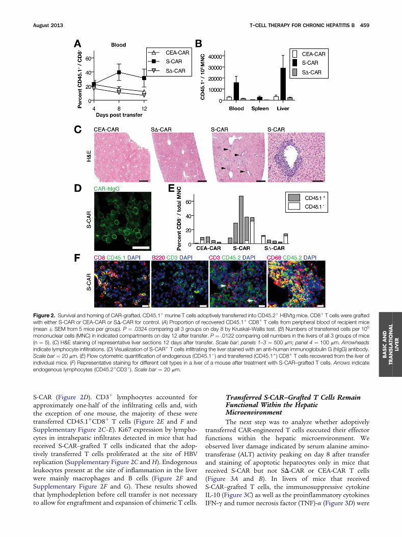
Figure 2. Survival and homing of CAR-grafted, CD45.1þ murine T cells adoptively transferred into CD45.2þ HBVtg mice. CD8þ T cells were graftedwith either S-CAR or CEA-CAR or SD-CAR for control. (A) Proportion of recovered CD45.1þ CD8þ T cells from peripheral blood of recipient mice(mean � SEM from 5 mice per group). P ¼ .0324 comparing all 3 groups on day 8 by Kruskal–Wallis test. (B) Numbers of transferred cells per 105
mononuclear cells (MNC) in indicated compartments on day 12 after transfer. P ¼ .0122 comparing cell numbers in the livers of all 3 groups of mice(n ¼ 5). (C) H&E staining of representative liver sections 12 days after transfer. Scale bar: panels 1–3 ¼ 500 mm; panel 4 ¼ 100 mm. Arrowheadsindicate lymphocyte infiltrations. (D) Visualization of S-CARþ T cells infiltrating the liver stained with an anti-human immunoglobulin G (hIgG) antibody.Scale bar ¼ 20 mm. (E) Flow cytometric quantification of endogenous (CD45.1�) and transferred (CD45.1þ) CD8þ T cells recovered from the liver ofindividual mice. (F) Representative staining for different cell types in a liver of a mouse after treatment with S-CAR–grafted T cells. Arrows indicateendogenous lymphocytes (CD45.2þCD3þ). Scale bar ¼ 20 mm.
August 2013 T–CELL THERAPY FOR CHRONIC HEPATITIS B 459
BASICAND
TRANSL
ATIONAL
LIVER
S-CAR (Figure 2D). CD3þ lymphocytes accounted forapproximately one-half of the infiltrating cells and, withthe exception of one mouse, the majority of these weretransferred CD45.1þCD8þ T cells (Figure 2E and F andSupplementary Figure 2C–E). Ki67 expression by lympho-cytes in intrahepatic infiltrates detected in mice that hadreceived S-CAR–grafted T cells indicated that the adop-tively transferred T cells proliferated at the site of HBVreplication (Supplementary Figure 2C and H). Endogenousleukocytes present at the site of inflammation in the liverwere mainly macrophages and B cells (Figure 2F andSupplementary Figure 2F and G). These results showedthat lymphodepletion before cell transfer is not necessaryto allow for engraftment and expansion of chimeric T cells.
Transferred S-CAR–Grafted T Cells RemainFunctional Within the HepaticMicroenvironment
The next step was to analyze whether adoptivelytransferred CAR-engineered T cells executed their effectorfunctions within the hepatic microenvironment. Weobserved liver damage indicated by serum alanine amino-transferase (ALT) activity peaking on day 8 after transferand staining of apoptotic hepatocytes only in mice thatreceived S-CAR but not SD-CAR or CEA-CAR T cells(Figure 3A and B). In livers of mice that receivedS-CAR–grafted T cells, the immunosuppressive cytokineIL-10 (Figure 3C) as well as the proinflammatory cytokinesIFN-g and tumor necrosis factor (TNF)-a (Figure 3D) were
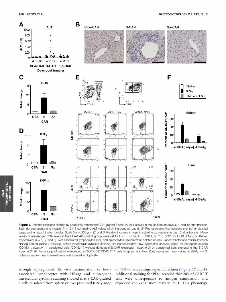
Figure 3. Effector functions exerted by adoptively transferred CAR-grafted T cells. (A) ALT activity in mouse sera on days 4, 8, and 12 after transfer.Each dot represents one mouse. P ¼ .0113 comparing ALT values of all 3 groups on day 8. (B) Representative liver sections stained for cleavedcaspase-3 on day 12 after transfer. Scale bar ¼ 200 mm. (C and D) Relative increase in hepatic cytokine expression on day 12 after transfer. Meanvalues of messenger RNA levels in the CEA-CAR control group were set to 1. P ¼ .0189, P ¼ .0041, or P < .0001 for IL-10, IFN-g, or TNF-a,respectively (n ¼ 5). (E and F) Liver-associated lymphocytes (liver) and splenocytes (spleen) were isolated on day 8 after transfer and restimulated onHBsAg-coated plates (þHBsAg) before intracellular cytokine staining. (E) Representative flow cytometric analysis gated on endogenous cells(CD45.1�, column 1), transferred cells (CD45.1þ) without detectable S-CAR expression (column 2) or transferred cells expressing the S-CAR(column 3). (F) Percentage of cytokine-secreting S-CARþCD8þCD45.1þ T cells in spleen and liver. Data represent mean values � SEM, n ¼ 3.Splenocytes from each animal were restimulated in duplicate.
460 KREBS ET AL GASTROENTEROLOGY Vol. 145, No. 2
BASIC
AND
TRANSLA
TIONAL
LIVER
strongly up-regulated. Ex vivo restimulation of liver-associated lymphocytes with HBsAg and subsequentintracellular cytokine staining showed that S-CAR–graftedT cells reisolated from spleen or liver produced IFN-g and/
or TNF-a in an antigen-specific fashion (Figure 3E and F).Additional staining for PD-1 revealed that 20% of CARþ Tcells were unresponsive to antigen stimulation andexpressed the exhaustion marker PD-1. This phenotype

August 2013 T–CELL THERAPY FOR CHRONIC HEPATITIS B 461
was observed less frequently when S-CAR T cells weretransferred into wild-type mice, in which antigen stimuluswas missing (Supplementary Figure 3). These resultsshowed that the majority of transferred S-CAR–graftedT cells remain functional even within the immunoregula-tory hepatic microenvironment.
Adoptively Transferred T Cells Redirected bythe HBV-Specific S-CAR Exhibit a StrongAntiviral Effect In Vivo
A profound reduction of the number of hepato-cytes with cytoplasmic expression of HBV core protein(Figure 4A) showed the antiviral activity of the adoptivelytransferred S-CAR–grafted T cells. Moreover, the numberof virions circulating in the bloodstream rapidly decreased100-fold (Figure 4B) and replicative forms of HBV DNAalmost completely disappeared from the liver within12 days (Figure 4C and D). Lacking antiviral activity ofCEA-CAR and SD-CAR engineered T cells proved thatantigen recognition and T-cell activation via the S-CARwere essential to stimulate the antiviral activity of adop-tively transferred T cells.
Treatment With S-CAR T Cells Causes OnlyMinor Side Effects
Figure 4. Antiviral effect of adoptively transferred T cells grafted withS-CAR, CEA-CAR, or SD-CAR. (A) Staining of HBV core–positivehepatocytes in liver sections from HBVtg mice 12 days after T-celltransfer. Scale bar ¼ 500 mm. (B) HBV viremia determined by quan-titative polymerase chain reaction. P ¼ .0242 or P ¼ .0122 comparingHBV DNA copies of all 3 groups on day 8 or 12, respectively. (C andD) HBV replication in the liver on day 12 after transfer. (C) HBV DNAcopy numbers determined by quantitative polymerase chain reactionwere normalized to the single copy gene Nid2. P ¼ .009 comparingS-CAR with CEA-CAR and SD-CAR. All data are presented as meanvalues � SEM (n ¼ 5). (D) Southern blot analysis of pooled total liverDNA. Equal amounts of liver DNA from each animal were pooled pergroup and probed for HBV DNA. Relaxed circular (rc), single-stranded(ss), and other replicative intermediate (ri) forms of HBV DNA areindicated.
BASICAND
TRANSL
ATIONAL
LIVER
Animals injected with 4 � 106 S-CAR–grafted Tcells did not lose weight over 34 days of treatment(Figure 5A) and did not show any obvious signs ofdistress, although serum TNF-a, IFN-g, MCP-1, IL-10,and IL-6 levels increased significantly (Figure 5B). Levelsof immunoglobulin G1 antibodies increased, but levelsof other immunoglobulin subtypes were not altered(Figure 5C). Twelve days after transfer, the relativeamount of CD4þ T cells and B cells decreased in thespleen and liver while B cells and NK cells increased inblood (Figure 5D, left panel). The relative amount ofmyeloid immune cells such as inflammatory monocytes,dendritic cells (DC), and neutrophils increased, especiallyin the liver (Figure 5D, right panel). Thirty-four days aftertreatment, the composition of immune cells in all an-alyzed compartments resembled that of untreated miceagain.
S-CAR–Grafted T Cells Exert StrongerAntiviral Activity Than Natural HBV-SpecificT Cells
To compare the efficacy of S-CAR T cells with“natural” S-specific T cells, wild-type mice were immu-nized with recombinant HBsAg and boosted withmodified vaccinia Ankara (MVA) virus expressing S-Protein to induce S-specific T cells for adoptive transfer(Supplementary Figure 4). A total of 1 � 106 S-specificCD8þ T cells and 1 � 106 and 4 � 106 S-CAR T cells wereinjected into HBVtg mice. Most of the vaccine-induced S-specific T cells accumulated in lymph nodes (Figure 6A),whereas S-CAR T cells preferentially homed to the liver(Figures 2B and 6A). ALT levels were not elevated on day 7in animals that received 1 � 106 S-specific T cells. Transfer
of the same amount of S-CAR T cells led to an increase inALT activity to approximately 150 U/L. Transfer of 4 �106 S-CAR T cells led to an ALT activity of approximately800 U/L (Figure 6B). Accordingly, S-CAR T cells reducedcytoplasmic hepatitis B core antigen expression moreprofoundly than vaccine-induced T cells (Figure 6C).However, the effect on HBV viremia (Figure 6D) and HBVreplication in the liver (Figure 6E) was not significantlydifferent among the 3 treatment groups, indicating adominant role of noncytopathic antiviral mediators.17,18
Cytoplasmic hepatitis B core antigen and virus DNA inthe serum increased to levels before treatment, when S-CAR T cells had almost vanished from the liver on day 34(Figure 6C–E) and HBV replication was driven again by thestable transgene, which cannot be eliminated. Takentogether, vaccine-induced T cells elicited their effect

Figure 5. Potential side effects of 4 � 106 adoptively transferred S-CAR–grafted T cells. (A) Body weight of mice relative to their individual weightbefore transfer. (B) Cytokines measured in sera on different days after T-cell transfer, compared over all days (n ¼ 3–7): IL-12 (P ¼ .1163), TNF-a(P ¼ .0049), IFN-g (P ¼ .0032), MCP-1 (P ¼ .0061), IL-10 (P ¼ .0238), or IL-6 (P ¼ .0047). (C) Immunoglobulin clonotypes in sera on different daysafter T-cell transfer. P ¼ .0041 for IgG1, comparing all days (n ¼ 3–7). Data represent mean values � SEM. (D) Relative amounts of different cell typesin blood, liver, and spleen on days 12 and 34 after T-cell transfer and in untreated mice. Each symbol represents one mouse.
462 KREBS ET AL GASTROENTEROLOGY Vol. 145, No. 2
BASIC
AND
TRANSLA
TIONAL
LIVER
mainly in a noncytopathic fashion, whereas S-CAR T cellskilled HBV-replicating hepatocytes in addition.
Discussion
Currently there is no cure for chronic hepatitis B.Novel antiviral agents very efficiently control HBV butcannot eliminate it. Immunotherapy using T cells that aregenetically modified to express an HBsAg-specific receptorseems a promising addition to current antiviral therapy.This therapy could cure chronic hepatitis B, which is apremalignant condition, but may also be applied to treatHBsAg-positive HCC.
Our study showed that T cells redirected by an HBV-specific CAR, when transferred into immunocompetentmice, (1) recognize HBV envelope proteins on the surfaceof HBV-replicating hepatocytes, (2) engraft and (3)expand in vivo, (4) infiltrate the liver, and (5) effectivelycontrol HBV replication. This new immunotherapyapproach proved safe and did not lead to excessive liverdamage after contact of T cells with circulating viral an-tigen or to functional exhaustion of the adoptivelytransferred T cells. Our results strongly suggest thatS-CAR–engineered T cells will be able to cure HBVinfection. However, this cannot be proven in the HBVtg
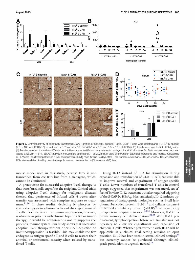
Figure 6. Antiviral activity of adoptively transferred S-CAR–grafted or natural S-specific T cells. CD8þ T cells were isolated and 1 � 106 S-specific(2.5 � 107 total CD45.1þ) as well as 1 � 106 and 4 � 106 S-CAR (1.4 � 106 and 5.5 � 106 total CD45.1þ) T cells were injected into HBVtg mice.(A) Relative amount of transferred T cells per total leukocytes in different compartments on days 12 and 34 after transfer. Data are presented as meanvalues � SEM (n ¼ 3–4). (B) ALT activity in mouse sera before and 7, 12, 20, and 34 days after transfer. Each dot represents one mouse. (C) Stainingof HBV core–positive hepatocytes in liver sections fromHBVtgmice 12 and 34 days after T-cell transfer.Scale bar¼ 200 mm, inset¼ 100 mm. (D and E)HBV viremia determined by quantitative polymerase chain reaction in (D) serum and (E) liver.
August 2013 T–CELL THERAPY FOR CHRONIC HEPATITIS B 463
BASICAND
TRANSL
ATIONAL
LIVER
mouse model used in this study, because HBV is nottranscribed from cccDNA but from a transgene, whichcannot be eliminated.
A prerequisite for successful adoptive T-cell therapy isthat transferred cells engraft in the recipient. Clinical trialsusing adoptive T-cell therapy for malignant diseasesshowed that persistence of infused cells 4 weeks aftertransfer was associated with complete response to treat-ment.11,16 In these studies, depleting lymphocytes bychemotherapy or irradiation facilitated the engraftment ofT cells. T-cell depletion or immunosuppression, however,is obsolete in patients with chronic hepatitis B. For tumortherapy, it would be advantageous not to suppress thepatient’s immune system. Our study shows that successfuladoptive T-cell therapy without prior T-cell depletion orimmunosuppression is feasible. This may enable the fewendogenous antigen-specific T and B cells to restore theirantiviral or antitumoral capacity when assisted by trans-ferred T cells.
Using IL-12 instead of IL-2 for stimulation duringexpansion and transduction of CD8þ T cells, we were ableto improve survival and engraftment of antigen-specificT cells. Lower numbers of transferred T cells in controlgroups suggested that engraftment was not merely an ef-fect of in vitro IL-12 treatment but also required triggeringof the S-CAR by HBsAg. Mechanistically, IL-12 induces up-regulation of antiapoptotic molecules such as B-cell lym-phoma 3-encoded protein (Bcl-3)19 and cellular caspase-8(FLICE)-like inhibitory protein (c-FLIP)20 while reducingproapoptotic caspase activation.20,21 Moreover, IL-12 im-proves memory cell differentiation.21,22 With IL-12 pre-treatment, lymphodepletion before cell transfer was notnecessary to allow for engraftment and expansion ofchimeric T cells. Whether pretreatment with IL-12 will beapplicable in a clinical trial setting remains an openquestion. IL-12 has been used in several clinical settings23
but currently cannot be purchased although clinical-grade production is urgently needed.24
464 KREBS ET AL GASTROENTEROLOGY Vol. 145, No. 2
BASIC
AND
TRANSLA
TIONAL
LIVER
An advantage of not administering immunosuppressivetherapy before adoptive T-cell therapy is that the regula-tory function of immune cells in the liver and other organsis preserved. In our experiments, the increasing ALT ac-tivity in the serum selectively after transfer of S-CAR–engineered T cells suggested that the S-CAR medi-ated the killing of HBV-positive hepatocytes in vivo andthus induced liver damage. Liver damage, however, wastransient. This may be explained by either increased levelsof the immunosuppressive cytokine IL-10 in the liver,inducing an exhausted phenotype, or contraction of theeffector T-cell population after massive clonal expan-sion,25,26 resulting in low-level cytotoxicity.27,28 Restrictionof liver damage by IL-10 was observed in several models ofimmune-mediated liver damage.29,30 The cellular source ofIL-10 may be liver-resident T-helper 2 or regulatoryT cells,31 Kupffer cells,32,33 or even transferred, IL-12–primed CD8þ T cells.34 Self-limitation of immune-mediated damage in the liver by any of these means willensure organ integrity but may limit the efficiency ofimmunotherapy.11 The rapid decrease of HBV replicationwithout severe liver disease is very likely due to the fact thatS-CAR–grafted T cells, like natural HBV-specificT cells,18,35 control HBV in transgenic mice in a non-cytopathic fashion via antiviral cytokines in addition todirectly killing HBV-replicating hepatocytes. This ideais supported by the fact that ALT levels in mice treated with1 � 106 T cells were much lower but the antiviral activitywas comparable to animals that received 4 times more cells.
Development of T-cell therapy for hepatitis B has beenencouraged by several observations. Control of HBVreplication is obtained after transfer of splenocytes fromimmunized wild-type mice into HBVtg mice.18,27 Moreimportantly, cure of HBV infection in patients has beenreported after transfer of specific immunity against HBVthrough allogeneic bone marrow transplantation.36,37
Bone marrow transplantation, however, is complex andcannot be broadly applied for treatment of an infectiousdisease because it requires an exact major histocompati-bility complex match of cells derived from an immunizeddonor as well as profound immunosuppression to allowengraftment. Using the patient’s own T cells and redi-recting them with an HBV-specific receptor seems a morefeasible approach to treat chronic hepatitis B or HBsAg-positive HCC. CAR-grafted T cells, which function inde-pendently of the patient’s HLA haplotype and recognizedifferent HBsAg subtypes, seem to be particularly suitedbecause they will in principle be applicable to almost allHBV-infected patients.38
Our preclinical model has similar levels of circulatingHBsAg (approximately 1000–1200 IU/mL) as detected inthe low-replicative phase of chronic hepatitis B.39 In thismodel, we observed elevation of cytokines but no severeside effects during T-cell therapy. However, in a patientwith high replication, preexisting liver inflammation,and tissue damage, the situation may be different. Pro-nounced elevation of ALT levels was observed in trans-plant recipients with cleared HBV infection,37 indicating
that hepatocyte killing was needed for elimination. S-CART cells and T cells induced by immunization of donormice showed comparable antiviral efficacy in our model,but elevation of ALT levels and clearance of hepatitis Bcore–positive hepatocytes indicating elimination of HBV-positive hepatocytes was only observed after S-CART-cell transfer. To avoid or reduce potential hepatotoxicityin a clinical setting, patients will be pretreated with anti-viral agents before T-cell transfer to reduce the amount ofHBsAg-positive hepatocytes and the grade of inflamma-tion and increase selection pressure on the virus to mini-mize the risk for emergence of viral variants, which couldescape CAR recognition.40 In addition, redirected T cellscan be specifically eliminated by a safeguard mechanism.For clinical use, we have added a truncated version of theepidermal growth factor receptor to the CAR construct,which allows for depletion of CAR transduced cells withthe clinically approved antibody cetuximab.41
We have previously reported that human T cells that areengrafted with the S-CAR can eliminate the nuclear persis-tence form of HBV, the cccDNA, from HBV-infected hepa-tocytes.12 In an alternative approach, Gehring et al42
generated 2 HBV-specific, HLA-A2–restricted T-cell re-ceptors for grafting and showed that HBV-specific T cellsgenerated from peripheral blood mononuclear cells of pa-tients with chronic HBV and HBV-related HCC becamemultifunctional and capable of recognizingHBV-replicatinghepatoma cells and HCC tumor cells expressing viral anti-gens from naturally integrated HBV DNA. We also haveestablished a series of such recombinant T-cell receptors ofdiverse receptor avidity (unpublished data; October 2011)and are currently comparing these with respect to optimalfunctionality.
The in vivo study presented here showed thatS-CAR–grafted T cells (although vast amounts of subviralparticles are present in the blood of HBVtg mice) infiltratethe liver, remain functional, and lead to a profound reduc-tion of viral load. From the data obtained, we conclude thatimmunotherapy with S-CAR–grafted T cells is a feasible andpromising approach to treat chronic HBV infection,providing proof of concept for further translation ofadoptiveT-cell therapy for chronic hepatitis B into the clinic.
Supplementary Material
Note: To access the supplementary materialaccompanying this article, visit the online version ofGastroenterology at www.gastrojournal.org, and at http://dx.doi.org/10.1053/j.gastro.2013.04.047.
References
1. Lavanchy D. Worldwide epidemiology of HBV infection, diseaseburden, and vaccine prevention. J Clin Virol 2005;34(Suppl 1):S1–S3.
2. Kwon H, Lok AS. Hepatitis B therapy. Nat Rev Gastroenterol Hepatol2011;8:275–284.
3. Moraleda G, Saputelli J, Aldrich CE, et al. Lack of effect of antiviraltherapy in nondividing hepatocyte cultures on the closed circularDNA of woodchuck hepatitis virus. J Virol 1997;71:9392–9399.
August 2013 T–CELL THERAPY FOR CHRONIC HEPATITIS B 465
BASICAND
TRANSL
ATIONAL
LIVER
4. Dandri M, Burda MR, Will H, et al. Increased hepatocyte turnover andinhibition of woodchuck hepatitis B virus replication by adefovirin vitro do not lead to reduction of the closed circular DNA. Hep-atology 2000;32:139–146.
5. Boettler T, Panther E, Bengsch B, et al. Expression of the interleukin-7receptor alpha chain (CD127) on virus-specific CD8þ Tcells identifiesfunctionally and phenotypically defined memory T cells during acuteresolving hepatitis B virus infection. J Virol 2006;80:3532–3540.
6. Maini MK, Boni C, Ogg GS, et al. Direct ex vivo analysis of hepatitis Bvirus-specific CD8(þ) T cells associated with the control of infection.Gastroenterology 1999;117:1386–1396.
7. Nayersina R, Fowler P, Guilhot S, et al. HLA A2 restricted cytotoxic Tlymphocyte responses to multiple hepatitis B surface antigen epi-topes during hepatitis B virus infection. J Immunol 1993;150:4659–4671.
8. Rehermann B, Fowler P, Sidney J, et al. The cytotoxic T lymphocyteresponse to multiple hepatitis B virus polymerase epitopes duringand after acute viral hepatitis. J Exp Med 1995;181:1047–1058.
9. Thimme R, Wieland S, Steiger C, et al. CD8(þ) T cells mediate viralclearance and disease pathogenesis during acute hepatitis B virusinfection. J Virol 2003;77:68–76.
10. Chu CM, Liaw YF. Membrane staining for hepatitis B surfaceantigen on hepatocytes: a sensitive and specific marker of activeviral replication in hepatitis B. J Clin Pathol 1995;48:470–473.
11. Protzer U, Maini MK, Knolle PA. Living in the liver: hepatic infections.Nat Rev Immunol 2012;12:201–213.
12. Bohne F, Chmielewski M, Ebert G, et al. T cells redirected againsthepatitis B virus surface proteins eliminate infected hepatocytes.Gastroenterology 2008;134:239–247.
13. Dumortier J, Schonig K, Oberwinkler H, et al. Liver-specific expres-sion of interferon gamma following adenoviral gene transfer controlshepatitis B virus replication in mice. Gene Ther 2005;12:668–677.
14. Engels B, Cam H, Schuler T, et al. Retroviral vectors for high-leveltransgene expression in T lymphocytes. Hum Gene Ther 2003;14:1155–1168.
15. Morita S, Kojima T, Kitamura T. Plat-E: an efficient and stable system fortransient packaging of retroviruses. Gene Ther 2000;7:1063–1066.
16. Rosenberg SA, Yang JC, Sherry RM, et al. Durable complete responsesin heavily pretreted patients with metastatic melanoma using T celltransfer immunotherapy. Clin Cancer Res 2011;17:4550–4557.
17. Guidotti LG, McClary H, Loudis JM, et al. Nitric oxide inhibits hepatitisB virus replication in the livers of transgenic mice. J Exp Med 2000;191:1247–1252.
18. Guidotti LG, Ishikawa T, HobbsMV, et al. Intracellular inactivation of thehepatitis B virus by cytotoxic T lymphocytes. Immunity 1996;4:25–36.
19. Valenzuela JO, Hammerbeck CD, Mescher MF. Cutting edge: Bcl-3up-regulation by signal 3 cytokine (IL-12) prolongs survival ofantigen-activated CD8 T cells. J Immunol 2005;174:600–604.
20. Lee SW, Park Y, Yoo JK, et al. Inhibition of TCR-induced CD8 T celldeath by IL-12: regulation of Fas ligand and cellular FLIP expressionand caspase activation by IL-12. J Immunol 2003;170:2456–2460.
21. Chang J, Cho JH, Lee SW, et al. IL-12 priming during in vitro antigenicstimulation changes properties of CD8 T cells and increases genera-tion of effector and memory cells. J Immunol 2004;172:2818–2826.
22. Diaz-Montero CM, Naga O, Zidan AA, et al. Synergy of brief activationof CD8 T-cells in the presence of IL-12 and adoptive transfer intolymphopenic hosts promotes tumor clearance and anti-tumormemory. Am J Cancer Res 2011;1:882–896.
23. Del Vecchio M, Bajetta E, Canova S, et al. Interleukin-12: biologicalproperties and clinical application. Clin Cancer Res 2007;13:4677–4685.
24. National Cancer Institute Immunotherapy Agent Workshop, Minutesfrom 7/12/2007. Frederick, MD: National Cancer Institute; NationalInstitutes of Health.
25. Badovinac VP, Porter BB, Harty JT. Programmed contraction ofCD8(þ) T cells after infection. Nat Immunol 2002;3:619–626.
26. Hoshino Y, Morishima T, Kimura H, et al. Antigen-driven expansionand contraction of CD8þ-activated T cells in primary EBV infection.J Immunol 1999;163:5735–5740.
27. Isogawa M, Furuichi Y, Chisari FV. Oscillating CD8(þ) T cell effectorfunctions after antigen recognition in the liver. Immunity 2005;23:53–63.
28. Stabenow D, Frings M, Truck C, et al. Bioluminescence imaging al-lows measuring CD8 T cell function in the liver. Hepatology 2010;51:1430–1437.
29. Swain MG, Appleyard C, Wallace J, et al. Endogenous glucocorticoidsreleased during acute toxic liver injury enhance hepatic IL-10 syn-thesis and release. Am J Physiol 1999;276:G199–G205.
30. von Freyend MJ, Untergasser A, Arzberger S, et al. Sequential controlof hepatitis B virus in a mouse model of acute, self-resolving hep-atitis B. J Viral Hepat 2010;18:216–226.
31. Mosser DM, Zhang X. Interleukin-10: new perspectives on an oldcytokine. Immunol Rev 2008;226:205–218.
32. Erhardt A, Biburger M, Papadopoulos T, et al. IL-10, regulatory T cells,and Kupffer cells mediate tolerance in concanavalin A-induced liverinjury in mice. Hepatology 2007;45:475–485.
33. Knolle PA, Loser E, Protzer U, et al. Regulation of endotoxin-inducedIL-6 production in liver sinusoidal endothelial cells and Kupffer cellsby IL-10. Clin Exp Immunol 1997;107:555–561.
34. Lee JB, Lee KA, Chang J. Phenotypic changes induced by IL-12priming regulate effector and memory CD8 T cell differentiation.Int Immunol 2007;19:1039–1048.
35. Guidotti LG, Rochford R, Chung J, et al. Viral clearance withoutdestruction of infected cells during acute HBV infection. Science1999;284:825–829.
36. Ilan Y, Nagler A, Adler R, et al. Ablation of persistent hepatitis B bybone marrow transplantation from a hepatitis B-immune donor.Gastroenterology 1993;104:1818–1821.
37. Lau GK, Lok AS, Liang RH, et al. Clearance of hepatitis B surfaceantigen after bone marrow transplantation: role of adoptive immunitytransfer. Hepatology 1997;25:1497–1501.
38. Courouce-Pauty AM, Plancon A, Soulier JP. Distribution of HBsAgsubtypes in the world. Vox Sang 1983;44:197–211.
39. Jaroszewicz J, Calle Serrano B, Wursthorn K, et al. Hepatitis B surfaceantigen (HBsAg) levels in the natural history of hepatitis B virus (HBV)-infection: a European perspective. J Hepatol 2010;52:514–522.
40. Protzer-Knolle U, Naumann U, Bartenschlager R, et al. HepatitisB virus with antigenically altered hepatitis B surface antigen isselected by high-dose hepatitis B immune globulin after livertransplantation. Hepatology 1998;27:254–263.
41. Wang X, Chang WC, Wong CW, et al. A transgene-encoded cell sur-face polypeptide for selection, in vivo tracking, and ablation ofengineered cells. Blood 2011;118:1255–1263.
42. Gehring AJ, Xue SA, Ho ZZ, et al. Engineering virus-specific T cellsthat target HBV infected hepatocytes and hepatocellular carcinomacell lines. J Hepatol 2011;55:103–110.
Received August 19, 2012. Accepted April 17, 2013.
Reprint requestsAddress requests for reprints to: Ulrike Protzer, MD, Institute of
Virology, Trogerstrasse 30, 81675 Munich, Germany. e-mail:[email protected]; fax: (49) 89-4140-6823.
AcknowledgmentsThe authors thank Theresa Asen, Chantal Gotthier, Raindy
Tedjokusumo, Judith Seebach, Daniel Kull, and Ruth Hillermann forexcellent technical assistance; ElisabethKremmer for providing antibodies;Elisa Kieback for sharing protocols; Stephan Haug for help with statisticalanalyses; and Irene Esposito for support with histological staining.
Dr Gasteiger’s current affiliation is: Memorial Sloan-Kettering CancerCenter, New York, New York.
Conflicts of interestThe authors disclose no conflicts.
FundingSupported by the Helmholtz Alliance on Immunotherapy of Cancer
and in part by the Deutsche Forschungsgemeinschaft SFB-TR36.