Embed Size (px)
Citation preview

1
Learning Module 3 – Lymphoid SystemLearning component 1 – Basic Responses and Normal Histology
2
Normal Bursa of Fabricius (BF)
A. Normal Bursa showing a plical fold with numerous lymphoid follicles.B. Higher magnification of A show the epithelium beneath which are lymphoid follicles with an outer cortex and inner medulla. Note the relatively small amount of interfollicular connective (interstitial) tissue.C. Higher magnification of a bursal lymphoid follicle.D. Shows the region of specialized epithelium that has pinocytotic properties and is closely associated with an underlying follicle.

3
Thymus Normal
A. Normal Thymus showing outer cortex and inner medulla.B. Normal thymus.C. Higher magnification of B shows the cortex and a small portion of medulla
(bottom enter).D. Normal thymus – higher magnification of B.
4
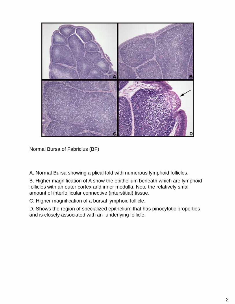
Thymus – Normal and Atrophy
A. Normal thymus.B. Thymus with severe atrophy.C. Normal thymus – higher magnification of A.D. Atrophic thymus – higher magnification of B.
5
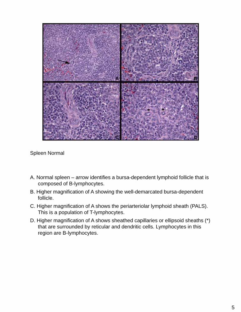
Spleen Normal
A. Normal spleen – arrow identifies a bursa-dependent lymphoid follicle that is composed of B-lymphocytes.
B. Higher magnification of A showing the well-demarcated bursa-dependent follicle.
C. Higher magnification of A shows the periarteriolar lymphoid sheath (PALS). This is a population of T-lymphocytes.
D. Higher magnification of A shows sheathed capillaries or ellipsoid sheaths (*) that are surrounded by reticular and dendritic cells. Lymphocytes in this region are B-lymphocytes.
6
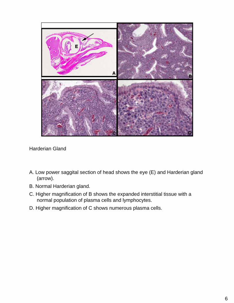
Harderian Gland
A. Low power saggital section of head shows the eye (E) and Harderian gland (arrow).
B. Normal Harderian gland.C. Higher magnification of B shows the expanded interstitial tissue with a
normal population of plasma cells and lymphocytes.D. Higher magnification of C shows numerous plasma cells.
7
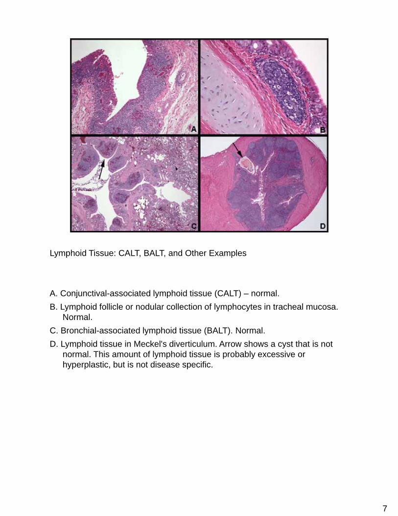
Lymphoid Tissue: CALT, BALT, and Other Examples
A. Conjunctival-associated lymphoid tissue (CALT) – normal.B. Lymphoid follicle or nodular collection of lymphocytes in tracheal mucosa.
Normal.C. Bronchial-associated lymphoid tissue (BALT). Normal.D. Lymphoid tissue in Meckel's diverticulum. Arrow shows a cyst that is not
normal. This amount of lymphoid tissue is probably excessive or hyperplastic, but is not disease specific.
8
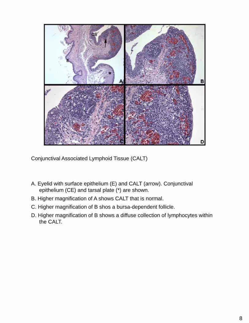
Conjunctival Associated Lymphoid Tissue (CALT)
A. Eyelid with surface epithelium (E) and CALT (arrow). Conjunctival epithelium (CE) and tarsal plate (*) are shown.
B. Higher magnification of A shows CALT that is normal.C. Higher magnification of B shos a bursa-dependent follicle.D. Higher magnification of B shows a diffuse collection of lymphocytes within
the CALT.

9
Normal Lymphoid Tissue
A. Shows a normal nodular and diffuses accumulation of lymphocytes at the esophageal – proventricular junction. Epithelium of the esophagus (E) and an esophageal mucous gland (*) are shown. Arrow identifies a bursa-dependent or nodular collection of lymphocytes.
B. Higher magnification of A at arrow shows the bursa-dependent follicle. Normal.
C. Bone marrow with a nodular collection of lymphocytes – normal finding.D. Higher magnification of C shows the normal nodular collection of
lymphocytes.

10
Apoptosis in Thymus and BF
A. Thymus with many apoptotic cells. A pyknotic nucleus is shown (arrow). This is normal or programmed cell death defined as apoptosis and is expected to be seen in lymphoid organs.
B. Higher magnification of A shows pyknotic nucleus (arrow). The pattern of apoptosis in lymphoid organs create the “starry sky” appearance.
C. Apoptosis in the medulla (arrow) of a bursal follicle is normal.D. Many apoptotic cells are shown in this higher magnification of C with a
pyknotic nucleus (arrow) identified.
11
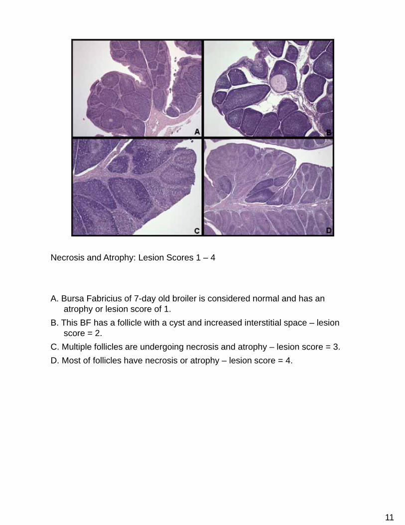
Necrosis and Atrophy: Lesion Scores 1 – 4
A. Bursa Fabricius of 7-day old broiler is considered normal and has an atrophy or lesion score of 1.
B. This BF has a follicle with a cyst and increased interstitial space – lesion score = 2.
C. Multiple follicles are undergoing necrosis and atrophy – lesion score = 3.D. Most of follicles have necrosis or atrophy – lesion score = 4.
12
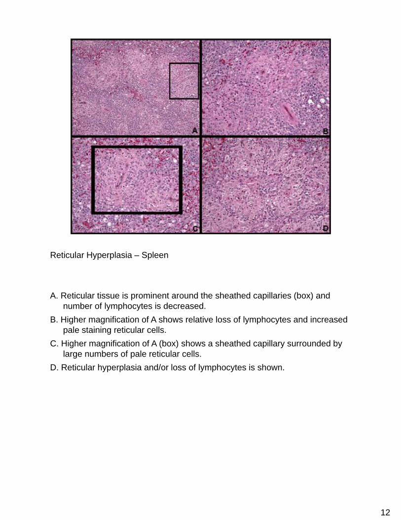
Reticular Hyperplasia – Spleen
A. Reticular tissue is prominent around the sheathed capillaries (box) and number of lymphocytes is decreased.
B. Higher magnification of A shows relative loss of lymphocytes and increased pale staining reticular cells.
C. Higher magnification of A (box) shows a sheathed capillary surrounded by large numbers of pale reticular cells.
D. Reticular hyperplasia and/or loss of lymphocytes is shown.