Embed Size (px)
Citation preview

Limbic system concept
Explanatory figure for the developmental concept of the limbic system (Szentagothai)
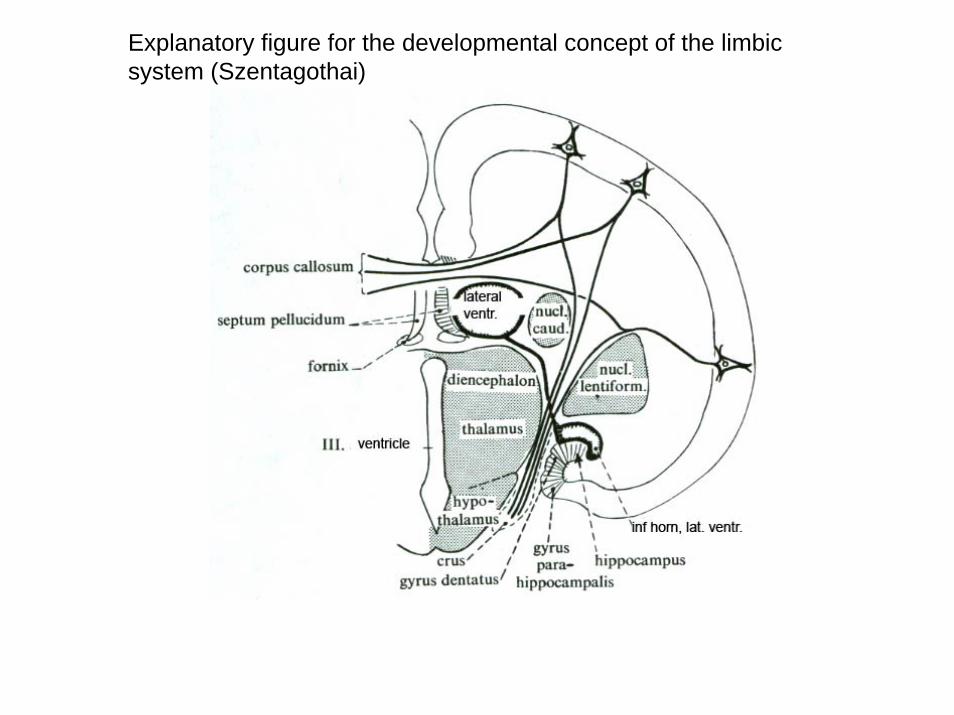
THE DEVELOPMENT OF THE LIMBIC SYSTEM CONCEPT
A: The great limbic lobe of Broca (1878); B: Papez’s circuit (ca 1938); C: Yakovlev(1948); D: MacLean (1949)
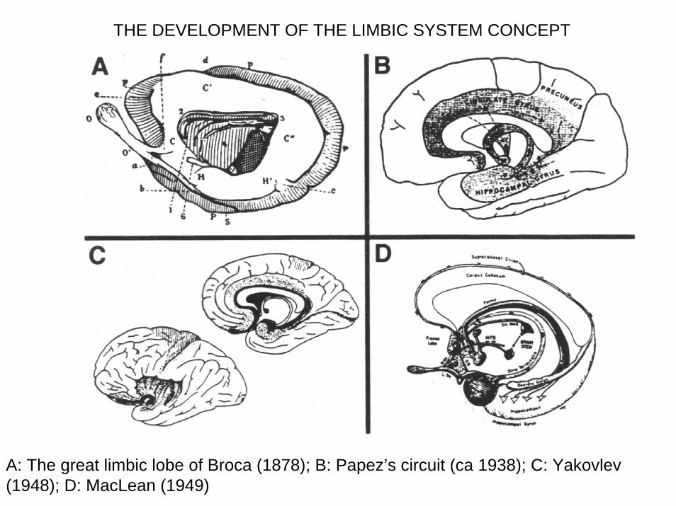
interconnections between different component of the limbic system (Nieuwenhyus, 1988).
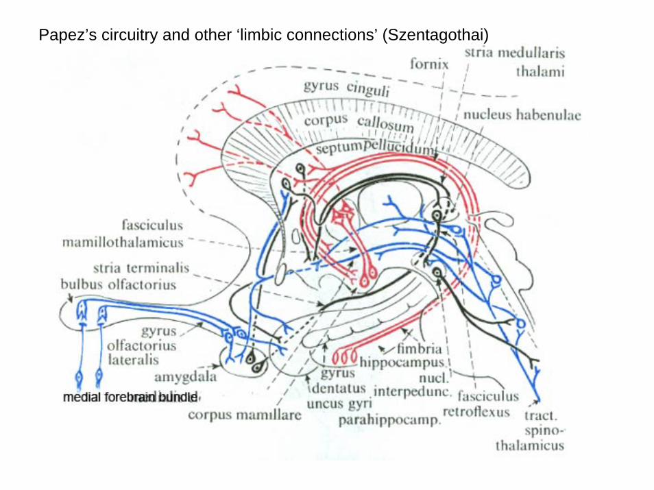
Papez’s circuitry and other ‘limbic connections’ (Szentagothai)
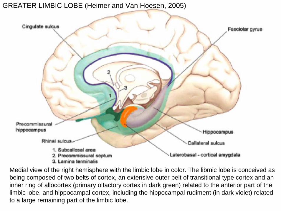
Medial view of the right hemisphere with the limbic lobe in color. The libmic lobe is conceived as being composed of two belts of cortex, an extensive outer belt of transitional type cortex and an inner ring of allocortex (primary olfactory cortex in dark green) related to the anterior part of the limbic lobe, and hippocampal cortex, including the hippocampal rudiment (in dark violet) related to a large remaining part of the limbic lobe.
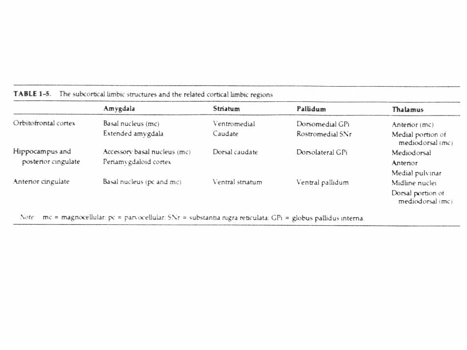
GREATER LIMBIC LOBE (Heimer and Van Hoesen, 2005)

Basal view of the human brain with the limbic lobe in color as inprevious figure. Dark green primary olfactory cortex. The anterior part of thetemporal lobe and the operculaecovering the insula have been removed in order to appreciate the continuity between the insula and the caudal part of the orbital cortex (from Heimer and van Hoesen, 2005).
GREATER LIMBIC LOBE (Heimer and Van Hoesen, 2005)

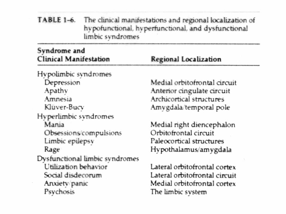
Medial Temporal Lobe. Amygdala and hippocampus
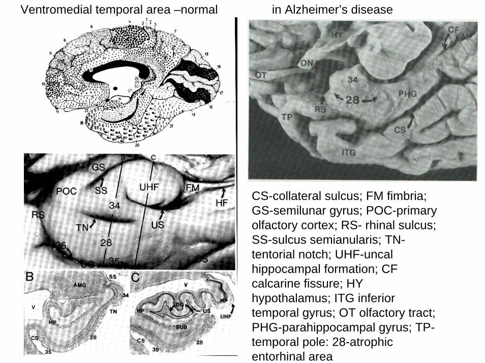
Ventromedial temporal area –normal in Alzheimer’s disease
CS-collateral sulcus; FM fimbria; GS-semilunar gyrus; POC-primary olfactory cortex; RS- rhinal sulcus; SS-sulcus semianularis; TN-tentorial notch; UHF-uncalhippocampal formation; CF calcarine fissure; HY hypothalamus; ITG inferior temporal gyrus; OT olfactory tract; PHG-parahippocampal gyrus; TP-temporal pole: 28-atrophic entorhinal area
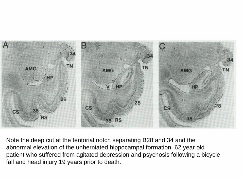
Note the deep cut at the tentorial notch separating B28 and 34 and the abnormal elevation of the unherniated hippocampal formation. 62 year old patient who suffered from agitated depression and psychosis following a bicycle fall and head injury 19 years prior to death.
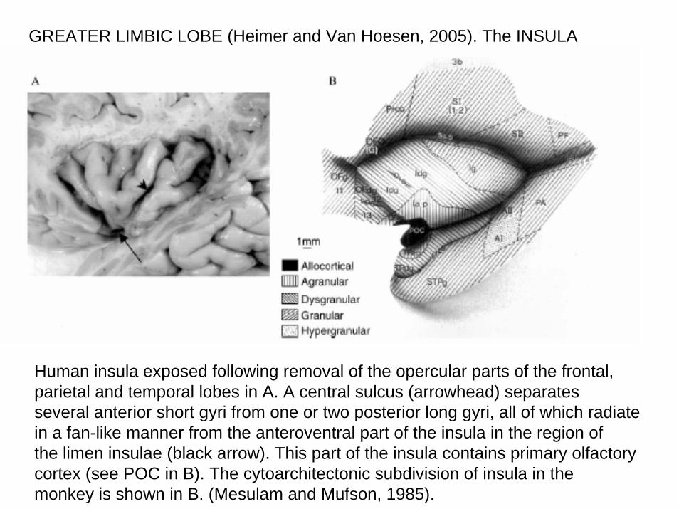
GREATER LIMBIC LOBE (Heimer and Van Hoesen, 2005). The INSULA
Human insula exposed following removal of the opercular parts of the frontal, parietal and temporal lobes in A. A central sulcus (arrowhead) separatesseveral anterior short gyri from one or two posterior long gyri, all of which radiate in a fan-like manner from the anteroventral part of the insula in the region ofthe limen insulae (black arrow). This part of the insula contains primary olfactory cortex (see POC in B). The cytoarchitectonic subdivision of insula in themonkey is shown in B. (Mesulam and Mufson, 1985).
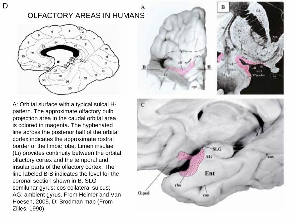
DOLFACTORY AREAS IN HUMANS
A: Orbital surface with a typical sulcal H-pattern. The approximate olfactory bulb projection area in the caudal orbital area is colored in magenta. The hyphenated line across the posterior half of the orbital cortex indicates the approximate rostral border of the limbic lobe. Limen insulae(Li) provides continuity between the orbital olfactory cortex and the temporal and insular parts of the olfactory cortex. The line labeled B-B indicates the level for the coronal section shown in B. SLG semilunar gyrus; cos collateral sulcus; AG: ambient gyrus. From Heimer and Van Hoesen, 2005. D: Brodman map (From Zilles, 1990)
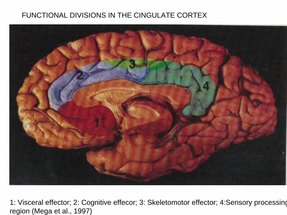
FUNCTIONAL DIVISIONS IN THE CINGULATE CORTEX
1: Visceral effector; 2: Cognitive effecor; 3: Skeletomotor effector; 4:Sensory processingregion (Mega et al., 1997)
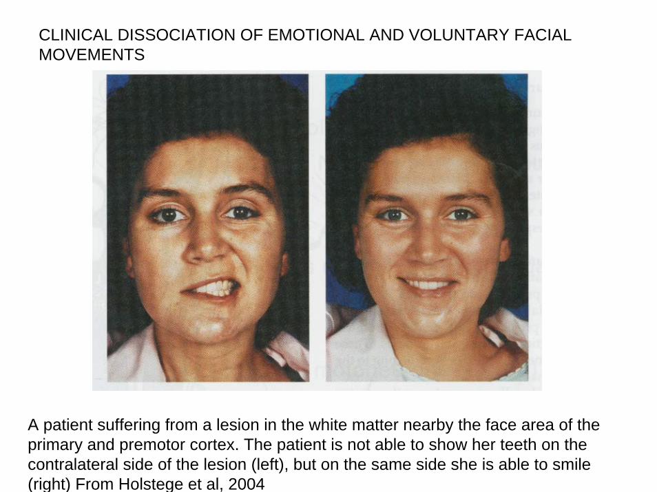
CLINICAL DISSOCIATION OF EMOTIONAL AND VOLUNTARY FACIAL MOVEMENTS
A patient suffering from a lesion in the white matter nearby the face area of the primary and premotor cortex. The patient is not able to show her teeth on the contralateral side of the lesion (left), but on the same side she is able to smile (right) From Holstege et al, 2004
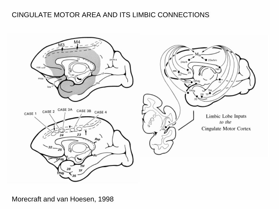
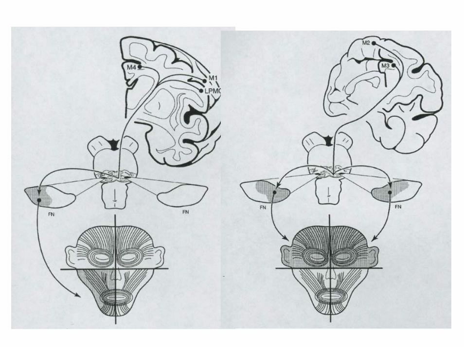
CINGULATE MOTOR AREA AND ITS LIMBIC CONNECTIONS
Morecraft and van Hoesen, 1998