Embed Size (px)
Citation preview

www.sciencedirect.com
c o r t e x 6 2 ( 2 0 1 5 ) 1 1 9e1 5 7
Available online at
ScienceDirect
Journal homepage: www.elsevier.com/locate/cortex
Special issue: Review
Limbic systems for emotion and for memory, butno single limbic system
Edmund T. Rolls a,b,*,1
aOxford Centre for Computational Neuroscience, Oxford, UKbUniversity of Warwick, Department of Computer Science, Coventry, UK
a r t i c l e i n f o
Article history:
Received 12 October 2013
Reviewed 4 December 2013
Revised 5 December 2013
Accepted 13 December 2013
Published online 24 December 2013
Keywords:
Limbic system
Orbitofrontal cortex
Emotion
Cingulate cortex
Amygdala
Episodic memory
Hippocampus
Reward value
* University of Warwick, Department of CoE-mail address: [email protected]
1 www.oxcns.org.0010-9452/$ e see front matter ª 2013 Elsevhttp://dx.doi.org/10.1016/j.cortex.2013.12.005
a b s t r a c t
The concept of a (single) limbic system is shown to be outmoded. Instead, anatomical,
neurophysiological, functional neuroimaging, and neuropsychological evidence is
described that anterior limbic and related structures including the orbitofrontal cortex
and amygdala are involved in emotion, reward valuation, and reward-related decision-
making (but not memory), with the value representations transmitted to the anterior
cingulate cortex for actioneoutcome learning. In this ‘emotion limbic system’ a compu-
tational principle is that feedforward pattern association networks learn associations
from visual, olfactory and auditory stimuli, to primary reinforcers such as taste, touch,
and pain. In primates including humans this learning can be very rapid and rule-based,
with the orbitofrontal cortex overshadowing the amygdala in this learning important
for social and emotional behaviour. Complementary evidence is described showing that
the hippocampus and limbic structures to which it is connected including the posterior
cingulate cortex and the fornix-mammillary body-anterior thalamus-posterior cingulate
circuit are involved in episodic or event memory, but not emotion. This ‘hippocampal
system’ receives information from neocortical areas about spatial location, and objects,
and can rapidly associate this information together by the different computational
principle of autoassociation in the CA3 region of the hippocampus involving feedback.
The system can later recall the whole of this information in the CA3 region from any
component, a feedback process, and can recall the information back to neocortical areas,
again a feedback (to neocortex) recall process. Emotion can enter this memory system
from the orbitofrontal cortex etc., and be recalled back to the orbitofrontal cortex etc.
during memory recall, but the emotional and hippocampal networks or ‘limbic systems’
operate by different computational principles, and operate independently of each other
except insofar as an emotional state or reward value attribute may be part of an episodic
memory.
ª 2013 Elsevier Ltd. All rights reserved.
mputer Science, Coventry CV4 7AL, UK..
ier Ltd. All rights reserved.
c o r t e x 6 2 ( 2 0 1 5 ) 1 1 9e1 5 7120
1. Introduction
The concept of the limbic system has a long history, and is a
concept that has endured to the present day (Catani,
Dell’acqua, & Thiebaut de Schotten, 2013; Mesulam, 2000).
In this paper I describe evidence that there are separate
systems in the brain for emotion and for memory, each
involving limbic structures, but that there is no single limbic
system.Wemight term the system for emotion the ‘emotional
limbic system’, and the system for memory the ‘memory
limbic system’, but there are non-limbic components to both
systems. The important concept I advance here is that the
systems for emotion and for episodic memory involve largely
different brain structures and connections, and different
computational principles of operation, which are described. I
argue here that of course some links from the emotional
system into the memory system are present, for often an
emotional state is part of an episodic memory, and when that
episodic memory is recalled, the emotional state must be
included inwhat is recalled. These concepts are important not
only within neuroscience, but also for neurology (Catani et al.,
2013; Mesulam, 2000), neuropsychology (Aggleton, 2012), and
psychiatry.
2. Historical background to the concept of alimbic system
The use of the term ‘limbic’ has changed over time, but the
concept of a limbic system is still in use (Catani et al., 2013).
The term ‘limbic’ was introduced by Thomas Willis (1664) to
designate a cortical border encircling the brainstem (limbus,
Latin for ‘border’). Paul Broca (1878) held the view that ‘le
grand lobe limbique’ was mainly an olfactory structure com-
mon to all mammalian brains, although he argued that its
functions were not limited to olfaction. Limbic structures are
frequently taken to include cortical structures such as the
hippocampus and cingulate cortex, and structures to which
they are connected such as the mammillary bodies, septal
area, and amygdala (Isaacson, 1982). After Broca’s publication,
the accumulation of experimental evidence from ablation
studies in animals broadened the role of limbic structures to
include other aspects of behaviour such as controlling social
interactions and behaviour (Brown & Schafer, 1888), consoli-
dating memories (Bechterew, 1900), and forming emotions
(Cannon, 1927). Anatomical and physiological advances led
James Papez (1937) to describe a neural circuit for linking ac-
tion and perception to emotion. The Papez circuit consists of
the hippocampus connecting via the fornix to themammillary
body, which connects via the mammillo-thalamic tract to the
anterior nuclei of the thalamus and thus back to the cingulate
cortex. According to Papez, emotion arises either from
cognition entering the circuit from the cortex through the
hippocampus, or from visceral and somatic perceptions
entering the circuit through the hypothalamus. Some of
Papez’ evidence on his circuit and emotion was that in rabies
where the disease appears to have a predilection for the hip-
pocampus and cerebellum, the patient is subject to anxiety,
apprehensiveness, and paroxysms of rage or terror. Papez
held that ‘the cortex of the cingular gyrusmay be looked on as
the receptive region for the experiencing of emotion as the
result of impulses coming from the hypothalamic region or
the hippocampal formation’ (Papez, 1937). A decade later, Paul
Yakovlev (1948) proposed that the orbitofrontal cortex, insula,
amygdala, and anterior temporal lobe form a network un-
derlying emotion and motivation. Paul MacLean crystallised
previous works by incorporating both Papez’ and Yakovlev’s
views into amodel of the limbic system (MacLean, 1949, 1952).
MacLean concluded that the limbic cortex, together with the
limbic subcortical structures, is a functionally integrated
system involved especially in emotion. Robert Isaacson
assembled evidence on the functions of this system in
emotion and memory in a book entitled The limbic system
(Isaacson, 1982).
In the remainder of this paper I describe evidence that
there are separate systems in the brain for emotion and for
episodic memory, each involving limbic structures; introduce
a hypothesis about the nature of the links between these
systems; show that the computations in the two systems are
very different; and argue that there is no (single) limbic
system.
3. Brain systems involved in emotion: theorbitofrontal cortex, amygdala, and anteriorcingulate cortex (ACC)
3.1. Emotions defined
A very useful working definition of emotions is that they are
states elicited by rewards and punishers, that is, by instru-
mental reinforcers (Gray, 1975; Rolls, 2005, 2014; Weiskrantz,
1968). Instrumental reinforcers are rewards and punishers
that are obtained as a result of an action instrumental in
gaining the reward or avoiding the punisher. This approach is
supported by many considerations (Rolls, 2014), including the
following three. First, the definition is conceptually accept-
able, in that it is difficult to think of exceptions to the rule that
rewards and punishers are associated with emotional states,
and to the rule that emotional states are produced by rewards
and punishers (Rolls, 2014). Second, the definition is powerful
in an evolutionary and explanatory sense, in that the func-
tions of emotion can be conceived of as related to processes
involved in obtaining goals, and in states that are produced
when goals are received. Indeed, my evolutionary Darwinian
account states that the adaptive value of rewards and pun-
ishers is that they are gene-specified goals for action, and that
it is much more effective for genes to specify rewards and
punishers, the goals for action, than to attempt to specify
actions (Rolls, 2014). Examples of such primary (i.e., unlearned
or gene-specified) reinforcers include the taste of food, pain,
stimuli that promote reproductive success, and face expres-
sion. Other stimuli become secondary reinforcers by learned
associations with primary reinforcers in parts of the brain
involved in emotion such as the orbitofrontal cortex and
amygdala. An example is the sight of food, which by learned
association with a primary reinforcer, taste, becomes a sec-
ondary reinforcer. Third, this approach provides a principled
way to analyse the brain mechanisms of emotion, by
c o r t e x 6 2 ( 2 0 1 5 ) 1 1 9e1 5 7 121
examination of where in the brain stimuli are represented by
their reward value (Rolls, 2014).
3.2. An anatomical and functional framework forunderstanding the neural basis of emotion
I now provide a framework for understanding some of the
brain structures involved in emotion, and at the same time
contrast them with the structures that in terms of connec-
tivity and function precede them and succeed them in the
anatomical and functional hierarchymoving from left to right
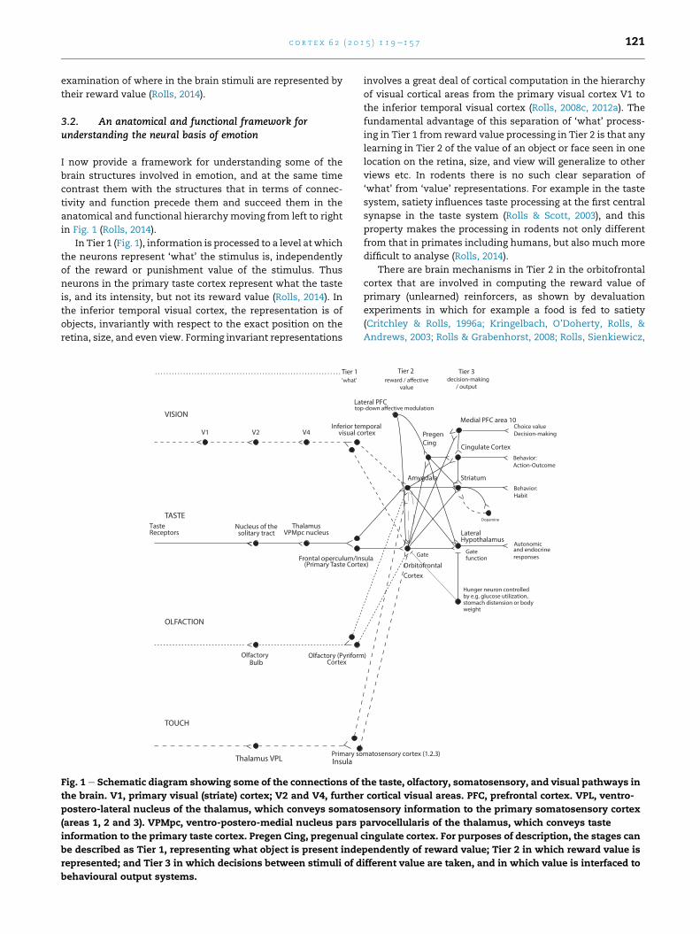
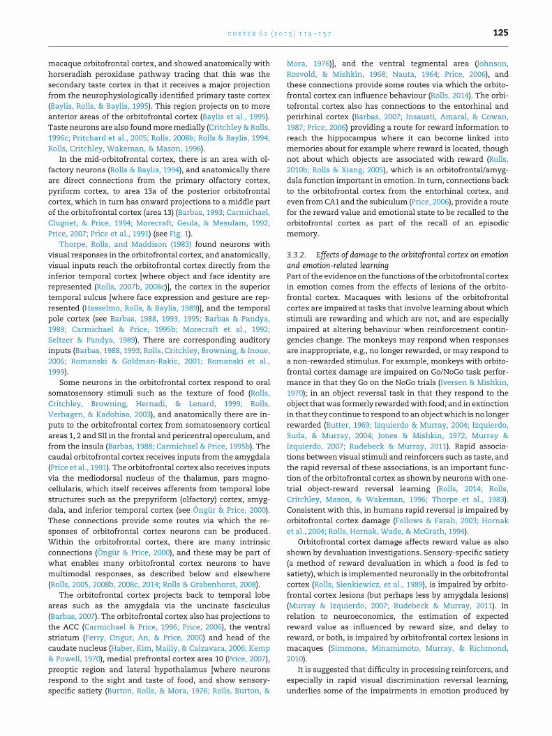
in Fig. 1 (Rolls, 2014).
In Tier 1 (Fig. 1), information is processed to a level at which
the neurons represent ‘what’ the stimulus is, independently
of the reward or punishment value of the stimulus. Thus
neurons in the primary taste cortex represent what the taste
is, and its intensity, but not its reward value (Rolls, 2014). In
the inferior temporal visual cortex, the representation is of
objects, invariantly with respect to the exact position on the
retina, size, and even view. Forming invariant representations
V1 V2 V4
ThalamusReceptors solitary tract VPMpc nucleus
VISION
TasteTASTE
Bulb
Frontal operculum/In
visual coInferior te
(Primary Taste Corte
Nucleus of the
TOUCH
OLFACTION
Thalamus VPL
Olfactory
Primary so
Olfactory (PyriformCortex
Insula
'what'. . . . . . . . . . . . . . . . . . . . . . . . . . . . . . . . . . . . . . . . . . . . . . . . . . . . . . . . . . . . . . . . . . Tier 1
La
Fig. 1 e Schematic diagram showing some of the connections of
the brain. V1, primary visual (striate) cortex; V2 and V4, further
postero-lateral nucleus of the thalamus, which conveys somato
(areas 1, 2 and 3). VPMpc, ventro-postero-medial nucleus pars p
information to the primary taste cortex. Pregen Cing, pregenual
be described as Tier 1, representing what object is present inde
represented; and Tier 3 in which decisions between stimuli of d
behavioural output systems.
involves a great deal of cortical computation in the hierarchy
of visual cortical areas from the primary visual cortex V1 to
the inferior temporal visual cortex (Rolls, 2008c, 2012a). The
fundamental advantage of this separation of ‘what’ process-
ing in Tier 1 from reward value processing in Tier 2 is that any
learning in Tier 2 of the value of an object or face seen in one
location on the retina, size, and view will generalize to other
views etc. In rodents there is no such clear separation of
‘what’ from ‘value’ representations. For example in the taste
system, satiety influences taste processing at the first central
synapse in the taste system (Rolls & Scott, 2003), and this
property makes the processing in rodents not only different
from that in primates including humans, but also much more
difficult to analyse (Rolls, 2014).
There are brain mechanisms in Tier 2 in the orbitofrontal
cortex that are involved in computing the reward value of
primary (unlearned) reinforcers, as shown by devaluation
experiments in which for example a food is fed to satiety
(Critchley & Rolls, 1996a; Kringelbach, O’Doherty, Rolls, &
Andrews, 2003; Rolls & Grabenhorst, 2008; Rolls, Sienkiewicz,
Behavior:Habit
Autonomicand endocrineresponses
Cingulate CortexBehavior:Action-Outcome
sula
rtexmporal
x)
Amygdala
Gate
Lateral
function
by e.g. glucose utilization,stomach distension or bodyweight
Gate
OrbitofrontalCortex
Hypothalamus
Hunger neuron controlled
matosensory cortex (1.2.3)
)
Striatum
PregenCing
Medial PFC area 10Choice valueDecision-making
decision-making / output
Tier 2 Tier 3
teral PFC
the taste, olfactory, somatosensory, and visual pathways in
cortical visual areas. PFC, prefrontal cortex. VPL, ventro-
sensory information to the primary somatosensory cortex
arvocellularis of the thalamus, which conveys taste
cingulate cortex. For purposes of description, the stages can
pendently of reward value; Tier 2 in which reward value is
ifferent value are taken, and in which value is interfaced to
c o r t e x 6 2 ( 2 0 1 5 ) 1 1 9e1 5 7122
& Yaxley, 1989), and by neuroeconomics experiments which
show that the amount and quality of each commodity is
encoded by orbitofrontal cortex neurons (Grabenhorst & Rolls,
2011; Padoa-Schioppa, 2011; Padoa-Schioppa & Assad, 2008).
The primary reinforcers include taste, touch (both pleasant
touch and pain), and to some extent smell, and perhaps
certain visual stimuli such as face expression. There is evi-
dence that there is a representation of the (reward/punish-
ment) value of many primary reinforcers in the orbitofrontal
cortex, including taste, positive touch and pain, face expres-
sion, face beauty, and auditory consonance/dissonance. In
neuroeconomics, these are termed ‘outcome value’ repre-
sentations (Rolls, 2014). Further evidence for value represen-
tations is that orbitofrontal cortex activations in humans to
these stimuli are linearly related to the subjectively reported
pleasantness of stimuli (medially), or to their unpleasantness
(laterally) (Rolls, 2014).
Brain regions in Tier 2 are also concerned with learning
associations between previously neutral stimuli, such as the
sight of objects or of individuals’ faces, with primary re-
inforcers. These brain regions include the amygdala and
orbitofrontal cortex, with the orbitofrontal cortex being
especially important in the rapid, one-trial, learning and
reversal of stimulus-reinforcer associations. In neuro-
economics, these are termed ‘expected value’ representa-
tions. Once the Tier 2 brain regions have determined whether
the input is reinforcing, whether primary or secondary, the
signal is passed directly to output regions of the brain, with no
need to produce and then feed back peripheral body or auto-
nomic responses to the brain.
In the orbitofrontal cortex in Tier 2, the representation is of
the value of stimuli, and actions are not represented. The
values of very many different types of stimuli, events or goals
are represented separately at the neuronal level, providing the
basis for choice between stimuli, and the selection at later
stages of processing of an appropriate action to obtain the
chosen goal.
Whereas the orbitofrontal cortex in Tier 2 represents the
value of stimuli (potential goals for action) on a continuous
scale, an area anterior to this, medial prefrontal cortex area 10
(in Tier 3), is implicated in decision-making between stimuli,
in which a selection or choicemust bemade,moving beyond a
representation of value on a continuous scale towards a de-
cision between goods based on their value (Grabenhorst, Rolls,
& Margot, 2011; Rolls, 2014; Rolls, Grabenhorst, & Parris, 2008).
The brain regions in which the reinforcing, and hence
emotional, value of stimuli are represented interface to three
main types of output system:
The first is the autonomic and endocrine system, for pro-
ducing such changes as increased heart rate and release of
adrenaline, which prepare the body for action. Structures
receiving from the orbitofrontal cortex, amygdala, and ACC
that provide a route for these autonomic effects include the
hypothalamus and parts of the anterior insula close to the
insular taste cortex (Critchley &Harrison, 2013; Rolls, 2014).
The second type of output is to brain systems
concerned with performing actions unconsciously or
implicitly, in order to obtain rewards or avoid punishers.
These brain systems include the basal ganglia for habit
(‘stimuluseresponse’) behaviour, and the ACC for
actioneoutcome learning (The ‘outcome’ is the reward or
punisher that is or is not obtained when the action is per-
formed.). The ACC contains representations of reward and
punisher value, and thus of outcome, which are essential
for learning associations between actions and the out-
comes that follow actions. The midcingulate area contains
representations of actions.
The third type of output is to a system capable of planning
many steps ahead, and for example deferring short-term
rewards in order to execute a long-term plan. This sys-
temmay use syntactic processing to perform the planning,
and is therefore part of a linguistic systemwhich performs
explicit (conscious) processing, as described more fully
elsewhere (Rolls, 2014).
It is notable that the orbitofrontal cortex and amygdala do
not receive inputs from the dorsal visual ‘where’ processing
areas such as the parietal cortex including the retrosplenial
cortex (which is part of the posterior cingulate cortex) that
provide inputs via parahippocampal areas TF/TH to the hip-
pocampus for its spatial (‘where’) functions inmemory, which
are described in Section 4. In a complementary way, the hip-
pocampus and parahippocampal areas do not contain value
representations of stimuli, except insofar as valuemay be part
of amemory such as reward-placememory (Rolls, 2010b; Rolls
& Xiang, 2005, 2006). This is part of the evidence that the
emotional and episodic memory systems have different con-
nections and functions, as described in Section 4 and else-
where (Rolls, 2008c, 2010b; Rolls & Xiang, 2005, 2006), and thus
that there is no single and unified limbic system.
Because of the intended relevance to understanding
human emotion and its disorders, the focus of the research
described here is on humans and macaques. This is impor-
tant, for many of the brain systems that are involved in
emotion have undergone considerable development in pri-
mates (e.g., monkeys and humans) (Rolls, 2014), as summa-
rized next.
First, the temporal lobe has undergone great development
in primates, and several systems in the temporal lobe are
either involved in emotion (e.g., the amygdala), or provide
some of the main sensory inputs to brain systems involved in
emotion and motivation. For example, the amygdala and the
orbitofrontal cortex, key brain structures in emotion, both
receive inputs from the highly developed primate temporal
lobe cortical areas, including those involved in invariant vi-
sual object recognition, and face identity and expression
processing (Rolls, 2000a, 2008c, 2011a, 2012a).
Second, there are many topological, cytoarchitectural, and
probably connectional similarities between macaques and
humans with respect to the orbitofrontal cortex (see Fig. 1 and
Carmichael & Price, 1994; Kringelbach & Rolls, 2004; Ongur &
Price, 2000; Petrides & Pandya, 1995; Price, 2006, 2007).
Third, the prefrontal cortex has also undergone great
development in primates, and one part of it, the orbitofrontal
cortex, is very little developed in rodents, yet is one of the
major brain areas involved in emotion and motivation in
primates including humans. Indeed, it has been argued that
the granular prefrontal cortex is a primate innovation, and the
implication of the argument is that any areas that might be
c o r t e x 6 2 ( 2 0 1 5 ) 1 1 9e1 5 7 123
termed orbitofrontal cortex in rats (Schoenbaum, Roesch,
Stalnaker, & Takahashi, 2009) are homologous only to the
agranular parts of the primate orbitofrontal cortex (shaded
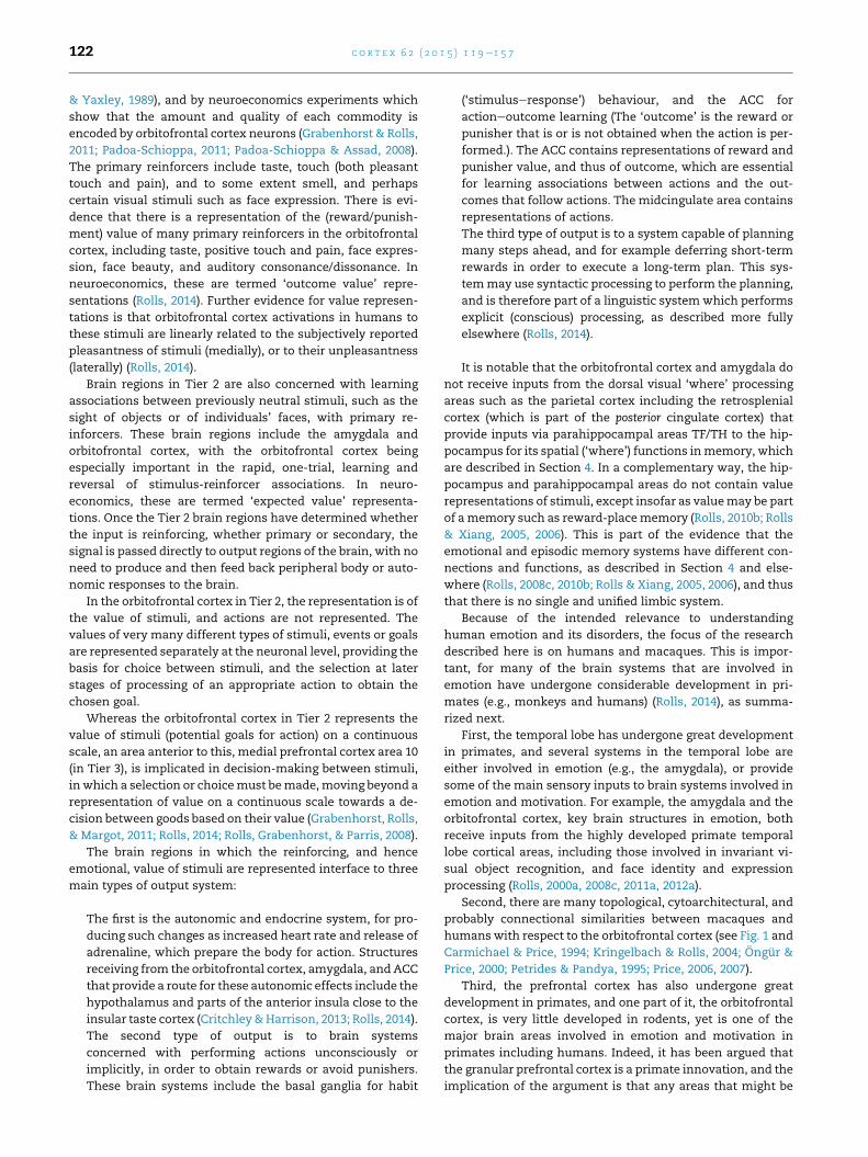
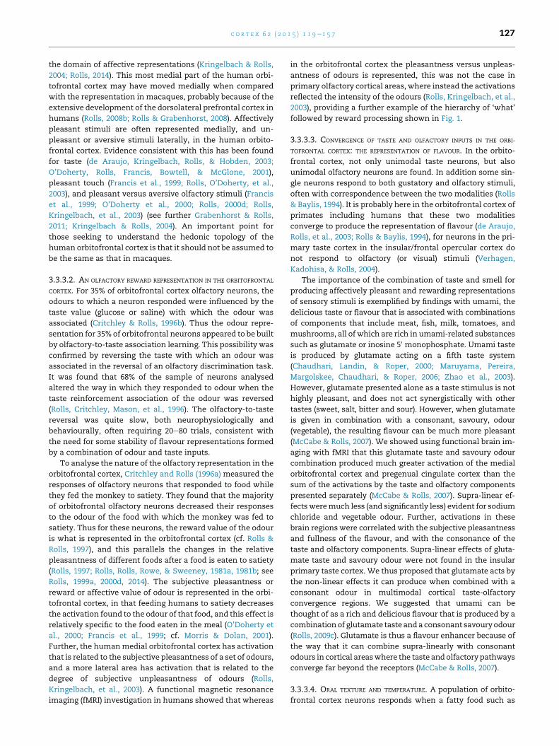
mid grey in Fig. 2), that is to areas 13a, 14c, and the agranular
insular areas labelled Ia in Fig. 2 (Passingham & Wise, 2012;
Wise, 2008). It follows from that argument that for most
areas of the orbitofrontal and medial prefrontal cortex in
humans and macaques (those shaded light grey in Fig. 2),
special consideration must be given to research in macaques
and humans. As shown in Fig. 2, there may be no cortical area
9
10p10r
10m
11m 14r14c
32/PL
24
32
8
6
BA acaMnamuH
24/AC
14
10m
agranular cortegranular cortex
allocortex
Iapm
10p
12r
45
12m
Iai
Pir13l
13m 13a
13b
11m 14r 14c
Iam
12s
11l
1
10o1
cc
25/IL
AON
Fig. 2 e Comparison of the orbitofrontal (below) and medial pref
and rats. (A) Medial (top) and orbital (bottom) areas of the human
(bottom) areas of the macaque frontal cortex (Carmichael & Pric
frontal cortex (Palomero-Gallagher & Zilles, 2004). Rostral is to th
Bottom row: in (A) and (B), lateral is up; in (C), dorsal is up. Not t
corpus callosum; Fr2, second frontal area; Ia, agranular insular
lateral orbital cortex; MO, medial orbital cortex: OB, olfactory bu
tenia tecta; VO, ventral orbital cortex; Subdivisions of areas are
(o), posterior or polar (p), rostral (r), or by arbitrary designation (a
Ongur, Amon T. Ferry, & Joseph L. Price. Architectonic subdivisio
of Comparative Neurology, 460(3), 425e49 Copyright 2003 Wiley-
Architectonic subdivision of the orbital and medial prefrontal c
Neurology, 346(3), 366e402 Copyright 1994 Wiley-Liss, Inc. (c) Ad
G. Paxinos (Ed.) The rat nervous system (3rd ed., pp. 729e57) cop
in rodents that is homologous to most of the primate
including human orbitofrontal cortex (Passingham & Wise,
2012; Preuss, 1995; Wise, 2008).
Fourth, even the taste system (which might have been
supposed to be phylogenetically old and preserved) of pri-
mates and rodents may be different, with obligatory pro-
cessing from the nucleus of the solitary tract via the thalamus
to the cortex in primates, but a subcortical pathway in rodents
via a pontine taste area to the amygdala, and differences in
where satiety influences taste-responsive neurons in
C taRyeknom euq
IG24a
24b/AC24c/AC
9
32/PL
25/IL
14cr
24a/ACcc
tt
ig
x
12l
12o
2r
1m
11l
12m13l
13m
13b 13a
Iam
IapmPir
AON
IaiIalIapl
G
14r 14c
Fr2 M1
Iad LO
Iav Iap
Par
Fr2
AC1
PL
IL
MO
VO
AC2 ccOB
tt
ig
Pir
AON
rontal (above) cortical areas in humans, macaque monkeys,
frontal codex (Ongur et al., 2003). (B) Medial (top) and orbital
e, 1994). (C) Medial (top) and lateral (bottom) areas of rat
e left in all drawings. Top row: dorsal is up in all drawings.
o scale. Abbreviations: AON, anterior olfactory ‘nucleus’; cc,
cortex; ig, indusium griseum; IL, infralimbic cortex; LO,
lb; Pr, piriform (olfactory) cortex; PL, prelimbic cortex; tt,
labelled caudal (c); inferior (i), lateral (l), medial (m); orbital
, b). (After Passingham & Wise, 2012). (a) Adapted from Dost
n of the human orbital andmedial prefrontal cortex. Journal
Liss, Inc. (b) Adapted from Carmichael, S. T., & Price, J. L.
ortex in the macaque monkey. Journal of Comparative
apted from Palomero-Gallagher, N. & Zilles, K., Isocortex, In
yright 2004, Elsevier Academic Press.
c o r t e x 6 2 ( 2 0 1 5 ) 1 1 9e1 5 7124
primates and rodents (Norgren, 1984; Rolls, 2014; Rolls & Scott,
2003; Small & Scott, 2009).
Fifth, with the great development of the orbitofrontal cor-
tex in primates, the amygdala may become relatively less
important in humans in emotion than in other vertebrates
(Rolls, 2014) (see Section 3.4).
To understand the functions of the orbitofrontal cortex and
connected areas in humans, the majority of the studies
described here were therefore performed with macaques or
with humans.
3.3. The orbitofrontal cortex
The first structure considered is the orbitofrontal cortex, and
although not a limbic structure such as the amygdala, has
similar connections to the amygdala, is connected to the
amygdala, and has considerably eclipsed in primates
including humans the functions of the amygdala (Rolls, 2014).
The amygdala has evolutionarily old origins and can be
ACC
10 14
11
12
13
as
fros
ps
rh
46
TG
Ventral view oforbitofrontal cortex
TGlos
mos
osolfactory
visualTE20taste
auditory
oryamostosens
51
ros
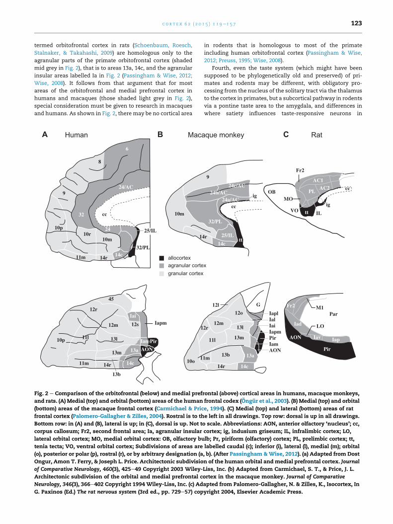
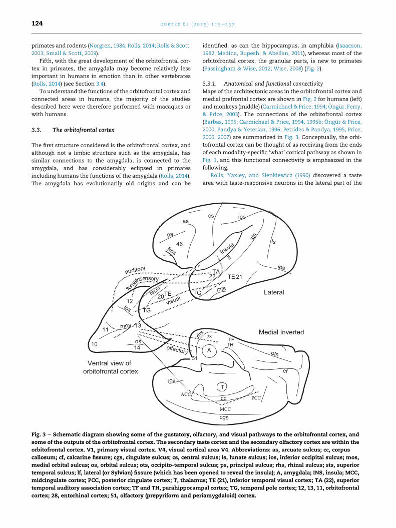
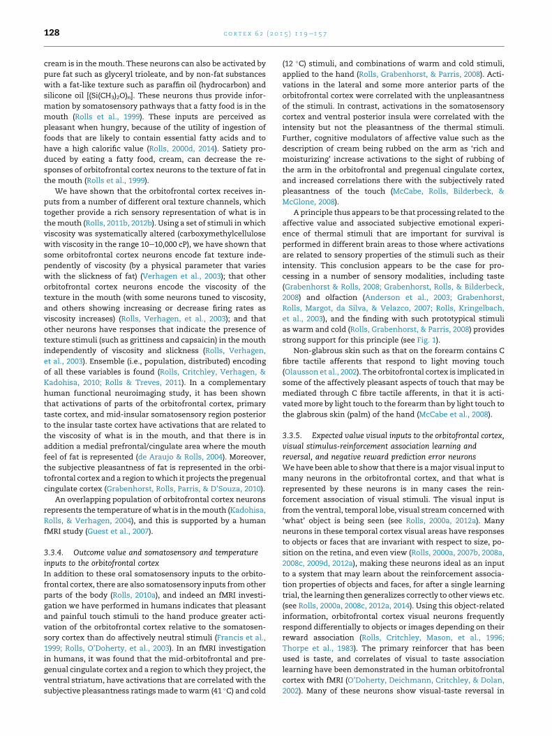
Fig. 3 e Schematic diagram showing some of the gustatory, olfa
some of the outputs of the orbitofrontal cortex. The secondary ta
orbitofrontal cortex. V1, primary visual cortex. V4, visual cortic
callosum; cf, calcarine fissure; cgs, cingulate sulcus; cs, central
medial orbital sulcus; os, orbital sulcus; ots, occipito-temporal s
temporal sulcus; lf, lateral (or Sylvian) fissure (which has been o
midcingulate cortex; PCC, posterior cingulate cortex; T, thalamu
temporal auditory association cortex; TF and TH, parahippocam
cortex; 28, entorhinal cortex; 51, olfactory (prepyriform and per
identified, as can the hippocampus, in amphibia (Isaacson,
1982; Medina, Bupesh, & Abellan, 2011), whereas most of the
orbitofrontal cortex, the granular parts, is new to primates
(Passingham & Wise, 2012; Wise, 2008) (Fig. 2).
3.3.1. Anatomical and functional connectivityMaps of the architectonic areas in the orbitofrontal cortex and
medial prefrontal cortex are shown in Fig. 2 for humans (left)
andmonkeys (middle) (Carmichael & Price, 1994; Ongur, Ferry,
& Price, 2003). The connections of the orbitofrontal cortex
(Barbas, 1995; Carmichael & Price, 1994, 1995b; Ongur & Price,
2000; Pandya & Yeterian, 1996; Petrides & Pandya, 1995; Price,
2006, 2007) are summarized in Fig. 3. Conceptually, the orbi-
tofrontal cortex can be thought of as receiving from the ends
of each modality-specific ‘what’ cortical pathway as shown in
Fig. 1, and this functional connectivity is emphasized in the
following.
Rolls, Yaxley, and Sienkiewicz (1990) discovered a taste
area with taste-responsive neurons in the lateral part of the
28
MCC
PCCcc
sts
ios
mts
cgs
ots
cf
s
cs ips
TA
lf
TE 21
ls
Medial Inverted
Lateral
A
T
TFTH
22
Insula
ctory, and visual pathways to the orbitofrontal cortex, and
ste cortex and the secondary olfactory cortex are within the
al area V4. Abbreviations: as, arcuate sulcus; cc, corpus
sulcus; ls, lunate sulcus; ios, inferior occipital sulcus; mos,
ulcus; ps, principal sulcus; rhs, rhinal sulcus; sts, superior
pened to reveal the insula); A, amygdala; INS, insula; MCC,
s; TE (21), inferior temporal visual cortex; TA (22), superior
pal cortex; TG, temporal pole cortex; 12, 13, 11, orbitofrontal
iamygdaloid) cortex.
c o r t e x 6 2 ( 2 0 1 5 ) 1 1 9e1 5 7 125
macaque orbitofrontal cortex, and showed anatomically with
horseradish peroxidase pathway tracing that this was the
secondary taste cortex in that it receives a major projection
from the neurophysiologically identified primary taste cortex
(Baylis, Rolls, & Baylis, 1995). This region projects on to more
anterior areas of the orbitofrontal cortex (Baylis et al., 1995).
Taste neurons are also foundmoremedially (Critchley & Rolls,
1996c; Pritchard et al., 2005; Rolls, 2008b; Rolls & Baylis, 1994;
Rolls, Critchley, Wakeman, & Mason, 1996).
In the mid-orbitofrontal cortex, there is an area with ol-
factory neurons (Rolls & Baylis, 1994), and anatomically there
are direct connections from the primary olfactory cortex,
pyriform cortex, to area 13a of the posterior orbitofrontal
cortex, which in turn has onward projections to a middle part
of the orbitofrontal cortex (area 13) (Barbas, 1993; Carmichael,
Clugnet, & Price, 1994; Morecraft, Geula, & Mesulam, 1992;
Price, 2007; Price et al., 1991) (see Fig. 1).
Thorpe, Rolls, and Maddison (1983) found neurons with
visual responses in the orbitofrontal cortex, and anatomically,
visual inputs reach the orbitofrontal cortex directly from the
inferior temporal cortex [where object and face identity are
represented (Rolls, 2007b, 2008c)], the cortex in the superior
temporal sulcus [where face expression and gesture are rep-
resented (Hasselmo, Rolls, & Baylis, 1989)], and the temporal
pole cortex (see Barbas, 1988, 1993, 1995; Barbas & Pandya,
1989; Carmichael & Price, 1995b; Morecraft et al., 1992;
Seltzer & Pandya, 1989). There are corresponding auditory
inputs (Barbas, 1988, 1993; Rolls, Critchley, Browning, & Inoue,
2006; Romanski & Goldman-Rakic, 2001; Romanski et al.,
1999).
Some neurons in the orbitofrontal cortex respond to oral
somatosensory stimuli such as the texture of food (Rolls,
Critchley, Browning, Hernadi, & Lenard, 1999; Rolls,
Verhagen, & Kadohisa, 2003), and anatomically there are in-
puts to the orbitofrontal cortex from somatosensory cortical
areas 1, 2 and SII in the frontal and pericentral operculum, and
from the insula (Barbas, 1988; Carmichael & Price, 1995b). The
caudal orbitofrontal cortex receives inputs from the amygdala
(Price et al., 1991). The orbitofrontal cortex also receives inputs
via the mediodorsal nucleus of the thalamus, pars magno-
cellularis, which itself receives afferents from temporal lobe
structures such as the prepyriform (olfactory) cortex, amyg-
dala, and inferior temporal cortex (see Ongur & Price, 2000).
These connections provide some routes via which the re-
sponses of orbitofrontal cortex neurons can be produced.
Within the orbitofrontal cortex, there are many intrinsic
connections (Ongur & Price, 2000), and these may be part of
what enables many orbitofrontal cortex neurons to have
multimodal responses, as described below and elsewhere
(Rolls, 2005, 2008b, 2008c, 2014; Rolls & Grabenhorst, 2008).
The orbitofrontal cortex projects back to temporal lobe
areas such as the amygdala via the uncinate fasciculus
(Barbas, 2007). The orbitofrontal cortex also has projections to
the ACC (Carmichael & Price, 1996; Price, 2006), the ventral
striatum (Ferry, Ongur, An, & Price, 2000) and head of the
caudate nucleus (Haber, Kim, Mailly, & Calzavara, 2006; Kemp
& Powell, 1970), medial prefrontal cortex area 10 (Price, 2007),
preoptic region and lateral hypothalamus [where neurons
respond to the sight and taste of food, and show sensory-
specific satiety (Burton, Rolls, & Mora, 1976; Rolls, Burton, &
Mora, 1976)], and the ventral tegmental area (Johnson,
Rosvold, & Mishkin, 1968; Nauta, 1964; Price, 2006), and
these connections provide some routes via which the orbito-
frontal cortex can influence behaviour (Rolls, 2014). The orbi-
tofrontal cortex also has connections to the entorhinal and
perirhinal cortex (Barbas, 2007; Insausti, Amaral, & Cowan,
1987; Price, 2006) providing a route for reward information to
reach the hippocampus where it can become linked into
memories about for example where reward is located, though
not about which objects are associated with reward (Rolls,
2010b; Rolls & Xiang, 2005), which is an orbitofrontal/amyg-
dala function important in emotion. In turn, connections back
to the orbitofrontal cortex from the entorhinal cortex, and
even fromCA1 and the subiculum (Price, 2006), provide a route
for the reward value and emotional state to be recalled to the
orbitofrontal cortex as part of the recall of an episodic
memory.
3.3.2. Effects of damage to the orbitofrontal cortex on emotionand emotion-related learningPart of the evidenceon the functionsof the orbitofrontal cortex
in emotion comes from the effects of lesions of the orbito-
frontal cortex. Macaques with lesions of the orbitofrontal
cortex are impaired at tasks that involve learning about which
stimuli are rewarding and which are not, and are especially
impaired at altering behaviour when reinforcement contin-
gencies change. The monkeys may respond when responses
are inappropriate, e.g., no longer rewarded, or may respond to
a non-rewarded stimulus. For example, monkeys with orbito-
frontal cortex damage are impaired on Go/NoGo task perfor-
mance in that they Go on the NoGo trials (Iversen & Mishkin,
1970); in an object reversal task in that they respond to the
object thatwas formerly rewardedwith food; and in extinction
in that they continue to respond to anobjectwhich is no longer
rewarded (Butter, 1969; Izquierdo & Murray, 2004; Izquierdo,
Suda, & Murray, 2004; Jones & Mishkin, 1972; Murray &
Izquierdo, 2007; Rudebeck & Murray, 2011). Rapid associa-
tions between visual stimuli and reinforcers such as taste, and
the rapid reversal of these associations, is an important func-
tion of the orbitofrontal cortex as shown by neuronswith one-
trial object-reward reversal learning (Rolls, 2014; Rolls,
Critchley, Mason, & Wakeman, 1996; Thorpe et al., 1983).
Consistent with this, in humans rapid reversal is impaired by
orbitofrontal cortex damage (Fellows & Farah, 2003; Hornak
et al., 2004; Rolls, Hornak, Wade, & McGrath, 1994).
Orbitofrontal cortex damage affects reward value as also
shown by devaluation investigations. Sensory-specific satiety
(a method of reward devaluation in which a food is fed to
satiety), which is implemented neuronally in the orbitofrontal
cortex (Rolls, Sienkiewicz, et al., 1989), is impaired by orbito-
frontal cortex lesions (but perhaps less by amygdala lesions)
(Murray & Izquierdo, 2007; Rudebeck & Murray, 2011). In
relation to neuroeconomics, the estimation of expected
reward value as influenced by reward size, and delay to
reward, or both, is impaired by orbitofrontal cortex lesions in
macaques (Simmons, Minamimoto, Murray, & Richmond,
2010).
It is suggested that difficulty in processing reinforcers, and
especially in rapid visual discrimination reversal learning,
underlies some of the impairments in emotion produced by

c o r t e x 6 2 ( 2 0 1 5 ) 1 1 9e1 5 7126
damage to the orbitofrontal cortex (Rolls, 2014). In humans,
euphoria, irresponsibility, lack of affect, and impulsiveness
can follow frontal lobe damage (Damasio, 1994; Kolb &
Whishaw, 2003; Rolls, 1999a; Zald & Rauch, 2006), particu-
larly orbitofrontal cortex damage (Berlin, Rolls, & Iversen,
2005; Berlin, Rolls, & Kischka, 2004; Hornak et al., 2003;
Hornak, Rolls, & Wade, 1996; Rolls, 1999a, 2014; Rolls et al.,
1994). These emotional changes may be related at least in
part to a failure to rapidly update the reinforcement associa-
tions of stimuli when the contingencies are changed as in a
visual discrimination reversal task (Berlin et al., 2004; Fellows,
2007, 2011; Fellows & Farah, 2003; Hornak et al., 2004; Rolls,
1999b, 2014; Rolls et al., 1994). Similar mechanisms may
contribute at least in part to the poor performance of humans
with ventromedial prefrontal cortex damage on the Iowa
Gambling Task (Bechara, Damasio, & Damasio, 2000; Maia &
McClelland, 2004). It is of interest that the patients with
bilateral orbitofrontal cortex damage who were impaired at
the visual discrimination reversal task had high scores on
parts of a Social Behaviour Questionnaire in which the pa-
tients were rated on behaviours such as emotion recognition
in others (e.g., their sad, angry, or disgusted mood); in inter-
personal relationships (such as not caring what others think,
and not being close to the family); emotional empathy (e.g.,
when others are happy, is not happy for them); interpersonal
relationships (e.g., does not care what others think, and is not
close to his family); public behaviour (is uncooperative);
antisocial behaviour (is critical of and impatient with others);
impulsivity (does things without thinking); and sociability (is
not sociable, and has difficulty making or maintaining close
relationships) (Hornak et al., 2003, 2004), all of which could
reflect less behavioural sensitivity to different types of pun-
ishment and reward. Further, in a Subjective Emotional
Change Questionnaire in which the patients reported on any
changes in the intensity and/or frequency of their own expe-
rience of emotions, the bilateral orbitofrontal cortex lesion
patientswith deficits in the visual discrimination reversal task
reported a number of changes, including changes in sadness,
anger, fear and happiness (Hornak et al., 2003).
3.3.3. Reward outcome value for taste, olfaction, flavour, oraltexture, and oral temperature in the orbitofrontal cortex3.3.3.1. TASTE AND ORAL TEXTURE. One of the discoveries that
havehelpedus tounderstand the functionsof theorbitofrontal
cortex in behaviour is that it contains a major cortical repre-
sentation of taste (see Kadohisa, Rolls, & Verhagen, 2005a;
Rolls, 1995a, 1997, 2014; Rolls & Scott, 2003; Rolls et al., 1990)
(cf. Fig. 1). Given that taste can act as a primary reinforcer, that
is without learning as a reward or punisher, we now have the
start for a fundamental understanding of the function of the
orbitofrontal cortex in stimulus-reinforcer association
learning (Rolls, 1999a, 2004, 2008c, 2014). We know how one
class of primary reinforcers reaches and is represented in the
orbitofrontal cortex. A representation of primary reinforcers is
essential for a system that is involved in learning associations
between previously neutral stimuli and primary reinforcers,
e.g., between the sight of an object and its taste.
The representation in the orbitofrontal cortex (shown by
analysing the responses of single neurons in macaques) is for
the majority of neurons the reward value of taste (Baylis &
Rolls, 1991; Kadohisa et al., 2005a; Rolls, 1995a, 1997, 2000c;
Rolls, Critchley, Browning, & Hernadi, 1998; Rolls, Critchley,
Wakeman, et al., 1996; Rolls & Scott, 2003; Rolls et al., 1990)
and oral texture including viscosity (Rolls, Verhagen, et al.,
2003), fat texture (Rolls et al., 1999; Verhagen, Rolls, &
Kadohisa, 2003), and astringency as exemplified by tannic
acid (Critchley & Rolls, 1996c). The evidence for this is that the
responses of orbitofrontal cortex taste neurons aremodulated
by hunger (as is the reward value or palatability of a taste). In
particular, it has been shown that orbitofrontal cortex taste
neurons gradually stop responding to the taste of a food as the
monkey is fed to satiety, but not to the taste of other foods,
revealing a mechanism for sensory-specific satiety and
reward devaluation (Rolls, Critchley, Wakeman, et al., 1996;
Rolls, Sienkiewicz, et al., 1989). In contrast, the representa-
tion of taste in the primary taste cortex (Scott, Yaxley,
Sienkiewicz, & Rolls, 1986; Yaxley, Rolls, & Sienkiewicz,
1990) is not modulated by hunger (Rolls, Scott, Sienkiewicz,
& Yaxley, 1988; Yaxley, Rolls, & Sienkiewicz, 1988). Thus in
the primate including human primary taste cortex, the reward
value of taste is not represented, and instead the identity and
intensity of the taste are represented (Grabenhorst & Rolls,
2008; Grabenhorst, Rolls, & Bilderbeck, 2008; Rolls, 2008c,
2014).
Additional evidence that the reward value of food is rep-
resented in the orbitofrontal cortex is that monkeys work for
electrical stimulation of this brain region if they are hungry,
but not if they are satiated (Mora, Avrith, Phillips, & Rolls,
1979; Rolls, 2005). Further, neurons in the orbitofrontal cor-
tex are activated from many brain-stimulation reward sites
(Mora, Avrith, & Rolls, 1980; Rolls, Burton, & Mora, 1980). Thus
there is clear evidence that it is the reward value of taste that
is represented in the orbitofrontal cortex (see further Rolls,
1999a, 2000d, 2014), and this is further supported by the
finding that feeding to satiety decreases the activation of the
human orbitofrontal cortex to the food eaten to satiety in a
sensory-specific way (Kringelbach et al., 2003). Some orbito-
frontal cortex neurons respond to the ‘taste’ of water in the
mouth (Rolls et al., 1990), and their responses occur only when
thirsty and not when satiated (Rolls, Sienkiewicz, et al., 1989);
and correspondingly in humans the subjective pleasantness
or affective value of the taste of water in the mouth is repre-
sented in the orbitofrontal cortex (de Araujo, Kringelbach,
Rolls, & McGlone, 2003). This is part of the evidence for the
separation of a ‘what’ tier of processing, which in this case is
the primary taste cortex, from a reward and affect-related
representation in the orbitofrontal cortex tier of processing,
as shown in Fig. 1.
Functional neuroimaging studies in humans have shown
that themost medial part of the human orbitofrontal cortex is
activated by taste, oral texture, and olfactory stimuli (de
Araujo, Kringelbach, Rolls, & Hobden, 2003; de Araujo &
Rolls, 2004; de Araujo, Rolls, Kringelbach, McGlone, &
Phillips, 2003; de Araujo, Rolls, Velazco, Margot, & Cayeux,
2005; Francis et al., 1999; Gottfried, Small, & Zald, 2006;
McCabe & Rolls, 2007; O’Doherty et al., 2000; Rolls,
Kringelbach, & de Araujo, 2003; Rolls & McCabe, 2007; Small,
Gerber, Mak, & Hummel, 2005; Small, Zatorre, Dagher,
Evans, & Jones-Gotman, 2001), and that the activations
correlate with ratings of subjective pleasantness and so are in
c o r t e x 6 2 ( 2 0 1 5 ) 1 1 9e1 5 7 127
the domain of affective representations (Kringelbach & Rolls,
2004; Rolls, 2014). This most medial part of the human orbi-
tofrontal cortex may have moved medially when compared
with the representation in macaques, probably because of the
extensive development of the dorsolateral prefrontal cortex in
humans (Rolls, 2008b; Rolls & Grabenhorst, 2008). Affectively
pleasant stimuli are often represented medially, and un-
pleasant or aversive stimuli laterally, in the human orbito-
frontal cortex. Evidence consistent with this has been found
for taste (de Araujo, Kringelbach, Rolls, & Hobden, 2003;
O’Doherty, Rolls, Francis, Bowtell, & McGlone, 2001),
pleasant touch (Francis et al., 1999; Rolls, O’Doherty, et al.,
2003), and pleasant versus aversive olfactory stimuli (Francis
et al., 1999; O’Doherty et al., 2000; Rolls, 2000d; Rolls,
Kringelbach, et al., 2003) (see further Grabenhorst & Rolls,
2011; Kringelbach & Rolls, 2004). An important point for
those seeking to understand the hedonic topology of the
human orbitofrontal cortex is that it should not be assumed to
be the same as that in macaques.
3.3.3.2. AN OLFACTORY REWARD REPRESENTATION IN THE ORBITOFRONTAL
CORTEX. For 35% of orbitofrontal cortex olfactory neurons, the
odours to which a neuron responded were influenced by the
taste value (glucose or saline) with which the odour was
associated (Critchley & Rolls, 1996b). Thus the odour repre-
sentation for 35% of orbitofrontal neurons appeared to be built
by olfactory-to-taste association learning. This possibility was
confirmed by reversing the taste with which an odour was
associated in the reversal of an olfactory discrimination task.
It was found that 68% of the sample of neurons analysed
altered the way in which they responded to odour when the
taste reinforcement association of the odour was reversed
(Rolls, Critchley, Mason, et al., 1996). The olfactory-to-taste
reversal was quite slow, both neurophysiologically and
behaviourally, often requiring 20e80 trials, consistent with
the need for some stability of flavour representations formed
by a combination of odour and taste inputs.
To analyse the nature of the olfactory representation in the
orbitofrontal cortex, Critchley and Rolls (1996a) measured the
responses of olfactory neurons that responded to food while
they fed the monkey to satiety. They found that the majority
of orbitofrontal olfactory neurons decreased their responses
to the odour of the food with which the monkey was fed to
satiety. Thus for these neurons, the reward value of the odour
is what is represented in the orbitofrontal cortex (cf. Rolls &
Rolls, 1997), and this parallels the changes in the relative
pleasantness of different foods after a food is eaten to satiety
(Rolls, 1997; Rolls, Rolls, Rowe, & Sweeney, 1981a, 1981b; see
Rolls, 1999a, 2000d, 2014). The subjective pleasantness or
reward or affective value of odour is represented in the orbi-
tofrontal cortex, in that feeding humans to satiety decreases
the activation found to the odour of that food, and this effect is
relatively specific to the food eaten in the meal (O’Doherty et
al., 2000; Francis et al., 1999; cf. Morris & Dolan, 2001).
Further, the humanmedial orbitofrontal cortex has activation
that is related to the subjective pleasantness of a set of odours,
and a more lateral area has activation that is related to the
degree of subjective unpleasantness of odours (Rolls,
Kringelbach, et al., 2003). A functional magnetic resonance
imaging (fMRI) investigation in humans showed that whereas
in the orbitofrontal cortex the pleasantness versus unpleas-
antness of odours is represented, this was not the case in
primary olfactory cortical areas, where instead the activations
reflected the intensity of the odours (Rolls, Kringelbach, et al.,
2003), providing a further example of the hierarchy of ‘what’
followed by reward processing shown in Fig. 1.
3.3.3.3. CONVERGENCE OF TASTE AND OLFACTORY INPUTS IN THE ORBI-
TOFRONTAL CORTEX: THE REPRESENTATION OF FLAVOUR. In the orbito-
frontal cortex, not only unimodal taste neurons, but also
unimodal olfactory neurons are found. In addition some sin-
gle neurons respond to both gustatory and olfactory stimuli,
often with correspondence between the two modalities (Rolls
& Baylis, 1994). It is probably here in the orbitofrontal cortex of
primates including humans that these two modalities
converge to produce the representation of flavour (de Araujo,
Rolls, et al., 2003; Rolls & Baylis, 1994), for neurons in the pri-
mary taste cortex in the insular/frontal opercular cortex do
not respond to olfactory (or visual) stimuli (Verhagen,
Kadohisa, & Rolls, 2004).
The importance of the combination of taste and smell for
producing affectively pleasant and rewarding representations
of sensory stimuli is exemplified by findings with umami, the
delicious taste or flavour that is associated with combinations
of components that include meat, fish, milk, tomatoes, and
mushrooms, all of which are rich in umami-related substances
such as glutamate or inosine 50 monophosphate. Umami taste
is produced by glutamate acting on a fifth taste system
(Chaudhari, Landin, & Roper, 2000; Maruyama, Pereira,
Margolskee, Chaudhari, & Roper, 2006; Zhao et al., 2003).
However, glutamate presented alone as a taste stimulus is not
highly pleasant, and does not act synergistically with other
tastes (sweet, salt, bitter and sour). However, when glutamate
is given in combination with a consonant, savoury, odour
(vegetable), the resulting flavour can be much more pleasant
(McCabe & Rolls, 2007). We showed using functional brain im-
aging with fMRI that this glutamate taste and savoury odour
combination produced much greater activation of the medial
orbitofrontal cortex and pregenual cingulate cortex than the
sum of the activations by the taste and olfactory components
presented separately (McCabe & Rolls, 2007). Supra-linear ef-
fects weremuch less (and significantly less) evident for sodium
chloride and vegetable odour. Further, activations in these
brain regions were correlated with the subjective pleasantness
and fullness of the flavour, and with the consonance of the
taste and olfactory components. Supra-linear effects of gluta-
mate taste and savoury odour were not found in the insular
primary taste cortex. We thus proposed that glutamate acts by
the non-linear effects it can produce when combined with a
consonant odour in multimodal cortical taste-olfactory
convergence regions. We suggested that umami can be
thought of as a rich and delicious flavour that is produced by a
combinationof glutamate taste and a consonant savouryodour
(Rolls, 2009c). Glutamate is thus a flavour enhancer because of
the way that it can combine supra-linearly with consonant
odours in cortical areaswhere the taste andolfactorypathways
converge far beyond the receptors (McCabe & Rolls, 2007).
3.3.3.4. ORAL TEXTURE AND TEMPERATURE. A population of orbito-
frontal cortex neurons responds when a fatty food such as
c o r t e x 6 2 ( 2 0 1 5 ) 1 1 9e1 5 7128
cream is in themouth. These neurons can also be activated by
pure fat such as glyceryl trioleate, and by non-fat substances
with a fat-like texture such as paraffin oil (hydrocarbon) and
silicone oil [(Si(CH3)2O)n]. These neurons thus provide infor-
mation by somatosensory pathways that a fatty food is in the
mouth (Rolls et al., 1999). These inputs are perceived as
pleasant when hungry, because of the utility of ingestion of
foods that are likely to contain essential fatty acids and to
have a high calorific value (Rolls, 2000d, 2014). Satiety pro-
duced by eating a fatty food, cream, can decrease the re-
sponses of orbitofrontal cortex neurons to the texture of fat in
the mouth (Rolls et al., 1999).
We have shown that the orbitofrontal cortex receives in-
puts from a number of different oral texture channels, which
together provide a rich sensory representation of what is in
themouth (Rolls, 2011b, 2012b). Using a set of stimuli in which
viscosity was systematically altered (carboxymethylcellulose
with viscosity in the range 10e10,000 cP), we have shown that
some orbitofrontal cortex neurons encode fat texture inde-
pendently of viscosity (by a physical parameter that varies
with the slickness of fat) (Verhagen et al., 2003); that other
orbitofrontal cortex neurons encode the viscosity of the
texture in the mouth (with some neurons tuned to viscosity,
and others showing increasing or decrease firing rates as
viscosity increases) (Rolls, Verhagen, et al., 2003); and that
other neurons have responses that indicate the presence of
texture stimuli (such as grittiness and capsaicin) in themouth
independently of viscosity and slickness (Rolls, Verhagen,
et al., 2003). Ensemble (i.e., population, distributed) encoding
of all these variables is found (Rolls, Critchley, Verhagen, &
Kadohisa, 2010; Rolls & Treves, 2011). In a complementary
human functional neuroimaging study, it has been shown
that activations of parts of the orbitofrontal cortex, primary
taste cortex, and mid-insular somatosensory region posterior
to the insular taste cortex have activations that are related to
the viscosity of what is in the mouth, and that there is in
addition a medial prefrontal/cingulate area where the mouth
feel of fat is represented (de Araujo & Rolls, 2004). Moreover,
the subjective pleasantness of fat is represented in the orbi-
tofrontal cortex and a region towhich it projects the pregenual
cingulate cortex (Grabenhorst, Rolls, Parris, & D’Souza, 2010).
An overlapping population of orbitofrontal cortex neurons
represents the temperature ofwhat is in themouth (Kadohisa,
Rolls, & Verhagen, 2004), and this is supported by a human
fMRI study (Guest et al., 2007).
3.3.4. Outcome value and somatosensory and temperatureinputs to the orbitofrontal cortexIn addition to these oral somatosensory inputs to the orbito-
frontal cortex, there are also somatosensory inputs fromother
parts of the body (Rolls, 2010a), and indeed an fMRI investi-
gation we have performed in humans indicates that pleasant
and painful touch stimuli to the hand produce greater acti-
vation of the orbitofrontal cortex relative to the somatosen-
sory cortex than do affectively neutral stimuli (Francis et al.,
1999; Rolls, O’Doherty, et al., 2003). In an fMRI investigation
in humans, it was found that the mid-orbitofrontal and pre-
genual cingulate cortex and a region towhich they project, the
ventral striatum, have activations that are correlated with the
subjective pleasantness ratingsmade to warm (41 �C) and cold
(12 �C) stimuli, and combinations of warm and cold stimuli,
applied to the hand (Rolls, Grabenhorst, & Parris, 2008). Acti-
vations in the lateral and some more anterior parts of the
orbitofrontal cortex were correlated with the unpleasantness
of the stimuli. In contrast, activations in the somatosensory
cortex and ventral posterior insula were correlated with the
intensity but not the pleasantness of the thermal stimuli.
Further, cognitive modulators of affective value such as the
description of cream being rubbed on the arm as ‘rich and
moisturizing’ increase activations to the sight of rubbing of
the arm in the orbitofrontal and pregenual cingulate cortex,
and increased correlations there with the subjectively rated
pleasantness of the touch (McCabe, Rolls, Bilderbeck, &
McGlone, 2008).
A principle thus appears to be that processing related to the
affective value and associated subjective emotional experi-
ence of thermal stimuli that are important for survival is
performed in different brain areas to those where activations
are related to sensory properties of the stimuli such as their
intensity. This conclusion appears to be the case for pro-
cessing in a number of sensory modalities, including taste
(Grabenhorst & Rolls, 2008; Grabenhorst, Rolls, & Bilderbeck,
2008) and olfaction (Anderson et al., 2003; Grabenhorst,
Rolls, Margot, da Silva, & Velazco, 2007; Rolls, Kringelbach,
et al., 2003), and the finding with such prototypical stimuli
as warm and cold (Rolls, Grabenhorst, & Parris, 2008) provides
strong support for this principle (see Fig. 1).
Non-glabrous skin such as that on the forearm contains C
fibre tactile afferents that respond to light moving touch
(Olausson et al., 2002). The orbitofrontal cortex is implicated in
some of the affectively pleasant aspects of touch that may be
mediated through C fibre tactile afferents, in that it is acti-
vatedmore by light touch to the forearm than by light touch to
the glabrous skin (palm) of the hand (McCabe et al., 2008).
3.3.5. Expected value visual inputs to the orbitofrontal cortex,visual stimulus-reinforcement association learning andreversal, and negative reward prediction error neuronsWehave been able to show that there is amajor visual input to
many neurons in the orbitofrontal cortex, and that what is
represented by these neurons is in many cases the rein-
forcement association of visual stimuli. The visual input is
from the ventral, temporal lobe, visual stream concernedwith
‘what’ object is being seen (see Rolls, 2000a, 2012a). Many
neurons in these temporal cortex visual areas have responses
to objects or faces that are invariant with respect to size, po-
sition on the retina, and even view (Rolls, 2000a, 2007b, 2008a,
2008c, 2009d, 2012a), making these neurons ideal as an input
to a system that may learn about the reinforcement associa-
tion properties of objects and faces, for after a single learning
trial, the learning then generalizes correctly to other views etc.
(see Rolls, 2000a, 2008c, 2012a, 2014). Using this object-related
information, orbitofrontal cortex visual neurons frequently
respond differentially to objects or images depending on their
reward association (Rolls, Critchley, Mason, et al., 1996;
Thorpe et al., 1983). The primary reinforcer that has been
used is taste, and correlates of visual to taste association
learning have been demonstrated in the human orbitofrontal
cortex with fMRI (O’Doherty, Deichmann, Critchley, & Dolan,
2002). Many of these neurons show visual-taste reversal in
c o r t e x 6 2 ( 2 0 1 5 ) 1 1 9e1 5 7 129
one or a very few trials [In a visual discrimination task, they
will reverse the stimulus to which they respond, from e.g., a
triangle to a square, in one trial when the taste delivered for a
behavioural response to that stimulus is reversed (Thorpe
et al., 1983).]. This reversal learning probably occurs in the
orbitofrontal cortex, for it does not occur one synapse earlier
in the visual inferior temporal cortex (Rolls, Judge, &
Sanghera, 1977), and it is in the orbitofrontal cortex that
there is convergence of visual and taste pathways onto the
same single neurons (Rolls & Baylis, 1994; Rolls, Critchley,
Mason et al., 1996; Thorpe et al., 1983).
The probablemechanism for this learning is an associative
modification of synapses conveying visual input onto taste-
responsive neurons, implementing a pattern association
network (Rolls, 2008c, 2014; Rolls & Deco, 2002; Rolls & Treves,
1998) (see Fig. 8), with the reversal facilitated by a rule for
which stimulus is currently rewarded held in short-term
memory (Deco & Rolls, 2005c).
The visual and olfactory neurons in primates that respond
to the sight or smell of stimuli that are primary reinforcers
such as taste clearly signal an expectation of reward that is
based on previous stimulus-reinforcement associations (Rolls,
Critchley, Mason, et al., 1996; Thorpe et al., 1983). So do the
conditional reward neurons which reflect the reward value
only for one of a pair of stimuli (Rolls, Critchley, Mason, et al.,
1996; Rolls & Grabenhorst, 2008; Thorpe et al., 1983). With
visual-taste association learning and reversal in primates, in
which the orbitofrontal cortex neurons and the behaviour can
change in one trial (Rolls, Critchley, Mason, et al., 1996; Thorpe
et al., 1983), the changing responses of the orbitofrontal cortex
neurons can contribute to the reversed behaviour, a view of
course supported by the impaired reversal learning produced
in primates including humans by orbitofrontal cortex damage
(e.g., Berlin et al., 2004; Fellows & Farah, 2003; Hornak et al.,
2004; Murray & Izquierdo, 2007; Rolls et al., 1994).
To analyse the nature of the visual representation of food-
related stimuli in the orbitofrontal cortex, Critchley and Rolls
(1996a) measured the responses of neurons that responded to
the sight of food while they fed the monkey to satiety in a
devaluation investigation. They found that the majority of orbi-
tofrontal visual food-related neurons decreased their responses
to the sight of the foodwithwhich themonkeywas fed to satiety.
Thus for theseneurons, the expected rewardvalueof the sight of
food is what is represented in the orbitofrontal cortex.
In addition to these neurons that encode the reward as-
sociation of visual stimuli, other, ‘error’, neurons in the orbi-
tofrontal cortex detect non-reward, in that they respond for
example when an expected reward is not obtained when a
visual discrimination task is reversed (Thorpe et al., 1983), or
when reward is no longer made available in a visual
discrimination task. These may be called “negative reward
prediction error neurons” (Rolls, 2014; Rolls & Grabenhorst,
2008). Evidence that there may be similar error neurons in
the human orbitofrontal cortex is that in a model of social
learning, orbitofrontal cortex activation occurred in a visual
discrimination reversal task at the time when the face of one
person no longer was associated with a smile, but became
associated with an angry expression, indicating on such error
trials that reversal of choice to the other individual’s face
should occur (Kringelbach & Rolls, 2003).
The orbitofrontal cortex negative reward prediction error
neurons respond to amismatch between the reward expected
and the reward that is obtained. Both signals are represented
in the orbitofrontal cortex, in the form of for example neurons
that respond to the sight of a learned reinforcer such as the
sight of a stimulus paired with taste, and neurons that
respond to the primary reinforcer, the taste (or texture or
temperature). The orbitofrontal cortex is the probable brain
region for this computation, because both the signals required
to compute negative reward prediction error are present in the
orbitofrontal cortex, so are the negative reward prediction
error neurons, and lesions of the orbitofrontal cortex impair
tasks such as visual discrimination reversal in which this type
of negative reward prediction error is needed [see above and
Rolls (2014)].
3.3.6. Orbitofrontal cortex neurons compared to dopamineneuronsThe dopamine neurons in the midbrain that respond to pos-
itive reward prediction error (a greater reward than expected)
may not be able to provide a good representation of negative
reward prediction error, because their spontaneous firing
rates are so low (Schultz, 2004) that much further reduction
would provide only a small signal. In any case, the dopamine
neurons would not appear to be in a position to compute a
negative reward prediction error, as they are not known to
receive inputs that signal expected reward, and the actual
reward (outcome) that is obtained, and indeed do not repre-
sent the reward obtained (or ‘outcome’), in that they stop
responding to a taste reward outcome if it is predictable.
Although some dopamine neurons do appear to represent a
positive reward prediction error signal (responding if a greater
than expected reward is obtained) (Schultz, 2004, 2006, 2013),
they do not appear to have the signals required to compute
this, that is, the expected reward, and the reward outcome
obtained, so even a positive reward prediction error must be
computed elsewhere. The orbitofrontal cortex does contain
representations of these two signals, the expected reward and
the reward outcome, and has projections to the ventral
striatum, which in turn projects to the region of the midbrain
dopamine neurons, and so this is one possible pathway along
which the firing of positive reward prediction error might be
computed (see Fig. 1) (Rolls, 2014). Consistent with this, acti-
vations in parts of the human ventral striatum are related to
positive reward prediction error (Hare, O’Doherty, Camerer,
Schultz, & Rangel, 2008; Rolls, McCabe, & Redoute, 2008c).
Thus the dopamine projections to the prefrontal cortex and
other areas are not likely to convey information about reward
to the prefrontal cortex, which instead is likely to be decoded
by the neurons in the orbitofrontal cortex that represent pri-
mary reinforcers, and the orbitofrontal cortex neurons that
learn associations of other stimuli to the primary reinforcers
to represent expected value (Rolls, 2008c; Rolls, Critchley,
Mason, et al., 1996; Rolls, McCabe, et al., 2008; Thorpe et al.,
1983). Although it has been suggested that the firing of dopa-
mine neurons may reflect the earliest signal in a task that
indicates reward and could be used as a reward prediction
error signal during learning (see Schultz, 2006; Schultz,
Tremblay, & Hollerman, 2000), it is likely, partly on the basis
of the above evidence, though an interesting topic for future

c o r t e x 6 2 ( 2 0 1 5 ) 1 1 9e1 5 7130
investigation, that any error information to which dopamine
neurons fire originates from representations in the orbito-
frontal cortex that encode expected value and reward
outcome, and which connect to the ventral striatum (Rolls,
2008c, 2009b, 2014). A further problem is that some dopa-
mine neurons respond to aversive or salient stimuli
(Bromberg-Martin, Matsumoto, & Hikosaka, 2010; Matsumoto
& Hikosaka, 2009), and overall the population may not code a
reward prediction error (Rolls, 2014).
3.3.7. Face-selective processing in the orbitofrontal cortexAnother type of visual information represented in the orbi-
tofrontal cortex is information about faces. There is a popu-
lation of orbitofrontal cortex neurons that respond in many
ways similarly to those in the temporal cortical visual areas
(Rolls, 1984, 1992a, 1996a, 2000a, 2007b, 2008a, 2008c, 2011a,
2012a; Rolls & Deco, 2002). The orbitofrontal cortex face-
responsive neurons, first observed by Thorpe et al. (1983),
then by Rolls, Critchley, et al. (2006), tend to respond with
longer latencies than temporal lobe neurons (140e200 msec
typically, compared to 80e100 msec); also convey information
about which face is being seen, by having different responses
to different faces; and are typically rather harder to activate
strongly than temporal cortical face-selective neurons, in that
many of them respond much better to real faces than to two-
dimensional images of faces on a video monitor (Rolls, 2011a;
Rolls, Critchley, et al., 2006) (cf. Rolls & Baylis, 1986). Some of
the orbitofrontal cortex face-selective neurons are responsive
to face expression, gesture or movement (Rolls, Critchley,
et al., 2006). The findings are consistent with the likelihood
that these neurons are activated via the inputs from the
temporal cortical visual areas in which face-selective neurons
are found (see Fig. 1). The significance of the neurons is likely
to be related to the fact that faces convey information that is
important in social reinforcement in at least two ways that
could be implemented by these neurons. The first is that some
may encode face expression (Rolls, Critchley, et al., 2006) (cf.
Hasselmo et al., 1989), which can indicate reinforcement. The
second way is that they encode information about which in-
dividual is present (Rolls, Critchley, et al., 2006), which by
stimulus-reinforcement association learning is important in
evaluating and utilising learned reinforcing inputs in social
situations, e.g., about the current reinforcement value as
decoded by stimulus-reinforcement association, to a partic-
ular individual. Between them, these neurons represent
whose face has a particular expression, and this is important
in social situations.
This system has also been shown to be present in humans.
For example, Kringelbach and Rolls (2003) showed that acti-
vation of a part of the human orbitofrontal cortex occurs
during a face discrimination reversal task. In the task, the
faces of two different individuals are shown, and when the
correct face is selected, the expression turns into a smile (The
expression turns to angry if the wrong face is selected.). After
a period of correct performance, the contingencies reverse,
and the other face must be selected to obtain a smile
expression as a reinforcer. It was found that activation of a
part of the orbitofrontal cortex occurred specifically in relation
to the reversal, that is when a formerly correct face was
chosen, but an angry face expression was obtained. In a
control task, it was shown that the activations were not
related just to showing an angry face expression. Thus in
humans, there is a part of the orbitofrontal cortex that re-
sponds selectively in relation to face expression specifically
when it indicates that behaviour should change, and this
activation is error-related (Kringelbach & Rolls, 2003) and oc-
curs when the error neurons in the orbitofrontal cortex
become active (Thorpe et al., 1983).
Also prompted by the neuronal recording evidence of face
and auditory neurons in the orbitofrontal cortex (Rolls,
Critchley, et al., 2006), it has further been shown that there
are impairments in the identification of facial and vocal
emotional expression in a group of patients with ventral
frontal lobe damagewho had socially inappropriate behaviour
(Hornak et al., 1996). The expression identification impair-
ments could occur independently of perceptual impairments
in facial recognition, voice discrimination, or environmental
sound recognition. Poor performance on both expression tests
was correlated with the degree of alteration of emotional
experience reported by the patients. There was also a strong
positive correlation between the degree of altered emotional
experience and the severity of the behavioural problems (e.g.,
disinhibition) found in these patients (Hornak et al., 1996). A
comparison group of patients with brain damage outside the
ventral frontal lobe region, without these behavioural prob-
lems, was unimpaired on the face expression identification
test, was significantly less impaired at vocal expression
identification, and reported little subjective emotional change
(Hornak et al., 1996). It has further been shown that patients
with discrete surgical lesions of restricted parts of the orbi-
tofrontal cortex may have face and/or voice expression iden-
tification impairments, and these are likely to contribute to
their difficulties in social situations (Hornak et al., 2003).
3.3.8. Topedown effects of cognition and attention on taste,olfactory, flavour, somatosensory, and visual processing:cognitive enhancement of the value of affective stimuliHow does cognition influence affective value? How does
cognition influence the way that we feel emotionally? Do
cognition and emotion interact in regions that are high in the
brain’s hierarchy of processing, for example in areas where
language processing occurs, or do cognitive influences
descend down anatomically to influence the first regions that
represent the affective value of stimuli?
An fMRI study to address these fundamental issues in
brain design has shown that cognitive effects can reach down
into the human orbitofrontal cortex and influence activations
produced by odours (de Araujo et al., 2005). In this study, a
standard test odour, isovaleric acid with a small amount of
cheese flavour, was delivered through an olfactometer (The
odour alone, like the odour of brie, might have been inter-
preted as pleasant, or perhaps as unpleasant.). On some trials
the test odour was accompanied with the visually presented
word label “cheddar cheese”, and on other trials with theword
label “body odour”. It was found that the activation in the
medial orbitofrontal cortex to the standard test odour was
much greater when the word label was cheddar cheese than
when it was body odour (Controls with clean air were run to
show that the effect could not be accounted for by the word
label alone.). Moreover, the word labels influenced the
c o r t e x 6 2 ( 2 0 1 5 ) 1 1 9e1 5 7 131
subjective pleasantness ratings to the test odour, and the
changing pleasantness ratings were correlated with the acti-
vations in the human medial orbitofrontal cortex. Part of the
interest and importance of this finding is that it shows that
cognitive influences, originating here purely at the word level,
can reach down and modulate activations in the first stage of
cortical processing that represents the affective value of sen-
sory stimuli (de Araujo et al., 2005; Rolls, 2014).
Also important is how cognition influences the affective
brain representations of the taste and flavour of a food. This is
important not only for understanding topedown influences in
the brain, but also in relation to the topical issues of appetite
control and obesity (Rolls, 2007c, 2007d, 2010c, 2011c, 2012b).
In an fMRI study it was shown that activations related to the
affective value of umami taste and flavour (as shown by cor-
relationswith pleasantness ratings) in the orbitofrontal cortex
were modulated by word-level descriptors (e.g., “rich and de-
licious flavour”) (Grabenhorst, Rolls, & Bilderbeck, 2008).
Affect-related activations to taste were modulated in a region
that receives from the orbitofrontal cortex, the pregenual
cingulate cortex, and to taste and flavour in another region
that receives from the orbitofrontal cortex, the ventral stria-
tum. Affect-related cognitive modulations were not found in
the insular taste cortex, where the intensity but not the
pleasantness of the taste was represented. Thus the tope-
down language-level cognitive effects reach far down into the
earliest cortical areas that represent the appetitive value of
taste and flavour. This is an important way anatomically in
which cognition influences the neural mechanisms that con-
trol appetite and emotion.
When we see a person being touched, we may empathize
the feelings being produced by the touch. Interestingly,
cognitive modulation of this effect can be produced. When
subjects were informed by word labels that a cream seen
being rubbed onto the forearm was a “Rich moisturising
cream” versus “Basic cream”, these cognitive labels influenced
activations in the orbitofrontal/pregenual cingulate cortex
and ventral striatum to the sight of touch and their correla-
tionswith the pleasantness ratings (McCabe et al., 2008). Some
evidence for topedown cognitive modulation of the somato-
sensory effects produced by the subject being rubbed with the
cream was found in brain regions such as the orbitofrontal
and pregenual cingulate cortex and ventral striatum, but
some effects were found in other brain regions, perhaps
reflecting backprojections from the orbitofrontal cortex
(McCabe et al., 2008; Rolls, 2010a).
What may be a fundamental principle of how topedown
attention can influence affective versus non-affective pro-
cessing has recently been discovered. For an identical taste
stimulus, paying attention to pleasantness activated some
brain systems (including emotion-related limbic structures),
and paying attention to intensity, which reflected the physical
and not the affective properties of the stimulus, activated
other brain systems (Grabenhorst & Rolls, 2008). In an fMRI
investigation, when subjects were instructed to remember
and rate the pleasantness of a taste stimulus, .1 M mono-
sodium glutamate, activations were greater in the medial
orbitofrontal and pregenual cingulate cortex than when sub-
jects were instructed to remember and rate the intensity of
the taste.When the subjects were instructed to remember and
rate the intensity, activations were greater in the insular taste
cortex. Thus, depending on the context in which tastes are
presented and whether affect is relevant, the brain responds
to a taste differently. These findings show thatwhen attention
is paid to affective value, the brain systems engaged to
represent the sensory stimulus of taste are different from
those engaged when attention is directed to the physical
properties of a stimulus such as its intensity. This differential
biasing of brain regions engaged in processing a sensory
stimulus depending on whether the attentional demand is for
affect-related versus more sensory-related processing may be
an important aspect of cognition and attention. This has
many implications for understanding attentional effects to
affective value not only on taste, but also on other sensory
stimuli (Ge, Feng, Grabenhorst, & Rolls, 2012; Luo, Ge,
Grabenhorst, Feng, & Rolls, 2013; Rolls, 2013a, 2014).
Indeed, the concept has been validated in the olfactory
system too. In an fMRI investigation, when subjects were
instructed to remember and rate the pleasantness of a jasmin
odour, activations were greater in themedial orbitofrontal and
pregenual cingulate cortex thanwhen subjectswere instructed
to remember and rate the intensity of the odour (Rolls,
Grabenhorst, Margot, da Silva, & Velazco, 2008). When the
subjects were instructed to remember and rate the intensity,
activations were greater in the inferior frontal gyrus. These
topedown effects occurred not only during odour delivery, but
started in a preparation period after the instruction before
odour delivery, and continued after termination of the odour in
a short-term memory period. Thus, depending on the context
in which odours are presented and whether affect is relevant,
the brain prepares itself, responds to, and remembers an odour
differently. These findings show that when attention is paid to
affective value, the brain systems engaged to prepare for,
represent, and remember a sensory stimulus are different
from those engaged when attention is directed to the physical
properties of a stimulus such as its intensity. This differential
biasing of brain regions engaged in processing a sensory
stimulus depending on whether the cognitive/attentional de-
mand is for affect-related versus more sensory-related pro-
cessing may be important for understanding how the context
can influence how we process stimuli that may have affective
properties, how different people may respond differently to
stimuli if they process the stimuli in different ways, and more
generally, how attentional set can influence the processing of
affective stimuli by influencing processing in for example the
orbitofrontal cortex and related areas (Rolls, 2013a, 2014).
The principle thus appears to be that topedown attentional
and cognitive effects on affective value influence representa-
tions selectively in cortical areas that process the affective
value and associated subjective emotional experience of taste
(Grabenhorst & Rolls, 2008; Grabenhorst, Rolls, & Bilderbeck,
2008) and olfactory (Anderson et al., 2003; Grabenhorst et al.,
2007; Rolls, Kringelbach, et al., 2003) stimuli in brain regions
such as the orbitofrontal cortex; whereas topedown atten-
tional and cognitive effects on intensity influence represen-
tations in brain areas that process the intensity and identity of
the stimulus such as the primary taste and olfactory cortical
areas (Anderson et al., 2003; Grabenhorst & Rolls, 2008;
Grabenhorst, Rolls, & Bilderbeck, 2008; Grabenhorst et al.,
2007; Rolls, Kringelbach, et al., 2003). This is computationally
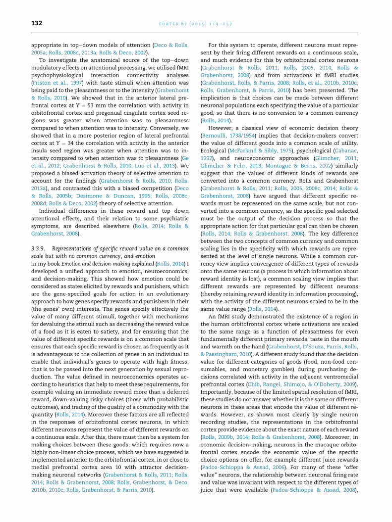
c o r t e x 6 2 ( 2 0 1 5 ) 1 1 9e1 5 7132
appropriate in topedown models of attention (Deco & Rolls,
2005a; Rolls, 2008c, 2013a; Rolls & Deco, 2002).
To investigate the anatomical source of the topedown
modulatory effects on attentional processing, we utilised fMRI
psychophysiological interaction connectivity analyses
(Friston et al., 1997) with taste stimuli when attention was
being paid to the pleasantness or to the intensity (Grabenhorst
& Rolls, 2010). We showed that in the anterior lateral pre-
frontal cortex at Y ¼ 53 mm the correlation with activity in
orbitofrontal cortex and pregenual cingulate cortex seed re-
gions was greater when attention was to pleasantness
compared to when attention was to intensity. Conversely, we
showed that in a more posterior region of lateral prefrontal
cortex at Y ¼ 34 the correlation with activity in the anterior
insula seed region was greater when attention was to in-
tensity compared to when attention was to pleasantness (Ge
et al., 2012; Grabenhorst & Rolls, 2010; Luo et al., 2013). We
proposed a biased activation theory of selective attention to
account for the findings (Grabenhorst & Rolls, 2010; Rolls,
2013a), and contrasted this with a biased competition (Deco
& Rolls, 2005b; Desimone & Duncan, 1995; Rolls, 2008c,
2008d; Rolls & Deco, 2002) theory of selective attention.
Individual differences in these reward and topedown
attentional effects, and their relation to some psychiatric
symptoms, are described elsewhere (Rolls, 2014; Rolls &
Grabenhorst, 2008).
3.3.9. Representations of specific reward value on a commonscale but with no common currency, and emotionIn my book Emotion and decision-making explained (Rolls, 2014) I
developed a unified approach to emotion, neuroeconomics,
and decision-making. This showed how emotion could be
considered as states elicited by rewards and punishers, which
are the gene-specified goals for action in an evolutionary
approach to how genes specify rewards and punishers in their
(the genes’ own) interests. The genes specify effectively the
value of many different stimuli, together with mechanisms
for devaluing the stimuli such as decreasing the reward value
of a food as it is eaten to satiety, and for ensuring that the
value of different specific rewards is on a common scale that
ensures that each specific reward is chosen as frequently as it
is advantageous to the collection of genes in an individual to
enable that individual’s genes to operate with high fitness,
that is to be passed into the next generation by sexual repro-
duction. The value defined in neuroeconomics operates ac-
cording to heuristics that help tomeet these requirements, for
example valuing an immediate reward more than a deferred
reward, down-valuing risky choices (those with probabilistic
outcomes), and trading of the quality of a commodity with the
quantity (Rolls, 2014). Moreover these factors are all reflected
in the responses of orbitofrontal cortex neurons, in which
different neurons represent the value of different rewards on
a continuous scale. After this, there must then be a system for
making choices between these goods, which requires now a
highly non-linear choice process, which we have suggested is
implemented anterior to the orbitofrontal cortex, in or close to
medial prefrontal cortex area 10 with attractor decision-
making neuronal networks (Grabenhorst & Rolls, 2011; Rolls,
2014; Rolls & Grabenhorst, 2008; Rolls, Grabenhorst, & Deco,
2010b, 2010c; Rolls, Grabenhorst, & Parris, 2010).
For this system to operate, different neurons must repre-
sent by their firing different rewards on a continuous scale,
and much evidence for this by orbitofrontal cortex neurons
(Grabenhorst & Rolls, 2011; Rolls, 2005, 2014; Rolls &
Grabenhorst, 2008) and from activations in fMRI studies
(Grabenhorst, Rolls, & Parris, 2008; Rolls, et al., 2010b, 2010c;
Rolls, Grabenhorst, & Parris, 2010) has been presented. The
implication is that choices can be made between different
neuronal populations each specifying the value of a particular
good, so that there is no conversion to a common currency
(Rolls, 2014).
However, a classical view of economic decision theory
(Bernoulli, 1738/1954) implies that decision-makers convert
the value of different goods into a common scale of utility.
Ecological (McFarland & Sibly, 1975), psychological (Cabanac,
1992), and neuroeconomic approaches (Glimcher, 2011;
Glimcher & Fehr, 2013; Montague & Berns, 2002) similarly
suggest that the values of different kinds of rewards are
converted into a common currency. Rolls and Grabenhorst
(Grabenhorst & Rolls, 2011; Rolls, 2005, 2008c, 2014; Rolls &
Grabenhorst, 2008) have argued that different specific re-
wards must be represented on the same scale, but not con-
verted into a common currency, as the specific goal selected
must be the output of the decision process so that the
appropriate action for that particular goal can then be chosen
(Rolls, 2014; Rolls & Grabenhorst, 2008). The key difference
between the two concepts of common currency and common
scaling lies in the specificity with which rewards are repre-
sented at the level of single neurons. While a common cur-
rency view implies convergence of different types of rewards
onto the same neurons (a process in which information about
reward identity is lost), a common scaling view implies that
different rewards are represented by different neurons
(thereby retaining reward identity in information processing),
with the activity of the different neurons scaled to be in the
same value range (Rolls, 2014).
An fMRI study demonstrated the existence of a region in
the human orbitofrontal cortex where activations are scaled
to the same range as a function of pleasantness for even
fundamentally different primary rewards, taste in the mouth
and warmth on the hand (Grabenhorst, D’Souza, Parris, Rolls,
& Passingham, 2010). A different study found that the decision
value for different categories of goods (food, non-food con-
sumables, and monetary gambles) during purchasing de-
cisions correlated with activity in the adjacent ventromedial
prefrontal cortex (Chib, Rangel, Shimojo, & O’Doherty, 2009).
Importantly, because of the limited spatial resolution of fMRI,
these studies do not answerwhether it is the same or different
neurons in these areas that encode the value of different re-
wards. However, as shown most clearly by single neuron
recording studies, the representations in the orbitofrontal
cortex provide evidence about the exact nature of each reward
(Rolls, 2009b, 2014; Rolls & Grabenhorst, 2008). Moreover, in
economic decision-making, neurons in the macaque orbito-
frontal cortex encode the economic value of the specific
choice options on offer, for example different juice rewards
(Padoa-Schioppa & Assad, 2006). For many of these “offer
value” neurons, the relationship between neuronal firing rate
and value was invariant with respect to the different types of
juice that were available (Padoa-Schioppa & Assad, 2008),
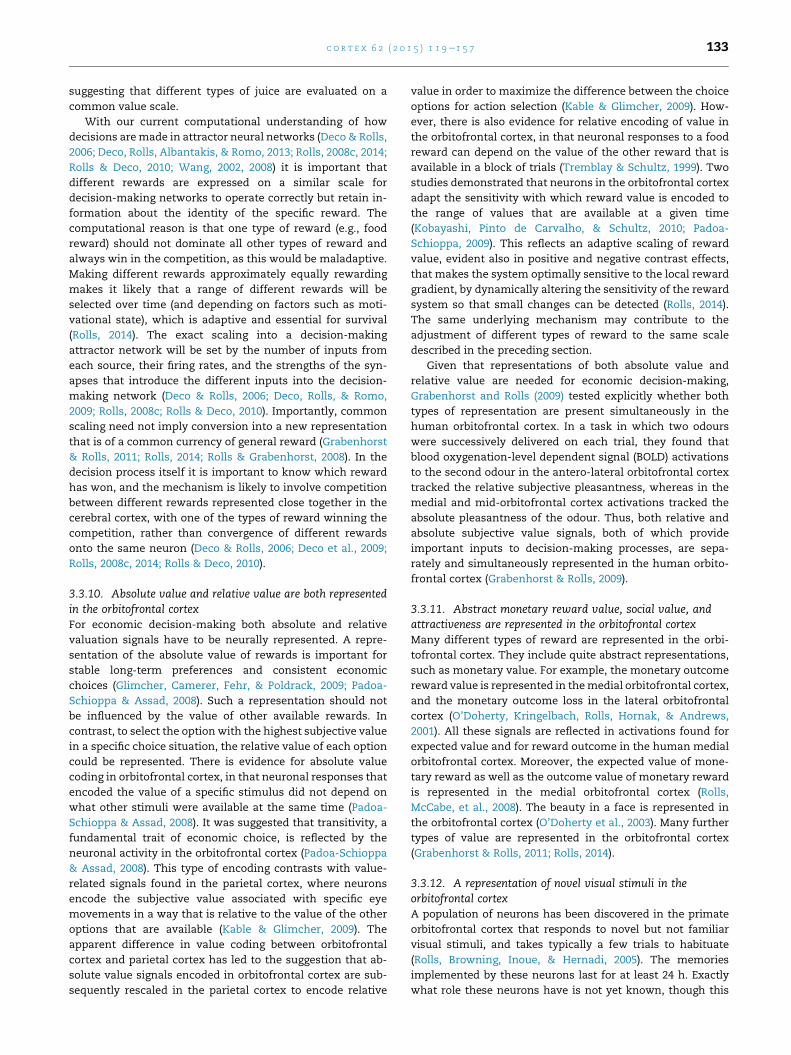
c o r t e x 6 2 ( 2 0 1 5 ) 1 1 9e1 5 7 133
suggesting that different types of juice are evaluated on a
common value scale.
With our current computational understanding of how
decisions aremade in attractor neural networks (Deco & Rolls,
2006; Deco, Rolls, Albantakis, & Romo, 2013; Rolls, 2008c, 2014;
Rolls & Deco, 2010; Wang, 2002, 2008) it is important that
different rewards are expressed on a similar scale for
decision-making networks to operate correctly but retain in-
formation about the identity of the specific reward. The
computational reason is that one type of reward (e.g., food
reward) should not dominate all other types of reward and
always win in the competition, as this would be maladaptive.
Making different rewards approximately equally rewarding
makes it likely that a range of different rewards will be
selected over time (and depending on factors such as moti-
vational state), which is adaptive and essential for survival
(Rolls, 2014). The exact scaling into a decision-making
attractor network will be set by the number of inputs from
each source, their firing rates, and the strengths of the syn-
apses that introduce the different inputs into the decision-
making network (Deco & Rolls, 2006; Deco, Rolls, & Romo,
2009; Rolls, 2008c; Rolls & Deco, 2010). Importantly, common
scaling need not imply conversion into a new representation
that is of a common currency of general reward (Grabenhorst
& Rolls, 2011; Rolls, 2014; Rolls & Grabenhorst, 2008). In the
decision process itself it is important to know which reward
has won, and the mechanism is likely to involve competition
between different rewards represented close together in the
cerebral cortex, with one of the types of reward winning the
competition, rather than convergence of different rewards
onto the same neuron (Deco & Rolls, 2006; Deco et al., 2009;
Rolls, 2008c, 2014; Rolls & Deco, 2010).
3.3.10. Absolute value and relative value are both representedin the orbitofrontal cortexFor economic decision-making both absolute and relative
valuation signals have to be neurally represented. A repre-
sentation of the absolute value of rewards is important for
stable long-term preferences and consistent economic
choices (Glimcher, Camerer, Fehr, & Poldrack, 2009; Padoa-
Schioppa & Assad, 2008). Such a representation should not
be influenced by the value of other available rewards. In
contrast, to select the option with the highest subjective value
in a specific choice situation, the relative value of each option
could be represented. There is evidence for absolute value
coding in orbitofrontal cortex, in that neuronal responses that
encoded the value of a specific stimulus did not depend on
what other stimuli were available at the same time (Padoa-
Schioppa & Assad, 2008). It was suggested that transitivity, a
fundamental trait of economic choice, is reflected by the
neuronal activity in the orbitofrontal cortex (Padoa-Schioppa
& Assad, 2008). This type of encoding contrasts with value-
related signals found in the parietal cortex, where neurons
encode the subjective value associated with specific eye
movements in a way that is relative to the value of the other
options that are available (Kable & Glimcher, 2009). The
apparent difference in value coding between orbitofrontal
cortex and parietal cortex has led to the suggestion that ab-
solute value signals encoded in orbitofrontal cortex are sub-
sequently rescaled in the parietal cortex to encode relative
value in order to maximize the difference between the choice
options for action selection (Kable & Glimcher, 2009). How-
ever, there is also evidence for relative encoding of value in
the orbitofrontal cortex, in that neuronal responses to a food
reward can depend on the value of the other reward that is
available in a block of trials (Tremblay & Schultz, 1999). Two
studies demonstrated that neurons in the orbitofrontal cortex
adapt the sensitivity with which reward value is encoded to
the range of values that are available at a given time
(Kobayashi, Pinto de Carvalho, & Schultz, 2010; Padoa-
Schioppa, 2009). This reflects an adaptive scaling of reward
value, evident also in positive and negative contrast effects,
that makes the system optimally sensitive to the local reward
gradient, by dynamically altering the sensitivity of the reward
system so that small changes can be detected (Rolls, 2014).
The same underlying mechanism may contribute to the
adjustment of different types of reward to the same scale
described in the preceding section.
Given that representations of both absolute value and
relative value are needed for economic decision-making,
Grabenhorst and Rolls (2009) tested explicitly whether both
types of representation are present simultaneously in the
human orbitofrontal cortex. In a task in which two odours
were successively delivered on each trial, they found that
blood oxygenation-level dependent signal (BOLD) activations
to the second odour in the antero-lateral orbitofrontal cortex
tracked the relative subjective pleasantness, whereas in the
medial and mid-orbitofrontal cortex activations tracked the
absolute pleasantness of the odour. Thus, both relative and
absolute subjective value signals, both of which provide
important inputs to decision-making processes, are sepa-
rately and simultaneously represented in the human orbito-
frontal cortex (Grabenhorst & Rolls, 2009).
3.3.11. Abstract monetary reward value, social value, andattractiveness are represented in the orbitofrontal cortexMany different types of reward are represented in the orbi-
tofrontal cortex. They include quite abstract representations,
such as monetary value. For example, the monetary outcome
reward value is represented in themedial orbitofrontal cortex,
and the monetary outcome loss in the lateral orbitofrontal
cortex (O’Doherty, Kringelbach, Rolls, Hornak, & Andrews,
2001). All these signals are reflected in activations found for
expected value and for reward outcome in the human medial
orbitofrontal cortex. Moreover, the expected value of mone-
tary reward as well as the outcome value of monetary reward
is represented in the medial orbitofrontal cortex (Rolls,
McCabe, et al., 2008). The beauty in a face is represented in
the orbitofrontal cortex (O’Doherty et al., 2003). Many further
types of value are represented in the orbitofrontal cortex
(Grabenhorst & Rolls, 2011; Rolls, 2014).
3.3.12. A representation of novel visual stimuli in theorbitofrontal cortexA population of neurons has been discovered in the primate
orbitofrontal cortex that responds to novel but not familiar
visual stimuli, and takes typically a few trials to habituate
(Rolls, Browning, Inoue, & Hernadi, 2005). The memories
implemented by these neurons last for at least 24 h. Exactly
what role these neurons have is not yet known, though this
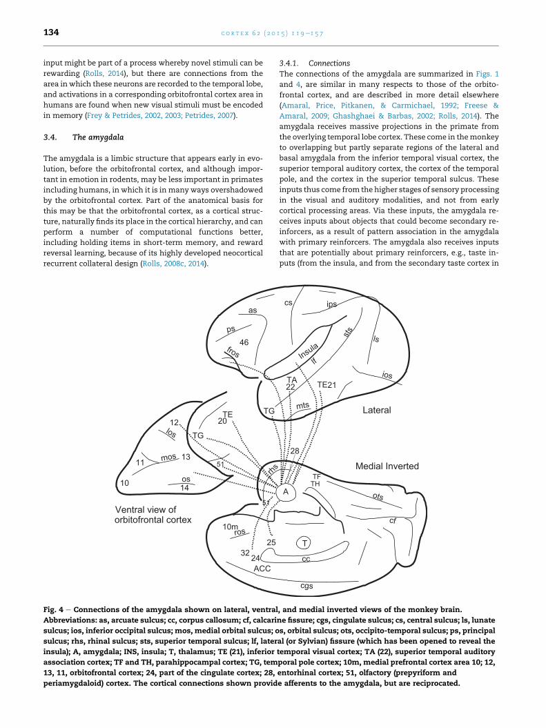
c o r t e x 6 2 ( 2 0 1 5 ) 1 1 9e1 5 7134
input might be part of a process whereby novel stimuli can be
rewarding (Rolls, 2014), but there are connections from the
area in which these neurons are recorded to the temporal lobe,
and activations in a corresponding orbitofrontal cortex area in
humans are found when new visual stimuli must be encoded
in memory (Frey & Petrides, 2002, 2003; Petrides, 2007).
3.4. The amygdala
The amygdala is a limbic structure that appears early in evo-
lution, before the orbitofrontal cortex, and although impor-
tant in emotion in rodents, may be less important in primates
including humans, in which it is in many ways overshadowed
by the orbitofrontal cortex. Part of the anatomical basis for
this may be that the orbitofrontal cortex, as a cortical struc-
ture, naturally finds its place in the cortical hierarchy, and can
perform a number of computational functions better,
including holding items in short-term memory, and reward
reversal learning, because of its highly developed neocortical
recurrent collateral design (Rolls, 2008c, 2014).
10m
ACC
14
11
12
13
as
fros
ps
ros
rhs
46
TG
Ventral view of
TG
TE
los
mos
os
20
orbitofrontal cortex
51
51
25
2432
10
Fig. 4 e Connections of the amygdala shown on lateral, ventral
Abbreviations: as, arcuate sulcus; cc, corpus callosum; cf, calcari
sulcus; ios, inferior occipital sulcus; mos, medial orbital sulcus; o
sulcus; rhs, rhinal sulcus; sts, superior temporal sulcus; lf, later
insula); A, amygdala; INS, insula; T, thalamus; TE (21), inferior
association cortex; TF and TH, parahippocampal cortex; TG, tem
13, 11, orbitofrontal cortex; 24, part of the cingulate cortex; 28,
periamygdaloid) cortex. The cortical connections shown provid
3.4.1. ConnectionsThe connections of the amygdala are summarized in Figs. 1
and 4, are similar in many respects to those of the orbito-
frontal cortex, and are described in more detail elsewhere
(Amaral, Price, Pitkanen, & Carmichael, 1992; Freese &
Amaral, 2009; Ghashghaei & Barbas, 2002; Rolls, 2014). The
amygdala receives massive projections in the primate from
the overlying temporal lobe cortex. These come in themonkey
to overlapping but partly separate regions of the lateral and
basal amygdala from the inferior temporal visual cortex, the
superior temporal auditory cortex, the cortex of the temporal
pole, and the cortex in the superior temporal sulcus. These
inputs thus come from the higher stages of sensory processing
in the visual and auditory modalities, and not from early
cortical processing areas. Via these inputs, the amygdala re-
ceives inputs about objects that could become secondary re-
inforcers, as a result of pattern association in the amygdala
with primary reinforcers. The amygdala also receives inputs
that are potentially about primary reinforcers, e.g., taste in-
puts (from the insula, and from the secondary taste cortex in
cc
sts
ios
mts
cgs
ots
cf
cs ips
lf
ls
Medial Inverted
Lateral
A
T
TFTH
TA22 21TE
Insula
28
, and medial inverted views of the monkey brain.
ne fissure; cgs, cingulate sulcus; cs, central sulcus; ls, lunate
s, orbital sulcus; ots, occipito-temporal sulcus; ps, principal
al (or Sylvian) fissure (which has been opened to reveal the
temporal visual cortex; TA (22), superior temporal auditory
poral pole cortex; 10m, medial prefrontal cortex area 10; 12,
entorhinal cortex; 51, olfactory (prepyriform and
e afferents to the amygdala, but are reciprocated.
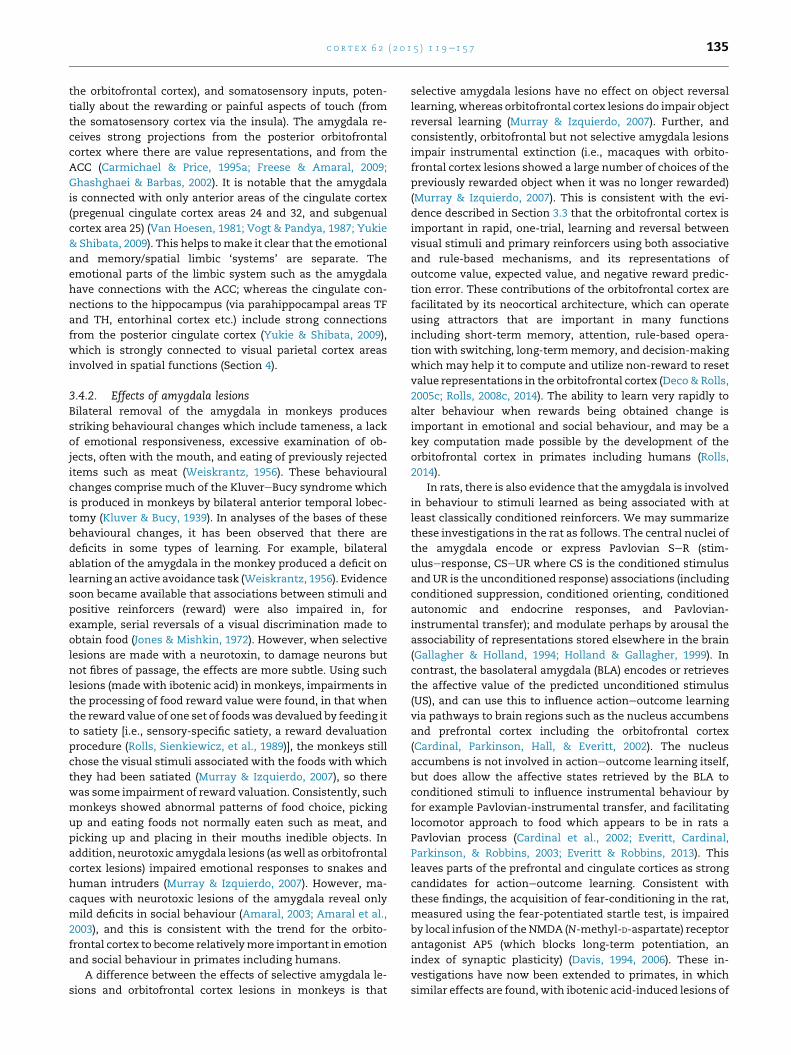
c o r t e x 6 2 ( 2 0 1 5 ) 1 1 9e1 5 7 135
the orbitofrontal cortex), and somatosensory inputs, poten-
tially about the rewarding or painful aspects of touch (from
the somatosensory cortex via the insula). The amygdala re-
ceives strong projections from the posterior orbitofrontal
cortex where there are value representations, and from the
ACC (Carmichael & Price, 1995a; Freese & Amaral, 2009;
Ghashghaei & Barbas, 2002). It is notable that the amygdala
is connected with only anterior areas of the cingulate cortex
(pregenual cingulate cortex areas 24 and 32, and subgenual
cortex area 25) (Van Hoesen, 1981; Vogt & Pandya, 1987; Yukie
& Shibata, 2009). This helps tomake it clear that the emotional
and memory/spatial limbic ‘systems’ are separate. The
emotional parts of the limbic system such as the amygdala
have connections with the ACC; whereas the cingulate con-
nections to the hippocampus (via parahippocampal areas TF
and TH, entorhinal cortex etc.) include strong connections
from the posterior cingulate cortex (Yukie & Shibata, 2009),
which is strongly connected to visual parietal cortex areas
involved in spatial functions (Section 4).
3.4.2. Effects of amygdala lesionsBilateral removal of the amygdala in monkeys produces
striking behavioural changes which include tameness, a lack
of emotional responsiveness, excessive examination of ob-
jects, often with the mouth, and eating of previously rejected
items such as meat (Weiskrantz, 1956). These behavioural
changes comprise much of the KluvereBucy syndrome which
is produced in monkeys by bilateral anterior temporal lobec-
tomy (Kluver & Bucy, 1939). In analyses of the bases of these
behavioural changes, it has been observed that there are
deficits in some types of learning. For example, bilateral
ablation of the amygdala in the monkey produced a deficit on
learning an active avoidance task (Weiskrantz, 1956). Evidence
soon became available that associations between stimuli and
positive reinforcers (reward) were also impaired in, for
example, serial reversals of a visual discrimination made to
obtain food (Jones & Mishkin, 1972). However, when selective
lesions are made with a neurotoxin, to damage neurons but
not fibres of passage, the effects are more subtle. Using such
lesions (made with ibotenic acid) in monkeys, impairments in
the processing of food reward value were found, in that when
the reward value of one set of foodswas devalued by feeding it
to satiety [i.e., sensory-specific satiety, a reward devaluation
procedure (Rolls, Sienkiewicz, et al., 1989)], the monkeys still
chose the visual stimuli associated with the foods with which
they had been satiated (Murray & Izquierdo, 2007), so there
was some impairment of reward valuation. Consistently, such
monkeys showed abnormal patterns of food choice, picking
up and eating foods not normally eaten such as meat, and
picking up and placing in their mouths inedible objects. In
addition, neurotoxic amygdala lesions (as well as orbitofrontal
cortex lesions) impaired emotional responses to snakes and
human intruders (Murray & Izquierdo, 2007). However, ma-
caques with neurotoxic lesions of the amygdala reveal only
mild deficits in social behaviour (Amaral, 2003; Amaral et al.,
2003), and this is consistent with the trend for the orbito-
frontal cortex to become relativelymore important in emotion
and social behaviour in primates including humans.
A difference between the effects of selective amygdala le-
sions and orbitofrontal cortex lesions in monkeys is that
selective amygdala lesions have no effect on object reversal
learning, whereas orbitofrontal cortex lesions do impair object
reversal learning (Murray & Izquierdo, 2007). Further, and
consistently, orbitofrontal but not selective amygdala lesions
impair instrumental extinction (i.e., macaques with orbito-
frontal cortex lesions showed a large number of choices of the
previously rewarded object when it was no longer rewarded)
(Murray & Izquierdo, 2007). This is consistent with the evi-
dence described in Section 3.3 that the orbitofrontal cortex is
important in rapid, one-trial, learning and reversal between
visual stimuli and primary reinforcers using both associative
and rule-based mechanisms, and its representations of
outcome value, expected value, and negative reward predic-
tion error. These contributions of the orbitofrontal cortex are
facilitated by its neocortical architecture, which can operate
using attractors that are important in many functions
including short-term memory, attention, rule-based opera-
tion with switching, long-termmemory, and decision-making
which may help it to compute and utilize non-reward to reset
value representations in the orbitofrontal cortex (Deco& Rolls,
2005c; Rolls, 2008c, 2014). The ability to learn very rapidly to
alter behaviour when rewards being obtained change is
important in emotional and social behaviour, and may be a
key computation made possible by the development of the
orbitofrontal cortex in primates including humans (Rolls,
2014).
In rats, there is also evidence that the amygdala is involved
in behaviour to stimuli learned as being associated with at
least classically conditioned reinforcers. We may summarize
these investigations in the rat as follows. The central nuclei of
the amygdala encode or express Pavlovian SeR (stim-
uluseresponse, CSeUR where CS is the conditioned stimulus
and UR is the unconditioned response) associations (including
conditioned suppression, conditioned orienting, conditioned
autonomic and endocrine responses, and Pavlovian-
instrumental transfer); and modulate perhaps by arousal the
associability of representations stored elsewhere in the brain
(Gallagher & Holland, 1994; Holland & Gallagher, 1999). In
contrast, the basolateral amygdala (BLA) encodes or retrieves
the affective value of the predicted unconditioned stimulus
(US), and can use this to influence actioneoutcome learning
via pathways to brain regions such as the nucleus accumbens
and prefrontal cortex including the orbitofrontal cortex
(Cardinal, Parkinson, Hall, & Everitt, 2002). The nucleus
accumbens is not involved in actioneoutcome learning itself,
but does allow the affective states retrieved by the BLA to
conditioned stimuli to influence instrumental behaviour by
for example Pavlovian-instrumental transfer, and facilitating
locomotor approach to food which appears to be in rats a
Pavlovian process (Cardinal et al., 2002; Everitt, Cardinal,
Parkinson, & Robbins, 2003; Everitt & Robbins, 2013). This
leaves parts of the prefrontal and cingulate cortices as strong
candidates for actioneoutcome learning. Consistent with
these findings, the acquisition of fear-conditioning in the rat,
measured using the fear-potentiated startle test, is impaired
by local infusion of the NMDA (N-methyl-D-aspartate) receptor
antagonist AP5 (which blocks long-term potentiation, an
index of synaptic plasticity) (Davis, 1994, 2006). These in-
vestigations have now been extended to primates, in which
similar effects are found, with ibotenic acid-induced lesions of
c o r t e x 6 2 ( 2 0 1 5 ) 1 1 9e1 5 7136
the amygdala preventing the acquisition of fear-potentiated
startle (Antoniadis, Winslow, Davis, & Amaral, 2009).
3.4.3. Amygdala neuronal activityIn the rat, in classical (Pavlovian) conditioning of fear, for
some classes of stimulus such as pure tones, the association
between the tone and an aversive US (a footshock) is reflected
in the responses of neurons in the amygdala (LeDoux, 1995,
2000b). The auditory inputs reach the amygdala both from
the subcortical, thalamic, auditory nucleus, the medial
geniculate (medial part), and from the auditory cortex. These
auditory inputs project to the lateral nucleus of the amygdala
(LA), which in turn projects to the central nucleus of the
amygdala (Ce) both directly and via the basal (B) and accessory
basal nuclei of the amygdala. LeDoux has emphasized the role
of the subcortical inputs to the amygdala in this type of con-
ditioning, based on the observations that the conditioning to
pure tones can take place without the cortex, and that the
shortest latencies of the auditory responses in the amygdala
are too short to be mediated via the auditory cortex (LeDoux,
1995, 2000b). This ‘low road’ from subcortical structures
bypassing cortical processing is unlikely to be a route for most
emotions in primates including humans (Rolls, 2014), for
stimuli requiring complex analysis (e.g., of face identity and
expression and gaze) and view, translation, and size invari-
ance require massive cortical computation which is now
starting to become understood (Rolls, 2008c, 2012a); and the
response latencies of amygdala neurons to such stimuli are
longer than those of neurons in the inferior temporal cortex
(Rolls, 1984).
There are separate output pathways in the rat from the
amygdala for different fear-related classically conditioned
responses. Lesions of the lateral hypothalamus (which re-
ceives from the central nucleus of the amygdala) blocked
conditioned heart rate (autonomic) responses. Lesions of the
central gray of the midbrain (which also receives from the
central nucleus of the amygdala) blocked the conditioned
freezing but not the conditioned autonomic response, and
lesions of the stria terminalis blocked the neuroendocrine
responses (LeDoux, 2000a).
In primates, the amygdala contains neurons that respond
to taste, and oral texture including viscosity, fat texture, and
capsaicin, and to somatosensory stimuli (Kadohisa et al.,
2005a; Kadohisa, Rolls, & Verhagen, 2005b; Rolls, 1992b,
2000b; Sanghera, Rolls, & Roper-Hall, 1979). These are all
potentially primary reinforcers, and these neurons could
represent outcome value. Neurons in the primate amygdala
also respond to the sight of instrumental reinforcers in a vi-
sual discrimination task (Sanghera et al., 1979; Wilson & Rolls,
2005), and to odours. These neurons could signal expected
value. However, in reward reversal learning, many amygdala
neurons do not reverse their responses (Sanghera et al., 1979;
Wilson & Rolls, 2005), and if any reversal is found, it is slow,
taking many trials (Paton, Belova, Morrison, & Salzman, 2006;
Sanghera et al., 1979; Wilson & Rolls, 2005), whereas orbito-
frontal neurons show rule-based reversal in one trial (Thorpe
et al., 1983). Further, devaluation by feeding to satiety pro-
duced only a partial reduction in responses to taste (Yan &
Scott, 1996), and little reduction in responses to visual stim-
uli associated with food (Sanghera et al., 1979), compared to
the complete reduction found for orbitofrontal cortex taste
(Rolls, Sienkiewicz, et al., 1989) and visual food (Critchley &
Rolls, 1996a) neurons. Thus by both reversal learning and deval-
uation measures of neuronal responses, the amygdala in primates
appears to make a less important contribution to reward value
representations than the orbitofrontal cortex.
Other amygdala neurons respond to faces (Leonard, Rolls,
Wilson, & Baylis, 1985; Rolls, 1992b, 2000b, 2011a; Sanghera
et al., 1979), including face expression and identity (Gothard,
Battaglia, Erickson, Spitler, & Amaral, 2007), both of which
are represented in the inferior temporal cortex areas that
project into the amygdala (Hasselmo et al., 1989). The pres-
ence of these neurons in the primate amygdala does empha-
size that as the amygdala has evolved in primates, it does
represent information that becomes highly developed in the
primate inferior temporal cortical areas, and that is important
for social and emotional responses to individuals, though the
orbitofrontal cortex of course also represents similar infor-
mation (Rolls, Critchley, et al., 2006).
3.4.4. Amygdala damage in humansThe greater importance of the orbitofrontal cortex in emotion
in humans is emphasized by a comparison with the effects of
bilateral amygdala damage in humans, which although pro-
ducing demonstrable deficits in face processing (Adolphs
et al., 2005; Spezio, Huang, Castelli, & Adolphs, 2007),
decision-making with linked autonomic deficits (Bechara,
Damasio, Damasio, & Lee, 1999; Brand, Grabenhorst, Starcke,
Vandekerckhove, & Markowitsch, 2007), and autonomic con-
ditioning (Phelps & LeDoux, 2005), may not (in contrast with
the orbitofrontal cortex) produce major changes in emotion
that are readily apparent in everyday behaviour (Phelps &
LeDoux, 2005; Rolls, 2008c; Seymour & Dolan, 2008; Whalen
& Phelps, 2009).
3.5. The anterior cingulate cortex (ACG)
The ACC is a limbic structure involved in emotion, with major
inputs from structures such as the amygdala and orbitofrontal
cortex, and activations that are correlated with the pleasant-
ness or unpleasantness of stimuli. The ACC uses these value
(including outcome value) representations in its function with
themidcingulate cortex in actioneoutcome learning. The ACC
can be conceived as a system that in primates links the orbi-
tofrontal cortex representations of the value of stimuli
(including reward and punisher outcomes) with actions (Rolls,
2014).
3.5.1. ConnectionsThe connections of the ACC (Vogt, 2009; Yukie & Shibata, 2009)
are illustrated in Fig. 5. The ACC includes area 32, area 25 the
subgenual cingulate cortex, and part of area 24. The cortex
anterior to the genu (knee, at the front) of the corpus callosum
is referred to as the pregenual cingulate cortex. The caudal
orbitofrontal cortex and amygdala project to the ACC
(Carmichael & Price, 1996; Price, 2006), and especially the
orbitofrontal cortex appears to influence the ACC strongly, for
activations related to the pleasantness or unpleasantness of
many stimuli are present in both (Grabenhorst & Rolls, 2011;
Rolls, 2014). It is of interest that the anterior but not
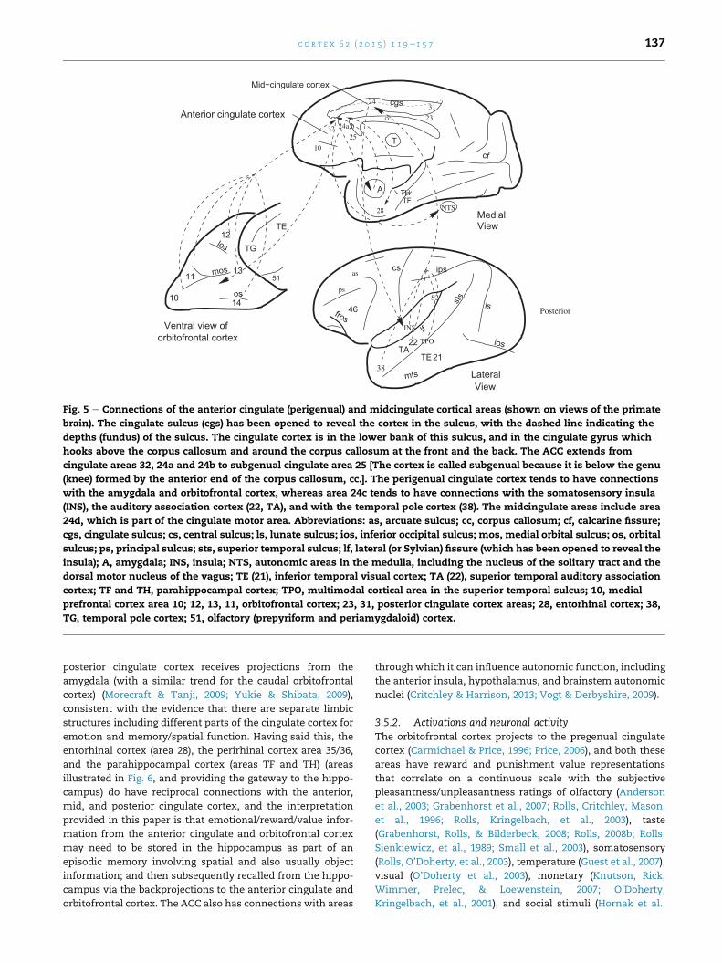
32
10
28
Anterior cingulate cortex
Mid−cingulate cortex
31
10 14
11
12
13
Ventral view of
TG
TE
los
mos
os
51
cf
THTF
ios
sts
mts
cs ips
TA
lf
21
22
lsfros
46
cc
T
ps
TPO
S2
INS
A
cgs
38
NTS
TE
Lateral
Posterior
View
View
as
Medial
24a,b
25
24
orbitofrontal cortex
23
Fig. 5 e Connections of the anterior cingulate (perigenual) and midcingulate cortical areas (shown on views of the primate
brain). The cingulate sulcus (cgs) has been opened to reveal the cortex in the sulcus, with the dashed line indicating the
depths (fundus) of the sulcus. The cingulate cortex is in the lower bank of this sulcus, and in the cingulate gyrus which
hooks above the corpus callosum and around the corpus callosum at the front and the back. The ACC extends from
cingulate areas 32, 24a and 24b to subgenual cingulate area 25 [The cortex is called subgenual because it is below the genu
(knee) formed by the anterior end of the corpus callosum, cc.]. The perigenual cingulate cortex tends to have connections
with the amygdala and orbitofrontal cortex, whereas area 24c tends to have connections with the somatosensory insula
(INS), the auditory association cortex (22, TA), and with the temporal pole cortex (38). The midcingulate areas include area
24d, which is part of the cingulate motor area. Abbreviations: as, arcuate sulcus; cc, corpus callosum; cf, calcarine fissure;
cgs, cingulate sulcus; cs, central sulcus; ls, lunate sulcus; ios, inferior occipital sulcus; mos, medial orbital sulcus; os, orbital
sulcus; ps, principal sulcus; sts, superior temporal sulcus; lf, lateral (or Sylvian) fissure (which has been opened to reveal the
insula); A, amygdala; INS, insula; NTS, autonomic areas in the medulla, including the nucleus of the solitary tract and the
dorsal motor nucleus of the vagus; TE (21), inferior temporal visual cortex; TA (22), superior temporal auditory association
cortex; TF and TH, parahippocampal cortex; TPO, multimodal cortical area in the superior temporal sulcus; 10, medial
prefrontal cortex area 10; 12, 13, 11, orbitofrontal cortex; 23, 31, posterior cingulate cortex areas; 28, entorhinal cortex; 38,
TG, temporal pole cortex; 51, olfactory (prepyriform and periamygdaloid) cortex.
c o r t e x 6 2 ( 2 0 1 5 ) 1 1 9e1 5 7 137
posterior cingulate cortex receives projections from the
amygdala (with a similar trend for the caudal orbitofrontal
cortex) (Morecraft & Tanji, 2009; Yukie & Shibata, 2009),
consistent with the evidence that there are separate limbic
structures including different parts of the cingulate cortex for
emotion and memory/spatial function. Having said this, the
entorhinal cortex (area 28), the perirhinal cortex area 35/36,
and the parahippocampal cortex (areas TF and TH) (areas
illustrated in Fig. 6, and providing the gateway to the hippo-
campus) do have reciprocal connections with the anterior,
mid, and posterior cingulate cortex, and the interpretation
provided in this paper is that emotional/reward/value infor-
mation from the anterior cingulate and orbitofrontal cortex
may need to be stored in the hippocampus as part of an
episodic memory involving spatial and also usually object
information; and then subsequently recalled from the hippo-
campus via the backprojections to the anterior cingulate and
orbitofrontal cortex. The ACC also has connections with areas
through which it can influence autonomic function, including
the anterior insula, hypothalamus, and brainstem autonomic
nuclei (Critchley & Harrison, 2013; Vogt & Derbyshire, 2009).
3.5.2. Activations and neuronal activityThe orbitofrontal cortex projects to the pregenual cingulate
cortex (Carmichael & Price, 1996; Price, 2006), and both these
areas have reward and punishment value representations
that correlate on a continuous scale with the subjective
pleasantness/unpleasantness ratings of olfactory (Anderson
et al., 2003; Grabenhorst et al., 2007; Rolls, Critchley, Mason,
et al., 1996; Rolls, Kringelbach, et al., 2003), taste
(Grabenhorst, Rolls, & Bilderbeck, 2008; Rolls, 2008b; Rolls,
Sienkiewicz, et al., 1989; Small et al., 2003), somatosensory
(Rolls, O’Doherty, et al., 2003), temperature (Guest et al., 2007),
visual (O’Doherty et al., 2003), monetary (Knutson, Rick,
Wimmer, Prelec, & Loewenstein, 2007; O’Doherty,
Kringelbach, et al., 2001), and social stimuli (Hornak et al.,
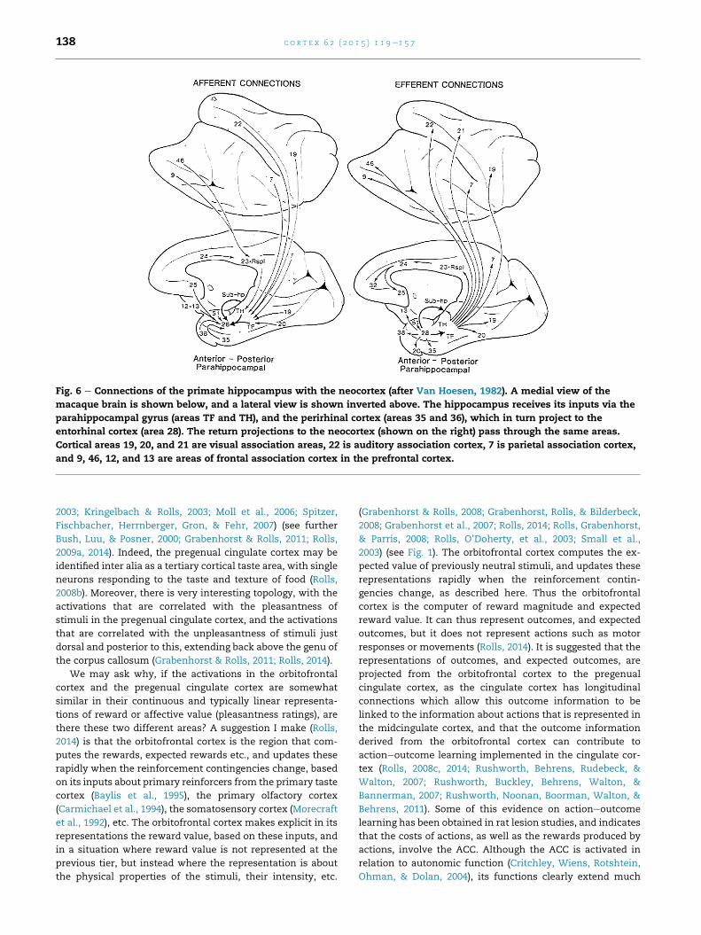
Fig. 6 e Connections of the primate hippocampus with the neocortex (after Van Hoesen, 1982). A medial view of the
macaque brain is shown below, and a lateral view is shown inverted above. The hippocampus receives its inputs via the
parahippocampal gyrus (areas TF and TH), and the perirhinal cortex (areas 35 and 36), which in turn project to the
entorhinal cortex (area 28). The return projections to the neocortex (shown on the right) pass through the same areas.
Cortical areas 19, 20, and 21 are visual association areas, 22 is auditory association cortex, 7 is parietal association cortex,
and 9, 46, 12, and 13 are areas of frontal association cortex in the prefrontal cortex.
c o r t e x 6 2 ( 2 0 1 5 ) 1 1 9e1 5 7138
2003; Kringelbach & Rolls, 2003; Moll et al., 2006; Spitzer,
Fischbacher, Herrnberger, Gron, & Fehr, 2007) (see further
Bush, Luu, & Posner, 2000; Grabenhorst & Rolls, 2011; Rolls,
2009a, 2014). Indeed, the pregenual cingulate cortex may be
identified inter alia as a tertiary cortical taste area, with single
neurons responding to the taste and texture of food (Rolls,
2008b). Moreover, there is very interesting topology, with the
activations that are correlated with the pleasantness of
stimuli in the pregenual cingulate cortex, and the activations
that are correlated with the unpleasantness of stimuli just
dorsal and posterior to this, extending back above the genu of
the corpus callosum (Grabenhorst & Rolls, 2011; Rolls, 2014).
We may ask why, if the activations in the orbitofrontal
cortex and the pregenual cingulate cortex are somewhat
similar in their continuous and typically linear representa-
tions of reward or affective value (pleasantness ratings), are
there these two different areas? A suggestion I make (Rolls,
2014) is that the orbitofrontal cortex is the region that com-
putes the rewards, expected rewards etc., and updates these
rapidly when the reinforcement contingencies change, based
on its inputs about primary reinforcers from the primary taste
cortex (Baylis et al., 1995), the primary olfactory cortex
(Carmichael et al., 1994), the somatosensory cortex (Morecraft
et al., 1992), etc. The orbitofrontal cortex makes explicit in its
representations the reward value, based on these inputs, and
in a situation where reward value is not represented at the
previous tier, but instead where the representation is about
the physical properties of the stimuli, their intensity, etc.
(Grabenhorst & Rolls, 2008; Grabenhorst, Rolls, & Bilderbeck,
2008; Grabenhorst et al., 2007; Rolls, 2014; Rolls, Grabenhorst,
& Parris, 2008; Rolls, O’Doherty, et al., 2003; Small et al.,
2003) (see Fig. 1). The orbitofrontal cortex computes the ex-
pected value of previously neutral stimuli, and updates these
representations rapidly when the reinforcement contin-
gencies change, as described here. Thus the orbitofrontal
cortex is the computer of reward magnitude and expected
reward value. It can thus represent outcomes, and expected
outcomes, but it does not represent actions such as motor
responses or movements (Rolls, 2014). It is suggested that the
representations of outcomes, and expected outcomes, are
projected from the orbitofrontal cortex to the pregenual
cingulate cortex, as the cingulate cortex has longitudinal
connections which allow this outcome information to be
linked to the information about actions that is represented in
the midcingulate cortex, and that the outcome information
derived from the orbitofrontal cortex can contribute to
actioneoutcome learning implemented in the cingulate cor-
tex (Rolls, 2008c, 2014; Rushworth, Behrens, Rudebeck, &
Walton, 2007; Rushworth, Buckley, Behrens, Walton, &
Bannerman, 2007; Rushworth, Noonan, Boorman, Walton, &
Behrens, 2011). Some of this evidence on actioneoutcome
learning has been obtained in rat lesion studies, and indicates
that the costs of actions, as well as the rewards produced by
actions, involve the ACC. Although the ACC is activated in
relation to autonomic function (Critchley, Wiens, Rotshtein,
Ohman, & Dolan, 2004), its functions clearly extend much
c o r t e x 6 2 ( 2 0 1 5 ) 1 1 9e1 5 7 139
beyond this, as shown also for example by the emotional
changes that follow damage to the ACC and related areas in
humans (Hornak et al., 2003).
In responding when the reward obtained is less than that
expected, the orbitofrontal cortex negative reward prediction
error neurons are working in a domain that is related to the
sensory inputs being received (expected reward and reward
obtained). There are also error neurons in the ACC that
respond when errors are made (Niki & Watanabe, 1979), or
when rewards are reduced (Shima & Tanji, 1998) (and in
similar imaging studies, Bush et al., 2002). Some of these
neurons may be influenced by the projections from the orbi-
tofrontal cortex, and reflect a mismatch between the reward
expected and the reward that is obtained. However, some
error neurons in the ACC may reflect errors that arise when
particular behavioural responses or actions are in error, and
this type of error may be important in helping an action sys-
tem to correct itself, rather than, as in the orbitofrontal cortex,
when a reward prediction system about stimuli needs to be
corrected. Consistent with this, many studies provide evi-
dence that errors made in many tasks activate the anterior/
midcingulate cortex, whereas tasks with response conflict
activate the superior frontal gyrus (Matsumoto, Matsumoto,
Abe, & Tanaka, 2007; Rushworth & Behrens, 2008;
Rushworth, Walton, Kennerley, & Bannerman, 2004; Vogt,
2009).
A neuroimaging study in humans illustrates how non-
reward/error signals relevant to emotion and social behav-
iour are found in the ACC. Kringelbach et al. (2003) used the
faces of two different people, and if one facewas selected then
that face smiled, and if the other was selected, the face
showed an angry expression. After good performance was
acquired, there were repeated reversals of the visual
discrimination task. Kringelbach et al. (2003) found that acti-
vation of a lateral part of the orbitofrontal cortex and in a
dorsal part of the ACC in the fMRI study was produced on the
error trials, that is when the human chose a face, and did not
obtain the expected reward. An interesting aspect of this
study thatmakes it relevant to human social behaviour is that
the conditioned stimuli were faces of different individuals,
and the USs were face expressions. Thus the association that
was being reversed in this study was between a representa-
tion of face identity and a representation of face expression.
Moreover, the study reveals that the human orbitofrontal
cortex and ACC are very sensitive to social feedback when it
must be used to change behaviour (Kringelbach & Rolls, 2003,
2004).
3.5.3. Effects of ACC lesions in humansCardinal et al. (2002) and Devinsky, Morrell, and Vogt (1995)
review evidence that anterior cingulate lesions in humans
produce apathy, autonomic dysregulation, and emotional
instability.
An investigation in patients with selective surgical lesions
has shown that patients with unilateral lesions of the antero-
ventral part of the ACC and/or medial area 9 were in some
cases impaired on voice and face expression identification,
had some change in social behaviour (such as inappropriate-
ness, being less likely to notice when other people were angry,
not being close to his or her family, and doing things without
thinking), and had significant changes in their subjective
emotional state (Hornak et al., 2003). Unilateral lesions were
sufficient to produce these effects, and there were no strong
laterality effects.
The results of Hornak et al. (2003) also confirmed that
damage restricted to the orbitofrontal cortex can produce
impairments in face and voice expression identification,
which may be primary reinforcers. The system is sensitive, in
that even patients with unilateral orbitofrontal cortex lesions
may be impaired. The impairment is not a generic impairment
of the ability to recognize any emotions in others, in that
frequently voice but not face expression identification was
impaired, and vice versa. This implies some functional
specialization for visual versus auditory emotion-related
processing in the human orbitofrontal cortex. The results
also show that the changes in social behaviour can be pro-
duced by damage restricted to the orbitofrontal cortex. The
patients were particularly likely to be impaired on emotion
recognition (they were less likely to notice when others were
sad, or happy, or disgusted); on emotional empathy (theywere
less likely to comfort those who are sad, or afraid, or to feel
happy for others who are happy); on interpersonal relation-
ships (not caring what others think, and not being close to his/
her family); andwere less likely to cooperatewith others; were
impatient and impulsive; and had difficulty in making and
keeping close relationships. The results also show that
changes in subjective emotional state (including frequently
sadness, anger and happiness) can be produced by damage
restricted to the orbitofrontal cortex (Hornak et al., 2003). In
addition, the patients with bilateral orbitofrontal cortex le-
sionswere impaired on the probabilistic reversal learning task
(Hornak et al., 2004). The findings overall thus make clear the
types of deficit found in humans with orbitofrontal cortex
damage, and can be easily related to underlying fundamental
processes in which the orbitofrontal cortex is involved as
described by Rolls (1999a, 2005), including decoding and rep-
resenting primary reinforcers, being sensitive to changes in
reinforcers, and rapidly readjusting behaviour to stimuli when
the reinforcers available change. The implication is that some
of the inputs to the ACC which produce similar deficits may
come from the orbitofrontal cortex. Consistent with this,
unilateral lesions of the ACC (including some of medial area 9)
can produce voice and/or face expression identification defi-
cits, changes in social behaviour, and marked changes in
subjective emotional state (Hornak et al., 2003).
In summary, the primate including human ACC has con-
nections with other limbic structures such as the amygdala
and limbic-related structures such as the caudal orbitofrontal
cortex, receives reward and punishment-related information
from these brain regions, and appears to be involved in using
this reward and punishment information to learn about ac-
tions to obtain goals, taking into account the costs of the ac-
tions (Grabenhorst & Rolls, 2011; Rolls, 2009a, 2014; Rushworth
et al., 2011). Appropriate actions to emotion-provoking stimuli
may not bemade after ACC damage. The ACC can thus be seen
as a limbic structure involved in emotion that forms part of a
system with other limbic structures involved in emotion, the
amygdala and orbitofrontal cortex. Structures that receive
input from these three structures are involved in linking to
habit responses (striatum including ventral striatum and

c o r t e x 6 2 ( 2 0 1 5 ) 1 1 9e1 5 7140
nucleus accumbens), autonomic and endocrine output
(insula, hypothalamus, brainstem autonomic nuclei), etc.
(Rolls, 2014).
These three limbic and related structures, the amygdala,
orbitofrontal cortex, and ACC, could be thought of as an
emotion limbic system, but not the limbic system, as different
limbic structures related to the hippocampus are involved
primarily in episodic memory and related spatial function,
and not in emotion. Damage to the emotion limbic system or
structures is not described as impairing episodic memory or
spatial function.
3.6. The insular cortex
The insula has sometimes been lumped in with limbic struc-
tures (Catani et al., 2013; Yakovlev, 1948), and parts of it are
involved in autonomic/visceral function (Critchley &
Harrison, 2013), so it is considered briefly here, and in more
detail elsewhere (Rolls, 2014).
3.6.1. The insular primary taste cortexThe primary taste cortex is in the dorsal part of the anterior
insula and adjoining frontal operculum (Pritchard, Hamilton,
Morse, & Norgren, 1986), and this region projects to the orbi-
tofrontal cortex (Baylis et al., 1995). Neurons in the primary
taste cortex represent what the taste is (including sweet, salt,
sour, bitter, and umami) (Baylis & Rolls, 1991; Rolls, 2009c;
Scott et al., 1986; Yaxley et al., 1990), but do not represent
reward value in that their responses are not decreased by
feeding to satiety (Rolls et al., 1988; Yaxley et al., 1988). Neu-
rons in the insular primary taste cortex do represent oral
texture including fat texture (de Araujo & Rolls, 2004;
Kadohisa et al., 2005a; Verhagen et al., 2004) and oral tem-
perature (Guest et al., 2007; Kadohisa et al., 2005a; Verhagen
et al., 2004). Neurons in the macaque primary taste cortex do
not have olfactory responses (Verhagen et al., 2004), and
consistently in a human fMRI investigation of olfactory and
taste convergence in the brain, it was shown that an agranular
more anterior past of the insula does show convergence be-
tween taste and odour to represent flavour (de Araujo, Rolls,
et al., 2003).
3.6.2. The visceral/autonomic insular cortexA region of the anterior insular cortex just ventral to the pri-
mary taste cortex has strong projections to the orbitofrontal
cortex (Baylis et al., 1995), and is putatively the visceral/auto-
nomic region of the insula (Critchley & Harrison, 2013). This
anterior insular region probably receives inputs from the
orbitofrontal cortex and ACC (Price, 2006, 2007), which decode
and represent the reward andpunishment-related signals that
can produce autonomic/visceral responses (Rolls, 2014). It is
suggested (Rolls, 2014) that when the anterior insula is acti-
vated by emotion-related stimuli and events such as face ex-
pressions of disgust (Phillips et al., 1998, 2004) andunfair offers
(which are aversive) in an ultimatum game (Sanfey, Rilling,
Aronson, Nystrom, & Cohen, 2003), the activations reflect the
effects produced by regions such as the orbitofrontal and
anterior cingulate cortices that can produce emotional and
autonomic responses, rather than face expression decoding
[which is performed elsewhere, in the cortex in the superior
temporal sulcus and in the orbitofrontal cortex (Hasselmo
et al., 1989; Rolls, 2007b, 2012a, 2011a)] or economic computa-
tions. Consistent with this, in a neuroeconomics study with
monetary reward, it was found that expected value was
negatively correlated with activations in the anterior insula
[�38 24 16] in a region that has been implicated in disgust, and
interestingly, the activations here were also correlated with
the uncertainty of the magnitude of the reward that would be
obtained (Rolls, McCabe, et al., 2008). Effectively there was
more insula activation in situations thatmight be described as
aversive. This part of the insula has activations that are related
to visceral/autonomic function, for example to heart and
stomach responses during disgust-associated nausea
(Critchley & Harrison, 2013). Further, electrical stimulation in
the antero-ventral insula produced feelings related to disgust,
including viscero-autonomic feelings (Krolak-Salmon et al.,
2003). Moreover, it is of course to be expected, and is the
case, that the autonomic output and the corresponding
visceral insular activitywill bedifferent fordifferent emotional
states, e.g., when eating a food versus when reacting to the
disgusting bitter taste of quinine or to pain or the sight of
aversive or unpleasant stimuli. Menon and Uddin (2010) have
suggested that the insula is part of a ‘saliency’ network. They
postulate that the insula is sensitive to salient events, and that
its core function is to mark such events for additional pro-
cessing and initiate appropriate control signals. However,
given the inputs to the anterior insula from the orbitofrontal
and anterior cingulate cortices, which decode stimuli and
events such as rewards, punishers, non-reward, and novel
stimuli, it is suggested (Rolls, 2014) that the anterior insulamay
respond to though not compute such “salient” stimuli and
events, and its activation may reflect autonomic and related
responses which are of course elicited by these “salient”
stimuli. A related point (Rolls, 2014) is relevant to Damasio’s
somatic marker hypothesis (Damasio, 1996).
3.6.3. The somatosensory insulaThe mid- and posterior insula has somatosensory represen-
tations of the body (Mufson &Mesulam, 1982). A property that
may be special about these somatosensory cortical represen-
tations is that activations are produced by touch to the body
but, in contrast to many other somatosensory cortical areas,
not by the sight of touch (McCabe et al., 2008). It was therefore
suggested that insular cortex activation thus allows an indi-
vidual to know that it is touch to the person’s body, and not
that someone else’s body is about to be touched (McCabe et al.,
2008). The insular somatosensory cortex may thus provide
evidence about what is happening to one’s own body (Rolls,
2010a). The same might be said of the insular primary taste
cortex, which when activated leaves no doubt that one is
tasting, and not seeing someone else tasting. So, in a sense,
feelings associated with activations of the insular cortex [and
they are: the subjective intensity of taste is linearly related to
the activation of the primary taste cortex (Grabenhorst &
Rolls, 2008)] do inform one about the state of one’s own
body, and this relates to Craig’s suggestions (Craig, 2009, 2011)
about the importance of the insula in interoceptive feelings.
However, this does not mean that the insular cortex is
necessary for body feelings, and indeed that seems to be ruled
out by the finding that a patient with extensive bilateral
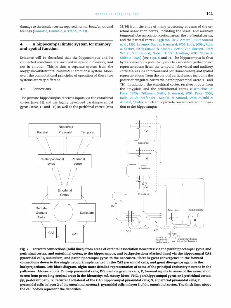
c o r t e x 6 2 ( 2 0 1 5 ) 1 1 9e1 5 7 141
damage to the insular cortex reported normal body/emotional
feelings (Damasio, Damasio, & Tranel, 2013).
4. A hippocampal limbic system for memoryand spatial function
Evidence will be described that the hippocampus and its
connected structures are involved in episodic memory, and
not in emotion. This is thus a separate system from the
amygdala/orbitofrontal cortex/ACC emotional system. More-
over, the computational principles of operation of these two
systems are very different.
4.1. Connections
The primate hippocampus receives inputs via the entorhinal
cortex (area 28) and the highly developed parahippocampal
gyrus (areas TF and TH) as well as the perirhinal cortex (area
CortexEntorhinal
DentateGranule
CellsSubiculum
CA1
Neocortex
CA3
gyrusParahippocampal
cortexPerirhinal
Parietal Prefrontal Temporal
DG
Fig. 7 e Forward connections (solid lines) from areas of cerebral
perirhinal cortex, and entorhinal cortex, to the hippocampus; an
pyramidal cells, subiculum, and parahippocampal gyrus to the
connections down to the single network implemented in the C
backprojections. Left: block diagram. Right: more detailed repres
pathways. Abbreviations: D, deep pyramidal cells; DG, dentate
cortex from preceding cortical areas in the hierarchy; mf, mossy
pp, perforant path; rc, recurrent collateral of the CA3 hippocam
pyramidal cells in layer 2 of the entorhinal cortex; 3, pyramidal c
the cell bodies represent the dendrites.
35/36) from the ends of many processing streams of the ce-
rebral association cortex, including the visual and auditory
temporal lobe association cortical areas, the prefrontal cortex,
and the parietal cortex (Aggleton, 2012; Amaral, 1987; Amaral
et al., 1992; Lavenex, Suzuki, & Amaral, 2004; Rolls, 2008c; Rolls
& Kesner, 2006; Suzuki & Amaral, 1994b; Van Hoesen, 1982;
Witter, Wouterlood, Naber, & Van Haeften, 2000; Yukie &
Shibata, 2009) (see Figs. 6 and 7). The hippocampus is thus
by its connections potentially able to associate together object
representations (from the temporal lobe visual and auditory
cortical areas via entorhinal and perirhinal cortex), and spatial
representations (from the parietal cortical areas including the
posterior cingulate cortex via parahippocampal areas TF and
TH). In addition, the entorhinal cortex receives inputs from
the amygdala and the orbitofrontal cortex (Carmichael &
Price, 1995a; Pitkanen, Kelly, & Amaral, 2002; Price, 2006;
Rolls, 2010b; Stefanacci, Suzuki, & Amaral, 1996; Suzuki &
Amaral, 1994a), which thus provide reward-related informa-
tion to the hippocampus.
rc
Fo nixr
23
rhinalEntopp
mf
Neocortex
FS
D
DS
5
accumbens,medial septum
nucleus
CA1CA3
& PerirhinalPHG
ant. nuc. of the thalamusmammillary bodies
Subiculum Presubiculum
association neocortex via the parahippocampal gyrus and
d backprojections (dashed lines) via the hippocampal CA1
neocortex. There is great convergence in the forward
A3 pyramidal cells; and great divergence again in the
entation of some of the principal excitatory neurons in the
granule cells; F, forward inputs to areas of the association
fibres; PHG, parahippocampal gyrus and perirhinal cortex;
pal pyramidal cells; S, superficial pyramidal cells; 2,
ells in layer 3 of the entorhinal cortex. The thick lines above
c o r t e x 6 2 ( 2 0 1 5 ) 1 1 9e1 5 7142
The primary output from the hippocampus to neocortex
originates in CA1 and projects to subiculum, entorhinal cor-
tex, and parahippocampal structures (areas TFeTH) as well as
prefrontal cortex (Delatour & Witter, 2002; van Haeften, Baks-
te-Bulte, Goede, Wouterlood, & Witter, 2003; Van Hoesen,
1982; Witter, 1993) (see Figs. 6 and 7). These are the pathways
that are likely to be involved in the recall of information from
the hippocampus. There are other outputs (Rolls & Kesner,
2006), including subicular complex connections to the orbi-
tofrontal cortex and ACC, and even direct connections from
CA1 to the orbitofrontal cortex and ACC (Price, 2006; Vogt &
Pandya, 1987; Yukie & Shibata, 2009), which are likely it is
suggested to be involved in the recall of emotional and
reward-related components of episodic memory; and sub-
icular complex projections to the posterior cingulate cortex
(Vogt & Pandya, 1987; Yukie & Shibata, 2009), which are likely
it is suggested to be involved in the recall of spatial compo-
nents of episodic memory.
In addition, there are subcortical connections that form
Papez’ circuit (Papez, 1937). The subiculum (subicular com-
plex) projects via the fornix to the mammillary bodies (Fig. 7),
which then project via the mammillo-thalamic tract to the
anterior thalamic nuclei, which project most strongly to the
posterior cingulate cortex (Shibata & Yukie, 2009), which in
turn via the cingulum projects back towards the hippocampus
via the parahippocampal cortex (Yukie & Shibata, 2009). The
fornix also conveys the cholinergic input from the septal
nuclei, classed as limbic structures, to the hippocampus, and
this must be taken into account when considering the effects
on memory of damage to the hippocampus, for acetylcholine
facilitates synaptic modification and regulates recurrent
collateral efficacy in the CA3 system (Giocomo & Hasselmo,
2007; Rolls, 2010b).
4.2. Effects of hippocampal system damage in primatesincluding humans
In humans, episodic memory, the memory of a particular
episode, requires the ability to remember particular events,
and to distinguish them from other events, and is impaired by
damage to the hippocampal system. An event consists of a set
of items that occur together, such as seeing a particular object
or person’s face in a particular place. An everyday example
might be remembering where one was for dinner, who was
present, what was eaten, what was discussed, and the time at
which it occurred. The spatial context is almost always an
important part of an episodic memory (Dere, Easton, Nadel, &
Huston, 2008), and it may be partly for this reason that
episodic memory is linked to the functions of the hippocam-
pal system, which is involved in spatial processing and
memory. A famous case, is that of H.M., who after surgery for
epilepsy that removed bilaterally parts of the temporal lobe
including parts of the hippocampus could no longer form
memories of events that occurred after the damage (antero-
grade amnesia), but could recall memories of events prior to
the hippocampal damage (Corkin, 2002), and similar impair-
ments are found in other patients with damage to the hip-
pocampus and connected structures (Squire & Wixted, 2011).
Indeed, section of the fornix in humans, at one time a side
effect of surgery on a third ventricle cyst, produces similar
amnesia (Gaffan & Gaffan, 1991), as can damage to the
mammillary body/mammillo-thalamic tract/anterior nucleus
of the thalamus pathway (see Aggleton, 2012). In humans,
functional neuroimaging shows that the hippocampal system
is activated by allocentric spatial processing and episodic
memory (Burgess, 2008; Burgess, Maguire, & O’Keefe, 2002;
Chadwick, Hassabis, Weiskopf, & Maguire, 2010; Hassabis
et al., 2009).
Damage to the hippocampus or to some of its connections
such as the fornix in monkeys produces deficits in learning
about the places of objects and about the places where re-
sponses should be made (Buckley & Gaffan, 2000). For
example, macaques and humans with damage to the hippo-
campal system or fornix are impaired in object-place memory
tasks in which not only the objects seen, but where they were
seen, must be remembered (Burgess et al., 2002; Crane &
Milner, 2005; Gaffan, 1994; Gaffan & Saunders, 1985;
Parkinson, Murray, & Mishkin, 1988; Smith & Milner, 1981).
Posterior parahippocampal lesions inmacaques impair even a
simple type of object-place learning in which the memory
load is just one pair of trial-unique stimuli (Malkova &
Mishkin, 2003) (It is further predicted that a more difficult
object-place learning task with non-trial-unique stimuli and
with many object-place pairs would be impaired by neuro-
toxic hippocampal lesions.). Further, neurotoxic lesions that
selectively damage the primate hippocampus impair spatial
scene memory, tested by the ability to remember where in a
scene to touch to obtain reward (Murray, Baxter, & Gaffan,
1998). Also, fornix lesions impair conditional lefteright
discrimination learning, in which the visual appearance of an
object specifies whether a response is to be made to the left or
the right (Rupniak & Gaffan, 1987). A comparable deficit is
found in humans (Petrides, 1985). Fornix sectioned monkeys
are also impaired in learning on the basis of a spatial cue
which object to choose (e.g., if two objects are on the left,
choose object A, but if the two objects are on the right, choose
object B) (Gaffan & Harrison, 1989a). Monkeys with fornix
damage are also impaired in using information about their
place in an environment. For example, there are learning
impairments when which of two or more objects the monkey
had to choose depended on the position of the monkey in the
room (Gaffan & Harrison, 1989b).
More recently, Lavenex et al. have described deficits pro-
duced by hippocampal damage in monkeys performing allo-
centric spatial memory tasks (Banta Lavenex & Lavenex,
2009). One such task involved freely moving in an environ-
ment using allocentric spatial room cues to remember the
locations of inverted cups that contained food. This is a food
reward e allocentric place association task. In contrast, the
perirhinal cortex, area 35, with its connections to inferior
temporal cortex areas involved in object perception (Rolls,
2008c, 2012a), is involved in recognition memory (Buckley,
2005), and indeed contains neurons related to long-term fa-
miliarity memory (Holscher, Rolls, & Xiang, 2003; Rolls, 2008c;
Rolls, Franco, & Stringer, 2005).
Rats with hippocampal lesions are also impaired in using
environmental spatial cues to remember particular places
(Cassaday & Rawlins, 1997; Jarrard, 1993; Kesner, Lee, &
Gilbert, 2004; Kesner, Morris, & Weeden, 2012; Martin,
Grimwood, & Morris, 2000; O’Keefe & Nadel, 1978), to utilize
c o r t e x 6 2 ( 2 0 1 5 ) 1 1 9e1 5 7 143
spatial cues or to bridge delays (Kesner, 1998; Kesner et al.,
2004; Kesner & Rolls, 2001; Rawlins, 1985; Rolls & Kesner,
2006), to perform object-place memory (Kesner et al., 2012;
Rolls & Kesner, 2006), or to perform relational operations on
remembered material (Eichenbaum, 1997).
It is notable that emotional changes, reward-related
learning and reversal impairments, and devaluation impair-
ments of stimuli are not produced by damage to this hippo-
campal system. This is further evidence for one of the theses
of this paper, that we should no longer be considering the
operation of limbic and related structures as being part of a
single limbic system. There is a double dissociation of func-
tions, with the orbitofrontal/amygdala/ACC limbic system
being involved in emotion but not episodic memory, and the
hippocampal system being involved in memory but not in
emotion.
4.3. Neuronal representations in the primatehippocampus
The systems-level neurophysiology of the hippocampus
shows what information could be stored or processed by the
hippocampus. To understand how the hippocampus works it
is not sufficient to state just that it can store informatione one
needs to know what information. The systems-level neuro-
physiology of the primate hippocampus has been reviewed by
Rolls and Xiang (2006), and a summary is provided here
because it provides a perspective relevant to understanding
the function of the human hippocampus that is somewhat
different from that provided by the properties of place cells in
rodents, which have been reviewed elsewhere (Jeffery,
Anderson, Hayman, & Chakraborty, 2004; Jeffery & Hayman,
2004; McNaughton, Barnes, & O’Keefe, 1983; Muller, Kubie,
Bostock, Taube, & Quirk, 1991; O’Keefe, 1984).
4.3.1. Spatial view neurons in the primate hippocampusWe have shown that the primate hippocampus contains
spatial cells that respond when the monkey looks at a certain
part of space, for example at one quadrant of a video monitor
while the monkey is performing an object-place memory task
in which he must remember where on the monitor he has
seen particular images (Rolls, 1999c; Rolls, Miyashita, et al.,
1989). Approximately 9% of hippocampal neurons have such
spatial view fields, and approximately 2.4% combine infor-
mation about the position in spacewith information about the
object that is in that position in space (Rolls, Miyashita, et al.,
1989). The representation of space is for the majority of hip-
pocampal neurons in allocentric not egocentric coordinates
(Feigenbaum & Rolls, 1991). These spatial view cells can be
recorded while monkeys move themselves round the test
environment by walking (or running) on all fours (Georges-
Francois, Rolls, & Robertson, 1999; Robertson, Rolls, &
Georges-Francois, 1998; Rolls, Robertson, & Georges-
Francois, 1997; Rolls, Treves, Robertson, Georges-Francois, &
Panzeri, 1998). These hippocampal ‘spatial view neurons’
respond significantly differently for different allocentric
spatial views and have information about spatial view in their
firing rate, but do not respond differently just on the basis of
eye position, head direction, or place (Georges-Francois et al.,
1999). If the view details are obscured by curtains and
darkness, then some spatial view neurons (especially those in
CA1 and less those in CA3) continue to respond when the
monkey looks towards the spatial view field, showing that
these neurons can be updated for at least short periods by
idiothetic (self-motion) cues including eye position and head
direction signals (Robertson et al., 1998; Rolls, Treves, Foster, &
Perez-Vicente, 1997).
4.3.2. Object-place neurons in the primate hippocampusA fundamental question about the function of the primate
including human hippocampus in relation to episodic mem-
ory is whether object as well as allocentric spatial information
is represented. To investigate this, Rolls, Xiang, and Franco
(2005) made recordings from single hippocampal formation
neurons while macaques performed an object-place memory
task that required the monkeys to learn associations between
objects, and where they were shown in a room. Some neurons
(10%) responded differently to different objects independently
of location; other neurons (13%) responded to the spatial view
independently of which object was present at the location;
and some neurons (12%) responded to a combination of a
particular object and the place where it was shown in the
room. These results show that there are separate as well as
combined representations of objects and their locations in
space in the primate hippocampus. This is a property required
in an episodic memory system, for which associations be-
tween objects and the places where they are seen, are proto-
typical. The results thus show that a requirement for a human
episodic memory system, separate and combined neuronal
representations of objects andwhere they are seen “out there”
in the environment, are present in the primate hippocampus
(Rolls, Xiang, et al., 2005).
What may be a corresponding finding in rats is that some
rat hippocampal neurons respond on the basis of the
conjunction of location and odour (Wood, Dudchenko, &
Eichenbaum, 1999). Results consistent with our object-place
neurons in primates are that Diamond et al. have now
shown using the vibrissa somatosensory input for the ‘object’
system, that rat hippocampal neurons respond to object-place
combinations, objects, or places, and there is even a reward-
place association system in rats (Itskov, Vinnik, & Diamond,
2011) similar to that in primates described below. This
brings the evidence from rats closely into line with the evi-
dence from primates of hippocampal neurons useful for
object-place episodic associative memory.
Spatial view cells and object-place cells, are also present in
the parahippocampal areas (Georges-Francois et al., 1999;
Robertson et al., 1998; Rolls, Robertson, et al., 1997; Rolls,
Treves, et al., 1998; Rolls, Xiang, et al., 2005). There are back-
projections from the hippocampus to the entorhinal cortex
and thus to parahippocampal areas, and these back-
projections could enable the hippocampus to influence the
spatial representations found in the entorhinal cortex and
parahippocampal gyrus. On the other hand, some of the
spatial functions may be provided for in these para-
hippocampal areas, which will in turn influence the hippo-
campus. However, it is argued below that the hippocampus
may be able tomake a special contribution to event or episodic
memory, by enabling in the CA3 network with its very wide-
spread recurrent collateral connections an association
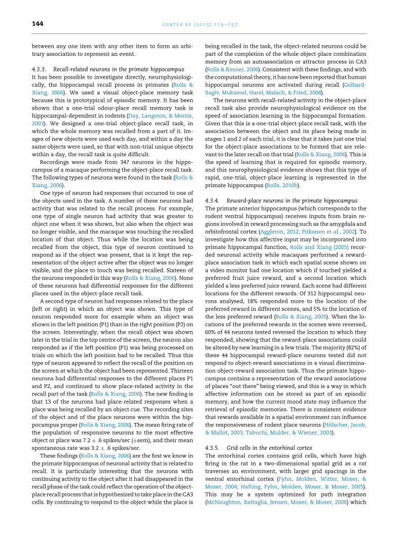
c o r t e x 6 2 ( 2 0 1 5 ) 1 1 9e1 5 7144
between any one item with any other item to form an arbi-
trary association to represent an event.
4.3.3. Recall-related neurons in the primate hippocampusIt has been possible to investigate directly, neurophysiologi-
cally, the hippocampal recall process in primates (Rolls &
Xiang, 2006). We used a visual object-place memory task
because this is prototypical of episodic memory. It has been
shown that a one-trial odour-place recall memory task is
hippocampal-dependent in rodents (Day, Langston, & Morris,
2003). We designed a one-trial object-place recall task, in
which the whole memory was recalled from a part of it. Im-
ages of new objects were used each day, and within a day the
same objects were used, so that with non-trial unique objects
within a day, the recall task is quite difficult.
Recordings were made from 347 neurons in the hippo-
campus of a macaque performing the object-place recall task.
The following types of neurons were found in the task (Rolls &
Xiang, 2006).
One type of neuron had responses that occurred to one of
the objects used in the task. A number of these neurons had
activity that was related to the recall process. For example,
one type of single neuron had activity that was greater to
object one when it was shown, but also when the object was
no longer visible, and the macaque was touching the recalled
location of that object. Thus while the location was being
recalled from the object, this type of neuron continued to
respond as if the object was present, that is it kept the rep-
resentation of the object active after the object was no longer
visible, and the place to touch was being recalled. Sixteen of
the neurons responded in this way (Rolls & Xiang, 2006). None
of these neurons had differential responses for the different
places used in the object-place recall task.
A second type of neuron had responses related to the place
(left or right) in which an object was shown. This type of
neuron responded more for example when an object was
shown in the left position (P1) than in the right position (P2) on
the screen. Interestingly, when the recall object was shown
later in the trial in the top centre of the screen, the neuron also
responded as if the left position (P1) was being processed on
trials on which the left position had to be recalled. Thus this
type of neuron appeared to reflect the recall of the position on
the screen at which the object had been represented. Thirteen
neurons had differential responses to the different places P1
and P2, and continued to show place-related activity in the
recall part of the task (Rolls & Xiang, 2006). The new finding is
that 13 of the neurons had place-related responses when a
place was being recalled by an object cue. The recording sites
of the object and of the place neurons were within the hip-
pocampus proper (Rolls & Xiang, 2006). Themean firing rate of
the population of responsive neurons to the most effective
object or place was 7.2 � .6 spikes/sec (�sem), and their mean
spontaneous rate was 3.2 � .6 spikes/sec.
These findings (Rolls & Xiang, 2006) are the first we know in
the primate hippocampus of neuronal activity that is related to
recall. It is particularly interesting that the neurons with
continuing activity to the object after it had disappeared in the
recall phaseof the task could reflect the operation of the object-
placerecall process that ishypothesized to takeplace in theCA3
cells. By continuing to respond to the object while the place is
being recalled in the task, the object-related neurons could be
part of the completion of the whole object-place combination
memory from an autoassociation or attractor process in CA3
(Rolls & Kesner, 2006). Consistent with these findings, andwith
thecomputational theory, ithasnowbeenreported thathuman
hippocampal neurons are activated during recall (Gelbard-
Sagiv, Mukamel, Harel, Malach, & Fried, 2008).
The neurons with recall-related activity in the object-place
recall task also provide neurophysiological evidence on the
speed of association learning in the hippocampal formation.
Given that this is a one-trial object-place recall task, with the
association between the object and its place being made in
stages 1 and 2 of each trial, it is clear that it takes just one trial
for the object-place associations to be formed that are rele-
vant to the later recall on that trial (Rolls & Xiang, 2006). This is
the speed of learning that is required for episodic memory,
and this neurophysiological evidence shows that this type of
rapid, one-trial, object-place learning is represented in the
primate hippocampus (Rolls, 2010b).
4.3.4. Reward-place neurons in the primate hippocampusThe primate anterior hippocampus (which corresponds to the
rodent ventral hippocampus) receives inputs from brain re-
gions involved in reward processing such as the amygdala and
orbitofrontal cortex (Aggleton, 2012; Pitkanen et al., 2002). To
investigate how this affective input may be incorporated into
primate hippocampal function, Rolls and Xiang (2005) recor-
ded neuronal activity while macaques performed a reward-
place association task in which each spatial scene shown on
a video monitor had one location which if touched yielded a
preferred fruit juice reward, and a second location which
yielded a less preferred juice reward. Each scene had different
locations for the different rewards. Of 312 hippocampal neu-
rons analysed, 18% responded more to the location of the
preferred reward in different scenes, and 5% to the location of
the less preferred reward (Rolls & Xiang, 2005). When the lo-
cations of the preferred rewards in the scenes were reversed,
60% of 44 neurons tested reversed the location to which they
responded, showing that the reward-place associations could
be altered by new learning in a few trials. Themajority (82%) of
these 44 hippocampal reward-place neurons tested did not
respond to object-reward associations in a visual discrimina-
tion object-reward association task. Thus the primate hippo-
campus contains a representation of the reward associations
of places “out there” being viewed, and this is a way in which
affective information can be stored as part of an episodic
memory, and how the current mood state may influence the
retrieval of episodic memories. There is consistent evidence
that rewards available in a spatial environment can influence
the responsiveness of rodent place neurons (Holscher, Jacob,
& Mallot, 2003; Tabuchi, Mulder, & Wiener, 2003).
4.3.5. Grid cells in the entorhinal cortexThe entorhinal cortex contains grid cells, which have high
firing in the rat in a two-dimensional spatial grid as a rat
traverses an environment, with larger grid spacings in the
ventral entorhinal cortex (Fyhn, Molden, Witter, Moser, &
Moser, 2004; Hafting, Fyhn, Molden, Moser, & Moser, 2005).
This may be a system optimized for path integration
(McNaughton, Battaglia, Jensen, Moser, & Moser, 2006) which
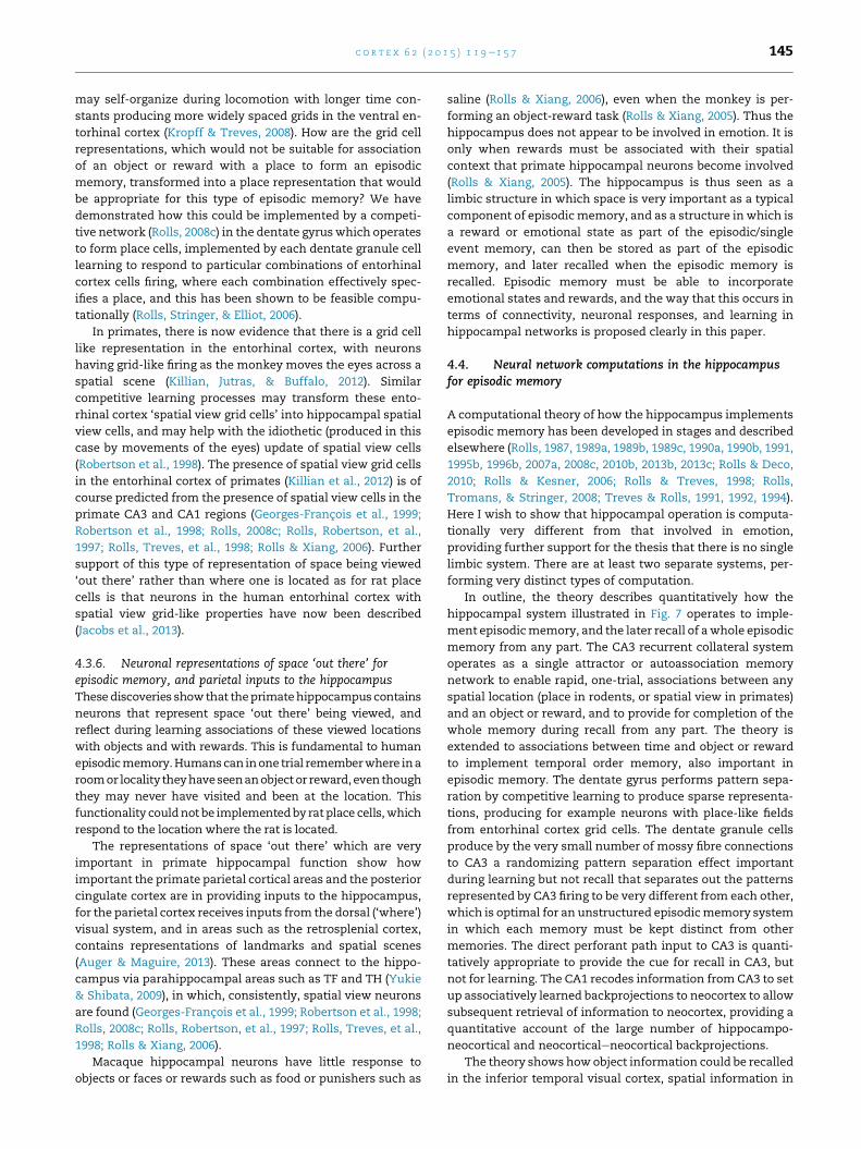
c o r t e x 6 2 ( 2 0 1 5 ) 1 1 9e1 5 7 145
may self-organize during locomotion with longer time con-
stants producing more widely spaced grids in the ventral en-
torhinal cortex (Kropff & Treves, 2008). How are the grid cell
representations, which would not be suitable for association
of an object or reward with a place to form an episodic
memory, transformed into a place representation that would
be appropriate for this type of episodic memory? We have
demonstrated how this could be implemented by a competi-
tive network (Rolls, 2008c) in the dentate gyruswhich operates
to form place cells, implemented by each dentate granule cell
learning to respond to particular combinations of entorhinal
cortex cells firing, where each combination effectively spec-
ifies a place, and this has been shown to be feasible compu-
tationally (Rolls, Stringer, & Elliot, 2006).
In primates, there is now evidence that there is a grid cell
like representation in the entorhinal cortex, with neurons
having grid-like firing as the monkey moves the eyes across a
spatial scene (Killian, Jutras, & Buffalo, 2012). Similar
competitive learning processes may transform these ento-
rhinal cortex ‘spatial view grid cells’ into hippocampal spatial
view cells, and may help with the idiothetic (produced in this
case by movements of the eyes) update of spatial view cells
(Robertson et al., 1998). The presence of spatial view grid cells
in the entorhinal cortex of primates (Killian et al., 2012) is of
course predicted from the presence of spatial view cells in the
primate CA3 and CA1 regions (Georges-Francois et al., 1999;
Robertson et al., 1998; Rolls, 2008c; Rolls, Robertson, et al.,
1997; Rolls, Treves, et al., 1998; Rolls & Xiang, 2006). Further
support of this type of representation of space being viewed
‘out there’ rather than where one is located as for rat place
cells is that neurons in the human entorhinal cortex with
spatial view grid-like properties have now been described
(Jacobs et al., 2013).
4.3.6. Neuronal representations of space ‘out there’ forepisodic memory, and parietal inputs to the hippocampusThesediscoveries showthat theprimatehippocampus contains
neurons that represent space ‘out there’ being viewed, and
reflect during learning associations of these viewed locations
with objects and with rewards. This is fundamental to human
episodicmemory.Humanscan inone trial rememberwhere ina
roomor locality theyhaveseenanobject or reward,even though
they may never have visited and been at the location. This
functionality couldnot be implementedby rat place cells,which
respond to the location where the rat is located.
The representations of space ‘out there’ which are very
important in primate hippocampal function show how
important the primate parietal cortical areas and the posterior
cingulate cortex are in providing inputs to the hippocampus,
for the parietal cortex receives inputs from the dorsal (‘where’)
visual system, and in areas such as the retrosplenial cortex,
contains representations of landmarks and spatial scenes
(Auger & Maguire, 2013). These areas connect to the hippo-
campus via parahippocampal areas such as TF and TH (Yukie
& Shibata, 2009), in which, consistently, spatial view neurons
are found (Georges-Francois et al., 1999; Robertson et al., 1998;
Rolls, 2008c; Rolls, Robertson, et al., 1997; Rolls, Treves, et al.,
1998; Rolls & Xiang, 2006).
Macaque hippocampal neurons have little response to
objects or faces or rewards such as food or punishers such as
saline (Rolls & Xiang, 2006), even when the monkey is per-
forming an object-reward task (Rolls & Xiang, 2005). Thus the
hippocampus does not appear to be involved in emotion. It is
only when rewards must be associated with their spatial
context that primate hippocampal neurons become involved
(Rolls & Xiang, 2005). The hippocampus is thus seen as a
limbic structure in which space is very important as a typical
component of episodicmemory, and as a structure in which is
a reward or emotional state as part of the episodic/single
event memory, can then be stored as part of the episodic
memory, and later recalled when the episodic memory is
recalled. Episodic memory must be able to incorporate
emotional states and rewards, and the way that this occurs in
terms of connectivity, neuronal responses, and learning in
hippocampal networks is proposed clearly in this paper.
4.4. Neural network computations in the hippocampusfor episodic memory
A computational theory of how the hippocampus implements
episodic memory has been developed in stages and described
elsewhere (Rolls, 1987, 1989a, 1989b, 1989c, 1990a, 1990b, 1991,
1995b, 1996b, 2007a, 2008c, 2010b, 2013b, 2013c; Rolls & Deco,
2010; Rolls & Kesner, 2006; Rolls & Treves, 1998; Rolls,
Tromans, & Stringer, 2008; Treves & Rolls, 1991, 1992, 1994).
Here I wish to show that hippocampal operation is computa-
tionally very different from that involved in emotion,
providing further support for the thesis that there is no single
limbic system. There are at least two separate systems, per-
forming very distinct types of computation.
In outline, the theory describes quantitatively how the
hippocampal system illustrated in Fig. 7 operates to imple-
ment episodicmemory, and the later recall of awhole episodic
memory from any part. The CA3 recurrent collateral system
operates as a single attractor or autoassociation memory
network to enable rapid, one-trial, associations between any
spatial location (place in rodents, or spatial view in primates)
and an object or reward, and to provide for completion of the
whole memory during recall from any part. The theory is
extended to associations between time and object or reward
to implement temporal order memory, also important in
episodic memory. The dentate gyrus performs pattern sepa-
ration by competitive learning to produce sparse representa-
tions, producing for example neurons with place-like fields
from entorhinal cortex grid cells. The dentate granule cells
produce by the very small number of mossy fibre connections
to CA3 a randomizing pattern separation effect important
during learning but not recall that separates out the patterns
represented by CA3 firing to be very different from each other,
which is optimal for an unstructured episodicmemory system
in which each memory must be kept distinct from other
memories. The direct perforant path input to CA3 is quanti-
tatively appropriate to provide the cue for recall in CA3, but
not for learning. The CA1 recodes information from CA3 to set
up associatively learned backprojections to neocortex to allow
subsequent retrieval of information to neocortex, providing a
quantitative account of the large number of hippocampo-
neocortical and neocorticaleneocortical backprojections.
The theory shows howobject information could be recalled
in the inferior temporal visual cortex, spatial information in
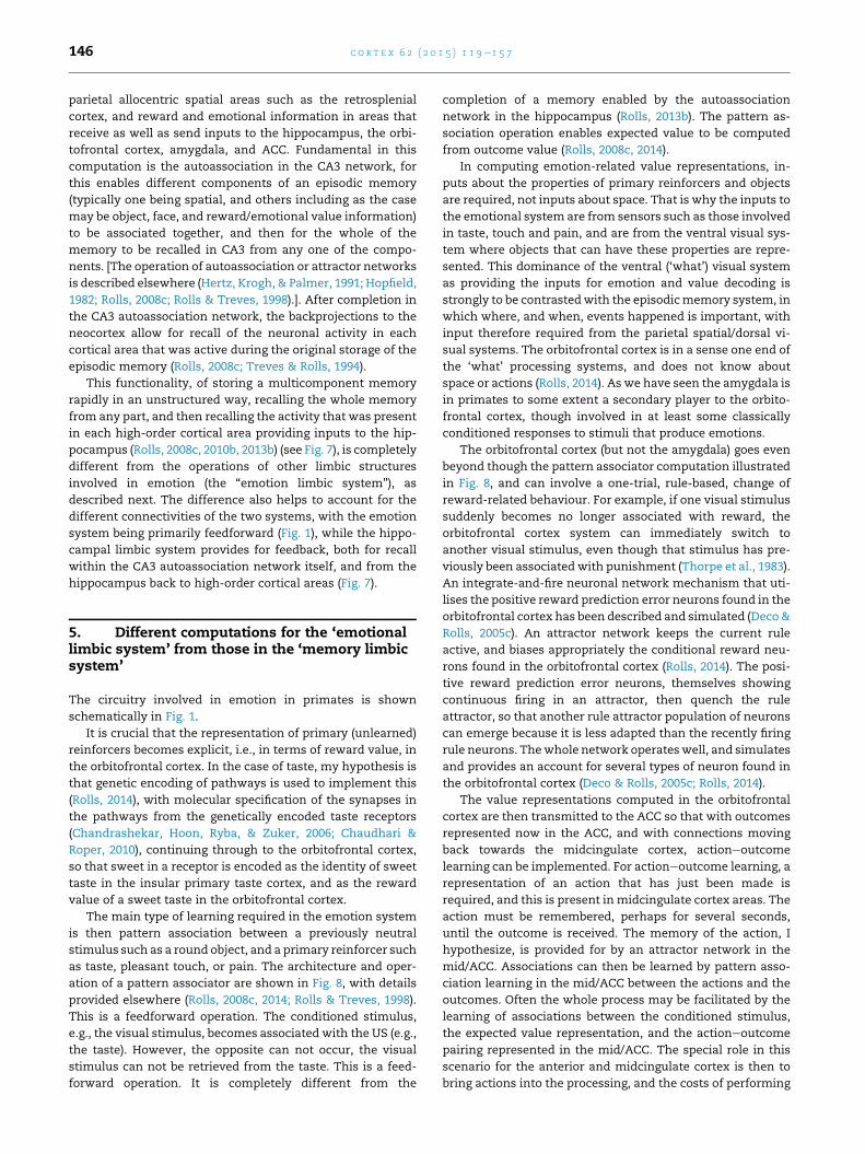
c o r t e x 6 2 ( 2 0 1 5 ) 1 1 9e1 5 7146
parietal allocentric spatial areas such as the retrosplenial
cortex, and reward and emotional information in areas that
receive as well as send inputs to the hippocampus, the orbi-
tofrontal cortex, amygdala, and ACC. Fundamental in this
computation is the autoassociation in the CA3 network, for
this enables different components of an episodic memory
(typically one being spatial, and others including as the case
may be object, face, and reward/emotional value information)
to be associated together, and then for the whole of the
memory to be recalled in CA3 from any one of the compo-
nents. [The operation of autoassociation or attractor networks
is described elsewhere (Hertz, Krogh, & Palmer, 1991; Hopfield,
1982; Rolls, 2008c; Rolls & Treves, 1998).]. After completion in
the CA3 autoassociation network, the backprojections to the
neocortex allow for recall of the neuronal activity in each
cortical area that was active during the original storage of the
episodic memory (Rolls, 2008c; Treves & Rolls, 1994).
This functionality, of storing a multicomponent memory
rapidly in an unstructured way, recalling the whole memory
from any part, and then recalling the activity that was present
in each high-order cortical area providing inputs to the hip-
pocampus (Rolls, 2008c, 2010b, 2013b) (see Fig. 7), is completely
different from the operations of other limbic structures
involved in emotion (the “emotion limbic system”), as
described next. The difference also helps to account for the
different connectivities of the two systems, with the emotion
system being primarily feedforward (Fig. 1), while the hippo-
campal limbic system provides for feedback, both for recall
within the CA3 autoassociation network itself, and from the
hippocampus back to high-order cortical areas (Fig. 7).
5. Different computations for the ‘emotionallimbic system’ from those in the ‘memory limbicsystem’
The circuitry involved in emotion in primates is shown
schematically in Fig. 1.
It is crucial that the representation of primary (unlearned)
reinforcers becomes explicit, i.e., in terms of reward value, in
the orbitofrontal cortex. In the case of taste, my hypothesis is
that genetic encoding of pathways is used to implement this
(Rolls, 2014), with molecular specification of the synapses in
the pathways from the genetically encoded taste receptors
(Chandrashekar, Hoon, Ryba, & Zuker, 2006; Chaudhari &
Roper, 2010), continuing through to the orbitofrontal cortex,
so that sweet in a receptor is encoded as the identity of sweet
taste in the insular primary taste cortex, and as the reward
value of a sweet taste in the orbitofrontal cortex.
The main type of learning required in the emotion system
is then pattern association between a previously neutral
stimulus such as a round object, and a primary reinforcer such
as taste, pleasant touch, or pain. The architecture and oper-
ation of a pattern associator are shown in Fig. 8, with details
provided elsewhere (Rolls, 2008c, 2014; Rolls & Treves, 1998).
This is a feedforward operation. The conditioned stimulus,
e.g., the visual stimulus, becomes associated with the US (e.g.,
the taste). However, the opposite can not occur, the visual
stimulus can not be retrieved from the taste. This is a feed-
forward operation. It is completely different from the
completion of a memory enabled by the autoassociation
network in the hippocampus (Rolls, 2013b). The pattern as-
sociation operation enables expected value to be computed
from outcome value (Rolls, 2008c, 2014).
In computing emotion-related value representations, in-
puts about the properties of primary reinforcers and objects
are required, not inputs about space. That is why the inputs to
the emotional system are from sensors such as those involved
in taste, touch and pain, and are from the ventral visual sys-
tem where objects that can have these properties are repre-
sented. This dominance of the ventral (‘what’) visual system
as providing the inputs for emotion and value decoding is
strongly to be contrastedwith the episodicmemory system, in
which where, and when, events happened is important, with
input therefore required from the parietal spatial/dorsal vi-
sual systems. The orbitofrontal cortex is in a sense one end of
the ‘what’ processing systems, and does not know about
space or actions (Rolls, 2014). As we have seen the amygdala is
in primates to some extent a secondary player to the orbito-
frontal cortex, though involved in at least some classically
conditioned responses to stimuli that produce emotions.
The orbitofrontal cortex (but not the amygdala) goes even
beyond though the pattern associator computation illustrated
in Fig. 8, and can involve a one-trial, rule-based, change of
reward-related behaviour. For example, if one visual stimulus
suddenly becomes no longer associated with reward, the
orbitofrontal cortex system can immediately switch to
another visual stimulus, even though that stimulus has pre-
viously been associated with punishment (Thorpe et al., 1983).
An integrate-and-fire neuronal network mechanism that uti-
lises the positive reward prediction error neurons found in the
orbitofrontal cortex has been described and simulated (Deco&
Rolls, 2005c). An attractor network keeps the current rule
active, and biases appropriately the conditional reward neu-
rons found in the orbitofrontal cortex (Rolls, 2014). The posi-
tive reward prediction error neurons, themselves showing
continuous firing in an attractor, then quench the rule
attractor, so that another rule attractor population of neurons
can emerge because it is less adapted than the recently firing
rule neurons. Thewhole network operateswell, and simulates
and provides an account for several types of neuron found in
the orbitofrontal cortex (Deco & Rolls, 2005c; Rolls, 2014).
The value representations computed in the orbitofrontal
cortex are then transmitted to the ACC so that with outcomes
represented now in the ACC, and with connections moving
back towards the midcingulate cortex, actioneoutcome
learning can be implemented. For actioneoutcome learning, a
representation of an action that has just been made is
required, and this is present in midcingulate cortex areas. The
action must be remembered, perhaps for several seconds,
until the outcome is received. The memory of the action, I
hypothesize, is provided for by an attractor network in the
mid/ACC. Associations can then be learned by pattern asso-
ciation learning in the mid/ACC between the actions and the
outcomes. Often the whole process may be facilitated by the
learning of associations between the conditioned stimulus,
the expected value representation, and the actioneoutcome
pairing represented in the mid/ACC. The special role in this
scenario for the anterior and midcingulate cortex is then to
bring actions into the processing, and the costs of performing
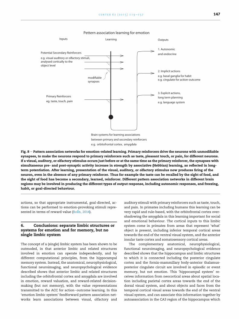
Fig. 8 e Pattern association networks for emotion-related learning. Primary reinforcers drive the neurons with unmodifiable
synapses, to make the neurons respond to primary reinforcers such as taste, pleasant touch, or pain, for different neurons.
If a visual, auditory, or olfactory stimulus occurs just before or at the same time as the primary reinforcer, the synapses with
simultaneous pre- and post-synaptic activity increase in strength by associative (Hebbian) learning, as reflected in long-
term potentiation. After learning, presentation of the visual, auditory, or olfactory stimulus now produces firing of the
neuron, even in the absence of any primary reinforcer. Thus for example the taste can be recalled by the sight of food, and
the sight of food has become a secondary, learned, reinforcer. Different pattern association networks in different brain
regions may be involved in producing the different types of output response, including autonomic responses, and freezing,
habit, or goal-directed behaviour.
c o r t e x 6 2 ( 2 0 1 5 ) 1 1 9e1 5 7 147
actions, so that appropriate instrumental, goal-directed, ac-
tions can be performed to emotion-provoking stimuli repre-
sented in terms of reward value (Rolls, 2014).
6. Conclusions: separate limbic structures orsystems for emotion and for memory, but nosingle limbic system
The concept of a (single) limbic system has been shown to be
outmoded, in that anterior limbic and related structures
involved in emotion can operate independently, and by
different computational principles, from the hippocampal
memory system. Instead, the anatomical, neurophysiological,
functional neuroimaging, and neuropsychological evidence
described shows that anterior limbic and related structures
including the orbitofrontal cortex and amygdala are involved
in emotion, reward valuation, and reward-related decision-
making (but not memory), with the value representations
transmitted to the ACC for actioneoutcome learning. In this
‘emotion limbic system’ feedforward pattern association net-
works learn associations between visual, olfactory and
auditory stimuli with primary reinforcers such as taste, touch,
and pain. In primates including humans this learning can be
very rapid and rule-based, with the orbitofrontal cortex over-
shadowing the amygdala in this learning important for social
and emotional behaviour. The cortical inputs to this limbic
system come in primates from areas that represent ‘what’
object is present, including inferior temporal cortical areas
towards the end of the ventral visual system, and the anterior
insular taste cortex and somatosensory cortical areas.
The complementary anatomical, neurophysiological,
functional neuroimaging, and neuropsychological evidence
described shows that the hippocampus and limbic structures
to which it is connected including the posterior cingulate
cortex and the fornix-mammillary body-anterior thalamus-
posterior cingulate circuit are involved in episodic or event
memory, but not emotion. This ‘hippocampal system’ re-
ceives information from neocortical areas about spatial loca-
tion including parietal cortex areas towards the end of the
dorsal visual system, and about objects and faces from the
temporal cortical visual areas towards the end of the ventral
visual system, and can associate this information together by
autoassociation in the CA3 region of the hippocampus which

c o r t e x 6 2 ( 2 0 1 5 ) 1 1 9e1 5 7148
involves feedback in the recurrent collateral system. The
hippocampalmemory system can later recall thewhole of this
information in the CA3 region from any component, a feed-
back process, and can recall that information back to
neocortical areas, again a feedback (to neocortex) recall pro-
cess. The nature of the computation in this hippocampal
system is thus very different from the feedforward pattern
association involved in stimulus-reward association learning
in the emotion system (Rolls, 2013b). Emotion and reward
signals can enter the hippocampal memory system from the
orbitofrontal cortex, amygdala and ACC (Rolls, 2014; Rolls &
Xiang, 2005), and can be recalled back to the orbitofrontal
cortex, amygdala and ACC during memory recall (Smith,
Stephan, Rugg, & Dolan, 2006), as emotion can provide a
component of episodic memory.
Thus, the emotional and hippocampal networks or ‘limbic
systems’ operate by different principles, and operate inde-
pendently of each other except insofar as emotional statemay
be part of an episodic memory. The concept of a single limbic
system may no longer appropriately reflect how the brain
operates. Instead, separate systems, which might be desig-
nated the emotional limbic system and the episodic memory
hippocampal system,arepresent in thebrain, and the separate
and different principles of their operation have been described
here and elsewhere (Rolls, 2008c, 2010b, 2013b, 2014).
Acknowledgements
The author has worked on some of the investigations
described here with I. De Araujo, G.C. Baylis, L.L. Baylis, A.
Berthoz, A. Bilderbeck, M.C.A. Booth, R. Bowtell, A.D. Brown-
ing, H.D. Critchley, J.D. Feigenbaum, S. Francis, L. Franco, P.
Georges-Francois, F. Grabenhorst, M.E. Hasselmo, C. Holscher,
J. Hornak, M. Kadohisa, R.P. Kesner, M. Kringelbach, C.M.
Leonard, C. Margot, C. McCabe, F. McGlone, F. Mora,
J. O’Doherty, B.A. Parris, D.I. Perrett, J. Redoute, R.G. Robertson,
M.K. Sanghera, T.R. Scott, Z. Sienkiewicz, S.J. Stringer,
S.J. Thorpe, A. Treves, J. Tromans, M.I. Velazco, J.V. Verhagen,
E.A. Wakeman, F.A.W. Wilson, J.-Z. Xiang, and S. Yaxley, and
their collaboration is sincerely acknowledged. Some of the
research described was supported by the Medical Research
Council, Wellcome Trust, Human Frontier Science Program,
and James S. McDonnell Foundation. Although in this paper
on “Limbic systems for emotion and formemory, but no single
limbic system” I have written for the first time on this topic,
I do note that some of the presentation of the evidence on the
functions of the orbitofrontal cortex etc. in emotion in Section
3 of this paper follows in part what I wrote in my Chapter 38
“The emotional systems” in J. K. Mai and G. Paxinos (Eds.), The
human nervous system (3rd ed., pp. 1315e1337). Elsevier:
London.
r e f e r e n c e s
Adolphs, R., Gosselin, F., Buchanan, T. W., Tranel, D., Schyns, P.,& Damasio, A. R. (2005). A mechanism for impaired fearrecognition after amygdala damage. Nature, 433, 68e72.
Aggleton, J. P. (2012). Multiple anatomical systems embeddedwithin the primate medial temporal lobe: implications forhippocampal function. Neuroscience and Biobehavioural Reviews,36, 1579e1596.
Amaral, D. G. (1987). Memory: anatomical organization ofcandidate brain regions. In V. B. Mountcastle (Ed.), Higherfunctions of the brain: Vol. V. Handbook of physiology. Section 1, Thenervous system (pp. 211e294). Washington DC: AmericanPhysiological Society.
Amaral, D. G. (2003). The amygdala, social behavior, and dangerdetection. Annals of the New York Academy of Sciences, 1000,337e347.
Amaral, D. G., Bauman, M. D., Capitanio, J. P., Lavenex, P.,Mason, W. A., Mauldin-Jourdain, M. L., et al. (2003). Theamygdala: is it an essential component of the neural networkfor social cognition? Neuropsychologia, 41, 517e522.
Amaral, D. G., Price, J. L., Pitkanen, A., & Carmichael, S. T. (1992).Anatomical organization of the primate amygdaloid complex.In J. P. Aggleton (Ed.), The amygdala (pp. 1e66). New York:Wiley-Liss.
Anderson, A. K., Christoff, K., Stappen, I., Panitz, D.,Ghahremani, D. G., Glover, G., et al. (2003). Dissociated neuralrepresentations of intensity and valence in human olfaction.Nature Neuroscience, 6, 196e202.
Antoniadis, E. A., Winslow, J. T., Davis, M., & Amaral, D. G. (2009).The nonhuman primate amygdala is necessary for theacquisition but not the retention of fear-potentiated startle.Biological Psychiatry, 65, 241e248.
de Araujo, I. E. T., Kringelbach, M. L., Rolls, E. T., & Hobden, P.(2003). The representation of umami taste in the human brain.Journal of Neurophysiology, 90, 313e319.
de Araujo, I. E. T., Kringelbach, M. L., Rolls, E. T., & McGlone, F.(2003). Human cortical responses to water in the mouth, andthe effects of thirst. Journal of Neurophysiology, 90, 1865e1876.
de Araujo, I. E. T., & Rolls, E. T. (2004). The representation in thehuman brain of food texture and oral fat. Journal ofNeuroscience, 24, 3086e3093.
de Araujo, I. E. T., Rolls, E. T., Kringelbach, M. L., McGlone, F., &Phillips, N. (2003). Taste-olfactory convergence, and therepresentation of the pleasantness of flavour, in the humanbrain. European Journal of Neuroscience, 18, 2059e2068.
de Araujo, I. E. T., Rolls, E. T., Velazco, M. I., Margot, C., &Cayeux, I. (2005). Cognitive modulation of olfactoryprocessing. Neuron, 46, 671e679.
Auger, S. D., & Maguire, E. A. (2013). Assessing the mechanism ofresponse in the retrosplenial cortex of good and poornavigators. Cortex, 49, 2904e2913.
Banta Lavenex, P., & Lavenex, P. (2009). Spatial memory and themonkey hippocampus: not all space is created equal.Hippocampus, 19, 8e19.
Barbas, H. (1988). Anatomic organization of basoventral andmediodorsal visual recipient prefrontal regions in the rhesusmonkey. Journal of Comparative Neurology, 276, 313e342.
Barbas, H. (1993). Organization of cortical afferent input to theorbitofrontal area in the rhesus monkey. Neuroscience, 56,841e864.
Barbas, H. (1995). Anatomic basis of cognitive-emotionalinteractions in the primate prefrontal cortex. Neuroscience andBiobehavioural Reviews, 19, 499e510.
Barbas, H. (2007). Specialized elements of orbitofrontal cortex inprimates. Annals of the New York Academy of Sciences, 1121,10e32.
Barbas, H., & Pandya, D. N. (1989). Architecture and intrinsicconnections of the prefrontal cortex in the rhesus monkey.Journal of Comparative Neurology, 286, 353e375.
Baylis, L. L., & Rolls, E. T. (1991). Responses of neurons in theprimate taste cortex to glutamate. Physiology and Behavior, 49,973e979.

c o r t e x 6 2 ( 2 0 1 5 ) 1 1 9e1 5 7 149
Baylis, L. L., Rolls, E. T., & Baylis, G. C. (1995). Afferent connectionsof the orbitofrontal cortex taste area of the primate.Neuroscience, 64, 801e812.
Bechara, A., Damasio, H., & Damasio, A. R. (2000). Emotion,decision making and the orbitofrontal cortex. Cerebral Cortex,10, 295e307.
Bechara, A., Damasio, H., Damasio, A. R., & Lee, G. P. (1999).Different contributions of the human amygdala andventromedial prefrontal cortex to decision-making. Journal ofNeuroscience, 19, 5473e5481.
Bechterew, W. (1900). Demonstration eines gehirns mit Zestorungder vorderenund inneren Theile der Hirnrinde beiderSchlafenlappen. Neurol. Centralbl., 20, 990e991.
Berlin, H., Rolls, E. T., & Iversen, S. D. (2005). Borderlinepersonality disorder, impulsivity and the orbitofrontal cortex.American Journal of Psychiatry, 162, 2360e2373.
Berlin, H., Rolls, E. T., & Kischka, U. (2004). Impulsivity, timeperception, emotion, and reinforcement sensitivity in patientswith orbitofrontal cortex lesions. Brain, 127, 1108e1126.
Bernoulli, J. (1738/1954). Exposition of a new theory on themeasurement of risk. Econometrica, 22, 23e36.
Brand, M., Grabenhorst, F., Starcke, K., Vandekerckhove, M. M., &Markowitsch, H. J. (2007). Role of the amygdala in decisionsunder ambiguity and decisions under risk: evidence frompatients with Urbach-Wiethe disease. Neuropsychologia, 45,1305e1317.
Broca, P. (1878). Anatomie comparee des circonvolutionscerebrales: le grand lobe limbique et la scissure limbique dansla serie des mammiferes. Revue Anthropologique, 1, 385e498.
Bromberg-Martin, E. S., Matsumoto, M., & Hikosaka, O. (2010).Dopamine in motivational control: rewarding, aversive, andalerting. Neuron, 68, 815e834.
Brown, S., & Schafer, E. A. (1888). An investigation into thefunctions of the occipital and temporal lobes of the monkey’sbrain. Philosophical Transactions of the Royal Society of LondonSeries B, 179, 303e327.
Buckley, M. J. (2005). The role of the perirhinal cortex andhippocampus in learning, memory, and perception. QuarterlyJournal of Experimental Psychology B, 58, 246e268.
Buckley, M. J., & Gaffan, D. (2000). The hippocampus, perirhinalcortex, and memory in the monkey. In J. J. Bolhuis (Ed.), Brain,perception, and memory: Advances in cognitive neuroscience (pp.279e298). Oxford: Oxford University Press.
Burgess, N. (2008). Spatial cognition and the brain. Annals of theNew York Academy of Sciences, 1124, 77e97.
Burgess, N., Maguire, E. A., & O’Keefe, J. (2002). The humanhippocampus and spatial and episodic memory. Neuron, 35,625e641.
Burton, M. J., Rolls, E. T., & Mora, F. (1976). Effects of hunger on theresponses of neurones in the lateral hypothalamus to thesight and taste of food. Experimental Neurology, 51, 668e677.
Bush, G., Luu, P., & Posner, M. I. (2000). Cognitive and emotionalinfluences in anterior cingulate cortex. Trends in CognitiveSciences, 4, 215e222.
Bush, G., Vogt, B. A., Holmes, J., Dale, A. M., Greve, D.,Jenike, M. A., et al. (2002). Dorsal anterior cingulate cortex: arole in reward-based decision making. Proceedings of theNational Academy of Sciences United States of America, 99,523e528.
Butter, C. M. (1969). Perseveration in extinction and indiscrimination reversal tasks following selective prefrontalablations in Macaca mulatta. Physiology and Behavior, 4,163e171.
Cabanac, M. (1992). Pleasure: the common currency. Journal ofTheoretical Biology, 155, 173e200.
Cannon, W. B. (1927). The James-Lange theory of emotion: acritical examination and an alternative theory. AmericanJournal of Psychology, 39, 106e124.
Cardinal, N., Parkinson, J. A., Hall, J., & Everitt, B. J. (2002). Emotionand motivation: the role of the amygdala, ventral striatum,and prefrontal cortex. Neuroscience and Biobehavioural Reviews,26, 321e352.
Carmichael, S. T., Clugnet, M.-C., & Price, J. L. (1994). Centralolfactory connections in the macaque monkey. Journal ofComparative Neurology, 346, 403e434.
Carmichael, S. T., & Price, J. L. (1994). Architectonic subdivision ofthe orbital and medial prefrontal cortex in the macaquemonkey. Journal of Comparative Neurology, 346, 366e402.
Carmichael, S. T., & Price, J. L. (1995a). Limbic connections of theorbital and medial prefrontal cortex in macaque monkeys.Journal of Comparative Neurology, 346, 403e434.
Carmichael, S. T., & Price, J. L. (1995b). Sensory and premotorconnections of the orbital and medial prefrontal cortex ofmacaque monkeys. Journal of Comparative Neurology, 363,642e664.
Carmichael, S. T., & Price, J. L. (1996). Connectional networkswithin the orbital and medial prefrontal cortex of macaquemonkeys. Journal of Comparative Neurology, 371, 179e207.
Cassaday, H. J., & Rawlins, J. N. (1997). The hippocampus, objects,and their contexts. Behavioural Neuroscience, 111, 1228e1244.
Catani, M., Dell’acqua, F., & Thiebaut de Schotten, M. (2013). Arevised limbic system model for memory, emotion andbehaviour. Neuroscience and Biobehavioural Reviews, 37,1724e1737.
Chadwick, M. J., Hassabis, D., Weiskopf, N., & Maguire, E. A.(2010). Decoding individual episodic memory traces in thehuman hippocampus. Current Biology, 20, 544e547.
Chandrashekar, J., Hoon, M. A., Ryba, N. J., & Zuker, C. S. (2006).The receptors and cells for mammalian taste. Nature, 444,288e294.
Chaudhari, N., Landin, A. M., & Roper, S. D. (2000). A metabotropicglutamate receptor variant functions as a taste receptor.Nature Neuroscience, 3, 113e119.
Chaudhari, N., & Roper, S. D. (2010). The cell biology of taste.Journal of Cell Biology, 190, 285e296.
Chib, V. S., Rangel, A., Shimojo, S., & O’Doherty, J. P. (2009).Evidence for a common representation of decision values fordissimilar goods in human ventromedial prefrontal cortex.Journal of Neuroscience, 29, 12315e12320.
Corkin, S. (2002). What’s new with the amnesic patient H.M.?Nature Reviews Neuroscience, 3, 153e160.
Craig, A. D. (2009). How do you feeldnow? The anteriorinsula and human awareness. Nature Reviews Neuroscience, 10,59e70.
Craig, A. D. (2011). Significance of the insula for the evolution ofhuman awareness of feelings from the body. Annals of the NewYork Academy of Sciences, 1225, 72e82.
Crane, J., & Milner, B. (2005). What went where? Impaired object-location learning in patients with right hippocampal lesions.Hippocampus, 15, 216e231.
Critchley, H. D., & Harrison, N. A. (2013). Visceral influences onbrain and behavior. Neuron, 77, 624e638.
Critchley, H. D., & Rolls, E. T. (1996a). Hunger and satiety modifythe responses of olfactory and visual neurons in the primateorbitofrontal cortex. Journal of Neurophysiology, 75, 1673e1686.
Critchley, H. D., & Rolls, E. T. (1996b). Olfactory neuronalresponses in the primate orbitofrontal cortex: analysis in anolfactory discrimination task. Journal of Neurophysiology, 75,1659e1672.
Critchley, H. D., & Rolls, E. T. (1996c). Responses of primate tastecortex neurons to the astringent tastant tannic acid. ChemicalSenses, 21, 135e145.
Critchley, H. D., Wiens, S., Rotshtein, P., Ohman, A., & Dolan, R. J.(2004). Neural systems supporting interoceptive awareness.Nature Neuroscience, 7, 189e195.
Damasio, A. R. (1994). Descartes’ error. New York: Putnam.
c o r t e x 6 2 ( 2 0 1 5 ) 1 1 9e1 5 7150
Damasio, A. R. (1996). The somatic marker hypothesis and thepossible functions of the prefrontal cortex. PhilosophicalTransactions of the Royal Society B: Biological Sciences, 351,1413e1420.
Damasio, A., Damasio, H., & Tranel, D. (2013). Persistence offeelings and sentience after bilateral damage of the insula.Cerebral Cortex, 23, 833e846.
Davis, M. (1994). The role of the amygdala in emotional learning.International Review of Neurobiology, 36, 225e266.
Davis, M. (2006). Neural systems involved in fear and anxietymeasured with fear-potentiated startle. American Psychologist,61, 741e756.
Day, M., Langston, R., & Morris, R. G. (2003). Glutamate-receptor-mediated encoding and retrieval of paired-associate learning.Nature, 424, 205e209.
Deco, G., & Rolls, E. T. (2005a). Attention, short-term memory, andaction selection: a unifying theory. Progress in Neurobiology, 76,236e256.
Deco, G., & Rolls, E. T. (2005b). Neurodynamics of biasedcompetition and co-operation for attention: a model withspiking neurons. Journal of Neurophysiology, 94, 295e313.
Deco, G., & Rolls, E. T. (2005c). Synaptic and spiking dynamicsunderlying reward reversal in orbitofrontal cortex. CerebralCortex, 15, 15e30.
Deco, G., & Rolls, E. T. (2006). Decision-making and Weber’s law: aneurophysiological model. European Journal of Neuroscience, 24,901e916.
Deco, G., Rolls, E. T., Albantakis, L., & Romo, R. (2013). Brainmechanisms for perceptual and reward-related decision-making. Progress in Neurobiology, 103, 194e213.
Deco, G., Rolls, E. T., & Romo, R. (2009). Stochastic dynamics as aprinciple of brain function. Progress in Neurobiology, 88, 1e16.
Delatour, B., & Witter, M. P. (2002). Projections from theparahippocampal region to the prefrontal cortex in the rat:evidence of multiple pathways. European Journal ofNeuroscience, 15, 1400e1407.
Dere, E., Easton, A., Nadel, L., & Huston, J. P. (Eds.). (2008).Handbook of episodic memory. Amsterdam: Elsevier.
Desimone, R., & Duncan, J. (1995). Neural mechanisms of selectivevisual attention. Annual Review of Neuroscience, 18, 193e222.
Devinsky, O., Morrell, M. J., & Vogt, B. A. (1995). Contributions ofanterior cingulate cortex to behaviour. Brain, 118, 279e306.
Eichenbaum, H. (1997). Declarative memory: insights fromcognitive neurobiology. Annual Review of Psychology, 48,547e572.
Everitt, B. J., Cardinal, R. N., Parkinson, J. A., & Robbins, T. W.(2003). Appetitive behavior: impact of amygdala-dependentmechanisms of emotional learning. Annals of the New YorkAcademy of Sciences, 985, 233e250.
Everitt, B. J., & Robbins, T. W. (2013). From the ventral to the dorsalstriatum: Devolving views of their roles in drug addiction.Neuroscience and Biobehavioural Reviews, 37, 1946e1954.
Feigenbaum, J. D., & Rolls, E. T. (1991). Allocentric and egocentricspatial information processing in the hippocampal formationof the behaving primate. Psychobiology, 19, 21e40.
Fellows, L. K. (2007). The role of orbitofrontal cortex in decisionmaking: a component process account. Annals of the New YorkAcademy of Sciences, 1121, 421e430.
Fellows, L. K. (2011). Orbitofrontal contributions to value-baseddecision making: evidence from humans with frontal lobedamage. Annals of the New York Academy of Sciences, 1239,51e58.
Fellows, L. K., & Farah, M. J. (2003). Ventromedial frontal cortexmediates affective shifting in humans: evidence from areversal learning paradigm. Brain, 126, 1830e1837.
Ferry, A. T., Ongur, D., An, X., & Price, J. L. (2000). Prefrontalcortical projections to the striatum in macaque monkeys:
evidence for an organization related to prefrontal networks.Journal of Comparative Neurology, 425, 447e470.
Francis, S., Rolls, E. T., Bowtell, R., McGlone, F., O’Doherty, J.,Browning, A., et al. (1999). The representation of pleasanttouch in the brain and its relationship with taste and olfactoryareas. NeuroReport, 10, 453e459.
Freese, J. L., & Amaral, D. G. (2009). Neuroanatomy of the primateamygdala. In P. J. Whalen, & E. A. Phelps (Eds.), The humanamygdala (pp. 3e42). New York: Guilford.
Frey, S., & Petrides, M. (2002). Orbitofrontal cortex and memoryformation. Neuron, 36, 171e176.
Frey, S., & Petrides, M. (2003). Greater orbitofrontal activitypredicts better memory for faces. European Journal ofNeuroscience, 17, 2755e2758.
Friston, K. J., Buechel, C., Fink, G. R., Morris, J., Rolls, E. T., &Dolan, R. J. (1997). Psychophysiological and modulatoryinteractions in neuroimaging. NeuroImage, 6, 218e229.
Fyhn, M., Molden, S., Witter, M. P., Moser, E. I., & Moser, M. B.(2004). Spatial representation in the entorhinal cortex. Science,305, 1258e1264.
Gaffan, D. (1994). Scene-specific memory for objects: a model ofepisodic memory impairment in monkeys with fornixtransection. Journal of Cognitive Neuroscience, 6, 305e320.
Gaffan, D., & Gaffan, E. A. (1991). Amnesia in man followingtransection of the fornix. A review. Brain, 114, 2611e2618.
Gaffan, D., & Harrison, S. (1989a). A comparison of the effects offornix section and sulcus principalis ablation upon spatiallearning by monkeys. Behavioural Brain Research, 31, 207e220.
Gaffan, D., & Harrison, S. (1989b). Place memory and scenememory: effects of fornix transection in the monkey.Experimental Brain Research, 74, 202e212.
Gaffan, D., & Saunders, R. C. (1985). Running recognition ofconfigural stimuli by fornix transected monkeys. QuarterlyJournal of Experimental Psychology (Hove), 37B, 61e71.
Gallagher, M., & Holland, P. C. (1994). The amygdala complex:multiple roles in associative learning and attention.Proceedings of the National Academy of Sciences United States ofAmerica, 91, 11771e11776.
Ge, T., Feng, J., Grabenhorst, F., & Rolls, E. T. (2012). ComponentialGranger causality, and its application to identifying the sourceand mechanisms of the top-down biased activation thatcontrols attention to affective vs sensory processing.NeuroImage, 59, 1846e1858.
Gelbard-Sagiv, H., Mukamel, R., Harel, M., Malach, R., & Fried, I.(2008). Internally generated reactivation of single neurons inhuman hippocampus during free recall. Science, 322, 96e101.
Georges-Francois, P., Rolls, E. T., & Robertson, R. G. (1999). Spatialview cells in the primate hippocampus: allocentric view nothead direction or eye position or place. Cerebral Cortex, 9,197e212.
Ghashghaei, H. T., & Barbas, H. (2002). Pathways for emotion:interactions of prefrontal and anterior temporal pathways inthe amygdala of the rhesus monkey. Neuroscience, 115,1261e1279.
Giocomo, L. M., & Hasselmo, M. E. (2007). Neuromodulation byglutamate and acetylcholine can change circuit dynamics byregulating the relative influence of afferent input andexcitatory feedback. Molecular Neurobiology, 36, 184e200.
Glimcher, P. W. (2011). Foundations of neuroeconomic analysis.Oxford: Oxford University Press.
Glimcher, P. W., Camerer, C. F., Fehr, E., & Poldrack, R. A. (Eds.).(2009). Neuroeconomics: Decision-making and the brain. London:Academic Press.
Glimcher, P. W., & Fehr, E. (Eds.). (2013). Neuroeconomics: Decision-making and the brain (2nd ed.). New York: Academic Press.
Gothard, K. M., Battaglia, F. P., Erickson, C. A., Spitler, K. M., &Amaral, D. G. (2007). Neural responses to facial expression and

c o r t e x 6 2 ( 2 0 1 5 ) 1 1 9e1 5 7 151
face identity in the monkey amygdala. Journal ofNeurophysiology, 97, 1671e1683.
Gottfried, J. A., Small, D. M., & Zald, D. H. (2006). The chemicalsenses. In D. H. Zald, & S. L. Rauch (Eds.), The orbitofrontal cortex(pp. 125e171). Oxford: Oxford University Press.
Grabenhorst, F., D’Souza, A., Parris, B. A., Rolls, E. T., &Passingham, R. E. (2010). A common neural scale for thesubjective pleasantness of different primary rewards.NeuroImage, 51, 1265e1274.
Grabenhorst, F., & Rolls, E. T. (2008). Selective attention toaffective value alters how the brain processes taste stimuli.European Journal of Neuroscience, 27, 723e729.
Grabenhorst, F., & Rolls, E. T. (2009). Different representations ofrelative and absolute value in the human brain. NeuroImage,48, 258e268.
Grabenhorst, F., & Rolls, E. T. (2010). Attentional modulation ofaffective vs sensory processing: functional connectivity and atop-down biased activation theory of selective attention.Journal of Neurophysiology, 104, 1649e1660.
Grabenhorst, F., & Rolls, E. T. (2011). Value, pleasure, and choicein the ventral prefrontal cortex. Trends in Cognitive Sciences, 15,56e67.
Grabenhorst, F., Rolls, E. T., & Bilderbeck, A. (2008). How cognitionmodulates affective responses to taste and flavor: top downinfluences on the orbitofrontal and pregenual cingulatecortices. Cerebral Cortex, 18, 1549e1559.
Grabenhorst, F., Rolls, E. T., & Margot, C. (2011). A hedonicallycomplex odor mixture captures the brain’s attention.NeuroImage, 55, 832e843.
Grabenhorst, F., Rolls, E. T., Margot, C., da Silva, M. A. A. P., &Velazco, M. I. (2007). How pleasant and unpleasant stimulicombine in different brain regions: odor mixtures. Journal ofNeuroscience, 27, 13532e13540.
Grabenhorst, F., Rolls, E. T., & Parris, B. A. (2008). From affectivevalue to decision-making in the prefrontal cortex. EuropeanJournal of Neuroscience, 28, 1930e1939.
Grabenhorst, F., Rolls, E. T., Parris, B. A., & D’Souza, A. (2010). Howthe brain represents the reward value of fat in the mouth.Cerebral Cortex, 20, 1082e1091.
Gray, J. A. (1975). Elements of a two-process theory of learning.London: Academic Press.
Guest, S., Grabenhorst, F., Essick, G., Chen, Y., Young, M.,McGlone, F., et al. (2007). Human cortical representation oforal temperature. Physiology and Behavior, 92, 975e984.
Haber, S. N., Kim, K. S., Mailly, P., & Calzavara, R. (2006). Reward-related cortical inputs define a large striatal region in primatesthat interface with associative cortical connections, providinga substrate for incentive-based learning. Journal ofNeuroscience, 26, 8368e8376.
van Haeften, T., Baks-te-Bulte, L., Goede, P. H., Wouterlood, F. G.,&Witter, M. P. (2003). Morphological and numerical analysis ofsynaptic interactions between neurons in deep and superficiallayers of the entorhinal cortex of the rat. Hippocampus, 13,943e952.
Hafting, T., Fyhn, M., Molden, S., Moser, M. B., & Moser, E. I. (2005).Microstructure of a spatial map in the entorhinal cortex.Nature, 436, 801e806.
Hare, T. A., O’Doherty, J., Camerer, C. F., Schultz, W., & Rangel, A.(2008). Dissociating the role of the orbitofrontal cortex and thestriatum in the computation of goal values and predictionerrors. Journal of Neuroscience, 28, 5623e5630.
Hassabis, D., Chu, C., Rees, G., Weiskopf, N., Molyneux, P. D., &Maguire, E. A. (2009). Decoding neuronal ensembles in thehuman hippocampus. Current Biology, 19, 546e554.
Hasselmo, M. E., Rolls, E. T., & Baylis, G. C. (1989). The role ofexpression and identity in the face-selective responses ofneurons in the temporal visual cortex of the monkey.Behavioural Brain Research, 32, 203e218.
Hertz, J., Krogh, A., & Palmer, R. G. (1991). An introduction to thetheory of neural computation. Wokingham: Addison-Wesley.
Holland, P. C., & Gallagher, M. (1999). Amygdala circuitry inattentional and representational processes. Trends in CognitiveSciences, 3, 65e73.
Holscher, C., Jacob, W., & Mallot, H. A. (2003). Reward modulatesneuronal activity in the hippocampus of the rat. BehaviouralBrain Research, 142, 181e191.
Holscher, C., Rolls, E. T., & Xiang, J.-Z. (2003). Perirhinal cortexneuronal activity related to long-term familiarity memory inthe macaque. European Journal of Neuroscience, 18, 2037e2046.
Hopfield, J. J. (1982). Neural networks and physical systems withemergent collective computational abilities. Proceedings of theNational Academy of Sciences United States of America, 79,2554e2558.
Hornak, J., Bramham, J., Rolls, E. T., Morris, R. G., O’Doherty, J.,Bullock, P. R., et al. (2003). Changes in emotion aftercircumscribed surgical lesions of the orbitofrontal andcingulate cortices. Brain, 126, 1691e1712.
Hornak, J., O’Doherty, J., Bramham, J., Rolls, E. T., Morris, R. G.,Bullock, P. R., et al. (2004). Reward-related reversal learningafter surgical excisions in orbitofrontal and dorsolateralprefrontal cortex in humans. Journal of Cognitive Neuroscience,16, 463e478.
Hornak, J., Rolls, E. T., & Wade, D. (1996). Face and voiceexpression identification in patients with emotional andbehavioural changes following ventral frontal lobe damage.Neuropsychologia, 34, 247e261.
Insausti, R., Amaral, D. G., & Cowan, W. M. (1987). The entorhinalcortex of the monkey. II. Cortical afferents. Journal ofComparative Neurology, 264, 356e395.
Isaacson, R. L. (1982). The limbic system (2nd ed.). New York:Plenum.
Itskov, P. M., Vinnik, E., & Diamond, M. E. (2011). Hippocampalrepresentation of touch-guided behavior in rats: persistentand independent traces of stimulus and reward location. PLoSONE, 6, e16462.
Iversen, S. D., & Mishkin, M. (1970). Perseverative interference inmonkeys following selective lesions of the inferior prefrontalconvexity. Experimental Brain Research, 11, 376e386.
Izquierdo, A., & Murray, E. A. (2004). Combined unilateral lesionsof the amygdala and orbital prefrontal cortex impair affectiveprocessing in rhesus monkeys. Journal of Neurophysiology, 91,2023e2039.
Izquierdo, A., Suda, R. K., & Murray, E. A. (2004). Bilateral orbitalprefrontal cortex lesions in rhesus monkeys disrupt choicesguided by both reward value and reward contingency. Journalof Neuroscience, 24, 7540e7548.
Jacobs, J., Weidemann, C. T., Miller, J. F., Solway, A., Burke, J. F.,Wei, X. X., et al. (2013). Direct recordings of grid-like neuronalactivity in human spatial navigation. Nature Neuroscience, 16,1188e1190.
Jarrard, E. L. (1993). On the role of the hippocampus in learningand memory in the rat. Behavioral and Neural Biology, 60, 9e26.
Jeffery, K. J., Anderson, M. I., Hayman, R., & Chakraborty, S. (2004).A proposed architecture for the neural representation ofspatial context. Neuroscience and Biobehavioural Reviews, 28,201e218.
Jeffery, K. J., & Hayman, R. (2004). Plasticity of the hippocampalplace cell representation. Reviews in the Neurosciences, 15,309e331.
Johnson, T. N., Rosvold, H. E., & Mishkin, M. (1968). Projectionsfrom behaviorally defined sectors of the prefrontal cortex tothe basal ganglia, septum and diencephalon of the monkey.Experimental Neurology, 21, 20e34.
Jones, B., & Mishkin, M. (1972). Limbic lesions and the problem ofstimulus-reinforcement associations. Experimental Neurology,36, 362e377.
c o r t e x 6 2 ( 2 0 1 5 ) 1 1 9e1 5 7152
Kable, J. W., & Glimcher, P. W. (2009). The neurobiology ofdecision: consensus and controversy. Neuron, 63, 733e745.
Kadohisa, M., Rolls, E. T., & Verhagen, J. V. (2004). Orbitofrontalcortex neuronal representation of temperature and capsaicinin the mouth. Neuroscience, 127, 207e221.
Kadohisa, M., Rolls, E. T., & Verhagen, J. V. (2005a). Neuronalrepresentations of stimuli in the mouth: the primate insulartaste cortex, orbitofrontal cortex, and amygdala. ChemicalSenses, 30, 401e419.
Kadohisa, M., Rolls, E. T., & Verhagen, J. V. (2005b). The primateamygdala: neuronal representations of the viscosity, fattexture, temperature, grittiness and taste of foods.Neuroscience, 132, 33e48.
Kemp, J. M., & Powell, T. P. S. (1970). The cortico-striateprojections in the monkey. Brain, 93, 525e546.
Kesner, R. P. (1998). Neural mediation of memory for time: role ofhippocampus and medial prefrontal cortex. PsychologicalBulletin Reviews, 5, 585e596.
Kesner, R. P., Lee, I., & Gilbert, P. (2004). A behavioral assessmentof hippocampal function based on a subregional analysis.Reviews in the Neurosciences, 15, 333e351.
Kesner, R. P., Morris, A. M., & Weeden, C. S. S. (2012). Spatial,temporal, and associative behavioral functions associatedwith different subregions of the hippocampus. In T. R. Zentall,& E. A. Wasserman (Eds.), Oxford handbook of comparativecognition (pp. 322e346). Oxford: Oxford University Press.
Kesner, R. P., & Rolls, E. T. (2001). Role of long term synapticmodification in short termmemory. Hippocampus, 11, 240e250.
Killian, N. J., Jutras, M. J., & Buffalo, E. A. (2012). A map of visualspace in the primate entorhinal cortex. Nature, 491, 761e764.
Kluver, H., & Bucy, P. C. (1939). Preliminary analysis of functionsof the temporal lobes in monkeys. Archives of Neurology andPsychiatry, 42, 979e1000.
Knutson, B., Rick, S., Wimmer, G. E., Prelec, D., & Loewenstein, G.(2007). Neural predictors of purchases. Neuron, 53, 147e156.
Kobayashi, S., Pinto de Carvalho, O., & Schultz, W. (2010).Adaptation of reward sensitivity in orbitofrontal neurons.Journal of Neuroscience, 30, 534e544.
Kolb, B., & Whishaw, I. Q. (2003). Fundamentals of humanneuropsychology (5th ed.). New York: Worth.
Kringelbach, M. L., O’Doherty, J., Rolls, E. T., & Andrews, C. (2003).Activation of the human orbitofrontal cortex to a liquid foodstimulus is correlated with its subjective pleasantness.Cerebral Cortex, 13, 1064e1071.
Kringelbach, M. L., & Rolls, E. T. (2003). Neural correlates of rapidreversal learning in a simple model of human socialinteraction. NeuroImage, 20, 1371e1383.
Kringelbach, M. L., & Rolls, E. T. (2004). The functionalneuroanatomy of the human orbitofrontal cortex: evidencefrom neuroimaging and neuropsychology. Progress inNeurobiology, 72, 341e372.
Krolak-Salmon, P., Henaff, M. A., Isnard, J., Tallon-Baudry, C.,Guenot, M., Vighetto, A., et al. (2003). An attention modulatedresponse to disgust in human ventral anterior insula. Annals ofNeurology, 53, 446e453.
Kropff, E., & Treves, A. (2008). The emergence of grid cells:intelligent design or just adaptation? Hippocampus, 18,1256e1269.
Lavenex, P., Suzuki, W. A., & Amaral, D. G. (2004). Perirhinal andparahippocampal cortices of the macaque monkey: intrinsicprojections and interconnections. Journal of ComparativeNeurology, 472, 371e394.
LeDoux, J. E. (1995). Emotion: clues from the brain. Annual Reviewof Psychology, 46, 209e235.
LeDoux, J. E. (2000a). The amygdala, fear conditioning andemotion. In J. P. Aggleton (Ed.), The amygdala: A functionalanalysis. Oxford: Oxford University Press.
LeDoux, J. E. (2000b). Emotion circuits in the brain. Annual Reviewof Neuroscience, 23, 155e184.
Leonard, C. M., Rolls, E. T., Wilson, F. A. W., & Baylis, G. C. (1985).Neurons in the amygdala of the monkey with responsesselective for faces. Behavioural Brain Research, 15, 159e176.
Luo, Q., Ge, T., Grabenhorst, F., Feng, J., & Rolls, E. T. (2013).Attention-dependent modulation of cortical taste circuitsrevealed by Granger causality with signal-dependent noise.PLoS Computational Biology, 9, e1003265.
MacLean, P. D. (1949). Psychosomatic disease and the ‘visceralbrain’: recent developments bearing on the Papez theory ofemotion. Psychosomatic Medicine, 11, 338e353.
MacLean, P. D. (1952). Some psychiatric implications ofphysiological studies on frontotemporal portion of limbicsystem (visceral brain). Electroencephalography and ClinicalNeurophysiology, 4, 407e418.
Maia, T. V., & McClelland, J. L. (2004). A reexamination of theevidence for the somatic marker hypothesis: whatparticipants really know in the Iowa gambling task. Proceedingsof the National Academy of Sciences United States of America, 101,16075e16080.
Malkova, L., & Mishkin, M. (2003). One-trial memory for object-place associations after separate lesions of hippocampus andposterior parahippocampal region in the monkey. Journal ofNeuroscience, 23, 1956e1965.
Martin, S. J., Grimwood, P. D., & Morris, R. G. (2000). Synapticplasticity and memory: an evaluation of the hypothesis.Annual Review of Neuroscience, 23, 649e711.
Maruyama, Y., Pereira, E., Margolskee, R. F., Chaudhari, N., &Roper, S. D. (2006). Umami responses in mouse taste cellsindicate more than one receptor. Journal of Neuroscience, 26,2227e2234.
Matsumoto, M., & Hikosaka, O. (2009). Two types of dopamineneuron distinctly convey positive and negative motivationalsignals. Nature, 459, 837e841.
Matsumoto, M., Matsumoto, K., Abe, H., & Tanaka, K. (2007).Medial prefrontal selectivity signalling prediction errors ofaction values. Nature Neuroscience, 10, 647e656.
McCabe, C., & Rolls, E. T. (2007). Umami: a delicious flavor formedby convergence of taste and olfactory pathways in the humanbrain. European Journal of Neuroscience, 25, 1855e1864.
McCabe, C., Rolls, E. T., Bilderbeck, A., & McGlone, F. (2008).Cognitive influences on the affective representation of touchand the sight of touch in the human brain. Social, Cognitive andAffective Neuroscience, 3, 97e108.
McFarland, D. J., & Sibly, R. M. (1975). The behavioural finalcommon path. Philosophical Transactions of the Royal Society B:Biological Sciences, 270, 265e293.
McNaughton, B. L., Barnes, C. A., & O’Keefe, J. (1983). Thecontributions of position, direction, and velocity to single unitactivity in the hippocampus of freely-moving rats.Experimental Brain Research, 52, 41e49.
McNaughton, B. L., Battaglia, F. P., Jensen, O., Moser, E. I., &Moser, M.-B. (2006). Path integration and the neural basis ofthe ‘cognitive map’. Nature Reviews Neuroscience, 7, 663e678.
Medina, L., Bupesh, M., & Abellan, A. (2011). Contribution ofgenoarchitecture to understanding forebrain evolution anddevelopment, with particular emphasis on the amygdala.Brain, Behavior and Evolution, 78, 216e236.
Menon, V., & Uddin, L. Q. (2010). Saliency, switching, attentionand control: a network model of insula function. BrainStructure & Function, 214, 655e667.
Mesulam, M. M. (2000). Behavioral neuroanatomy: large-scalenetworks, association cortex, frontal syndromes, the limbicsystem, and hemispheric specializations. In M. M. Mesulam(Ed.), Principles of behavioral and cognitive neurology (2nd ed.) (pp.1e120). Oxford: Oxford University Press.
c o r t e x 6 2 ( 2 0 1 5 ) 1 1 9e1 5 7 153
Moll, J., Krueger, F., Zahn, R., Pardini, M., de Oliveira-Souza, R., &Grafman, J. (2006). Human fronto-mesolimbic networks guidedecisions about charitable donation. Proceedings of the NationalAcademy of Sciences United States of America, 103, 15623e15628.
Montague, P. R., & Berns, G. S. (2002). Neural economics and thebiological substrates of valuation. Neuron, 36, 265e284.
Mora, F., Avrith, D. B., Phillips, A. G., & Rolls, E. T. (1979). Effects ofsatiety on self-stimulation of the orbitofrontal cortex in themonkey. Neuroscience Letters, 13, 141e145.
Mora, F., Avrith, D. B., & Rolls, E. T. (1980). An electrophysiologicaland behavioural study of self-stimulation in the orbitofrontalcortex of the rhesus monkey. Brain Research Bulletin, 5,111e115.
Morecraft, R. J., Geula, C., & Mesulam, M.-M. (1992).Cytoarchitecture and neural afferents of orbitofrontal cortexin the brain of the monkey. Journal of Comparative Neurology,232, 341e358.
Morecraft, R. J., & Tanji, J. (2009). Cingulofrontal interaction andthe cingulate motor areas. In B. A. Vogt (Ed.), Cingulateneurobiology and disease (pp. 113e144). Oxford: OxfordUniversity Press.
Morris, J. S., & Dolan, R. J. (2001). Involvement of human amygdalaand orbitofrontal cortex in hunger-enhanced memory for foodstimuli. Journal of Neuroscience, 21, 5304e5310.
Mufson, E. J., & Mesulam, M.-M. (1982). Insula of the old worldmonkey: II: afferent cortical input and comments on theclaustrum. Journal of Comparative Neurology, 212, 23e37.
Muller, R. U., Kubie, J. L., Bostock, E. M., Taube, J. S., & Quirk, G. J.(1991). Spatial firing correlates of neurons in the hippocampalformation of freely moving rats. In J. Paillard (Ed.), Brain andspace (pp. 296e333). Oxford: Oxford University Press.
Murray, E. A., Baxter, M. G., & Gaffan, D. (1998). Monkeys withrhinal cortex damage or neurotoxic hippocampal lesions areimpaired on spatial scene learning and object reversals.Behavioral Neuroscience, 112, 1291e1303.
Murray, E. A., & Izquierdo, A. (2007). Orbitofrontal cortex andamygdala contributions to affect and action in primates.Annals of the New York Academy of Sciences, 1121, 273e296.
Nauta, W. J. H. (1964). Some efferent connections of the prefrontalcortex in the monkey. In J. M. Warren, & K. Akert (Eds.), Thefrontal granular cortex and behavior (pp. 397e407). New York:McGraw Hill.
Niki, H., & Watanabe, M. (1979). Prefrontal and cingulate unitactivity during timing behavior in the monkey. Brain Research,171, 213e224.
Norgren, R. (1984). Central neural mechanisms of taste. In I. Darien-Smith (Ed.), Handbook of physiology e The nervous system III.Sensory processes 1 (pp. 1087e1128). Washington, DC: AmericanPhysiological Society.
O’Doherty, J. P., Deichmann, R., Critchley, H. D., & Dolan, R. J.(2002). Neural responses during anticipation of a primary tastereward. Neuron, 33, 815e826.
O’Doherty, J., Kringelbach, M. L., Rolls, E. T., Hornak, J., &Andrews, C. (2001). Abstract reward and punishmentrepresentations in the human orbitofrontal cortex. NatureNeuroscience, 4, 95e102.
O’Doherty, J., Rolls, E. T., Francis, S., Bowtell, R., & McGlone, F.(2001). The representation of pleasant and aversive taste inthe human brain. Journal of Neurophysiology, 85, 1315e1321.
O’Doherty, J., Rolls, E. T., Francis, S., Bowtell, R., McGlone, F.,Kobal, G., et al. (2000). Sensory-specific satiety relatedolfactory activation of the human orbitofrontal cortex.NeuroReport, 11, 893e897.
O’Doherty, J., Winston, J., Critchley, H., Perrett, D., Burt, D. M., &Dolan, R. J. (2003). Beauty in a smile: the role of medialorbitofrontal cortex in facial attractiveness. Neuropsychologia,41, 147e155.
O’Keefe, J. (1984). Spatial memory within and without thehippocampal system. In W. Seifert (Ed.), Neurobiology of thehippocampus (pp. 375e403). London: Academic Press.
O’Keefe, J., & Nadel, L. (1978). The hippocampus as a cognitive map.Oxford: Clarendon Press.
Olausson, H., Lamarre, Y., Backlund, H., Morin, C., Wallin, B. G.,Starck, G., et al. (2002). Unmyelinated tactile afferents signaltouch and project to insular cortex. Nature Neuroscience, 5,900e904.
Ongur, D., Ferry, A. T., & Price, J. L. (2003). Architectonic divisionof the human orbital and medial prefrontal cortex. Journal ofComparative Neurology, 460, 425e449.
Ongur, D., & Price, J. L. (2000). The organisation of networkswithin the orbital and medial prefrontal cortex of rats,monkeys and humans. Cerebral Cortex, 10, 206e219.
Padoa-Schioppa, C. (2009). Range-adapting representation ofeconomic value in the orbitofrontal cortex. Journal ofNeuroscience, 29, 14004e14014.
Padoa-Schioppa, C. (2011). Neurobiology of economic choice: agood-based model. Annual Review of Neuroscience, 34, 333e359.
Padoa-Schioppa, C., & Assad, J. A. (2006). Neurons in theorbitofrontal cortex encode economic value. Nature, 441,223e226.
Padoa-Schioppa, C., & Assad, J. A. (2008). The representation ofeconomic value in the orbitofrontal cortex is invariant forchanges of menu. Nature Neuroscience, 11, 95e102.
Palomero-Gallagher, N., & Zilles, K. (2004). Isocortex. In G. Paxinos(Ed.), The rat nervous system (pp. 729e757). San Diego, CA:Elsevier Academic Press.
Pandya, D. N., & Yeterian, E. H. (1996). Comparison of prefrontalarchitecture and connections. Philosophical Transactions of theRoyal Society of London Series B, 351, 1423e1431.
Papez, J. W. (1937). A proposed mechanism for emotion. Archivesof Neurology and Psychiatry, 38, 725e743.
Parkinson, J. K., Murray, E. A., & Mishkin, M. (1988). A selectivemnemonic role for the hippocampus in monkeys: memory forthe location of objects. Journal of Neuroscience, 8, 4159e4167.
Passingham, R. E. P., & Wise, S. P. (2012). The neurobiology of theprefrontal cortex. Oxford: Oxford University Press.
Paton, J. J., Belova, M. A., Morrison, S. E., & Salzman, C. D. (2006).The primate amygdala represents the positive and negativevalue of visual stimuli during learning. Nature, 439, 865e870.
Petrides, M. (1985). Deficits on conditional associative-learningtasks after frontal- and temporal-lobe lesions in man.Neuropsychologia, 23, 601e614.
Petrides, M. (2007). The orbitofrontal cortex: novelty, deviationfrom expectation, and memory. Annals of the New YorkAcademy of Sciences, 1121, 33e53.
Petrides, M., & Pandya, D. N. (1995). Comparative architectonicanalysis of the human and macaque frontal cortex. In F. Boller, &J. Grafman (Eds.), Handbook of neuropsychology (Vol. 9); (pp.17e58). Amsterdam: Elsevier Science.
Phelps, E. A., & LeDoux, J. E. (2005). Contributions of the amygdalato emotion processing: from animal models to humanbehavior. Neuron, 48, 175e187.
Phillips, M. L., Williams, L. M., Heining, M., Herba, C. M.,Russell, T., Andrew, C., et al. (2004). Differential neuralresponses to overt and covert presentations of facialexpressions of fear and disgust. NeuroImage, 21, 1484e1496.
Phillips, M. L., Young, A. W., Scott, S. K., Calder, A. J., Andrew, C.,Giampietro, V., et al. (1998). Neural responses to facial andvocal expressions of fear and disgust. Proceedings of the RoyalSociety of London. Series B, 265, 1809e1817.
Pitkanen, A., Kelly, J. L., & Amaral, D. G. (2002). Projections fromthe lateral, basal, and accessory basal nuclei of the amygdalato the entorhinal cortex in the macaque monkey.Hippocampus, 12, 186e205.
c o r t e x 6 2 ( 2 0 1 5 ) 1 1 9e1 5 7154
Preuss, T. M. (1995). Do rats have prefrontal cortex? The Rose-Woolsey-Akert program reconsidered. Journal of CognitiveNeuroscience, 7, 1e24.
Price, J. L. (2006). Connections of orbital cortex. In D. H. Zald, &S. L. Rauch (Eds.), The orbitofrontal cortex (pp. 39e55). Oxford:Oxford University Press.
Price, J. L. (2007). Definition of the orbital cortex in relation tospecific connections with limbic and visceral structures andother cortical regions. Annals of the New York Academy ofSciences, 1121, 54e71.
Price, J. L., Carmichael, S. T., Carnes, K. M., Clugnet, M.-C.,Kuroda, M., & Ray, J. P. (1991). Olfactory input to the prefrontalcortex. In J. L. Davis, & H. Eichenbaum (Eds.), Olfaction: A modelsystem for computational neuroscience (pp. 101e120). Cambridge,Mass.: MIT Press.
Pritchard, T. C., Edwards, E. M., Smith, C. A., Hilgert, K. G.,Gavlick, A. M., Maryniak, T. D., et al. (2005). Gustatory neuralresponses in the medial orbitofrontal cortex of the old worldmonkey. Journal of Neuroscience, 25, 6047e6056.
Pritchard, T. C., Hamilton, R. B., Morse, J. R., & Norgren, R. (1986).Projections of thalamic gustatory and lingual areas in themonkey, Macaca fascicularis. Journal of Comparative Neurology,244, 213e228.
Rawlins, J. N. P. (1985). Associations across time: thehippocampus as a temporary memory store. Behavioral andBrain Sciences, 8, 479e496.
Robertson, R. G., Rolls, E. T., & Georges-Francois, P. (1998). Spatialview cells in the primate hippocampus: effects of removal ofview details. Journal of Neurophysiology, 79, 1145e1156.
Rolls, E. T. (1984). Neurons in the cortex of the temporal lobe andin the amygdala of the monkey with responses selective forfaces. Human Neurobiology, 3, 209e222.
Rolls, E. T. (1987). Information representation, processing andstorage in the brain: analysis at the single neuron level. In J.-P. Changeux, & M. Konishi (Eds.), The neural and molecular basesof learning (pp. 503e540). Chichester: Wiley.
Rolls, E. T. (1989a). Functions of neuronal networks in thehippocampus and cerebral cortex in memory. InR. M. J. Cotterill (Ed.), Models of brain function (pp. 15e33).Cambridge: Cambridge University Press.
Rolls, E. T. (1989b). Functions of neuronal networks in thehippocampus and neocortex in memory. In J. H. Byrne, &W. O. Berry (Eds.), Neural models of plasticity: Experimental andtheoretical approaches (pp. 240e265). San Diego: Academic Press.
Rolls, E. T. (1989c). The representation and storage of informationin neuronal networks in the primate cerebral cortex andhippocampus. In R. Durbin, C. Miall, & G. Mitchison (Eds.), Thecomputing neuron (pp. 125e159). Wokingham, England:Addison-Wesley.
Rolls, E. T. (1990a). Functions of the primate hippocampus inspatial processing and memory. In D. S. Olton, & R. P. Kesner(Eds.), Neurobiology of comparative cognition (pp. 339e362).Hillsdale, N.J.: L. Erlbaum.
Rolls, E. T. (1990b). Theoretical and neurophysiological analysis ofthe functions of the primate hippocampus in memory. ColdSpring Harbor Symposia in Quantitative Biology, 55, 995e1006.
Rolls, E. T. (1991). Functions of the primate hippocampus inspatial and non-spatial memory. Hippocampus, 1, 258e261.
Rolls, E. T. (1992a). Neurophysiological mechanisms underlyingface processing within and beyond the temporal corticalvisual areas. Philosophical Transactions of the Royal Society ofLondon Series B, 335, 11e21.
Rolls, E. T. (1992b). Neurophysiology and functions of the primateamygdala. In J. P. Aggleton (Ed.), The amygdala (pp. 143e165).New York: Wiley-Liss.
Rolls, E. T. (1995a). Central taste anatomy and neurophysiology. InR. L. Doty (Ed.), Handbook of olfaction and gustation (pp. 549e573).New York: Dekker (Chapter 24).
Rolls, E. T. (1995b). A model of the operation of the hippocampusand entorhinal cortex in memory. International Journal of NeuralSystems, 6, 51e70.
Rolls, E. T. (1996a). The orbitofrontal cortex. PhilosophicalTransactions of the Royal Society of London Series B, 351,1433e1444.
Rolls, E. T. (1996b). A theory of hippocampal function in memory.Hippocampus, 6, 601e620.
Rolls, E. T. (1997). Taste and olfactory processing in the brain andits relation to the control of eating. Critical Reviews inNeurobiology, 11, 263e287.
Rolls, E. T. (1999a). The brain and emotion. Oxford: OxfordUniversity Press.
Rolls, E. T. (1999b). The functions of the orbitofrontal cortex.Neurocase, 5, 301e312.
Rolls, E. T. (1999c). Spatial view cells and the representation ofplace in the primate hippocampus. Hippocampus, 9, 467e480.
Rolls, E. T. (2000a). Functions of the primate temporal lobe corticalvisual areas in invariant visual object and face recognition.Neuron, 27, 205e218.
Rolls, E. T. (2000b). Neurophysiology and functions of the primateamygdala, and the neural basis of emotion. In J. P. Aggleton(Ed.), The amygdala: A functional analysis (2nd ed.) (pp. 447e478).Oxford: Oxford University Press.
Rolls, E. T. (2000c). The representation of umami taste in the tastecortex. Journal of Nutrition, 130, S960eS965.
Rolls, E. T. (2000d). Taste, olfactory, visual and somatosensoryrepresentations of the sensory properties of foods in the brain,and their relation to the control of food intake. InH.-R. Berthoud,& R. J. Seeley (Eds.), Neural and metabolic control of macronutrientintake (pp. 247e262). Boca-Raton, Florida: CRC Press.
Rolls, E. T. (2004). The functions of the orbitofrontal cortex. Brainand Cognition, 55, 11e29.
Rolls, E. T. (2005). Emotion explained. Oxford: Oxford UniversityPress.
Rolls, E. T. (2007a). An attractor network in the hippocampus:theory and neurophysiology. Learning and Memory, 14,714e731.
Rolls, E. T. (2007b). The representation of information about facesin the temporal and frontal lobes. Neuropsychologia, 45,125e143.
Rolls, E. T. (2007c). Sensory processing in the brain related to thecontrol of food intake. Proceedings of the Nutrition Society, 66,96e112.
Rolls, E. T. (2007d). Understanding the mechanisms of food intakeand obesity. Obesity Reviews, 8, 67e72.
Rolls, E. T. (2008a). Face processing in different brain areas, andcritical band masking. Journal of Neuropsychology, 2, 325e360.
Rolls, E. T. (2008b). Functions of the orbitofrontal and pregenualcingulate cortex in taste, olfaction, appetite and emotion. ActaPhysiologica Hungarica, 95, 131e164.
Rolls, E. T. (2008c). Memory, attention, and decision-making: Aunifying computational neuroscience approach. Oxford: OxfordUniversity Press.
Rolls, E. T. (2008d). Top-down control of visual perception:attention in natural vision. Perception, 37, 333e354.
Rolls, E. T. (2009a). The anterior and midcingulate cortices andreward. In B. A. Vogt (Ed.), Cingulate neurobiology and disease(pp. 191e206). Oxford: Oxford University Press.
Rolls, E. T. (2009b). From reward value to decision-making:neuronal and computational principles. In J.-C. Dreher, &L. Tremblay (Eds.), Handbook of reward and decision-making (pp.95e130). New York: Academic Press.
Rolls, E. T. (2009c). Functional neuroimaging of umami taste: whatmakes umami pleasant. American Journal of Clinical Nutrition,90, 803Se814S.
Rolls, E. T. (2009d). The neurophysiology and computationalmechanisms of object representation. In S. Dickinson, M. Tarr,
c o r t e x 6 2 ( 2 0 1 5 ) 1 1 9e1 5 7 155
A. Leonardis, & B. Schiele (Eds.), Object categorization: Computerand human vision perspectives (pp. 257e287). Cambridge:Cambridge University Press.
Rolls, E. T. (2010a). The affective and cognitive processing oftouch, oral texture, and temperature in the brain. Neuroscienceand Biobehavioural Reviews, 34, 237e245.
Rolls, E. T. (2010b). A computational theory of episodic memoryformation in the hippocampus. Behavioural Brain Research, 205,180e196.
Rolls, E. T. (2010c). Taste, olfactory and food texture processing inthe brain and the control of appetite. In L. Dube, A. Bechara,A. Dagher, A. Drewnowski, J. LeBel, P. James, et al. (Eds.),Obesity prevention (pp. 41e56). London: Academic Press.
Rolls, E. T. (2011a). Face neurons. In A. J. Calder, G. Rhodes,M. H. Johnson, & J. V. Haxby (Eds.), The Oxford handbook of faceperception (pp. 51e75). Oxford: Oxford University Press.
Rolls, E. T. (2011b). The neural representation of oral textureincluding fat texture. Journal of Texture Studies, 42, 137e156.
Rolls, E. T. (2011c). Taste, olfactory, and food texture rewardprocessing in the brain and obesity. International Journal ofObesity, 35, 550e561.
Rolls, E. T. (2012a). Invariant visual object and face recognition:neural and computational bases, and a model, VisNet.Frontiers in Computational Neuroscience, 6(35), 1e70.
Rolls, E. T. (2012b). Taste, olfactory, and food texture rewardprocessing in the brain and the control of appetite. Proceedingsof the Nutrition Society, 71, 488e501.
Rolls, E. T. (2013a). A biased activation theory of the cognitive andattentional modulation of emotion. Frontiers in HumanNeuroscience, 7(74), 1e15.
Rolls, E. T. (2013b). The mechanisms for pattern completion andpattern separation in the hippocampus. Frontiers in SystemsNeuroscience, 7, 74.
Rolls, E. T. (2013c). A quantitative theory of the functions of thehippocampal CA3 network in memory. Frontiers in CellularNeuroscience, 7, 98.
Rolls, E. T. (2014). Emotion and decision-making explained. Oxford:Oxford University Press.
Rolls, E. T., & Baylis, G. C. (1986). Size and contrast have only smalleffects on the responses to faces of neurons in the cortex ofthe superior temporal sulcus of the monkey. Experimental BrainResearch, 65, 38e48.
Rolls, E. T., & Baylis, L. L. (1994). Gustatory, olfactory, and visualconvergence within the primate orbitofrontal cortex. Journal ofNeuroscience, 14, 5437e5452.
Rolls, E. T., Browning, A. S., Inoue, K., & Hernadi, S. (2005). Novelvisual stimuli activate a population of neurons in the primateorbitofrontal cortex. Neurobiology of Learning and Memory, 84,111e123.
Rolls, E. T., Burton, M. J., & Mora, F. (1976). Hypothalamic neuronalresponses associated with the sight of food. Brain Research,111, 53e66.
Rolls, E. T., Burton, M. J., & Mora, F. (1980). Neurophysiologicalanalysis of brain-stimulation reward in the monkey. BrainResearch, 194, 339e357.
Rolls, E. T., Critchley, H. D., Browning, A., & Hernadi, I. (1998). Theneurophysiology of taste and olfaction in primates, and umamiflavor. Annals of the New York Academy of Sciences, 855, 426e437.
Rolls, E. T., Critchley, H. D., Browning, A. S., Hernadi, A., &Lenard, L. (1999). Responses to the sensory properties of fat ofneurons in the primate orbitofrontal cortex. Journal ofNeuroscience, 19, 1532e1540.
Rolls, E. T., Critchley, H. D., Browning, A. S., & Inoue, K. (2006).Face-selective and auditory neurons in the primateorbitofrontal cortex. Experimental Brain Research, 170, 74e87.
Rolls, E. T., Critchley, H. D., Mason, R., & Wakeman, E. A. (1996).Orbitofrontal cortex neurons: role in olfactory and visualassociation learning. Journal of Neurophysiology, 75, 1970e1981.
Rolls, E. T., Critchley, H. D., Verhagen, J. V., & Kadohisa, M. (2010).The representation of information about taste and odor in theorbitofrontal cortex. Chemosensory Perception, 3, 16e33.
Rolls, E. T., Critchley, H., Wakeman, E. A., & Mason, R. (1996).Responses of neurons in the primate taste cortex to theglutamate ion and to inosine 50-monophosphate. Physiologyand Behavior, 59, 991e1000.
Rolls, E. T., & Deco, G. (2002). Computational neuroscience of vision.Oxford: Oxford University Press.
Rolls, E. T., & Deco, G. (2010). The noisy brain: Stochastic dynamics asa principle of brain function. Oxford: Oxford University Press.
Rolls, E. T., Franco, L., & Stringer, S. M. (2005). The perirhinalcortex and long-term familiarity memory. Quarterly Journal ofExperimental Psychology B, 58, 234e245.
Rolls, E. T., & Grabenhorst, F. (2008). The orbitofrontal cortex andbeyond: from affect to decision-making. Progress inNeurobiology, 86, 216e244.
Rolls, E. T., Grabenhorst, F., & Deco, G. (2010). Choice, difficulty,and confidence in the brain. NeuroImage, 53, 694e706.
Rolls, E. T., Grabenhorst, F., & Deco, G. (2010). Decision-making,errors, and confidence in the brain. Journal of Neurophysiology,104, 2359e2374.
Rolls, E. T., Grabenhorst, F., Margot, C., da Silva, M. A. A. P., &Velazco, M. I. (2008). Selective attention to affective valuealters how the brain processes olfactory stimuli. Journal ofCognitive Neuroscience, 20, 1815e1826.
Rolls, E. T., Grabenhorst, F., & Parris, B. A. (2008). Warm pleasantfeelings in the brain. NeuroImage, 41, 1504e1513.
Rolls, E. T., Grabenhorst, F., & Parris, B. A. (2010). Neural systemsunderlying decisions about affective odors. Journal of CognitiveNeuroscience, 22, 1069e1082.
Rolls, E. T., Hornak, J., Wade, D., & McGrath, J. (1994). Emotion-related learning in patients with social and emotional changesassociated with frontal lobe damage. Journal of Neurology,Neurosurgery and Psychiatry, 57, 1518e1524.
Rolls, E. T., Judge, S. J., & Sanghera, M. (1977). Activity of neuronesin the inferotemporal cortex of the alert monkey. BrainResearch, 130, 229e238.
Rolls, E. T., & Kesner, R. P. (2006). A computational theory ofhippocampal function, and empirical tests of the theory.Progress in Neurobiology, 79, 1e48.
Rolls, E. T., Kringelbach, M. L., & de Araujo, I. E. T. (2003). Differentrepresentations of pleasant and unpleasant odors in thehuman brain. European Journal of Neuroscience, 18, 695e703.
Rolls, E. T., & McCabe, C. (2007). Enhanced affective brainrepresentations of chocolate in cravers vs non-cravers.European Journal of Neuroscience, 26, 1067e1076.
Rolls, E. T., McCabe, C., & Redoute, J. (2008). Expected value, rewardoutcome, and temporal difference error representations in aprobabilistic decision task. Cerebral Cortex, 18, 652e663.
Rolls, E. T., Miyashita, Y., Cahusac, P. M. B., Kesner, R. P., Niki, H.,Feigenbaum, J., et al. (1989). Hippocampal neurons in themonkey with activity related to the place in which a stimulusis shown. Journal of Neuroscience, 9, 1835e1845.
Rolls, E. T., O’Doherty, J., Kringelbach, M. L., Francis, S.,Bowtell, R., & McGlone, F. (2003). Representations of pleasantand painful touch in the human orbitofrontal and cingulatecortices. Cerebral Cortex, 13, 308e317.
Rolls, E. T., Robertson, R. G., & Georges-Francois, P. (1997). Spatialview cells in the primate hippocampus. European Journal ofNeuroscience, 9, 1789e1794.
Rolls, E. T., & Rolls, J. H. (1997). Olfactory sensory-specific satietyin humans. Physiology and Behavior, 61, 461e473.
Rolls, B. J., Rolls, E. T., Rowe, E. A., & Sweeney, K. (1981a). Howsensory properties of foods affect human feeding behaviour.Physiology and Behavior, 29, 409e417.
Rolls, B. J., Rolls, E. T., Rowe, E. A., & Sweeney, K. (1981b). Sensoryspecific satiety in man. Physiology and Behavior, 27, 137e142.
c o r t e x 6 2 ( 2 0 1 5 ) 1 1 9e1 5 7156
Rolls, E. T., & Scott, T. R. (2003). Central taste anatomy andneurophysiology. In (2nd ed.), Handbook of olfaction and gustation(Vol. Chap., 32); (pp. 679e705) New York: Dekker.
Rolls, E. T., Scott, T. R., Sienkiewicz, Z. J., & Yaxley, S. (1988). Theresponsiveness of neurones in the frontal opercular gustatorycortex of the macaque monkey is independent of hunger.Journal of Physiology, 397, 1e12.
Rolls, E. T., Sienkiewicz, Z. J., & Yaxley, S. (1989). Hungermodulates the responses to gustatory stimuli of singleneurons in the caudolateral orbitofrontal cortex of themacaque monkey. European Journal of Neuroscience, 1, 53e60.
Rolls, E. T., Stringer, S. M., & Elliot, T. (2006). Entorhinal cortex gridcells can map to hippocampal place cells by competitivelearning. Network: Computation in Neural Systems, 17, 447e465.
Rolls, E. T., & Treves, A. (1998). Neural networks and brain function.Oxford: Oxford University Press.
Rolls, E. T., & Treves, A. (2011). The neuronal encoding ofinformation in the brain. Progress in Neurobiology, 95, 448e490.
Rolls, E. T., Treves, A., Foster, D., & Perez-Vicente, C. (1997).Simulation studies of the CA3 hippocampal subfield modelledas an attractor neural network. Neural Networks, 10,1559e1569.
Rolls, E. T., Treves, A., Robertson, R. G., Georges-Francois, P., &Panzeri, S. (1998). Information about spatial view in anensemble of primate hippocampal cells. Journal ofNeurophysiology, 79, 1797e1813.
Rolls, E. T., Tromans, J., & Stringer, S. M. (2008). Spatial scenerepresentations formed by self-organizing learning in ahippocampal extension of the ventral visual system. EuropeanJournal of Neuroscience, 28, 2116e2127.
Rolls, E. T., Verhagen, J. V., & Kadohisa, M. (2003). Representationsof the texture of food in the primate orbitofrontal cortex:neurons responding to viscosity, grittiness and capsaicin.Journal of Neurophysiology, 90, 3711e3724.
Rolls, E. T., & Xiang, J.-Z. (2005). Reward-spatial viewrepresentations and learning in the hippocampus. Journal ofNeuroscience, 25, 6167e6174.
Rolls, E. T., & Xiang, J.-Z. (2006). Spatial view cells in the primatehippocampus, and memory recall. Reviews in the Neurosciences,17, 175e200.
Rolls, E. T., Xiang, J.-Z., & Franco, L. (2005). Object, space andobject-space representations in the primate hippocampus.Journal of Neurophysiology, 94, 833e844.
Rolls, E. T., Yaxley, S., & Sienkiewicz, Z. J. (1990). Gustatoryresponses of single neurons in the caudolateral orbitofrontalcortex of the macaque monkey. Journal of Neurophysiology, 64,1055e1066.
Romanski, L. M., & Goldman-Rakic, P. S. (2001). An auditorydomain in primate prefrontal cortex. Nature Neuroscience, 5,15e16.
Romanski, L. M., Tian, B., Fritz, J., Mishkin, M., Goldman-Rakic, P. S., & Rauschecker, J. P. (1999). Dual streams ofauditory afferents target multiple domains in the primateorbitofrontal cortex. Nature Neuroscience, 2, 1131e1136.
Rudebeck, P. H., & Murray, E. A. (2011). Dissociable effects ofsubtotal lesions within the macaque orbital prefrontal cortexon reward-guided behavior. Journal of Neuroscience, 31,10569e10578.
Rupniak, N. M. J., & Gaffan, D. (1987). Monkey hippocampus andlearning about spatially directed movements. Journal ofNeuroscience, 7, 2331e2337.
Rushworth, M. F., & Behrens, T. E. (2008). Choice, uncertainty andvalue in prefrontal and cingulate cortex. Nature Neuroscience,11, 389e397.
Rushworth, M. F., Behrens, T. E., Rudebeck, P. H., & Walton, M. E.(2007). Contrasting roles for cingulate and orbitofrontal cortexin decisions and social behaviour. Trends in Cognitive Sciences,11, 168e176.
Rushworth, M. F., Buckley, M. J., Behrens, T. E., Walton, M. E., &Bannerman, D. M. (2007). Functional organization of themedial frontal cortex. Current Opinion in Neurobiology, 17,220e227.
Rushworth, M. F., Noonan, M. P., Boorman, E. D., Walton, M. E., &Behrens, T. E. (2011). Frontal cortex and reward-guidedlearning and decision-making. Neuron, 70, 1054e1069.
Rushworth, M. F., Walton, M. E., Kennerley, S. W., &Bannerman, D. M. (2004). Action sets and decisions inthemedial frontal cortex. Trends in Cognitive Sciences, 8, 410e417.
Sanfey, A. G., Rilling, J. K., Aronson, J. A., Nystrom, L. E., &Cohen, J. D. (2003). The neural basis of economic decision-making in the ultimatum game. Science, 300, 1755e1758.
Sanghera, M. K., Rolls, E. T., & Roper-Hall, A. (1979). Visualresponses of neurons in the dorsolateral amygdala of the alertmonkey. Experimental Neurology, 63, 610e626.
Schoenbaum, G., Roesch, M. R., Stalnaker, T. A., &Takahashi, Y. K. (2009). A new perspective on the role of theorbitofrontal cortex in adaptive behaviour. Nature ReviewsNeuroscience, 10, 885e892.
Schultz, W. (2004). Neural coding of basic reward terms of animallearning theory, game theory, microeconomics andbehavioural ecology. Current Opinion in Neurobiology, 14,139e147.
Schultz, W. (2006). Behavioral theories and the neurophysiologyof reward. Annual Review of Psychology, 57, 87e115.
Schultz, W. (2013). Updating dopamine reward signals. CurrentOpinion in Neurobiology, 23, 229e238.
Schultz, W., Tremblay, L., & Hollerman, J. R. (2000). Rewardprocessing in primate orbitofrontal cortex and basal ganglia.Cerebral Cortex, 10, 272e284.
Scott, T. R., Yaxley, S., Sienkiewicz, Z. J., & Rolls, E. T. (1986).Gustatory responses in the frontal opercular cortex of the alertcynomolgus monkey. Journal of Neurophysiology, 56, 876e890.
Seltzer, B., & Pandya, D. N. (1989). Intrinsic connections andarchitectonics of the superior temporal sulcus in the rhesusmonkey. Journal of Comparative Neurology, 290, 451e471.
Seymour, B., & Dolan, R. (2008). Emotion, decision making, andthe amygdala. Neuron, 58, 662e671.
Shibata, H., & Yukie, M. (2009). Thalamocingulate connections inthe monkey. In B. A. Vogt (Ed.), Cingulate neurobiology anddisease (pp. 95e111). Oxford: Oxford University Press.
Shima, K., & Tanji, J. (1998). Role for cingulate motor area cells involuntary movement selection based on reward. Science, 282,1335e1338.
Simmons, J. M., Minamimoto, T., Murray, E. A., & Richmond, B. J.(2010). Selective ablations reveal that orbital and lateralprefrontal cortex play different roles in estimating predictedreward value. Journal of Neuroscience, 30, 15878e15887.
Small, D. M., Gerber, J. C., Mak, Y. E., & Hummel, T. (2005).Differential neural responses evoked by orthonasal versusretronasal odorant perception in humans. Neuron, 47,593e605.
Small, D. M., Gregory, M. D., Mak, Y. E., Gitelman, D.,Mesulam, M. M., & Parrish, T. (2003). Dissociation of neuralrepresentation of intensity and affective valuation in humangustation. Neuron, 39, 701e711.
Small, D. M., & Scott, T. R. (2009). Symposium overview: whathappens to the pontine processing? Repercussions ofinterspecies differences in pontine taste representation fortasting and feeding. Annals of the New York Academy of Sciences,1170, 343e346.
Small, D. M., Zatorre, R. J., Dagher, A., Evans, A. C., & Jones-Gotman, M. (2001). Changes in brain activity related to eatingchocolate: from pleasure to aversion. Brain, 124, 1720e1733.
Smith, M. L., & Milner, B. (1981). The role of the righthippocampus in the recall of spatial location. Neuropsychologia,19, 781e793.
c o r t e x 6 2 ( 2 0 1 5 ) 1 1 9e1 5 7 157
Smith, A. P., Stephan, K. E., Rugg, M. D., & Dolan, R. J. (2006). Taskand content modulate amygdala-hippocampal connectivity inemotional retrieval. Neuron, 49, 631e638.
Spezio, M. L., Huang, P. Y., Castelli, F., & Adolphs, R. (2007).Amygdala damage impairs eye contact during conversationswith real people. Journal of Neuroscience, 27, 3994e3997.
Spitzer, M., Fischbacher, U., Herrnberger, B., Gron, G., & Fehr, E.(2007). The neural signature of social norm compliance.Neuron, 56, 185e196.
Squire, L. R., & Wixted, J. T. (2011). The cognitive neuroscience ofhuman memory since H.M. Annual Review of Neuroscience, 34,259e288.
Stefanacci, L., Suzuki, W. A., & Amaral, D. G. (1996). Organizationof connections between the amygdaloid complex and theperirhinal and parahippocampal cortices in macaquemonkeys. Journal of Comparative Neurology, 375, 552e582.
Suzuki, W. A., & Amaral, D. G. (1994a). Perirhinal andparahippocampal cortices of the macaque monkey e corticalafferents. Journal of Comparative Neurology, 350, 497e533.
Suzuki, W. A., & Amaral, D. G. (1994b). Topographic organizationof the reciprocal connections between the monkey entorhinalcortex and the perirhinal and parahippocampal cortices.Journal of Neuroscience, 14, 1856e1877.
Tabuchi, E., Mulder, A. B., & Wiener, S. I. (2003). Reward valueinvariant place responses and reward site associated activityin hippocampal neurons of behaving rats. Hippocampus, 13,117e132.
Thorpe, S. J., Rolls, E. T., & Maddison, S. (1983). Neuronal activityin the orbitofrontal cortex of the behaving monkey.Experimental Brain Research, 49, 93e115.
Tremblay, L., & Schultz, W. (1999). Relative reward preference inprimate orbitofrontal cortex. Nature, 398, 704e708.
Treves, A., & Rolls, E. T. (1991). What determines the capacity ofautoassociative memories in the brain? Network, 2, 371e397.
Treves, A., & Rolls, E. T. (1992). Computational constraints suggestthe need for two distinct input systems to the hippocampalCA3 network. Hippocampus, 2, 189e199.
Treves, A., & Rolls, E. T. (1994). A computational analysis ofthe role of the hippocampus in memory. Hippocampus, 4,374e391.
Van Hoesen, G. W. (1981). The differential distribution, diversityand sprouting of cortical projections to the amygdala in therhesus monkey. In Y. Ben-Ari (Ed.), The amygdaloid complex (pp.77e90). Amsterdam: Elsevier.
Van Hoesen, G. W. (1982). The parahippocampal gyrus. Newobservations regarding its cortical connections in the monkey.Trends in Neuroscience, 5, 345e350.
Verhagen, J. V., Kadohisa, M., & Rolls, E. T. (2004). The primateinsular/opercular taste cortex: neuronal representations of theviscosity, fat texture, grittiness, temperature and taste offoods. Journal of Neurophysiology, 92, 1685e1699.
Verhagen, J. V., Rolls, E. T., & Kadohisa, M. (2003). Neurons in theprimate orbitofrontal cortex respond to fat textureindependently of viscosity. Journal of Neurophysiology, 90,1514e1525.
Vogt, B. A. (Ed.). (2009). Cingulate neurobiology and disease. Oxford:Oxford University Press.
Vogt, B. A., & Derbyshire, S. W. G. (2009). Visceral circuits andcingulate-mediated autonomic functions. In B. A. Vogt (Ed.),Cingulate neurobiology and disease (pp. 219e235). Oxford: OxfordUniversity Press.
Vogt, B. A., & Pandya, D. N. (1987). Cingulate cortex of the rhesusmonkey: II. Cortical afferents. Journal of Comparative Neurology,262, 271e289.
Wang, X. J. (2002). Probabilistic decision making by slowreverberation in cortical circuits. Neuron, 36, 955e968.
Wang, X. J. (2008). Decision making in recurrent neuronal circuits.Neuron, 60, 215e234.
Weiskrantz, L. (1956). Behavioral changes associated withablation of the amygdaloid complex in monkeys. Journal ofComparative and Physiological Psychology, 49, 381e391.
Weiskrantz, L. (1968). Emotion. In L. Weiskrantz (Ed.), Analysis ofbehavioural change (pp. 50e90). New York and London: Harperand Row.
Whalen, P. J., & Phelps, E. A. (Eds.). (2009). The human amygdala.New York: Guilford.
Willis, T. (1664). Cerebri anatome. London: Martyn & Allestry.Wilson, F. A. W., & Rolls, E. T. (2005). The primate amygdala and
reinforcement: a dissociation between rule-based andassociatively-mediated memory revealed in amygdalaneuronal activity. Neuroscience, 133, 1061e1072.
Wise, S. P. (2008). Forward frontal fields: phylogeny andfundamental function. Trends in Neuroscience, 31, 599e608.
Witter, M. P. (1993). Organization of the entorhinal-hippocampalsystem: a review of current anatomical data. Hippocampus, 3,33e44.
Witter, M. P., Wouterlood, F. G., Naber, P. A., & Van Haeften, T.(2000). Anatomical organization of the parahippocampal-hippocampal network. Annals of the New York Academy ofSciences, 911, 1e24.
Wood, E. R., Dudchenko, P. A., & Eichenbaum, H. (1999). Theglobal record of memory in hippocampal neuronal activity.Nature, 397, 613e616.
Yakovlev, P. J. (1948). Motility, behavior and the brain;stereodynamic organization and neural coordinates ofbehavior. Journal of Nervous and Mental Disorders, 107, 313e335.
Yan, J., & Scott, T. R. (1996). The effect of satiety on responses ofgustatory neurons in the amygdala of alert cynomolgusmacaques. Brain Research, 740, 193e200.
Yaxley, S., Rolls, E. T., & Sienkiewicz, Z. J. (1988). Theresponsiveness of neurons in the insular gustatory cortex ofthe macaque monkey is independent of hunger. Physiology andBehavior, 42, 223e229.
Yaxley, S., Rolls, E. T., & Sienkiewicz, Z. J. (1990). Gustatoryresponses of single neurons in the insula of the macaquemonkey. Journal of Neurophysiology, 63, 689e700.
Yukie, M., & Shibata, H. (2009). Temporocingulate interactions inthe monkey. In B. A. Vogt (Ed.), Cingulate neurobiology anddisease (pp. 145e162). Oxford: Oxford University Press.
Zald, D. H., & Rauch, S. L. (Eds.). (2006). The orbitofrontal cortex.Oxford: Oxford University Press.
Zhao, G. Q., Zhang, Y., Hoon, M. A., Chandrashekar, J.,Erlenbach, I., Ryba, N. J., et al. (2003). The receptors formammalian sweet and umami taste. Cell, 115, 255e266.