Embed Size (px)
Citation preview
LIGAND-PROTEIN INTERACTIONS OF INHIBITORS WITH
DENGUE VIRUS TYPE 2 SERINE PROTEASE:
A STRUCTURAL STUDY
ROZANA OTHMAN
THESIS SUBMITTED IN FULFILMENT OF
THE REQUIREMENTS FOR THE DEGREE OF
DOCTOR OF PHILOSOPHY
FACULTY OF SCIENCE
UNIVERSITY OF MALAYA
KUALA LUMPUR
2010

ii
ABSTRACT
Dengue is a serious infectious disease that is endemic in over 100 countries.
There has been an estimate of 50 million infection per year globally, with more than 2.5
billion people are at risk for epidemic transmission. Two principal illnesses associated
with dengue are Dengue Fever (DF) and Dengue Haemorrhagic Fever (DHF). To date,
there is no licensed vaccine or therapeutic drug available for these illnesses, although
there have been reports of some vaccine candidates in clinical trials. Treatments have
only been supportive thus far. This thesis describes part of our ongoing effort to search
for a lead therapeutic agent for DF/DHF.
The early phase of this study involved the attempts to crystallize DEN-2 NS2B-
NS3 protease complex. Optimization of the protein expression and purification
procedures was carried out to yield protein purity suitable for crystallization trials.
Protein crystallization techniques involved the hanging drop and sitting drop methods.
The crystallization attempts were unsuccessful mainly due to the dynamic nature of the
dengue protease which underwent auto-cleavage upon folding into its active
conformation and produced degenerative products. The crystallized protease of DEN-2,
however, was published in 2006 by D’Arcy et al.
Following the report of the crystallized protease, we performed validation of the
in silico method that we established in predicting and building of the secondary
structure profile of NS3 protease. The approach adopted was able to yield good
prediction results and can be very useful, especially for cases where there are low
homology sequence relationships between query and template proteins.

iii
Subsequent experiments involved the computational docking of six non-
competitive and one competitive inhibitor onto the DEN-2 protease complex. For the
non-competitive inhibitors (ligands), blind docking was performed on the rigid ligand
structures followed by flexible-ligand docking. Results obtained showed Lys 74 to be
the most important residue in the binding site for interaction with the non-competitive
inhibitors. Other residues were involved in the interactions with the inhibitors via H-
bonds, van der Waals and hydrophobic interactions. Structure activity relationship study
yielded the important features of the inhibitors that are responsible for activities to be
the rigid structure of flavanone, the presence of the C5 hydroxyl and C7 methoxy
groups on ring A, and the phenyl ring (B) in the molecules.
Computational docking of the competitive inhibitor, 4-hydroxypanduratin A,
onto the protease active site, was done followed by QM/MM study using the ONIOM2
method. Results obtained proposed that the binding of the inhibitor to the active site to
be mediated by H-bonding to Ser135. Structural investigation of the protein-inhibitor
complex illustrated that the inhibitor was able to block the S1 pocket of the binding site,
thus inhibiting the recognition of the pocket for two basic amino acids at the P1 position
of a substrate. This blockade seemed to disable the substrate from entering the S1
pocket, rendering the protease inactive.
Information obtained from this study will be useful for the design of potential
anti-dengue therapeutic agents.
(483 words)

iv
ABSTRAK
Denggi adalah suatu penyakit berjangkit yang serius dan endemik di lebih dari
100 buah negara. Adalah dianggarkan 50 juta jangkitan setiap tahun secara global,
dengan lebih dari 2.5 billion orang berisiko untuk transmisi epidemik. Dua penyakit
utama dikaitkan dengan denggi adalah demam denggi (DF) dan demam denggi berdarah
(DHF). Sehingga kini, tiada vaksin berlesen atau ubat terapeutik bagi penyakit-penyakit
tersebut, walaupun terdapat laporan tentang beberapa calon vaksin dalam percubaan
klinikal. Selama ini, rawatan adalah berbentuk rawatan sokongan sahaja. Tesis ini
menerangkan sebahagian dari usaha berterusan kami untuk mencari satu agen terapeutik
‘lead’ bagi DF/DHF.
Bahagian awal kajian ini melibatkan usaha untuk menghablur kompleks proteas
NS2B-NS3 DEN-2. Prosedur bagi ekspresi dan penulenan protein telah dioptimumkan
bagi menghasilkan ketulenan protein yang sesuai untuk percubaan penghabluran.
Teknik-teknk penghabluran protein melibatkan kaedah-kaedah ‘hanging drop’ dan
‘sitting drop’. Percubaan-percubaan tersebut tidak berjaya menghasilkan hablur
disebabkan oleh sifat semulajadi proteas denggi yang dinamik, yang mana protein
tersebut mengalami pemotongan-auto sebaik sahaja ia melipat kepada konformasi aktif,
lantas menghasilkan produk-produk degeneratif. Walaubagaimanapun, hablur protease
bagi DEN-2 telah dilapurkan pada tahun 2006 oleh D’Arcy et al.
Susulan dari laporan tentang protease yang telah dihablur, pengesahan kaedah in
silico yang telah dibina dijalankan untuk menjangkakan dan membina profil struktur
sekunder bagi protease NS3. Pendekatan yang diambil telah dapat memberikan hasil
jangkaan yang baik dan boleh menjadi sangat berguna, terutama bagi kes-kes di mana
v
terdapat hubungkait jujukan homologi yang rendah di antara protein yang dikaji dengan
templat.
Kajian-kajian berikutnya melibatkan ‘docking’ berkomputer bagi enam perencat
tak-bersaing dan satu perencat bersaing ke atas kompleks proteas DEN-2. Bagi
perencat-perencat (ligan) tak-bersaing, ‘blind docking’ telah dijalankan ke atas struktur
tegar bagi ligan, diikuti dengan ‘flexible-ligand docking’. Hasil-hasil yang diperolehi
menunjukkan bahawa Lys74 adalah residu paling penting dalam tapak pengikatan untuk
interaksi dengan perencat-perencat tak-bersaing. Residu-residu lain terlibat dalam
interaksi dengan perencat-perencat melalui ikatan-H, interaksi van der Waals dan
interaksi hidrofobik. Kajian hubungkait aktiviti-struktur menghasilkan ciri-ciri penting
yang bertanggungjawab bagi aktiviti perencat iaitu: struktur tegar bagi flavanon,
kehadiran kumpulan hidroksil bagi C5 dan kumpulan metoksi C7 pada gelang A, dan
gelang (B) fenil.
‘Docking’ berkomputer bagi perencat bersaing, 4-hidroksipanduratin A, ke atas
tapak aktif proteas, telah dijalankan diikuti dengan kajian QM/MM menggunakan
kaedah ONIOM2. Hasil-hasil yang diperolehi mengesyorkan bahawa pengikatan
perencat kepada tapak aktif adalah melalui ikatan-H kepada Ser135. Kajian struktur
bagi kompleks protein-perencat menunjukkan bahawa perencat tersebut dapat
menghalang poket S1 dalam tapak pengikatan, seterusnya menghindarkan
pengenalpastian oleh poket tersebut terhadap dua asid amino berbes di kedudukan P1
pada suatu substrat. Halangan ini nampaknya menghindar substrat dari masuk ke dalam
poket S1, menyebabkan proteas menjadi tidak aktif. Informasi yang diperolehi dari
kajian ini adalah berguna bagi merekabentuk agen-agen terapeutik anti-denggi yang
berpotensi.

vi
Acknowledgements
I would like to express my special thanks and deepest gratitude to both my supervisors,
Prof Dr Noorsaadah Abd Rahman and Prof Dr Rohana Yusof, for their kind
supervision, guidance, patience, understanding and trust throughout the course of this
study. Special thanks are also due to Assoc Prof Dr Habibah Abd Wahab from USM,
for her kind help and expertise, and also for giving access to the facilities in her
computer lab. I also would like to thank Prof David Rice of the Kreb’s Institute, the
University of Sheffield, for being a good supervisor and teacher during my six month
attachment in his lab.
Special thanks to my lab mates especially Yean Kee, Hwee Ying, Teoh Kim Tat,
Hadinur, Shatrah, Mudiana and Azahemy. Also to others in the Drug Design and
Development Research Group (DDDRG), I thank you. I will cherish all the kind help
that was given to me, and for making me laugh during hard times. To Dr. Tan Siew
Kiat, thank you so much for your work has enabled me to continue with my study.
Not to forget, my appreciation is also for my lab friends from the Department of
Molecular Biology and Biotechnology, the University of Sheffield, for their help and
guidance during my attachment. In particular, to Berrisford for his help around the lab,
and to Natalie and Ling, for sharing happy and sweet memories with me. My deepest
appreciation is also due to my flat mates and others who had made my stay in Sheffield
easier and full of memories. To Kak Min, thank you for everything; you have done it
and now it is my turn.
vii
My appreciation also goes to friends from USM, UPM, UKM and UiTM for the
friendship, useful discussions and ideas. Special thanks to all staff from Universiti
Malaya and the University of Sheffield for their assistance. I would also like to
acknowledge the financial support provided by Universiti Malaya, Academy of Science
Malaysia, and Ministry of Science, Technology and Innovation.
Finally, I owe very much to my family; my husband, Hermanto, and my children,
Danial, Syauqee and Nabeel. You have been my main inspiration to persevere and
complete my PhD project. To my parents, special thanks for all the guidance and
teachings throughout my growing years.
And last, though not least, Alhamdulillah.

viii
CONTENTS
Page
Abstract ii
Abstrak iv
Acknowledgements vi
Contents viii
List of figures xiv
List of tables xviii
Appendices xx
Abbreviations xxi
Chapter 1 General Introduction 1
1.1 Aim and objectives 2
Chapter 2 Literature Review
2.1
Dengue
2.1.1 History and epidemiology
2.1.2 Dengue syndromes
2.1.3 Dengue virus
2.1.4 NS2B-NS3 protease complex
2.1.5 Dengue vaccine and antiviral drug development
4
4
6
7
10
12
2.2 Serine protease 15
2.3 Protein crystallization
2.3.1 History and background
2.3.2 The crystallization phase diagram
2.3.3 Techniques in protein crystallization
19
19
20
23

ix
2.3.3.1 The vapour diffusion technique 23
2.4 Molecular modelling studies of protein
2.4.1 Protein secondary structure prediction and homology modelling
2.4.2 Computational approaches to study ligand-protein interactions
2.4.2.1 Types of molecular interactions
2.4.2.2 Computational docking
2.4.2.3 Quantum mechanic/molecular mechanic (QM/MM)
method
26
26
29
31
32
33
Chapter 3 Attempts towards crystallization of DEN-2 protease complex
3.1 Protein crystallization of DEN-2 NS2B-NS3pro 35
3.2 Materials and Methods 35
3.2.1 Materials
3.2.1.1 Materials and instruments for protein overexpression and
purification
3.2.1.2 Media for bacterial cell growth
3.2.1.3 Stock solutions
3.2.1.4 Buffers for protein purification and dialysis
3.2.1.5 Solutions for Sodium Dodecyl Sulfate –
Polyacrylamide Gel Electrophoresis (SDS-PAGE)
3.2.1.6 12% SDS-PAGE gel
3.2.1.7 Dialysis tubing preparation
3.2.1.8 Precipitating buffers for crystallization trials
3.2.1.9 Cleaning and siliconizing cover slips
3.2.2 Methods
3.2.2.1 Starter scale culture and glycerol stock preparation
35
35
36
37
38
39
41
43
43
44
44
44

x
3.2.2.2 Large scale protein overexpression, harvesting and
extraction
3.2.2.3 Protein purification
3.2.2.4 Bio-Rad Protein assay
3.2.2.5 Bovine serum albumin (BSA) standard curve
3.2.2.6 7-Amino-4-methylcoumarin (AMC) standard plot for
protein assay
3.2.2.7 Preparation of fluorogenic peptide substrate
3.2.2.8 Determination of protease kinetic properties
3.2.2.9 Crystallization screens
3.2.2.9 (a) Hanging drop method
3.2.2.9 (b) Sitting drop method
46
47
49
49
49
50
50
50
51
53
3.3 Results
3.3.1 Expression and purification of active DEN-2 NS2B-NS3pro
3.3.2 Kinetic properties of DEN-2 NS2B-NS3pro
3.3.3 Crystallization screens of DEN-2 NS2B-NS3pro
53
53
62
68
3.4 Discussion 68
3.5 Conclusion 73
Chapter 4 Secondary structure prediction of DEN-2 protease
4.1 Secondary structure prediction 74
4.2 Materials and Methods 75
4.2.1 Multiple sequence alignment
4.2.2 Secondary structure prediction
75
77
4.3 Results 79
4.4 Discussion 84
xi
4.5 Conclusion 86
Chapter 5 Computational docking of non-competitive inhibitors to DEN-
2 NS2B-NS3
5.1 Introduction 88
5.2 Materials and Methods 89
5.2.1 Materials
5.2.1.1 Hardware
5.2.1.2 Softwares
5.2.2. Methods
5.2.2.1 Building and optimization of protein and ligand
structures.
5.2.2.2 Automated rigid-ligand docking
5.2.2.3 Automated flexible-ligand docking
5.2.2.4 Analysis of results
92
92
92
93
99
99
5.3 Results
5.3.1 Automated rigid-ligand docking
5.3.2 Automated flexible-ligand docking
100
103
5.4 Discussion
5.4.1 Non-competitive inhibition of DEN-2 NS2B-NS3
5.4.2 Predicted free energy of binding, docking energy and Ki values
5.4.3 Binding site and ligand conformations
5.4.4 Binding interactions
5.4.4.1 Pinostrobin
5.4.4.2 Pinostrobin chalcone
5.4.4.3 The other ligands
107
107
110
113
116
119

xii
5.4.5 Structure-activity relationship (SAR) analysis 121
5.5 Conclusion 124
Chapter 6 Computational docking of competitive inhibitor to DEN-2
NS2B-NS3
6.1 Introduction 126
6.2 Materials and Methods 126
6.2.1 Materials
6.2.1.1 Hardwares
6.2.1.2 Softwares
6.2.2 Methods
6.2.2.1 Building of protein and ligand structures
6.2.2.2 Automated protein-ligand docking
6.2.2.3 Calculation of binding energies using quantum
mechanics / molecular mechanics (QM/MM) methods
129
129
130
130
135
6.2.2.3.1 System setup 135
6.2.2.3.2 System optimization 136
6.2.2.3.3 ONIOM2 calculations 140
6.2.2.4 Analysis of results 145
6.3 Results 145
6.3.1 Automated protein-ligand docking
6.3.2 Calculation of binding energies using QM/MM methods
145
148
6.4 Discussions 148
6.4.1 Automated protein-ligand docking
6.4.2 Calculation of binding energies using QM/MM methods
6.4.3 Protein-ligand binding interactions
149
153
153

xiii
6.5 Conclusion 161
Chapter 7 General discussion and conclusion 163
References 167

xiv
LIST OF FIGURES
Page
2.1 World map showing countries / areas at risk of dengue transmission 5
2.2 Images of a dengue virus particle 8
2.3 Schematic representation of the dengue polyprotein processing 9
2.4 Schematic representation of a protein substrate binding to a protease 11
2.5 Charge-relay network in the catalytic triad of serine protease 17
2.6 Mechanism of peptide hydrolysis by serine protease catalytic triad 18
2.7 Crystallographic data collection 21
2.8 Schematic illustration of a typical protein crystallization phase diagram 22
2.9 Vapour diffusion techniques in protein crystallization trial 25
2.10 Steps in homology modelling of protein structure 28
2.11 Different strengths and computational costs of the different molecular
modelling methods
30
3.1 Flowchart of protocols involved in this study towards the crystallization
trials of DEN-2 protease
45
3.2 Diagram showing 24-well Linbro plate and the robotic system 52
3.3 Elution profiles of fractions obtained from purification of DEN-2
protease precursor on Nickel (Ni2+
) affinity column
55
3.4 Expression and purification profile of NS2B-NS3pro using 12 % SDS-
PAGE
56
3.5 Profile of gradient elution of NS2B-NS3pro using different
concentrations of imidazole as illustrated by 12 % SDS-PAGE
57
3.6 Profile of protein solution eluted from Ni2+
affinity column and dialysis
products illustrated by 12 % SDS-PAGE
58

List of figures
xv
3.7
Elution profiles of eluents from size exclusion chromatography column
using preparative SephadexTM
G-75
59
3.8 Elution profiles of eluents from size exclusion chromatography column
using pre-packed Hiload SuperdexTM
200 column (under denaturing
condition)
60
3.9 Elution profiles of eluents from size exclusion chromatography column
using pre-packed Hiload SuperdexTM
200 column (under native
condition)
61
3.10 The Bovine serum albumin (BSA) standard curve 63
3.11 The 7-amino-4-methylcoumarin (AMC) standard curve 65
3.12 The Lineweaver-Burk plot for DEN-2 NS2B-NS3pro 66
3.13 Pictures showing examples of the different forms of precipitates
developed in the crystallisation screens of DEN-2 NS2B-NS3pro
69
3.14 Amino acid sequences of NS2B-NS3pro as a result of DNA sequencing
experiment, and results from N-terminal protein sequencing of the
purified protein after dialysis
71
4.1 Flowchart of protocols involved in the secondary structure prediction
study of DEN-2 NS3 protease
76
4.2 Multiple sequence alignment of DEN-2 NS3 protease and other proteases
belonging to the genus Flavivirus
80
4.3 Profile of 1D secondary structure prediction of DEN-2 NS3 protease
based on automated prediction programme
81
4.4 Profile of 1D secondary structure prediction of DEN-2 NS3 protease
based on combinations of results
82
4.5 Alignment of secondary structure prediction consensus obtained from
Approaches 1 and 2, against the secondary structure of 2FOM
87

List of figures
xvi
5.1 Structures of the compounds extracted from Boesenbergia rotunda L. 90
5.2 Flowchart of protocols involved in the study of docking of non-
competitive inhibitors to DEN-2 NS2B-NS3
91
5.3 The main features of a grid map 94
5.4 Setting up of grid boxes in the building of the grid parameter files 97
5.5 Superimposition of pinostrobin conformations as a result of rigid-ligand
docking to DEN-2 NS2B-NS3
104
5.6 Superimposition of pinostrobin chalcone conformations as a result of
flexible-ligand docking to DEN-2 NS2B-NS3
105
5.7 Computer generated models illustrating the superimposition of the
flavanones
108
5.8 3D isosurface plots of the electrostatic potentials of the ligands 112
5.9 Representations of pinostrobin at the binding site 114
5.10 Diagram illustrating the interplanar angle 117
5.11 Views of pinostrobin chalcone at the binding site of DEN-2 protease 118
5.12 Models of ligands bound to DEN-2 protease at the binding sites 120
5.13 Schematic diagrams (2D) illustrating residues in the binding sites which
are involved in hydrophobic interactions
122
6.1 Structure of 4-hydroxypanduratin A isolated from Boesenbergia rotunda 127
6.2 Flowchart of protocols involved in the study of docking of 4-
hydroxypanduratin A to DEN-2 NS2B-NS3
128
6.3 Setting up of grid box in the building of the grid parameter files prior to
docking
133
6.4 Capping groups of the N- and C-terminal ends for the cut amino acid
residues
138

List of figures
xvii
6.5 Schematic 2D diagram of the model system of 4-hydroxypanduratin A
bound to DEN-2 protease binding site
143
6.6 Schematic illustration of the cutting of peptide bond between Thr134
(outer layer) and Ser135 (inner layer) for energy calculations using
ONIOM2 method
144
6.7 Computer generated models 149
6.8 Connolly surface representation of the binding site accommodating the
ligand, 4-hydroxypanduratin A
154
6.9 View of 4-hydroxypanduratin A at the binding site of DEN-2 protease 156
6.10 Diagrams to illustrate the difference in protein folding of DEN-2
protease, as obtained from crystallized protease and homology modelling
technique
158
6.11 4-hydroxypanduratin A bound to the active site of DEN-2 protease 159
A1 Illustration of the steps involved in the binding of a ligand to its target
protein
226

List of figures
xviii
LIST OF TABLES
Page
3.1 Table showing protein yield after each step of purification and the overall
protein yield
64
3.2 Kinetic properties of NS2B-NS3pro 67
5.1 An example of the grid parameter file (GPF) 95
5.2 An example of the docking parameter file (DPF) 98
5.3 Single-point energy values of ligands calculated using AM1 method 101
5.4 Estimated free energy of binding and final docked energy calculated
using AutoDock 3.0.5
102
5.5 Results of automated flexible-ligand docking to DEN-2 NS2B-NS3
calculated using AutoDock 3.0.5
106
6.1 The grid parameter file (GPF) containing the parameters and maps
required by AutoGrid 4 utility in the AutoDock 4.0 software
132
6.2 The docking parameter file (DPF) 134
6.3 Residues in the binding sites of DEN-2 protease complex representing
the model systems (in the presence of inhibitor) that are used in the
QM/MM energy calculation
137
6.4 Residues in the model systems which are capped to “cut” the bonds with
other amino acids in the chain
139
6.5 An example of the input file containing the command lines for the
Gaussian optimization job performed using ONIOM method
141
6.6 Single-point energy values of 4-hydroxypanduratin A calculated using
AM1 method
146

List of figures
xix
6.7 Results of the top two conformers generated from the automated flexible-
ligand docking of 4-hydroxypanduratin A to DEN-2 NS2B-NS3
147
6.8 Calculated energies of the optimized systems using ONIOM2 method 150
6.9 Binding energies of ligand to the binding site of DEN-2 protease, with
regards to the two systems investigated in this study
151
6.10 Residues in DEN-2 protease active site which are involved in the
interactions with the docked conformer of 4-hydroxypanduratin A
157

Docking of Noncompetitive Inhibitors into Dengue Virus Type 2 Protease:Understanding the Interactions with Allosteric Binding Sites
Rozana Othman,† Tan Siew Kiat,§ Norzulaani Khalid,| Rohana Yusof,‡ E. Irene Newhouse,#
James S. Newhouse,∇ Masqudul Alam,O and Noorsaadah Abdul Rahman*,⊥
Pharmacy Department and Department of Molecular Medicine, Faculty of Medicine and Institute ofBiological Sciences and Chemistry Department, Faculty of Science, Universiti Malaya, 50603 Kuala Lumpur,Malaysia, Sunway University College, Bandar Sunway, 46150 Petaling Jaya, Malaysia, Advanced Studies inGenomics, Proteomics and Bioinformatics, University of Hawaii at Manoa, 2565 McCarthy Mall, Keller 319,
Honolulu, Hawaii 96822, Maui High Performance Computing Center, 550 Lipoa Parkway, Kihei,Hawaii 96753, and Department of Microbiology, University of Hawaii at Manoa, 2538 McCarthy Mall,
Snyder 111, Honolulu, Hawaii 96822
Received October 26, 2007
A group of flavanones and their chalcones, isolated from Boesenbergia rotunda L., were previously reportedto show varying degrees of noncompetitive inhibitory activities toward Dengue virus type 2 (Den2) protease.Results obtained from automated docking studies are in agreement with experimental data in which theligands were shown to bind to sites other than the active site of the protease. The calculated Ki values arevery small, indicating that the ligands bind quite well to the allosteric binding site. Greater inhibition bypinostrobin, compared to the other compounds, can be explained by H-bonding interaction with the backbonecarbonyl of Lys74, which is bonded to Asp75 (one of the catalytic triad residues). In addition,structure-activity relationship analysis yields structural information that may be useful for designing moreeffective therapeutic drugs against dengue virus infections.
INTRODUCTION
Dengue virus belongs to the FlaViViridae family and is awidespread human pathogen that can cause diseases rangingfrom a harmless flulike illness to a severe hemorrhagic feverwith high mortality rate, especially in children.1 Dengueinfections place some 2.5 billion people (or 40% of the worldpopulation) at risk and are a significant cause of mortality,especially in the tropical and subtropical regions,2 and havebeen on the rise globally in recent years. In Malaysia, theHealth Ministry’s Parliamentary Secretary reported thatdengue fever killed 44 people in the first four months of2007, and a record number of suspected dengue casesoccurred in 2007, with 900 cases in the first week of Junealone. This is an increase of more than 100% compared tothe same period of 2006.3 Management of dengue fever islargely supportive, while its more severe hemorrhagicmanifestation may require blood transfusions.
The most prevalent of the four dengue serotypes is Denguevirus type 2 (Den2), which contains a single-stranded RNAof positive polarity. The RNA genome codes for a singlepolyprotein precursor of 3,391 amino acids, arranged in the
order C-prM-E-NS1-NS2A-NS2B-NS3-NS4A-NS4B-NS5,comprising three structural and seven nonstructural proteins.4
Flavivirus replication is dependent upon the correct cleavageof this polypeptide and requires both host cell proteases andthe virus-encoded, two-component protease, NS2B-NS3.5,6
NS3 contains a trypsin-like serine proteinase domain of 180amino acid residues at its N-terminal, suggesting its role asthe putative viral protease.7,8 Analysis of virus sequencealignments indicates the catalytic triad of Den2 protease isHis51, Asp75, and Ser135.9 The N-termini of severalnonstructural proteins are produced through cleavage byNS2B-NS3 at dibasic sites. Optimal catalytic activity of NS3depends on the presence of NS2B. Biochemical studies anddeletion analyses have mapped the required region on theNS2B to a central, hydrophilic 40 amino acid domain(Lys54-Glu93) in an otherwise relatively hydrophobicprotein.6,10,11 Hence, the NS2B-NS3 protease complex servesas a target in the development of antiviral drugs.
In spite of the efforts of many research groups,12–17 novaccines or antiviral drugs are currently available againstdengue viral infections.18 Thus, there is an immense ongoinginterest in developing new antiviral therapeutic agents to fightdiseases caused by dengue viruses.
Many research groups worldwide search for antiviraltherapeutic agents from natural products. Nevertheless, thisapproach continues to furnish investigators with new andinteresting findings. Several compounds from Boesenbergiarotunda (L.) Mansf. Kulturpfl. have shown noncompetitiveinhibitory activity against the ability of Den2 protease tocleave fluorogenic peptide substrates.12,19 Boesenbergiarotunda (L.) belongs to the ginger family (Zingiberaceae)
* Corresponding author phone: +603 7967 4254; fax: +603 7967 4193;e-mail: [email protected].
† Pharmacy Department, Faculty of Medicine, Universiti Malaya.§ Sunway University College.| Institute of Biological Sciences, Faculty of Science, Universiti Malaya.‡ Department of Molecular Medicine, Faculty of Medicine, Universiti
Malaya.# Advanced Studies in Genomics, Proteomics and Bioinformatics,
University of Hawaii at Manoa.∇ Maui High Performance Computing Center.O Department of Microbiology, University of Hawaii at Manoa.⊥ Chemistry Department, Faculty of Science, Universiti Malaya.
J. Chem. Inf. Model. 2008, 48, 1582–15911582
10.1021/ci700388k CCC: $40.75 2008 American Chemical SocietyPublished on Web 07/26/2008
and is a spice commonly used in Southeast Asia, especiallyin Malaysia and Indonesia. Some reports have indicated ithas medicinal properties, and it has been used traditionally,mainly for problems and diseases of women, such as inpostpartum protective medication, treatment for rheumatism,and as a tonic/lotion.20 Scientific reports on the anti-inflammatory and anti-HIV activities of extracts from thisplant have also been published.21–23
In this study, we report automated docking studiesperformed on the compounds which exhibited noncompeti-tive inhibitory activities toward NS2B-NS3 of Den2, usingAutoDock 3.0.5 and Glide24 software. The subjects of thisstudy are the flavanones: pinostrobin, pinocembrin, andalpinetin and their chalcone derivatives: pinostrobin chalcone,pinocembrin chalcone, and cardamonin, respectively (Figure1). The aim of this study is to understand the interactionsinvolved in the binding of these compounds (referred to asligands hereafter) to NS2B-NS3 of Den2 via computationaldocking methods and to gain insights into the experimentalinhibition pattern.19 It is hoped that information from thisstudy will provide further understanding of the mechanismof inhibition of Den2 protease and enable the design ofantiviral drugs which inhibit dengue virus replication.
EXPERIMENTAL METHODS
Purification and Screening of Ligands for InhibitionActivities. The purification techniques used to isolate theligands involved in this study and the methods used in theirbiological screening for inhibition of proteolytic activity ofDen2 NS2B-NS3 have been reported.12,19 The fluorogenicsubstrate used in these studies was Boc-Gly-Arg-Arg-MCA.Den2 NS3 protease has been shown to cleave this substrate.
Building Protein and Ligand Structures for Compu-tational Docking Experiments. The three-dimensionalstructure of Den2 NS2B-NS3 was retrieved from the ProteinData Bank (http://www.rcsb.org/pdb; accession code 2FOM).Chlorine atoms, water, and glycerol molecules were removed.Crystal coordinates of the flavanones were obtained fromthe Cambridge Crystallographic Data Centre and relatedpublications.25–27 Structures of the chalcones were built fromthe flavanones using Hyperchem Pro 6.0 software (Hyper-cube Inc.). Ligand structures were minimized with Hyper-chem using the steepest descent and conjugate gradientmethods (termination conditions set to a maximum of 500cycles or 0.1 kcal/Å mol rms gradient).
Automated Rigid-Ligand Docking. Docking files wereprepared using AutoDock Tools v.1.4 software.28 For theprotein molecule, polar hydrogen atoms were added and
nonpolar hydrogen atoms were merged and Kollman chargesand solvation parameters were assigned by default. For theligands, Gasteiger charges were added, nonpolar hydrogenatoms were merged, and all bonds were made nonrotatable.All docking calculations were done with the AutoDock 3.0.5software package using the Lamarckian genetic algorithm(LGA).29 A population size of 150 and 250,000 energyevaluations were used for 100 search runs. The grid box,with grid spacing of 0.375 Å and dimension of 128 × 100× 116 points along the x, y, and z axes, was centered on themacromolecule. After the docking searches were completed,clustering histogram analyses were performed, based on an rmsd(root-mean-square deviation) of not more than 1.0 Å. Theconformation with the lowest docked energy was chosen fromthe most populated cluster and put through the next stage.
Automated Flexible-Ligand Docking. The methodsinvolved were similar to the above, except that in ligand filepreparation, torsional degrees of freedom were defined byallowing rotatable bonds to remain flexible. In addition, thegrid box size was reduced to points, centered on the ligand.The number of energy evaluation was increased to 10 million.The conformations from the docking experiments wereanalyzed using Viewerlite 4.2 (http://www.accelrys.com),which also identified the H-bonding and van der Waalsinteractions between the protease and the ligands. LIG-PLOT30 was used to determine residues involved in hydro-phobic interactions with the ligands. The HBPLUS31 programin LIGPLOT calculates (by default) nonbonded/hydrophobiccontacts, with cutoff in the range of 2.9-4.3 Å. TheLIGPLOT algorithm30 reads the three-dimensional (3D)structure of the ligands from the PDB files, together withthe protein residues, and ‘unrolls’ each object about itsrotatable bonds, flattening them out onto the 2D page.
Electrostatic Potential Map. Hyperchem Pro 6.0 was usedto calculate single point energy of the ligands with the mostfavorable conformations using the semiempirical AM1 method.The electrostatic potentials of the ligands were then displayedas contour maps plotted in the form of 3-dimensional isosurfacesusing Hyperchem. By default, Mulliken charges were used inthe calculations to obtain the electrostatic potentials.
Glide Docking. The protein (PDB file as above) wasprepared using the Protein Preparation Wizard of Schro-edinger Suite 2007 (Schroedinger, Inc.). The suite’s Site Mappackage was used to identify the potential binding sites; thenoncompetitive site was selected for input into the GlideReceptor Grid Generation utility. The ligands were preparedeither from sdf files found in the NCI database32 or modifiedfrom similar compounds using the builder in the Maestropackage of Schroedinger Suite 2007 and optimized using a
Figure 1. Structures of the compounds extracted from Boesenbergia rotunda L.
DENGUE VIRUS TYPE 2 PROTEASE J. Chem. Inf. Model., Vol. 48, No. 8, 2008 1583

version of MacroModel also included. They underwentpreparation using the Ligand Preparation utility of Schro-edinger Suite 2007. The defaults, which were used, are setto generate states at pH 7 ( 2 and a maximum of 32tautomers and stereoisomers, retaining specified chiralities,using the OPLS 2005 force field.33 The outputs fromReceptor Grid generation and Ligand Preparation are theinputs to Glide, which was run with standard precision(default settings), flexible docking, permitting 5- and 6-mem-bered ring conformational interchange. Glide uses a gridapproximation for the binding site, and an all-atom treatmentof the ligands. Initial rough, rigid posing is followed byflexible energy minimization with an OPLS force field. Thebest poses are further refined using Monte Carlo methods.The output file is produced with best-scoring poses first andanalyzed with Maestro’s Pose Viewer utility.
RESULTS
Noncompetitive Inhibition of Den2 NS2B-NS3. Figure2 shows the percentage of inhibition by the ligands, testedat three different concentrations. Results indicated pinostrobinto be the most active inhibitor, with up to 85.4% inhibitionat 400 µg mL-1, while its chalcone derivative, pinostrobinchalcone, did not exhibit any activity. However, in the othertwo cases, the chalcones were more active than theircorresponding flavanones.
Tan et al. (2006) recently reported the noncompetitiveinhibitory activities of some of these ligands.12 The IC
50
(ligand concentration at 50% inhibition) values are shownin Table 1, arranged in decreasing activity from left to right.The IC
50value of pinostrobin (90.48 µg mL-1) is 2.6-3-
fold smaller than the other ligands, indicating that it is themost active noncompetitive inhibitor.
Predicted Free Energy of Binding, Docking Energy,and Ki values. AutoDock was recently reported to be themost popular docking program.34 Its high accuracy andversatility have expanded its use. In this study, rigid-liganddocking was initially performed using energetically optimizedligands at different potential binding sites of the wholeprotein (blind docking), since this reduces the time for thesimulation to complete. Hetenyi et al. (2002)35 reportedAutodock to be able to select the correct protein-ligandcomplexes, based on energy, without prior knowledge of thebinding site (blind docking). Autodock has been proven tobe efficient and robust in finding the binding pockets andbinding orientations of the ligands, whether they are rigid(up to at least 30 heavy atoms) or flexible. As the ligandsinvolved in this study are noncompetitive inhibitors, it wasanticipated that they would dock to sites other than the active,requiring blind docking. SiteMap, part of the SchroedingerSuite 2007 (see Experimental Methods), identified largelythe same noncompetitive binding site as Autodock. Glide24
uses a ChemScore-derived algorithm to rank ligand binding;the more negative the score, the stronger the binding. TheGlide scores are listed in Table 2.
Figure 2. Percentage inhibition of DEN-2 NS2B/NS3 cleavage of substrate by ligands extracted from Boesenbergia rotunda L.11,18 Thestandard 100 µL reaction mixtures comprised of 100 µM fluorogenic peptide subtrate Boc-Gly-Arg-Arg-MCA, 2 µM DEN-2 proteasecomplex and with or without ligands of varying concentrations, buffered at pH 8.5 by 200 mM Tris-HCl. Each ligand was assayed at threedifferent concentrations; 120 µg mL-1, 240 µg mL-1, and 400 µg mL-1. Each test was done in quadruplicate. Four readings were taken-eachat a time interval of 5 s per sample, and the three most consistent readings (% standard deviation <5%) were accepted.
Table 1. Automated Flexible-Ligand-Protein Docking Results Calculated Using AutoDock 3.0.5a
ligand pinostrobin cardamonin alpinetinpinocembrin
chalcone pinocembrinpinostrobin
chalcone
experimental IC50 (µg/mL)18 90.48 235.86 242.76 273.10 286.90 -min. estimated free energy of binding, ∆G
bind(kcal/mol) -8.30 -7.55 -8.84 -7.59 -9.25 -7.79
min. docked energy, Edock
(kcal/mol) -8.82 -9.04 -9.45 -8.85 -9.57 -8.71estimated inhibition constant, Ki (µM) 0.83 2.92 0.33 2.71 0.17 1.95final intermolecular energy, ∆G
inter(kcal/mol) -8.92 -8.80 -9.46 -8.53 -9.56 -9.03
final internal energy of ligand, ∆Gintra
(kcal/mol) +0.10 -0.25 +0.02 -0.32 0.00 +0.33torsional free energy, ∆G
tor(kcal/mol) +0.62 +1.25 +0.62 +0.93 +0.31 +1.25
rmsd (from ref structure) (Å) 0.61 3.15 0.60 2.05 0.88 4.60
a The IC50 values were obtained from experiments performed by Tan (2005).18 The ligands are arranged in decreasing inhibition activity(increasing IC50 values) from left to right of the table. Estimated energy and Ki values shown were obtained from flexible docking of eachligand conformation which was initially the most favorable conformation obtained from rigid docking procedures (refer to the ExperimentalMethods section).
1584 J. Chem. Inf. Model., Vol. 48, No. 8, 2008 OTHMAN ET AL.

Once the binding site for noncompetitive inhibition wasidentified, flexible-ligand docking was carried out at this siteusing Autodock. For this, a grid box of 60 × 60 × 60 numberof points (grid spacing of 0.375 Å) was built around thebinding region. Table 3 shows the calculated docking energy(E
dock) and free energy of binding (∆G
bind) of both the rigid
and flexible-ligand docking simulations. On the whole,energy values obtained from flexible docking were lowerthan those from rigid docking. Allowing the bonds in theligands to rotate generated different ligand conformations,enabling more refined search for preferred binding sites.
The calculated energies and Ki (inhibitory constant) valuesdo not follow the pattern of activity-ranking according tothe observed IC
50values. Pinostrobin, the most active
inhibitor, had the best Glide score. The remaining fourinhibitors all had IC50 values between 200 and 300 µg mL-1,so one might expect docking computations to be unable todiscriminate between them. This highlights the challengeassociated with scoring compound conformations and ac-curately predicting activity ranking using computationalmodeling techniques. Enthalpic and entropic effects driveligand-binding processes, and either of these effects candominate specific interactions.36 However, if binding inducesconformational changes in the protein, as often occurs inenzymatic interactions, using rigid binding sites limits thepredictive ability to link experimental activity-ranking withcalculated scoring functions.
In general, the estimated ∆Gbind
for the flavanones obtainedin this study were lower than those of their chalconederivatives (Table 1). This is mainly because the chalconeshave more rotatable bonds, increasing the torsional freeenergy (∆G
tor) and lowering binding affinity (higher Ki
values). On the other hand, the calculated Ki values for allthe ligands are reasonably small (within the µM range),indicating the formation of stable enzyme-inhibitor com-plexes. Quantitative explanation of the experimental inhibi-tory activity of the ligands toward substrate-binding by theprotease requires further structural insights.
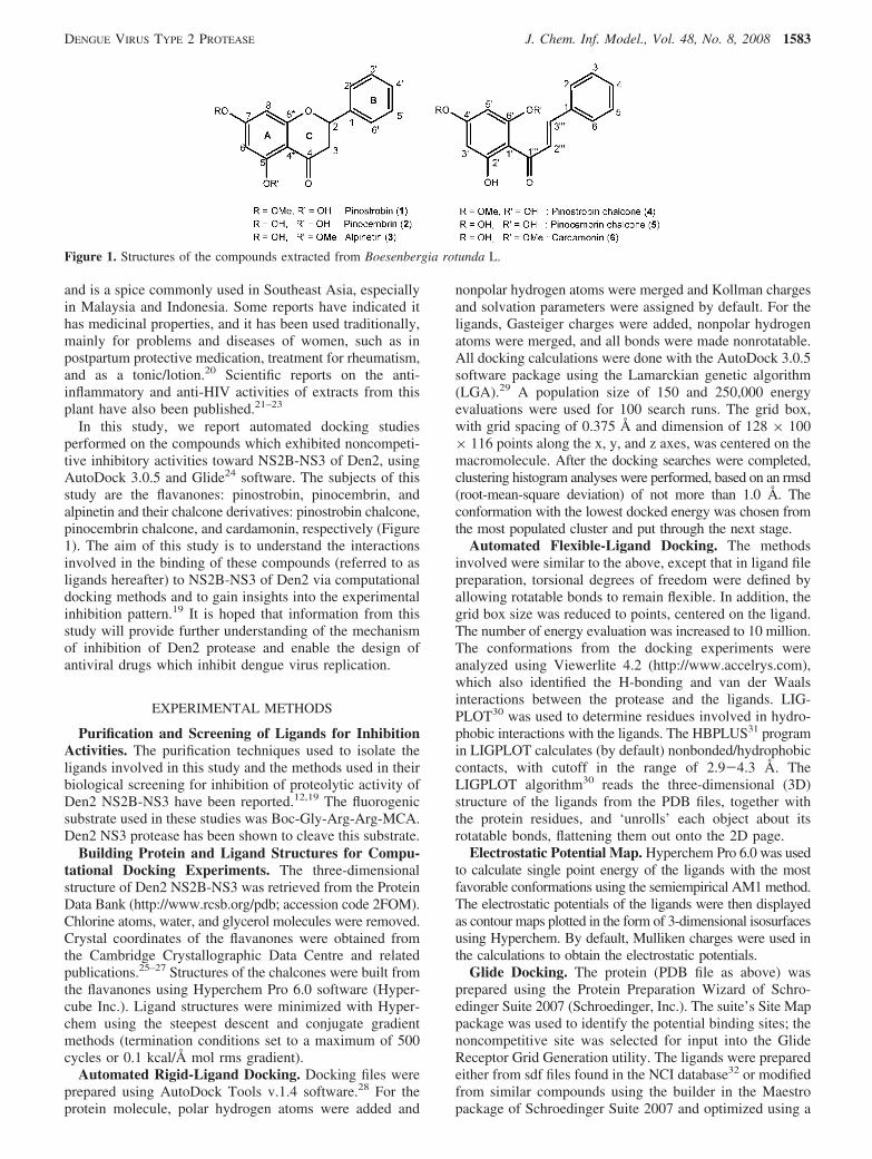
Binding Site and Ligand Conformations. As expected,the automated docking experiments showed that all theligands studied did not bind to the active site of the protease.Surprisingly, however, the binding site for these ligands wasconfined to a specific region of the protease (Figure 3a,b).The shape of the binding site can be divided into three parts
(Figure 3b): the left region constitutes a hydrophobic holewhich accommodates the aromatic rings of the ligands; themiddle allows placement of flavanone ring C or the chainscarrying the enone groups of the chalcones, enabling theseligands to interact with the surrounding residues; and theregion on the right, bigger than the other two, allows morevaried orientations of the ligands’ pharmacophores. Presum-ably, the shape of the binding site complements the shapesof the ligands. Figure 3c,d shows the superimposition of theconformations of the flavanones and chalcones, respectively,at the binding sites computed with Autodock. Of theflavanones, pinocembrin and alpinetin take up similar poses.However, although pinostrobin lies on an axis similar to theother two ligands, it is oriented in the opposite direction, sothat the phenyl ring projects into the region on the right sideof the binding site.
Figure 3e shows the superposition of all six ligandconformations computed with Glide. Unlike Autodock,pinostrobin’s orientation using Glide is similar to that of theother flavonoids. Like Autodock, pinostrobin chalcone bindsdifferently from the five other ligands. The conformationaldifference between pinostrobin and the other flavanonesobtained from Autodock is more clearly shown in Figure4a-c which illustrates the three-dimensional (3D) isosurfaceplots of the electrostatic potential of the ligands. Similar plotswere observed for alpinetin and pinocembrin, but thepotential map for pinostrobin (colored green to indi-cate neutral to positive potential) is more elongated due tothe presence of a methoxy group on ring A. Furthermore,the negative potentials (magenta) due to the carbonyl andhydroxyl groups on ring C and ring A, respectively, pointtoward the back of the plane of the model, while that due tothe ether group on ring C points more toward the front ofthe plane of the model. The reverse was observed withalpinetin and pinocembrin. These observations infer thedifferent chemical environment requirement of the bindingsite for docking of pinostrobin compared to alpinetin andpinocembrin. For the chalcones (Figure 4d-f), the shapesof the 3D isosurface plots of the electrostatic potential weredifferent for each ligand. This could be attributable to thehigher flexibility of the enone chains. This finding may alsoexplain the requirement of a different (chemical) environmentof the binding site by the ligands, hence the differentorientations adopted by the ligands in the binding site.
Table 2. Automated Flexible, Extra-Precision-Ligand-Protein Docking Results Calculated Using Glide
ligand pinostrobin cardamonin alpinetin pinocembrin pinocembrin chalcone pinostrobin chalcone
Glide score -7.14 -6.39 -6.86 -6.92 -5.55 -6.45
Table 3. Results of Rigid and Flexible Ligand Docking (AutoDock)a
no. of free torsions min. estimated ∆Gbind (kcal/mol) final Edock (kcal/mol)
compounds no. of atoms rigid flexible rigid flexible rigid flexible
pinostrobin 34 0 3 -8.23 -8.30 -8.23 -8.82pinocembrin 31 0 3 -8.63 -9.25 -8.63 -9.57alpinetin 34 0 3 -9.17 -8.84 -9.17 -9.45pinostrobin chalcone 34 0 6 -7.74 -7.79 -7.74 -8.71pinocembrin chalcone 31 0 6 -7.40 -7.59 -7.40 -8.85cardamonin 34 0 6 -8.00 -7.55 -8.00 -9.04
a The energy values shown were obtained from flexible docking of each ligand conformation which initially was the most favorableconformation obtained from rigid docking procedures (refer to the Materials and Methods section).
DENGUE VIRUS TYPE 2 PROTEASE J. Chem. Inf. Model., Vol. 48, No. 8, 2008 1585
DISCUSSION
Although both docking programs identified largely thesame binding site, and docked pinostrobin chalcone differ-ently from the other ligands, the details of the best posesfrom the two programs were different. Autodock’s scoringappears to be more heavily tilted toward maximizing the
number of hydrogen bonds than Glide. Glide was able toidentify the best inhibitor but not the worst.
Binding Interactions. Pinostrobin. As shown in Table 1,pinostrobin showed the highest inhibition among all theligands screened, with an IC
50value of 90.48 µg mL-1, while
pinostrobin chalcone was found to be inactive. Figure 5a,b
Figure 3. Computer generated models illustrating the superimposition of the flavanones, pinostrobin (orange), pinocembrin (purple), andalpinetin (yellow), and the chalcones, pinostrobin chalcone (pink), pinocembrin chalcone (blue), and cardamonin (green), at the bindingsite. The ligands are shown as sticks. (a) All the ligands are superimposed at the binding site. DEN-2 protease is represented as ribbons.The catalytic triads are labeled as His51, Asp75, and Ser135 and are shown as balls and sticks. (b) Connolly surface representation of thebinding site colored according to electrostatic potential spectrum. (c) Superimposition of the flavanone poses as found in the binding site.(d) Superimposition of the chalcone poses as found in the binding site. (e) Glide poses of all ligands. Note that, as with Autodock, pinostrobinchalcone binds quite differently. The orange molecule is pinostrobin chalcone.
Figure 4. 3D isosurface plots of the electrostatic potentials of the ligands, using Hyperchem Pro 6.0 software. Green surfaces indicateneutral to positive potentials, while magenta surfaces indicate negative potentials.
1586 J. Chem. Inf. Model., Vol. 48, No. 8, 2008 OTHMAN ET AL.

illustrated the binding site for pinostrobin. The shape of thebinding pocket was observed to complement the shape (pose)of the ligand. The phenyl ring is protruded out toward thesurface of the protein, while the rest of the molecule wasembedded into the inner part of the protein. The surroundingresidues involved in hydrogen bonding interactions withpinostrobin were Lys74, Leu149, and Asn152. The H-bonding interaction with Lys74 (Figure 5c,d) was notobserved with the other flavanones and chalcones studied.The interaction between the hydroxyl H atom on ring A ofpinostrobin with the backbone carbonyl O atom of Lys74could account for the relatively high inhibition activity ofpinostrobin. Since Lys74 is directly bonded to Asp75, theformation of H-bond between Lys74 and pinostrobin couldhave directly induced conformational change on Asp75, inparticular, or the catalytic triad region, in general. This,presumably, could disrupt the electron transfer processrequired for substrate binding at the active site, henceaffecting the activity of the protease.
Studies have shown that the formation of low-barrierhydrogen bond between Asp and His (of the catalytic triad)facilitates the nucleophilic attack by the �-OH group of Seron the acyl carbonyl group of substrates.37–39 Besides Lys74,Leu149 might play a role toward the activity of pinostrobinin two ways. First, Leu149 could be blocking the entry ofthe ligand into the active site due to its position in theprotease, as observed in Figure 5c,d. Second, Leu149 wasseen to protrude toward the adjacent �-barrel which carriedthe residues Asp75 and His51. Erbel et al. (2006)40 reportedthat the NS3 protease domain adopts a chymotrypsin-likefold with two �-barrels and that the catalytic triad is locatedat the cleft between the two �-barrels. Thus, upon binding
Leu149 with pinostrobin, a conformational change of theresidue could occur in order to reduce steric clashes, affectingthe spatial conformation of the surrounding residues, inparticular the catalytic triad. In conjunction with this, theelectron transfer process involved between Asp75 and His51might be affected, reducing the capability of the active siteto bind to the substrate. This process could further increasethe inhibition capability of pinostrobin.
Figure 5f shows the residues involved in hydrophobicinteractions with pinostrobin, obtained by the Ligplotprogram. Besides the hydrophobic interactions, Trp83 alsoexhibited nonclassical hydrogen bonding interaction betweenthe H atom on Nε1 of its indole ring with the phenyl ring(B) of pinostrobin (Figure 5e). In this case of nonclassicalhydrogen bonding, the hydrogen bond acceptor is thearomatic ring.41 Brocchieri and Karlin (1994) reported thatthe interplanar angle (dihedral angle between the extendedplanes of interacting planar groups, R) between the phenylring of Phe and the aromatic ring of Trp to be favorable at30° < R < 90°.42 They also reported that, generally, planarinteractions of Trp involved mostly the five-atom ring whichis capable of forming a hydrogen bond (involving its iminogroup), engaging π-cloud electrostatic interactions, and alsoundergoing hydrophobic interactions. Electrostatic chargesassociated with the phenyl ring include a weak negativecharge about the center of the aromatic ring and a weakpositive charge projected at the ring pheriphery.43 In thisstudy, interactions were also observed between Trp83 andpinostrobin, where the bond distance between the H atomon Nε1 of Trp83 and the center of the ring of the ligand(Rcentroid) was 2.67 Å, and R was approximately 61.0°.
Figure 5. (a) Transparent Connolly surface representation of pinostrobin at the binding site. (b) View of pinostrobin in the binding sitefrom a different angle. (c) View of pinostrobin (orange) at the binding site of Den2 protease (ribbons). Residues interacting with the ligandare shown as sticks. Catalytic triads are shown as balls and sticks. (d) Simplified view of pinostrobin interacting with surrounding residues.Residues labeled in green interact with the ligand via H-bonds, while those in red interact via van der Waals contacts. The catalytic triadsare labeled in black. (e) Nonclassical H-bonding between Trp83 and phenyl ring B of pinostrobin. (f) 2D schematic diagram of residues inthe binding site which exhibit hydrophobic interactions with pinostrobin, obtained using the Ligplot program. Keys for the plot are thefollowing: (blue ball-purple stick-black ball) ligand bond; (His51 with curved railroad-like ties) nonligand residue involved in hydrophobiccontact; and (black ball with railroad-like ties) corresponding atom involved in hydrophobic contact.
DENGUE VIRUS TYPE 2 PROTEASE J. Chem. Inf. Model., Vol. 48, No. 8, 2008 1587

Pinostrobin Chalcone. Previous study had shown thatpinostrobin chalcone did not exhibit inhibition activity (Table1).19 Investigation into the electrostatic interactions ofpinostrobin chalcone with the binding site revealed that thesurrounding amino acid residues of the protease involved inthe interactions, via van der Waals and H-bond, wereconfined to the C-terminal region of the protease, withresidues ranging from Asn152 to Asn167 (Figure 6a). Thisbinding mode may not have any structural effect on thecatalytic triad in promoting the disruption of electron transferfor the initiation of proteolytic processing. Presumably,binding activity of the ligand with its surrounding residuesis confined to a region which may not impose any confor-mational change to the active site. Figure 6b illustrates theresidues involved in H-bonding and van der Waals interac-tions with pinostrobin chalcone, in which a nonclassicalH-bond is observed between Asn167 and the phenyl ring(B) of the ligand (Rcentroid ) 3.64 Å, R ) 40°). Figure 6cshows the residues involved in hydrophobic interactions withthe ligand obtained from the Ligplot program. In additionto the interactions, the shape of the binding site was alsocomplementary to the ligand’s pose. From Figure 6d,observation of the hydrophobic pocket which accommodatedthe phenyl ring (B) of the ligand seemed to indicate that thering was not placed directly in the center of the pocket, ratherit was bent toward the left. This orientation may be attributedto the nonclassical H-bond interaction between the ring andAsn167.
The Other Ligands. Figure 7a-d shows the orientationsof cardamonin, alpinetin, pinocembrin, and pinocembrin
chalcone in the binding site of the protease, respectively.Except for pinocembrin chalcone, these ligands experiencedH-bond and van der Waals interactions with almost the samesurrounding residues. The common residues forming H-bondwith the three ligands were Leu149 and Asn152. In addition,pinocembrin formed an additional H-bond with Val147 andIle165. In terms of the van der Waals interactions, carda-monin interacted with Ile165, while alpinetin interacted withVal147 and Ile165, and pinocembrin with Asn167. Pinocem-brin chalcone, however, did not demonstrate van der Waalsinteraction with any of the surrounding residues but interactedwith Lys73 and Asn167 via H-bond.
The degree of inhibition offered by these four ligands was,in actual fact, similar to each other, even though bothcardamonin and alpinetin exhibited slightly higher activitiesthan pinocembrin chalcone and pinocembrin (Figure 2 andTable 1). The similarity in the degree of activities shownmay be explained by the similar axis of orientation in thebinding site (except for cardamonin; Figure 3) and similarmode of interactions as described above (except for pi-nocembrin chalcone; Figure 7). Unlike pinostrobin, theseligands did not form H-bond with Lys74, which may be thecause of the reduced activity observed when compared topinostrobin. Nevertheless, Lys74 was involved in hydropho-bic interaction with alpinetin, pinocembrin, and pinocembrinchalcone. Figure 8 illustrates the residues involved inhydrophobic interactions with the ligands using the Ligplotprogram. The observed activities of cardamonin, alpinetin,and pinocembrin could be due to the interactions of theligands with Leu149, as shown in parts a, b, and d,
Figure 6. (a) View of pinostrobin chalcone (pink) at the binding site of Den2 protease (ribbons). Residues interacting with the ligand areshown as sticks. Catalytic triads are shown as balls and sticks. The yellow circle highlights the binding mode of the ligand which isconfined to the C-terminal region of the protease, away from the catalytic triads. (b) Simplified view of pinostrobin chalcone interactingwith surrounding residues. Residues labeled in green interact with the ligand via H-bonds, while those in blue exhibit both H-bond and vander Waals interactions. (c) 2D schematic diagram of residues in the binding site which exhibit hydrophobic interactions with pinostrobinchalcone obtained using the Ligplot program. Keys for the plot are the following: (blue ball-purple stick-black ball) ligand bond; (His51with curved railroad-like ties) nonligand residue involved in hydrophobic contact; and (black ball with railroad-like ties) correspondingatom involved in hydrophobic contact. (d) Connolly surface representation of pinostrobin chalcone at the binding site nonligand residueinvolved in hydrophobic contact.
1588 J. Chem. Inf. Model., Vol. 48, No. 8, 2008 OTHMAN ET AL.

respectively, of Figure 7. The effect of Leu149 on substrate-binding capability of the protease upon its binding with theligands has been discussed earlier.
A different mode of interaction was observed withpinocembrin chalcone which did not bind to Leu149 toexhibit a similar inhibition effect. Rather, its H-bondinginteraction with Lys73 could be the cause of the observedactivity (Figure 7c). Lys73 is two residues downstream fromAsp75, and interaction between the ligand with this residuemay contribute directly to the conformational change of theactive site, the extent of which, however, was not as greatas that compared to the interaction between pinostrobin andLys74. The fact that pinocembrin chalcone did not exhibitvan der Waals interactions with the surrounding residues maybe the cause for the lower activity seen in the pinocembrinchalcone than that of pinostrobin.
Structure-Activity Relationship (SAR) Analysis. Thedocking studies performed gave better structural insights andunderstanding on how the various ligands interacted withthe protease in acting as noncompetitive inhibitors towardDen2 protease activity. Analysis of the structure-activityrelationships of the ligands in this study may shed light onthe important structure and conformation which could beapplied in the design of new compounds.
This study affirmed that rigid conformation of the fla-vanones would ensure ligand activities. Opening up of thering C, as found in the chalcones, would introduce higherflexibility to the ligands leading to a more extendedconformation, thus requiring a different environment of thebinding site (Figure 4). Higher flexibility of the chalconesalso meant higher ∆G
torcompared to the corresponding
flavanones and, hence, higher ∆Gbind
. This would result inthe chalcone binding being less favorable to the site when
compared to the corresponding flavanones (Table 2). Pi-nocembrin showed the lowest values for the E
dockand ∆G
bind,
followed by alpinetin and pinostrobin, indicating the parentstructure of 7-hydroxyflavanone to have good bindingcapabilities with the protease.
The methoxy group at C7 on the ring A of pinostrobinmay be important in creating the preferable electrostaticpotential surface of the molecule in the ‘search’ for anoptimal chemical environment within the binding site. Ingeneral, an electron-donating group at this position wouldbe preferable for the design of new inhibitory compounds.The hydroxyl group at C5 on the ring A of pinostrobin couldbe considered to be an important pharmacophore. Thishydroxyl group is very useful in forming H-bonds with thesurrounding residues, particularly, between pinostrobin andLys74 and between pinocembrin chalcone and Lys73 (theOH group is on C2′ of ring A for pinocembrin chalcone).For pinocembrin chalcone, the number of electrostaticinteractions involved was the lowest among all the ligandsreported. Thus, one might expect pinocembrin chalcone toexhibit a much lower inhibitory activity than the other ligands(except pinostrobin chalcone). However, from experiment,its activity was observed to be comparable to those ofpinocembrin, cardamonin, and alpinetin. This could beattributed to its H-bonding interaction with Lys73 via thehydroxyl group, where Lys73 is positioned two residuesdownstream from Asp75. The conclusions derived from thisstudy with regards to the importance of the methoxy andhydroxyl groups in determining the activity of the ligandsare in accordance with those postulated by Tan (2005).19
The presence of the phenyl ring (B) in all the ligands isnot of little importance and is essential in contributing tothe hydrophobicity of the ligands. The entropic effects of
Figure 7. Models of ligands bound to the protease (ribbons) at the binding sites. The ligands shown are (a) cardamonin (green), (b)alpinetin (yellow), (c) pinocembrin chalcone (blue), and (d) pinocembrin (purple). Residues interacting with the ligands are shown assticks. Catalytic triads are shown as balls and sticks. Residues labeled in green interact with the ligands via H-bonds, while those in redinteract via van der Waals contacts.
DENGUE VIRUS TYPE 2 PROTEASE J. Chem. Inf. Model., Vol. 48, No. 8, 2008 1589

ligand binding vary with the degree of hydrophobic interac-tions in the system, contributing to the determination of the∆G
bindvalue, and, as discussed earlier, the phenyl ring of
pinostrobin and pinostrobin chalcone formed nonclassicalH-bonding with Trp and Asn, respectively.
CONCLUSIONS
Three flavanones and three chalcones isolated from theplant Boesenbergia rotunda L. were docked onto the Den2NS2B-NS3 protease using the AutoDock 3.0.5 software.Results obtained from this study are consistent with theexperimental results illustrating the noncompetitive inhibitoryactivities for most of the ligands.19 As expected, these ligandswere found to bind to sites other than the active site of theDen2 serine protease. The calculated Ki values of theseligands were very small indicating that they bound consider-ably well to the allosteric binding site. The higher noncom-petitive inhibitory activity shown by pinostrobin comparedto the other compounds could be accounted for by H-bondinginteraction with the backbone carbonyl of Lys74, which isbonded to Asp75 (one of the catalytic triad residues). Thisinteraction was not observed with the other ligands. SARanalysis yielded some structural features which may be usefulfor the design of new compounds with potential inhibitoryactivities. These features are the rigid structure of flavanone,the C5 hydroxyl, and C7 methoxy groups on ring A and thephenyl ring (B).
Docking experiments were also performed with rigidligands as well as with flexible ligands on the rigid protein
structures (obtained from the PDB). To ensure that theobserved interactions in the resulting structures were sus-tained, molecular dynamics calculations were often per-formed. Although molecular dynamics on the system wasnot carried out in the study, this additional technique couldbe carried out as a potential method of justification for theresulting structures. The results obtained did not illustrateany conformational changes that could occur after the ligand-protein binding process. To overcome such limitation,flexible-protein docking would be preferable. However, thedevelopment of computational strategies for this purpose isstill in its infancy.34 Several methods have been reported,and promising results have emerged from the application ofcombined methods such as the ensemble docking approachand an induced fit.44
Flexible-protein docking could be a way forward towarddeeper insights into the present system which could aid inthe design of new compounds for a therapeutic drug againstdengue virus infections.
ACKNOWLEDGMENT
This work was supported in part by the Malaysian Ministryof Science, Technology and Innovation under the Top DownNational Biotechnology Directory grant number 09-02-04-001BTK/TH/004 [UM 36-02-03-6008], the Academy ofScience, Malaysia, under the Scientific Advancement FundAllocation (SAGA 66-02-03-0049), and Universiti Malayaunder the F-Vote grant number F0171/2005D.
Figure 8. Schematic diagrams (2D) illustrating residues in the binding sites which are involved in hydrophobic interactions with (a) cardamonin,(b) alpinetin, (c) pinocembrin chalcone, and (d) pinocembrin. Keys for the plot are the following: (blue ball-purple stick-black ball) ligandbond; (His51 with curved railroad-like ties) nonligand residue involved in hydrophobic contact; and (black ball with railroad-like ties)corresponding atom involved in hydrophobic contact.
1590 J. Chem. Inf. Model., Vol. 48, No. 8, 2008 OTHMAN ET AL.

REFERENCES AND NOTES
(1) Kautner, I.; Robinson, M. J.; Kuhnle, U. Dengue virus infection:epidemiology, pathogenesis, clinical presentation, diagnosis andprevention. J. Pediatr. 1997, 131, 516–524.
(2) Gubler, D. J. The global emergence/resurgence of arboviral diseasesas public health problems. Arch Med Res. 2002, 33, 330–42. (WHO.2007. http://www.who.int/csr/disease/dengue/impact/en/index.html (ac-cessed May 20, 2008))
(3) Dengue kills 44 people in Malaysia, Sun Malaysia, 2007; May 20,2007; http://story.malaysiasun.com/index.php/ct/9/cid/48cba686fe041718/id/250213/cs/1/ (accessed May 20, 2008).
(4) Irie, K.; Mohan, P. M.; Sasaguri, Y.; Putnak, R.; Padmanabhan, R.Sequence analysis of cloned dengue virus type 2 genome (NewGuinea-C strain). Gene 1989, 75, 197–211.
(5) Falgout, B.; Pethel, M.; Zhang, Y. M.; Lai, C. J. Both nonstructuralproteins NS2B and NS3 are required for the proteolytic processing ofdengue virus nonstructural proteins. J. Virol. 1991, 65, 2467–2475.
(6) Yusof, R.; Clum, S.; Wetzel, M.; Murthy, H. M.; Padmanabhan, R.Purified NS2B-NS3 serineprotease of dengue virus type 2 exhibitscofactor NS2B dependence for cleavage of substrates with dibasicamino acids in vitro. J. Biol. Chem. 2000, 275, 9963–9969.
(7) Gorbalenya, A. E.; Donchenko, A. P.; Koonin, E. V.; Blinov, V. M.N-terminal domains of putative helicases of flavi- and pestivirusesmay be serine proteases. Nucleic Acids Res. 1989, 17, 3889–3897.
(8) Bazan, J. F.; Fletterick, R. J. Detection of a trypsin-like serine proteasedomain in flaviviruses and pestiviruses. Virology 1989, 171, 637–639.
(9) Brinkworth, R. I.; Fairlie, D. P.; Leung, D.; Young, P. R. Homologymodel of the dengue 2 virus NS3 protease: putative interactions withboth substrate and NS2B cofactor. J. Gen. Virol. 1999, 80, 1167–1177.
(10) Arias, C. F.; Preugschat, F.; Strauss, J. H. Dengue 2 virus NS2B andNS3 form a stable complex that can cleave NS3 within the helicasedomain. Virology 1993, 193, 888–899.
(11) Clum, S.; Ebner, K. E.; Padmanabhan, R. Cotranslational membraneinsertion of the serine proteinase precursor NS2B-NS3(Pro) of denguevirus type 2 is required for efficient in vitro processing and is mediatedthrough the hydrophobic regions of NS2B. J. Biol. Chem. 1997, 272,30715–30723.
(12) Tan, S. K.; Pippen, R.; Yusof, R.; Ibrahim, H.; Khalid, N.; AbdRahman, N. Inhibitory activity of cycohexenyl chalcone derivativesand flavonoids of fingerroot, Boesenbergia rotunda (L.), towardsdengue-2 virus NS3 protease. Bioorg. Med. Chem. Lett. 2006, 16,3337–3340.
(13) Whitby, K.; Pierson, T. C.; Geiss, B.; Lane, K.; Engle, M.; Yi, Z.;Doms, R. W.; Diamond, M. S. Castanospermine, a potent inhibitor ofDengue virus infection in vitro and in vivo. J. Virol. 2005, 79, 8698–8706.
(14) Hrobowski, Y. M.; Garry, R. F.; Michael, S. F. Peptide inhibitors ofdengue virus and West Nile virus infectivity. Virol. J. 2005, 2, 49.
(15) Putnak, J. R.; Coller, B.-A.; Voss, G.; Vaughn, D. W.; Clements, D.;Peters, I.; Bignami, G.; Hounga, H.-S.; Chena, R. C.-M.; Barvir, D. A.;Seriwatana, J.; Cayphas, S.; Garcon, N.; Gheysen, D.; Kanesa-thasan,N.; McDonell, M.; Humphreys, T.; Eckels, K. H.; Prieels, J.-P.; Innis,B. L. An evaluation of dengue type-2 inactivated, recombinant subunit,and live-attenuated vaccine candidates in the rhesus macaque model.Vaccine 2005, 23, 4442–4452.
(16) Yin, Z.; Patel, S. J.; Wang, W.-L.; Wang, G.; Chan, W.-L.; RangaRao, K. R.; Alam, J.; Jeyaraj, D. A.; Ngew, X.; Patel, V.; Beer, D.;Lim, S. P.; Vasudevan, S. G.; Keller, T. H. Peptide inhibitors of denguevirus NS3 protease. Part 1: Warhead. Bioorg. Med. Chem. Lett. 2006,16, 36–39.
(17) Diamond, M. S.; Zachariah, M.; Harris, E. Mycophenolic acid inhibitsdengue virus infection by preventing replication of viral RNA. Virology2002, 304, 211–221.
(18) Ray, D.; Shi, P.-Y. Recent Advances in Flavivirus Antiviral DrugDiscovery and Vaccine Development. Recent Pat. Anti-InfectiVe DrugDiscoVery 2006, 1, 45–55.
(19) Tan, S. K. FlaVonoids from Boesenbergia rotunda (L). Mansf.:Chemistry, bioactiVity and accumulation; Ph.D. Thesis, UniversitiMalaya, Kuala Lumpur 2005.
(20) Ibrahim, H.; Rahman, A. A. SeVeral ginger plants (Zingiberaceae) ofpotential Value. In Malaysian Traditional Medicine: Proceedings of
the Seminar on Malaysian Traditional Medicine; Soepadmo, E., Goh,S. H., Wong, W. H., Din, L., Chuah, C. H., Eds.; Institute of AdvancedStudies, University of Malaya, and Malaysian Institute of Chemistry:Kuala Lumpur, 1989; pp 159-161.
(21) Tuchinda, P.; Reutrakul, V.; Claeson, P.; Pongprayoon, U.; Sematong,T.; Santisuk, T.; Taylor, W. Anti-inflammatory cyclohexenyl chalconederivative in Boesenbergia pandurata. Phytochemistry 2002, 59, 169–173.
(22) Tewtrakul, S.; Subhadhirasakul, S.; Kummee, S. HIV-1 proteaseinhibitory effects of medicinal plants used as self medication by AIDSpatients. Songklanakarin J. Sci. Technol. 2003, 25, 239–243.
(23) Tewtrakul, S.; Subhadhirasakul, S.; Puripattanavong, J.; Panphadung,T. HIV-1 protease inhibitory substances from the rhizomes ofBoesenbergia pandurata Holtt. Songklanakarin J. Sci. Technol. 2003,25, 503–508.
(24) Friesner, R. A.; Bank, J. L.; Murphy, R. B.; Halgren, T. A.; Klicic,J. J.; Mainz, D. T.; Repasky, M. P.; Knoll, E. H.; Shaw, D. E.; Shelley,M.; Perry, J. K.; Francis, P.; Shenking, P. S. Glide: A New Approachfor Rapid, Accurate Docking and Scoring. 1. Method and Assessmentof Docking Accuracy. J. Med. Chem. 2004, 47, 1739–1749.
(25) Brown, M. J.; Henderson, D. E.; Hunt, C. Comparison of antioxidantproperties of supercritical fluid extracts of herbs and the confirmationof pinocembrin as a principle antioxidant in Mexican oregano (Lippagravolens). Electron. J. EnViron. Agric. Food Chem. 2006, 5, 1265–1277.
(26) Jiang, R.-W.; He, Z.-D.; But, P. P.-H.; Chan, Y.-M.; Ma, S.-C.; Mak,T. C. W. A Novel 1:1 Complex of Potassium Mikanin-3-O-sulfatewith Methanol. Chem. Pharm. Bull. 2001, 49, 1166–1169.
(27) Shoja, M. 5-Hydroxy-7-methoxyflavone. Acta Crystallogr. 1989, C45,828–829.
(28) Sanner, M. F. Python: A programming language for software integra-tion and development. J. Mol. Graphics Model. 1999, 17, 57–61.
(29) Morris, G. M.; Goodsell, D. S.; Halliday, R. S.; Huey, R.; Hart, W. E.;Belew, R. K.; Olson, A. J. Automated Docking Using a LamarckianGenetic Algorithm and an Empirical Binding Free Energy Function.J. Comput. Chem. 1998, 19, 1639–1662.
(30) Wallace, A. C.; Laskowski, R. A.; Thornton, J. M. LIGPLOT: aprogram to generate schematic diagrams of protein-ligand interactions.Protein Eng. 1995, 8, 127–134.
(31) McDonald, I. K.; Thornton, J. M. Satisfying hydrogen-bondingpotential in proteins. J. Mol. Biol. 1994, 238, 777–793.
(32) National Cancer Institute, Developmental Therapeutics Program,Enhanced Database Browser. http://129.43.27.140/ncidb2/ (accessedMay 20, 2008).
(33) Jorgensen, W. L.; Tirado-Rives, J. Potential energy functions foratomic-leVel simulations of water and organic and biomolecularsystems PNAS 2005, 102, 6665–6670.
(34) Sousa, S. F.; Fernandes, P. A.; Ramos, M. J. Protein-Ligand Docking:Current Status and Future Challenges. Proteins 2006, 65, 15–26.
(35) Hetenyi, C.; van der Spoel, D. Efficient docking of peptides to proteinswithout prior knowledge of the binding site. Protein Sci. 2002, 11,1729–1737.
(36) Kitchen, D. B.; Decornez, H.; Furr, J. R.; Bajorath, J. Docking andscoring in virtual screening for drug discovery: Methods and applica-tion. Nature ReV. Drug DiscoVery 2004, 3, 935–949.
(37) Frey, P. A.; Whitt, S. A.; Tobin, J. B. A Low-Barrier Hydrogen Bondin the Catalytic Triad of Serine Proteases. Science 1994, 264, 1927–1930.
(38) Santis, L. D.; Carloni, P. Serine Proteases: An Ab Initio MolecularDynamics Study. Proteins 1999, 37, 611–618.
(39) Hunkapiller, M. W.; Forgac, M. D.; Richards, J. H. Mechanism ofAction of Serine Proteases: Tetrahedral Intermediate and ConcertedProton Transfer. Biochemistry 1976, 15, 5581.
(40) Erbel, P.; Schiering, N.; D’Arcy, A.; Renatus, M.; Kroemer, M.; Lim,S. P.; Yin, Z.; Keller, T. H.; Vasudevan, S. G.; Hommel, U. Structuralbasis for the activation of flaviviral NS3 proteases from dengue andWest Nile virus. Nat. Struct. Mol. Biol. 2006, 13, 372–373.
(41) Gervasio, F. L.; Chelli, R.; Procacci, P.; Schettino, V. The Nature ofIntermolecular Interactions between Aromatic Amino Acid Residues.Proteins 2002, 48, 117–125.
(42) Brocchieri, L.; Karlin, S. Geometry of interplanar residue contacts inprotein structures. Proc. Natl. Acad. Sci. U.S.A. 1994, 91, 9297–9301.
(43) Burley, S. K.; Petsko, G. A. Weakly polar interactions in proteins.AdV. Protein Chem. 1988, 39, 125–189.
(44) Cavasotto, C. N.; Kovacs, J. A.; Abagyan, R. A. Representing ReceptorFlexibility in Ligand Docking through Relevant Normal Modes. J. Am.Chem. Soc. 2005, 127, 9632–9640.
CI700388K
DENGUE VIRUS TYPE 2 PROTEASE J. Chem. Inf. Model., Vol. 48, No. 8, 2008 1591

In Silico Biology 7, 0022 (2007); ©2007, Bioinformation Systems e.V.
Analysis of secondary structure predictions of Dengue virus type 2 NS2B/NS3 against crystal structure to
evaluate the predictive power of the in silico methods
Rozana Othman1,5*, Habibah Abdul Wahab2,3, Rohana Yusof4 and Noorsaadah Abd Rahman5
1 Department of Pharmacy, Faculty of Medicine, Universiti Malaya, 50603 Kuala Lumpur, Malaysia
2 School of Pharmaceutical Sciences, Universiti Sains Malaysia, 11800, USM, Penang, Malaysia
3 Laboratory of Biocrystallography and Bioinformatics Structure, Universiti Sains Malaysia, 11800, USM, Penang, Malaysia
4 Department of Molecular Medicine, Faculty of Medicine, Universiti Malaya, 50603 Kuala Lumpur, Malaysia
5 Department of Chemistry, Faculty of Science, Universiti Malaya, 50603 Kuala Lumpur, Malaysia
* Corresponding author Email: [email protected]; Phone: +60-3-79675796, +60-3-79674959; Fax: +60-3-79674964
Edited by E. Wingender; received September 18, 2006; revised February 08 and March 02, 2007; accepted March 03, 2007; published March 27, 2007
Abstract
Multiple sequence alignment was performed against eight proteases from the Flaviviridae
family using ClustalW to illustrate conserved domains. Two sets of prediction approaches were
applied and the results compared. Firstly, secondary structure prediction was performed using
available structure prediction servers. The second approach made use of the information on the
secondary structures extracted from structure prediction servers, threading techniques and
DSSP database of some of the templates used in the threading techniques. Consensus on the
one-dimensional secondary structure of Den2 protease was obtained from each approach and
evaluated against data from the recently crystallised Den2 NS2B/NS3 obtained from the Protein
Data Bank (PDB). Results indicated the second approach to show higher accuracy compared
to the use of prediction servers only. Thus, it is plausible that this approach is applicable to the
initial stage of structural studies of proteins with low amino acid sequence homology against
other available proteins in the PDB.
Keywords: Dengue virus type 2, serine protease, secondary structure prediction, consensus,
protein structure
Introduction

Dengue virus belongs to the Flaviviridae family and is a widespread human pathogen that can
cause haemorrhagic fevers [Kautner et al., 1997]. Dengue infections place some 2.5 billion
people (or 40% of the world population) at risk and are a significant cause of mortality,
especially in the tropical and subtropical regions [Yin et al., 2006]. In the recent years, dengue
fever has been on the rise globally. The most prevalent of the four dengue serotypes is the
dengue virus type 2 (Den2). Den2 contains a single-stranded RNA of positive polarity. This
RNA genome codes for a single polyprotein precursor arranged in the order C-prM-E-NS1-
NS2A-NS2B-NS3-NS4A-NS4B-NS5 [Irie et al., 1989]. Flavivirus replication is dependent upon
the correct cleavage of this polypeptide and requires both host cell proteases and a virus-
encoded, two-component protease, NS2B/NS3 [Falgout et al., 1991; Yusof et al., 2000]. Hence,
this protease complex serves to be a target for the development of antiviral drugs. Although
there have been effort by many research groups, no vaccines or antiviral drugs are currently
available against dengue virus infections. Thus, there is an immense interest in developing new
antiviral therapeutic agents to fight diseases caused by dengue viruses.
Several studies were carried out by various research groups to gain structural insight into the
protease complex of Dengue virus type 2 (Den2). Until very recently [D'Arcy et al., 2006; Erbel
et al., 2006], no data on the crystal structure of NS2B/NS3 protease complex of the dengue
virus was available. A homology model of the NS2B/NS3 of Den2 was built by Brinkworth et al.,
1999, based on the crystal structure coordinates of the hepatitis C virus NS3/NS4A as template
(PDBid: 1JXP). The overall identity between the two sequences is only 14.8%. However,
regions surrounding the putative catalytic residues, as defined by Bazan and Fletterick, 1989,
indicate a high level of identity. The lack of structural details of the active protease from
experiments did not offer substantial insights into its interaction with substrates. Thus, the
design of inhibitors were mainly based on either kinetic studies, such as that reported by Kiat et
al., 2006, or theoretical understandings and explanations from in silico simulations [Brinkworth
et al., 1999; Lee et al. 2006].
Knowledge on the three-dimensional (3D) structure of a protein is crucial towards
understanding its function. Since it is often difficult or impossible to determine a structure
experimentally, computational techniques have become very popular in generating models of
proteins. Comparative, or homology, modelling remains the only method that can reliably
predict the 3D structure of a protein with accuracy comparable to that of a protein structure
resolved at low-resolution via experimental means [Marti-Renom et al., 2000]. This technique
relies upon the alignment of a protein sequence of unknown structure (target) to a homologue
of known structure (template). However, potential problems can occur in structural
determination when the target protein and template have less than 25% sequence identity
(based on an average domain length of 80 amino acids) [Sander and Schneider, 1991; Marti-
Renom et al., 2003]. Nevertheless, sufficiently long alignments can still infer structural similarity,
even when the sequence similarity is below 25% [Lund et al., 1997].
With no homologue of known structure from which to make a 3D model, a logical next step is to
predict secondary structure, which aims to provide the location of α-helices and β-strands within
a protein or protein family. Once the secondary structure of a protein has been determined, the
protein fold recognition is carried out, followed by a prediction of the tertiary structure. There are
many methods (web servers) available to perform secondary structure prediction. Kabsch and
Sander, 1984, reported that low level of success of prediction without additional information
was to be commonly attributed to the neglect of long-range interactions within a protein.
However, Pan et al., 1999, attributed the low levels in prediction accuracy to the limitation of the
available protein database size or prediction algorithm. Nevertheless, the field of secondary

structure has achieved a break-through by combining algorithms from artificial intelligence with
evolutionary information [Rost, 2003], boosting the current height of prediction accuracy to
around 77%. Thus, secondary structure prediction can be accurate enough to be taken
seriously as a tool to assist in the design of experiments to probe protein structure and function
[Barton, 1995].
A secondary structure prediction study of 175 N-terminal amino acids of the NS3 protease
using a combination of several prediction tools available over the website has been carried out
in our lab, a couple of years before the publication of crystal structure of Den2 NS2B/NS3
protease complex (PDBid: 2FOM) [D'Arcy et al., 2006; Erbel et al., 2006]. The aim of this work
is to map out the secondary structure of the different regions in the protease with the
knowledge of structurally conserved regions obtained from multiple sequence alignment against
NS3 proteases of other viruses from the Flaviviridae family. The structural data obtained from
the recently crystallised Den2 NS2B/NS3 has enabled us to make an evaluation of the
prediction and thus reported herein.
Methods
Multiple sequence alignment
Protein sequence alignments and comparisons were done with a BLAST (Basic Local
Alignment Search Tool) program, blastp, against database specification of non-redundant
protein which were available at the National Center for Biotechnology Information (NCBI) Web
server, [Altschul et al., 1997; http://www.ncbi.nlm.nih.gov/blast/]. Viruses for proteases used in
this study were checked against the Universal Virus Database, ICTVdb (International
Committee on Taxonomy of Viruses) [Büchen-Osmond, 2003]. Amino acid sequences were
obtained from NCBI sequence viewer 2.0 available at http://www.ncbi.nlm.nih.gov. Multiple
sequence alignments were done using ClustalW 1.82 [Thompson et al. 1994], available at the
European Bioinformatic Institute (EBI) Web server. Consensus of amino acid sequence was
obtained from Boxshade available at the European Molecular Biology Network (EMBnet) Web
server (http://www.ch.embnet.org).
Secondary structure prediction
The 175 amino acid sequence of Den2 NS3 protease was submitted for automated prediction
of secondary structures to the following programs via their web site interfaces: PSIPRED
[Jones, 1999; McGuffin et al., 2000; Bryson et al., 2005; http://bioinf.cs.ucl.ac.uk/psipred/],
PROF on PredictProtein Web server [Rost et al., 2004; http://www.predictprotein.org], PHD on
PredictProtein Web server [Rost, 1996], APSSP2 [Raghava, 2002;
http://www.imtech.res.in/raghava/apssp2/] and Jnet on Jpred Web server [Cuff and Barton,
2000; http://www.compbio.dundee.ac.uk/~www-jpred/jnet/].
Threading programs used via their web site interfaces were: 123D+ [Alexandrov et al., 1995;
http://123d.ncifcrf.gov/123D+.html], 3D-PSSM [Kelley et al., 2000;
http://www.sbg.bio.ic.ac.uk/servers/3dpssm/] and LOOPP [Meller and Elber, 2001; Teodorescu
et al. 2004; http://cbsuapps.tc.cornell.edu/loopp.aspx]. The secondary structure informations of
some templates used in the threading programs were obtained from DSSP database available

online [Kabsch and Sander, 1983; http://swift.cmbi.ru.nl/gv/dssp/]. One-dimensional (1D)
secondary structure predictions performed in this study employed two approaches:
1. By utilising available structure prediction servers only. Alignment of secondary
structures obtained from prediction programs was performed to result in a consensus.
2. By gaining information on the secondary structures extracted from structure
prediction servers, threading techniques and DSSP database of some of the templates
used in the threading techniques. An alignment of all secondary structures obtained
was performed to result in another consensus.
Comparison of the results obtained from the two tool sets (approach 1 and approach 2) were
then carried out against the observed secondary structure of the recently crystallised
NS2B/NS3 obtained from the Protein Data Bank [Berman et al., 2000] (PDBid: 2FOM) and the
percentage accuracy, A, was calculated as follows:
where c is the number of residues predicted correctly, and N is the total number of residues
with predicted secondary structures aligned against structures from crystal data.
Percentage difference, D, in secondary structure between both approaches was calculated as
follows:
where d is the number of residues with different secondary structure from both approaches, and
Nt is the total number of residues.
The implementation of approach 2 was out of curiosity to see if more exhaustive techniques of
secondary structure prediction would give rise to more reliable results.
Results and discussion
Fig. 1 illustrates the multiple sequence alignment of Den2 NS3 protease (EC 3.4.21.91) and
other proteases belonging to the genus Flavivirus of the Flaviviridae family. Consensus of
sequences obtained from the Boxshade program is shown and it can be seen that regions
surrounding the catalytic triads are highly conserved [Bazan and Fletterick, 1989; Brinkworth,
1999]. Secondary structure profiles built following the two approaches (as stated in the methods
section) are illustrated in Figs. 2 and 3. Consensus of the secondary structures is given as E (β-
strand), H (α-helix) or C (coil). It can be seen that both approaches yield mainly β-strands,
39.43% from the whole structure using the first approach, while the latter results in 40% of β-
strands. These results are in accordance with the fact that Den2 NS3 protease belongs to fold
family of trypsin-like serine-proteases (S07.001; according to MEROPS) [Rawlings et al., 2006],
which falls into the all-β proteins class of protein structure (according to SCOP) [Bazan and
Fletterick, 1989; Murzin, 1995].

Figure 1: Multiple sequence alignment of Den2 NS3 protease and other proteases belonging to the genus Flavivirus. The catalytic triad residues: His51, Asp75 and Ser135, are labelled with the symbol # and are found in boxes labelled A, B and C. (The numbers in the diagram may not represent the exact positions of residues in the actual protein due to the insertion of gaps during alignment). The boxes labelled A, B, C and D identify regions of significant similarity surrounding the catalytic triad residues and residues that might form the substrate-binding pocket [Bazan and Fletterick, 1989]. Consensus of residues throughout the different viruses are indicated by Boxshade, with those having 100% similarity written in the upper case font. Sequences used in the analyses: Den1 - Dengue virus type 1, Den3 - Dengue virus type 3, Den4 - Dengue virus type 4, KUN - Kunjin virus, WNV - West Nile virus, JEV - Japanese encephalitis virus, SLEV - St. Louis encephalitis virus and YFV - Yellow fever virus.
Figure 2: Profile of 1D secondary structure prediction of Den2 NS3 protease based on Approach 1 (see Method section): combination of automated prediction online programs. Consensus of secondary structure
are given as E = β-strand, H = α-helix or C = coil. The
catalytic triad residues: His51, Asp75 and Ser135, are labelled with the symbol #.
Figure 3: Profile of 1D secondary structure prediction of Den2 NS3 protease based on Approach 2 (see Method section): combination of automated prediction tools, threading programs and observed secondary structure information (taken from structural templates) used in threading methods (obtained from DSSP
database). The templates were chosen based on E-
values (E-value < 0.05, highly confident; E-value ≤
1.00, worthy of attention) or Z-scores (Z-score > 3,
high confidence). Template sequences used in the analyses: 1dy9 - Hepatitis C virus NS3 protease/helicase, 1svp - Sindbid virus capsid protein, 1vcp - Semiliki Forest virus capsid protein, 1jxp - Human Hepatitis C virus NS3 protease, 1ep5 - Venezuelan Equine Encephalitis virus capsid protein, and 1cqq - Type 2 Rhinovirus 3C protease. Codes for
secondary structures are given by: E = extended β-
strand, B = residue in isolated β-bridge, H = α-helix, G
= 3/10 helix, T = hydrogen-bonded turn, S = bend, L = loop and C = coil [Kabsch and Sander, 1983]. Regions with no codings are noted as coils. Consensus of secondary structure is given as E, H or C. Underlined secondary structures in the consensus line refers to regions which differ from 2FOM. (2FOM: secondary structure of crystallised Den2 NS3 protease available on PDB).

Comparison of the prediction results with secondary structure of crystallised NS3 protease of
Den2 (2FOM), shows that the second approach yields higher accuracy (A = 76%) compared to
the use of prediction servers only (A = 72.67%). These results show that the application of
programs used in the second approach gives a more reliable 1D secondary structure profile.
Difference in secondary structure between both approaches was calculated to be 7.43%. The
alignment of various predictions provides much more structural information than a pairwise
alignment. As opposed to the suggestions made by Russell, 2002, the techniques used in this
study do not involve the usage of human insight to reach the consensus. With only a 'palmful' of
knowledge on the Den2 NS3 protein structure during the early part of this study, we wondered
about the closeness of the prediction results to the true secondary profile of the system.
However, the recently published crystal model of Den2 NS2B/NS3 protease [Erbel et al., 2006],
which resolved to 1.5 Å, assured the quality of the prediction results at 76% accuracy for the
results from the second approach against the secondary profile of the crystal structure. This
accuracy value is reasonably high. The application of the second approach towards the building
up of secondary structure profile of proteins can also be suggested to other groups of proteins
from the same family of virus, i. e. Flaviviridae (which can further be classified into 3 genus).
Nevertheless, the percentage of accuracy of the prediction results compared with the true
structure will also depend on the prediction tools chosen. The selection of the appropriate
prediction tools can be determined by referring to CASP (the Critical Assessment of Structure
Prediction) [Bourne, 2003] experiments.
The crystal structure of Den2 NS2B/NS3 revealed that the protease adopt a β-barrel fold [Erbel
et al., 2006]. It was also reported that residues 51-57 of the cofactor NS2B contributed one β-
strand to the N-terminal β-barrel. Furthermore, the crystal structure of West Nile virus
NS2B/NS3, complexed with inhibitor, revealed direct interactions of NS2B with active site of
NS3, thus underlining the work by Yusof et al., 2000, which indicated the dependence of the
protease on the cofactor for cleavage of substrates with dibasic amino acids. However, this
study only concentrated on the secondary structure of the protease domain (NS3) and it was
assumed, at this stage, that the involvement (or not) of NS2B cofactor in the prediction will not
affect the building up of 1D secondary structure profile of the NS3 protease, since alignment of
the protein was made with other proteins of the same domain (when using prediction tools).
Analysis of conservation in the protein families is effective in secondary structure predictions
performed before knowledge of the protein structure [Cuff and Barton, 1999]. A strong
correlation can be made between structurally conserved regions (Fig. 3) and regions with highly
conserved sequences across the different proteases (Fig. 1), especially regions surrounding
the catalytic triad. However, two regions were missed by the prediction methods, where 2FOM
defined residues Trp50-Arg54 and Ser131-Ser135 (each carrying residues comprising the
catalytic triad, i.e. His51 and Ser135, respectively) to be α-helices. On the other hand, it is quite
unexpected for α-helix to span the region Ser131-Ser135 which also included Pro132, and
proline is well-known to be a 'helix-breaker' due to its rigid ring conformation [Garrett and
Grisham, 1997]. Looking at the prediction results in Fig. 3 for region Ser131-Ser135, there is
100% consensus for this region to be coiled. Perhaps the α-helices will be recognised after the
secondary structure profile is put through fold recognition procedures.
Conclusions
This study illustrates the application of automated prediction servers and information of
secondary structures from threading programs to build a consensus of secondary structure (1D)

of Den2 protease. In an early stage, the secondary structure prediction was carried out before
the PDB deposition of the crystal structure (2FOM) for NS2B/NS3. Soon after the publication of
the 3D-structure in the year 2006, we conducted a validation of the predictive power of the
tools. The conclusions were drawn by comparing a combination of tools (approaches 1 and 2)
against the observed secondary structure of 2FOM. The consensus obtained in the comparison
studies showed higher similarity in approach 2 than approach 1 to 2FOM (Fig. 4). In the present
case and presumably other cases of low homology sequence relationships, a significantly
better prediction can be attributed to approach 2 because it comprises heuristic (similarity of
amino acid properties), probabilistic (from structural data collections) and more sophisticated
overlaying techniques, i.e. threading the query into the template backbone to detect hidden
phylogenetic resemblance.
Figure 4: Alignment of secondary structure prediction consensus obtained from approaches 1 and 2, against the secondary structure of 2FOM. Underlined secondary structures in the consensus lines refer to regions which differ from 2FOM. The catalytic triad residues: His51, Asp75 and Ser135, are labelled with the symbol #.
Acknowledgements
The authors thank Mohd Firdaus Raih from the National Institute for Genomics and Molecular
Biology (Interim Laboratory), and School of Biosciences and Biotechnology, Universiti
Kebangsaan Malaysia, Malaysia, for his kind intellectual contribution towards this study. This
work was supported in part by the Malaysian Ministry of Science, Technology and Innovation
under the Top Down National Biotechnology Directory grant number 09-02-04-001BTK/TH/004
[UM 36-02-03-6008], and the Academy of Science, Malaysia, under the Scientific Advancement
Fund Allocation (SAGA 66-02-03-0049).
References
Alexandrov, N. N., Nussinov, R. and Zimmer, R. M. (1995). Fast protein fold recognition via sequence to structure alignment and contact capacity potentials. In: Pacific Symposium on Biocomputing '96, Hunter, L. & Klein, T. E. (eds), World Scientific Publishing Co., Singapore, pp.53-72.
Altschul, S. F., Madden, T. L., Schäffer, A. A., Zhang, J., Zhang, Z., Miller, W. and Lipman, D. J. (1997). Gapped BLAST and PSI-BLAST: a new generation of protein database search programs. Nucleic Acids Res. 25, 3389-3402.
Barton, G. J. (1995). Protein secondary structure prediction. Curr. Opin. Struct. Biol. 5, 372-376.

Bazan, J. F. and Fletterick, R. J. (1989). Detection of a trypsin-like serine protease domain in flaviviruses and pestiviruses. Virology 171, 637-639.
Berman, H. M., Westbrook, J., Feng, Z., Gilliland, G., Bhat, T. N., Weissig, H., Shindyalov, I. N. and Bourne, P. E. (2000). The Protein Data Bank, Nucleic Acids Res. 28, 235-242.
Bourne, P. E. (2003). CASP and CAFASP experiments and their findings. Methods Biochem. Anal. 44, 501-507.
Brinkworth, R. I., Fairlie, D. P., Leung, D. and Young, P. R. (1999). Homology model of the dengue 2 virus NS3 protease: putative interactions with both substrate and NS2B cofactor. J. Gen. Virol. 80, 1167-1177.
Bryson, K., McGuffin, L. J., Marsden, R. L., Ward, J. J., Sodhi, J. S. and Jones, D. T. (2005). Protein structure prediction servers at University College London. Nucleic Acids Res. 33, W36-W38.
Büchen-Osmond, C. (2003). The Universal Virus Database ICTVdB. Comput. Science Engineer. 5(3), 16-25.
Cuff, J. A. and Barton, G. J. (1999). Evaluation and improvement of multiple sequence methods for protein secondary structure prediction. Proteins 34, 508-519.
Cuff, J. A. and Barton, G. J. (2000). Application of enhanced multiple sequence alignment profiles to improve protein secondary structure prediction. Proteins 40, 502-511.
D'Arcy, A., Chaillet, M., Schiering, N., Villard, F., Lim, S. P., Lefeuvre, P. and Erbel, P. (2006). Purification and crystallization of dengue and West Nile virus NS2B-NS3 complexes. Acta Cryst. Sec. F 62, 157-162.
Erbel, P., Schiering, N., D'Arcy, A., Renatus, M., Kroemer, M., Lim, S. P., Yin, Z., Keller, T. H., Vasudevan, S. G. and Hommel, U. (2006). Structural basis for the activation of flaviviral NS3 proteases from dengue and West Nile Virus. Nat. Struct. Mol. Biol. 13, 372-373.
Falgout, B., Pethel, M., Zhang, Y.-M. and Lai, C.-J. (1991). Both nonstructural proteins NS2B and NS3 are required for the proteolytic processing of dengue virus nonstructural proteins. J. Virol. 65, 2467-2475.
Garrett, R. H. and Grisham, C. M. (1997). Proteins: Secondary, tertiary, and quarternary structure. In: Principles of biochemistry, with a human focus, Vondeling, J. and Kiselica, S. (eds.), Thomson Learning, Inc. Thomson Learning, USA, pp. 113-152.
Irie, K., Mohan, P. M., Sasaguri, Y., Putnak, R. and Padmanabhan, R. (1989). Sequence analysis of cloned dengue virus type 2 genome (New Guinea-C strain). Gene 75, 191-211.
Jones, D. T. (1999). Protein secondary structure prediction based on position-specific scoring matrices. J. Mol. Biol. 292, 195-202.
Kabsch, W. and Sander, C. (1983). Dictionary of protein secondary structure: pattern recognition of hydrogen-bonded and geometrical features. Biopolymers 22, 2577-2637.
Kabsch, W. and Sander, C. (1984). On the use of sequence homologies to predict protein structure: Identical pentapeptides can have completely different conformations. Proc. Natl. Acad. Sci. USA 81, 1075-1078.
Kautner, I., Robinson, M. J. and Kuhnle, U. (1997). Dengue virus infection: epidemiology, pathogenesis, clinical presentation, diagnosis and prevention. J. Pediatr. 131, 516-524.
Kelley, L. A., MacCallum, R. M. and Sternberg, M. J. E. (2000). Enhanced genome annotation using structural profiles in the program 3D-PSSM. J. Mol. Biol. 299, 499-520.
Kiat, T. S., Pippen, R., Yusof, R., Ibrahim, H., Khalid, N. and Rahman, N. A. (2006). Inhibitory activity of cyclohexenyl chalcone derivatives and flavonoids of fingerroot, Boesenbergia rotunda (L.), towards dengue-2 virus NS3 protease. Bioorg. Med. Chem. Lett. 16, 3337-3340.
Lee, Y. K., Othman, R., Abdul Wahab, H., Yusof, R. and Rahman, N. A. (2006). A revisit into the Den2 NS2B/NS3 virus protease homology model: Structural verification and comparison with crystal structure of HCV NS3/4A and Den2 NS3. Malaysian J. Sci. 25, 15-22.
Lund, O., Frimand, K., Gorodkin, J., Bohr, H., Bohr, J., Hansen, J. and Brunak, S. (1997). Protein distance constraints predicted by neural networks and probability density functions. Protein Eng. 10, 1241-1248.
Martí-Renom, M. A., Stuart, A. C., Fiser, A., Sánchez, R., Melo, F. and Sali, A. (2000). Comparative protein structure modeling of genes and genomes. Annu. Rev. Biophys. Biomol. Struct. 29, 291-325.
Marti-Renom, M. A., Fiser, A., Madhusudhan, M. S., John, B., Stuart, A., Eswar, N., Pieper, U., Shen, M.-Y. and Sali, A. (2003). Modeling protein structure from sequence. Curr. Prot. Bioinfo., John Wiley & Sons, Inc. 5(3), 5.1.1-5.1.42.
McGuffin, L. J., Bryson, K. and Jones, D. T. (2000). The PSIPRED protein structure prediction server. Bioinformatics 16, 404-405.
Meller, J. and Elber, R. (2001). Linear optimisation and a double statistical filter for protein threading protocols. Proteins 45, 241-261.
Murzin, A. G., Brenner, S. E., Hubbard, T. and Chothia, C. (1995). SCOP: A structural classification of proteins database for the investigation of sequences and structures. J. Mol. Biol. 247, 536-540.
Pan, X.-M., Niu, W.-D. and Wang, Z.-X. (1999). What is the minimum number of residues to determine the secondary structural state? J. Protein Chem. 18, 579-584.
Raghava, G. P. S. (2002). APSSP2: A combination method for protein secondary structure prediction based on neural network and example based learning. CASP5, A-132.
Rawlings, N. D., Morton, F. R. and Barrett, A. J. (2006) MEROPS: the peptidase database. Nucleic Acids Res. 34, D270-D272.

Rost, B. (1996). PHD: predicting 1D protein structure byprofile based neural networks. Methods Enzymol. 266, 525-539.
Rost, B. (2003). Rising accuracy of protein secondary structure prediction. In: Protein structure determination, analysis, and modeling for drug discovery, Chasman, D. (ed.), Dekker, New York, pp. 207-249.
Rost, B., Yachdav, G. and Liu, J. (2004). The PredictProtein Server. Nucleic Acids Res. 32, W321-W326.
Russell, R. (2002). A guide to structure prediction, v2.1. http://speedy.embl-heidelberg.de/gtsp/.
Sander, C. and Schneider, R. (1991). Database of homology derived protein structures and the structural meaning of sequence alignment. Proteins 9, 56-69.
Teodorescu, O., Galor, T., Pillardy, J. and Elber, R. (2004). Enriching the sequence substitution matrix by structural information. Proteins 54, 41-48.
Thompson, J. D., Higgins, D. G. and Gibson, T. (1994). CLUSTALW: improving the sensitivity of progressive multiple sequence alignment through sequence weighting, position-specific gap penalties and weight matrix choice. Nucleic Acids Res. 22, 4673-4680.
Yin, Z., Patel, S. J., Wang, W.-L., Wang, G., Chan, W.-L., Rao, K. R. R., Alam, J., Jeyaraj, D. A., Ngew, X., Patel, V., Beer, D., Lim, S. P., Vasudevan, S. G. and Keller, T. H. (2006). Peptide inhibitors of dengue virus NS3 protease. Part 1: Warhead. Bioorg. Med. Chem. Lett. 16, 36-39.
Yusof, R., Clum, S., Wetzel, M., Murthy, H. M. K. and Padmanabhan, R. (2000). Purified NS2B/NS3 serine protease of Dengue virus type 2 exhibits cofactor NS2B dependence for the cleavage of substrates with dibasic amino acids in vitro. J. Biol. Chem. 275, 9963-9969.

Malaysian Journal of Science 25(1): 15-22 (2006)
A Revisit into the DEN2 NS2B/NS3 Virus Protease Homology Model:
Structural Verification and Comparison with Crystal Structure of HCV
NS3/4A and DEN2 NS3
Lee Yean Kee,1 Rozana Othman,
1 Habibah Abdul Wahab,
2 Rohana Yusof
3 and Noorsaadah
Abd. Rahman.1,*
1. Dept. of Chemistry, Faculty of Science, Universiti Malaya, 50603 Kuala Lumpur 2. School of Pharmacy, Universiti Sains Malaysia, Pulau Pinang 3. Dept. of Mol. Medicine, Faculty of Medicine, Universiti Malaya, 50603 Kuala Lumpur.
ABSTRACT
Although the crystal structure of DEN2 NS3 serine protease has been reported, the
proteolytic mechanism of this enzyme in the presence of NS2B as co-factor which greatly
enhance its activity is not well-understood. Using the homology model of DEN2 NS3 co-
complexed with NS2B co-factor based on HCV NS3/4A as the proposed template, the model
of the DEN2 NS2B/3 complex was reproduced and its structure evaluated through
PROCHECK, VERIFY3D and ERRAT. Comparison of the homology model with the crystal
structure of DEN2 NS3 revealed that this homology model served as a better choice for the
NS2B/3 protein model.
Keywords: Dengue, NS3, serine protease, structural verification, HCV.
INTRODUCTION
Dengue fever and dengue haemorrhagic fever is a serious disease with almost 40%
of the world population currently at risk. This disease is easily spread by the Aedes aegypti
and Aedes albopictus mosquitoes which are highly populated in tropical country. Studies
revealed that about 50 million cases of dengue infection were reported every year. There is
currently no available drug or vaccine to combat this disease. However, various efforts are
currently being put into finding effective vaccines or inhibitors for this disease (Kautner et al.,
1997; Monath, 1994).
The dengue fever and dengue haemorrhagic fever is caused by the dengue virus that
grouped under the flavivirus family. Currently, 4 serotypes of dengue virus have been
discovered with the DEN2 serotype being the most prevalent amongst the four. The single-

Malaysian Journal of Science 25(1): 15-22 (2006)
stranded, positive-sense RNA of dengue virus has approximately 10,723 nucleotides (for New
Guinea-C strand). The genomic RNA has a single open reading frame that encodes a
polyprotein of 3,391 amino acids. These amino acids are processed into 3 structural (C, prM,
and E) proteins that are further assembled into the virion and seven non-structural proteins,
NS1 to NS5, and are expressed in infected cells (Irie et al., 1989). Studies has revealed that
the second largest protein encoded by virus, NS3 to contain a serine proteinase catalytic triad
within terminal region of 180 amino acid residues which require 40 amino acid residues of
NS2B for protease activity (Falgout et al., 1993).
The polyprotein precursor processing occurs co-translationally as well as post-
translationally and is performed by either the host signalase in association with the
membranes of the endoplasmic reticulum or the viral protease. The protease cleaves the viral
polyprotein at four junctions, NS2A-NS2B (Arg-Ser), NS2B-NS3 (Arg-Ala), NS3-NS4A (Lys-
Ser), and NS4B-NS5 (Arg-Gly) where a pair of dibasic amino acids at the P2 and P1 positions
followed by a small, non-branched amino acid at P1’ was found as the consensus of substrate
cleavage motif (Yusof et al., 2000 and references therein).
Many approaches have been employed to understand the mechanism, structure and
molecular interaction between the serine protease of NS2B/3 complex and its substrates.
The minimum domain size required for protease activity of the 69-kDa NS3 protein has been
mapped to 167 residues at the N terminus (Li et al., 1999). The virus sequence alignments
analyzed revealed that structural motifs as well as the characteristic catalytic triad (His-Asp-
Ser) of mammalian serine proteases are conserved in all flaviviruses (Bazan and Fletterick,
1989; Gorbalenya et al. 1989). Sequence comparison among the serine proteases and
mutational analysis verified that a catalytic triad of NS2B/3 comprised of the residues His51,
Asp75, and Ser135, and that replacement of the catalytic Ser135 residue by alanine resulted
in an enzymatically inactive NS3 protease (Yan et al., 1998). The presence of a peptide co-
factor is also found to be essential for optimal catalytic activity of the flaviviral proteases with
natural polyprotein substrates (Bartenschlager et al., 1995; Chambers, 1991).

Malaysian Journal of Science 25(1): 15-22 (2006)
Although the dengue virus NS3 protease exhibits NS2B-independent activity with
model substrates for serine proteases such as N--benzoyl-L-arginine-p-nitroanilide, the
enzymatic cleavage of dibasic peptides is markedly enhanced in NS2B/3 complex. In
addition, the presence of the NS2B co-factor has been shown to be an absolute requirement
for trans-cleavage of a cloned polyprotein substrate (Yusof et al, 2000). A genetically
engineered NS2B(H)-NS3pro protease containing a non-cleavable nonamer glycine linker
between the NS2B activation sequence and the protease moiety exhibited higher specific
activity with para-nitroanilide peptide substrates than the NS2B(H)-NS3pro molecule (Leung
et al., 2001). The NS2B-NS3pro protease incorporating a full-length NS2B cofactor sequence
could catalyze the cleavage of 12-mer peptide substrates representing native polyprotein
junctions (Khumthong et al., 2002; Khumthong et al., 2003). However, this protein appeared
to be completely resistant to proteolytic self-cleavage.
A model of the NS2B/NS3 dengue virus protease was first constructed through
homology modeling using the crystal structure of HCV NS3/4A complex as template by
Brinkworth and co-workers, with the suggestion that the 40 amino acid residues of dengue
virus NS2B co-factor could be reduced to 12 hydrophobic residues (Brinkworth et al., 1999).
Experimental data on hepatitis C virus protease showed some structural and mechanistic
explanations for the protease activation by its co-factor, where the NS4A co-factor was found
to affect the folding of the NS3 protease. This resulted in the conformational rearrangements
of the N-terminal 28 residues of the protease and a strand displacement that lead to the
formation of a well-ordered array of three -sheets with the co-factor as an integral part of the
protease fold (Kim et al., 1996; Yan et al., 1998). These conformational changes reorient the
residues of the catalytic triad making it more favorable for proton shuttling during proteolytic
process.
In the absence of the crystal structure for NS3 pro complexed with its co-factor NS2B,
it is rather difficult to understand the structure and conformation of the dengue virus serine
protease as well as its mechanism of action. Comparative modeling could provide an
alternative method to understand the structure and conformation of protease and further aid in

Malaysian Journal of Science 25(1): 15-22 (2006)
predicting the binding interactions of the substrate with competitive substrate-based or non-
substrate-based inhibitors.
Although the model of the NS2B/3 complex of the dengue virus protease has been
proposed based on a homology modeling on HCV (Brinkworth et al., 1999), the verifications
of this model were not extensively carried out. In addition, there was no detailed discussion
on the comparisons between the homology structure and the dengue NS3 crystal structure to
enable better insights into the role of NS2B co-factor in the influencing the function of NS3. In
this work, the dengue virus protease model was reproduced through homology modeling and
is subjected to structural verification and evaluation using server-based protein structure
verification program, namely PROCHECK, VERIFY 3D and ERRAT. The results of these
verifications are discussed in greater details in this paper. The similarities and differences
between the crystal structures and computer model of NS3 generated in this work as well as
that of Brinkworth’s (Brinkworth et al., 1999) are also discussed.
MATERIALS AND METHODS
Homology model of DEN2 NS2B/3 Serine Protease
Homology model of NS2B/3 of dengue virus type 2 was built using the HCV serine
protease NS3/4A (pdb ID: 1jxp) as the template. The Modeller (mod6v2) software package
was used to perform model building. The sequence alignment was done based on the
published results of Brinkworth et. al., 1999. The quality of the backbone of rough model
generated from Modeller was then evaluated using PROCHECK (Laskowski et. al., 1993),
VERIFY3D (Bowie et. al. 1991) and ERRAT (Colovos and Yeates, 1993) on the UCLA
bioinformatics server. Energy minimization (100 steps of steepest decent plus 50 steps of
conjugate gradient) was performed onto the model, using Hyperchem software package
(Hypercube, Inc.) to reduce the bumps and bad contacts while keeping the backbone of the
protein restrained. The model evaluation was then repeated.
Malaysian Journal of Science 25(1): 15-22 (2006)
Comparison of the homology model with crystal structures of and DEN2 NS3 and HCV
NS3/4A
The similarities and differences of the structure and conformation around the catalytic
triad in of the constructed homology model of DEN2 NS2B/3 serine protease were evaluated
using the crystal structures of DEN2 NS3 (pdb ID: 1bef) and the HCV NS3/4A (pdb id: 1jxp).
RESULTS
Homology model building and model evaluation
The Ramachandran plot obtained from PROCHECK (Figure 1) showed an overall
100 % non-glycine residue to be in the allowed region, which implies a good protein backbone
structure and folding, where the distribution of the / angle of the model were within the
allowed region. In addition, VERIFY 3D showed 90.4% of the residues having a 3D-1D score
greater than 0.2, suggesting a reasonable conformation of the residues in the model.
However, region with a 3D-1D score lower than 0.2 was found in the range of Glu91-Gln110,
indicating a lower confidence in its conformations and folding, implying a lower homology
between DEN2 serine protease and HCV serine protease in this particular region. ERRAT
was used to check the non-bonded structures of the queried structure and to compare with a
database of reliable high-resolution structures. The DEN2 N2SB/3 homology model gave
77.1% overall quality factor of the sequence to be below 95% rejection limit for each chain in
the input structure (see figure 1, 2 and 3 for more details) after several iterations of geometry
optimizations. This indicated a better three-dimensional profile of the protein from pre-
generated homology structure (data not shown). All these verification procedures performed
on the NS2B/3 protease model indicated that the model has reached a satisfactory fold
quality. Hence, no further loop modeling was carried out on the model.
DISCUSSIONS
Comparison of the homology model with crystal structures of and DEN2 NS3 and HCV
NS3/4A
Overall, the homology model showed almost the same folding of protein backbone as
observed in the DEN2 NS3 crystal structure where in the first domain (NH2-terminal domain)
of NS3, one alpha-helix and 6 beta sheets are found, both in the homology model as well as
Malaysian Journal of Science 25(1): 15-22 (2006)
in the DEN2 NS3 crystal structure (Figure 4). The differences between two models, however,
were observed in the second NS3 domain where more loop regions are found in the crystal
structure of NS3 compared to those observed in the homology model. In addition, the crystal
structure of NS3 indicated the presence of only one alpha helix and 7 beta sheets in C
terminal region, whilst one extra beta sheets in the same domain was observed in the
homology model.
It has been reported that in the crystal structure of NS3 with NS4A co-factor of HCV,
the incorporation of NS4A co-factor into the N-terminal domain -sheet led to a more rigid and
precise framework for “prime-side” substrate binding channel residues, thus providing a better
catalytic cavity that makes the NS3 enzyme active in proteolytic process (Kim et al., 1996).
Superimposition (Figure 2d) of the NS3 structure from the crystallographic data with that of
the homology model revealed a difference in the folding between Gly114-Val126 of crystals of
NS3 compared to the homology model. These may explain the importance of the NS2B co-
factor’s role in re-packing the NS3 protein into a more rigid and stabilized structure, especially
at the C- terminal domain, where more secondary structure was observed as compared to the
NS3 crystal in the absence of the NS2B co-factor.
The catalytic triad residues for HCV NS3/4A and DEN2 NS2B/3 serine proteases
were found to be in the structurally conserved region and there is no significant
conformational differences observed between them. The RMSD value found between the
catalytic triad residues of the HCV NS3/4A (His57, Asp81 and Ser139) crystal with the
homology model of DEN2 NS2B/3 (His51, Asp75 and Ser135) is 0.6 while that of the
homology model of DEN2 NS2B/3 and the DEN2 NS3 crystal structures is 1.1. The hydrogen
bonding between the hydroxyl group of Ser135 and cycloimine of His51 sidechain was
observed in the catalytic triad of the reported Den2 NS3 crystal structure (Figure 5a). The
side chain carboxyl oxygen of Asp75, however, is observed to be oriented away from His51
(Figure 5a). This caused it to be unable to form a hydrogen bond between carboxyl group of
Asp75 and cycloamine of His51 and hence, the proton transfer from Asp75 to Ser 135 which
is required to activate the proteolytic process could not transpire.

Malaysian Journal of Science 25(1): 15-22 (2006)
In the homology model, the carboxyl oxygen of Asp75 and His51, as well as that of
His75 and the hydroxyl of Ser135, was found to be at 1.6 Ǻ which is within the hydrogen
bonding distance (Figure 5b), suggesting a better arrangement of catalytic residues.
As demonstrated in Table 1, all the structures gave a reasonable reading of the
Ramachandran plot, where more than 90% of non-glycine residues were located in the
allowed region and no residues were located in disallowed region was observed in all the
protein structures. This indicated that the backbone of the serine protease of HCV and DEN2
have reasonably high degree of homology, in spite of their low sequence identity (Brinkworth
et al., 1999). Interestingly, the homology model for DEN2 NS2B/3 displayed a better reading
in VERIFY3D and ERRAT as compared to crystal structure of NS3, suggesting a better side
chain packing in the computer model. The absence of the NS2B co-factor fragment in the
crystal structure of NS3 is attributed to a lower quality 3D structure of the crystals. This
information has provided some insights into the role of the protease co-complexed with NS2B
co-factor, which seems to re-orientate the active pocket of the DEN2 NS3, exhibiting a better
side chain packing for a more efficient proteolytic cleavage (Yusof et al, 2000). However, it is
still not viable to use the NS3 crystal structure as a template for generating the model
structure of NS2B/NS3 dengue virus protease since the structural verification studies of
various methods performed showed low confidence in the structural information. On the other
hand, structural verifications performed on the crystal structure of HCV NS3/4A showed a
remarkably high level of confidence. Hence, the homology model generated using HCV
crystal structure as a template should provide a better and more accurate picture of the DEN2
serine protease structure.
CONCLUSIONS
In summary, the homology model of DEN2 NS2B/3 complex serine protease has
provided information on the similarities and differences between the structures of HCV
NS3/4A crystal and uncomplexed DEN2 NS3 crystal. The crystal structure of HCV NS3/4A
appears to be a better choice to be used as a template in order to generate a model for DEN2
serine protease since results indicated better structure verification values for HCV NS3/4A
than that of DEN2 NS3 crystal.

Malaysian Journal of Science 25(1): 15-22 (2006)
ACKNOWLEDGEMENT
The authors acknowledge financial support for this project provided by the Malaysian Ministry
of Science, Technology and Innovation under the Top Down National Biotechnology Directory
grant no 09-02-04-001BTK/TH/004 (UM 36-02-03-6008).
Figure 1: Ramachandran plot of built homology model of DEN2 NS2B/3 complex.

Malaysian Journal of Science 25(1): 15-22 (2006)
Figure 2: Verify 3D plot of NS2B/3 homology model
Figure 3: ERRAT analysis of NS2B/3 homology model

Malaysian Journal of Science 25(1): 15-22 (2006)
Figure 4: Structures of flavivirus serine proteases; a: DEN2 NS2B/3 complex homology model, b: DEN2 NS3 crystal structure, c: HCV NS3/4A complex crystal structure, d: superimposition of DEN2 NS3 crystal (green) and homology model (blue). In figure 2d, fragment that exhibiting the difference in the protein folding is shown in red.
Figure 5: Spatial arrangement of catalytic triad in a: DEN2 NS3 crystal structure (pdb id: 1BEF) and b: DEN2 NS2B/3 complex homology model. Distance between the carboxyl oxygen of Asp75 and His51, as well as that of His75 and the hydroxyl of Ser135 are indicated.
b a
c d
a b
1.6Ǻ 2.3Ǻ
2.5Ǻ
2.1Ǻ

Malaysian Journal of Science 25(1): 15-22 (2006)
Table 1: Structural verification (PROCHECK, VERIFY3D, ERRAT) comparison between structure of HCV NS3/4A crystal, homology model of DEN2 NS2B/3 and DEN2 NS3 crystal.
Structural Verification HCV crystal NS2B/3 Homology model NS3 crystal
Ramachandran Plot
Core 80.7 86.3 82.7
allowed 18.0 11.0 15.1
Generously allowed 1.3 2.7 2.2
Disallowed 0 0 0
VERIFY3D 97.4 90.4 52.2
ERRAT 92.0 77.1 49.4

Malaysian Journal of Science 25(1): 15-22 (2006)
REFERENCES
Bartenschlager, R., V. Lohman, T. Wilkinson, and J. O. Koch. (1995) Complex
formation between the NS3 serine-type proteinase of the hepatitis C virus and
NS4A and its importance for polyprotein maturation. J. Virol. 69:7519–7528.
Bazan, J. F., and Fletterick, R. J. (1989) Detection of a trypsin-like serine protease
domain in flaviviruses and pestiviruses. Virology 171, 637–639
Bowie JU, Luthy R, Eisenberg D (1991) A method to identify protein sequences that
fold into a known three-dimensional structure. Science, 253(5016):164-170
Brinkworth, R. I., D. P. Fairlie, D. Leung, and P. R. Young. (1999) Homology model of
the dengue 2 virus NS3 protease: putative interactions with both substrate and
NS2B cofactor. J. Gen. Virol. 80:1167–1177.
Chambers, T. J., A. Grakoui, and C. M. Rice. (1991) Processing of the yellow fever
virus nonstructural polyprotein: a catalytically active NS3 proteinase domain
and NS2B are required for cleavages at dibasic sites. J. Virol. 65 (11):6042–
6050.
Colovos C, Yeates TO. (1993) Verification of protein structures: patterns of nonbonded
atomic interactions. Protein Sci., 2(9):1511-1519.
Falgout, B., R. H. Miller, and C.H. Lai (1993) Deletion Analysis of Dengue Virus Type 4
Nonstructural Protein NS2B: Identification of a Domain Required for NS2B-NS3
Protease Activity. J. Virol. 67(4): 2034-2042.
Gorbalenya, A. E., Donchenko, A. P., Koonin, E. V., and Blinov, V. M. (1989) N-terminal
domains of putative helicases of flavi- and pestiviruses may be serine
proteases. Nucleic Acids Res. 17(10): 3889–3897.
Irie, K., Mohan, P. M., Sasaguri, Y., Putnak, R., and Padmanabhan, R. (1989)
Sequence analysis of cloned dengue virus type 2 genome (New Guinea-C
strain). Gene, 75(2): 197–211.
Kautner, I., Robinson, M. J., and Kuhnle, U. (1997) Dengue virus infection:
epidemiology, pathogenesis, clinical presentation, diagnosis, and prevention. J.
Pediatr. 131(4): 516–524.
Khumthong, R., C. Angsuthanasombat, S. Panyim, and G. Katzenmeier (2002) In vitro

Malaysian Journal of Science 25(1): 15-22 (2006)
determination of dengue virus type 2 NS2B-NS3 protease activity with
fluorescent peptide substrates. J. Biochem. Mol. Biol. 35(2): 206–212.
Khumthong, R., P. Niyomrattanakit, S. Chanprapaph, C. Angsuthanasombat, S.
Panyim, and G. Katzenmeier (2003) Steady-state cleavage kinetics for dengue
virus type 2 NS2B-NS3(pro) serine protease with synthetic peptides. Protein
Peptide Lett. 10(1): 19–26.
Kim, J. L., K. A. Morgenstern, C. Lin, T. Fox, M. D. Dwyer, J. A. Landro, S. P.
Chambers, W. Markland, C. A. Lepre, E. T. O’Malley, S. L. Harbeson, C. M.
Rice, M. A. Murcko, P. R. Caron, and J. A. Thomson. (1996) Crystal structure
of the hepatitis C virus NS3 protease domain complexed with a synthetic NS4A
cofactor peptide. Cell, 87(2): 343–355.
Laskowski R A, MacArthur M W, Moss D S & Thornton J M (1993). PROCHECK: a
program to check the stereochemical quality of protein structures. J. Appl.
Cryst., 26, 283-291.
Leung, D., K. Schroeder, H. White, N. X. Fang, M. J. Stoermer, G. Abbenante, J. L.
Martin, P. R. Young, and D. P. Fairlie. (2001) Activity of recombinant dengue 2
virus NS3 protease in the presence of a truncated NS2B co-factor, small
peptide substrates, and inhibitors. J. Biol. Chem. 276(49):45762–45771.
Li, H., S. Clum, S. You, K. E. Ebner, and R. Padmanabhan. (1999). The serine
protease and RNA-stimulated nucleoside triphosphatase and RNA helicase
functional domains of dengue virus type 2 NS3 converge within a region of 20
amino acids. J. Virol. 73(4):3108–3116.
Monath, T. P. (1994) Dengue: the risk to developed and developing countries. Proc. Natl. Acad. Sci. U. S. A. 91(7): 2395–2400
Valle, R. P. C., and B. Falgout. (1998) Mutagenesis of the NS3 protease of dengue
virus type 2. J. Virol. 72(1):624–632.
Yan, Y., Y. Li, S. Munshi, V. Sardana, J. L. Cole, M. Sardana, C. Steinkuehler, L.
Tomei, R. De Francesco, L. C. Kuo, and Z. Chen. (1998) Complex of NS3
protease and NS4A peptide of BK strain hepatitis C virus: a 2.2 A resolution
structure in a hexagonal crystal form. Protein Sci. 7(4): 837–847.
Yusof, R., S. Clum, M. Wetzel, H. M. Murthy, and R. Padmanabhan. (2000) Purified

Malaysian Journal of Science 25(1): 15-22 (2006)
NS2B/NS3 serine protease of dengue virus type 2 exhibits cofactor NS2B
dependence for cleavage of substrates with dibasic amino acids in vitro. J. Biol.
Chem. 275:9963–9969.