Embed Size (px)
Citation preview

Kor J Med Mycol 17(4), 2012
- 230 -
Korean J Med Mycol 17(4), 2012
Kerion Celsi Caused by Trichophyton verrucosum Probably Transmitted from Cattle
Myung Hoon Lee1, Ji Young Yoo1, Moo Kyu Suh1, Gyoung Yim Ha2, Jung Ran Kim3 and Hyo Jin Lee4
Departments of Dermatology1, Laboratory Medicine2 and Pathology3, Dongguk University College Medicine, Gyeongju, Korea, Department of Dermatology,
Yeungnam University College Medicine, Daegu, Korea4
= Abstract =
Kerion celsi is a severe inflammatory type of tinea capitis that presents as an inflammatory, boggy
mass with broken hairs and hair loss. It is usually occurred in children between the age of 4 and 14
years that caused by zoophilic or geophilic pathogens such as Microsporum(M.) canis, Trichophyton(T.)
mentagrophytes, T. verrucosum, M. gypseum, T. verrucosum was chiefly found from cattle which infect
the human through direct contact. We report a case of kerion celsi caused by T. verrucosum probably
transmitted from cattle in a 3-year-old boy. The patient had a solitary, tender, 6.0 × 5.5 cm sized,
erythematous boggy plaque and pustules with hair loss on the right side of occipital scalp for 2 weeks.
Chains of chlamydoconidia were observed in KOH mount and slide culture by light microscopy. The
nucleotide sequence of internal transcribed spacer (ITS) region for clinical isolate was identical to that
of T. verrucosum strain IFM 57570. He was treated with 125 mg of terbinafine daily for 12 weeks and
short term therapy of low dose of prednisolone. Skin lesion was cured without recurrence.
[Korean J Med Mycol 2012; 17(4): 230-235]
Key Words: Cattle, Kerion celsi, Trichophyton verrucosum
INTRODUCTION
Kerion celsi is a severe inflammatory type of
tinea capitis which is the result of a hypersensitivity
reaction to the infection. Clinically, kerion celsi
presents boggy inflammatory mass studded with
broken hairs and follicular orifices oozing with
pus. Most cases of kerion celsi develop in school-
age children and delayed treatment usually results
in scarring alopecia1. The causative pathogens are
zoophilic or geophilic dermatophytes such as
Microsporum(M.) canis, Trichophyton(T.) menta-
grophytes, M. gypseum, T. verrucosum, of which
T. verrucosum is transmitted to humans through
contact with cattle and rarely causes kerion celsi.
Since Kim et al2 reported a first case of kerion
celsi in Korea in 1986, 8 cases of kerion celsi have
been reported so far3~8. Although diagnoses of
dermatophytosis, including kerion celsi, depends
Case Report
Received: September 17, 2012 Revised: December 17, 2012, Accepted: December 18, 2012†Corresponding author: Moo Kyu Suh, Department of Dermatology, Gyeongju Hospital, Dongguk University College of Medicine, Seokjang-dong, Gyeongju 780-350, Korea. Tel: +82-54-770-8268, Fax: +82-54-773-1581, e-mail: [email protected]
Myung Hoon Lee, et al: Kerion Celsi Caused by Trichophyton verrucosum Probably Transmitted from Cattle
- 231 -
primarily on KOH mount and fungal cultures,
causative pathogens have recently been comple-
mentarily identified by molecular biologic analysis8.
Herein, we report the case of a 3-year-old boy
with kerion celsi caused by T. verrucosum possibly
transmitted through contact with cattle on the
authority of KOH mount, fungal culture, and
molecular biologic analysis.
CASE REPORT
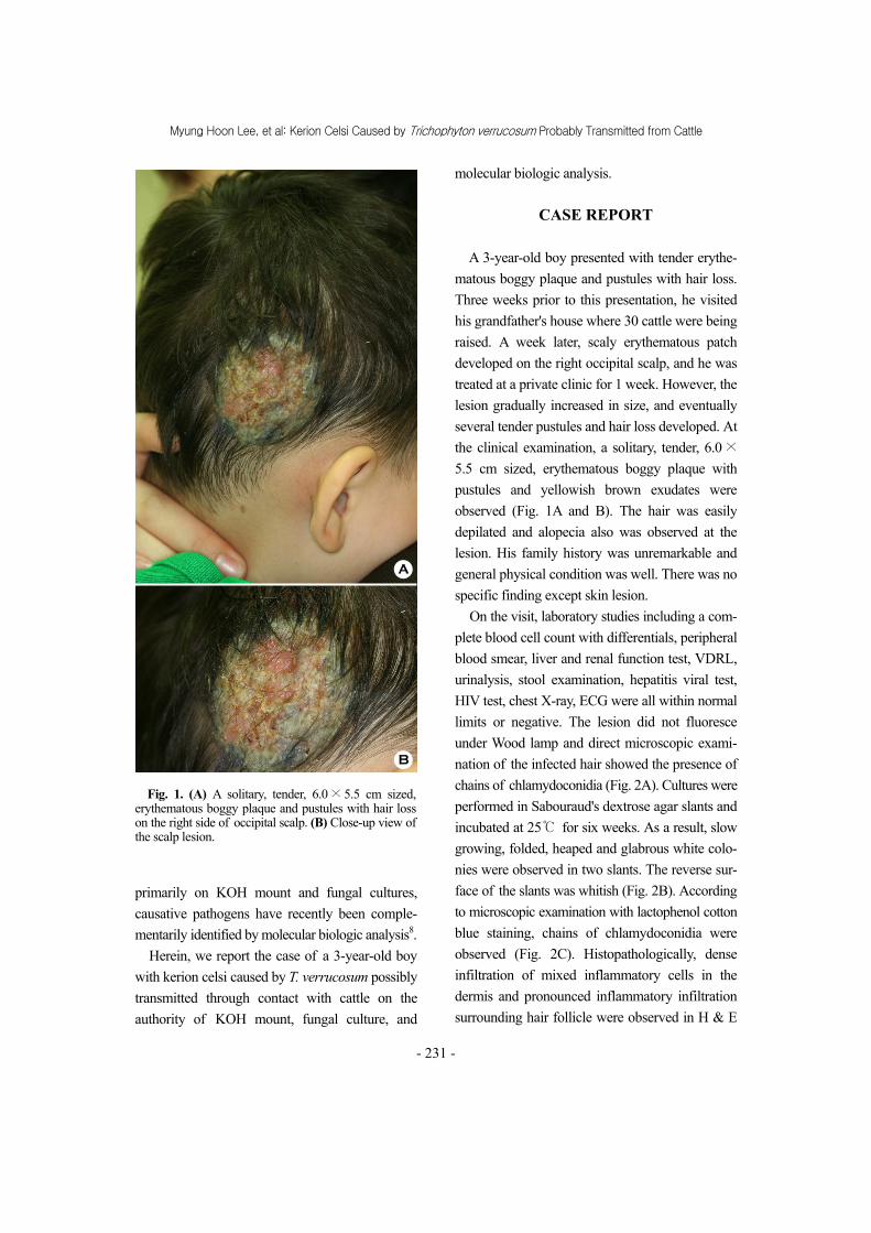
A 3-year-old boy presented with tender erythe-
matous boggy plaque and pustules with hair loss.
Three weeks prior to this presentation, he visited
his grandfather's house where 30 cattle were being
raised. A week later, scaly erythematous patch
developed on the right occipital scalp, and he was
treated at a private clinic for 1 week. However, the
lesion gradually increased in size, and eventually
several tender pustules and hair loss developed. At
the clinical examination, a solitary, tender, 6.0 ×
5.5 cm sized, erythematous boggy plaque with
pustules and yellowish brown exudates were
observed (Fig. 1A and B). The hair was easily
depilated and alopecia also was observed at the
lesion. His family history was unremarkable and
general physical condition was well. There was no
specific finding except skin lesion.
On the visit, laboratory studies including a com-
plete blood cell count with differentials, peripheral
blood smear, liver and renal function test, VDRL,
urinalysis, stool examination, hepatitis viral test,
HIV test, chest X-ray, ECG were all within normal
limits or negative. The lesion did not fluoresce
under Wood lamp and direct microscopic exami-
nation of the infected hair showed the presence of
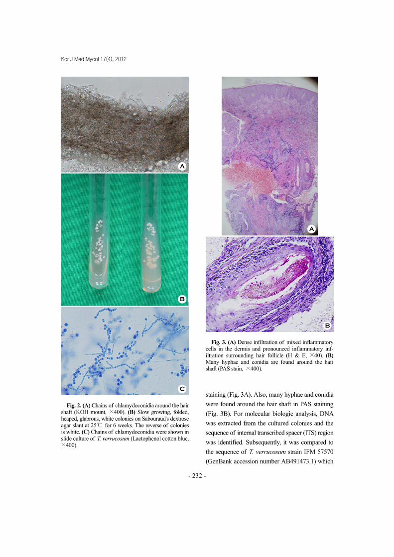
chains of chlamydoconidia (Fig. 2A). Cultures were
performed in Sabouraud's dextrose agar slants and
incubated at 25℃ for six weeks. As a result, slow
growing, folded, heaped and glabrous white colo-
nies were observed in two slants. The reverse sur-
face of the slants was whitish (Fig. 2B). According
to microscopic examination with lactophenol cotton
blue staining, chains of chlamydoconidia were
observed (Fig. 2C). Histopathologically, dense
infiltration of mixed inflammatory cells in the
dermis and pronounced inflammatory infiltration
surrounding hair follicle were observed in H & E
Fig. 1. (A) A solitary, tender, 6.0 × 5.5 cm sized, erythematous boggy plaque and pustules with hair loss on the right side of occipital scalp. (B) Close-up view ofthe scalp lesion.
A
B
Kor J Med Mycol 17(4), 2012
- 232 -
staining (Fig. 3A). Also, many hyphae and conidia
were found around the hair shaft in PAS staining
(Fig. 3B). For molecular biologic analysis, DNA
was extracted from the cultured colonies and the
sequence of internal transcribed spacer (ITS) region
was identified. Subsequently, it was compared to
the sequence of T. verrucosum strain IFM 57570
(GenBank accession number AB491473.1) which
Fig. 3. (A) Dense infiltration of mixed inflammatory cells in the dermis and pronounced inflammatory inf-iltration surrounding hair follicle (H & E, ×40). (B)Many hyphae and conidia are found around the hair shaft (PAS stain, ×400).
A
B
C
Fig. 2. (A) Chains of chlamydoconidia around the hairshaft (KOH mount, ×400). (B) Slow growing, folded,heaped, glabrous, white colonies on Sabouraud's dextroseagar slant at 25℃ for 6 weeks. The reverse of colonies is white. (C) Chains of chlamydoconidia were shown inslide culture of T. verrucosum (Lactophenol cotton blue,×400).
A
B

Myung Hoon Lee, et al: Kerion Celsi Caused by Trichophyton verrucosum Probably Transmitted from Cattle
- 233 -
stored in GenBank, using Blast program. The result
was 100% matched (Fig. 4).
The patient was treated with daily doses of
terbinafine 125 mg and predisolone 10 mg for the
first week. After than he was treated with 125 mg
oral terbinafine daily alone. Two weeks after starting
treatment, the inflammation and pustules were
remarkably improved. We performed repeated
KOH mount and fungal cultures for 2 times with
2 weeks interval and all were negative. Eight weeks
after starting medication, all skin lesions, including
alopecia, were improved.
DISCUSSION
Kerion celsi is a severe type of tinea capitis
caused by zoophilic or geophilic dermatophytes.
The distribution of causative pathogens varies
among regions and times1. Most cases of kerion
celsi reported in Korea are known to be caused by
M. canis9~11. Since kerion celsi caused by T.
verrucosum was first reported by Kim et al2 in
1986. Eight cases of kerion celsi have been
reported in Korea3~8 (Table 1). Of these 8 cases, 2
occurred in males and 6 in females; 3 in children
Table 1. Summary of the reported cases of kerion celsi caused by Trichophyton verrucosum in Korea
Author (year) Sex/ Age
Suspected animal Site Skin lesion Treatment
Kim et al2 (1986) F/9 Cattle Frontal scalp Boggy mass Ketoconazole, ampicilline
Kim et al3 (1989) M/4 Cattle Vertex Boggy masses Griseofulvin, prednisolone
Suh et al4 (1994) M/15 Cattle Frontal scalp Boggy mass Itraconazole, prednisolone,cephadroxil
Ro et al5 (1997) F/7 Cattle Left temporal scalp Boggy patch Griseofulvin, prednisolone
F/52 Cattle Vertex Indurated masses Terbinafine, prednisoloneYoun et al6 (2000)
F/50 Cattle Occipital scalp Purulent plaque Terbinafine, prednisolone
Kim et al7 (2000) F/67 Cattle Right parietal Boggy mass Terbinafine, prednisolone
Kim et al8 (2010) F/19 (-) Frontal scalp Boggy mass Itraconazole, methylprednisolone, cephradine
Present case (2012) M/3 Cattle Occipital scalp Boggy plaque Terbinafine, prednisolone
ATCATTAACGCGCAGGCCGGAGGCTGGCCCCCCACGATAGGGATCAGCGTTCCATCAGGGGTGTGCAGATGTGCGCCGGC [80]CTTACGCCCCATTCTTGTCTACCTTACTCGGTTGCCTCGGCGGGCCGCGCTCTCCCCGGAGAGTCGTCCGGCGAGCCTCT [160]TCGGGGGCTTTAGCTGGATCGCGCCCGCCGGAGGACAGACATCAAAAAATCTTGAAGAGCTGTCAGTCTGAGCGTTAGCA [240]AGCAAAATCAGTTAAAACTTTCAACAACGGATCTCTTGGTTCCGGCATCGATGAAGAACGCAGCGAAATGCGATAAGTAA [320]TGTGAATTGCAGAATTCCGTGAATCATCGAATCTTTGAACGCACATTGCGCCCTCTGGTATTCCGGGGGGCATGCCTGTT [400]CGAGCGTCATTTCAACCCCTCAAGCTCGGCTTGTGTGATGGACGACCGTCCGGCCCCCTCTTTCGGGGGCGGGACGCGCC [480]CGAAAAGCAGTGGCCAGGCCGCGATTCCGGCTTCCTGGGCGAATGGGCAATCAAACCAGCGCCCTCAGGACCGGCCGCTC [560]TGGCCTTCCCCCAAATCTCTCTGAGATTTTTTTCAGGTTGACC [603]
Fig. 4. Alignment of ITS sequences of the sample from patient and T. verrucosum strain IFM57570 (GenBank accession number AB491473.1). The sequences of ITS of clinical sample was 100% match to that of T. verrucosum strain IFM57570 (GenBank accession number AB491473.1).
Kor J Med Mycol 17(4), 2012
- 234 -
and 5 in adults; 7 were transmitted through contact
with cattle, while the transmission route of 1 case
was not identified. T. verrucosum is known to be
the most common causative pathogen of dermato-
phytosis caused by cattle which is usually trans-
mitted to other cattle through hay or bull pens
contaminated by the rubbing of infected skin
against surfaces and is sometimes transmitted to
humans by direct contact with infected cattle's
skin12~14. In this case, kerion celsi developed after
a 3-year-old boy visited his grandfather's house
where cattle were being raised, which suggests
that these cattles were the source of infection
A thorough history of contact with infected
patients, domestic animals or pets as well as the
long-term use of steroids or immunosuppressants
is neccessary in the diagnosis of kerion celsi.
Wood lamp, KOH mount and fungal cultures
should be performed in suspected cases. Since
fungal elements are relatively scarce in scales or
crusts, the infected hair in the lesion should be
obtained and examined whenever possible15,16.
Although Wood lamp is usually positive for T.
verrucosum in cattle, it is often negative in humans,
except for at the early stage of infection2,13. T.
verrucosum grows very slowly for at least 4 weeks
at 37℃. Because of this long cultivation time, it
cannot be isolated if culture media are contaminated
with other microorganisms5,7. Since T. verrucosum
is an ectothrix that involves the external surface of
hair shaft, it causes more severe infection than an
endothrix, and arthroconidia are not observed in
hair shaft of infected patients8. In severe cases,
kerion celsi should be discriminated from furuncle,
impetigo, folliculitis decalvans and chronic pyo-
derma of the scalp. In cases with resultant scars,
kerion celsi should be differentiated from discoid
lupus erythematosus, lichen planopilaris, pseu-
dopelade and radiation dermatitis1. Our case was
negative for wood lamp but positive for KOH
mount as well as 6-week fungal cultures. The
biopsy of the tissue obtained from the lesion
revealed numerous hyphae and spores around the
hair shaft. The patient was diagnosed with kerion
celsi based on these results. Although dermato-
phytosis is usually diagnosed based on the
morphologic feature of cultured colonies, various
molecular biologic analysis have recently been used
in the isolation of causative pathogens. In this case,
DNA was extracted from the cultured colonies
and the sequence of ITS region was identified.
Subsequently, it was compared to the sequence of
T. verrucosum strain IFM 57570 (GenBank
accession number AB491473.1) which stored in
GenBank, using Blast program. The result was
100% matched which suggests that the causative
pathogen was T. verrucosum.
Kerion celsi caused by zoophilic dermatophytes
responds well to medical treatment; however,
effective drugs must be carefully selected because
their susceptibility differs between pathogens17~19.
At first, causative pathogens should be treated by
oral and topical antifungal agents. In cases of
secondary infections, also antibiotics should be
administered. Combination therapy with antifungal
agents and corticosteroids is useful for the pre-
vention of permanent scarring alopecia. In addition,
terbinafine and itraconazole have recently been
used effectively, where griseofulvin was widely
used in the past5,7,8,15,16.
Also the patient in this case was treated with
daily doses of terbinafine 125 mg and predisolone
10 mg for the first week. After than he was treated
with 125 mg oral terbinafine daily alone for 7
weeks. Eight weeks after starting medication, all
skin lesions, including alopecia, were improved.
REFERENCES
1. Verma S, Heffernan MP. Superficial fungal infection:
dermatophytosis, onychomycosis, tinea nigra, piedra.
In: Wolff K, Goldsmith LA, Katz SI, Gilchrest BA,

Myung Hoon Lee, et al: Kerion Celsi Caused by Trichophyton verrucosum Probably Transmitted from Cattle
- 235 -
Paller AS, Leffell DJ, editors. Fitzpatrick's der-
matology in general medicine. 7th ed. New York:
McGraw-Hill, 2008:1807-1821
2. Kim YP, Chun IK, Kim SH. A case of kerion celsi
caused by Trichophyton verrucosum and its
epidemiologic study. Korean J Dermatol 1986;24:
687-691
3. Kim HS, Choi JS, Kim KH. A case of kerion celsi
caused by Trichophyton verrucosum. Korean J
Dermatol 1989;27:73-78
4. Suh MK, Sung YO, Kim JR. A case of kerion celsi
caused by Trichophyton verrucosum and treated
with itraconazole. Korean J Dermatol 1994;32:124
-129
5. Ro YW, Lee WJ, Suhr KB, Lee JH, Park JK. A case
of kerion celsi caused by Trichophyton verrucosum
in Chungcheong province. Korean J Dermatol 1997;
35:187-190
6. Youn CS, Moon SH, Kim JA, Kim KH, Cho KH,
Kang SH. Kerion celsi caused by Trichophyton
verrucosum. A report of two cases. Kor J Med Mycol
2000;5:60-65
7. Kim YJ, Choi JH, Bang JS, Suh MK, Lee JW, Kim
TH, et al. A case of kerion celsi in the elderly
caused by Trichophyton verrucosum and treated with
terbinafine. Kor J Med Mycol 2000;5:129-134
8. Kim JY, Hwang YJ, Ko JH, Oh BH, Lee YW, Choe
YB, et al. A case of kerion celsi caused by
Trichophyton verrucosum. Kor J Med Mycol 2010;
15:83-87
9. Oh SH, Kim SH, Suh SB. Tinea capitis of adults in
Daegu city for 11 years (1978-1988). Korean J
Dermatol 1989;27:666-679
10. Chun IK, Lim MH, Lee SC, Won YH. Clinical and
mycological studies of tinea capitis in Chonnam area
(1986-1995). Kor J Med Mycol 1996;1:83-89
11. Shin DH, Kim KS, Kim KH. Clinical and mycologic
studies of tinea capitis in Taegu. Kor J Med Mycol
1998;3:132-138
12. Chermette R, Ferreiro L, Guillot J. Dermatophytoses
in animals. Mycopathologia 2008;166:385-405
13. Rippon JW. Medical mycology: The pathogenic
fungi and the pathogenic actinomycetes, 3rd ed.
Philadelphia:WB Saunders, 1988:259-260
14. Friedman L, Derbes VJ, Tinea capitis. In: Demis J,
Mcguire J, eds. Clinical dermatology. 11th ed.
Philadelphia:Harper & Row Publisher, 1984;3:17-26
15. Jang BS, Jo JH, Oh CK, Jang HS, Kwon KS. A case
of kerion celsi caused by Trichophyton menta-
grophytes. Kor J Med Mycol 2002;7:86-91
16. Park MW, Chun BM, Park H, Kim SS, Lee JO, Lee
CJ. A case of kerion celsi caused by Trichophyton
mentagrophytes. Kor J Med Mycol 2006;11:191-194
17. Lee YW, Lim SH, Yim SM, Choe YB, Ahn KJ. A
clinical and mycological study of dermatophytosis
associated with animal contact. Kor J Med Mycol
2005;10:151-159
18. Kim YJ, Choi JH, Bang JS, Suh MK, Lee JW, Ha
GY. A case of Trichophyton mentagrophytes infection
probably transmitted from hamster. Kor J Med Mycol
2000;5:140-143
19. Kim SH, Suh MK, Kim JH, Ha GY, Kim JR. A case
of kerion celsi caused by Microsporum canis
probably transmitted from hamster. Kor J Med Mycol
2009;14:23-27