Embed Size (px)
Citation preview

General arthrology
Juncturae seu Systema articulare
Joint or Articular system
• synathrosis (immovable joints)
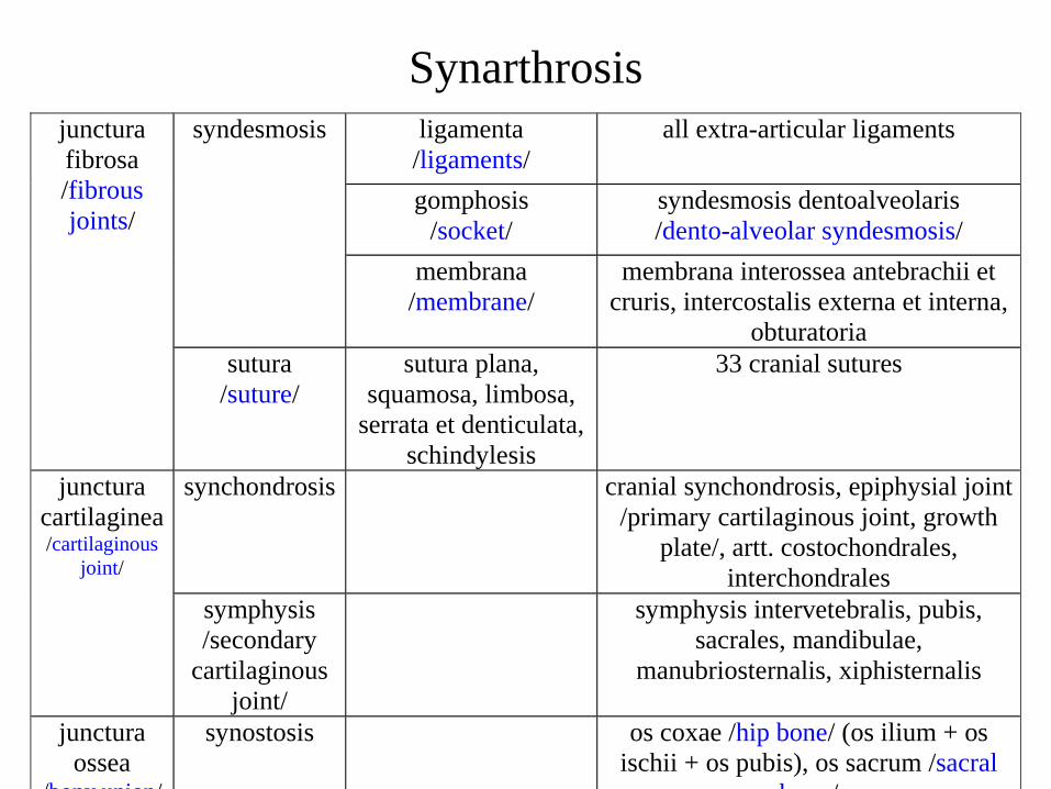
– connection by means of connective tissue• fibrous (junctura fibrosa) - syndesmosis
• cartilage (junctura cartilaginea) - synchondrosis, symphysis
• bony (junctura ossea) – synostosis
– no joint cavity
• diarthrosis (synovial joint)
– connecting surfaces with a cavity
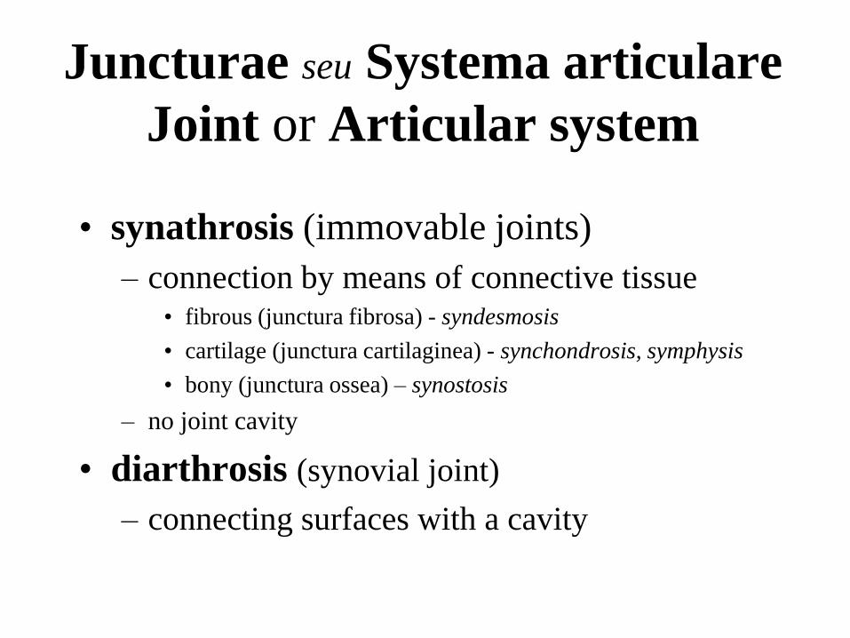
Synarthrosis
ligamenta
/ligaments/
all extra-articular ligaments
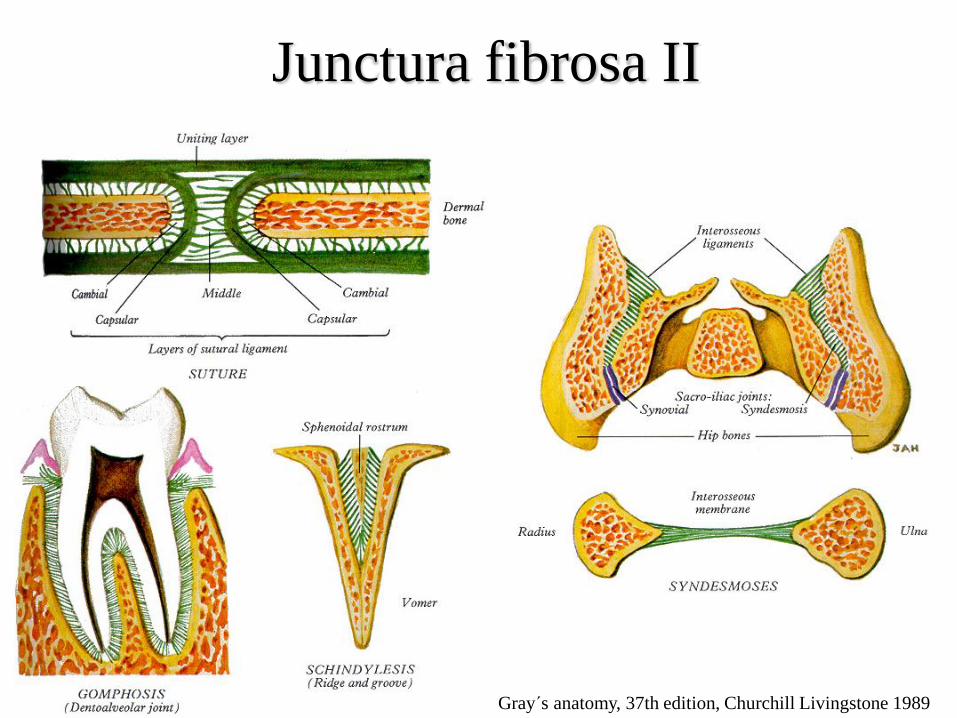
gomphosis
/socket/
syndesmosis dentoalveolaris
/dento-alveolar syndesmosis/
syndesmosis
membrana
/membrane/
membrana interossea antebrachii et
cruris, intercostalis externa et interna,
obturatoria
junctura
fibrosa
/fibrous
joints/
sutura
/suture/
sutura plana,
squamosa, limbosa,
serrata et denticulata,
schindylesis
33 cranial sutures
synchondrosis cranial synchondrosis, epiphysial joint
/primary cartilaginous joint, growth
plate/, artt. costochondrales,
interchondrales
junctura
cartilaginea /cartilaginous
joint/
symphysis
/secondary
cartilaginous
joint/
symphysis intervetebralis, pubis,
sacrales, mandibulae,
manubriosternalis, xiphisternalis
junctura
ossea /bony union/
synostosis os coxae /hip bone/ (os ilium + os
ischii + os pubis), os sacrum /sacral
bone/
Čihák R., Anatomie 1, Grada Publishing a.s. 2001
Junctura fibrosa I
Junctura fibrosa II
Gray´s anatomy, 37th edition, Churchill Livingstone 1989
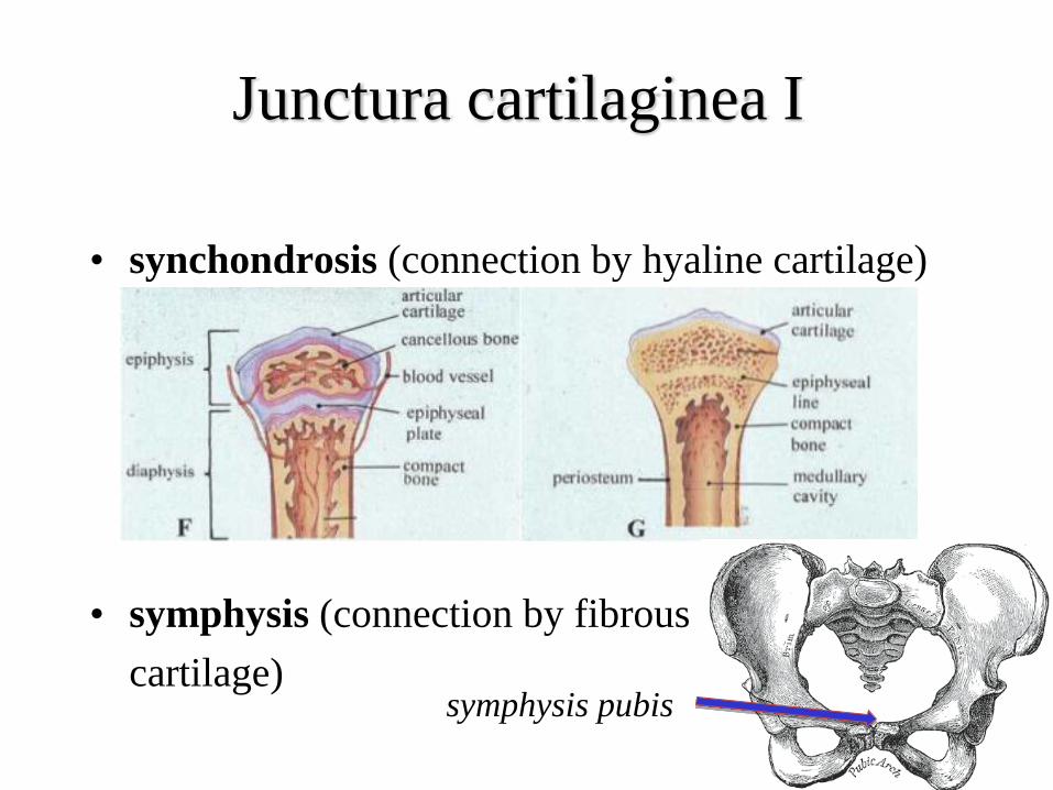
• synchondrosis (connection by hyaline cartilage)
• symphysis (connection by fibrous
cartilage)
Junctura cartilaginea I
symphysis pubis
Junctura cartilaginea II
Gray´s anatomy, 40th edition, Churchill Livingstone Elsevier 2008
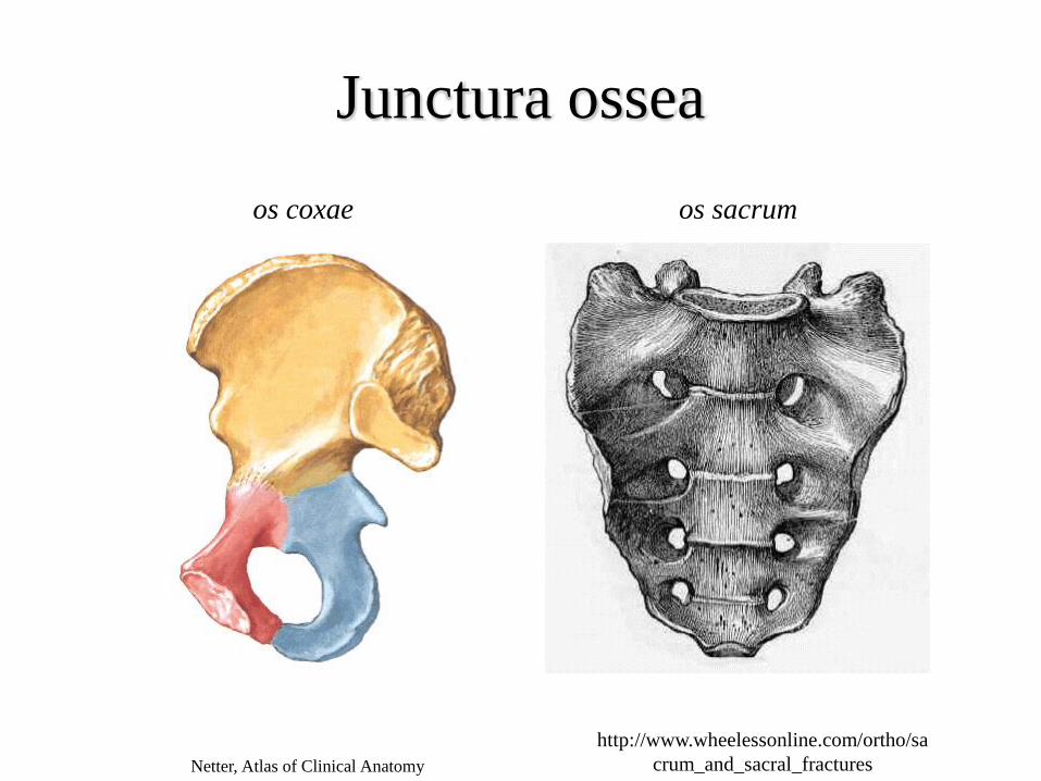
Junctura ossea
os coxae os sacrum
Netter, Atlas of Clinical Anatomy
http://www.wheelessonline.com/ortho/sa
crum_and_sacral_fractures
Diarthrosis = Junctura synovialis
= Articulatio = Synovial joint• facies articulares (articular surfaces)
– fossa (fossa articularis) x head (caput articulare)
• capsula articularis (joint capsule)
– stratum fibrosum (externally)
– stratum synoviale (little differentiated synovialocytes hyaluronic acid)
plicae synoviales (synovial folds), corpus adiposum intraarticulare (intraarticular fat pad)
• cavitas articularis (articular cavity)
– capillary slit
– contains synovia (synovial fluid) = plasma transsudate + hyaluronic acid + a few leukocytes
• special joint structures
Membrana synovialis (Synovial membrane)
• lines the whole articular cavity
– apart from articular surfaces
• protrudes in plicae synoviales and villi
synoviales
• well supplied by vessels and nerves
• 3 types
– fibrous
– areolar
– adipose
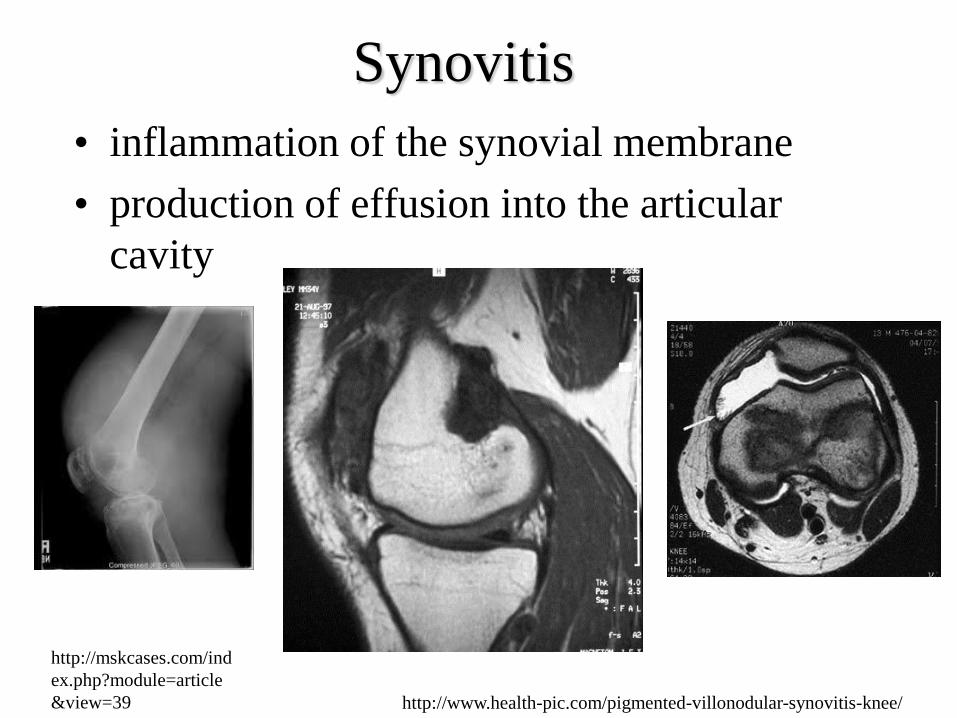
Synovitis
• inflammation of the synovial membrane
• production of effusion into the articular
cavity
http://www.health-pic.com/pigmented-villonodular-synovitis-knee/
http://mskcases.com/ind
ex.php?module=article
&view=39
Gray´s anatomy, 37th edition, Churchill Livingstone 1989
Special joint structures I
• labrum articulare (labrum)– enlarges the area of articular fossa
– art. humeri, art. coxae
• disci et menisci articulares (articular discs and menisci)– they level articular incongruations
– elastic liner/pad
– disc divides articular cavity in two• art. temporomandibularis, art. sternoclavicularis
– meniscus is becoming flatter in the inner direction, has free inner margin
• art. genus
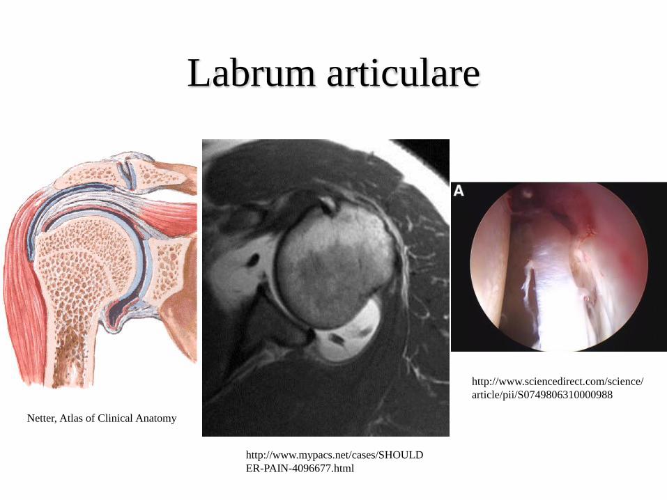
Labrum articulare
http://www.mypacs.net/cases/SHOULD
ER-PAIN-4096677.html
http://www.sciencedirect.com/science/
article/pii/S0749806310000988
Netter, Atlas of Clinical Anatomy
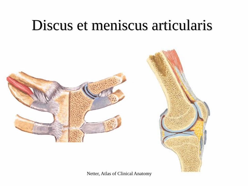
Discus et meniscus articularis
Netter, Atlas of Clinical Anatomy
Special joint structures II
• ligamenta (ligaments)– capsular (ligg. capsularia), extracapsular (ligg.
extracapsularia) and intracapsular (ligg. intracapsularia)
– strenghten the capsule
– support the movements of the joint
– limit the movement of the joint
• bursae synoviales (synovial bursae)– cavities lined by synovial membrane
– inside there is a fluid similar to synovia
– place of pathological changes
• musculi articulares (joint muscles)– prevent joint capsule strangulation
Netter, Atlas of Clinical Anatomy
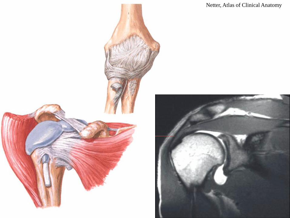
Special joint structures III
• fibrocartilago (fibrocartilage)– enlarge the articular fossa and strengthen the capsule
• corpus adiposum (fat pad)
• plica synovialis
– level incongruations of the articular surfaces
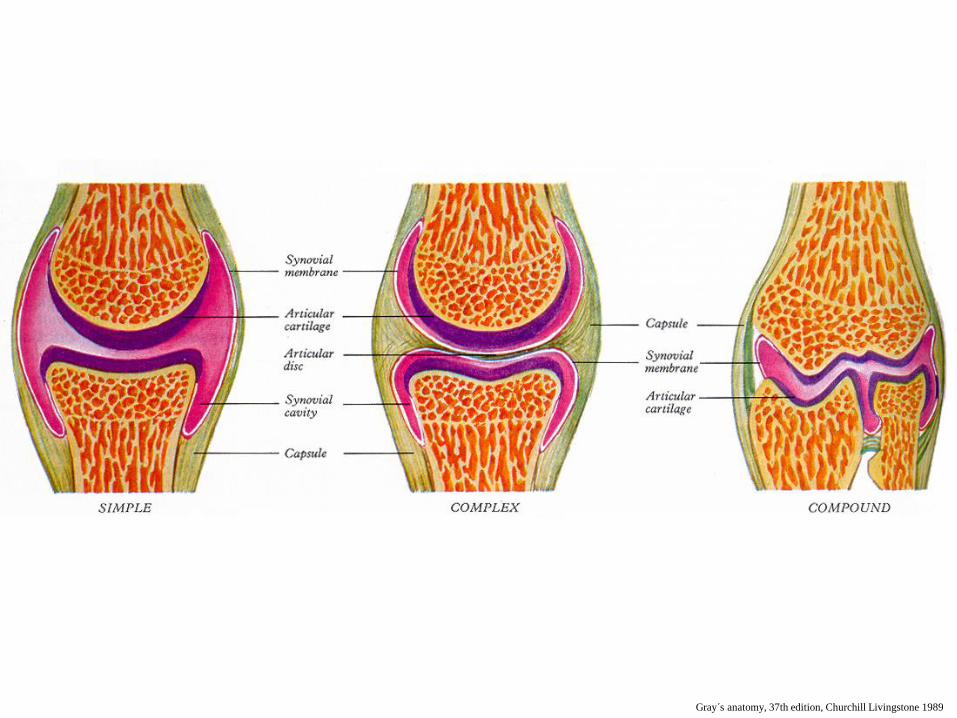
Classification of diarthrosis
• by part number:
– simple /art. simplices/ - 2 kosti
– compound /art. compositae/
• more than 2 bones
• 2 bones + disc or meniscus
• by movement extension
- amphiarthrosis (rigid)
- more movable (all others)
• by shape of connecting surfaces
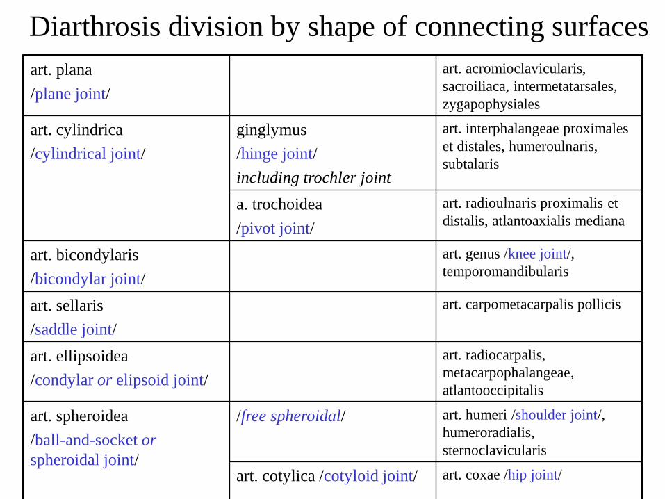
Diarthrosis division by shape of connecting surfaces
art. plana
/plane joint/
art. acromioclavicularis,
sacroiliaca, intermetatarsales,
zygapophysiales
art. cylindrica
/cylindrical joint/
ginglymus
/hinge joint/
including trochler joint
art. interphalangeae proximales
et distales, humeroulnaris,
subtalaris
a. trochoidea
/pivot joint/
art. radioulnaris proximalis et
distalis, atlantoaxialis mediana
art. bicondylaris
/bicondylar joint/
art. genus /knee joint/,
temporomandibularis
art. sellaris
/saddle joint/
art. carpometacarpalis pollicis
art. ellipsoidea
/condylar or elipsoid joint/
art. radiocarpalis,
metacarpophalangeae,
atlantooccipitalis
art. spheroidea
/ball-and-socket or
spheroidal joint/
/free spheroidal/ art. humeri /shoulder joint/,
humeroradialis,
sternoclavicularis
art. cotylica /cotyloid joint/ art. coxae /hip joint/
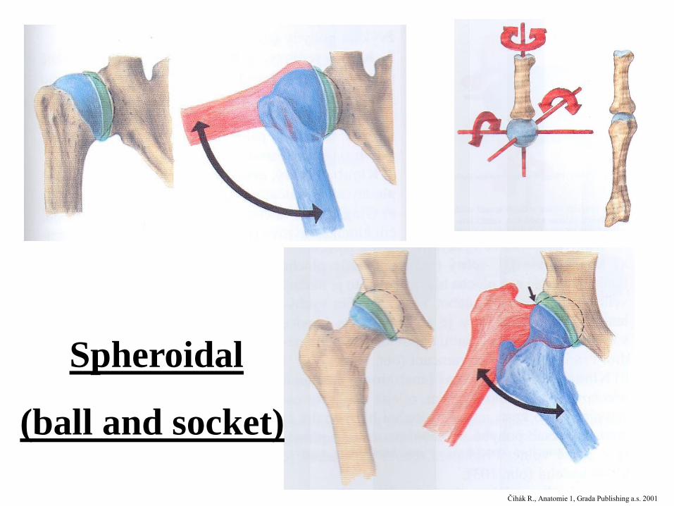
Spheroidal
(ball and socket)
Čihák R., Anatomie 1, Grada Publishing a.s. 2001
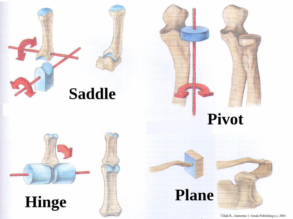
Saddle
Pivot
Hinge PlaneČihák R., Anatomie 1, Grada Publishing a.s. 2001
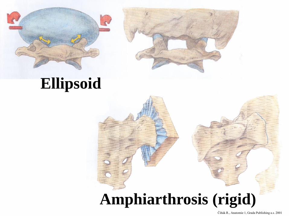
Ellipsoid
Amphiarthrosis (rigid)Čihák R., Anatomie 1, Grada Publishing a.s. 2001
Joint movements I
• according to axis
– mono-, bi- and polyaxial
• basic position
– reflects the basic anatomical position (palms ventrally)
• loose position– most relaxed articular capsule (releaving position)
• movement extension
– limited by
• shape of fossa and head
• ligaments
• close bony projections
• soft tissue size in the vicinity (muscles, fat)
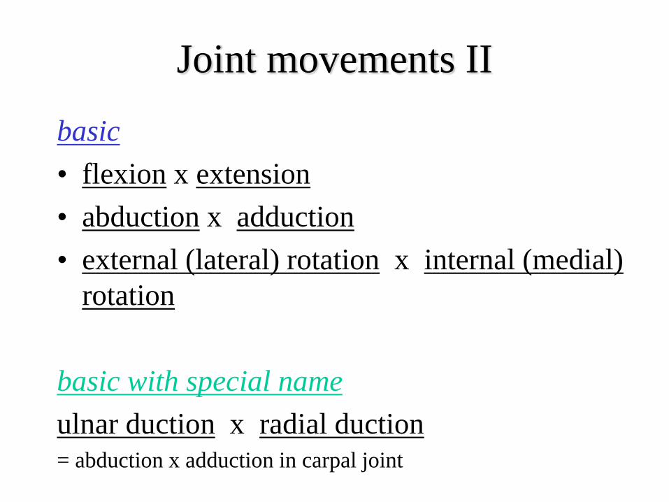
Joint movements II
basic
• flexion x extension
• abduction x adduction
• external (lateral) rotation x internal (medial)
rotation
basic with special name
ulnar duction x radial duction= abduction x adduction in carpal joint
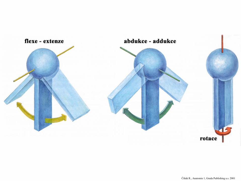
Čihák R., Anatomie 1, Grada Publishing a.s. 2001
Joint movements III
• pronation x supination= special type of radius rotation around ulna
• opposition x reposition= special thumb movement to face the other fingers
• elevation x depression
+ protraction x retraction= special movement in temporomandibular joint and in loose
connection between scapula and thorax (is not an
anatomical joint, just functional connection!)
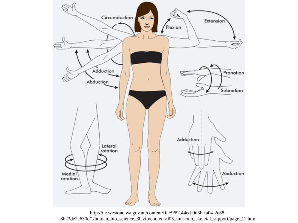
http://tle.westone.wa.gov.au/content/file/969144ed-0d3b-fa04-2e88-
8b23de2a630c/1/human_bio_science_3b.zip/content/003_musculo_skeletal_support/page_11.htm
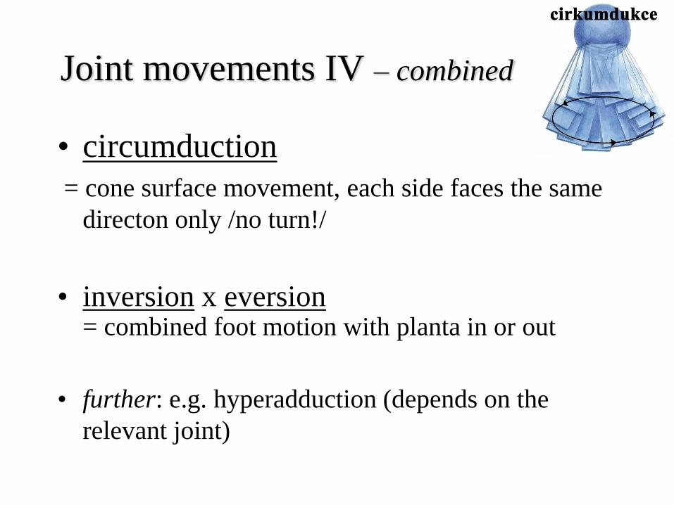
Joint movements IV – combined
• circumduction
= cone surface movement, each side faces the same
directon only /no turn!/
• inversion x eversion= combined foot motion with planta in or out
• further: e.g. hyperadduction (depends on the
relevant joint)
Vessels and nerve supply of the joint
• blood vessels: rete articulare from surrounding
arteries, capillaries close to the surface
• lymph vessels: blind beginnings (cul-de-sac),
deeper in the capsule
• nerves:
– centripetal sensory fibres
• information about joint position, movement direction and grade, angular movement speed, ligaments and capsule tension grade (= proprioception)
• pressure and pain informations
– centrifugal autonomic fibres (vessels´ lumen regulation)
Development of the joint
• plates of mesenchyme between adjacent skeletal elements =
interzonal mesenchyme
• interzonal mesenchyme becomes trilaminar
– 2 dense strata
– intermediate zone
• intermediate stratum merges with general mesenchyme →
a cuff condenses creating a fibrous capsule of the joint
• dense strata becomes cartilaginous
• cavitation of intermediate zone establishes the cavity of the
joint
• synovial mesenchyme forms synovial membrane and other
structures, such as tendons, ligaments, discs and menisci
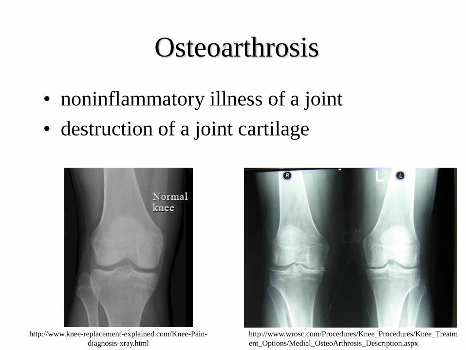
Osteoarthrosis
• noninflammatory illness of a joint
• destruction of a joint cartilage
http://www.wrosc.com/Procedures/Knee_Procedures/Knee_Treatm
ent_Options/Medial_OsteoArthrosis_Description.aspx
http://www.knee-replacement-explained.com/Knee-Pain-
diagnosis-xray.html
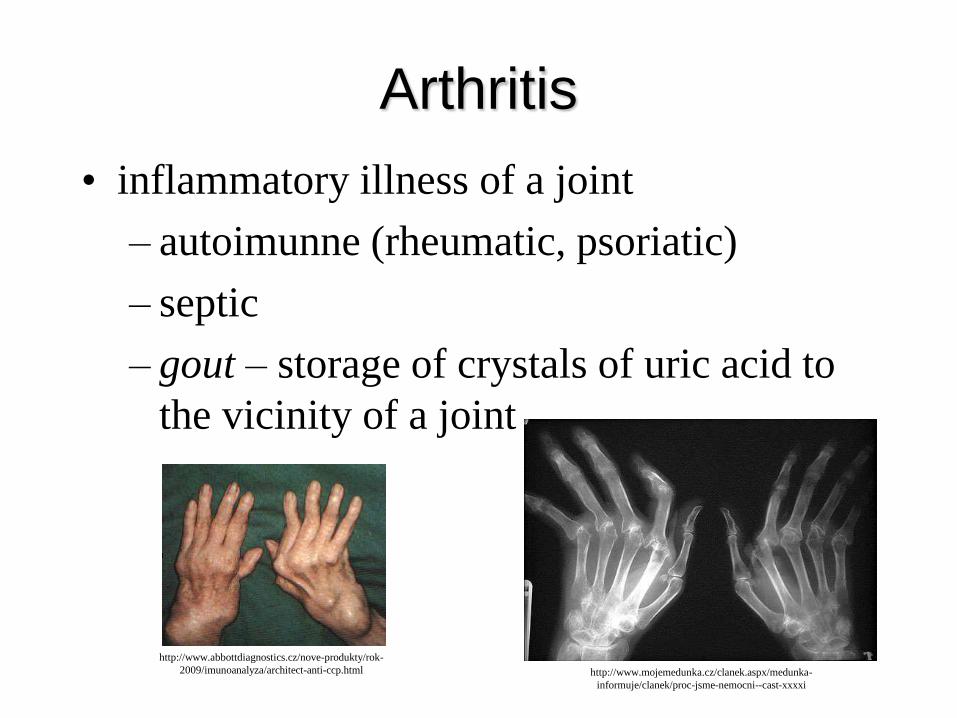
Arthritis
• inflammatory illness of a joint
– autoimunne (rheumatic, psoriatic)
– septic
– gout – storage of crystals of uric acid to
the vicinity of a joint
http://www.abbottdiagnostics.cz/nove-produkty/rok-
2009/imunoanalyza/architect-anti-ccp.html http://www.mojemedunka.cz/clanek.aspx/medunka-
informuje/clanek/proc-jsme-nemocni--cast-xxxxi
Joint description
!!! follow general rules !!!
• name (Latin, English)
• type – by part number, shape of connecting surfaces, movability, axis number
• head and fossa
• joint capsule insertion – close to connecting surfaces – several important exceptions !!!
• special joint structures – labrum, disc, meniscus, fibrocartilage, ligaments, synovial bursae, fat
pads
• basic and loose position
• movements (+ movements extension in degrees)
– passive
– active