Embed Size (px)
Citation preview

Morphology and Phylogenetic Position of Eucomonympha imla(Parabasalia: Hypermastigida)
KEVIN J. CARPENTER and PATRICK J. KEELING
Botany Department, Canadian Institute for Advanced Research, University of British Columbia, 3529-6270 University Boulevard,
Vancouver, BC, Canada V6T 1Z4
ABSTRACT. Eucomonympha imla is a hypermastigote parabasalian found in the gut of the wood-feeding cockroach Cryptocercuspunctulatus. It has received little attention since its original description in 1934 as the type species of the genus Eucomonympha and thefamily Eucomonymphidae. We used light and scanning electron microscopy to characterize surface morphology and organelles, withparticular attention to the form of the rostrum, operculum, nucleus, and parabasals. Two previously unrecognized groups of bacterialectobionts were observed—spirochetes that associate with the flagella and one or more types of rod-shaped bacteria that adhere to the cellsurface. The small subunit rRNA (SSU rRNA) sequence was determined from manually isolated cells, and phylogenetic analyses place E.imla in a strongly supported clade with the genera Teranympha and Pseudotrichonympha and three sequences from formally undescribedtermite symbionts provisionally assigned to Eucomonympha. Unexpectedly, the Eucomonympha isolates from termites are more closelyrelated to Teranympha than to the type species, suggesting these should not be classified as species of Eucomonympha, despite theirmorphological similarity to E. imla. Eucomonymphidae fall within a strongly supported Trichonymphida (also including Hoplonymph-idae, Trichonymphidae, and Staurojoeninidae), but this clade branches separately from other hypermastigote groups (lophomonads andspirotrichonymphids), suggesting that hypermastigotes are polyphyletic.
Key Words. Bacterial symbionts, Cryptocercus punctulatus, excavata, Pseudotrichonympha, scanning electron microscopy, symbiosis,Teranympha mirabilis, Trichonymphida.
PARABASALIA comprise a large and extremely diversegroup of anaerobic protists, including flagellate, amoebofla-
gellate, and amoeboid forms, distinguished by the presence ofhydrogenosomes (and the associated absence of canonical aerobicmitochondria), parabasal bodies (Golgi vesicles with an associat-ed complex of fibres), closed mitosis with an external spindle, anda pelta-axostyle complex (Brugerolle and Lee 2002). Parabasaliatraditionally comprise two classes—Trichomonadida and Hype-rmastigida. The former consist mostly of small and structurallyrelatively simple polymastigotes, although a few have attainedlarge size, sometimes through multiplication of the karyomasti-gont apparatus (Brugerolle and Lee 2002; Honigberg 1963). Incontrast, hypermastigotes are complex and typically large cells,with flagella numbering in the hundreds or thousands, but theirkaryomastigont system has not replicated and is associated with asingle nucleus. Hypermastigotes are found only in symbiotic as-sociation with termites and wood-feeding cockroaches of the ge-nus Cryptocercus, where they aid in digestion of ingested woodparticles in the hindgut (Cleveland 1923, 1924; Ohtoko et al.2000; Trager 1932), a symbiotic relationship that is of enormousecological importance in forest ecosystems, both in the decom-position of lignocellulose and in forming an important part of theterrestrial food chain (Ohkuma 2003).
The monophyly of both trichomonads and hypermastigotes hasrecently been questioned. Analyses of molecular data supportmultiple origins of the hypermastigote cell form and paraphylyof trichomonads, although some important nodes are weakly sup-ported (Hampl et al. 2006; Ohkuma et al. 2005). Reflecting this, ina recent classification of eukaryotes, Adl et al. (2005) assignedhypermastigote taxa to three different groups, one of which alsocontains some of the more structurally complex trichomonads(e.g. Calonymphids).
Eucomonympha imla, a hypermastigote found exclusively inthe gut of the wood-feeding cockroach Cryptocercus punctulatus,is a striking example of the extreme morphological complexityexhibited by parabasalians. First described by Cleveland et al.
(1934), E. imla is a large cell (over 100 microns in length) con-sisting of two roughly spheroidal portions: an anterior rostrum anda much larger posterior post-rostral area. With the exception of theextreme anterior and posterior ends, the cell is completely coveredin flagella, estimated to number over 52,000 in some specimens(Cleveland et al. 1934). Most swim freely in the lumen of thehindgut where they endocytose wood particles, while a minorityremains attached to the wall of the hindgut and obtains nutrientsthrough unknown means (Cleveland et al. 1934). Eucomonymphaimla is the type species of the genus Eucomonympha and the ge-nus is the type of the family Eucomonymphidae. The only otherknown occurrence of members of the genus Eucomonympha is inthe hindgut of the termite Hodotermopsis (Brugerolle and Bor-dereau 2004; Kitade, Maeyama, and Matsumoto 1997; Ohkumaet al. 2000). Cleveland et al. (1934) hypothesized a close rela-tionship with the genus Pseudotrichonympha from the termiteCoptotermes, based on the presence of flagella covering the entirebody surface and the manner of cell division. This hypothesis issupported by recent molecular phylogenies (Ohkuma et al. 2000,2005), the former of which claims that Eucomonymphidae may bethe sister group to the rest of the parabasalians.
As most parabasalians, especially hypermastigotes and the oth-er inhabitants of the complex wood-digesting insect gut environ-ment, are typically not cultivated, nearly all of the recent work onthese organisms has used molecular approaches to address ques-tions related to systematics and evolution (Keeling 2002; Ohkumaet al. 2000, 2005), their role in termite and cockroach gut metab-olism (Nakashima, Watanabe, and Azuma 2002; Ohtoko et al.2000), and their symbioses with ecto- and endosymbiotic bacteria(Noda et al. 2005, 2006). However, the extreme structural diver-sity of these extraordinary but still poorly studied organismsserves to emphasize the need for new morphological data as well.Cleveland et al. (1934) originally described E. imla with illustra-tions based on light microscopy (LM), and Hollande and Caruette-Valentin (1971) subsequently added ultrastructural data based ontransmission electron micrographs. Here we seek to complementdata from previous studies by examining surface morphology withscanning electron microscopy (SEM), as well as inferring itsphylogenetic position based on SSU rRNA sequence data, withthe goal of providing an integrated view of this species and theimplications of its phylogenetic placement.
Corresponding Author: P. Keeling, Botany Department, University ofBritish Columbia, 3529-6270 University Boulevard, Vancouver, BC,Canada V6T 1Z4—Telephone number: 1 604 822 2845 (lab); FAXnumber: 1 604 822 6089; e-mail: [email protected]
263
J. Eukaryot. Microbiol., 54(3), 2007 pp. 263–270r 2007 The Author(s)Journal compilation r 2007 by the International Society of ProtistologistsDOI: 10.1111/j.1550-7408.2007.00263.x

MATERIALS AND METHODS
Sampling. Individuals of C. punctulatus Scudder were col-lected by C. Nalepa from several locations in the Appalachianmountain range of the eastern United States. Insects were killedby severing the head with a razor blade, and the entire gut wasremoved by gently pulling on the posterior-most segment of theabdomen with forceps. The hindgut was then removed, immersedin a several drops of Trager Medium U, and opened with a scalpelunder a dissecting microscope. Hindgut contents were then col-lected using a 1,000-ml micropipette. The hindgut contents of 10individuals were examined using LM and SEM, but E. imla wasnot universally present. Data presented here come from a singleindividual from Mountain Lake Biological Station, Giles County,VA and one from South Mountains, Burke County, NC.
Light microscopy (LM). Samples of hindgut contents wereexamined and photographed live with a Zeiss Axioplan 2 com-pound microscope with Plan Apochromat objectives and Q-imag-ing Micro Imager II digital camera using differential interferencecontrast (DIC) illumination.
Scanning electron microscopy. Samples of hindgut contentswere placed in plastic Petri dishes, diluted with Trager Medium Uto a total volume of approximately 5 ml, and fixed with 4% (w/v)osmium tetroxide (OsO4) vapours for 30 min. Five or six drops of4% OsO4 were then added directly to the samples, which were leftfor an additional 30 min to complete fixation. Approximately200ml of the fixed contents were pipetted onto a Millipore Iso-pore membrane filter with 5-mm pore size (Billerica, MA) held ina Millipore Swinnex plastic cartridge (Billerica) affixed to a 10-mlplastic syringe. This was repeated (i.e. using additional filter/car-tridge/syringe assemblies) until all of the contents were exhausted.The material was then rinsed with 10 ml of Trager Medium U anddehydrated in an ethanol series (10 ml each of 50%, 70%, 90%,and two changes of 100%) for a minimum of 10 min at each stage.Filters were CO2 critical point dried with a Balzers CPD 020 crit-ical point drying apparatus (Liechtenstein), affixed to aluminumSEM stubs with Ted Pella double stick carbon adhesive (Redding,CA), and sputter coated with gold–palladium in a Nanotech (Man-chester, United Kingdom) SEM Prep 2 sputter coater. Stubs wereexamined and photographed with a Hitachi S4700 field emissionscanning electron microscope (Tokyo, Japan) at 1.5–5.0 kV.
DNA extraction, amplification, and sequencing. Cell sus-pensions in Trager’s Medium U were placed in a cavity slide andindividual cells matching the description of E. imla were manuallyisolated using a micropipette, as described by Keeling (2002). Apool of 30 E. imla cells was isolated in one tube, and three cellswere isolated into three individual tubes. DNA was isolated fromall four samples using a single chloroform extraction followed byethanol precipitation, as described (Keeling 2002). The SSUrRNA genes were amplified by rehydrating the DNA directly ina 10-ml reaction volume using the primers 50-GCGCTACCTGGTTGATCCTGCC-30 and 50-TGATCCTTCTGCAGGTTCACCTAC-30 and amplifying for 35 cycles with an annealing temper-ature of 45 1C and an extension time of 1.5 min. Products wereseparated by electrophoresis and cloned. Multiple clones weresequenced on both strands. As the level of variation betweenclones was low, a single clone was submitted to GenBank as Ac-cession Number DQ923125.
Phylogenetic analysis. The E. imla SSU rRNA sequence wasadded to an alignment including all known parabasalian SSUrRNA sequences. Manual inspection and phylogenetic analyses ofthis global alignment were carried out to identify sequences thatwere either nearly identical to other sequences, truncated, or high-ly divergent. These were removed, along with environmentalclones of unknown origin that fell in well-supported clades rep-resented by named species, resulting in an alignment of 49 se-
quences and 1,236 alignable sites. This alignment was analysedusing Bayesian, maximum likelihood, and distance methods.Maximum likelihood trees and 1,000 ml bootstrap replicates wereinferred by PHYML 2.4.4 (Guindon and Gascuel 2003) using theGTR substitution model and rate between sites modelled on a gdistribution with eight variable categories and invariable sites.The proportion of invariable sites and the a shape parameter wereestimated from the data (0.22 and 0.56, respectively). Bayesiananalyses were carried out using MrBayes 3.1 (Ronquist and Huel-senbeck 2003) with the same substitution and rate between sitesparameters; 1,000,000 generations were run with three hot chainsand one cold chain sampled every 10,000 generations with aburnin of 100,000 generations. Distances were calculated usingTREE-PUZZLE 5.2 (Schmidt et al. 2002) with the settings de-scribed for ML analysis and 1,000 distance bootstraps calculatingusing the shell script puzzleboot (by M. Holder and A. Roger:www.tree-puzzle.de). Trees were inferred from distances usingweighbor 1.2 (Bruno, Socci, and Halpern 2000). Bootstrap ana-lyses restricted to the Eucomonymphidae (six sequences) werealso carried out using PhyML with the same parameters and 1,000replicates.
The monophyly of Eucomonympha was specifically tested us-ing the approximately unbiased (AU) test (Shimodaira and Has-egawa 2001). The monophyly of E. imla and the three sequencesfrom Eucomonympha-like cells in termites were constrained, andthe ML tree optimized according to this constraint using PAUP4.0 b 10 using the site-to-site rate variation parameters estimatedabove, the GTR substitution model, and a heuristic search. Theresulting tree differed only in two nodes from the ML tree (both inthe trichomonads, far from the eucomonymphids). Site likeli-hoods were calculated for both trees by TREE-PUZZLE usingthe wsl option with the settings described above. AU tests werecarried out using CONSEL 1.19 (Shimodaira 2002).
RESULTS
Distribution. From 10 cockroaches investigated, E. imla wasfound in a single individual from each of two different popula-tions, and in individuals where it was present, it was never asabundant as other hypermastigotes, such as Trichonympha orBarbulanympha.
Morphology. Under LM, E. imla appears as a large (4100 mmlong), slowly swimming, densely flagellated cell exhibiting min-imal plasticity of shape. E. imla cells are composed of two distinctregions: an anterior hemispherical rostrum and an oblate-spheroi-dal to slightly elongate post-rostral portion (Fig. 1–7), which oc-casionally displays an encircling constriction or furrow (Fig. 5).Measurements taken from SEM micrographs indicate a mean celllength of 135.7 mm, including both rostral and post-rostral por-tions, and a mean cell width of 118.3 mm (Table 1). Except for asmall, anterior-most portion called the operculum (Fig. 8–10), andthe extreme posterior portion, the entire cell is covered with a verydense complement of flagella. Rostral flagella appear to be con-siderably longer than flagella on the post-rostral portion in LM(Fig. 2, 9). With SEM, however, it is extremely difficult to deter-mine unequivocally where even a single flagellum originates andterminates, especially on the post-rostrum, making accurate mea-surements and statistically significant comparisons of flagellarlength impractical. This difficulty is caused by the great density offlagella on this organism and the fact that, while rostral flagellatend to project outward from the cell in a more or less straight line(Fig. 8), post-rostral flagella often appear to run more parallel tothe cell surface where they become entangled among neighbour-ing flagella and, in some areas, spirochetes (Fig. 12–17). It may bethat the apparent difference in length between rostral and post-rostral flagella observed with LM and also noted by Cleveland
264 J. EUKARYOT. MICROBIOL., VOL. NO. 54, NO. 3, MAY–JUNE 2007

et al. (1934) is exaggerated due to these properties, which arevisible only under SEM.
The operculum was often obscured by rostral flagella in SEMbut was visible in some of the individuals from the South Moun-tains cockroach (Fig. 1, 8, 10). The operculum appears hemi-spherical in side view (Fig. 10), while in face view it is circularand bears approximately 20 clavate appendages more or less reg-ularly spaced around its circumference (Fig. 8). A circular toovoid nucleus is visible directly posterior to the rostrum (Fig. 2,9). In the original description of E. imla, Cleveland stated that
parabasals were not observed (Cleveland et al. 1934), but we ob-served numerous parabasals radiating from just under the rostrumusing Nomarski DIC microscopy (Fig. 11), likely dictating thedistribution of flagella. Endocytosed wood fragments are also vis-ible scattered throughout the post-rostral region (Fig. 2).
Bacterial ectobionts. Examination of E. imla with SEM andDIC microscopy revealed that it harbours at least two differentpopulations of bacterial ectobionts. The most conspicuous arespirochetes that associate with flagella and are clearly discernableeven with LM (Fig. 12). They were not observed on all cells, but
Fig. 1–7. The whole cell of Eucomonympha imla (SEM and LM micrographs). 1. SEM micrograph, side view showing rostrum (r) and post-rostralarea (p), South Mountains population. 2. LM micrograph, side view showing nucleus (n), Mountain Lake Biological Station population. 3–7. SEMmicrographs, Mountain Lake Biological Station population. 3. Side view. 4. Top view. 5. Oblique view. 6. Top view. 7. Side view. Bars 5 20 mm.
265CARPENTER & KEELING—EUCOMONYMPHA IMLA
when present they are abundant. Moreover, they are not evenlydistributed over the surface of the cell; we observed them to becommon on the anterior half of the post-rostral region (Table 1and Fig. 12–17), rare on the posterior of the post-rostral region,and completely absent on the rostrum, although they were ob-served right up to the junction between the rostrum and post-ros-tral region (Fig. 13). We did not observe any spirochetes to beanchored to the cell surface, but instead they seem only to asso-ciate with the flagella, in some cases appearing to be entangled inthem. Whether they are bound to the flagella is not clear, but theirassociation appears to be specific, as the flagella were not ob-served to attract other entanglements (Fig. 1, 3–7; 12–17), andalso relatively robust, as they were not observed to dislodge whencells were manipulated or isolated. Spirochetes were observedswimming freely in the gut contents, as is also common in termitegut environments, but were never observed associated with any ofthe other large, densely flagellated protists occurring in the gut ofC. punctulatus (e.g. Trichonympha, Spirotrichonympha, Barbula-nympha). The abundance of these spirochetes on E. imla cells andtheir corresponding absence on any other hypermastigote, togeth-er with their specific and restricted distribution to a post-rostralcollar on E. imla, all suggest this association is not by chance.
In addition to the spirochetes, SEM also revealed small rod-shaped bacteria sparsely distributed over the cell surface of thepost-rostral region between flagellar emergence points (Table 1and Fig. 15–17).
Phylogenetic analysis. The SSU rRNA gene was amplifiedfrom 30 manually isolated E. imla cells and eight clones were se-quenced. All clones were nearly identical (o1% variation wasobserved and always in a single clone) and similar to sequencesfrom Teranympha mirabilis and undescribed termite symbiontsassigned to the genus Eucomonympha. To confirm the identity ofthe isolated cells, three additional cells were isolated individually,and the SSU rRNA was amplified, cloned, and sequenced fromeach. Once again, all three cells yielded sequences nearly identicalto those amplified from the 30 cells and showed about the samelevel of variation (at all variable sites these clones were identicalto the consensus). Since the variation was so low, a single clonewas selected for use in phylogenetic analysis.
Phylogenetic analysis consistently placed E. imla within astrongly supported clade (100%) including the eucomonymphidPseudotrichonympha grassii, three sequences from Eucomo-nympha-like cells from termites, and the teranymphid T. mira-bilis (Fig. 18). Within the larger tree, the Eucomonymphidae/Teranymphidae group falls within a strongly supported (97%–98%) clade that corresponds to the order Trichonymphida (Bruge-rolle and Lee 2002), which also includes Hoplonymphidae,Trichonymphidae, and Staurojoenidae. Within the Eucomo-nymphidae/Teranymphidae group, T. mirabilis and the Eucomo-nympha-like cells from termites consistently formed a stronglysupported (100%) group to the exclusion of E. imla and P. grassi.E. imla consistently shares a common ancestor with T. mirabilisand the Eucomonympha-like cells from termites, to the exclusion
of P. grassii, although this node is not very strongly supported(Fig. 18). This suggests E. imla and the Eucomonympha-like cellsfrom termites are not monophyletic, despite their similarity inoverall appearance. We tested this in a reduced data set ofEucomonymphidae/Teranymphidae and it was again supportedat 100%. We also used AU tests to compare the ML tree with analternative where the monophyly of E. imla and the Eucomo-nympha-like cells from termites was enforced and found this al-ternative was rejected with a P-value of 6 � 10� 6.
DISCUSSION
Morphology. Many of the morphological characters observedhere were also noted in earlier descriptions of E. imla based onLM or transmission electron microscopy (TEM) (Cleveland et al.1934; Hollande and Caruette-Valentin 1971). The one major ex-ception to this is the association between E. imla and various pro-karyotes. Ecto- and endosymbiotic bacteria are commonly foundin association with numerous different termite and cockroach gutprotists not only with parabasalians (Brugerolle and Lee 2002;Noda et al. 2005, 2006), but also with oxymonads (Leander andKeeling 2004). Noda et al. (2006) determined that Gram-negative,rod-shaped bacterial ectosymbionts found in gut protists belong tothree different lineages of Bacteroidales. One of these lineages ischaracteristic of the order Trichonymphida (strongly supported asmonophyletic in the present study), to which E. imla belongs. Al-though Noda et al. (2005) found that bacterial endosymbionts(thought to be derived from ectosymbionts) of the genus Pseu-dotrichonympha (which is closely related to E. imla) actually be-long to a different group than these, they are still members ofBacteroidales. Thus, it is possible that the rod-shaped bacterialectobionts of E. imla may also be members of Bacteroidales, butthis will have to be tested directly.
Spirochetes, on the other hand, have been found to have lessspecific relationships with host protists; more than one species ofspirochete may associate with a given host protist and any givenspecies of spirochete may associate with more than one protisthost species. Although the nature of their interactions with thehost remains unknown, it seems clear that such bacterial sym-bionts are of some importance to their protist hosts and the func-tioning of the gut metabolism (see Noda et al. 2005, 2006). Itseems likely that this is true for E. imla as well, although the exactnature of this symbiosis remains to be investigated. At very least,the morphological evidence argues that the observed associationof spirochetes with E. imla is more than the result of mere acci-dental entanglement: when present, spirochetes are consistentlyrestricted to the upper portion of the post-rostral area; they remainattached when E. imla cells are manipulated; and they are not seenassociated with any of the other large, densely flagellated protistspresent in the same gut environment. Thus, we hypothesize thatthe spirochetes are genuine ectobionts.
Phylogeny. Our analysis of SSU rRNA places E. imla in aclade with Teranympha, Pseudotrichonympha, and sequences ob-tained from unidentified Eucomonympha-like cells from a termite.Ohkuma et al. (2005) inferred a relationship between theseEucomonympha-like cells and T. mirabilis, but with the additionof E. imla it is clear that the Eucomonympha-like isolates fromtermites share a more recent common ancestor with Teranymphathan they do with E. imla. As E. imla is the type species of thegenus Eucomonympha and the genus Teranympha (Koidzumi1921) predates Eucomonympha (Cleveland et al. 1934), we sug-gest that when the currently undescribed Eucomonympha-like or-ganisms from termites are formally named, they should not beincluded in the genus Eucomonympha because it would make thegenus paraphyletic. Instead, they could be included in Tera-nympha, if they fit its description, or in a new genus.
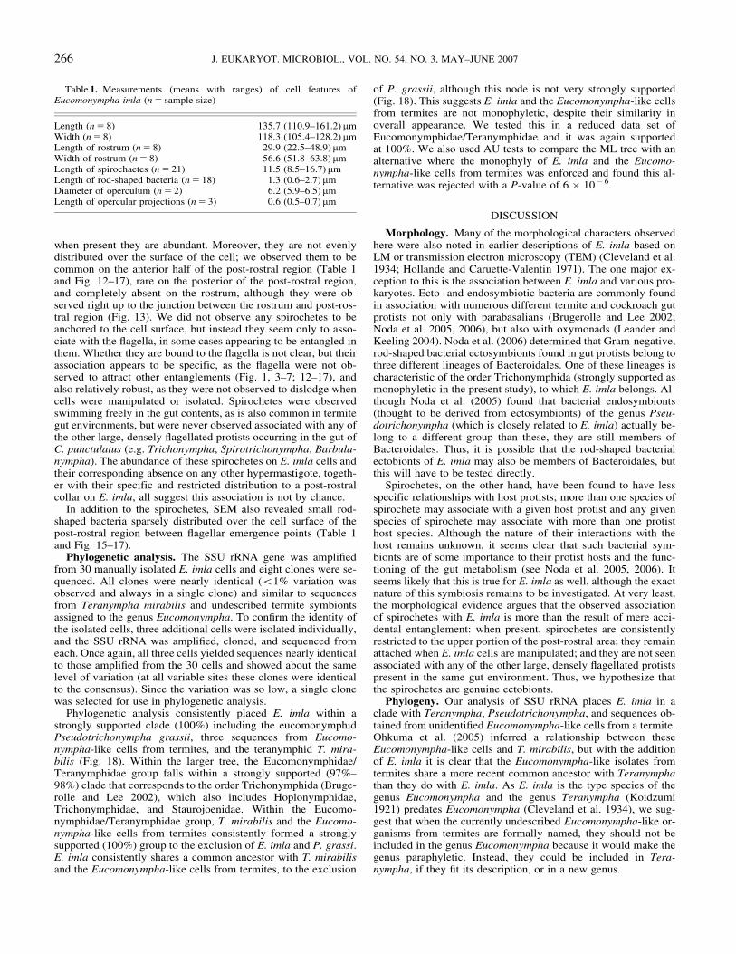
Table 1. Measurements (means with ranges) of cell features ofEucomonympha imla (n 5 sample size)
Length (n 5 8) 135.7 (110.9–161.2)mmWidth (n 5 8) 118.3 (105.4–128.2)mmLength of rostrum (n 5 8) 29.9 (22.5–48.9)mmWidth of rostrum (n 5 8) 56.6 (51.8–63.8)mmLength of spirochaetes (n 5 21) 11.5 (8.5–16.7)mmLength of rod-shaped bacteria (n 5 18) 1.3 (0.6–2.7) mmDiameter of operculum (n 5 2) 6.2 (5.9–6.5) mmLength of opercular projections (n 5 3) 0.6 (0.5–0.7) mm
266 J. EUKARYOT. MICROBIOL., VOL. NO. 54, NO. 3, MAY–JUNE 2007

This phylogenetic placement of E. imla also implies that thepresence of flagella covering nearly the entire cell surface is likelya synapomorphy of the Eucomonymphidae/Teranymphidae cladebecause all investigated members of this clade exhibit this featureexcept for T. mirabilis, which nests above P. grassii and E. imla.The Eucomonympha-like organisms from termites also appear tobe fully flagellated in light micrographs (Ohkuma et al. 2005). InT. mirabilis, the arrangement of flagella in rings separated bybands of cytoplasm, both of which run perpendicular to the long
axis of the cell, is thus likely to be an autapomorphy derived fromthe fully flagellated state in Eucomonympha and Pseudotricho-nympha. Likewise, depending on the resolution of its positionwith respect to E. imla, the highly elongate cell form of Pseudot-richonympha may represent an autapomorphy of this genus. At-tached forms of E. imla appear somewhat elongate (Cleveland etal. 1934), as does T. mirabilis (Brugerolle and Lee 2002), but notto the extreme seen in Pseudotrichonympha (Cleveland et al.1934).
Fig. 8–11. Micrographs (SEM and LM) of surface structures and organelles of Eucomonympha imla. 8. SEM micrograph showing operculum (o)with clavate projections (c), South Mountains population, bar 5 3mm. 9. LM micrograph showing rostrum (r), post-rostral area (p), operculum (o), andnucleus (n), Mountain Lake Biological Station population, bar 5 20 mm. 10. SEM micrograph showing operculum (o), South Mountains population,bar 5 2mm. 11. LM micrograph showing rostrum (r), nucleus (n), and parabasals (pb), Mountain Lake Biological Station population, bar 5 10 mm.
267CARPENTER & KEELING—EUCOMONYMPHA IMLA

Within the parabasalian tree as a whole, our results also sup-port the monophyly of the order Trichonymphida (includingfamilies Eucomonymphidae, Teranymphidae, Hoplonymphidae,Staurojoeninidae, and Trichonymphidae), whose putativesynapomorphies include bilateral symmetry (two symmetric-flagellated areas that separate during cell division), a rostral tubeand cap, and a cell body consisting of rostral and post-rostral areas(Brugerolle and Lee 2002). Thus, lack of any of these characters(e.g. the rostral tube), as seen in the families Staurojoeninidae andHoplonymphidae, probably represents a secondary loss. Othergroups previously or currently considered to be hypermastigotesthat branch elsewhere include lophomonads, which branch withdevescovinids and calonymphids, and spirotrichonymphids,
which are sister to monocercomonads. This is consistent withother recent analyses (Ohkuma et al. 2005) and at face valuesupports the possibility that hypermastigotes are polyphyletic,although the branches separating Trichonymphida and spirot-richonymphids are not supported.
A more thorough assessment of character evolution in Eucomo-nymphidae and Teranymphidae awaits investigation with both ofT. mirabilis SEM and TEM and the Eucomonympha-like termiteflagellates that are only known from SSU rRNA sequences andlight micrographs at present. Likewise, the task of understandingmorphological character evolution in all parabasalians awaits im-proved sampling of taxa with both molecular and morphologicalmethods and improved support for important nodes.
Fig. 12–17. Micrographs (SEM and LM) of bacterial ectosymbionts of Eucomonympha imla, Mountain Lake Biological Station population. 12. LMmicrograph of the anterior end of cell showing spirochetes (s) associated with flagella on the post-rostral area, bar 5 20 mm. 13. SEM micrograph showingportions of the rostrum (r) and post-rostral areas with spirochetes (s) associated with flagella of the latter, bar 5 10mm. 14. SEM micrograph of the post-rostral area showing spirochetes (s) associated with flagella (f), bar 5 5mm. 15–17. SEM micrographs of the post-rostral area showing spirochetes (s)associated with post-rostral flagella (f) and rod-shaped bacteria (e) on the cell surface (Bars: D 5 3mm; E 5 1mm; F 5 2mm).
268 J. EUKARYOT. MICROBIOL., VOL. NO. 54, NO. 3, MAY–JUNE 2007

687608 Ditrichomonas honigbergi
4071318 Monotrichomonas carabina ATCC50700
32263496 Monocercomonas ruminantium KOJ14
4455109 Tetratrichomonas gallinarum
37704017 Trichomonas vaginalis TV2
1132484 Trichomonas tenax ATCC30207
886700 Pentatrichomonoides scroa PM13
37954932 Cochlosoma anatis
4455110 Pentatrichomonas hominis PH KT
5921095 Reticulitermes speratus symbiont Rs16
28194502 Trichomitopsis termopsidis
3551805 Pseudotrypanosoma giganteum 2
687610 Monocercomonas sp ATCC 50210
687609 Metadevescovina polyspira
22074427 Metacoronympha senta
1340079 Devescovina sp D16
34850295 Koruga bonita
45580814 Deltotrichonympha nana
886684 Metadevescovina extranea PM15
45580813 Mixotricha paradoxa
22074430 Calonympha grassii
3551796 Cryptotermes brevis symbiont 77087
22074414 Snyderella tabogae
5921112 Neotermes koshunensis symbiont Nk4
12006862 Histomonas meleagridis
1408458 Dientamoeba fragilis ATCC30948
63147270 Spirotrichonymphella sp MO2004
5921115 Spirotrichonympha sp Hs1 gene
5921102 Spirotrichonympha leidyi
5921101 Holomastigotoides mirabile
4378001 Hypotrichomonas acosta ATCC30069
4378000 Trichomitus batrachorum BUB
3551798 Cryptotermes brevis symbiont 77089
3551795 Cryptotermes brevis symbiont 77086
63147271 Staurojoenina assimilis
5921119 Trichonympha sp Hs10
2116625 Trichonympha agilis EH11
2547167 Trichonympha cf collaris
5921118 Trichonympha sp Hs8
5921116 Trichonympha sp Hs3
5921128 Hodotermopsis sjoestedti symbiont HsS9
3551808 Trichonympha magna
63147269 Hoplonympha sp MO 2004 2
5921100 Pseudotrichonympha grassii Cf12
63147265 Teranympha mirabilis
63147267 Eucomonympha sp MO 2004 1
5921120 Eucomonympha sp HsL3
5921124 Eucomonympha sp HsL15
0.2
DQ923125 Eucomonympha imla
Eucomonymphids& Teranymphid
Trichonymphids
Spirotrichonymphids
Devescovinids& Calonymphids
Trichomonads
97100
100100
100100
100100
9897
−−
100100
74−
100100
7271
100100
100100
7569
9598
9794
−68
Lophomonads
Staurojoenids
Hoplonymphids
Monocercomonads
Monocercomonads
Monocercomonads?
Monocercomonad
Fig. 18. Small subunit rRNA phylogeny of Parabasalia. The tree shown is a Bayesian topology with maximum likelihood branch lengths. Subgroupsare named and bracketed to the right. Taxon names are preceded by GenBank locus indicators except in the case of Eucomonympha imla where theGenBank Accession Number is used. In cases of unidentified symbiont gene the name of the host is used with a strain designation. Numbers at nodescorrespond to bootstraps from maximum likelihood (top) and distance (bottom).
269CARPENTER & KEELING—EUCOMONYMPHA IMLA

ACKNOWLEDGMENTS
This work was supported by a grant form the Natural Sciencesand Engineering Research Council of Canada (227301). We thankC. Nalepa for generously sharing collections of C. punctulatus, R.Humayun for assistance constructing the alignment, G. Noble forassistance with cloning and sequencing, and B. Leander and E.Humphries for assistance with electron microscopy. P. J. K. is aFellow of the Canadian Institute for Advanced Research and aSenior Investigator of the Michael Smith Foundation for HealthResearch.
LITERATURE CITED
Adl, S. M., Simpson, A. G., Farmer, M. A., Andersen, R. A., Anderson, O.R., Barta, J. R., Bowser, S. S., Brugerolle, G., Fensome, R. A., Fred-ericq, S., James, T. Y., Karpov, S., Kugrens, P., Krug, J., Lane, C. E.,Lewis, L. A., Lodge, J., Lynn, D. H., Mann, D. G., McCourt, R. M.,Mendoza, L., Moestrup, Ø., Mozley-Standridge, S. E., Nerad, T. A.,Shearer, C. A., Smirnov, A. V., Spiegel, F. W. & Taylor, M. F. 2005.The new higher level classification of eukaryotes with emphasis on thetaxonomy of protists. J. Eukaryot. Microbiol., 52:399–451.
Brugerolle, G. & Bordereau, C. 2004. The flagellates of the termite Ho-dotermopsis sjoestedti with special reference to Hoplonympha, Ho-lomastigotes and Trichomonoides trypanoides n. comb. Eur. J.Protistol., 40:163–174.
Brugerolle, G. & Lee, J. J. 2002. Phylum parabasalia. In: Lee, J. J., Lee-dale, G. F. & Bradbury, P. (ed.), An Illustrated Guide to the Protozoa.2nd ed.. Allen Press Inc., Lawrence, Kansas. p. 1196–1250.
Bruno, W. J., Socci, N. D. & Halpern, A. L. 2000. Weighted neighborjoining: a likelihood-based approach to distance-based phylogeny re-construction. Mol. Biol. Evol., 17:189–197.
Cleveland, L. R. 1923. Symbiosis between termites and their intestinalprotozoa. Proc. Natl. Acad. Sci. USA, 9:424–428.
Cleveland, L. R. 1924. The physiological and symbiotic relationships be-tween the intestinal protozoa of termites and their host, with specialreference to Reticulitermes flavipes Kollar. Biol. Bull., 46:177–225.
Cleveland, L. R., Hall, S. R., Sanders, E. P. & Collier, J. 1934. The wood-feeding roach, Cryptocercus, its protozoa and the symbiosis betweenprotozoa and roach. Mem. Am. Acad. Arts Sci., 17:1–342.
Guindon, S. & Gascuel, O. 2003. A simple, fast, and accurate algorithmto estimate large phylogenies by maximum likelihood. Syst. Biol., 52:696–704.
Hampl, V., Vrlik, M., Cepicka, I., Pecka, Z., Kulda, J. & Tachezy, J. 2006.Affiliation of Cochlosoma to trichomonads confirmed by phylogeneticanalysis of the small-subunit rRNA gene and a new family concept ofthe order Trichomonadida. Int. J. Syst. Evol. Microbiol., 56:305–312.
Hollande, A. & Caruette-Valentin, J. 1971. Les atractophores, l’inductiondu fuseau et la division cellulaire chez les Hypermastigines. Etude in-frastructurale et revision systematique des Trichonymphines et desSpirotrichonymphines. Protistologica, 7:5–100.
Honigberg, B. M. 1963. Evolutionary and systematic relationships in theflagellate order Trichomonadida Kirby. J. Protozool., 10:20–63.
Keeling, P. J. 2002. Molecular phylogenetic position of Trichomitopsistermopsidis (Parabasalia) and evidence for the Trichomitopsiinae. Eur.J. Protistol., 38:279–286.
Kitade, O., Maeyama, T. & Matsumoto, T. 1997. Establishment of sym-biotic flagellate fauna of Hodotermopsis japonica. Sociobiology,30:161–167.
Koidzumi, M. 1921. Studies on the intestinal protozoa found in the ter-mites of Japan. Parasitology, 13:235–309.
Leander, B. S. & Keeling, P. J. 2004. Symbiotic innovation in the oxy-monad Streblomastix strix. J. Eukaryot. Microbiol., 51:291–300.
Nakashima, K. I., Watanabe, H. & Azuma, J. I. 2002. Cellulase genes fromthe parabasalian symbiont Pseudotrichonympha grassii in the hindgutof the wood-feeding termite Coptotermes formosanus. Cell. Mol. LifeSci., 59:1554–1560.
Noda, S., Iida, T., Kitade, O., Nakajima, H., Kudo, T. & Ohkuma, M.2005. Endosymbiotic Bacteroidales bacteria of the flagellated protistPseudotrichonympha grassii in the gut of the termite Coptotermes for-mosanus. Appl. Environ. Microbiol., 71:8811–8817.
Noda, S., Inoue, T., Hongoh, Y., Kawai, M., Nalepa, C. A., Vongkaluang,C., Kudo, T. & Ohkuma, M. 2006. Identification and characterization ofectosymbionts of distinct lineages in Bacteroidales attached to flagel-lated protists in the gut of termites and a wood-feeding cockroach. En-viron. Microbiol., 8:11–20.
Ohkuma, M. 2003. Termite symbiotic systems: efficient bio-recycling oflignocellulose. Appl. Microbiol. Biotechnol., 61:1–9.
Ohkuma, M., Iida, T., Ohtoko, K., Yuzawa, H., Noda, S., Viscogliosi, E. &Kudo, T. 2005. Molecular phylogeny of parabasalids inferred fromsmall subunit rRNA sequences, with emphasis on the Hypermastigea.Mol. Phylogenet. Evol., 35:646–655.
Ohkuma, M., Ohtoko, K., Iida, T., Tokura, M., Moriya, S., Usami, R.,Horikoshi, K. & Kudo, T. 2000. Phylogenetic identification of hype-rmastigotes, Pseudotrichonympha, Spirotrichonympha, Holomastigoto-ides, and parabasalian symbionts in the hindgut of termites. J. Eukaryot.Microbiol., 47:249–259.
Ohtoko, K., Ohkuma, M., Moriya, S., Inoue, T., Usami, R. & Kudo, T.2000. Diverse genes of cellulase homologues of glycosyl hydrolasefamily 45 from the symbiotic protists in the hindgut of the termite Ret-iculitermes speratus. Extremophiles, 4:343–349.
Ronquist, F. & Huelsenbeck, J. P. 2003. MrBayes 3: Bayesian phyloge-netic inference under mixed models. Bioinformatics, 19:1572–1574.
Schmidt, H. A., Strimmer, K., Vingron, M. & von Haeseler, A. 2002.TREE-PUZZLE: maximum likelihood phylogenetic analysis usingquartets and parallel computing. Bioinformatics, 18:502–504.
Shimodaira, H. 2002. An approximately unbiased test of phylogenetic treeselection. Syst. Biol., 51:492–508.
Shimodaira, H. & Hasegawa, M. 2001. CONSEL: for assessing the con-fidence of phylogenetic tree selection. Bioinformatics, 17:1246–1247.
Trager, W. 1932. A cellulase from the symbiotic intestinal flagellates oftermites and of the roach, Cryptocercus punctulatus. Biochem. J., 26:1762–1771.
Received: 12/01/06, 02/07/07; accepted: 02/21/07
270 J. EUKARYOT. MICROBIOL., VOL. NO. 54, NO. 3, MAY–JUNE 2007